Embed Size (px)
Citation preview
4/17/2015
1
DIAGNOSTIC CHALLENGES:Cytology Slide Seminar
Pancreas FNAB
Dr. M. Weir
May 2015
CONFLICT OF INTEREST DISCLOSURE
I have not had in the past 3 years, a financial interest, arrangement or affiliation with one or more organizations that could be perceived as a direct or indirect conflict of interest in the content of this presentation.
OBJECTIVES
Recognize diagnostic approaches to complex cytological problems
Expand knowledge & skills in interpretation of advanced cytology sampling techniques & ancillary tests
4/17/2015
2
AGENDA
2 cases
Pancreas eus fnabs
Posted online prior to session
Approach, diagnostic differentials
Photos: Hologic, PathologyOutlines, lcytology.wordpress.com
www.eurocytology.eu, www.joplink.net,www.pubcan.org
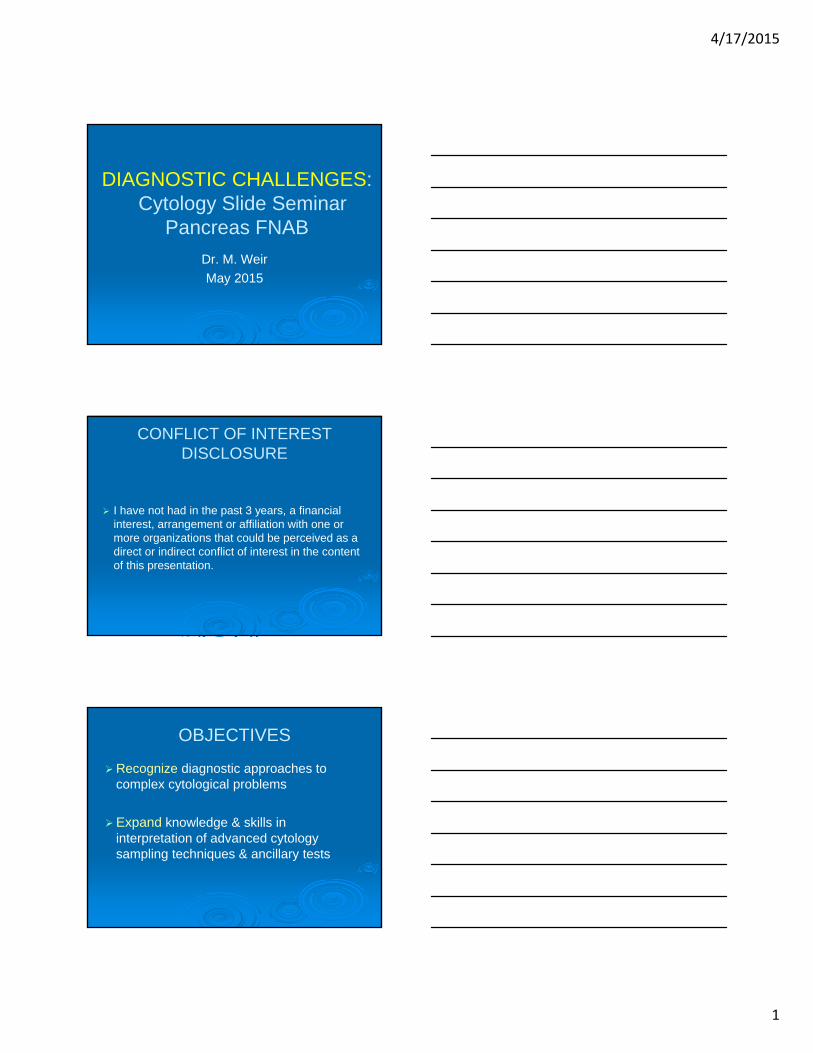
Case 1
55 year old female
Head of pancreas (HOP) mass
Solid, 5 x 4 cm
Transduodenal EUS FNAB
4/17/2015
3
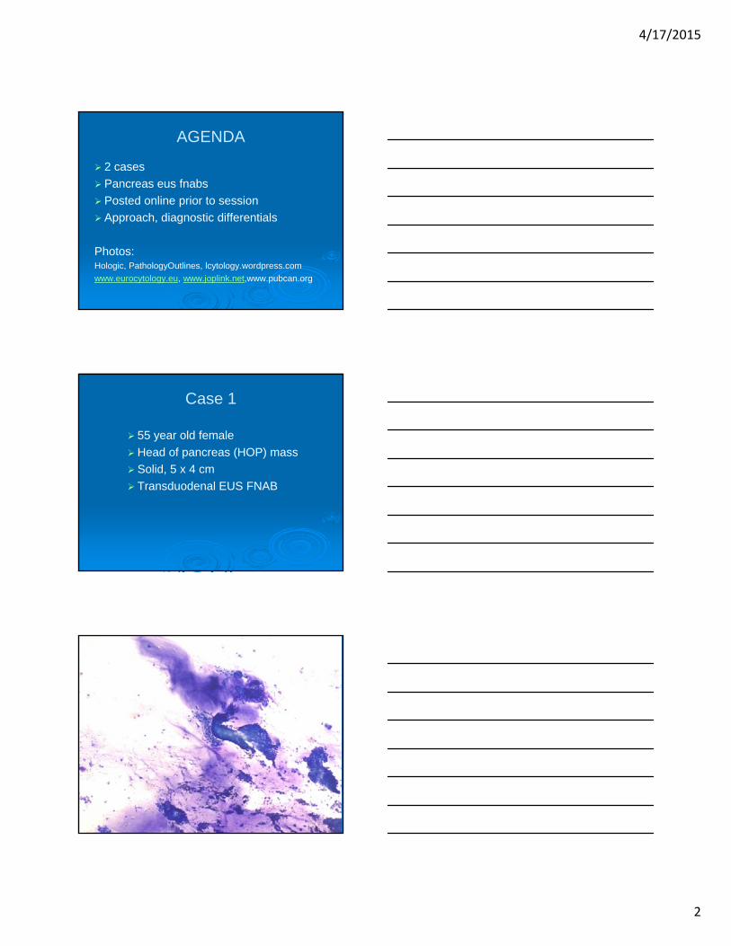
Approach
Clinical & imaging important- if solid – use algorithm for DDX
Microscopic approach- Adequacy
- Background- Contamination
- Diagnosis
4/17/2015
4
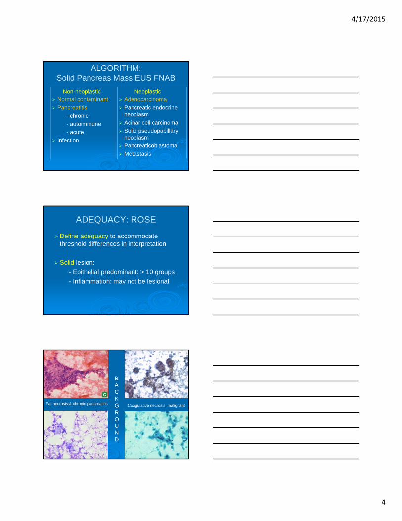
ALGORITHM:Solid Pancreas Mass EUS FNAB
Non-neoplastic
Normal contaminant
Pancreatitis
- chronic
- autoimmune
- acute
Infection
Neoplastic
Adenocarcinoma
Pancreatic endocrine neoplasm
Acinar cell carcinoma
Solid pseudopapillaryneoplasm
Pancreaticoblastoma
Metastasis
ADEQUACY: ROSE
Define adequacy to accommodate threshold differences in interpretation
Solid lesion:
- Epithelial predominant: > 10 groups
- Inflammation: may not be lesional
Coagulative necrosis: malignantFat necrosis & chronic pancreatitis
BACKGROUND
4/17/2015
5
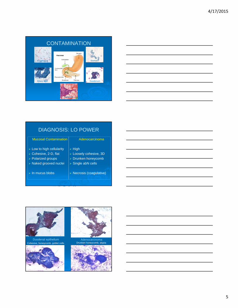
CONTAMINATION
Esophagus Stomach
DuodenumBiliary duct
Pancreas
DIAGNOSIS: LO POWER
Mucosal Contamination
Low to high cellularity
Cohesive, 2-D, flat
Polarized groups
Naked grooved nuclei
In mucus blobs
Adenocarcinoma
High
Loosely cohesive, 3D
Drunken honeycomb
Single abN cells
Necrosis (coagulative)
Duodenal epithelium Cohesive, honeycomb, goblet cells
AdenocarcinomaDrunken honeycomb, atypia
4/17/2015
6
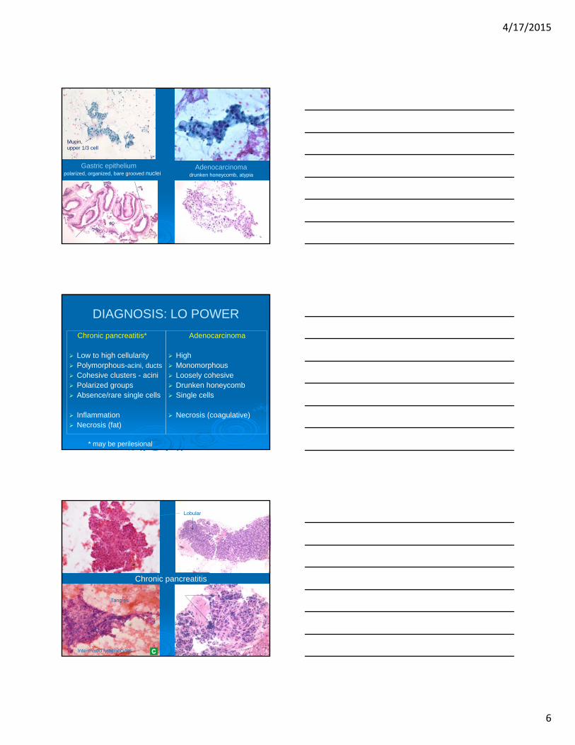
Gastric epitheliumpolarized, organized, bare grooved nuclei
Mucin, upper 1/3 cell
Adenocarcinomadrunken honeycomb, atypia
DIAGNOSIS: LO POWER
Chronic pancreatitis*
Low to high cellularity Polymorphous-acini, ducts
Cohesive clusters - acini Polarized groups Absence/rare single cells
Inflammation Necrosis (fat)
Adenocarcinoma
High Monomorphous Loosely cohesive Drunken honeycomb Single cells
Necrosis (coagulative)
* may be perilesional
Chronic pancreatitis
The worms
Intermixed lymphocytes
Tangles
Lobular
4/17/2015
7
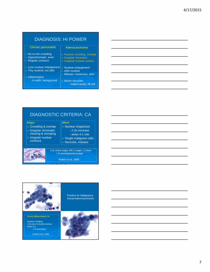
DIAGNOSIS: HI POWER
Chronic pancreatitis
No to min crowding Hypochromatic, even Regular contours
Less nuclear enlargement Tiny nucleoli, not abN
Inflammation- in epith, background
Adenocarcinoma
Nuclear crowding, overlap Irregular chromatin Irregular nuclear contour
Nuclear enlargement AbN nucleoli Mitoses: numerous, abN
Mucin vacuoles- indent nuclei, fill cell
DIAGNOSTIC CRITERIA: CA
Major Crowding & overlap Irregular chromatin
clearing & clumping Irregular nuclear
contours
Minor Nuclear shape/size
- 2-3x increase- aniso 4:1 rule
Single malignant cells Necrosis, mitoses
2 or more major OR 1 major, 3 minor= hi sens/spec/accuracy
Robins et al, 1995
Positive for MalignancyDuctal Adenocarcinoma
Poorly differentiated CA
Nuclear molding2-3x size N ductal nucleusAniso 4:1
= hi sens/spec
Cohen et al, 1991
4/17/2015
8
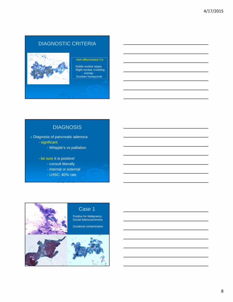
DIAGNOSTIC CRITERIA
Well differentiated CA
Subtle nuclear atypiaSlight nuclear crowding,
overlapDrunken honeycomb
DIAGNOSIS
Diagnosis of pancreatic adenoca
- significant
- Whipple’s vs palliation
- be sure it is positive!
- consult liberally
- internal or external
- LHSC: 40% rate
Case 1Positive for MalignancyDuctal Adenocarcinoma
Duodenal contamination
4/17/2015
9
Consider benign mimics for pancreas ca- pancreatitis, GI contaminants
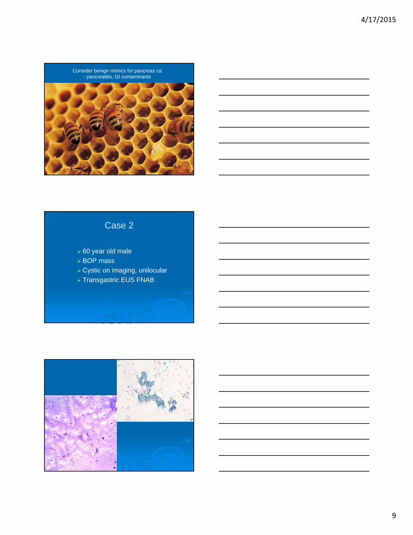
Case 2
60 year old male
BOP mass
Cystic on imaging, unilocular
Transgastric EUS FNAB
4/17/2015
10
APPROACH
Clinical & imaging important- if cystic - use algorithm for DDX
- check cyst chemistry
Microscopic approach- Adequacy
- Background- Contamination
- Diagnosis
ALGORITHM:Cystic Pancreas Mass EUS FNAB
Non-neoplastic
Normal contaminant
- mucus
- GI epithelium
Pseudocyst
Other benign cysts
Neoplastic
Neoplastic Mucinous Cyst - MCN, IPMN
Serous Neoplasm
Any solid neoplasm
- PanNET, SPN, AC
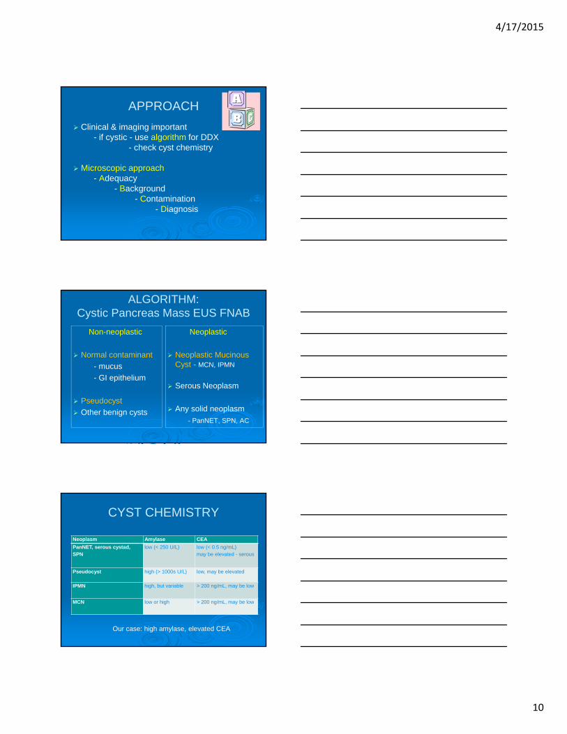
CYST CHEMISTRY
Neoplasm Amylase CEA
PanNET, serous cystad, SPN
low (< 250 U/L) low (< 0.5 ng/mL)may be elevated - serous
Pseudocyst high (> 1000s U/L) low, may be elevated
IPMN high, but variable > 200 ng/mL, may be low
MCN low or high > 200 ng/mL, may be low
Our case: high amylase, elevated CEA
4/17/2015
11
ADEQUACY: ROSE
Cystic lesion:- cyst fluid characteristics (vol, type)
- viscous (mucinous)- watery (serous)/ turbid (pseudocyst)
- cyst protocol: no microscopic ass’t- 4 smears: 1 air dried, 1 alcohol, Pap
2 mucin stains (PASd, AB2.5) - 1 ThinPrep +/- cell block
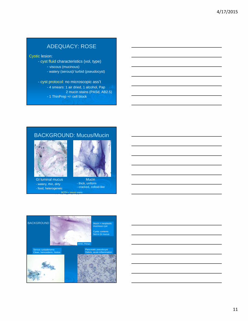
BACKGROUND: Mucus/Mucin
GI luminal mucus- watery, thin, dirty
- food, heterogeneic
Mucin- thick, uniform- cracked, colloid-like
BOTH + mucin stains
Serous cystadenomaClean, hemosiderin, histios
Pancreatic pseudocystDebris, acute inflammation
BACKGROUND
WHO, Pitman
Mucin = neoplastic mucinous cyst
Cystic contents Not in GI mucus
4/17/2015
12
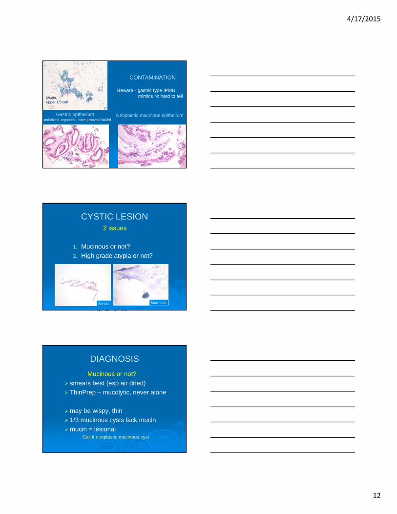
Gastric epitheliumpolarized, organized, bare grooved nuclei
Mucin, upper 1/3 cell
Neoplastic mucinous epithelium
CONTAMINATION
Beware - gastric type IPMNmimics N: hard to tell
CYSTIC LESION2 issues
1. Mucinous or not?
2. High grade atypia or not?
Serous Mucinous
DIAGNOSIS
Mucinous or not?
smears best (esp air dried)
ThinPrep – mucolytic, never alone
may be wispy, thin
1/3 mucinous cysts lack mucin
mucin = lesional• Call it neoplastic mucinous cyst
4/17/2015
13
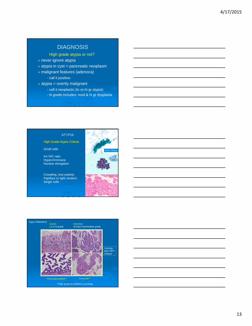
DIAGNOSISHigh grade atypia or not?
never ignore atypia
atypia in cyst = pancreatic neoplasm
malignant features (adenoca)
- call it positive
atypia < overtly malignant
- call it neoplastic (lo vs hi gr atypia)
- hi grade includes: mod & hi gr dysplasia
ATYPIA
High Grade Atypia Criteria
Small cells
Inc N/C ratioHyperchromasiaNuclear elongation
Crowding, loss polarityPapillary or tight clustersSingle cells
WHO, Pitman
GastricLo or hi grade
IntestinalAt least intermediate grade
Pancreaticobiliary** Oncocytic**
**High grade by definition (cytology)
Cytology does NOT subtype
Types IPMN/MCN
4/17/2015
14
Cyst contents: Present/Absent
Mucin: Present/AbsentMucin Stains: Positive/Negative/NA
Lesional Cells: Mucinous/Serous/OtherAtypia: Low/High grade
Mucosal Contamination: Gastric/Duodenal Present/Absent
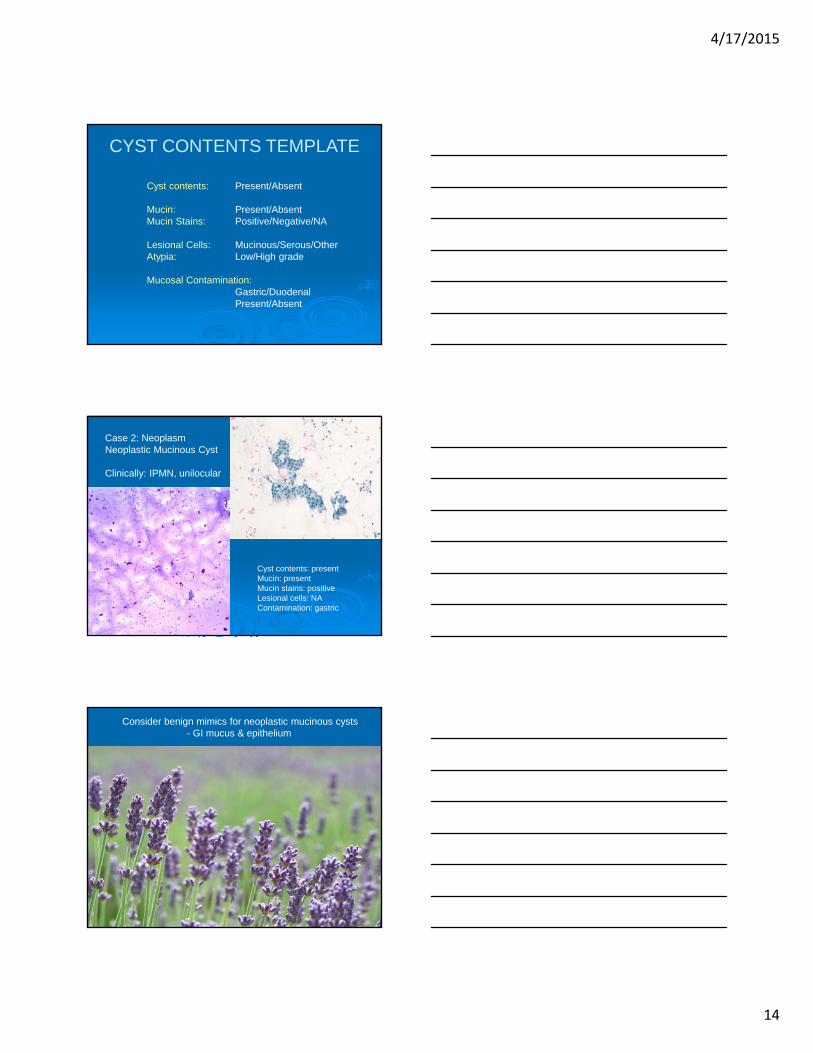
CYST CONTENTS TEMPLATE
Case 2: NeoplasmNeoplastic Mucinous Cyst
Clinically: IPMN, unilocular
Cyst contents: presentMucin: presentMucin stains: positiveLesional cells: NAContamination: gastric
Consider benign mimics for neoplastic mucinous cysts- GI mucus & epithelium
4/17/2015
15
Take home pointsFor solid pancreas eus fnab
- use malignancy criteria
- consider benign mimics
- consult liberally
For cystic pancreas eus fnab
- identify mucin –it’s enough for dx
- consider benign mimics
- look for mucinous epi, atypia
“Being challenged in life is inevitable, being defeated is optional.”
Roger Crawford