Embed Size (px)
DESCRIPTION
Diagnosis of Pregnancy. Departmentment of ob & gyn Renmin Hospital of Wuhan University Zhuoni Xiao. Woman’s reproductive period. Menarche. Menopause. 13-45 years. Duration of Pregnancy. 10 lunar months ; 9 calendar months and 7 days ; 280 days ; 40 weeks. - PowerPoint PPT Presentation
Citation preview

Diagnosis of PregnancyDiagnosis of Pregnancy
Departmentment of ob & gynDepartmentment of ob & gynRenmin Hospital of Wuhan UnivRenmin Hospital of Wuhan Univ
ersity ersity Zhuoni XiaoZhuoni Xiao
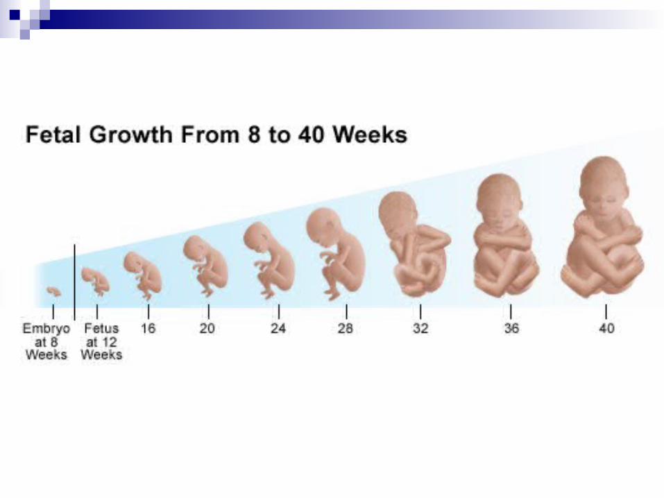

Woman’s reproductive periodWoman’s reproductive period
MenarcheMenarche MenopauMenopausese
13-45 years13-45 years


Duration of Pregnancy
10 lunar months ;10 lunar months ;9 calendar months and 7 days ;9 calendar months and 7 days ;280 days ;280 days ;40 weeks 40 weeks
Menstrual or gestational age Menstrual or gestational age

Fertilization or Ovulatory age Fertilization or Ovulatory age
280 days -14 days=266 days280 days -14 days=266 days

First trimester: first 12 weeksFirst trimester: first 12 weeks Second trimester: 13-28 weeksSecond trimester: 13-28 weeks Last trimester: 29-40 weeksLast trimester: 29-40 weeks
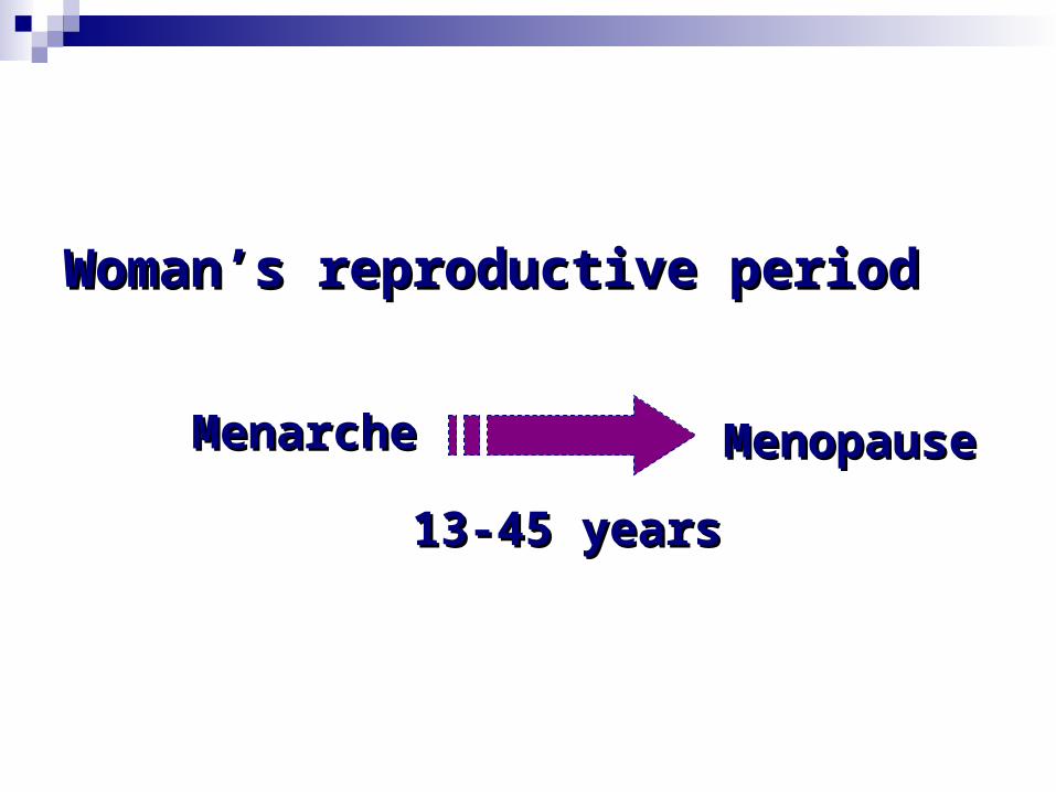
BladderBladderRectuRectumm
First trimesterFirst trimester
Subjective SymptomsSubjective Symptoms Objective SignsObjective Signs Immunological TestsImmunological Tests Ultra SonographUltra Sonograph
Pregnancy DiagnosisPregnancy Diagnosis

Subjective SymptomsSubjective Symptoms
AmenorrhoeaAmenorrhoea Warning :cyclic bleeding may last up to 12 weeks;Warning :cyclic bleeding may last up to 12 weeks; scanty and short time;scanty and short time; pathological bleeding ,such as miscarriagpathological bleeding ,such as miscarriag
e e
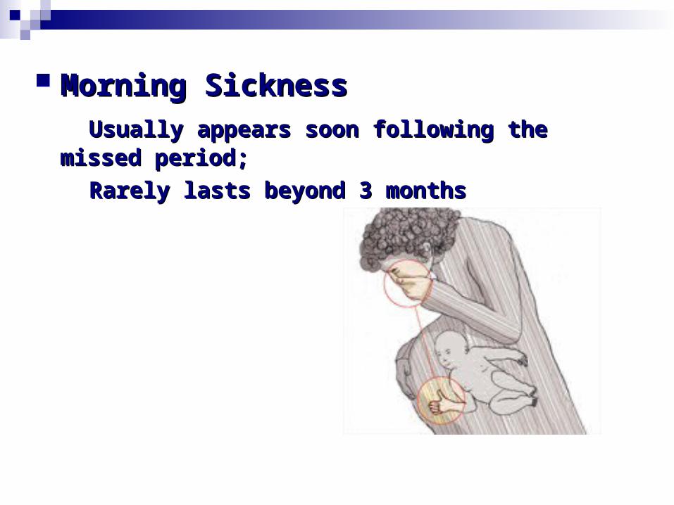
Morning SicknessMorning Sickness Usually appears soon following the missed Usually appears soon following the missed
period;period;
Rarely lasts beyond 3 monthsRarely lasts beyond 3 months


Frequence of micturitionFrequence of micturition troublesome symptom during 8-12 weekstroublesome symptom during 8-12 weeks enlarged uterusenlarged uterus congestion of bladdercongestion of bladder change in maternal osmoregulationchange in maternal osmoregulation

Breast discomfortBreast discomfort FullnessFullness
Pricking sensationPricking sensation

FatigueFatigue Occur early in pregnancyOccur early in pregnancy
Objective SignsObjective Signs
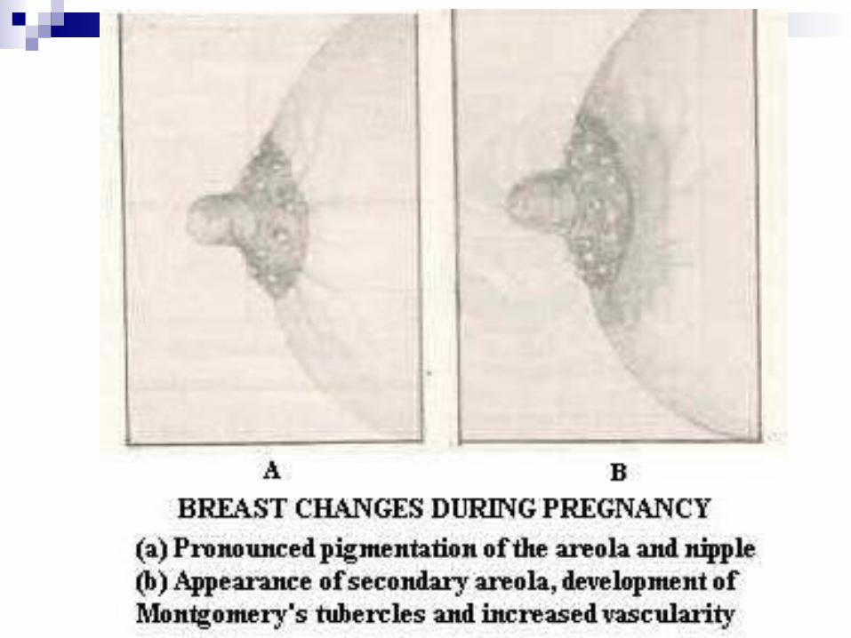
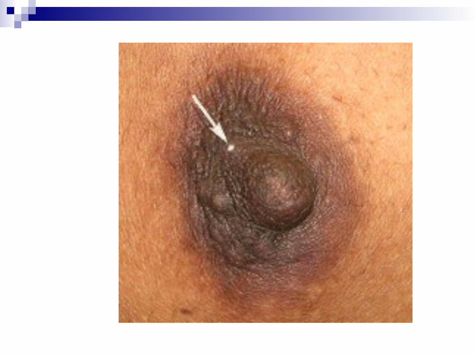
Breast changesBreast changes valuable only in primigravidaevaluable only in primigravidae breast changes are evident between 6-8 weeksbreast changes are evident between 6-8 weeks (vascular engorgement & nipple and areola pigm(vascular engorgement & nipple and areola pigm
ent)ent) Montgomery’s tubercleMontgomery’s tubercle colostrum expressed as early as 12th weekscolostrum expressed as early as 12th weeks

Per AbdomenPer Abdomen Uterus remains a pelvis organ until 12 Uterus remains a pelvis organ until 12
weeksweeks
Pelvic ChangesPelvic Changes Chadwick’ signChadwick’ sign
Vaginal signVaginal sign
Cervical signCervical sign
Uterine signUterine sign

Chadwick’ sign Chadwick’ sign is a bluish discoloration is a bluish discoloration of the of the cervix, vagina, and and labia caused by the hormone estrogen which results in . It can be observed as early as 6-8 weeks after conception, and its presence is an early sign of pregnancy.
Vaginal sign (Osiander’s sign)Vaginal sign (Osiander’s sign) Bluish discolouration of anterior vaginal wallBluish discolouration of anterior vaginal wall Vaginal wall softenVaginal wall soften Mucoid dischargeMucoid discharge Increased pulsationIncreased pulsation

Cervical sign (Goodell’ sign)Cervical sign (Goodell’ sign) Become soft as early as 6th weekBecome soft as early as 6th week
Non pregnant Non pregnant uterusuterus
Pregnant Pregnant uterusuterus

Uterine signUterine signSize, shape and consistency Size, shape and consistency
6th week6th week 8th week8th week 12th week12th week
Asymmetric→SymmetricAsymmetric→Symmetric
The pregnant uterus feels The pregnant uterus feels softsoft and and elasticelastic

Hegar’s sign Hegar’s sign demonstrated between 6-10 weeksdemonstrated between 6-10 weeks
Upper part of the body of the uterus is enlarged bUpper part of the body of the uterus is enlarged by growing fetus;y growing fetus;
Lower part of the body of the uterus is empty and Lower part of the body of the uterus is empty and extremely soft;extremely soft;
The cervix is comparatively firmThe cervix is comparatively firm

Two fingers in the anterior fTwo fingers in the anterior fornixornix
The abdominal fingers behind the uterusThe abdominal fingers behind the uterus
The abdominal and vaginalThe abdominal and vaginal
fingers seem oppose fingers seem oppose
below the body of uterusbelow the body of uterus
Palmer’s signPalmer’s sign Regular and rhythmic uterine contraction can be Regular and rhythmic uterine contraction can be
elicited doring biomanual examination as early aelicited doring biomanual examination as early as 4-8 weekss 4-8 weeks

Immunological TestImmunological Test

Principle of pregnancy testPrinciple of pregnancy test detection of the antigen of HCG present in tdetection of the antigen of HCG present in t
he maternal urine or serumhe maternal urine or serum Selectionn of time Selectionn of time 8-10 days after conception8-10 days after conception Collection of urineCollection of urine the first voided urine in the morning in a clthe first voided urine in the morning in a cl
ean containerean container

Ultra Sonograph Ultra Sonograph Gestation sac (GS) can be identified as early Gestation sac (GS) can be identified as early
as 29-35 days of gestation as 29-35 days of gestation
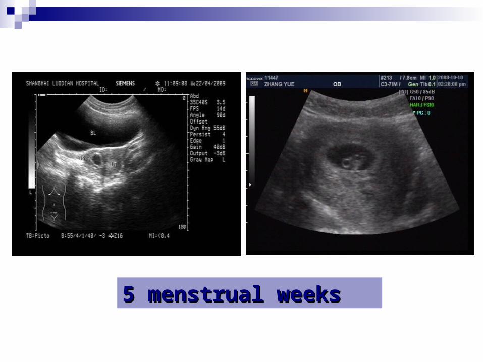
5 menstrual weeks5 menstrual weeks

6 menstrual weeks6 menstrual weeks

True and pseudo gestation sacTrue and pseudo gestation sac
CharacterCharacter True GSTrue GS Pseudo GSPseudo GSLocationLocation EccentricallyEccentrically CentrallyCentrally
ShapeShape Round & Round & regularregular
IrregularIrregular
Double ring signDouble ring sign PresentPresent AbsentAbsent
York sac and fetal York sac and fetal polepole
PresentPresent AbsentAbsent
Increase in sac Increase in sac sizesize
1 mm/ day1 mm/ day AbsentAbsent

First Trimester ReviewFirst Trimester Review
The Whole Period of Pregnancy The Whole Period of Pregnancy Can Be Divided Into Three StagesCan Be Divided Into Three StagesThe first trimester (early pregnancy): 1-12wThe first trimester (early pregnancy): 1-12wThe second trimester (middle pregnancy): The second trimester (middle pregnancy):
13-27 w13-27 wThe third trimester (late pregnancy): 28-40The third trimester (late pregnancy): 28-40
ww

1. History and symptoms1. History and symptoms
A. Cessation of menstruationA. Cessation of menstruation
This is the first frequent symptom of pregnancy, This is the first frequent symptom of pregnancy, although although
a few women may have slight bleeding after a few women may have slight bleeding after conception.conception. Amenorrhea is not only due to pregnancyAmenorrhea is not only due to pregnancy
but also other reasons. but also other reasons. Women of breast feeding may be pregnant Women of breast feeding may be pregnant before the recovery of mensesbefore the recovery of menses.
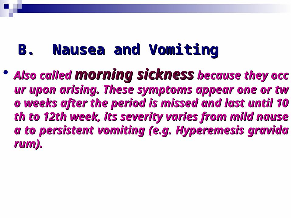
B. Nausea and VomitingB. Nausea and Vomiting Also called Also called morning sicknessmorning sickness because they because they
occur upon arising. These symptoms appear onoccur upon arising. These symptoms appear one or two weeks after the period is missed and lae or two weeks after the period is missed and last until 10th to 12th week, its severity varies frost until 10th to 12th week, its severity varies from mild nausea to persistent vomiting (e.g. Hypm mild nausea to persistent vomiting (e.g. Hyperemesis gravidarum).eremesis gravidarum).

C. Urinary symptomsC. Urinary symptoms
Increased frequency of urination is due to Increased frequency of urination is due to incincreased circulationreased circulation associated with the eff associated with the effect of ect of estrogen and progesteroneestrogen and progesterone on th on the bladder, combined with e bladder, combined with pressure by the pressure by the gradually enlarged uterus on the blagradually enlarged uterus on the bladder.dder.
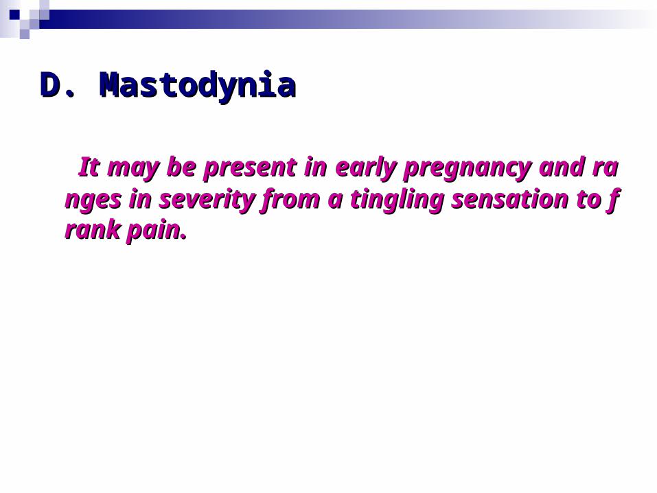
D. MastodyniaD. Mastodynia
It may be present in early pregnancy and It may be present in early pregnancy and ranges in severity from a tingling sensatioranges in severity from a tingling sensation to frank pain.n to frank pain.
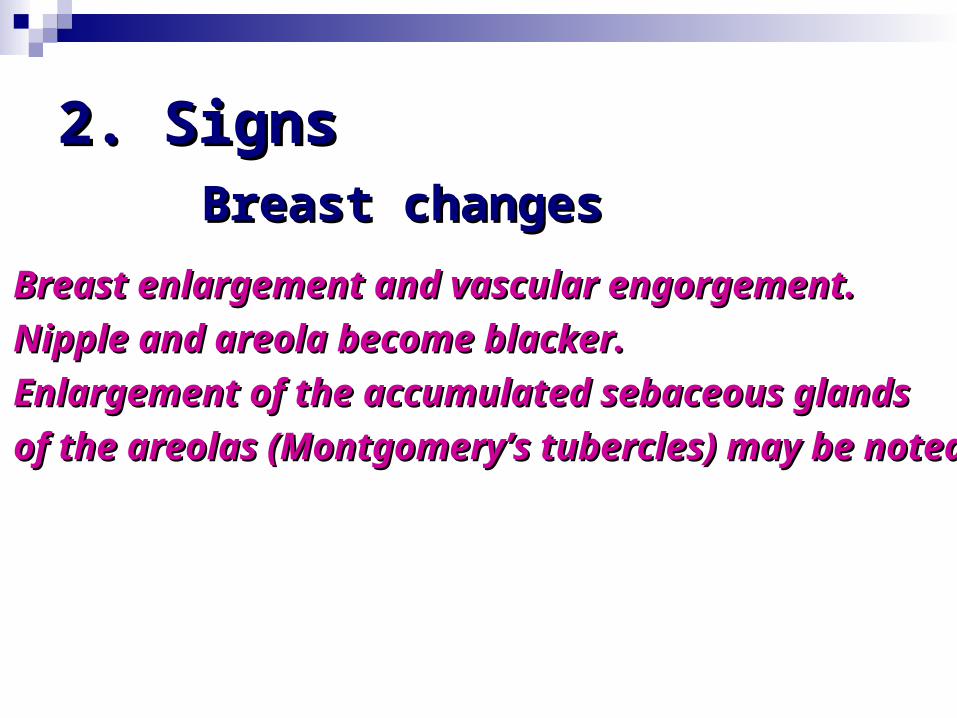
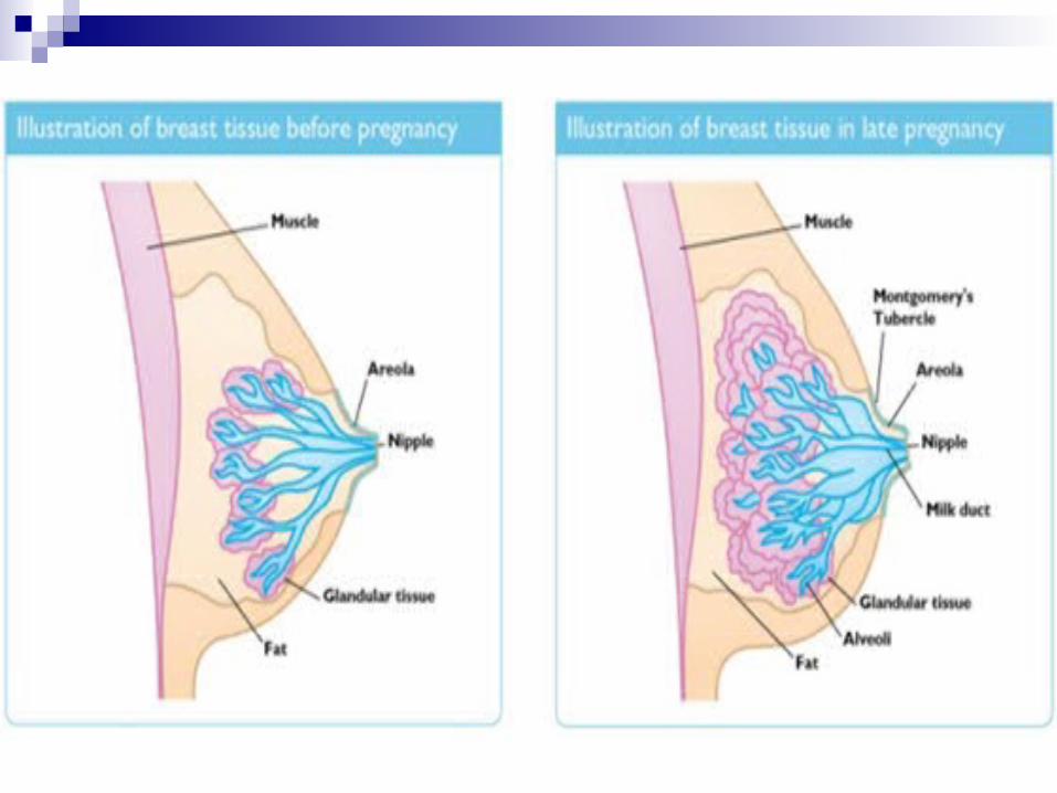
2. Signs2. SignsBreast changesBreast changes
Breast enlargement and vascular engorgement. Breast enlargement and vascular engorgement. Nipple and areola become blacker. Nipple and areola become blacker. Enlargement of the accumulated sebaceous glands Enlargement of the accumulated sebaceous glands of the areolas (Montgomery’s tubercles) may be noted.of the areolas (Montgomery’s tubercles) may be noted.

Changes of the reproductive Changes of the reproductive organsorgans
Vagina:Vagina: The vaginal wall become discol The vaginal wall become discoloration as the pelvic blood vessel becomes oration as the pelvic blood vessel becomes congested.congested.
Cervix:Cervix: Cyanosis and a gradual softenin Cyanosis and a gradual softening due to congestion.g due to congestion.
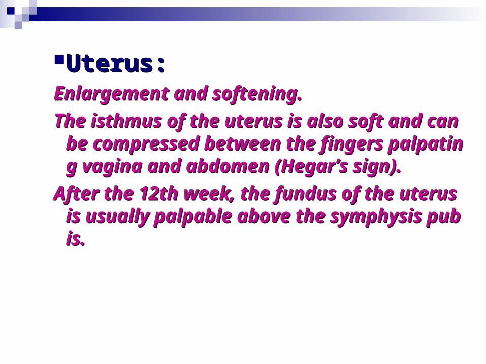
Uterus:Uterus: Enlargement and softening. Enlargement and softening. The isthmus of the uterus is also soft and cThe isthmus of the uterus is also soft and c
an be compressed between the fingers paan be compressed between the fingers palpating vagina and abdomen (Hegar’s silpating vagina and abdomen (Hegar’s sign). gn).
After the 12th week, the fundus of the uterAfter the 12th week, the fundus of the uterus is usually palpable above the symphysus is usually palpable above the symphysis pubis. is pubis.

3. Supplementary 3. Supplementary examinationexamination
Pregnancy test Pregnancy test The laboratory test for pregnancy are based on the The laboratory test for pregnancy are based on the identification of human chorionic gonadotropin (hCG), identification of human chorionic gonadotropin (hCG), which can be detected as early as 7-9 days after fertilizationwhich can be detected as early as 7-9 days after fertilizationby high sensitive technique. by high sensitive technique. The samples may be blood or urine.The samples may be blood or urine.
Basal body temperature (BBT)Basal body temperature (BBT)
A persistent elevation of BBT for longer than 18 days A persistent elevation of BBT for longer than 18 days may be presumptive evidence of pregnancy.may be presumptive evidence of pregnancy.

Progesterone testProgesterone test
ProgesteroneProgesterone is given to a women with amenorrhea. If she is pregnant, no bleeding will follow, otherwise, bleeding should occur within 7-10 days of progesterone administration. This is reliable in the nonpregnant patient only if there is adequate estrogen stimulation of the endometrium.

UltrasonographyUltrasonographyThere are trans-vaginal and abdominal Ultrasonagraphys. A gestational sac can usually be identified at 5-6 weeksafter the beginning of the last period. Fetal heart beating can be detected by about 7th week and the fetus itself can be seen by about the 8th week. Doppler is also an ultrasound technique, which diagnoses the pregnancy by revealing the heart beating.
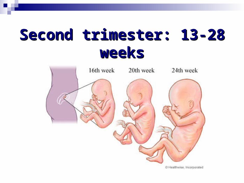
Second trimester: 13-28 Second trimester: 13-28 weeksweeks
SymptomsSymptoms General examinationGeneral examination Abdominal examinationAbdominal examination Vaginal examinationVaginal examination

SymptomsSymptoms Nausea, vomiting, frequency of micturition subsideNausea, vomiting, frequency of micturition subside Amenorrhea continuesAmenorrhea continues Quickening: perception of active fetal movement by Quickening: perception of active fetal movement by
womenwomen (From 18th week)(From 18th week) Progressive enlargement of lower abdomen by the gProgressive enlargement of lower abdomen by the g
rowing uterusrowing uterus


General examinationGeneral examination Chloasma 24th weekChloasma 24th week
Breast changes: more enlargedBreast changes: more enlarged
PigmentationPigmentation

Abdominal examinationAbdominal examination InspectionInspection
Palpation Palpation
AuscultationAuscultation

InspectionInspection Linea nigraLinea nigra StriaeStriae

Symphysis PubisSymphysis Pubis
Ensiform CartilageEnsiform Cartilage
StriaeStriae

PalpationPalpation Fundal height increasesFundal height increases Uterus soft and elastic, ovoid in shapeUterus soft and elastic, ovoid in shape Braxton-Hicks ContractionBraxton-Hicks Contraction Palpation of fetal parts: 20th weekPalpation of fetal parts: 20th week Active fetal movements: 20th weekActive fetal movements: 20th week External ballottement External ballottement
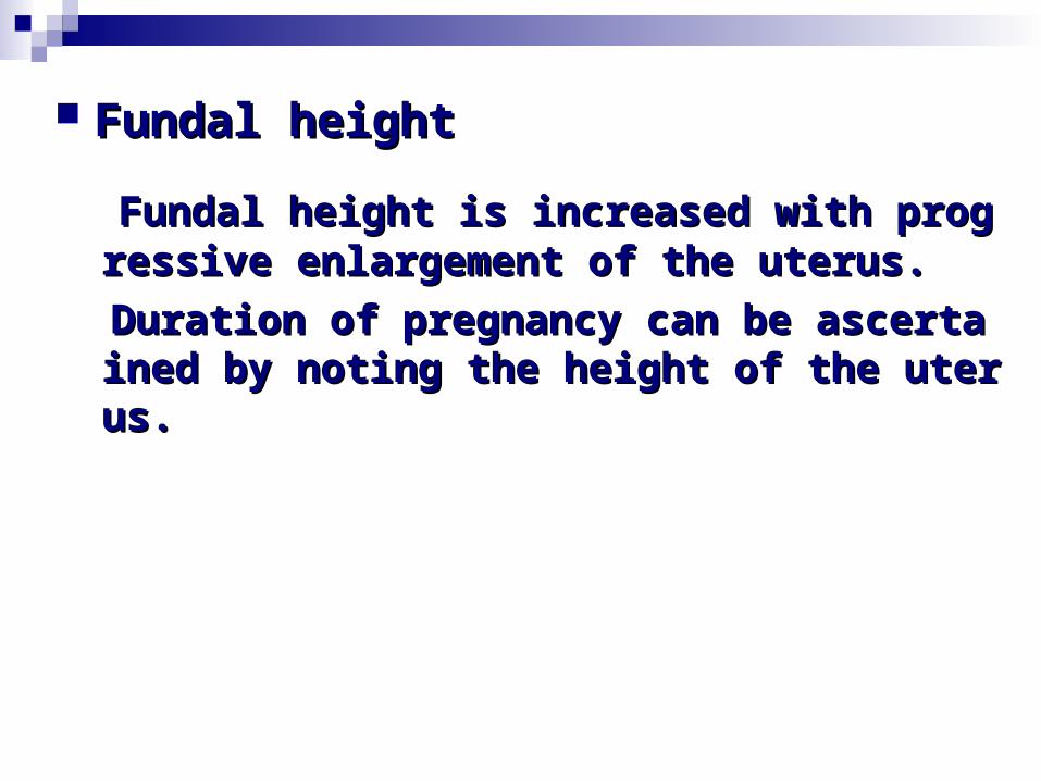
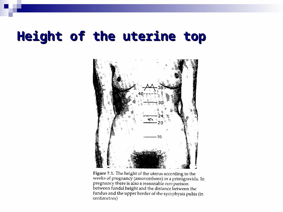
Fundal height is increased with progressive Fundal height is increased with progressive enlargement of the uterus.enlargement of the uterus.
Duration of pregnancy can be ascertained by Duration of pregnancy can be ascertained by noting the height of the uterus.noting the height of the uterus.
Fundal heightFundal height


16th week:16th week: midway between symphysis p midway between symphysis pubis and umbilicusubis and umbilicus
22~24th week:22~24th week: at the level of umbilicus at the level of umbilicus 28th week:28th week: at the junction of the lower 1/ at the junction of the lower 1/
3 and upper 2/3 of the distance between th3 and upper 2/3 of the distance between the umbilicus and ensiform cartilagee umbilicus and ensiform cartilage

Abnormal Fundal HeightAbnormal Fundal Height IUGR (intrauterine growth retardation)IUGR (intrauterine growth retardation) Multiple PregnancyMultiple Pregnancy Polyhydramnios(CNS or Cardiovascular DisfPolyhydramnios(CNS or Cardiovascular Disf
unction)unction) OligohydramniosOligohydramnios

Braxton-Hicks ContractionBraxton-Hicks Contraction
In 1872, John Braxton Hicks investigated the later In 1872, John Braxton Hicks investigated the later stages of pregnancy and noted that many women feltstages of pregnancy and noted that many women feltcontractions without being near birth.contractions without being near birth. This process was usually painless but caused women This process was usually painless but caused women
confusionconfusionas to whether or not they were going into actual labor as to whether or not they were going into actual labor
CauseCause
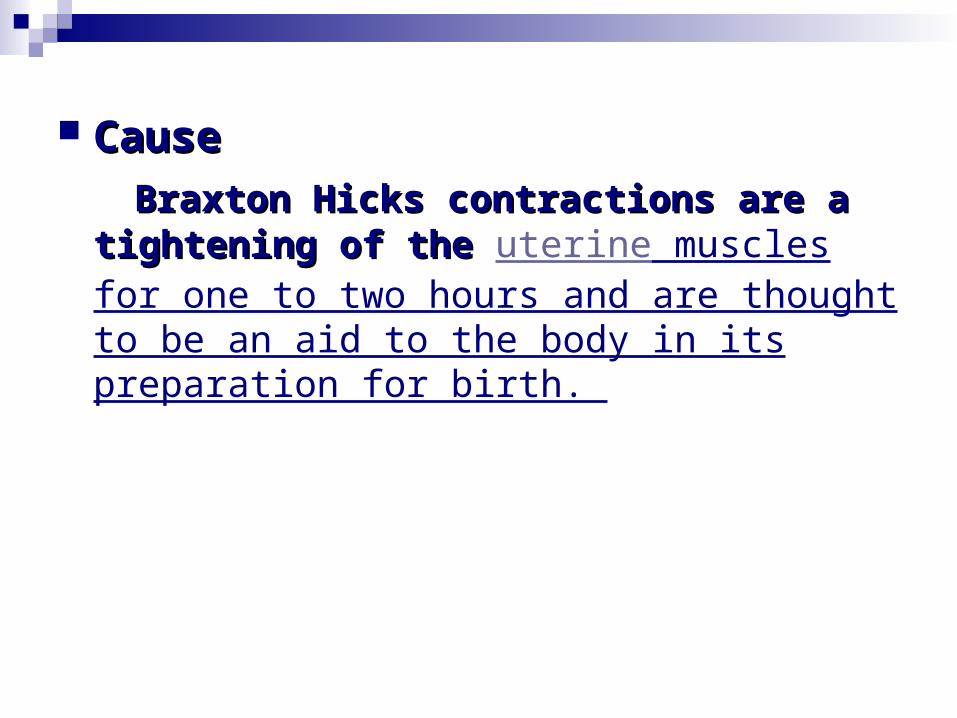
Braxton Hicks contractions are a Braxton Hicks contractions are a tightening of the tightening of the uterine muscles for one to two hours and are thought to be an aid to the body in its preparation for birth.

Alleviating factorsAlleviating factors Rhythmic breathing Rhythmic breathing
Lying down on the left side Lying down on the left side
A slight change in movement A slight change in movement
Urination Urination

Very early, the uterus undergoes spontaneous cVery early, the uterus undergoes spontaneous contractionontraction
Firmer at one moment and soft at anotherFirmer at one moment and soft at another Can be excited by rubbing the uterusCan be excited by rubbing the uterus Irregular,infrequent, spasmodic, and painessIrregular,infrequent, spasmodic, and painess Near term, frequent with increase in intensity, Near term, frequent with increase in intensity,
discomfortdiscomfort Merge with the labor Merge with the labor

Palpation of fetal partsPalpation of fetal parts Diagnosis of pregnancyDiagnosis of pregnancy Identify the presentation and position of fIdentify the presentation and position of f
etusetus


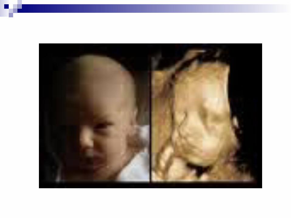
Active fetal movementsActive fetal movementsPositive evidence of pregnancy & live fetusPositive evidence of pregnancy & live fetusFaint flutter→stronger movementFaint flutter→stronger movement

External ballottementExternal ballottement
Be elicited as early as 20th weekBe elicited as early as 20th week Obese women & scanty liquor amniiObese women & scanty liquor amnii
by a push to the foetal parts with one hand abdominally by a push to the foetal parts with one hand abdominally and the other hand receiving the impulse and the other hand receiving the impulse

Ballottement Ballottement is a medical sign which indicatis a medical sign which indicates increased fluid in the suprapatellar pouch over es increased fluid in the suprapatellar pouch over the patella at the knee joint. To test ballottement tthe patella at the knee joint. To test ballottement the examiner would apply downward pressure tohe examiner would apply downward pressure towards the foot with one hand, while pushing the wards the foot with one hand, while pushing the patella backwards against the femur with one finpatella backwards against the femur with one finger of the opposite hand. ger of the opposite hand.

AscutationAscutation
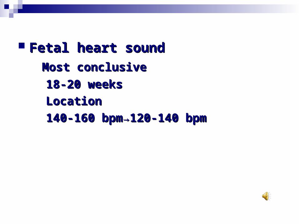
Fetal heart soundFetal heart sound Most conclusiveMost conclusive 18-20 weeks18-20 weeks LocationLocation 140-160 bpm→120-140 bpm140-160 bpm→120-140 bpm
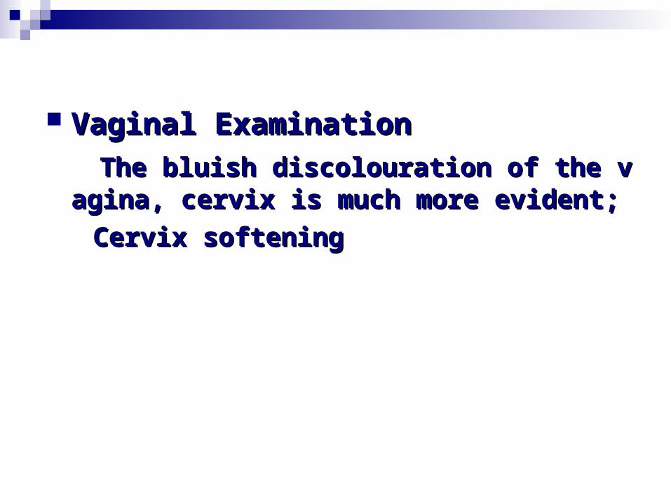
Vaginal ExaminationVaginal Examination The bluish discolouration of the vagina, ceThe bluish discolouration of the vagina, ce
rvix is much more evident;rvix is much more evident; Cervix softeningCervix softening

InvestigationInvestigation
Sonograph: 12-20 weeks; a detailed survey Sonograph: 12-20 weeks; a detailed survey of fetal anatomy, placenta localization, intof fetal anatomy, placenta localization, integrity of the cervical canalegrity of the cervical canal
Fetal organ anatomyFetal organ anatomy Radiologic examinationRadiologic examination

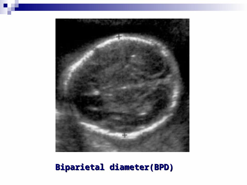
Biparietal diameter(BPD)Biparietal diameter(BPD)

BPDBPD
First Trimester: < 3cmFirst Trimester: < 3cm From 20th week: = pregnancy From 20th week: = pregnancy
month(28th week=7cm; 32th month(28th week=7cm; 32th week=8cmweek=8cm
From 32th week = 8cm: From 32th week = 8cm: 02.cm/week02.cm/week
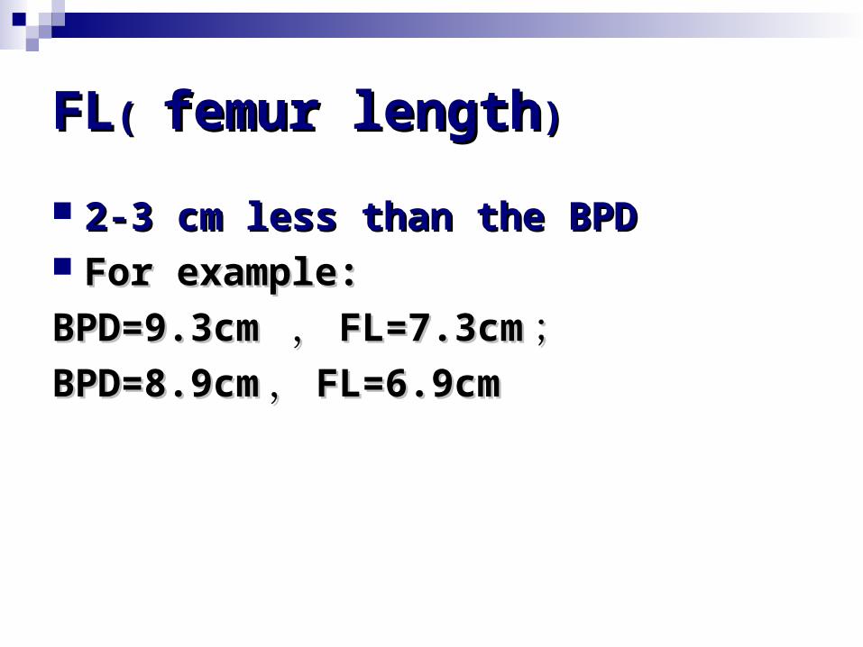
FLFL( ( femur lengthfemur length))
2-3 cm less than the BPD2-3 cm less than the BPD For example:For example:
BPD=9.3cm BPD=9.3cm ,, FL=7.3cmFL=7.3cm ;;BPD=8.9cmBPD=8.9cm ,, FL=6.9cmFL=6.9cm


Second Trimester ReviewSecond Trimester Review
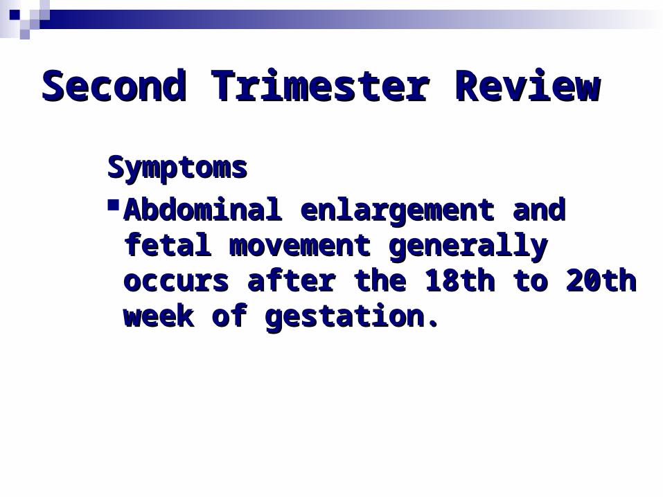
SymptomsSymptomsAbdominal enlargement and Abdominal enlargement and fetal movement generally fetal movement generally occurs after the 18th to 20th occurs after the 18th to 20th week of gestation.week of gestation.

SignsSigns The uterus continues to enlargeThe uterus continues to enlarge Fetal movement Fetal movement (quickening)(quickening) ca ca
n usually be seen or heard after 1n usually be seen or heard after 18th week of gestation8th week of gestation
Height of the uterine topHeight of the uterine top

SignsSigns
Fetal heart sound can be heard at Fetal heart sound can be heard at rate varies from rate varies from 120 to 160 beats 120 to 160 beats per minuteper minute..
The fetal body can usually be The fetal body can usually be palpated by the palpated by the 18th to 20th18th to 20th week week of gestation unless the patient is of gestation unless the patient is too fat, the abdomen is tender or too fat, the abdomen is tender or there is an excessive amount of there is an excessive amount of amniotic fluid.amniotic fluid.
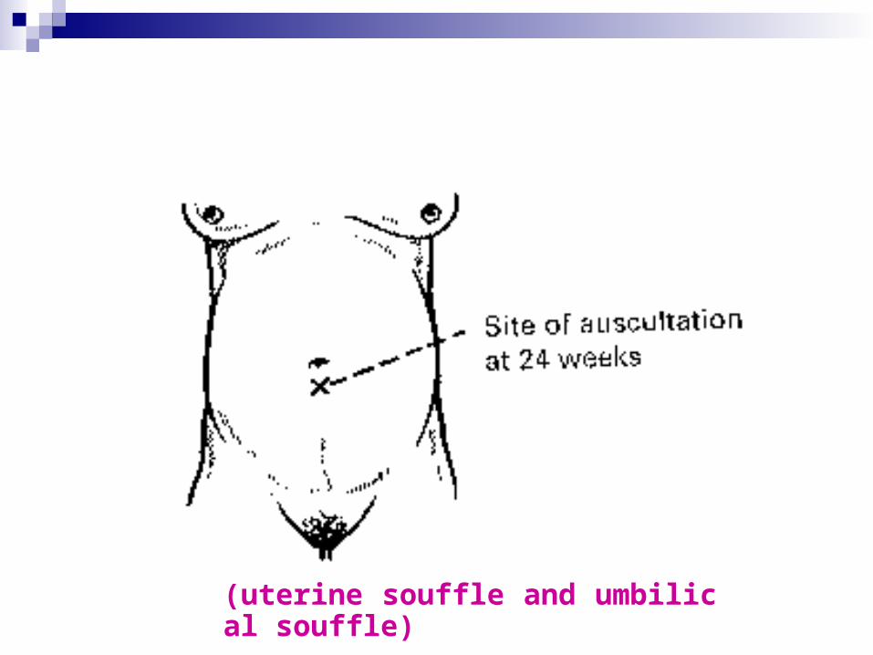
(uterine souffle and umbilical souffle)

ballottement


Third trimester: 29-40 Third trimester: 29-40 weeksweeks

Symptoms:Symptoms: AmenorrhoeaAmenorrhoea Enlargement of the abdomen Enlargement of the abdomen Lightening: due to the engagement of the presentiLightening: due to the engagement of the presenti
ng partng part Frequency of micturitionFrequency of micturition Fetal movementFetal movement
Sign:Sign: Cutaneous changes: increased pigmentation and strCutaneous changes: increased pigmentation and str
iaeiae Uterine shape: cylindrical to spherical beyond 36th Uterine shape: cylindrical to spherical beyond 36th
weekweek Fundal heightFundal height Braxton-Hicks contraction Braxton-Hicks contraction Fetal movementFetal movement Palpation of the fetal partsPalpation of the fetal parts


3232th week: the junction of the upper andth week: the junction of the upper and middle third between the distance middle third between the distance of umbilicus and ensiform cartilageof umbilicus and ensiform cartilage
3636th week: the level of the ensiform cartilageth week: the level of the ensiform cartilage
4040th week: down to the level of 32th th week: down to the level of 32th

Symphysis fundal height (SFH)Symphysis fundal height (SFH)
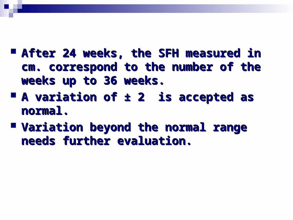
After 24 weeks, the SFH measured in After 24 weeks, the SFH measured in cm. correspond to the number of the cm. correspond to the number of the weeks up to 36 weeks.weeks up to 36 weeks.
A variation of ± 2 is accepted as A variation of ± 2 is accepted as normal.normal.
Variation beyond the normal range Variation beyond the normal range needs further evaluation.needs further evaluation.

More than More than ① ① Mistaken date of the last menstrual period;Mistaken date of the last menstrual period; ② ② Twins;Twins; ③ ③ Polyhydramnios;Polyhydramnios; ④ ④ Big baby;Big baby; ⑤ ⑤ Pelvic tumours;Pelvic tumours; ⑥ ⑥ Hydatidiform mole;Hydatidiform mole; ⑦ ⑦ Concealed accidental haemorrhageConcealed accidental haemorrhage
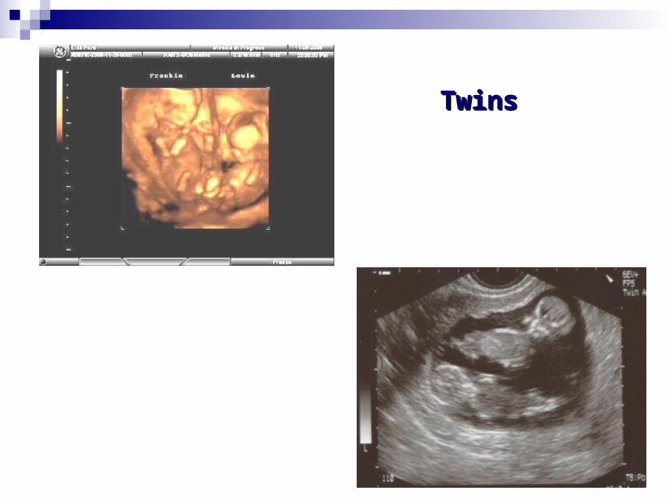
TwinsTwins

Hydatidiform moleHydatidiform mole
Less thanLess than
① ① Mistaken date of the last menstrual periMistaken date of the last menstrual period;od;
② ② Scanty liquor amnii;Scanty liquor amnii; ③ ③ Fetal growth retardation;Fetal growth retardation; ④ ④ Intrauterine fetal deathIntrauterine fetal death

Upper Upper partpart of the uterus of the uterus Lateral part of the uterusLateral part of the uterus
Presentation; Presentation; EngagementEngagement
Further confirmationFurther confirmation

→ → breechbreech
→ → headhead
Fundal gripFundal grip broad, soft, irregular mass broad, soft, irregular mass smooth, hard, globular smooth, hard, globular

Lateral or umbilical gripLateral or umbilical grip smooth curved and resistant →blacksmooth curved and resistant →black
comparatively empty and small knob comparatively empty and small knob → limb→ limb

First pelvic gripFirst pelvic grip Presentation: the part occupy the lower pole Presentation: the part occupy the lower pole
of the uterusof the uterus Attitude: the relative position of the sincipitAttitude: the relative position of the sincipit
al and occipital or different parts of the fetal and occipital or different parts of the fetus to one anotherus to one another
Engagement: convergence or divergence of tEngagement: convergence or divergence of the finger during palpationhe finger during palpation
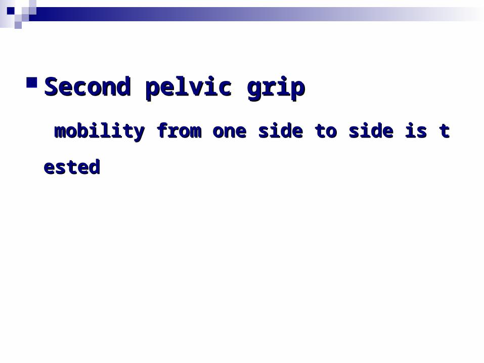
Second pelvic grip Second pelvic grip
mobility from one side to side is testedmobility from one side to side is tested
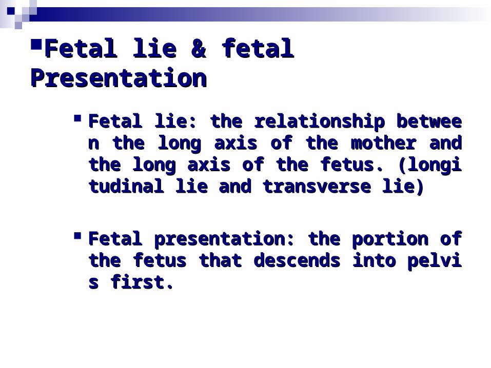
Fetal lie & fetal PresentationFetal lie & fetal Presentation
Fetal lie: the relationship between the Fetal lie: the relationship between the long axis of the mother and the long along axis of the mother and the long axis of the fetus. (longitudinal lie and txis of the fetus. (longitudinal lie and transverse lie)ransverse lie)
Fetal presentation: the portion of the fFetal presentation: the portion of the fetus that descends into pelvis first.etus that descends into pelvis first.
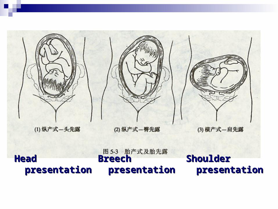
Head Head presentationpresentation
Breech Breech presentationpresentation
Shoulder Shoulder presentationpresentation
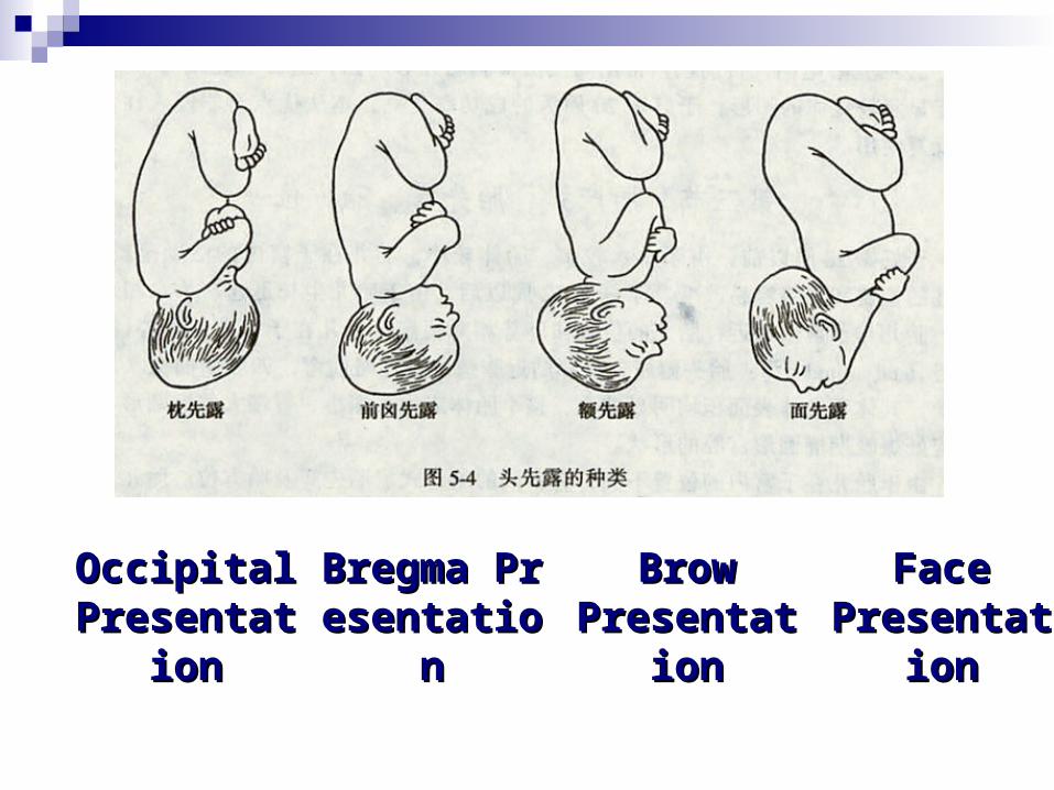
Face Face PresentatiPresentati
onon
Brow Brow PresentatiPresentati
onon
Occipital Occipital PresentatiPresentati
onon
Bregma PreBregma Presentationsentation
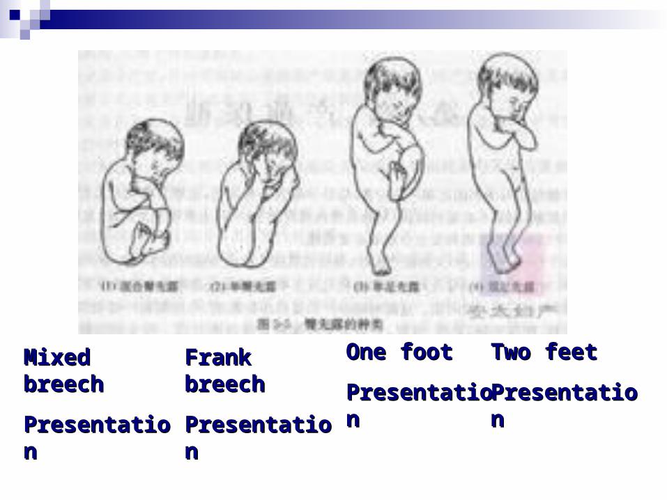
Mixed Mixed breechbreech
PresentatioPresentationn
Frank Frank breechbreech
PresentatioPresentationn
One footOne foot
PresentatioPresentationn
Two feetTwo feet
PresentationPresentation

Fetal positionFetal position
Fetal position: the relationship of somFetal position: the relationship of some guiding point of fetal presentation te guiding point of fetal presentation to a fined area of the maternal pelvis. o a fined area of the maternal pelvis. (LOA, left occipital anterior)(LOA, left occipital anterior)

PresentationPresentation
Occipital Presentation: Occipital, OOccipital Presentation: Occipital, OBreech Presentation: Sacrum, S Breech Presentation: Sacrum, S Face Presentation: Mentum, M Face Presentation: Mentum, M Shoulder Presentation: Scapula, Sc Shoulder Presentation: Scapula, Sc

sacrumsacrum ROPROP
LOALOA ROAROA
LOPLOP

Fetal Heart SoundFetal Heart Sound
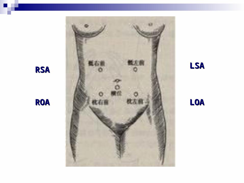
LOALOA
RSARSA LSALSA
ROAROA
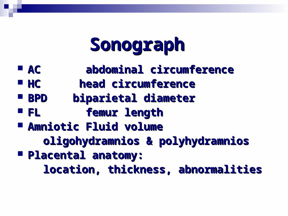
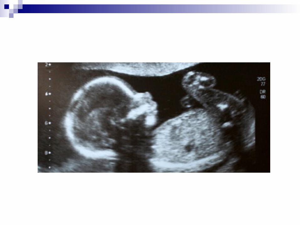
SonographSonograph AC abdominal circumferenceAC abdominal circumference HC head circumferenceHC head circumference BPD biparietal diameterBPD biparietal diameter FL femur lengthFL femur length Amniotic Fluid volume Amniotic Fluid volume oligohydramnios & polyhydramniosoligohydramnios & polyhydramnios Placental anatomy:Placental anatomy: location, thickness, abnormalitieslocation, thickness, abnormalities

Differential Diagnosis of Differential Diagnosis of PregnancyPregnancy
Pseudocyesis: Pseudocyesis: psychological disorder,psychological disorder, cessation of menstruationcessation of menstruation

Cystic ovarian tumourCystic ovarian tumour Swelling is slow;Swelling is slow; Amenorrhoea is absent;Amenorrhoea is absent; Feels cystic or tense cystic;Feels cystic or tense cystic; Absence of Braxton-Hicks contraction;Absence of Braxton-Hicks contraction; Absence of positive signs of pregnancy;Absence of positive signs of pregnancy; Ultrasonograph show absence of fetusUltrasonograph show absence of fetus

Uterine fibroid:Uterine fibroid: Slow growing;Slow growing; Amenorrhoea is absent;Amenorrhoea is absent; Feels firm , more towards hard;Feels firm , more towards hard; Absence of Braxton-Hicks contraction;Absence of Braxton-Hicks contraction; Absence of positive signs of pregnancy;Absence of positive signs of pregnancy; Ultrasonograph show absence of fetusUltrasonograph show absence of fetus
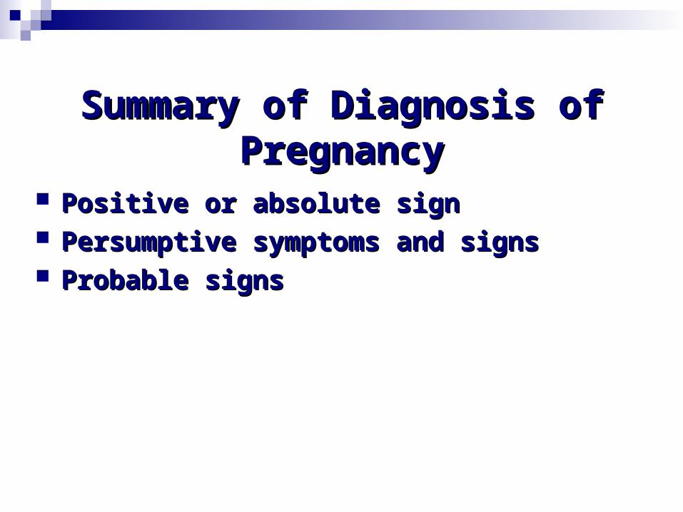
Summary of Diagnosis of Summary of Diagnosis of PregnancyPregnancy
Positive or absolute signPositive or absolute sign Persumptive symptoms and signsPersumptive symptoms and signs Probable signsProbable signs
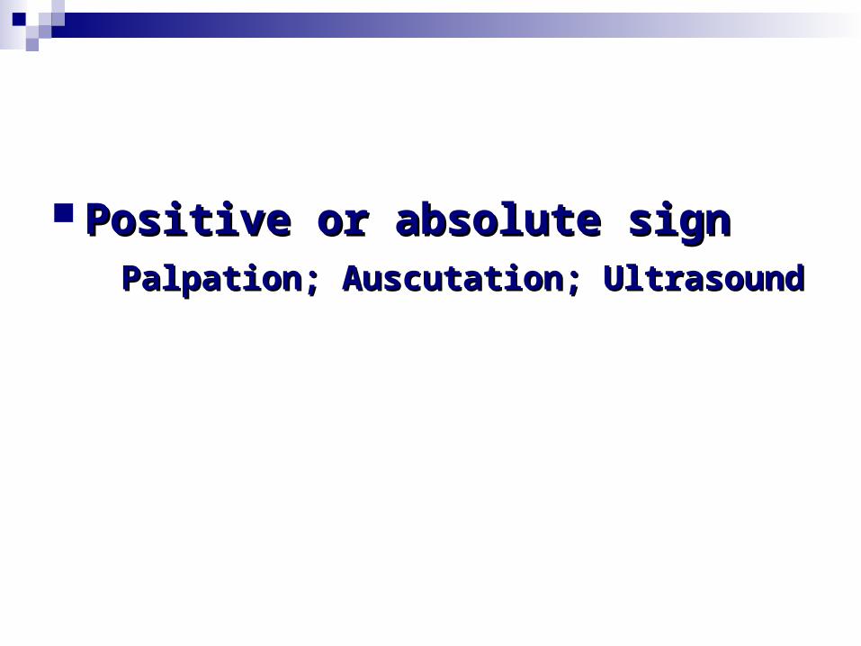
Positive or absolute signPositive or absolute sign Palpation; Auscutation; UltrasoundPalpation; Auscutation; Ultrasound
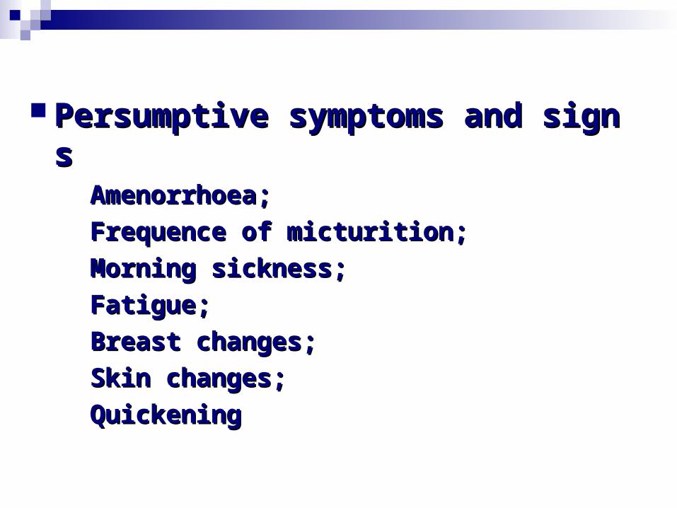
Persumptive symptoms and signsPersumptive symptoms and signs Amenorrhoea; Amenorrhoea; Frequence of micturition; Frequence of micturition; Morning sickness; Morning sickness; Fatigue;Fatigue; Breast changes; Breast changes; Skin changes; Skin changes; QuickeningQuickening
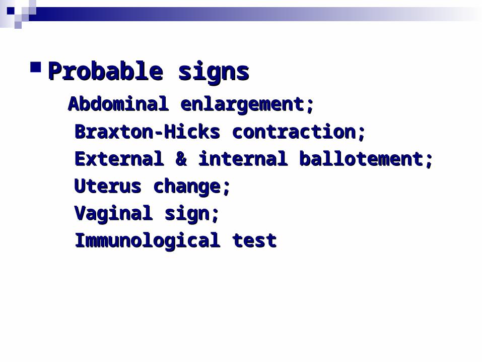
Probable signsProbable signs Abdominal enlargement;Abdominal enlargement; Braxton-Hicks contraction; Braxton-Hicks contraction; External & internal ballotement; External & internal ballotement; Uterus change;Uterus change; Vaginal sign; Vaginal sign; Immunological testImmunological test

Chronological AppearanceChronological Appearance At 6-8 weeksAt 6-8 weeks At 16th weekAt 16th week At 20th weekAt 20th week

Estimation of Gestation AgeEstimation of Gestation Age& &
Prediction of Excepted Date of Prediction of Excepted Date of DeliveryDelivery
Excepted Due Date Excepted Due Date
= LMP 3/ 9 month and 7 days﹣ ﹢ ﹢= LMP 3/ 9 month and 7 days﹣ ﹢ ﹢
LMP is 26th July, when is the EDD?LMP is 26th July, when is the EDD?

Patient’ statementPatient’ statement Date of coitusDate of coitus Naegele’s FormulaNaegele’s Formula Date of quickening: adding 22 -24 weeks Date of quickening: adding 22 -24 weeks

Excepted Due Date Excepted Due Date = LMP﹣3/﹢9 month and ﹢7 days= LMP﹣3/﹢9 month and ﹢7 days add seven daysadd seven days subtract 3 monthssubtract 3 months add one yearadd one year
LMP is 26th July, when is the EDD?LMP is 26th July, when is the EDD?

Previous recordPrevious record ClinicalClinical : : Size of the uterusSize of the uterus
Palpation of fetal partsPalpation of fetal parts
Investigation: Investigation: Ultrasonographic findingUltrasonographic finding
GS - 5th weekGS - 5th week
cardiac activity - 6th weekcardiac activity - 6th week
Embryo movement – 7th weekEmbryo movement – 7th week
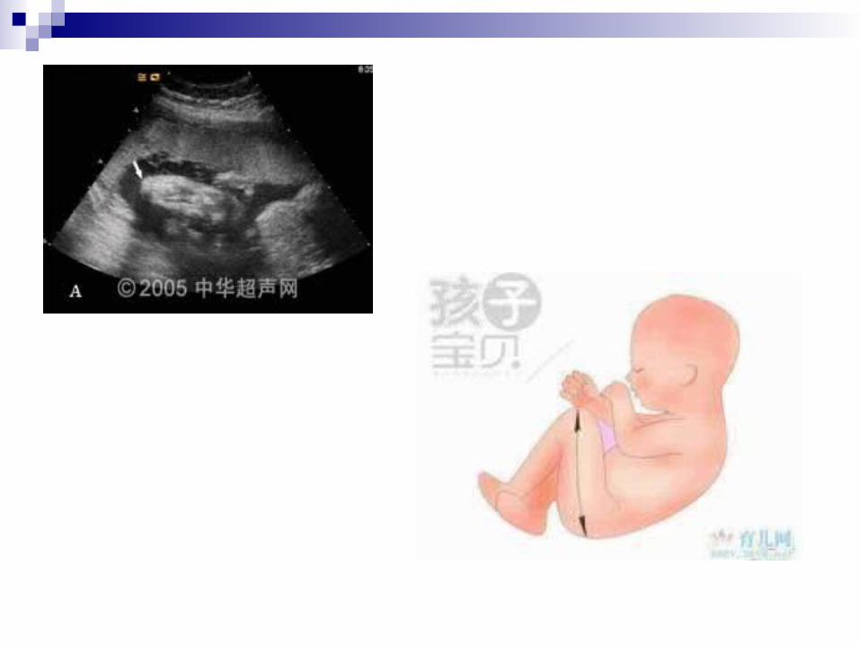
CRL in cm + 6.5 = week of pregnancyCRL in cm + 6.5 = week of pregnancy
(Crown-rump length)(Crown-rump length)
BPD in cm = month of pregnancy BPD in cm = month of pregnancy
(From 20th week)(From 20th week)

Objective signsObjective signs
Height of the uterusHeight of the uterus
LighteningLightening
Size of the fetusSize of the fetus
Vaginal examinationVaginal examination

QuestionQuestion
Hegar’s sign Hegar’s sign
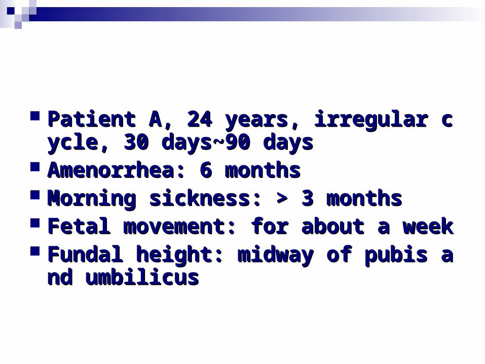
Patient A, 24 years, irregular cycle, 3Patient A, 24 years, irregular cycle, 30 days~90 days0 days~90 days
Amenorrhea: 6 monthsAmenorrhea: 6 months Morning sickness: > 3 monthsMorning sickness: > 3 months Fetal movement: for about a weekFetal movement: for about a week Fundal height: midway of pubis and Fundal height: midway of pubis and
umbilicusumbilicus