Embed Size (px)
Citation preview
1
Diagnosis of Ocular Sarcoidosis
Marina Papadia 1,2
Carl P Herbort 1,3
Manabu Mochizuki 4
1 Inflammatory and Retinal Eye Diseases,
Centre for Ophthalmic Specialised Care (COS)
Lausanne, Switzerland 2 Eye Clinic, Department of Neurosciences, Ophthalmology and Genetics,
University of Genova, Genova, Italy 3 University of Lausanne, Lausanne, Switzerland 4 Department of Ophthalmology & Visual Science, Tokyo Medical and Dental
University, Graduate School of Medical and Dental Sciences
1-5-45 Yushima, Bunkyo-ku, Tokyo 113-8519, Japan
e-mail: [email protected]
2
I. Background
Sarcoidosis is a chronic inflammatory disorder with an unknown etiology characterized by
non-caseating granulomas. [1-3] The disorder is a multisystem and affects many organs,
including the lung, lymph nodes, skin, heart, liver, muscles, and the eye. A very high proportion
(30-60%) of patients with sarcoidosis develop ocular changes, and bilateral granulomatous
uveitis is the most common presentation. [4-13] The characteristic ocular lesions may occur
without apparent systemic involvement.
It is universally accepted that the gold standard for the diagnosis of sarcoidosis is
histopathological proof on biopsy tissue showing non-caseating granulomas, and exclusion of
other diseases which produce granulomatous lesions, such as tuberculosis. [14] Skin,
peripheral lymph nodes, and lung are the common biopsy sites for sarcoidosis. However, biopsy
of intraocular tissues is not performed due to its high risk for vision and is hardly accepted by
uveitis patients. Therefore, many attempts have been made to establish diagnositic criteria for
sarcoidosis with and without biopsy. One example of the diagnostic criteria for systemic and
ocular sarcoidosis are those established by the Japanese Society of Sarcoidosis and Other
Granulomatous Disorders in 1991, and recently revised by the same group. [15,16] Another
example for international diagnostic criteria for ocular sarcoidosis are the proceedings of the 1st
International Workshop on Ocular Sarcoidosis (IWOS) which was held in 2006 in Tokyo, Japan.
[17] The diagnostic criteria focused on the diagnosis of ocular sarcoidosis when ocular changes
are present with or without apparent clinical signs of systemic sarcoidosis.
II. Basic concepts of international criteria for the diagnosis of ocular sarcoidosis put forward at
IWOS.
The IWOS diagnostic criteria are proposed to ophthalmologists to enable them to make the
diagnosis of ocular sarcoidosis without invasive investigations. The criteria consist of seven
clinical ocular signs (Table 1) and five laboratory tests/investigations (Table 2). Based on these
ocular signs and laboratory tests, four categories of certainty of sarcoidosis were esablished
relying on the combination of ocular signs and positive results of laboratory tests, resulting in
four levels of diagnostic certainty, namely (1) definite, (2) presumed, (3) probable, and (4)
possible ocular sarcoidosis (Table 3).
III. Ocular clinical signs suggestive of ocular sarcoidosis
Many reports in the literature described granulomatous uveitis as the hallmark of sarcoidosis.
It was the task of IWOS, consisting of a panel of international uveitis specialists and
pulmonologists, to define, based on their clinical experience, what clinical signs were most
suggestive for ocular sarcoidosis. Seven clinical signs or group of signs were identified.

3
1. Mutton-fat/granulomatous keratic precipitates (KPs) and/or iris nodules (Koeppe/ Busacca).
(figure 1)
These two sings are the expression of granulomatous changes in the anterior segment of the
eye. KPs can be large (mutton-fat KPs) or can take the form of smaller granulomatous KPs. The
nodules at the pupillary margin (Koeppe nodules) and those in the iris stroma (Busacca
nodules) are classic manifestations of chronic anterior granulomatous uveitis.
Figure 1. Granulomatous KPs and iris nodules. Several examples of granulomatous KPs from large
mutton-fat KPs (A) to medium-sized granulomatous KPs (B) and to smaller granulomatous KPs (C). On
the right (D) 2 Koeppe nodules at the pupillary margin.
2. Trabecular meshwork (TM) nodules and/or tent-shaped peripheral anterior synechiae
(PAS).
Small nodules are commonly seen on the surface of the TM. The small size of the nodules
and their color (white or whitish grey) on TM (white) make it difficult to find. Careful gonioscopic
examination with high magnification may help to detect the TM nodules. Tent-shaped PAS is
much easier to find by a gonioscopy. This tent-shaped PAS is considered to be the
consequence of the resolution and scarring of TM nodules representing the same pathology at a
different stage of inflammation. TM nodules can disappear soon after the treatment with topical
or systemic corticosteroids, while tent-shaped PAS stay there and can be found at any time
once they occured.
3. Snowball/string of pearls vitreous opacities.
These types of vitreous opacities represent granulomatous changes in the vitreous. These
vitreous opacities are usually located at the inferior segment of ocular fundus, and can be single
or multiple and forming a shape of “string of pearls”. They are commonly seen in sarcoidosis,

4
but also can be seen in pars planitis and intermediate uveitis associated with multiple sclerosis.
4. Multiple chorioretinal peripheral lesions (active and/or atrophic). (Figure 2)
Multiple chorioretinal lesions in sarcoidosis are round in shape, small in size, white or
whitish-yellow in color and present in clusters, located at random in the periphery up to 360
angle degrees.The lesions are more whitish when active and become more yellowish-greyish
with more sharp margins when cicatricial.
Figure 2. Peripheral chorioretinal granulomas.
5. Nodular and/or segmental periphlebitis (± candlewax drippings) (Figure 3)
Nodular or segmental sheathing of retinal veins is a typical manifestation of retinal vasculitis
in sarcoidosis. They can cause obstruction of the retinal veins, resulting in areas of non-
perfusion causing retinal neovascularization. In addition to these signs of periphlebitis, the
presence of macroaneurisms in an inflamed eye is considered to be a significant although rare
ocular sign of sarcoidosis. (Figure 4)
Figure 3. Segmental sheathing of retinal veins, shown on fundus photography (top, black arrows) and
fluorescein angiography (bottom)
5
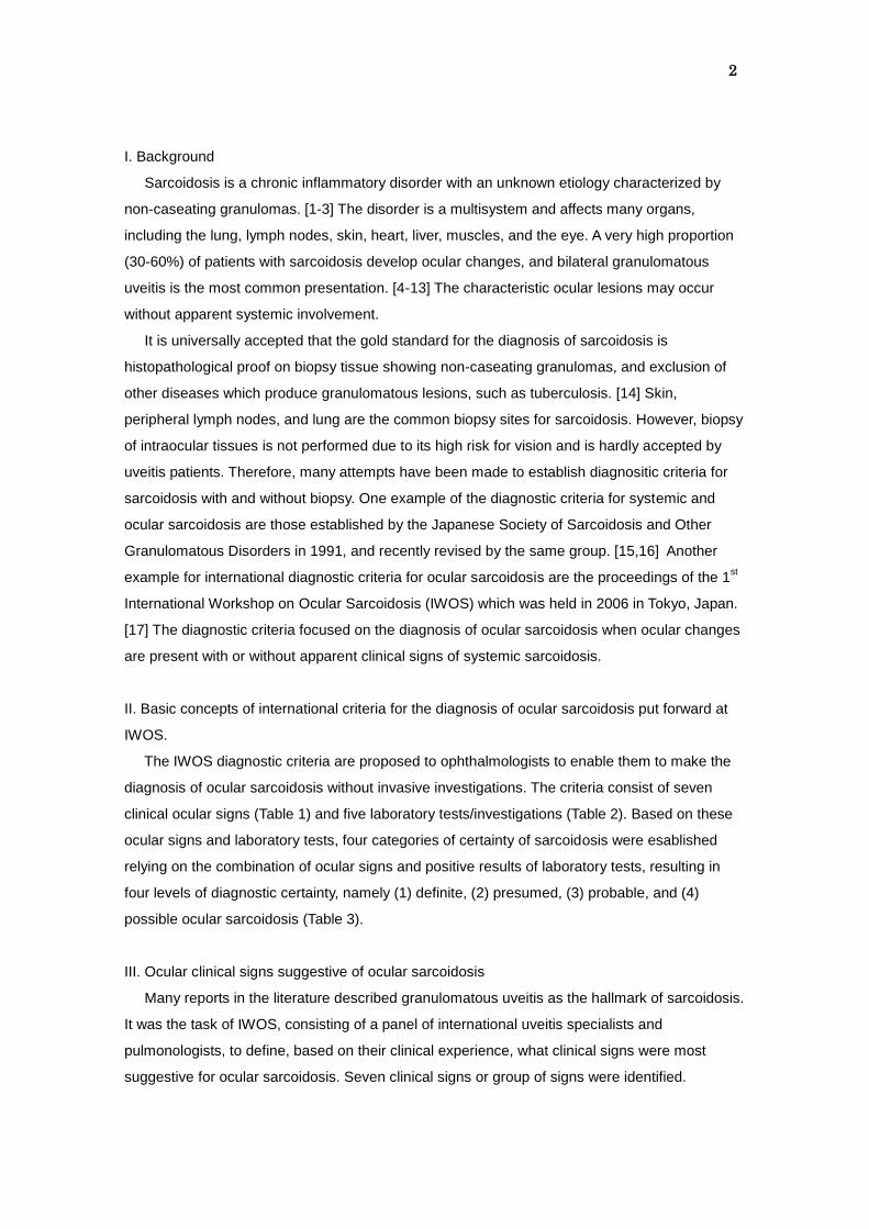
Figure 4. Retinal macroaneurism in a case of sarcoidosis. Macroaneurism (yellow arrows) shown on fundus
picture (top right), on FA intermediate angiographic phase (top left), in late FA phase (bottom left), on ICGA
intermediate phase (center picture) and ICGA late phase (right middle picture). Note also on ICGA frames numerous
hypofluorescent dark areas (black arrows) indicating occult granulomas not seen on fundus picture and FA
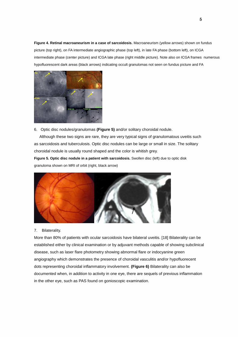
6. Optic disc nodules/granulomas (Figure 5) and/or solitary choroidal nodule.
Although these two signs are rare, they are very typical signs of granulomatous uveitis such
as sarcoidosis and tuberculosis. Optic disc nodules can be large or small in size. The solitary
choroidal nodule is usually round shaped and the color is whitish grey.
Figure 5. Optic disc nodule in a patient with sarcoidosis. Swollen disc (left) due to optic disk
granuloma shown on MRI of orbit (right, black arrow)
7. Bilaterality.
More than 80% of patients with ocular sarcoidosis have bilateral uveitis. [18] Bilaterality can be
established either by clinical examination or by adjuvant methods capable of showing subclinical
disease, such as laser flare photometry showing abnormal flare or indocyanine green
angiography which demonstrates the presence of choroidal vasculitis and/or hypofluorecent
dots representing choroidal inflammatory involvement. (Figure 6) Bilaterality can also be
documented when, in addition to activity in one eye, there are sequels of previous inflammation
in the other eye, such as PAS found on gonioscopic examination.
6
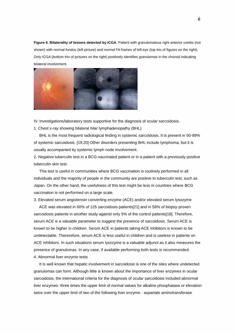
Figure 6. Bilaterality of lesions detected by ICGA. Patient with granulomatous right anterior uveitis (not
shown) with normal fundus (left picture) and normal FA frames of left eye (top trio of figures on the right).
Only ICGA (bottom trio of pictures on the right) positively identifies granulomas in the choroid indicating
bilateral involvement.
IV. Investigations/laboratory tests supportive for the diagnosis of ocular sarcoidosis.
1. Chest x-ray showing bilateral hilar lymphadenopathy (BHL)
BHL is the most frequent radiological finding in systemic sarcoidosis. It is present in 50-89%
of systemic sarcoidosis. [19,20] Other disorders presenting BHL include lymphoma, but it is
usually accompanied by systemic lymph node involvement.
2. Negative tuberculin test in a BCG-vaccinated patient or in a patient with a previously positive
tuberculin skin test.
This test is useful in communities where BCG vaccination is routinely performed in all
individuals and the majority of people in the community are positive to tuberculin test, such as
Japan. On the other hand, the usefulness of this test might be less in countries where BCG
vaccination is not performed on a large scale.
3. Elevated serum angiotensin converting enzyme (ACE) and/or elevated serum lysozyme
ACE was elevated in 60% of 125 sarcoidosis patients[21] and in 58% of biopsy-proven
sarcoidosis patients in another study against only 5% of the control patients[18]. Therefore,
serum ACE is a valuable parameter to suggest the presence of sarcoidosis. Serum ACE is
known to be higher in children. Serum ACE in patients taking ACE inhibitors is known to be
undetectable. Thererefore, serum ACE is less useful in children and is useless in patients on
ACE inhibitors. In such situations serum lysozyme is a valuable adjunct as it also measures the
presence of granulomas. In any case, if available performing both tests is recommended.
4. Abnormal liver enzyme tests
It is well known that hepatic involvement in sarcoidosis is one of the sites where undetected
granulomas can form. Although little is known about the importance of liver enzymes in ocular
sarcoidosis, the international criteria for the diagnosis of ocular sarcoidosis included abnormal
liver enzymes: three times the upper limit of normal values for alkaline phosphatase or elevation
twice over the upper limit of two of the following liver enzyme - aspartate aminotransferase

7
(ASAT), alanine aminotransferase (ALAT) or alkaline phosphatase.
5. Chest CT scan in patients with a negative chest x-ray
This test is not a first-line test, but used for patients where sarcoidosis is strongly suspected
but where the chest radiography is negative for BHL.
V. Interventional diagnostic tests
1. Broncho-alveolar lavage fluids (BALF)
Examination of BALF includes cytology and subset of lymphocytes in the fluids.
Lymphocytosis and an increase of CD4/CD8 ratio in non-smoking individuals are considered to
be characteristic in sarcoidosis. In fact, this is included in the diagnostic criteria for sarcoidosis
by the Japanese Society of Sarcoidosis and Other Granulomatous Disorders (16). BALF
samples can be used for various tests (smears, cultures) for mycobacterium in case of need to
differentiate from tuberculosis.
2. Transbronchial lung biopsy (TBLB)
TBLB is the most commonly performed biopsy in sarcoidosis. Non-caseating granulomas
with Langhans type epithelioid cells are present in sarcoidosis. However, unlike biopsies of the
skin or lymph nodes, it is not a lesion-guided biopsy. It can be false negative when the biopsy
site is not appropriate.
VI. Other investigational tests.
The IWOS criteria allow to reach a certain level of diagnostic certainty in a large proportion of
cases and have the merit to standardize diagnostic parameters in sarcoidosis apparently limited
to the eye. Nevertheless most of the laboratory tests/investigations used by IWOS criteria to
make the diagnosis of ocular sarcoidosis rely on the fact that there is systemic occult
involvement because purely ocular sarcoidosis cannot account for the fact for instance that
elevation of lysozyme and/or ACE in serum are caused by granulomas merely limited to the
eye. Additionally, by definition, if hilar adenopathy, one of the IWOS investigational tests, is
found, this automatically implies systemic (pulmonary) involvement. Moreover the low grade of
specificity and sensitivity of most of the investigational/laboratory tests at our disposal justifies to
resort to other tests at hand to increase the level of diagnostic certainty, eventhough the latter
are also of low sensitivity and specificity.
1. Gallium scan [22]
67Gallium is a radioactive isotope with a half-life of 78 hours that is taken up by activated
macrophages so marking areas where epitheloid cell granulomas are formed. During its decay
gamma photons are emitted and captured by a gamma camera from the sites of uptake. After
intravenous injection of 67
Gallium citrate, the radioisotope has a half-life in the blood pool of 12
8
hours. Gallium uptake is assessed and graded in the lacrimal gland, salivary glands, thorax,
spleen, liver and the eyes 48 hours after injection. In a retrospective study of 18 patients
diagnosed as ocular sarcoidosis a high sensitivity for Gallium scan was found. [23] In the
context of a disease lacking sensitive and specific tests, Gallium scan is an additional tool in
cases where diagnosis is hard to ascertain.
2. Polyclonal immunoglobulin activation
Sarcoidosis is at the origin of immunologic changes modifying the balance between the cellular
and humoral immunity that can serve as a diagnostic test. [24] CD4 lymphocytes are attracted
to the foci of inflammation in tissues with a decrease of efficiency of cellular immunity and a
polyclonal increase of antibodies due to a take over of humoral immunity [25, 26] which explains
the anergy found in sarcoidosis. [27] This immune dysfunction is well known by internists and
has been reported for patients with systemic sarcoidosis. [28-30] A clinical study used the fact
that a very high percentage of the adult population has been exposed to most herpes viruses to
search for increased titers to herpes viruses in sarcoidosis suspect patients. [31] This study
showed that a polyclonal activation score was significantly more elevated in a group with proven
sarcoidosis whan compared to control groups of HLA-B27 uveitis, pars planitis uveitis and
healthy controls.
3. Indocyanine green angiography (ICGA)
ICGA allows not only to detect bilaterality of disease as mentioned hereabove but allows also to
detect occult choroidal lesions when nothing lets suspect fundus or posterior segment
involvement. [31] In patients with scarce ocular signs and no apperent fundus lesions, ICGA can
be useful to reinforce the suspicion of sarcoidosis. It was shown that in a collective of 19
sarcoidosis patients all patients had choroidal involvement some of which were not suspected
by fundus examination or fluorescein angiography. [12] ICGA signs consisted of hypofluorescent
dark dots, fuzziness of large choroidal vessels and late diffuse choroidal hyperfluorescence. An
additional sign was hyperfluorescent pinpoints present in 89% of patients. These signs are not
specific for sarcoidosis but can be present in other granulomatous diseases of the choroid.
Several reports document the fact that ICGA allows to detect occult choroidal lesions in
sarcoidosis. [12,31,32] Therefore in patients with uveitis suspected to be due to sarcoidosis,
especially in those cases presenting with limited signs for granulomatous uveitis, ICGA
represents an additional help to establish the clinical picture and diagnosis of ocular sarcoidsis
and should be part of the recommended work-up. (Figures 6 & 7)
Although all these diagnostic modalities are of limited specificity and sensitivity they should be
considered in the armamentarium to diagnose ocular sarcoidosis.

9
Figure 7. Occult choroidal granulomas seen only on ICGA. 23-year-old patient presenting with right
irido-corneal synechiae (not shown) and several granulomatous KPs OD (not shown) and an increased
flare by laser flare photometry OU (32,4 ph/ms OD versus 17.9 ph/ms OS) with elevated serum ACE and
lysozyme. The top two duets of pictures are FA frames of both eyes which are practically normal except for
slight disc hyperfluorescence OD, whereas ICGA frames show extensive choroidal involvement in form of
numerous hypofluorescent dark dots (HDDs) (squares,circles and arrows) and fuzzy indistinc choroidal
vessels. ICGA was the only way to determine bilateral posterior inflammatory involvement.
VII. Exclusion of other entities
In addition to the clinical signs and laboratory tests indicative of sarcoidosis, exclusion of
other entities that can be mistaken for sarcoidosis is equally important for the diagnosis of
sarcoidosis. Some uveitis entities, particularly entities with granulomatous uveitis must be
excluded. Hereunder the principal entities to be excluded are listed and the specific clinical
signs to be looked for as well as the laboratory tests and investigations to be performed are
exposed..
(1) Exclusion of ocular tuberculosis
Tuberculosis is the first and most important entity to be excluded. It classically presents as
a granulomatous uveitis undistinguishable from sarcoid uveitis. Exclusion of Tuberculosis has
become more easy since the availability of gamma-interferon release assays (IGRA,
Quantiferon®-gold or TB Elispot® test) as false-negatives are very rare. Classical bacterial tests
for Mycobacterium tuberculosis should be performed in case of need.
(2) Exclusion of Vogt-Koyanagi-Harada disease
In certain areas and populations including the Asian continent, Hispanic populations and
Amerindians, Vogt-Koyanagi-Hadara (VKH) disease should be excluded. If acute VKH can be
differentiated easily from sarcoidosis, chronic VKH presents granulomatous anterior uveitis with
iris nodules, nodules at the trabecular meshwork and tent-shaped PAS resembling ocular
sarcoidosis. However, chronic VKH also presents typical depigmentation of the fundus which
can be seen in sarcoidosis when it is patchy as well as sunset-glow fundus, which is not seen in
10
sarcoidosis.
(3) Exclusion of HTLV-1 uveitis
In the southern shores of Japan and in the Caraibbes, human T-cell leukemia virus type 1
(HTLV-1) is an entity to be excluded. HTLV-1 uveitis is characterized by vitreous opacities
(intermediate uveitis) and mild retinal vasculiltis. In some patients with HTLV-1 uveitis can
present iris nodules as well as nodules at trabecular meshwork. Therefore, serological test to
search for anti-HTLV-1 antibodies is important in countries where HTLV-1 is endemic.
(4) Exclusion of herpetic anterior uveitis.
Anterior uveitis caused by human herpes viruses, such as herpes simplex (HSV), vallecella
zoster virus (VZV), and cytomegalovirus (CMV), are ubiquitous and present as a granulomatous
uveitis characterized by mutton-fat KPs, ocular hypertension and iris atrophy especially in case
HSV and VZV uveitis. These clinical signs are similar to those of ocular sarcoidosis. However,
the clinical manifestations of these two diseases are sufficiently distinctive to allow to exclude
herpetic conditions. Herpetic anterior uveitis is nearly always a unilateral condition whereas
ocular sarcoidosis is bilateral in approximately 80% of cases. [18] In addition, fine changes on
the iris surface in the acute stage followed by iris atrophy (diffuse or segmental) a few months
later are present in anterior uveitis caused by VZV or HSV.
(5) Exclusion of bilatral Fuchs’ uveitis (FU) (Figure 8)
When Fuchs’ uveitis (FU) is unilateral the diagnosis is easy. However when it presents as a
bilateral uveitis, which is the case in up to 8% of Fuchs’ cases [33], it can be mistaken for ocular
sarcoidosis especially when the microgranulomatous stellate Fuchs’ KPs are slightly larger than
usual. Fuchs' uveitis is also ubiquitous and should be one of the conditions to be excluded.
Characteristic FU signs should be identified to exclude this condition. They include spread of
KPs above the midline and allover the endothelium, iris structural changes, abnormal vessels in
the iridocorneal angle and absence of synechiae.
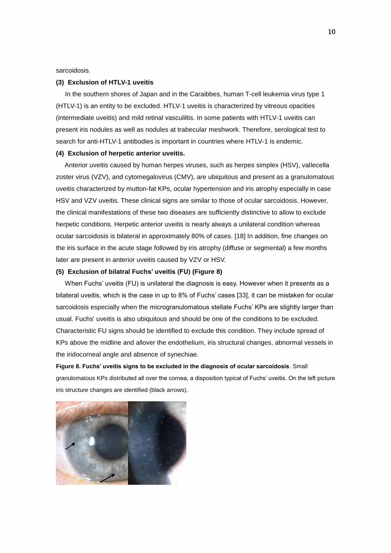
Figure 8. Fuchs’ uveitis signs to be excluded in the diagnosis of ocular sarcoidosis. Small
granulomatous KPs distributed all over the cornea, a disposition typical of Fuchs’ uveitis. On the left picture
iris structure changes are identified (black arrows).
11
(6) Exclusion of iIntermediate uveitis, especially of the pars planitis type.
Sarcoisosis can present as an intermediate uveitis . Therefore any type of intermediate
uveitis, particularly pars planitis, should be excluded. Pars planitis is a bilateral condition
presenting with snowballs and snowbanking, scarce anterior segment inflammation, absence of
synechiae and negativity of laboratory tests. Sarcoidosis can present with snowballs and
snowbanking in a similar fashion to pars planitis and the presence of granulomatous KPs (not
seen in pars planitis) and irido-crystalline synechiae allow to exclude pars planitis and strongly
speak for sarcoidosis or another granulomatous disease.
(7) Exclusion of multiple sclerosis
As indicated hereabove sarcoidosis can present as intermediate uveitis and peripheral
vasculitis. When it presents in this form, one disease that should be excluded is multiple
sclerosis, one of the systemic associations with intermediate uveitis. ln countries where multiple
sclerosis is a common clinical uveitis entity, oriented history taking and MRI are therefore
recommended to exclude or confirm multiple sclerosis.
(8) Exclusion of masquerade syndromes
Conditions masquerading for uveitis such as intraocular lymphoma and amyloidosis, present
vitreous opacities similar to those in ocular sarcoidosis. There are two types of clinical
presentations in primary intraocular lymphoma: one with multiple retinal/sub-retinal lesions and
the other with vitreous opacities with/without retinal lesions. There are some differences in the
clinical pictures of vitreous opacities between intraocular lymphoma and sarcoidosis. The
vitreous opacities in intraocular lymphoma are diffuse with large cells in the vitreous. The
characteristic features of vitreous opacities in ocular sarcoidosis are granulomatous opacities,
such as snow ball opacities or string of pearls opacities, together with diffuse vitreous opacities.
Vitreous opacities in lymphoma have poor response to corticosteroid therapy whereas those in
sarcoidosis respond well to corticosteroids. If intraocular lymphoma is suspected, cytological
examination of the vitreous cells, cytokine assay in the aqueous humor (interleukin-10) or
detection of rearrangement of immunoglobulin genes by polymerase chain reaction are
necessary. []
VIII. Practical processes of diagnosis
In a considerable proportion of cases it is the ophthalmologist who first sees patients
presenting with the ocular expression of sarcoidosis. When any of the ocular signs in Table 1
are present on ocular examination, we should suspect ocular sarcoidosis together with all
clinical entities to be excluded. In countries where sarcoidosis is very common and the leading

12
cause of uveitis, such as in Japan, all laboratory tests listed in Table 2 should be performed and
the patient is then sent for internal medecine work-up, often performed by the pulmonologist.
The pulmonologists perform all necessary systemic examination for systemic sarcoidosis which
include chest CT scan, gallium scintigraphy, bronchoalveolar fluid lavage, and lung biopsy.
However, when the clinician wants to by-pass such an invasive approach or in countries where
sarcoidosis is less common a non-invasive approach may be warranted at first.
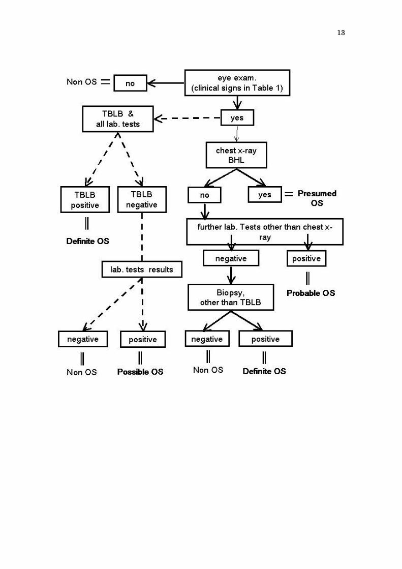
Figure 9 illustrates the practical approach on how diagnosis of ocular sarcoidosis can be
done when the primary invasive approach is avoided.
The first line of systemic examinations in such conditions is a standard chest x-ray,
tuberculin skin test, serum ACE (and/or serum lysozyme), and liver enzymes. After all other
diseases are ruled out, BHL is present on standard chest x-ray then the patients can be
diagnosed as “presumed ocular sarcoidosis”. However, if BHL is not detected by standard chest
x-ray, then further laboratory/investigational tests should be performed including CT scan
Gallium scan or search for polyclonal activation. If the results of the further
laboratoryinvestigational tests are positive (two positive tests in Table 2 and three of the ocular
signs in Table 1), the patients will be diagnosed as “probable ocular sarcoidosis”. However, if the
patients do not meet these criteria, it is recommended to send the patients to the pulmonologists
for further systemic examinations including biopsy (lung biopsy), or the dermatologists for skin
biopsy if skin lesions are present. If histopathological examination on the biopsy tissues is
positive, the patients are diagnosed as “definite ocular sarcoidosis” (biopsy-proven sarcoidosis).
However, if biopsy is negative, the patients are not diagnosed as ocular sarcoidosis, and other
causes of uveitis should be ruled out in those cases. Because lung biopsy is a blind biopsy and
can generate false negatives if the biopsy site is not appropriate, in patients with a negative lung
biopsy the disease can still be due to sarcoidosis. Following the IWOS criteria, in these rare
cases, when four of the suggestive ocular signs and two of the supportive
laboratory/investigational tests are positive, these patients will still be diagnosed as “possible”
ocular sarcoidosis.
Figure 9. Approach for the diagnosis of ocular sarcoidosis (OS) Dot line indicates a
diagnostic approach in countries where OS is very common, whereas solid line illustrates a
practical approach on how diagnosis of OS can be done when the primary invasive is avoided.
TBLB: transbrochial lung biopsy.
13

14
References
[1] Newman LS, Rose CS, Maier LA. Sarcoidosis. N Engl J Med. 1997; 336: 1224-1234.
[2] ATS/ ERS/ WASOG Committee. Statement on sarcoidosis. Am J Respir Crit Care
Med. 1999; 160: 736-755.
[3] Yamamoto M, Sharma OP, Hosoda Y. Special report: the 1991 descriptive definition
of sarcoidosis. Sarcoidosis. 1992; 9: 33-34.
[4] Crick RP, Hoyle C, Smellie H. The eyes in sarcoidosis. Br J Ophthalmol. 1961; 45:
461-481.
[5] Obenauf CD, Shaw HE, Syndor CF, Klintworth GK. Sarcoidosis and its ophthalmic
manifestations. Am J Ophthalmol. 1978; 86: 648-655.
[6] Jabs DA, Johns CJ. Ocular involvement in chronic sarcoidosis. Am J Ophthalmol.
1986; 102: 297-301.
[7] Ohara K, Okubo A, Sasaki H, Kamata K. Intraocular manifestations of systemic
sarcoidosis. Jpn J Ophthalmol. 1992; 36: 452-457.
[8] Chumbley LC, Kearns TP. Retinopathy of sarcoidosis. Am J Ophthalmol. 1972; 73:
123-131.
[9] Spalton DJ, Sanders MD. Fundus changes in histologically confirmed sarcoidosis. Br
J Ophthalmol. 1981; 65: 348-358.
[10] Cook BE Jr, Robertson DM. Confluent choroidal infiltrates with sarcoidosis. Retina.
2000; 20: 1-7.
[11] Thorne JE, Brucker AJ. Choroidal white lesions as an early manifestation of
sarcoidosis. Retina. 2000; 20: 8-15.
[12] Wolfensberger TJ, Herbort CP. Indocyanine green angiographic features in ocular
sarcoidosis. Ophthalmology. 1999; 106: 285-289.
[13] Desai UR, Tawansy KA, Joondeph BC, Schiffman RM, Choroidal granulomas in
systemic sarcoidosis. Retina. 2001; 21: 40-47.
[14] Richeldi L. An update on the diagnosis of tuberculosis infection. Am J Crit Care Med.
2006;174:736-742.
[15] Research Committee of Diffuse Pulmonary Disorders of the Ministry of Health and
Welfore of Japan. Diagnostic criteria of sarcoidosis. Jpn J Sarcoidosis and Other
Granulomatous Disorders. 1991;10:159-162 (in Japanese).
[16] Japan Society for Sarcoidosis and Other Granulomatous Disorders and Japanese
Inflammation Society. Diagnostic guidelines and criteria for sarcoidosis-2006. J Jpn
Ophthalmol Soc. 2007;111:114-118.
[17] Herbort CP, Mochizuki M, Rao NA. Internationl criteria for the diagnosis of ocular
sarcoidosis: Results of the first international workshop on ocular sarcoidosis (IWOS).
15
Ocul Immunol Inflamm. 2009; 17: 160-169.
[18] Kawaguchi T, Hanada A, Horie S, et al. Evaluation of characteristic ocular signs and
systemic investigations in ocular sarcoidosis patients. Jpn J Ophthalmol. 2007; 51:
121-126.
[19] Mana J, Teistein AS, Mendelson DS, Padilla ML, DePalo LR. Excessive thoracic
compute tomogrphic scanning in sarcoidosis. Thorax. 1995; 50:1264-1266.
[20] Pakhale SS, Unruh H, Tan L, Sharma S. Has mediastinoscopy still a role in
suspected stage 1 sarcoidosis? Sarcoidosis Vasc Diffuse Lung Dis. 2006; 23: 66-69.
[21] Hosoya S, Kataoka M, Nakata Y, et al. Clinical features of 125 patients with
sarcoidosis: Okayama University Hospital review of a recent 10-year period. Acta
Med Okayama. 1992; 46:31-36.
[22] Versari A, Cimino L.. 67-Gallium (67
Ga) Scan in sarcoid uveitis. (chapter 14c). In:Gupta A,
Gupta V, Herbort CP, Khairallah M. Uveitis.eds. Japee. New Dehli. 2009:301-6.
[23] Dios Castro E,Herreras Cantalapiedra JM, Calonge Cano M, Oses S. Ocular Sarcoidosis.
Retrospective study of 18 cases. Arch Soc Esp Oftalmol. 2002; 77:301-8.
[24] Daniele RP, Dauber JH, Rossmann MD. Immunologic abnormalities in sarcoidosis.
Ann Intern Med. 1980; 92:406-16.
[25] O’Connor, Fitzgerald MX. Speculations on sarcoidosis. Respir Med. 1992; 86:277-82.
[26] Semenzato G, James DG. The immunological approach to the enigma to the enigma
of sarcoidosis. Sarcoidosis. 1984; 1:24-35.
[27] James DG, Neville E. Pathobiology of sarcoidosis. Pathobiol Annu. 1977; 7:31-61.
[28] James DG, William WJ. Immunology of sarcoidosis. Am J Med. 1982; 72:5-8.
[29] Slitzbach LE, James DG, Neville E. Course and prognosis of sarcoidosis around the
world. Am J Med. 1974; 57:847-52.
[30] Berthoud-Kündig J, Keller A, Herbort CP. Polyclonal elevation of immunoglobulines :
a help rather than a trap in the diagnosis of uveitis caused by sarcoidosis. Klin
Monatsbl Augenheilk. 1994; 204:323-9.
[31] Herbort CP. Indocyanine green angiography (chapter 6). In:Gupta A, Gupta V,
Herbort CP, Khairallah M. Uveitis.eds. Japee. New Dehli. 2009:87-143.
[32] Herbort CP. Precise monitoring and differentiation of inflammatory events by indocyanine
green angiography in a case of recurrent posterior sarcoid uveitis. Ocular Immunol Inflam
2000; 8:303-306.
[33] Bouchenaki N, Herbort CP. Fuchs’ uveitis: failure to
associate vitritis and disc hyperfluorescence with the disease is the major factor for
misdiagnosis and diagnostic delay. MEAJO 2009; 16:239-44.

16
1. Mutton-fat KPs (large and small) and/or iris nodules at pupillary margin (Koeppe) or in stroma (Bussacca)
2. Trabecular meshwork (TM) nodules and/or tent-shaped peripheral anterior synecia (PAS)
3. Snowballs/string of pearls vitreous opacities.
4. Multiple chorioretinal peripheral lesions (active & atrophic)
5. Nodular and/or segmental peri-pherebitis (+ /- candlewax drippings) and/or macroaneurism in an inflamed eye
6. Optic disc nodule(s)/granuloma(s) and/or solitary choroidal nodule
7. Bilaterality (assessed by clinical examination or investigational tests showing subclinical inflammation).
Table 1. Clinical signs suggestive of ocular sarcoidosis xx
17
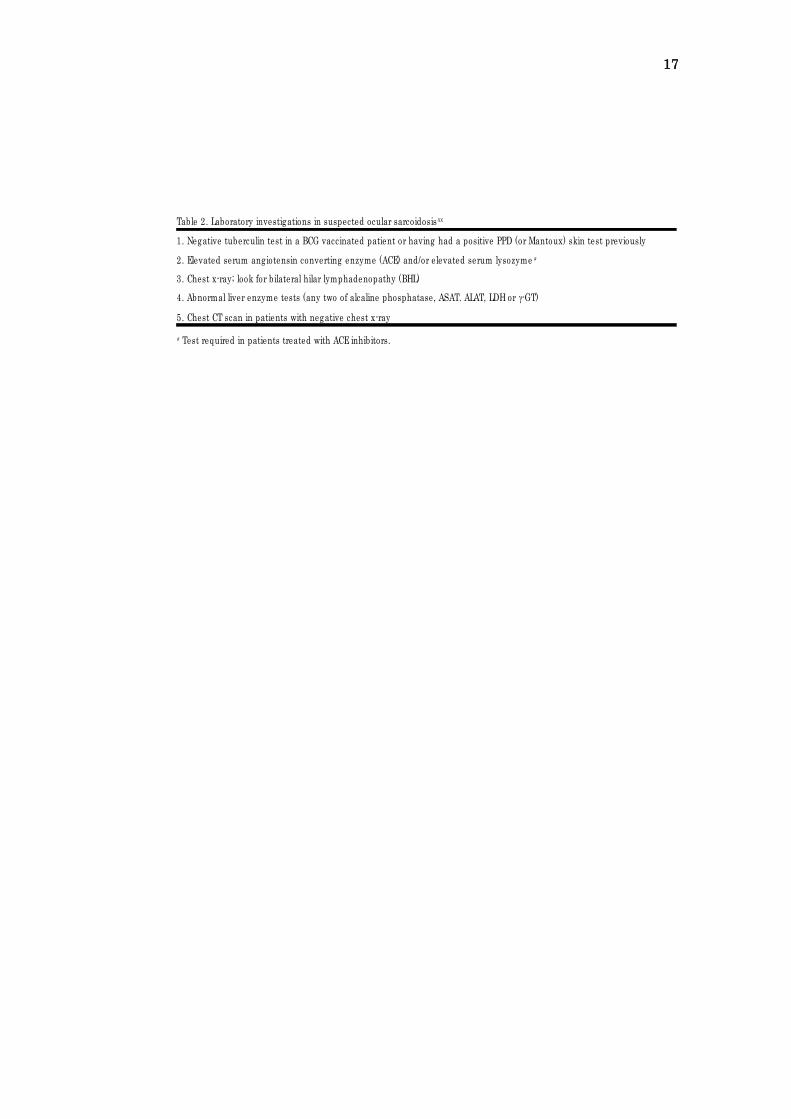
1. Negative tuberculin test in a BCG vaccinated patient or having had a positive PPD (or Mantoux) skin test previously
3. Chest x-ray; look for bilateral hilar lymphadenopathy (BHL)
5. Chest CT scan in patients with negative chest x-ray
Table 2. Laboratory investigations in suspected ocular sarcoidosis xx
2. Elevated serum angiotensin converting enzyme (ACE) and/or elevated serum lysozyme a
4. Abnormal liver enzyme tests (any two of alcaline phosphatase, ASAT. ALAT, LDH or g-GT)
a Test required in patients treated with ACE inhibitors.
18
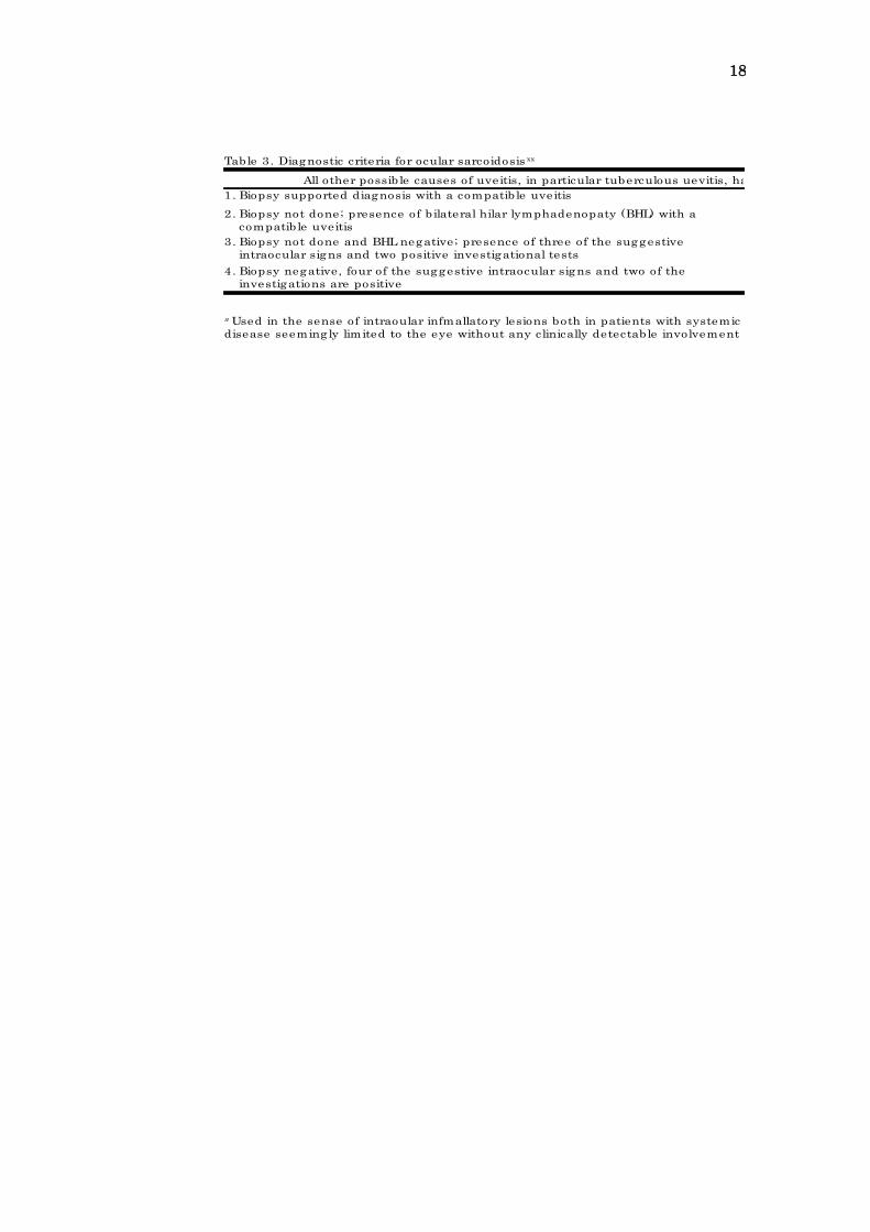
All other possible causes of uveitis, in particular tuberculous uevitis, have to be ruled out.
1. Biopsy supported diagnosis with a compatible uveitis
2.
3.
4.
Table 3. Diagnostic criteria for ocular sarcoidosis xx
Biopsy not done; presence of bilateral hilar lymphadenopaty (BHL) with a
compatible uveitis
Biopsy not done and BHL negative; presence of three of the suggestive
intraocular signs and two positive investigational tests
Biopsy negative, four of the suggestive intraocular signs and two of the
investigations are positive
a Used in the sense of intraoular infmallatory lesions both in patients with systemic disease and in patients with
disease seemingly limited to the eye without any clinically detectable involvement of another organ.