Embed Size (px)
Citation preview
7/27/2019 Diagnosis of Deep Vein Thrombosis (DVT) using Colour Duplex Imaging (CDI) versus D Dimer Test.
http://slidepdf.com/reader/full/diagnosis-of-deep-vein-thrombosis-dvt-using-colour-duplex-imaging-cdi-versus 1/4
International Journal of Science and Research (IJSR), India Online ISSN: 2319-7064
Volume 2 Issue 6, June 2013www.ijsr.net
Diagnosis of Deep Vein Thrombosis (DVT) usingColor Duplex Imaging (CDI) versus D Dimer Test
Nadia A. Salih 1, Bushra H. Almalik 2, Abu Naib BaderEldin 3
Abstract: Several diagnostic strategies using ultrasound imaging, measurement of D-dimer, and assessment of clinical probability of disease have proved safe in patients with suspected deep-vein thrombosis,. The purpose of this review is to discuss the utility of venousultrasonography as the foundation for diagnosis of lower extremity DVT. The effectiveness and practicality of venous ultrasonography
as a stand-alone examination versus D-dimer testing in the diagnosis of DVT. Inpatients and Outpatients presenting with suspected lower-extremity deep-vein thrombosis were potentially eligible. Using a clinical model, physicians evaluated the patients and categorized
them as likely or unlikely to have deep-vein thrombosis. The patients were then randomly assigned to undergo ultrasound imaging alone(control group) or to undergo D-dimer testing (D-dimer group) followed by ultrasound imaging unless the D-dimer test was negative and
the patient was considered clinically unlikely to have deep-vein thrombosis, in which case ultrasound imaging was not performed.The study consisted of 300 Saudi patients were 121 males (40.3%) and 179 females (59.7%).In our study D- Dimer test was positive in 286 (95.3 %) with sensitivity (95.3%) and specificity (83.3%). comparison with Color Duplex Imaging (CDI) showed that 274 positive
patients 91.3 % had sensitivity (91.7%) and specificity (97.1%).
Keywords: Diagnosis, Clinical, Ultra Sonography, Patients, D-Dimer group
1. Introduction
Deep veins thrombosis (DVT) is a blood clot in a deep vein,also known veins thromboembolism (DVT). DVT
predominantly occurs in the legs and may have nosymptoms. When symptoms are present, the non-specificsigns include pain, swelling, redness, warmness, and engorged superficial veins in the leg. DVT can go awaynaturally, but the most serious complication is when athrombosis dislodges and travels to the lungs to become alife-threatening pulmonary embolism. The term veinsthromboembolism is used to refer to DVT and/or pulmonaryembolism. The most frequent complication of DVT is the
post-thrombotic syndrome, which can cause swelling(edema), pain, and rarely, leg ulcers. These symptoms make
post-thrombotic syndrome a significant contributor to thehealth care costs of DVT. About 1 in 1000 adults developsDVT annually, and aging increases its rate of occurrence. [1]
DVT is a serious medical event associated with a substantialrisk of adverse outcomes [2]. The 30-day case fatality rate(i.e. proportion of patients who die) is about 5% for DVTand 10% for PE; the one-year case fatality rate isapproximately 20% for both DVT and PE[3]. The 10-year
recurrence rate of is 30%21-24. Predictors of DVTrecurrence are male sex, idiopathic DVT, and persistent risk factors. The recurrence rates of DVT and PE are similar [4].
The initial clinical presentation of DVT or PE predicts themanifestation of a recurrence; hence, patients that had a
previous PE event tend to have a recurrence of PE morefrequently than patients that had a previous DVT event [5].Furthermore, DVT is associated with long- termcomplications. The post-thrombotic syndrome is a chronic,
progressive condition that occurs despite optimalanticoagulant therapy. This syndrome occurs years after theDVT event, thus, it may not be interpreted as a result of
thrombosis. The symptoms include pain, heaviness,swelling, and cramping in the leg; these symptoms areaggravated during standing or walking. In severe cases, avenous ulcer may develop. The post-thrombotic syndromeoccurs in 20-50% of DVT patients within 10 years of the
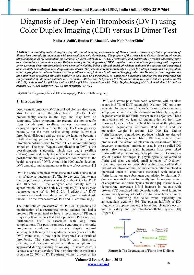
DVT, and severe post-thrombotic syndrome with an ulcer occurs in 3-7% of DVT patients[6]. D-dimer (DD) units aregenerated by the action of factor XIIIa on fibrin monomersand polymers and when the endogenous fibrinolytic systemdegrades cross-linked fibrin present in the organism. Theseunits consist of two identical subunits derived from twofibrin molecules. DD is the final fragment of the plasmin-mediated degradation of cross-linked fibrin and itsmolecular weight is around 180 000 Da. Unlikefibrin/fibrinogen degradation products, which are derived from both fibrinogen and fibrin, DD fragments are end
products of the action of plasmin on cross-linked fibrin;however, monoclonal antibodies used in the so-called DDassays also recognize many fragments from cross-linked fibrin without prior proteolysis by plasmin [7] Because 2 – 3% of plasma fibrinogen is physiologically converted tofibrin and then degraded, small amounts of D-dimer-containing species are detectable in the plasma of healthyindividuals. However, the D-dimer concentration in blood isincreased under all conditions associated with enhanced fibrin formation and subsequent degradation by plasmin. D-dimer represents the most frequently used laboratory marker of coagulation and fibrinolysis activation [8]. Plasma levelsdemonstrate anaverage 8-fold increase in patients with
proven VTE compared with controls, with a level falling toapproximately one-quarter of the initial value between week 1 and 2 following the acute event, especially onanticoagulant treatment [9]. The plasma half-life of DDfragments is approx- imately 8 hours and clearance occursvia the kidney and the reticuloendothelial system [10](Figure 1).
Figure 1: The Degradation of Fibrin into D-dimer
37
7/27/2019 Diagnosis of Deep Vein Thrombosis (DVT) using Colour Duplex Imaging (CDI) versus D Dimer Test.
http://slidepdf.com/reader/full/diagnosis-of-deep-vein-thrombosis-dvt-using-colour-duplex-imaging-cdi-versus 2/4
International Journal of Science and Research (IJSR), India Online ISSN: 2319-7064
Volume 2 Issue 6, June 2013www.ijsr.net
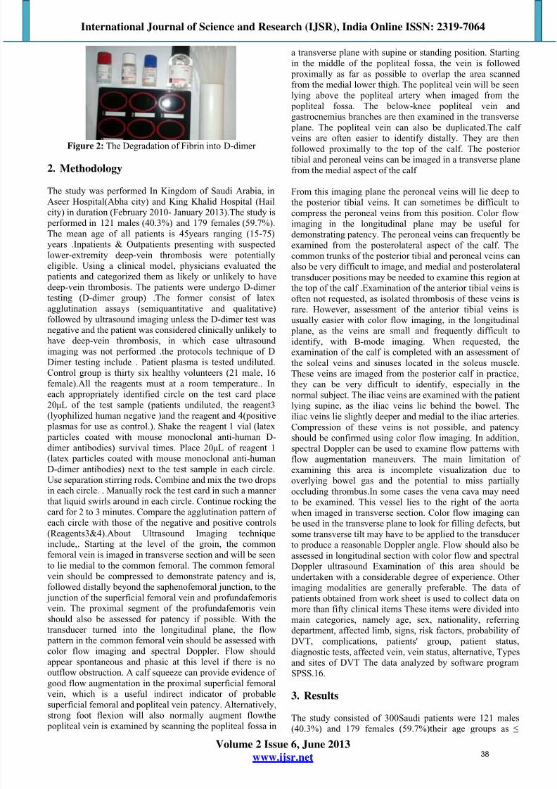
Figure 2: The Degradation of Fibrin into D-dimer
2. Methodology
The study was performed In Kingdom of Saudi Arabia, inAseer Hospital(Abha city) and King Khalid Hospital (Hailcity) in duration (February 2010- January 2013).The study is
performed in 121 males (40.3%) and 179 females (59.7%).The mean age of all patients is 45years ranging (15-75)years .Inpatients & Outpatients presenting with suspected lower-extremity deep-vein thrombosis were potentiallyeligible. Using a clinical model, physicians evaluated the
patients and categorized them as likely or unlikely to havedeep-vein thrombosis. The patients were undergo D-dimer testing (D-dimer group) .The former consist of latexagglutination assays (semiquantitative and qualitative)followed by ultrasound imaging unless the D-dimer test wasnegative and the patient was considered clinically unlikely tohave deep-vein thrombosis, in which case ultrasound imaging was not performed .the protocols technique of DDimer testing include . Patient plasma is tested undiluted.Control group is thirty six healthy volunteers (21 male, 16female).All the reagents must at a room temperature.. Ineach appropriately identified circle on the test card place20µL of the test sample (patients undiluted, the reagent3(lyophilized human negative )and the reagent and 4(positive
plasmas for use as control.). Shake the reagent 1 vial (latex particles coated with mouse monoclonal anti-human D-dimer antibodies) survival times. Place 20µL of reagent 1(latex particles coated with mouse monoclonal anti-humanD-dimer antibodies) next to the test sample in each circle.Use separation stirring rods. Combine and mix the two dropsin each circle. . Manually rock the test card in such a manner that liquid swirls around in each circle. Continue rocking thecard for 2 to 3 minutes. Compare the agglutination pattern of each circle with those of the negative and positive controls(Reagents3&4).About Ultrasound Imaging techniqueinclude,. Starting at the level of the groin, the commonfemoral vein is imaged in transverse section and will be seento lie medial to the common femoral. The common femoralvein should be compressed to demonstrate patency and is,followed distally beyond the saphenofemoral junction, to the
junction of the superficial femoral vein and profundafemorisvein. The proximal segment of the profundafemoris veinshould also be assessed for patency if possible. With thetransducer turned into the longitudinal plane, the flow
pattern in the common femoral vein should be assessed withcolor flow imaging and spectral Doppler. Flow should appear spontaneous and phasic at this level if there is nooutflow obstruction. A calf squeeze can provide evidence of good flow augmentation in the proximal superficial femoralvein, which is a useful indirect indicator of probablesuperficial femoral and popliteal vein patency. Alternatively,strong foot flexion will also normally augment flowthe
popliteal vein is examined by scanning the popliteal fossa in
a transverse plane with supine or standing position. Startingin the middle of the popliteal fossa, the vein is followed
proximally as far as possible to overlap the area scanned from the medial lower thigh. The popliteal vein will be seenlying above the popliteal artery when imaged from the
popliteal fossa. The below-knee popliteal vein and gastrocnemius branches are then examined in the transverse
plane. The popliteal vein can also be duplicated.The calf
veins are often easier to identify distally. They are thenfollowed proximally to the top of the calf. The posterior tibial and peroneal veins can be imaged in a transverse planefrom the medial aspect of the calf
From this imaging plane the peroneal veins will lie deep tothe posterior tibial veins. It can sometimes be difficult tocompress the peroneal veins from this position. Color flowimaging in the longitudinal plane may be useful for demonstrating patency. The peroneal veins can frequently beexamined from the posterolateral aspect of the calf. Thecommon trunks of the posterior tibial and peroneal veins canalso be very difficult to image, and medial and posterolateraltransducer positions may be needed to examine this region atthe top of the calf .Examination of the anterior tibial veins isoften not requested, as isolated thrombosis of these veins israre. However, assessment of the anterior tibial veins isusually easier with color flow imaging, in the longitudinal
plane, as the veins are small and frequently difficult toidentify, with B-mode imaging. When requested, theexamination of the calf is completed with an assessment of the soleal veins and sinuses located in the soleus muscle.These veins are imaged from the posterior calf in practice,they can be very difficult to identify, especially in thenormal subject. The iliac veins are examined with the patientlying supine, as the iliac veins lie behind the bowel. Theiliac veins lie slightly deeper and medial to the iliac arteries.Compression of these veins is not possible, and patencyshould be confirmed using color flow imaging. In addition,spectral Doppler can be used to examine flow patterns withflow augmentation maneuvers. The main limitation of examining this area is incomplete visualization due tooverlying bowel gas and the potential to miss partiallyoccluding thrombus.In some cases the vena cava may need to be examined. This vessel lies to the right of the aortawhen imaged in transverse section. Color flow imaging can
be used in the transverse plane to look for filling defects, butsome transverse tilt may have to be applied to the transducer to produce a reasonable Doppler angle. Flow should also beassessed in longitudinal section with color flow and spectralDoppler ultrasound Examination of this area should beundertaken with a considerable degree of experience. Other imaging modalities are generally preferable. The data of
patients obtained from work sheet is used to collect data onmore than fifty clinical items These items were divided intomain categories, namely age, sex, nationality, referringdepartment, affected limb, signs, risk factors, probability of DVT, complications, patients' group, patient status,diagnostic tests, affected vein, vein status, alternative, Typesand sites of DVT The data analyzed by software programSPSS.16.
3. Results
The study consisted of 300Saudi patients were 121 males(40.3%) and 179 females (59.7%)their age groups as ≤
38
7/27/2019 Diagnosis of Deep Vein Thrombosis (DVT) using Colour Duplex Imaging (CDI) versus D Dimer Test.
http://slidepdf.com/reader/full/diagnosis-of-deep-vein-thrombosis-dvt-using-colour-duplex-imaging-cdi-versus 3/4
International Journal of Science and Research (IJSR), India Online ISSN: 2319-7064
Volume 2 Issue 6, June 2013www.ijsr.net
20years 3 (0.1%), ≤ 30years 06 (2.0%), ≤ 40years 13(14.3%), ≤ 50years 76 (25.3%), ≤ 60years 126 (42.0%), ≥ 60years 76 (25.3%).with symptoms and signs suggestive of DVT. obese 246 (82 %), over 40 years 295 (98.3%),hypertension 161 (53.7%) smoker 108(36.0%), hormonetherapy o oralcontraceptive131 (43.7%), Pregnancy or post-
partum7 (2.3 %) and coagulation disorders, Patients withmajor joint replacement 34 (11.3%), and H-risk patients, not
received anticoagulant 34 (11.3%)among patients for whomdeep-vein thrombosis had been ruled out by the initialdiagnostic strategy, there were 274 (83%) is confirmed venous thromboembolic in the D-dimer group and in 20
patients (87%) is confirmed by Color Duplex Imaging.Ultrasound testing can be safely omitted in such patients. Inour study proximal DVT 14(64%), Distal DVT 8(36%).
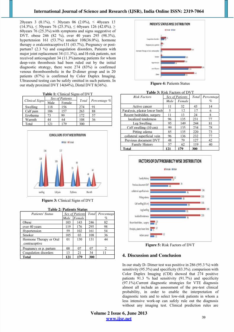
Table 1: Clinical Signs of DVT
Clinical SignsSex of Patients
Total Percentage %Male Female
Swelling 118 156 274 91Calf pain 106 157 263 88
Erythema 73 99 172 57Warmth 44 64 108 36Total 121 179 300
Figure 3: Clinical Signs of DVT
Table 2: Patients StatusPatients' Status Sex of Patients Total Percentage
% Male FemaleObese 103 143 246 82over 40 years 119 176 295 98
Hypertension 59 102 161 54Smoker 105 03 108 36Hormone Therapy or Oral.contraceptive
01 130 131 44
Pregnancy or p. partum 00 07 07 2Coagulation disorders 13 21 34 11Total 121 179 300
Figure 4: Patients Status
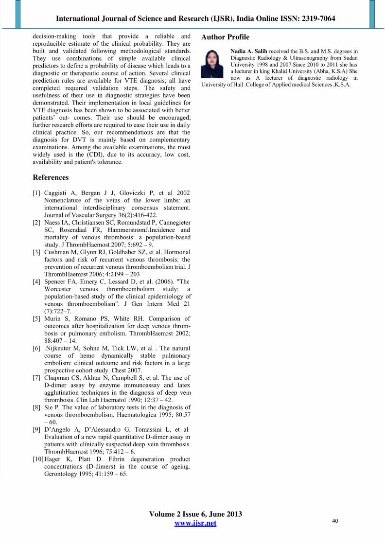
Table 3: Risk Factors of DVT Risk Factors Sex of Patients Total Percentage
% Male Female
Active cancer 11 32 43 14Paralysis, plaster lower limb 5 12 17 6
Recent bedridden, surgery 11 13 24 8localized tenderness 96 135 231 77Leg Swelling 95 149 244 81
Calf swelling (10 cm) 99 135 234 78Pitting edema 85 135 220 73
collateral superficial vein 96 136 232 77Previous document DVT 48 79 127 42
Family History 57 62 119 40Total 121 179 300
Figure 5: Risk Factors of DVT
4. Discussion and Conclusion
In our study D- Dimer test was positive in 286 (95.3 %) withsensitivity (95.3%) and specificity (83.3%). comparison withColor Duplex Imaging (CDI) showed that 274 positive
patients 91.3 % had sensitivity (91.7%) and specificity(97.1%).Current diagnostic strategies for VTE diagnosisalmost all include an assessment of the pre-test clinical
probability, in order to enable the interpretation of diagnostic tests and to select low-risk patients in whom aless intensive work-up can safely rule out the diagnosiswithout any imaging test. Clinical prediction rules are
39
7/27/2019 Diagnosis of Deep Vein Thrombosis (DVT) using Colour Duplex Imaging (CDI) versus D Dimer Test.
http://slidepdf.com/reader/full/diagnosis-of-deep-vein-thrombosis-dvt-using-colour-duplex-imaging-cdi-versus 4/4
International Journal of Science and Research (IJSR), India Online ISSN: 2319-7064
Volume 2 Issue 6, June 2013www.ijsr.net
decision-making tools that provide a reliable and reproducible estimate of the clinical probability. They are
built and validated following methodological standards.They use combinations of simple available clinical
predictors to define a probability of disease which leads to adiagnostic or therapeutic course of action. Several clinical
prediction rules are available for VTE diagnosis; all havecompleted required validation steps. The safety and
usefulness of their use in diagnostic strategies have beendemonstrated. Their implementation in local guidelines for VTE diagnosis has been shown to be associated with better
patients’ out- comes. Their use should be encouraged;further research efforts are required to ease their use in dailyclinical practice. So, our recommendations are that thediagnosis for DVT is mainly based on complementaryexaminations. Among the available examinations, the mostwidely used is the (CDI), due to its accuracy, low cost,availability and patient's tolerance.
References
[1] Caggiati A, Bergan J J, Gloviczki P, et al 2002 Nomenclature of the veins of the lower limbs: aninternational interdisciplinary consensus statement.Journal of Vascular Surgery 36(2):416-422.
[2] Naess IA, Christiansen SC, Romundstad P, Cannegieter SC, Rosendaal FR, HammerstrømJ.Incidence and mortality of venous thrombosis: a population-based study. J ThrombHaemost 2007; 5:692 – 9.
[3] Cushman M, Glynn RJ, Goldhaber SZ, et al. Hormonalfactors and risk of recurrent venous thrombosis: the
prevention of recurrent venous thromboembolism trial. JThrombHaemost 2006; 4:2199 – 203
[4] Spencer FA, Emery C, Lessard D, et al. (2006). "TheWorcester venous thromboembolism study: a
population-based study of the clinical epidemiology of venous thromboembolism". J Gen Intern Med 21(7):722–7.
[5] Murin S, Romano PS, White RH. Comparison of outcomes after hospitalization for deep venous throm-
bosis or pulmonary embolism. ThrombHaemost 2002;88:407 – 14.
[6] .Nijkeuter M, Sohne M, Tick LW, et al . The naturalcourse of hemo dynamically stable pulmonaryembolism: clinical outcome and risk factors in a large
prospective cohort study. Chest 2007.[7] Chapman CS, Akhtar N, Campbell S, et al. The use of
D-dimer assay by enzyme immunoassay and latexagglutination techniques in the diagnosis of deep veinthrombosis. Clin Lab Haematol 1990; 12:37 – 42.
[8] Sie P. The value of laboratory tests in the diagnosis of venous thromboembolism. Haematologica 1995; 80:57
– 60.[9] D’Angelo A, D’Alessandro G, Tomassini L, et al.
Evaluation of a new rapid quantitative D-dimer assay in patients with clinically suspected deep vein thrombosis.ThrombHaemost 1996; 75:412 – 6.
[10] Hager K, Platt D. Fibrin degeneration productconcentrations (D-dimers) in the course of ageing.Gerontology 1995; 41:159 – 65.
Author Profile
Nadia A. Salih received the B.S. and M.S. degrees inDiagnostic Radiology & Ultrasonography from SudanUniversity 1998 and 2007.Since 2010 to 2011 she hasa lecturer in king Khalid University (Abha, K.S.A) Shenow as A lecturer of diagnostic radiology in
University of Hail .College of Applied medical Sciences ,K.S.A.
40