Embed Size (px)
Citation preview
301
CEDirected Reading
This article is a Directed Reading. Your access to Directed Reading quizzes for continuing education credit is determined by your membership status and CE preference.
RADIOLOGIC TECHNOLOGY, January/February 2015, Volume 86, Number 3
The spine’s bony structure, made up of vertebrae, sup-ports the core of the body and endures the impact of nearly
every physical movement. The verte-bral column houses the spinal cord and branching nerves that enable movement and supports 80% of the body’s weight.1
Osteoporosis, malignancies, and trauma can affect the vertebral column, causing vertebral bodies to collapse. This injury is called a vertebral compression fracture. Unlike a traditional traumatic fracture, com-pression fractures occur when the bone condenses, much like a crushed aluminum can.2 Radiographs are standard diagnostic tools for vertebral compression fractures, and medical imaging is used throughout manage-ment of such injuries.1
Vertebral AnatomyAn adult vertebral column is made
up of 33 vertebrae categorized into 5 sections (see Figure 1)3:
After completing this article, the reader should be able to:Describe the causes of most vertebral compression fractures. List risk factors for vertebral compression fractures. Discuss psychological and physiological effects of vertebral compression fractures. Explain the role of medical imaging in diagnosing and treating vertebral compression
fractures. Discuss medications and treatment options for managing vertebral compression fractures. Describe complications associated with surgical procedures used to treat vertebral
compression fractures.
A healthy spine is an integral part of an individual’s overall well-being. The spinal column’s essential role in physiological and neurological function can be compromised when disease or trauma causes a vertebra to compress under the body’s weight, producing a vertebral compression fracture. This is a common ailment among adults older than 65 years of age, especially for those with low bone mass or osteoporosis. This article describes vertebral compression fractures, with a special emphasis on medical imaging.
Cherie Dewar, BS
Diagnosis and Treatment of Vertebral Compression Fractures
Lateral Spinal Column
Figure 1. Sections of the spine.
302
CEDirected Reading
RADIOLOGIC TECHNOLOGY, January/February 2015, Volume 86, Number 3
Diagnosis and Treatment of Vertebral Compression Fractures
The cervical section from the base of the head to the top of the shoulders is made up of 7 vertebrae. These are the smallest, and they bear the least amount of weight.
The thoracic section ranges from the shoulders to just above the lower back. The thoracic section is the longest, with 12 vertebrae.
The lumbar section is the lower back, with 5 ver-tebrae.
Below the lumbar region is the sacral section, located behind the pelvis. The 5 sacral vertebrae are fused in adults.
The coccygeal section, at the base of spinal column, comprises 4 vertebrae that fuse after 30 years of age.
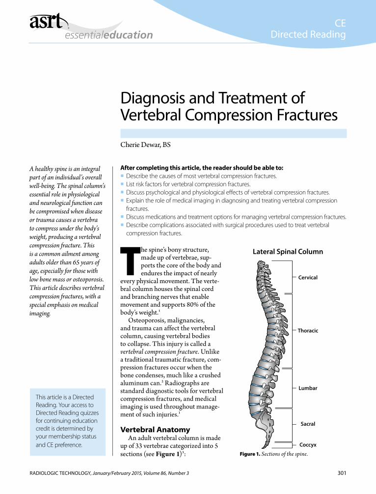
A vertebra is composed of a vertebral arch, a vertebral body, and projections called processes (see Figure 2).3 Each process forms a sort of “W” shape with 2 extra spikes in either valley, and together they form the vertebral arch. The oval-shaped vertebral body is attached anteriorly to the vertebral arch via pedicles and protects the anterior side of the spinal cord.3 The pedicles originate from the vertebral body on either side of the spinal cord and con-nect the vertebral body to the vertebral arch.
The vertebral body is composed of trabecular bone that has needlelike crossing projections that create vas-cular, spongy pores within which red marrow produces red blood cells.3 This trabecular bone is encased in a thin layer of cortical bone.3 The edges of the top and bottom of the vertebral body are layered with an end plate (see Figure 3).3
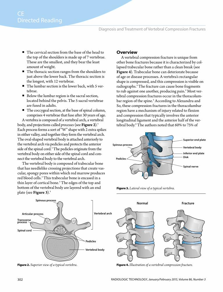
OverviewA vertebral compression fracture is unique from
other bone fractures because it is characterized by col-lapsed trabecular bone rather than a clean break (see Figure 4). Trabecular bone can deteriorate because of age or disease processes. A vertebra’s rectangular shape is compressed, and this compression is visible on radiographs.4 The fracture can cause bone fragments to rub against one another, producing pain.4 Most ver-tebral compression fractures occur in the thoracolum-bar region of the spine.5 According to Alexandru and So, these compression fractures in the thoracolumbar region have a mechanism of injury related to f lexion and compression that typically involves the anterior longitudinal ligament and the anterior half of the ver-tebral body.6 The authors noted that 60% to 75% of
Figure 4. Illustration of a vertebral compression fracture.
Figure 3. Lateral view of a typical vertebra.
Figure 2. Superior view of a typical vertebra.
Spinous process
Transverseprocess
Spinal cord
Vertebral body
Articular process
Pedicles
Vertebral arch
Spinous process
Spinal nerve
Vertebral body
Pedicles
Superior end plate
Inferior end plateDisk
Normal Fracture
303
CEDirected Reading
RADIOLOGIC TECHNOLOGY, January/February 2015, Volume 86, Number 3
Dewar
vertebral compression fractures occur between T12 and L2, where the vertebral column transitions from being rigid to mobile, which could make these vertebrae more vulnerable to compression fractures.6
Classification Vertebral compression fractures are classified into
3 types based on the portion of the vertebral body affected. The types are wedge, biconcave, and crush.6 Approximately half of all vertebral compression frac-tures are wedge types that involve compression of the anterior segment of the vertebral body; they typically occur in the midthoracic region. About 17% of ver-tebral compression fractures are biconcave, meaning the center portion of the vertebral body is compressed but its anterior and posterior walls remain unaffected. The rarest type of vertebral compression fracture is a crush fracture, which involves either compression of the entire anterior and posterior portions of the vertebral body6 or only the posterior portion.1 Crush fractures make up about 13% of vertebral compres-sion fractures, and complex fractures—fractures with significant soft tissue injury—constitute the remain-ing types.6
At times, the nature of vertebral compression frac-tures can be confused with developmental deformities such as Scheuermann disease in which the end plates degenerate and then re-form. The fractures also can be confused with degeneration from osteoarthritis.5
SymptomsThe onset of mechanical axial back pain caused by
a vertebral compression fracture can surprise a patient who has likely experienced no traumatic injury.1 The pain can be abrupt and in a single site, but patients have also reported pain that extends anteriorly to their heart or lung area.1 Lying down relieves the pain because it decreases the effect of gravity on the broken vertebra that occurs with sitting and standing.1
Causes Most vertebral compression fractures are caused by
diseases, but they also can occur as a result of physical trauma. A single compression fracture can lead to sub-sequent vertebral fractures.
Most patients who have vertebral compression frac-tures have weakened vertebrae from diseases or condi-tions such as osteoporosis, cancer, or as sequelae from infection, hyperparathyroidism, hypogonadism, or osteomalacia.1,6 Because osteoporosis usually is associ-ated with older patients, malignancy should be consid-ered as the cause of vertebral compression fracture in patients who are aged younger than 55 years.6
Osteoporosis is the predominant cause of vertebral compression fractures. The disease disrupts the struc-ture of the trabecular bones in the vertebrae and chang-es the contents of the noncollagenous proteins in the bone’s matrix.6 Approximately 44 million Americans have osteoporosis, and another 34 million have low bone mass. The high incidence of osteoporosis likely contributes to the 1.5 million vertebral compression fractures diagnosed annually in the United States.6
A decrease in bone density begins in both men and women after they reach 40 years of age, but the process accelerates in women who have undergone menopause.1 A patient’s bone mineral density loss is measured with dual-energy x-ray absorptiometry, or bone densitometry.6 The examination generates a T-score that represents the standard deviation from the mean peak value of bone density in young adults.6 The World Health Organization defines osteoporosis as a T-score of less than 2.5, and a T-score between 1 to 2.5 is defined as decreased bone density or osteopenia. A T-score of 1 and higher is considered normal.6 Low bone mass is a term used for a T-score between 2.5 and 1.4.1
If a patient’s bone density decreases one standard deviation below the average vertebral density, the risk of vertebral compression fracture doubles1; if the bone mineral density falls by 2 standard deviations, the risk of a compression fracture increases 4 to 6 times.6
Once a patient has one vertebral compression fracture, the risk of another fracture, despite the individual’s bone density, increases 5-fold6; having 2 or more of the frac-tures increases the risk of developing another vertebral compression fracture by 12-fold.6 Patients who have had a vertebral compression fracture also have a higher risk of fracture in other bones, such as the hip.2 Other predic-tors of vertebral fracture risk can include an individual’s bone geometry and the structure of the cortical bone sur-rounding the trabecular bone in the vertebrae.7
304
CEDirected Reading
RADIOLOGIC TECHNOLOGY, January/February 2015, Volume 86, Number 3
Diagnosis and Treatment of Vertebral Compression Fractures
Once an individual’s vertebrae are compromised by a bone-weakening condition, the structures cannot withstand normal amounts of pressure or strain.2 As a result, the smallest of movements such as bending over, turning quickly, lifting even light objects, sneezing, and coughing can cause a vertebral compression fracture.2,6 Traumatic injury caused by a fall from a height or a motor vehicle crash can cause compression fractures, but up to 30% of vertebral compression fractures can occur while a person is in bed.6 The reason that trivial motions can lead to injury might be explained by the contraction of the paraspinal muscles.6
Studies have shown that decreased extensor strength in the back muscles, reduced spinal mobility, altered trunk muscle control, and balance issues might con-tribute to incidence of vertebral compression fractures in individuals who have osteoporosis. The paraspinal muscles are responsible for compressive forces in the spinal area. Briggs et al showed that a pattern of paraspi-nal muscle recruitment occurs in individuals who have vertebral compression fractures that is different from the pattern in those who have osteoporosis and do not experience fractures. Motor recruitment is a neuromus-cular response that relies on motor, or muscular, units as resistance increases. The muscle recruitment changes occur at the thoracolumbar transition zone in which vertebral fractures are most common.7
Risk Factors Several risk factors predispose individuals to ver-
tebral compression fractures. These include alcohol consumption, tobacco use, estrogen deficiency, frailty, a history of falls, impaired eyesight, lack of physical activity, and a deficiency of calcium and vitamin D.5,6 Chronic obstructive pulmonary disease, seropositive rheumatoid arthritis, and Crohn disease are additional risk factors for vertebral fractures, as is the use of sys-tematic glucocorticoids, especially after cumulative exposure to such medications.5
Obesity is not considered a risk factor for vertebral compression fractures but instead decreases risk of bone loss.6 Obesity causes an increased production of hor-mones such as estrogen and of insulinlike growth factor binding protein-1, both of which stimulate osteoblast activity and increased bone production.6
SequleaePain is the chief result of a vertebral compres-
sion fracture, and pain emanating from the back can make standing, walking, or even lying down difficult.6 Compression fractures in the lumbar region might cause greater disability than those in the thoracic region.5
When the vertebrae compress and the spine tilts for-ward, the gap between the ribs and pelvis closes and can impair balance.1 This forward-tilting spinal alignment causes a patient to pull back for counterbalance, which can cause pain.2
Pain from vertebral fractures has additional con-sequences, particularly if subsequent compression fractures occur.6 These consequences include loss of stature, fatigued muscles, disproportionate thoracic kyphosis (curvature of the thoracic region, resulting in concave curve of the upper back), and lumbar lordosis (convex curvature of the lower back).6 If kyphosis is severe, as seen in patients with osteoporosis, it can exert so much pressure on the pelvis that the individual expe-riences pulmonary impairment, a protruding abdomen, satiety, poor nutrition, and weight loss.6 Constipation, bowel obstruction, and a reduced amount of activity also are possible results of a vertebral compression frac-ture, and reduced activity only expedites weakening of the bones and muscles or can cause complications such as deep vein thrombosis.6
The sequelae related to vertebral fractures nega-tively affects a patient’s quality of life, independence, self-esteem, emotions, and social life.6 Approximately 40% of patients with a vertebral compression fracture develop depression.2
Even after a vertebral compression fracture has healed, an individual’s loss of height can lead to focal or sagittal imbalance,1 requiring the use of a cane or walk-er for mobility or causing difficulty with activities such as riding in an automobile.2 Patients who have vertebral compression fractures have a 15% greater risk of death.6
The 2005 direct medical economic burden of osteo-porosis in the United States is estimated to be between $13.7 billion and $20.3 billion, and vertebral compres-sion fractures account for about $1.1 billion of these costs.8 It is projected that this cost will increase by 50% by 2025.9 The long-term morbidities that result from
305
CEDirected Reading
RADIOLOGIC TECHNOLOGY, January/February 2015, Volume 86, Number 3
Dewar
vertebral compression fractures are reflected in the fact that, in the first year after a painful fracture, the rate of patients who require primary care services is 14 times higher than that of the general population.1 The indi-rect costs are difficult to estimate, but they are likely much higher when patients’ time off work and other issues like pain, insomnia, and depression are factored into the cost of vertebral compression fractures.9
EpidemiologyThe risk factors that contribute to vertebral compres-
sion factors also affect incidence, leading to a higher number of the fractures among women who have passed menopause. In fact, 25% of postmenopausal women in the United States experience a vertebral compression fracture.6
Vertebral compression fractures are most preva-lent in adults aged 80 years and older, at a rate of 30%. Nearly 50% of the bone mass in the axial skeleton is lost when a woman reaches 80 years of age. Women aged 50 to 69 years have the fractures at a rate of 5% to 10%.1 The difference in incidence according to sex is nearly double for women, particularly as they age. In general, 10.7 per 1000 women have a vertebral compression frac-ture annually in the United States, compared with 5.7 fractures per 1000 men.6 Some reports say that women who are Asian or African American have a lower preva-lence of the fractures than do Caucasian women.5
An explanation for incidence might lie in the associa-tion of vertebral compression factors with osteoporosis and osteopenia. Nearly 50% of the 1.5 million fractures related to osteoporosis each year in the United States are vertebral fractures.2 Likewise, approximately 50% of all patients who have a vertebral compression frac-ture also have osteoporosis, and 40% have osteopenia.1 Approximately 14% to 18% of women who are aged older than 60 years and have low bone mass have a ver-tebral compression fracture,5 and 20% of women who are postmenopausal and have osteoporosis will develop a second vertebral fracture within a year of their first compression fracture.1 An increasingly aging popula-tion will lead to higher incidence of the fractures associ-ated with loss of bone mass.2
Cancer also causes vertebral compression fractures. Approximately 30% of patients with various neoplastic
conditions develop a spinal metastasis that causes pain.10 Approximately 60% of spinal metastases origi-nate from breast or prostate cancer.11 The vertebral body is more susceptible to invasion by tumor cells because of its vascular nature as opposed to the avascu-lar nature of a disk; about 80% of the time, the cancer invades the vertebral body, followed by the pedicles.11
Diagnosis A patient’s physical examination might reveal hyper-
kyphosis, or excessive thoracic spine curvature. The finding indicates likelihood of vertebral compression fractures.5 The patient’s medical history should provide additional information such as osteoarthritic pain, pathologic pain from a tumor, or lumbar strain to aid in differential diagnosis of vertebral compression fracture.1
A definitive diagnosis of vertebral compression frac-ture usually is accomplished using a number of medical imaging modalities. However, most vertebral compression fractures are not diagnosed when they occur. In 2 studies in the United States, only 25% to 33% of radiographically identified vertebral fractures were clinically diagnosed.5 The same is true in Europe, where nearly 66% of osteopo-rotic vertebral compression fractures are missed.12
Many vertebral compression fractures are identified incidentally on chest radiographs but not addressed by the treating clinician.5 In addition, vertebral compression fractures have been described as insidious and might not be visible on a first radiograph.1 Unlike imaging of bone metastases, nuclear bone scans are used infrequently to diagnose vertebral compression fractures. A compression fracture might be revealed incidentally on a bone scan by increased uptake of radionuclides.1
A physician might not order radiography or ver-tebral fracture assessment, a technology using bone densitometry of the thoracic and lumbar spine, when a patient has been diagnosed with osteoporosis or has a bone density T-score of 1.5 or higher. The reason is that confirming a vertebral compression fracture likely will not change the selected course of medical therapy.5 However, if the patient is a postmenopausal woman with a T-score of between 1.5 and 2.4 who could benefit from drugs such as bisphosphonates to prevent further fractures following an initial vertebral compres-sion fracture, radiography is warranted. In addition,

306
CEDirected Reading
RADIOLOGIC TECHNOLOGY, January/February 2015, Volume 86, Number 3
Diagnosis and Treatment of Vertebral Compression Fractures
diagnosis of fractures using radiography or vertebral fracture assessment is justified in terms of cost-effectiveness when compared to not treating a vertebral compression fracture and risking additional fractures or long-term morbidity.1,5
RadiographyA radiograph is the first diagnostic tool used in iden-
tifying a vertebral compression fracture,6 with a lateral projection of the thoracic and lumbar spine being the most cost-effective approach (see Figure 5).1 According
to Craig St George, R.T.(R)(VI), if a cervical collar has been applied, a minimum of anteroposterior and lateral views are performed to rule out fracture before the col-lar is removed (written communication, October 2014). If a patient has undergone severe trauma, the patient’s entire spine should be assessed to check all vertebrae for injuries. Five percent to 20% of patients who are diagnosed with vertebral compression fracture have additional vertebral compression fractures.6
A radiograph provides the following diagnostic information1,6: Identification of a vertebral compression fracture,
including type (wedge, biconcave, or crush). Measurement of a vertebra’s height loss; a mini-
mum of 20% must be lost compared with normal portions of the vertebral body for a vertebral com-pression fracture to be diagnosed.
Measurement of increased distance between the processes or pedicles, indicating vertebral disrup-tion.
Estimation of how much a vertebra has moved out of alignment along its anterior and posterior lines can be seen on erect projections.
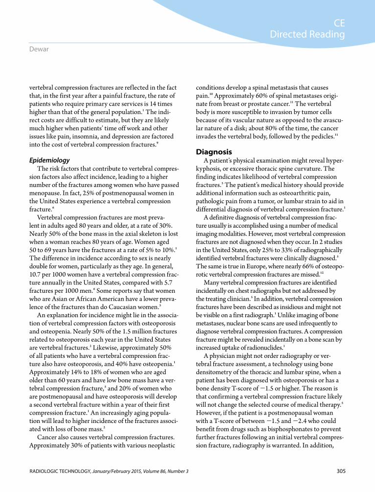
A patient who has osteopenia could be more prone to vertebral compression fractures, and certain charac-teristics on radiographs suggest osteopenia, including increased radiolucence and loss of horizontal trabecu-lae. Wong and McGirt also list decreased cortical thick-ness accompanied by increased relative opacity of the end plates and vertical trabeculae as signs of osteope-nia.1 The authors also noted that the age of a fracture can be estimated if prior radiographs with no signs of fracture are available for comparison. In addition, radiologists also measure the kyphotic angle, or the angle between the superior end plate one level above the injured segment and the inferior end plate one level below the injured segment, to reach a diagnosis of verte-bral compression fracture (see Figure 6).6
In 2013, Cho et al published the results of their study comparing the usefulness of the prone cross-table lateral projection to the standing extension lateral pro-jection in preoperative and postoperative radiographs of 62 patients with vertebral compression fractures (see Figure 7).13 The authors measured the degree of patients’ pain associated with each position and the
Figure 5. Lateral radiograph of a vertebral compression fracture showing anterior wedging (arrow). Reprinted with permission from Alexandru D, So W. Evaluation and management of verte-bral compression fractures. Perm J. 2012;16(4):46-51.
307
CEDirected Reading
RADIOLOGIC TECHNOLOGY, January/February 2015, Volume 86, Number 3
Dewar
accuracy with which each position and projection could display the restoration of the postoperative wedge angle and vertebral heights. The authors found that the prone cross-table lateral radiographs were more accurate than the erect images in helping radiologists determine the degree of restoration of vertebral heights and wedge angles following surgery. In addition, the prone cross-table positioning caused less pain during the examina-tion. The authors also noted that the prone cross-table lateral radiograph can be used effectively in diagnosing intravertebral clefts or intravertebral dynamic mobil-ity.13 Intravertebral clefts are a radiographic sign of avas-cular necrosis in the vertebral body. When intraverte-bral clefts are present, a delay of fracture healing called dynamic mobility can occur.14 Eventually, dynamic mobility can result in nonunion of the fractured verte-brae, called Kümmel disease.13
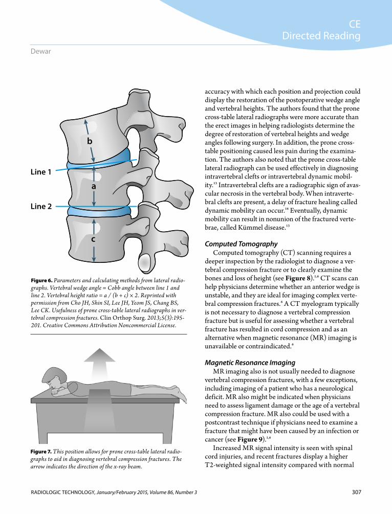
Computed TomographyComputed tomography (CT) scanning requires a
deeper inspection by the radiologist to diagnose a ver-tebral compression fracture or to clearly examine the bones and loss of height (see Figure 8).1,6 CT scans can help physicians determine whether an anterior wedge is unstable, and they are ideal for imaging complex verte-bral compression fractures.6 A CT myelogram typically is not necessary to diagnose a vertebral compression fracture but is useful for assessing whether a vertebral fracture has resulted in cord compression and as an alternative when magnetic resonance (MR) imaging is unavailable or contraindicated.6
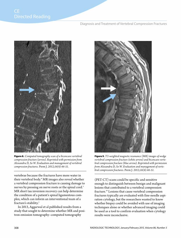
Magnetic Resonance ImagingMR imaging also is not usually needed to diagnose
vertebral compression fractures, with a few exceptions, including imaging of a patient who has a neurological deficit. MR also might be indicated when physicians need to assess ligament damage or the age of a vertebral compression fracture. MR also could be used with a postcontrast technique if physicians need to examine a fracture that might have been caused by an infection or cancer (see Figure 9).1,6
Increased MR signal intensity is seen with spinal cord injuries, and recent fractures display a higher T2-weighted signal intensity compared with normal
Figure 6. Parameters and calculating methods from lateral radio-graphs. Vertebral wedge angle = Cobb angle between line 1 and line 2. Vertebral height ratio = a / (b + c) × 2. Reprinted with permission from Cho JH, Shin SI, Lee JH, Yeom JS, Chang BS, Lee CK. Usefulness of prone cross-table lateral radiographs in ver-tebral compression fractures. Clin Orthop Surg. 2013;5(3):195-201. Creative Commons Attribution Noncommercial License.
Line 1
Line 2
b
a
c
Figure 7. This position allows for prone cross-table lateral radio-graphs to aid in diagnosing vertebral compression fractures. The arrow indicates the direction of the x-ray beam.
308
CEDirected Reading
RADIOLOGIC TECHNOLOGY, January/February 2015, Volume 86, Number 3
Diagnosis and Treatment of Vertebral Compression Fractures
vertebrae because the fractures have more water in their vertebral body.6 MR images also reveal whether a vertebral compression fracture is causing damage to nerves by pressing on nerve roots or the spinal cord.1 MR short tau inversion recovery can help determine the condition of a patient’s spinal ligamentous com-plex, which can inform an interventional team of a fracture’s stability.1
In 2013, Aggarwal et al published results from a study that sought to determine whether MR and posi-tron emission tomography–computed tomography
(PET-CT) scans could be specific and sensitive enough to distinguish between benign and malignant lesions that contributed to a vertebral compression fracture.15 Lesions that cause vertebral compression fractures typically are evaluated with fine-needle aspi-ration cytology, but the researchers wanted to know whether biopsy could be avoided with use of imaging techniques alone or whether advanced imaging could be used as a tool to confirm evaluation when cytology results were inconclusive.
Figure 9. T2-weighted magnetic resonance (MR) images of wedge vertebral compression fracture (white arrow) and biconcave verte-bral compression fracture (blue arrow). Reprinted with permission from Alexandru D, So W. Evaluation and management of verte-bral compression fractures. Perm J. 2012;16(4):46-51.
Figure 8. Computed tomography scan of a biconcave vertebral compression fracture (arrow). Reprinted with permission from Alexandru D, So W. Evaluation and management of vertebral compression fractures. Perm J. 2012;16(4):46-51.
309
CEDirected Reading
RADIOLOGIC TECHNOLOGY, January/February 2015, Volume 86, Number 3
Dewar
Imaging was conducted on 24 sub-jects with vertebral compression fractures and no history of osteoporosis, degen-erative disease, or disk herniations. MR images were acquired with a 1.5T Siemens MAGNETOM with and without gadolini-um contrast (0.1 mmol/kg). CT scans were acquired at 140 kV 10 mA, followed by PET scans on GE Discovery PET-CT equip-ment after the injection of 10 mCi to 20 mCi of fluorodeoxyglucose F 10.15 All patients also underwent fine-needle aspiration biopsy of primary lesions or of other sites noted by PET-CT if the initial cytology results were uncertain.
Researchers found that the diagnosing specificity for MR and PET-CT combined was 100% for benign lesions, and the sensi-tivity of the 2 imaging modalities combined was 100% for malignant tumors. The speci-ficity for MR and PET-CT combined was not 100% for malignant tumors. The researchers found that PET-CT scans were helpful in determining additional biopsy locations.15
Vertebral Fracture AssessmentA vertebral fracture assessment can divulge an occult
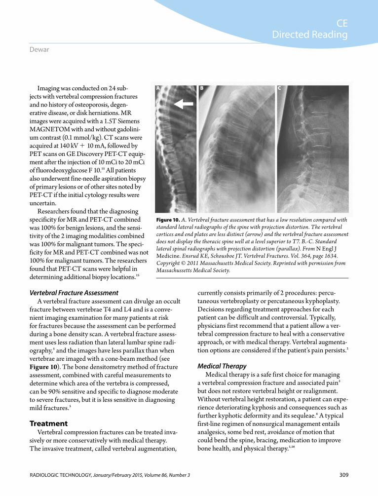
fracture between vertebrae T4 and L4 and is a conve-nient imaging examination for many patients at risk for fractures because the assessment can be performed during a bone density scan. A vertebral fracture assess-ment uses less radiation than lateral lumbar spine radi-ography,5 and the images have less parallax than when vertebrae are imaged with a cone-beam method (see Figure 10). The bone densitometry method of fracture assessment, combined with careful measurements to determine which area of the vertebra is compressed, can be 90% sensitive and specific to diagnose moderate to severe fractures, but it is less sensitive in diagnosing mild fractures.5
TreatmentVertebral compression fractures can be treated inva-
sively or more conservatively with medical therapy. The invasive treatment, called vertebral augmentation,
currently consists primarily of 2 procedures: percu-taneous vertebroplasty or percutaneous kyphoplasty. Decisions regarding treatment approaches for each patient can be difficult and controversial. Typically, physicians first recommend that a patient allow a ver-tebral compression fracture to heal with a conservative approach, or with medical therapy. Vertebral augmenta-tion options are considered if the patient’s pain persists.2
Medical TherapyMedical therapy is a safe first choice for managing
a vertebral compression fracture and associated pain2 but does not restore vertebral height or realignment. Without vertebral height restoration, a patient can expe-rience deteriorating kyphosis and consequences such as further kyphotic deformity and its sequleae.6 A typical first-line regimen of nonsurgical management entails analgesics, some bed rest, avoidance of motion that could bend the spine, bracing, medication to improve bone health, and physical therapy.1,16
Figure 10. A. Vertebral fracture assessment that has a low resolution compared with standard lateral radiographs of the spine with projection distortion. The vertebral cortices and end plates are less distinct (arrow) and the vertebral fracture assessment does not display the thoracic spine well at a level superior to T7. B.-C. Standard lateral spinal radiographs with projection distortion (parallax). From N Engl J Medicine. Ensrud KE, Schousboe JT. Vertebral Fractures. Vol. 364, page 1634. Copyright © 2011 Massachusetts Medical Society. Reprinted with permission from Massachussetts Medical Society.
A B C
310
CEDirected Reading
RADIOLOGIC TECHNOLOGY, January/February 2015, Volume 86, Number 3
Diagnosis and Treatment of Vertebral Compression Fractures
MedicationsA combination of medications can be recommended
for patients who have vertebral compression fractures to treat the pain. The type and level of analgesic chosen might depend on pain level, potential adverse effects, and the medication’s effects on a patient’s comorbidi-ties. Pain relief options range from nonsteroidal anti-inflammatory drugs to opiates.1,2 Patients on opiates should be monitored carefully for addiction to these drugs. Other medication options to treat vertebral compression fracture pain include muscle relaxants and local analgesic patches.1 Tricyclic antidepressants also are commonly prescribed for patients with vertebral compression fractures and neuropathic pain.5
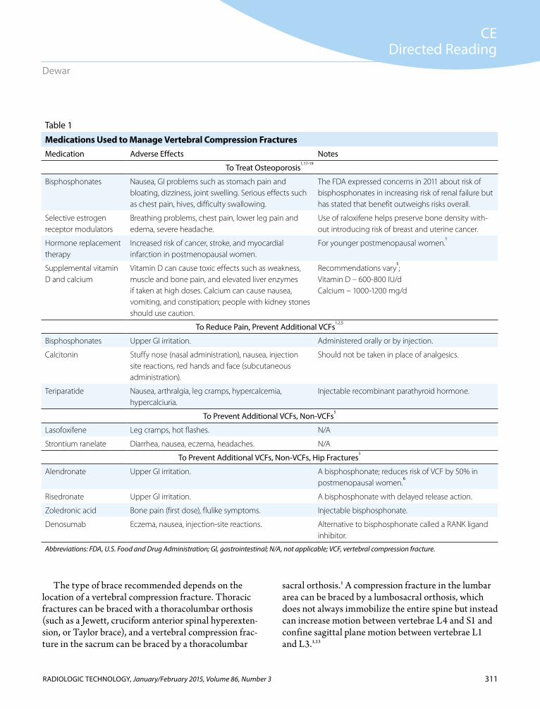
If a vertebral compression fracture leads to the diagno-sis of osteoporosis, a patient could receive one of several drugs that have been vetted in large, randomized trials to treat osteoporosis, reduce pain from the fracture, and prevent further fractures.5 Other drugs have even been shown to prevent additional nonvertebral fractures such as hip fractures. Table 1 lists several medications used to manage pain, osteoporosis, and fractures.
Physical ActivityOften, a physician orders bed rest for a patient with
a new vertebral compression fracture but only for a limited period. Long periods of immobility can increase bone mineral density loss (0.25%-1.0% per week of bed rest) and the risk of other health issues such as venous thrombosis, pulmonary embolism, pressure ulcers, and urinary tract infections.2,6
When a patient is ready, performing gentle exercises such as swimming or walking, perhaps with a walking aid, is recommended.2,20 Patients also may be referred to physical therapy and rehabilitation programs to improve recovery from a vertebral compression frac-ture.20 Physical therapy should strengthen a patient’s axial musculature, including the spinal extensors, and educate the patient how to improve posture and gait to avoid falls.1
Strengthening spinal extensors also reduces verte-bral compression fracture pain.1 Randomized trials led by Malmros et al and Bennell et al followed patients who participated in the authors’ respective 10-week physical therapy programs. Malmros et al emphasized
strength and balance training, with stabilization of the lumbar area, twice a week. Bennell et al emphasized once-a-week soft tissue massage therapy and exercises such as standing wall pushups.21,22 Both programs led to reduced back pain from compression fractures and improved the patients’ quality-of-life scores. A third research group led by Papaioannou conducted a randomized controlled trial that lasted 6 months and included stretching, strength training, and aerobics. Although participants in the Papaioannou study had improved quality-of-life scores and better balance, their bone mineral density remained unchanged.23
Nonmedication Pain Relief MethodsPhysicians might recommend applying ice packs or
heat packs over the area of the vertebral compression fracture to help relieve pain.2 Other pain relief methods include intercostal nerve blocks and use of transcutane-ous nerve stimulation units.1 Radiation therapy can help relieve pain for patients whose vertebral compression fracture is caused by cancer. Radiation therapy has been shown to provide pain relief for half of the patients whose fractures were caused by myeloma, prostate can-cer, or breast cancer.6
Back BracingA back brace can help reduce the amount of pres-
sure on a patient’s compression fracture.2 Braces hold the spine straight, stabilize the fracture, and prevent further compression.2 A patient could require use of a brace for up to 3 months, but using a brace beyond 3 months is not recommended because it can weaken the core muscles.2 A prospective randomized study with patients using thoracolumbar orthotic braces found that patients’ core strength, posture, and height improved, leading to a better quality of life for the patients.1
Another study with bracing found that patients’ core muscles were actually more active with a brace, though the brace should have been limiting their f lexion.1 Yet another small, randomized, unblinded study involved 2 groups, one with patients who wore a rigid brace dur-ing their waking hours for 6 weeks and a second group of patients who wore a nonrigid brace for only 2 hours a day for 24 weeks. Participants in both groups experi-enced less self-reported pain.5
311
CEDirected Reading
RADIOLOGIC TECHNOLOGY, January/February 2015, Volume 86, Number 3
Dewar
The type of brace recommended depends on the location of a vertebral compression fracture. Thoracic fractures can be braced with a thoracolumbar orthosis (such as a Jewett, cruciform anterior spinal hyperexten-sion, or Taylor brace), and a vertebral compression frac-ture in the sacrum can be braced by a thoracolumbar
sacral orthosis.1 A compression fracture in the lumbar area can be braced by a lumbosacral orthosis, which does not always immobilize the entire spine but instead can increase motion between vertebrae L4 and S1 and confine sagittal plane motion between vertebrae L1 and L3.1,13
Table 1
Medications Used to Manage Vertebral Compression FracturesMedication Adverse Effects Notes
To Treat Osteoporosis1,17-19
Bisphosphonates Nausea, GI problems such as stomach pain and bloating, dizziness, joint swelling. Serious effects such as chest pain, hives, difficulty swallowing.
The FDA expressed concerns in 2011 about risk of bisphosphonates in increasing risk of renal failure but has stated that benefit outweighs risks overall.
Selective estrogen receptor modulators
Breathing problems, chest pain, lower leg pain and edema, severe headache.
Use of raloxifene helps preserve bone density with-out introducing risk of breast and uterine cancer.
Hormone replacement therapy
Increased risk of cancer, stroke, and myocardial infarction in postmenopausal women.
For younger postmenopausal women.1
Supplemental vitamin D and calcium
Vitamin D can cause toxic effects such as weakness, muscle and bone pain, and elevated liver enzymes if taken at high doses. Calcium can cause nausea, vomiting, and constipation; people with kidney stones should use caution.
Recommendations vary5;
Vitamin D – 600-800 IU/dCalcium – 1000-1200 mg/d
To Reduce Pain, Prevent Additional VCFs1,2,5
Bisphosphonates Upper GI irritation. Administered orally or by injection.
Calcitonin Stuffy nose (nasal administration), nausea, injection site reactions, red hands and face (subcutaneous administration).
Should not be taken in place of analgesics.
Teriparatide Nausea, arthralgia, leg cramps, hypercalcemia, hypercalciuria.
Injectable recombinant parathyroid hormone.
To Prevent Additional VCFs, Non-VCFs5
Lasofoxifene Leg cramps, hot flashes. N/A
Strontium ranelate Diarrhea, nausea, eczema, headaches. N/A
To Prevent Additional VCFs, Non-VCFs, Hip Fractures5
Alendronate Upper GI irritation. A bisphosphonate; reduces risk of VCF by 50% in postmenopausal women.
6
Risedronate Upper GI irritation. A bisphosphonate with delayed release action.
Zoledronic acid Bone pain (first dose), flulike symptoms. Injectable bisphosphonate.
Denosumab Eczema, nausea, injection-site reactions. Alternative to bisphosphonate called a RANK ligand inhibitor.
Abbreviations: FDA, U.S. Food and Drug Administration; GI, gastrointestinal; N/A, not applicable; VCF, vertebral compression fracture.
312
CEDirected Reading
RADIOLOGIC TECHNOLOGY, January/February 2015, Volume 86, Number 3
Diagnosis and Treatment of Vertebral Compression Fractures
Complications with bracing include discomfort, skin irritation, and breathing interference. Softer, light-weight braces could reduce these complications and improve compliance, but a more stringent brace might be needed if a patient’s spine is severely deformed.1
Vertebral AugmentationTypically, the augmentation options from which a
patient can choose are percutaneous vertebroplasty (also called acrylic vertebroplasty) and percutaneous kyphoplasty (also called balloon-assisted vertebro-plasty).23 Kyphoplasty is a type of vertebroplasty, and both procedures can potentially stabilize the vertebra, prevent further loss of vertebral height, correct kyphotic deformity, and reduce pain. Between 85% and 95% of patients who have undergone one of the procedures have experienced immediate pain relief. A study of 1309 patients reported that both techniques reduced patients’ pain by half.2 As with any invasive procedure, risks are involved with both options, and augmentation should be carefully considered for use in elderly patients, who have a higher incidence of comorbidities.6,25 The proce-dures for vertebral compression fractures are conducted by interventional radiologists, anesthesiologists and other pain management specialists, and orthopedic surgeons.26 Medical imaging is used before, during, and after vertebroplasty and kyphoplasty.2
A vertebroplasty involves a small incision that allows a needle, guided by real-time CT or f luoroscopy, to inject liquid cement into a compressed vertebra. The cement is injected under pressure into the vertebra’s spaces and crevices.2,25,27 Two interventional neuroradi-ologists in Amiens, France, performed the first percuta-neous vertebroplasty procedure in 1984.25 The doctors, Galibert and Deramond, performed the procedure on a patient who had a C2 vertebra compromised by a verte-bral hemangioma. The patient’s pain subsided for a long period following the injection.25
After vertebroplasty gained notice, an orthopedic surgeon named Reiley developed a new vertebroplasty technique in an effort to reduce the risk of cement leak-age during the procedure and to reinstate vertebral height to the damaged vertebra.25 Instead of squeezing liquid cement between the cracks of a compressed ver-tebra, Reiley first inserted and inflated a balloon (also
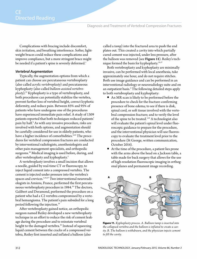
called a tamp) into the fractured area to push the end plates out. This created a cavity into which partially cured cement was injected, under less pressure, after the balloon was removed (see Figure 11). Reiley’s tech-nique formed the basis for kyphoplasty.25,27
Both vertebroplasty and kyphoplasty are minimally invasive, can be performed with local anesthesia, take approximately one hour, and do not require stitches. Both use image guidance and can be performed in an interventional radiology or neuroradiology suite and on an outpatient basis.4 The following detailed steps apply to both vertebroplasty and kyphoplasty: An MR scan is likely to be performed before the
procedure to check for the fracture-confirming presence of bone edema; to see if there is disk, spinal cord, or soft tissue involved with the verte-bral compression fracture; and to verify the level of the spine to be treated.2,25 A technologist also will evaluate the patient’s spinal level under f luo-roscopic guidance to prepare for the procedure, and the interventional physician will use f luoros-copy to evaluate the treatment level prior to the procedure (St George, written communication, October 2014).
At the time of the procedure, a patient lies prone with the arms above the head on a Jackson table, a table made for back surgery that allows for the use of high-resolution f luoroscopic imaging in orthog-onal planes and permanent image recording.
Figure 11. Kyphoplasty process. A. Balloon tamp is inserted into the collapsed vertebra and the balloon is inflated to create a cav-ity. B. The balloon is withdrawn, and the physician injects cement to fill the cavity.
A B
313
CEDirected Reading
RADIOLOGIC TECHNOLOGY, January/February 2015, Volume 86, Number 3
Dewar
Using the Jackson table minimizes repositioning and potential infection of the injection site.1,24
A patient’s vital signs such as heart rate, blood pressure, and pulse are reviewed and recorded before the procedure begins, and the incision area is cleaned and shaved.4
A patient’s vital signs are checked regularly during the procedure, and a facility performing vertebral augmentation should ensure access to CT or rapid MR within 30 to 45 minutes in the event of com-plications. Most patients receive local anesthesia under the skin and in the deep tissues near the fracture, but patients who are in poor health or of advanced age, who cannot tolerate lying prone, or who have multiple vertebral compression frac-tures requiring treatment might receive anesthe-sia intravenously.2,4
The physician makes a small incision near the fracture and inserts a trocar (hollow needle) through the patient’s muscles and into the frac-tured vertebra under f luoroscopic guidance.4,25 The lateral and anteroposterior f luoroscopic images should display the pedicles clearly for tro-car position. Patients who are awake might feel a tapping sensation when the trocar is inserted into the vertebra.4 The procedure occasionally is per-formed under CT guidance.
Some physicians perform venography before injecting the cement mixed with a contrast mate-rial. Fewer physicians are performing venography and some debate whether venography is effective, but its use might minimize cement leakage and display the venous channels near the trocar.25
In vertebroplasty, cement is injected into the verte-bra.4,25 In kyphoplasty, the physician inserts a bal-loon tamp through the trocar, and the balloon is inflated to create a cavity, then deflated and with-drawn before cement is injected into the cavity.4,25
After the injection, the trocar is removed, and a bandage is placed over the incision.4
The cement takes approximately 15 to 20 minutes to harden and will be at 90% of its strength within 24 hours.2,25
After the procedure, a radiograph or CT scan might be performed to check the cement’s
location. The length of observation time deemed appropriate varies for each patient. Rarely, a patient is admitted as an inpatient.4
Pain relief can be immediate or take up to 48 hours.2,4 The cement’s mechanism of action for pain relief is not totally understood, but the hypothesis is that it immobi-lizes the end plates and the tiny trabecular fractures that caused pain during movement. Kyphoplasty is consider-ably more expensive than vertebroplasty.25
Vertebral Augmentation CementMost cement used for vertebroplasty is polymethyl
methacrylate, which was approved by the U.S. Food and Drug Administration (FDA) in 2004.25 Polymethyl methacrylate usually is shipped as 2 components, a powder and a liquid, and then mixed in the operating room or interventional suite just before injecting the cement into a patient.25 The cement hardens slowly and has proved its compatibility with the human body in dentistry, hip and joint replacements, and filling gaps between prostheses and bone.25
The powder portion of polymethyl methacrylate is made up of beads or a similar acrylic polymer, along with a filler that includes a contrast agent, most often barium sulfate. Other contrast agents that can be used are tantalum powder, tungsten, or zirconium dioxide.25 The liquid part is made of methyl methacrylate mono-mer (concentration 95% weight).25
When the 2 parts are mixed together, the polymer-ization is initiated by a chemical in the liquid, but some hardening inhibitors such as hydroquinine might be added if necessary.25 The person mixing the cement should carefully calculate the ratio, which typically is 2.18 parts solid to 1 part liquid. If a staff person makes an error in the ratio and the mixture is not properly bal-anced, the cement could harden prematurely or insuf-ficiently, leading to inadequate strength in the final compound, or an irregularity in the time required for hardening, all of which could lead to serious adverse effects in a patient.25
It is recommended that a vertebroplasty cement have a longer liquid phase working time than that used for kyphoplasty. The cement used in vertebroplasty must be squeezed into the tight spaces of a fracture while under high pressure, but a cement used during
314
CEDirected Reading
RADIOLOGIC TECHNOLOGY, January/February 2015, Volume 86, Number 3
Diagnosis and Treatment of Vertebral Compression Fractures
kyphoplasty should have a shorter liquid phase and take on a more doughy appearance to fill the cavity under less pressure.25
After the procedure, the hardened cement is vis-ible on diagnostic imaging. Some practices have added antibiotics to the cement to minimize infection risk, but antibiotics such as gentamicin might compromise the mechanical strength of the cement.25 To avoid this, anti-biotics can be administered intravenously to a patient before the procedure.
VertebroplastyThe use of vertebroplasty to manage osteoporotic
vertebral compression fractures increased rapidly after the first vertebroplasty was performed in the United States in the 1990s.2 Claims for Medicare Part B fee-for-service enrollees nearly doubled for vertebroplasty procedures between 2001 and 2005.26
Typically, 2 trocars are needed for a bipedicular approach to a vertebral compression fracture for a verte-broplasty; this allows more cement to be evenly spread into the fracture.1 The upper region’s vertebral pedicles are smaller, which might necessitate an extrapedicular approach between the medial rib head and lateral edge of the pedicle.1
The procedure can relieve a patient’s pain,25 with up to 75% to 100% of patients reporting good to moder-ate pain relief shortly after vertebroplasty.6 A 2006 literature review identified 4 studies that reported reduction in pain lasting up to one year.28 As many as 75% of patients have regained their mobility following vertebroplasty.4
KyphoplastyKyphoplasty differs from vertebroplasty in the use of
a balloon tamp to first create a cavity in the broken ver-tebra, which compacts the spongy bone and pushes the end plates as close as possible into their original posi-tions.1 During a kyphoplasty procedure, the physician can insert the needle using an extrapedicular approach or use 2 needles.25
A pressure gauge informs the physician when to stop inflating the balloon during the procedure. This point normally occurs at about 220 psi, not to exceed 300 psi. The physician also gauges the balloon’s inflation volume
(4-6 mL) and the appearance of the balloon on fluoros-copy.1 Sequential images are acquired during inflation to ensure that the end plates are not disturbed by the balloon.1 Multiple images also are acquired during the cement injection process to assess when the cavity is properly filled and whether the cement has leaked into the spinal cavity.1 The mean volume of cement injected into patients undergoing kyphoplasty has been reported at 3.9 mL, compared with 2.2 mL for vertebroplasty injection.16
An early study of kyphoplasty reported that 95% of patients experienced considerable pain relief.27 A review article mentioned that kyphoplasty can restore vertebral height by 50% to 70% and improve segmental kyphosis by 6° to 10°.6 The chances of restoring the vertebral height are best when kyphoplasty is per-formed within 3 months of the vertebral compression fracture occurrence.6
Complications of Vertebroplasty and Kyphoplasty Because both procedures use plastic cement to fill
a vertebral body, both vertebroplasty and kyphoplasty can put patients at risk for symptomatic cement leakage, or extravasation. In 2002, the FDA warned that extrav-asation can occur from both procedures.27
Extravasation of the cement material can cause an embolism, neurological deficits, spinal cord compres-sion, osteomyelitis, hematoma, infection, digestive track bleeding, and adjacent vertebral compression.25 Extravasation could cause radiculopathy (a disorder of the spinal nerve roots) if the cement leaks into the spinal canal or neural foramina of the spine, leading to weakness.1 The cement’s seepage through the venous channel in the vertebral body could cause an embolism in the lungs, kidneys, or heart.2,25,29
Fortunately, a physician can observe the cement entering a vertebra with the use of real-time f luoroscopy so extravasation can be avoided.25 A number of studies have found statistical differences for complication risk between vertebroplasty and kyphoplasty techniques. Some of these statistics are reviewed in Table 2.25
The force of high pressure on the slowly filling cement required for vertebroplasty is a problematic aspect of the procedure compared with kyphoplasty, which does not require the same degree of pressure
315
CEDirected Reading
RADIOLOGIC TECHNOLOGY, January/February 2015, Volume 86, Number 3
Dewar
to fill an empty cavity.25 Extravasation rates have been reported as higher in osteoporotic-associated vertebral compression fractures compared with those associated with malignant tumors. The overall complication rate for both vertebral augmentation procedures is less than 2% for fractures caused by osteoporosis and 10% for fractures caused by malignant tumors.25
As shown in Table 2, a wide range of adjacent verte-bral fracture rates are observed with vertebroplasty and kyphoplasty.25 Yimin et al noted that science has not determined whether the cement, which is stiffer than vertebral bone, affects neighboring vertebrae, or wheth-er the subsequent fractures are simply part of the natu-ral progression of osteoporosis.25 A subsequent vertebral compression fracture rate of 10% has been reported for both procedures.4
In 2012, Qian et al published the results of a study that examined early-stage adjacent disk degeneration follow-ing vertebroplasty (n 9) and kyphoplasty (n 53), compared with a control group (n 35).16 There were no significant differences between age, sex, body weight, or smoking status among the patient groups selected for the study. Those who remained in the study (90.7%) had MR imaging performed to evaluate the condition of their disks at baseline, or during their first appointment, and at their last appointment 2 years later.
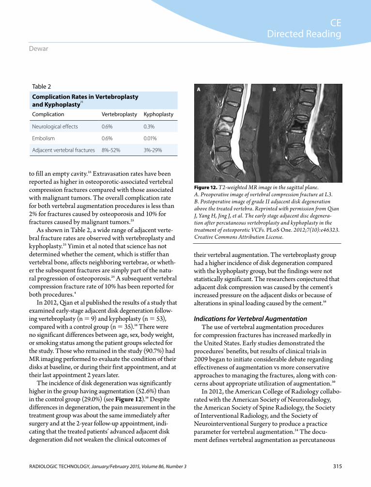
The incidence of disk degeneration was significantly higher in the group having augmentation (52.6%) than in the control group (29.0%) (see Figure 12).16 Despite differences in degeneration, the pain measurement in the treatment group was about the same immediately after surgery and at the 2-year follow-up appointment, indi-cating that the treated patients’ advanced adjacent disk degeneration did not weaken the clinical outcomes of
their vertebral augmentation. The vertebroplasty group had a higher incidence of disk degeneration compared with the kyphoplasty group, but the findings were not statistically significant. The researchers conjectured that adjacent disk compression was caused by the cement’s increased pressure on the adjacent disks or because of alterations in spinal loading caused by the cement.16
Indications for Vertebral AugmentationThe use of vertebral augmentation procedures
for compression fractures has increased markedly in the United States. Early studies demonstrated the procedures’ benefits, but results of clinical trials in 2009 began to initiate considerable debate regarding effectiveness of augmentation vs more conservative approaches to managing the fractures, along with con-cerns about appropriate utilization of augmentation.30
In 2012, the American College of Radiology collabo-rated with the American Society of Neuroradiology, the American Society of Spine Radiology, the Society of Interventional Radiology, and the Society of Neurointerventional Surgery to produce a practice parameter for vertebral augmentation.24 The docu-ment defines vertebral augmentation as percutaneous
Table 2
Complication Rates in Vertebroplasty and Kyphoplasty25
Complication Vertebroplasty Kyphoplasty
Neurological effects 0.6% 0.3%
Embolism 0.6% 0.01%
Adjacent vertebral fractures 8%-52% 3%-29%
Figure 12. T2-weighted MR image in the sagittal plane. A. Preoperative image of vertebral compression fracture at L3. B. Postoperative image of grade II adjacent disk degeneration above the treated vertebra. Reprinted with permission from Qian J, Yang H, Jing J, et al. The early stage adjacent disc degenera-tion after percutaneous vertebroplasty and kyphoplasty in the treatment of osteoporotic VCFs. PLoS One. 2012;7(10):e46323. Creative Commons Attribution License.
A B
316
CEDirected Reading
RADIOLOGIC TECHNOLOGY, January/February 2015, Volume 86, Number 3
Diagnosis and Treatment of Vertebral Compression Fractures
techniques used to stabilize a vertebral body, and it deals primarily with vertebroplasty and kyphoplasty.
In the document, which has been renamed as a practice parameter and revised in 2014, the authoring committee described the indications and timing for when augmentation is warranted after the first line of treatment for vertebral compression fractures, medi-cal therapy, fails. Typically, medical therapy for verte-bral compression fractures is deemed as failed when a patient has pain that persists at a level that leads to the patient not being ambulatory for periods as long as 24 hours, even though the patient is receiving pain control from analgesics. In other cases, a patient cannot toler-ate physical therapy, or therapy to relieve pain from weakened or fractured vertebral bodies leads to adverse effects such as confusion, oversedation, or constipation. Patients who have augmentation procedures should be symptomatic and have fractures confirmed by imaging or vertebral bodies confirmed as weakened by tumors. Prevention of future fractures is not considered an appropriate indication for the procedures.24
The practice parameters also clarify several contrain-dications to vertebral augmentation. Patients who have osteomyelitis (active infection) of the affected vertebra should not have the procedure. A systemic infection (septicemia) also is considered a contraindication, as is allergy to cement or contrast materials. Patients with certain clotting disorders that cannot be corrected with medications prior to or during the procedure should not have vertebral augmentation.
Other possible contraindications should be consid-ered by physicians recommending vertebral augmenta-tion to relieve symptoms in patients with compression fractures. Some patients have pain that extends beyond local vertebral pain that is unrelated to the vertebral collapse. Pathology such as extension of a tumor into the spinal canal or a fracture fragment compromising the spinal canal could contraindicate augmentation. Further, some patients improve with medical therapy during preparation for augmentation.24
Physicians also must consider whether an individual patient can tolerate anesthesia if required, along with a patient’s cardiac and pulmonary reserve, which must be considered for the procedure length, anesthesia, and the prone position typically used for the procedure.1 If
70% of a vertebra has been compressed, an insufficient amount of bone remains in which the procedure can take place.2 Young patients with healthy bones who have a vertebral compression fracture from an accident should not have vertebral augmentation. Their bones are able to heal more readily than those of elderly patients, and no data on the long-term effects of verte-broplasty or kyphoplasty exist.4 These procedures also are not meant for patients with scoliosis or kyphosis caused by diseases other than osteoporosis, nor for patients with herniated disks or spinal stenosis.4
Alternative Augmentation ProceduresNew bioactive cements that can induce bone devel-
opment have been introduced to the marketplace.24 One of these is CORTOSS (Orthovita), a composite that has resin and bioactive glass fibers in it; patients who have been treated with this cement have had fewer sub-sequent vertebral compression fractures.6 Another new cement additive is radiopaque strontium. In studies, strontium has been shown to prompt new bone devel-opment and prevent bone resorption; the substance’s radiopaque properties can improve its visibility under image guidance for cement injection.25
New tools also have been developed in recent years. An expandable polymer bone tamp called SKy Bone Expander (Disc-O-Tech Medical Technologies Ltd) uses a polymer instead of a balloon to expand a defined cavity during the procedure.6,31
A new technique called radiofrequency-targeted ver-tebral augmentation modifies the kyphoplasty proce-dure.25 The physician directs warm, viscous polymethyl methacrylate using radiofrequency activation through an osteotome (chisellike instrument) into the middle third of the vertebral body. The radiofrequency-activated cement does not harden prematurely and improves pre-cision of delivery. Further research is needed for radio-frequency injection of bone cement.25
Vesselplasty is an additional modification of kypho-plasty that involves leaving a balloon in the patient’s vertebra to minimize the risk of cement leakage.6 A 2013 article published the results of vesselplasty to treat tumor-associated vertebral compression fractures in 9 patients. None of the patients experienced cement leak-age and their pain and disability improved significantly
317
CEDirected Reading
RADIOLOGIC TECHNOLOGY, January/February 2015, Volume 86, Number 3
Dewar
despite initially having fractures that caused major pos-terior wall deficiency. A limitation of the study was its small sample size.32
Another procedure uses VerteLift (SpineAlign Medical Inc), a nitinol (nickel-titanium alloy) device. A compressed nitinol cage is inserted into the verte-bra after a space has been drilled out with a coaxial manual drill. Once in place, the cage is opened and filled with polymethyl methacrylate.25 The technique has demonstrated effectiveness in preventing future vertebral height loss, and no cement leakage has been observed.25
Finally, the Kiva VCF Treatment System (Benvenue Medical) is a coillike nitinol insert filled with cement to maintain the latticelike, spongy bone architecture of the vertebra and raise the endplates. A study comparing the device (n 26) to kyphoplasty (n 26) in patients with osteoporotic vertebral com-pression fractures found that the Kiva system required less procedure time, produced fewer subsequent frac-tures, and resulted in about the same rate of cement leakage as the kyphoplasty procedures.33 The reduc-tion in subsequent vertebral compression fractures could be attributed to the lower volume of cement required to fill the coils (2.2-2.6 mL) compared to kyphoplasty (4.7-7.5 mL). The increased volume used in kyphoplasty possibly equates to more force applied to the adjacent vertebrae.33
Medical Therapy vs AugmentationVertebroplasty, kyphoplasty, and nonsurgical
approaches currently are used to manage vertebral compression fractures. Determining which approach is optimal for each patient can present clinical challenges. In 2009, research articles that compared vertebroplasty to a feigned surgery (sham procedure) were published by Buchbinder et al and Kallmes et al in the New England Journal of Medicine.34,35 Each trial concluded that patients with osteoporosis-associated vertebral compression fractures who had vertebroplasty did not benefit from the procedure compared with patients who underwent the sham procedure. These conclusions elicited a debate, including claims of the following f laws with the studies1,36: Small sample sizes.
At the time of the actual or sham procedure, most participants’ fractures were chronic (the average age of fractures was 4 to 5 months for the Kallmes et al study and 2 months for the Buchbinder et al study) as opposed to acute ( 4 weeks old) when vertebroplasty is thought to be the most effective.
Failure to include vertebroplasty for fractures caused by malignancies.
Failure to compare vertebroplasty to medical therapy options.
Additional research published since 2009 addresses the continuing discussion regarding treatment options for vertebral compression fractures. The VERTOS IV is an ongoing trial that compares vertebroplasty to a sham procedure.37 Study results are expected in late 2014; however, the information was unavailable prior to publi-cation of this Directed Reading article.38
In 2013, Van Meirhaeghe et al published a report that compared patients undergoing kyphoplasty (n 149) to patients undergoing medical therapy (n 151) and found that the patients who had kyphoplasty reported a better quality of life and less pain than those who underwent medical therapy only.20 Patients who received vertebral augmentation had significantly greater restoration of their kyphotic angulation and vertebral height. The authors also noted that mounting evidence supports kyphoplasty and vertebroplasty as more cost-effective than medical treatment,20 perhaps because patients who have received augmentation have fewer subsequent visits to their physi-cians because of back pain.25
A study published by Kim et al in 2013 surveyed 430 patients in Korea whose osteoporotic vertebral compression fractures had been managed either medi-cally (63%) or with vertebroplasty (37%).39 The authors found no significant difference between the 2 groups’ scores for patients’ self-reported pain assessment or dis-ability. In addition, 75% of patients from both groups were satisfied with their treatment outcomes.
Kim et al’s findings were similar to the findings of Buchbinder et al and Kallmes et al, but Kim et al sup-ported vertebroplasty for certain patients, such as those who are elderly and have both osteoporotic fractures and chronic lung diseases. Kim et al also expressed a need for more patients to defend against vertebral compression fractures with medication that inhibits or
318
CEDirected Reading
RADIOLOGIC TECHNOLOGY, January/February 2015, Volume 86, Number 3
Diagnosis and Treatment of Vertebral Compression Fractures
delays osteoporosis, stating that 87% of their study’s patients had not taken any preventive medications before the first vertebral compression fracture, 35% of which occurred without trauma.39
In 2012, Papanastassiou et al published the results of a literature review that included 27 studies, each with at least 20 patients who were involved in prospective mul-tiarm studies comparing kyphoplasty, vertebroplasty, or medical therapy. The authors found the following40: Pain relief was superior for patients receiving ver-
tebral augmentation compared with those who had medical therapy only.
No difference in pain relief was seen between patients who had vertebroplasty and kyphoplasty.
More subsequent fractures occurred in the group managed with no augmentation (22%) compared with those having vertebral augmentation (11%).
Kyphoplasty resulted in greater kyphosis reduc-tion (4.88) than vertebroplasty (1.78).
Quality of life scores were better for patients undergoing kyphoplasty than for those who had vertebroplasty.
Kyphoplasty was associated with fewer incidents of cement extravasation than was vertebroplasty.
Augmentation within 7 weeks of incurring a vertebral compression fracture provided the best results vs waiting longer.
In 2012, Ma et al came to similar conclusions after the authors conducted a literature search and graded the quality of studies, including a randomized trial and 11 nonrandomized trials for a total of 1081 patients.41 The authors concluded that both kyphoplasty and ver-tebroplasty are safe and effective techniques for osteo-porotic vertebral compression fractures and that kypho-plasty is slightly better for patients with large kyphosis angles, vertebral fissures, breaks in the posterior edge of a vertebral body, and significant vertebral height loss.41
PreventionBecause osteoporosis is the main cause of vertebral
compression fractures, gentle exercises to strengthen the bones might help prevent fractures and have been the subject of many studies. Improved back extension strength and lumbar mobility are important for post-menopausal women with osteoporosis. Back extension
strength has been shown to have a higher significance in maintaining quality of life than other influential fac-tors such as kyphotic angle and bone mineral density.1
Physicians and therapists must carefully choose weight-bearing exercises for patients with compression fractures and osteoporosis, ensuring that the exercise does not overstrain the spine and cause a new fracture.1 One study found that postmenopausal women who performed abdominal f lexion exercises had an 89% rate of additional fractures associated with the exercise, compared with just 16% who experienced additional fractures after performing back extension exercises.1
Wong and McGirt recommended isometric contrac-tion of paraspinal muscles and weight-bearing exercises for the upper body. They also recommended the spinal proprioception extension exercise dynamic (developed by Mehrsheed Sinaki) that can be performed twice daily for 20 minutes each.1 Women with osteoporosis who participated in the workout during a study con-ducted by Sinaki et al reported less back pain, more back strength, and a reduced risk of falling or fear of falling. Computer analysis of the study’s participants confirmed that they had improved gait and posture.1
Yoga is an additional option to help patients with osteoporosis avoid inactivity that can lead to progres-sive bone loss. Benefits of yoga include strengthening of the trunk muscles to support vertebrae, improved balance, and loosening of hip extensors to address tight hamstrings and prevent hyperkyphosis. In a 2013 arti-cle, authors Smith and Boser noted that yoga teachers should pay careful attention to clients with osteoporosis because f lexion and twisting motions can cause ver-tebral compression fractures.42 Their literature review found that patients with osteoporosis could benefit from strengthening their spinal extensor muscles and improve their posture by doing gentle prone or standing yoga postures. Individuals with severe hyperkyphosis who cannot perform these postures with a straight spine should remain in supine positions without weight-bearing spinal movements.
ConclusionVertebral compression fractures are a problem-
atic, underdiagnosed condition affecting millions of Americans each year. Radiologic technologists play a
319
CEDirected Reading
RADIOLOGIC TECHNOLOGY, January/February 2015, Volume 86, Number 3
Dewar
pivotal role in helping to identify these fractures and assist physicians in determining appropriate manage-ment of symptoms. Medical imaging also is critical to image guidance for vertebral augmentation and the patient’s follow-up radiologic assessment.
Cherie Dewar, BS, is a freelance medical writer and
president of Hummingbird Medical Communications. She has written a previous Radiologic Technology Directed Reading and contributed to ASRT Scanner. Dewar specializes in writing and editing patient education materials, manuscripts, pharmaceutical marketing material, drug dossiers, and meeting summaries. She is the president-elect of the American Medical Writers Association’s mid-Atlantic chapter.
Reprint requests may be mailed to the American Society of Radiologic Technologists, Communications Department, at 15000 Central Ave SE, Albuquerque, NM 87123-3909, or e-mailed to [email protected].
© 2015 American Society of Radiologic Technologists
References1. Wong CC, McGirt MJ. Vertebral compression fractures: a
review of current management and multimodal therapy. J Multidiscip Healthc. 2013;6:205-214. doi:10.2147/JMDH .S31659.
2. Harvard Women’s Health Watch. Treating osteoporotic frac-tures of the spine. http://www.health.harvard.edu/newslet ters/Harvard_Womens_Health_Watch/2008/December /Treating_osteoporotic_fractures_of_the_spine. Published December 2008. Accessed March 1, 2014.
3. Moore KL, Dalley AF. Back (chapter 4). In: Sun B, Scogna K, Glazer J, Odyniec C, eds. Clinically Oriented Anatomy. 5th ed: Lippincott Williams & Wilkins; 2006: http://dermatologic .com.ar/4.htm. Accessed February 16, 2014.
4. Radiological Society of North America. Vertebroplasty & kyphoplasty. RadiologyInfo.org. http://www.radiologyinfo .org/en/info.cfm?pg=vertebro. Reviewed August 5, 2013. Accessed March 1, 2014.
5. Ensrud KE, Schousboe JT. Vertebral fractures. N Engl J Med. 2011;364(17):1634-1642. doi:10.1056/NEJMcp1009697.
6. Alexandru D, So W. Evaluation and management of vertebral compression fractures. Perm J. 2012;16(4):46-51.
7. Briggs AM, Geig AM, Hodges PW. Paraspinal muscle control in people with osteoporotic vertebral fracture. Eur Spine J. 2007;16(8):1137-1144. doi:10.1007/s00586-006-0276-8.
8. Burge R, Dawson-Hughes B, Solomon DH, Wong JB, King A, Tosteson A. Incidence and economic burden of osteoporosis-related fractures in the United States, 2005-2025. J Bone Miner Res. 2007;22(3):465-475. doi:10.1359 /jbmr.061113.
9. Kondo KL. Osteoporotic vertebral compression fractures and vertebral augmentation. Semin Intervent Radiol. 2008; 25(4):413-424. doi:10.1055/s-0028-1103000.
10. Fourney DR, Schomer DF, Nader R, et al. Percutaneous ver-tebroplasty and kyphoplasty for painful vertebral body frac-tures in cancer patients. J Neurosurg. 2003;98(1 suppl):21-30.
11. Aebi M. Spinal metastasis in the elderly [published online ahead of print September 23, 2003]. Eur Spine J. 2003;12 (suppl 2):S202-213.
12. Anselmetti GC, Bernard J, Blattert T, et al. Criteria for the appropriate treatment of osteoporotic vertebral compression fractures. Pain Physician. 2013;16(5):E519-E530.
13. Cho JH, Shin SI, Lee JH, Yeom JS, Chang BS, Lee CK. Usefulness of prone cross-table lateral radiographs in verte-bral compression fractures. Clin Orthop Surg. 2013;5(3):195-201. doi:10.4055/cios.2013.5.3.195.
14. Jang JS, Kim DY, Lee SH. Efficacy of percutaneous vertebro-plasty in the treatment of intravertebral pseudarthrosis asso-ciated with noninfected avascular necrosis of the vertebral body. Spine (Phila PA 1976). 2003;28(14):1588-1592.
15. Aggarwal A, Salunke P, Shekhar BR, et al. The role of mag-netic resonance imaging and positron emission tomography-computed tomography combined in differentiating benign from malignant lesions contributing to vertebral compression fractures. Surg Neurol Int. 2013;4(suppl 5):S323-S326.
16. Qian J, Yang H, Jing J, et al. The early stage adjacent disc degeneration after percutaneous vertebroplasty and kypho-plasty in the treatment of osteoporotic VCFs. PLoS One. 2012;7(10):e46323. doi:10.1371/journal.pone.0046323.
17. National Library of Medicine. Drugs, supplements, and herbal information. MedlinePlus. http://www.nlm.nih.gov/medline plus/druginformation.html. Accessed September 10, 2014.
18. Bisphosphonates (marketed as Actonel, Actonel+Ca, Aredia, Boniva, Didronel, Fosamax, Fosamax+D, Reclast, Skelid, and Zometa) Information. U.S. Food and Drug Administration Web site. http://www.fda.gov/drugs/drugsafety/postmarket drugsafetyinformationforpatientsandproviders/ucm101551 .htm. Updated September 1, 2011. Accessed September 9, 2014.
19. Understanding osteoporosis medications. eMedicineHealth Web site. http://www.emedicinehealth.com/understand ing_osteoporosis_medications/page7_em.htm. Accessed September 10, 2014.
20. Van Meirhaeghe J, Bastain L, Boonen S, Ranstam J, Tillman JB, Wardlaw D. A randomized trial of balloon kyphoplasty
320
CEDirected Reading
RADIOLOGIC TECHNOLOGY, January/February 2015, Volume 86, Number 3
Diagnosis and Treatment of Vertebral Compression Fractures
and nonsurgical management for treating acute vertebral compression fractures: vertebral body kyphosis correc-tion and surgical parameters. Spine. 2013;38(12):971-983. doi:10.1097/BRS.0b013e31828e8e22.
21. Malmros B, Mortensen L, Jensen MB, Charles P. Positive effects of physiotherapy on chronic pain and performance in osteoporosis. Osteoporos Int. 1998;8(3):215-221.
22. Bennell KL, Matthews B, Greig A, et al. Effects of an exercise and manual therapy program on physical impairments, func-tion and quality-of-life in people with osteoporotic vertebral fracture: a randomised, single-blind controlled pilot trial. BMC Musculoskelet Disord. 2010;11:36.
23. Papaioannou A, Adachi JD, Winegard K, et al. Efficacy of home-based exercise for improving quality of life among elderly women with symptomatic osteoporosis-related verte-bral fractures. Osteoporos Int. 2003;14(8):677-682.
24. American College of Radiology. ACR–ASNR–ASSR–SIR–SNIS practice parameter for the performance of vertebral augmentation. http://www.acr.org/~/media/ACR/Docu ments/PGTS/guidelines/Vertebral_Augmentation.pdf. Amended 2014. Accessed March 1, 2014.
25. Yimin Y, Zhiwei R, Wei M, Jha R. Current status of percu-taneous vertebroplasty and percutaneous kyphoplasty--a review. Med Sci Monit. 2013;19:826-836. doi:10.12659/MSM .889479.
26. Gray DT, Hollingworth W, Onwudiwe N, Deyo RA, Jarvik JG. Thoracic and lumbar vertebroplasties performed in US Medicare enrollees, 2001-2005. JAMA. 2007;298(15):1760-1762.
27. Harvard Health Publications. Osteoporosis: A guide to prevention and treatment. Boston, MA: Harvard Medical School; 2010.
28. Satre TJ, Mackler L, Birch JT Jr. Clinical inquiries. Who should receive vertebroplasty? J Fam Pract. 2006;55(7): 637-638.
29. Chung SE, Lee SH, Kim TH, Yoo KH, Jo BJ. Renal cement embolism during percutaneous vertebroplasty [published online ahead of print December 14, 2005]. Eur Spine J. 2006; 15(suppl 5):590-594. doi:10.1007/s00586-005-0037-0.
30. Machikanti L, Pampati V, Hirsch JA. Analysis of utiliza-tion patterns of vertebroplasty and kyphoplasty in the Medicare population. J NeuroIntervent Surg. 2013;5:467-472. doi:10.1136/neurintsurg-2012-010337.
31. Seel EH, Davies EM. A biomechanical comparison of kypho-plasty using a balloon bone tamp versus an expandable poly-mer bone tamp in a deer spine model. J Bone Joint Surg Br. 2007;89(2):253-257.
32. Klingler JH, Sircar R, Deininger MH, Scheiwe C, Kogias E, Hubbe U. Vesselplasty: A new minimally invasive approach to treat pathological vertebral fractures in selected tumor
patients - preliminary results. Rofo. 2013;185(4):340-350. doi:10.1055/s-0032-1330443.
33. Otten LA, Bornemnn R, Jansen TR, et al. Comparison of balloon kyphoplasty with the new kiva VCF system for the treatment of vertebral compression fractures. Pain Physician. 2013;16(5):E505-E512.
34. Buchbinder R, Osborne RH, Ebeling PR, et al. A randomized trial of vertebroplasty for painful osteoporotic vertebral frac-tures. N Engl J Med. 2009;361(6):557-568. doi:10.1056/NEJ Moa0900429.
35. Kallmes DF, Comstock BA, Heagerty PJ, et al. A randomized trial of vertebroplasty for osteoporotic spinal fractures. N Engl J Med. 2009;361(6):569-579. doi:10.1056/NEJMoa0900563.
36. Kinkade S, Stevermer JJ. Vertebroplasty for osteoporotic frac-ture? Think twice. J Fam Pract. 2009;58(12):654-656.
37. Firanescu C, Lohle PN, de Vries J, et al. A randomised sham controlled trial of vertebroplasty for painful acute osteopo-rotic vertebral fractures (VERTOS IV). Trials. 2011;12:93.
38. Spinal News International. VERTOS IV study uses strict inclusion criteria. http://www.spinalnewsinternational.com /sn-features/spinal-news---features/vertos-iv-study-uses -strict-inclusion-criteria. Published January 9, 2014. Accessed March 22, 2014.
39. Kim KW, Cho KJ, Kim SW, Lee SH, An MH, Im JH. A nation-wide, outpatient-based survey on the pain, disabil-ity, and satisfaction of patients with osteoporotic vertebral compression fractures. Asian Spine J. 2013;7(4):301-307. doi:10.4184/asj.2013.7.4.301.
40. Papanastassiou ID, Phillips FM, Van Meirhaeghe J, et al. Comparing effects of kyphoplasty, vertebroplasty, and non-surgical management in a systematic review of random-ized and non-randomized controlled studies. Eur Spine J. 2012;21(9):1826-1843.
41. Ma XL, Xing D, Ma JX, Xu WG, Wang J, Chen Y. Balloon kyphoplasty versus percutaneous vertebroplasty in treating osteoporotic vertebral compression fracture: grading the evidence through a systematic review and meta-analysis. Eur Spine J. 2012;21(9):1844-1859.
42. Smith EN, Boser A. Yoga, vertebral fractures, and osteo-porosis: research and recommendations. Int J Yoga Therap. 2013;23(1):17-23.

321RADIOLOGIC TECHNOLOGY, January/February 2015, Volume 86, Number 3
Directed Reading Quiz
continued on next page
Diagnosis and Treatment of Vertebral Compression Fractures
1. A vertebral compression fracture is best described as a ______ bone.a. snapped b. condensed c. ruptured d. shattered
2. An adult vertebra is made up of which of the following?
1. vertebral body2. vertebral arch3. spinous process
a. 1 and 2b. 1 and 3c. 2 and 3d. 1, 2, and 3
3. The ______ is the oval-shaped structure attached anteriorly to the vertebral arch that protects the spinal cord. a. superior end plateb. vertebral bodyc. inferior end plated. spinal nerve
4. Most vertebral compression fractures occur in the ______ region of the vertebral column.a. cervical b. thoracolumbar c. sacral d. coccygeal
5. What is the predominant cause of vertebral compression fractures?a. traumab. cancerc. osteoporosisd. infection
Read the preceding Directed Reading and choose the answer that is most correct based on the article.
Renewed through Mar. 1, 2020*
To earn continuing education credit: Take this Directed Reading quiz online at asrt.org/drquiz. Or, transfer your responses to the answer sheet on Page 326 and mail to:
Processing Center, 2908 Stewart Creek Blvd., Charlotte, NC 28216.
New and rejoining members are ineligible to take DRs from journal issues published prior to their most recent join date unless they have purchased access to the quiz from the ASRT. To purchase access to other quizzes, go to asrt.org/store.
* Your answer sheet for this Directed Reading must be received in the ASRT office on or before this date. Some quizzes are renewed and the expiration date extended. Check online at asrt.org/drquiz or call Member Services at 800-444-2778.
15801-02 1.0 Category A+ creditsOriginal Expiration Date: Feb. 28, 2017*
RADIOLOGIC TECHNOLOGY, January/February 2015, Volume 86, Number 3
Directed Reading Quiz
continued on next page
322
6. A T-score between 21 and 22.5 is defined by the World Health Organization as:a. osteoarthritis.b. osteomalacia.c. osteoporosis.d. osteopenia.
7. Which of the following are risk factors for developing a vertebral compression fracture?
1. chronic obstructive pulmonary disease2. impaired eyesight3. obesity
a. 1 and 2b. 1 and 3c. 2 and 3d. 1, 2, and 3
8. Which of the following is not an effect of severe kyphosis? a. pulmonary impairmentb. protruding abdomenc. back soresd. poor nutrition
9. Which part of the population is most likely to experience a vertebral compression fracture?a. menb. postmenopausal womenc. African American womend. young adults
10. Which diagnostic imaging examination is used most often to diagnose vertebral compression fractures?a. lateral radiograph b. magnetic resonance (MR) scanc. computed tomography (CT) scand. nuclear medicine bone scan
11. A ______ % decrease in vertebral height is needed to diagnose a vertebral compression fracture.a. 10b. 15c. 20d. 25
12. Which imaging examination can help determine the condition of the spinal ligamentous complex and inform a surgical or interventional team of a fracture’s stability?a. lateral radiograph b. MR short tau inversion recoveryc. helical CT scand. nuclear medicine bone scan
13. When a patient is ready, performing gentle exercises such as swimming or walking is recommended.
a. trueb. false
14. What could happen if a patient wears a brace for more than 3 months?a. weakened core musclesb. tightened back musclesc. additional vertebral compression fractures d. altered posture
15. Before vertebral augmentation, which type of imaging is used to confirm a vertebral compression fracture and display possible disk, spinal cord, or soft tissue involvement? a. lateral radiograph b. MR scan c. CT scand. nuclear medicine bone scan
RADIOLOGIC TECHNOLOGY, January/February 2015, Volume 86, Number 3
Directed Reading Quiz
323
16. According to the article, during a vertebroplasty, the lateral and anteroposterior fluoroscopic images should display the ______ clearly for trocar position.a. intravertebral foramenb. balloonc. pediclesd. spreader
17. Which contrast agent is most often used in the cement for vertebroplasty or kyphoplasty?a. barium sulfateb. tantalum powderc. tungstend. zirconium dioxide
18. The chances of restoring vertebral height are best when kyphoplasty is performed ______ the vertebral compression fracture occurrence.a. within 3 weeks ofb. within 3 months ofc. after 3 months have passed sinced. after 6 months have passed since
19. According to the American College of Radiology and collaborating societies, which is not an indication for vertebral augmentation? a. failure of medical therapy to ease symptomsb. vertebral bodies weakened by neoplasmc. symptomatic fractured. prevention of future fractures
20. Patients with herniated disks and scoliosis can be treated with kyphoplasty or vertebroplasty.a. trueb. false


![1 16000939-01 Vertebral Compression Fracture Management Series ADDRESSING THE BURDEN OF VERTEBRAL COMPRESSION FRACTURES Presented by: [Name] [Title] [Institution]](https://img.pdfslide.us/doc/110x75/56649dbb5503460f94aad300/1-16000939-01-vertebral-compression-fracture-management-series-addressing-the.jpg)