Embed Size (px)
Citation preview

18F-FDG PET/CT in the Diagnosis and Staging of Breast Diagnosis and Staging of Breast
Cancer
David Groheux, Elif Hindié, Marc Espié
Diagnosis of Breast cancer:
Is PET(/CT) useful?

References Nb patientes sensitivity Specificity Accuracy
Adler 1993 28 96% 100% ~~
Dehdashti 1995 32 88% 100% 91%
Avril 1996 72 83% 84% 83%
Palmedo 1997 20 92% 86% 90%
Hubner 2000 35 96% 91% 94%
Breast lesions screening
Yutani 2000 40 79% ~~ 80%
Schirrmeister 2001 117 93% 75% 89%
Samson 2002 [1] 606 88% 79% ~~
Heinisch 2003 36 68% ~~ ~~
Kumar 2006 [2] 111 48% 97% 61%
[1] Should FDG PET be used to decide whether a patient with an abnormal mammogram or breast finding at physical examination should undergo biopsy? Samson DJ Acad Radiol 2002;9:773-83.
[2] Clinicopathologic factors associated with false negative FDG-PET in primary breast cancer. Kumar R, et al. Breast Cancer Res Treat. 2006;98:267-74.

• Prospective study
Eur J Nucl Med Mol Imaging 2011;38:426-35.
• Prospective study
• 132 consecutive patients with a large (>2cm) and/or locally advanced breast cancer.
• 18F-FDG PET-CT examination was performed before starting neoadjuvant chemotherapy.

Variables % Median SUVmax P-value
Menopaused No 54 6.7 0.008
Yes 46 5.5
T-Stage T2 44 6.3 0.073
T3 28 5.3
Results: Univariate analysis
T4 28 7.6
Node status N0 31 5.7 0.43
N1, N2, N3 69 6.6
Histology IDC 82 6.6 <0.0001
ILC 11 3.4
metaplastic 5 12.9

Variables % Median SUVmax P-value
Histological grade 1-2 59 4.8 <0.0001
3 41 9.7
ER - 38 7.6 0.003
+ 62 5.5
PR - 64 7.0 0.003
Univariate analysis (continue)
PR - 64 7.0 0.003
+ 36 5.2
c-erbB2 - 82 6.2 0.76
+ 18 6.7
Triple negativity TN 27 9.2 0.0005
non-TN 73 5.8
p53 Wild type 54 5.0 <0.0001
Mutated 46 7.8

Patient 21. 53 years old, IDC, 52mm,SBR1, ER +++, PR +++, c-erbB2-, p53 wild type,
SUV max: 2.5
Patient 10. 64 years old, IDC, 52mm,SBR 3, triple negative, mutated p53,
SUV max: 12.9

• Low FDG uptake :1- « small » lesion (<1-2cm)2- DCIS, ILC3- Biochemical and biological tumor characteristics
(low grade, low proliferation, well-differentiated
PET and Diagnosis: Conclusions
(low grade, low proliferation, well-differentiated œstrogene-positive tumors…)
⇒Whole body PET/CT is not indicated for breast cancer diagnosis.
• In the future :PEM ?

Initial Work-up
Stage I Breast Cancer

Axillary Staging
Hodgson et al. J Clin Oncol. 2008 Feb 10;26(5):712-20.
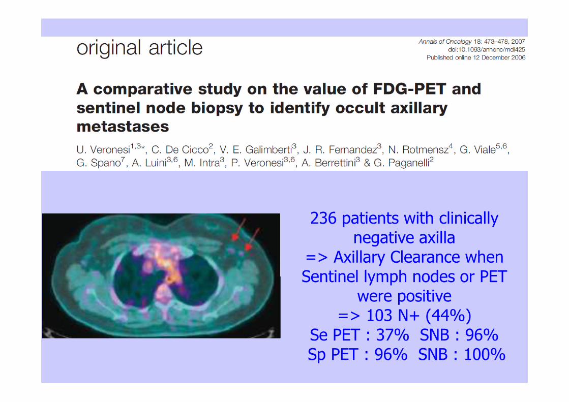
236 patients with clinically negative axilla
=> Axillary Clearance when Sentinel lymph nodes or PET
were positive=> 103 N+ (44%)
Se PET : 37% SNB : 96%Sp PET : 96% SNB : 100%
Initial Work-up: Stage I Breast Cancer
• FDG PET/CT has no indication:
- Performances of PET/CT << SNB
- Group with low risk of distant metastases and - Group with low risk of distant metastases and potential risk of false-positive PET-findings

Initial Work-up
Locally Advanced and Locally Advanced and inflammatory Breast Cancer
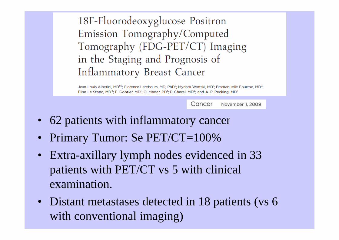
• 62 patients with inflammatory cancer• 62 patients with inflammatory cancer
• Primary Tumor: Se PET/CT=100%
• Extra-axillary lymph nodes evidenced in 33 patients with PET/CT vs 5 with clinical examination.
• Distant metastases detected in 18 patients (vs 6 with conventional imaging)

Saint Louis Hospital Experience between 2006-2011
LABC was defined as a T4 primary tumor and/or a N2 or N3 lymph node disease according to the AJCC V7 classification

Findings with 18FDG-PET/CT in three different groups: non inflammatory LABC, inflammatory carcinoma, and the whole population. Results expressed per patient basis

Saint Louis Hospital Experience between 2006-2011

Saint Louis Hospital Experience between 2006-2011
Kaplan-Meier Disease-specific Survival for 104 patients with recent follow-up.

Initial Work-up
Is there a role for PET/CT Is there a role for PET/CT between Stage I and Inflammatory Breast Cancer ?

• 60 Patients (T >3cm)
• Staging Modification for 42 % of patients
• Extra-axillary lymph nodes: 3 patients
• Distant metastases: Se PET = 100% (60% for CI)
Sp PET = 98% (83% for CI)
CI: Conventional Imaging

⇒ Study assessing the yield of PET/CT for initial ⇒ Study assessing the yield of PET/CT for initial work-up of 131 breast cancer patients clinically stage IIA, IIB or IIIA

Consecutive patients with breast cancer examined at the breast disease unitof Saint-Louis hospital from Mai 2006 to December 2010
History and physical examination,mammography,
breast and axilla US, breast MRI
131 Patients classified Stages IIA-IIB-IIIA:
18F-FDG PET/CTworkup
Conventional Imaging workup(chest examination by radiography and/or CT,
abdomino-pelvic examination by US and/or CT,and bone scan)
131 Patients classified Stages IIA-IIB-IIIA:- 36 Stage IIA (2 T1 N1, 34 T2 N0)
- 48 Stage IIB (28 T2 N1, 20 T3 N0)- 47 Stage IIIA (9 T2 N2, 29 T3 N1 and 9 T3 N2)

⇒ No difference in the yield between stage IIB (T3 N0, T2 N1) and T3 N1 of stage IIIA (7/48 vs 3/29 ; p=0.739).
⇒ Staging modifications for 5.5% (2/36) in the stage IIA group, 13% (10/77) in the stage IIB + T3 N1 group and 56% (10/18) in
The Yield of 18FDG-PET/CT in Patients with Clinical Stage IIA, IIB, or IIIA Breast Cancer: A Prospective Study.
13% (10/77) in the stage IIB + T3 N1 group and 56% (10/18) in the stage IIIA group with N2 disease (P < 0.0001).
⇒ Accuracy: PET-CT > Bone scan (P = 0.036).

Conclusions
• Diagnosis of malignancy: PET/CT is not indicated
• Stage I Breast Cancer Staging: No role for PET/CT; SNB >> FDG-PET/CTPET/CT; SNB >> FDG-PET/CT
• Stage III locally advanced and inflammatory breast cancer: Recognized role for PET/CT
• Stage IIB (T2N1, T3N0) and T3 N1 breast cancer: A new emerging role for PET/CT

• Thank you for your attention