Embed Size (px)
Citation preview
Sveriges lantbruksuniversitet Swedish university of Agricultural Sciences Faculty of Veterinary Medicine and Animal Science Department of Biomedical Science and Veterinary Public Health
Diagnosis and Molecular Epidemiology of Bovine Rotavirus and Coronavirus in Brazil
Samuel Jakobsson
Uppsala
2013
Degree project within the Veterinary Medicine Program ISSN 1652-8697
Examensarbete 2013:22

SLU Sveriges lantbruksuniversitet
Diagnostik och Molekylär Epidemiologi av Bovint Rotavirus och Coronavirus i Brasilien
Samuel Jakobsson
Handledare: Mikael Berg, Institutionen för Biomedicin och Veterinär Folkhälsovetenskap
Examinator: Susanna Sternberg Lewerin, Institutionen för Biomedicin och Veterinär Folkhälsovetenskap
Examensarbete inom veterinärprogrammet, Uppsala 2013 Fakulteten för veterinärmedicin och husdjursvetenskap
Institutionen för Biomedicin och Veterinär Folkhälsovetenskap Kurskod: EX0751, Nivå A2E, 30hp
Key words: molecular epidemiology, rotavirus, coronavirus, calf, diarrhea, Brazil
Nyckelord: molekylär epidemiologi, rotavirus, coronavirus, kalvar, diarré, Brasilien
Online publication of this work: http://epsilon.slu.se ISSN 1652-8697
Examensarbete 2013:22

Abstract ........................................................................................................................................... 1
Sammanfattning .............................................................................................................................. 1
Introduction ..................................................................................................................................... 2
Scope of the study ........................................................................................................................... 2
Litterature study .............................................................................................................................. 3
Brazil’s agricultural sector .................................................................................................................... 3
Neonatal diarrhea in calves .................................................................................................................. 3
The disease ....................................................................................................................................... 3
Economic impact .............................................................................................................................. 4
Management of diarrheic calves ...................................................................................................... 4
Prevention and disease control ........................................................................................................ 4
Diagnosis ........................................................................................................................................... 5
Rotavirus ................................................................................................................................................... 5
Taxonomy ............................................................................................................................................. 5
Pathogenesis ......................................................................................................................................... 6
Host spectrum ...................................................................................................................................... 6
Morphology .......................................................................................................................................... 7
Genetic structure .................................................................................................................................. 7
Replication ............................................................................................................................................ 8
Prevalence in Brazil and other parts of the world ................................................................................ 8
Molecular epidemiology ....................................................................................................................... 9
G-genotypes ..................................................................................................................................... 9
P-genotypes ...................................................................................................................................... 9
Combinations .................................................................................................................................... 9
Difference between dairy and beef calves ..................................................................................... 10
Mixed genotypes ............................................................................................................................ 10
Bovine Coronavirus ................................................................................................................................. 10
Taxonomy ........................................................................................................................................... 10
Pathogenesis ....................................................................................................................................... 11
Host spectrum .................................................................................................................................... 11
Morphology ........................................................................................................................................ 12
Genetic structure ................................................................................................................................ 12
Replication .......................................................................................................................................... 12
Prevalence in Brazil and other parts of the world .............................................................................. 14
Molecular epidemiology ..................................................................................................................... 14
Materials and methods .................................................................................................................. 15
Lab description and equipment: ......................................................................................................... 15
Sample collection and preparation .................................................................................................... 15
RNA extraction .................................................................................................................................... 15
Reverse Transcription ......................................................................................................................... 16
Multiplex semi-nested Reverse Transcription Polymerase Chain Reaction ....................................... 16
VP4 ...................................................................................................................................................... 17
VP7 ...................................................................................................................................................... 17
NSP4 .................................................................................................................................................... 18
S-gene ................................................................................................................................................. 18
Amplicon purification and sequencing ............................................................................................... 18
Sequence editing and building of phylogenetic trees ........................................................................ 19
Results ........................................................................................................................................... 20
MSN RT-PCR ........................................................................................................................................ 20
Rotavirus batch one ............................................................................................................................ 20
Rotavirus batch two ............................................................................................................................ 20
Coronavirus batch one ....................................................................................................................... 20
Coronavirus batch two ....................................................................................................................... 21
Sequencing results .................................................................................................................................. 21
Batch one ............................................................................................................................................ 21
Batch two ............................................................................................................................................ 21
Phylogenetic trees .................................................................................................................................. 22
Discussion ..................................................................................................................................... 25
Rotavirus ................................................................................................................................................. 25
Rotavirus genotypes ........................................................................................................................... 25
Rotavirus amplification problems ...................................................................................................... 26
Coronavirus............................................................................................................................................. 27
Coronavirus genus .............................................................................................................................. 27
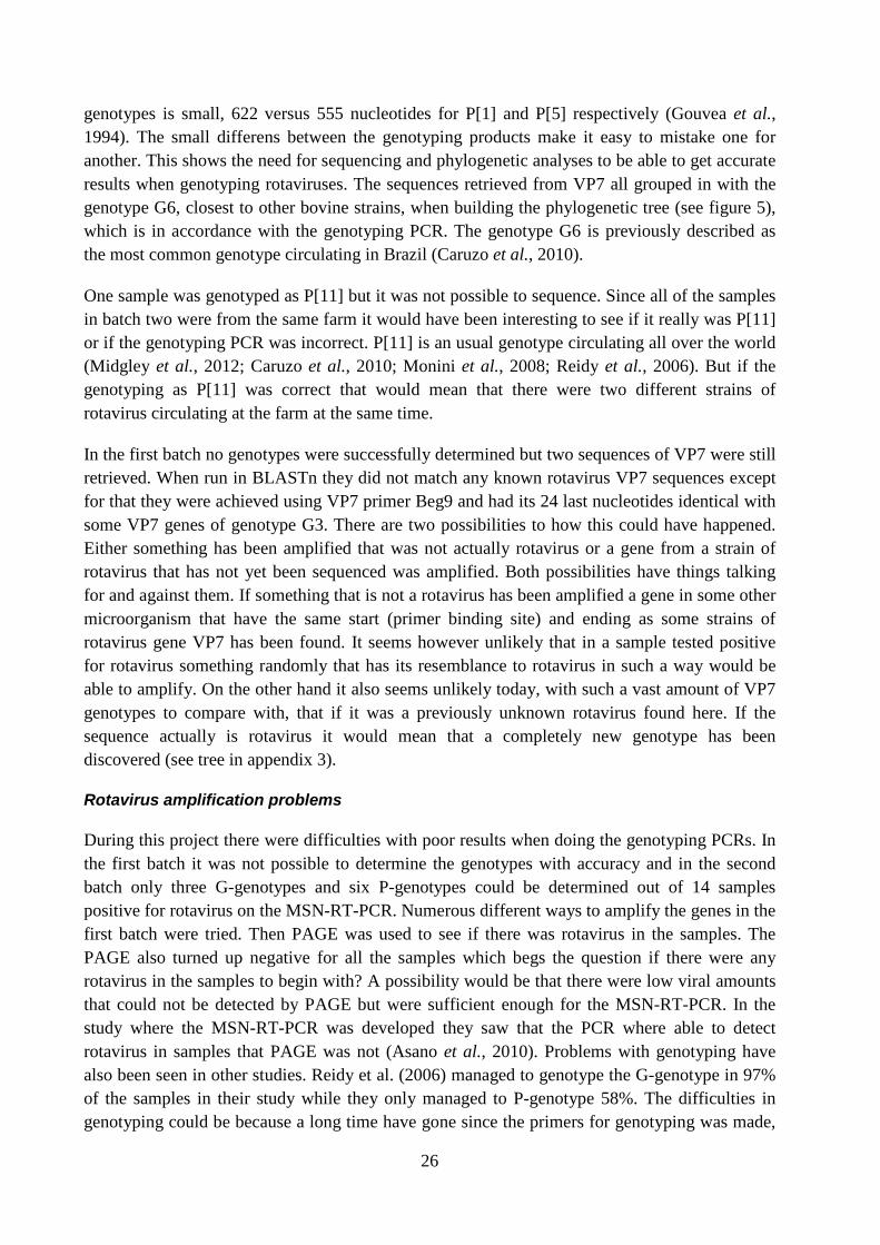
Relations to other Brazilian strains ..................................................................................................... 27
Coronavirus amplification problems .................................................................................................. 27
Conclusion .................................................................................................................................... 28
Acknowledgement ........................................................................................................................ 28
References ..................................................................................................................................... 29
Appendix 1 .......................................................................................................................................... 33

Appendix 2 .......................................................................................................................................... 34
Appendix 3 .......................................................................................................................................... 35
1
ABSTRACT
Rotavirus and coronavirus are the two most common viral causes of neonatal calf diarrhea and their presence causes a lot of economic damage to the farmers as well as suffering to the animal. By getting better knowledge about the viruses we may help in tracing transmission and in producing new vaccines. This paper served to study the molecular epidemiology of rotavirus and coronavirus in fecal samples collected from two different farms in the state of São Paulo, Brazil. These samples have been studied with focus on the gene VP4, VP7, NSP4 in rotavirus and the gene S1 in coronavirus. From the first farm the screening PCR showed that 12/48 samples were positive for rotavirus and 4/48 positive for coronavirus. No determination of genotypes of rotavirus was achieved and the sequences retrieved did not match any known rotavirus. Nor was any amplification and sequencing of the positive samples of coronavirus successful. From the second farm 14/22 samples tested positive for rotavirus and 2/22 samples positive for coronavirus. Initial genotyping PCR for determination of G- and P-genotype identified 3 samples as G6, 5 samples as P[1] and 1 sample as P[11]. Successful sequencing confirmed that the 3 samples were of genotype G6 but identified the, by genotyping PCR determined, P[1] samples as P[5]. From the coronavirus positive samples one sequence which clustered within the genus betacoronaviruses were retrieved.
SAMMANFATTNING
Rotavirus och coronavirus är de två vanligaste virala orsakerna till neonatal diarré hos kalvar och orsakar stora ekonomiska skador för djurägare samt ett lidande för djuren. Genom att få mer kunskap om hur dessa virus är uppbyggda på RNA-nivå kan man hjälpa i smittspårning och i utveckling av nya vacciner. Detta är en studie på molekylär epidemiologi hos dessa virus i fekala prover insamlade från två olika gårdar i delstaten São Paulo, Brasilien. Dessa prover har undersökts med fokus på generna VP4, VP7, NSP4 hos rotavirus samt genen S1 hos coronavirus. I proverna från första gården var 14/48 prover positiva för rotavirus och 4/48 positiva för coronavirus. Genotypning av rotavirus lyckades inte i denna uppsättning och de sekvenser som erhölls liknade inte några andra kända sekvenser av rotavirus. Amplifiering och sekvensering av coronavirus i denna uppsättning lyckades inte. Från den andra gården var 14/22 prover positiva för rotavirus och 2/22 positiva för coronavirus. I genotypande PCR för G- och P-genotyp identifierades 3 prover som G6, 5 prover som P[1] och 1 prov som P[11]. Sekvensering av dessa prover konfirmerade genotypningen som G6 men visade att de tidigare genotypade P[1] istället var av genotyp P[5]. Sekvensering av genen S1 i ett av de två positiva proverna av coronavirus lyckades och denna sekvens placerades i genus betacoronavirus vid en fylogenetisk analys.
2
INTRODUCTION
The population of the world is growing and with it the demand for good quality food. To prevent and decrease the now current amount of people living in starvation the resources of food must be optimized. Brazil is one of the top producers and the biggest exporter of meat from cattle in the world and the production is increasing. But diseases like neonatal calf diarrhea cause a lot of problems to the production of beef. The prevention of disease amongst neonatal calves is important and better understanding of the genetics and molecular epidemiology of the causative pathogens is crucial. Rotavirus and Coronavirus are the most common viral pathogens causing diarrhea in young cattle. Molecular epidemiology is an important tool for tracing the transmission of the diseases and helps in gaining knowledge about the viruses in order to produce effective vaccines. This is a Minor Field Study focusing on the molecular epidemiology of rotavirus and coronavirus amongst calves with diarrhea in the state of São Paulo, Brazil.
SCOPE OF THE STUDY
The scope of this study was to isolate and determine genetic sequences of rotavirus genes VP4, VP7, NSP4 and coronavirus gene S1 in fecal samples collected from calves presenting diarrhea in the state of São Paulo, Brazil. The aims were to determine the genotypes of rotavirus and for coronavirus assess the genus and its relation with other Brazilian strains in order to determine whether the strains in Brazil were divergent from known isolates. For rotavirus genotyping PCRs were performed and phylogenetic trees constructed to assess the genotypes of rotavirus in the samples by comparing them to known sequences from different hosts. For Coronavirus phylogenetic trees were constructed to assess which genus the sample virus belonged to by comparing them to other viral strain sequences with known genuses, and comparing it to other strains originating in Brazil.
3
LITTERATURE STUDY Brazil’s agricultural sector
Brazil has one of the biggest populations of cattle in the world with a national herd size of an estimated 200 million heads. The gross value of the milk and meat produced is estimated at $ 67 billion a year. The tropical climate with its good conditions for pastures is accredited a large part of the success of Brazilian cattle production as the need for housing and daily care is low (AGRICULTURA).
Brazil is also the number one exporter of meat of beef and veal in the world (FAO, 2010). And the production of meat in Brazil is growing, especially the production of beef, pork and chicken. By 2020 the Brazilian production of meat is estimated to supply 44,5 % of the global market maintaining Brazil as the one of the top producers of meat (AGRICULTURA).
The production of meat is currently thought to increase by roughly 12,6 million tons by 2018/2019. This represents an increase of 51% compared to the meat production in 2009, half of which is accredited to the domestic consumption. The increase and is an effect of better use of pastures through modern agronomic techniques, better knowledge of nutrition and investment in genetics. These combined will reduce the age of slaughter for the Brazilian cattle herd and thereby increase the amount of meat produced (AGRICULTURA).
Neonatal diarrhea in calves
The disease
Diarrhea among neonatal calves is a common disease. The form of the disease varies from calf to calf, some suffering acute dehydration and death whilst others suffer from sub-acute forms with malnutrition that lasts for several days (Gay et al., 2012). Neonatal diarrhea is a worldwide problem and is seen as one of the biggest challenges for both the beef and dairy industry (Lorenz et al., 2011).
Several different pathogens have been associated with neonatal calf diarrhea with the most common being Escherichia coli, rotavirus, coronavirus and Cryptosporidium parvum (Gay et al., 2012). These pathogens often occur in fecal samples from healthy calves as well making the development of disease a relationship between the resistance of the calf combined with the infection pressure (Lorenz et al., 2011). Co-infection with more than one of these pathogens is usual and often worsens the symptoms. Depending on the age of the calf, some pathogens are more likely to be the cause of diarrhea. E. coli most often affects calves 3-5 days old, Corona and rotavirus 5-15 days old although it can affect calves up to several months of age and Cryptosporidiosis most commonly affect calves aged 5-35 days. Common for all the pathogens is that if they do not result in death they cause need for extra care, sometimes intensive care, and weight loss in the calves (Gay et al., 2012).
4
Economic impact
Neonatal diarrhea is an important disease and causes a lot of economic damage to the farmers through need for increased management, veterinary treatment, reduced growth and deaths of the calves. In 1998 Donovan et al. showed that the occurrence of diarrhea during the first six months caused reduction on growth and argued that it causes a prolonging of the time from birth to when the heifers have their first calving (Donovan et al., 1998). In an economic model of the cost of rearing young cattle in the Netherlands in 2012 it was found that occurrence of diarrhea had a big impact on the economy by influencing the mortality. A higher incidence rate of diarrhea caused a higher mortality amongst the calves. Decreased growth was also seen as a big factor influencing the economy (Mohd Nor et al., 2012). The cost for the unwanted death of a calf was calculated to roughly $ 60 UD and an reduction in mortality in farms in Kuwait was seen to have a big positive impact on the gross margins (Razzaque et al., 2009).
Management of diarrheic calves
When an animal has developed diarrhea they require an extra need of management with the most important part being oral rehydration therapy. The loss of water and electrolytes is important to replace to avoid dehydration and acidosis. Continuous feeding with milk or milk replacer is also important to prevent malnourishment although force-feeding is not advised because of the risk dysfunction of the esophageal groove. This leads to fermentation in the reticulorumen contributing further to the acidosis. The continued feeding does not only provide energy for growth but also provides nutrients needed for the recovery of the intestinal mucosa. If the animal is severely depressed and too weak to drink by itself intravenous fluid therapy is recommended (Lorenz et al., 2011).
Prevention and disease control
Some of the management practices found to play an important role in how resistant the calves are to infection include the prevention of dystocia, the reception of adequate amounts of colostrum of good quality and an appropriate diet thereafter. The pressure of the infectious agents can be decreased by proper hygiene practices in the calving space, feeding space, housing area and during general handling of the calves (Lorenz et al., 2011). Several risk factors have been identified, influencing the prevalence of the pathogens and often the prevalence of one of the pathogens increases the risk of another pathogen also being present, supporting the previous statement that co-infection is common. Other risk factors included hygiene practices, herd size and age (Bartels et al., 2010). Some vaccines against rotavirus have been developed and can be given to the dam in the late stages of pregnancy. This gives a high level of antibodies in the colostrum. The main effect of the antibodies is presented in the lumen of the small intestine while antibodies in the bloodstream seem to have little effect on preventing the symptoms of disease (Dhama et al., 2009). Other commercially available vaccines also often contain antigen against E. coli F5 and coronavirus. The levels of antibodies are well increased in the colostrum for most these vaccines but often the clinical efficiency varies between studies (Lorenz et al., 2011).
5
Diagnosis
There are a variety of diagnostic methods available for the detection of rotavirus and coronavirus including PCR, ELISA, Electron microscope and Immune electron microscope. Rotavirus may also be detected by agglutination and polyacrylamide gel electrophoresis. For rotavirus sample material that may be used for detection are feces and biopsies of small intestine. Biopsy being an alternative if the animal has died and necropsy is performed. PCR have been shown to have a higher sensitivity than both antigen detection methods and EM and some of the commercial ELISA kits have shown a low sensitivity and specificity. PCR are therefore an increasingly common way of diagnosis (Blanchard, 2012). Bovine coronavirus can be diagnosed using secretions or excretions from animals. Usually the collection of material for diagnosis is performed by nasal swabbing with a sterile swab or collection of feces from the rectum in a sterile cup. The diagnosis is confirmed by detection of virus, viral antigen or viral RNA. The virus detection, which includes replication in cell culture, is not an ideal way of diagnosis though since some bovine coronavirus strains fail to grow in cell culture. Coronavirus antigens are most commonly detected using ELISA which has the good qualities of being fast and having the capability to handle a big number of samples at the same time. Detection methods focusing on the viral RNA are becoming a more widely used way of diagnosis. These include RT-PCR and the more sensitive nested RT-PCR and realtime-qPCR (Saif, 2010).
Rotavirus Taxonomy
According to the International Committee on Taxonomy of Viruses (ICTV) Rotaviruses belongs to the family Reoviridae, subfamily Sedoreoviridae and genus Rotavirus where the genus Rotavirus include five different species, Rotavirus A-E ((ICTV), 2011).
The current classification of rotaviruses was established by Matthjinsen et al. (2008). The then previous way of classifying group A rotavirus by its serological aspects was gradually replaced when sequencing became a more popular way of analyzing viruses. The sequencing made it possible to compare the genomics of the virus on nucleotide level and a classification system that compared the genome of the virus in the genes VP4 and VP7 was established. In 2008 the classification system was elongated to include all 11 of the rotavirus gene segments. This made it possible to have a classification where the whole genome of the virus was considered. The classification system is as follows: Gx-P[x]-Ix-Rx-Cx-Mx-Ax-Nx-Tx-Ex-Hx and are used for the VP7-VP4-VP6-VP1-VP2-VP3-NSP1-NSP2-NSP3-NSP4-NSP5/SNP6 genes respectively.
A Rotavirus Classification Working Group was established to ensure the accuracy, maintain, evaluate and develop the new classification system. The group receives nucleotide sequences from potential new rotavirus genotypes and performs their own phylogenetic analysis of the viruses. The result is then sent back to the submitter who can publish the new strain (Matthijnssens et al., 2011).
6
Pathogenesis
Rotavirus that infects calves causes an often severe and sometimes life-threatening diarrhea. The diarrhea is caused by several factors. The virus replicates in mature enterocytes on the villi of the small intestines. The replication eventually causes lysis of the cells. The mature enterocytes are then replaced by immature enterocytes from the crypts of the villi. The balance between absorption and secretion of fluid is then changed resulting in an accumulation of fluid in the small intestine. Loss of mature enterocytes also contributes to a systemic insufficiency of bicarbonates, sodium, potassium, chloride and water causing acidosis. The loss of enterocytes reduces the ability to digest milk and the undigested milk is further fermented by microorganisms, which also contributes to cause acidosis. Low lactase, due to the loss of enterocytes, in the intestinal lumen further contributes to fluid accumulation by a failed osmotic regulation.
The histological picture shows short and blunt villi in the small intestine. The columnar epithelial cells are substituted by cuboidal or squamous cells from the crypts and infiltration of inflammatory cells in the lamina propria is seen (Dhama et al., 2009).
Recent studies have also shown that the viral protein NSP4 may act like an enterotoxin. After cell lysis the protein binds to cells and causes secretion of chloride into the intestinal lumen, causing osmotic diarrhea (Dhama et al., 2009). This is supported by the fact that diarrhea often set in even before histological changes are visible (International Committee on Taxonomy of Viruses. & King, 2012). On top of that, inflammatory changes in the small intestine caused by the infection gives the intestine a hypermobility that results in less absorption of fluid. The end result of all the factors is a watery and most often blood and mucus free diarrhea. If mucus and blood are present it probably originates from secondary bacterial infection (Dhama et al., 2009).
The incubation period of the virus is 12-24 hours and the diarrheic calves are most often feverless. The disease may result in severe dehydration and mortality rates differ, but reports have estimated it to be in average 5-20 %. The diarrhea is self-limiting and animals most often recover if not to severe dehydration have occurred. Animals that recover properly usually return to normal bodyweight within 10-28 days post infection (Dhama et al., 2009).
Host spectrum
Rotaviruses have a large host range and are able to infect a wide array of mammals and birds. The virus transmits through a fecal-oral route and calves are most often infected by contact with other calves, primarily or secondary through objects, feed and water. It has been proposed that calves can also be infected by virus shed by the dam at birth. The infected calves shed virus through the feces from the second day of infection and the shedding may last for 7-8 days. The virus primarily affects neonatal individuals, and calves more than 3 months of age are usually not affected (Dhama et al., 2009). Rotaviruses were long thought to be host specific but several different studies have shown rotavirus to have a cross species potential. In vivo tests trying to infect mice with both a strain of simian rotavirus as well as bovine rotavirus and recombinants of the two showed that some of the recombinants were able to infect and replicate in the mice
7
several times better than the original viruses. Some genes were shown to have a greater effect on the ability of the virus to infect and replicate. The origin of VP4, VP7 and NSP1 were found to have a significant effect on the capability of the virus to infect and replicate (Feng et al., 2011). Kim et al. (2011) also showed that a triple reassortant virus found in Korea containing several human and porcine-like genes were able to infect calves and cause severe diarrhea. Here the genes VP4 and NSP1 where of porcine origin while the VP7 was of bovine origin (G6P[7]) .
Morphology
Rotaviruses have a “wheel-like” appearance which explains the origin of its name. The virion consists of a triple layer capsid covering a genome of double stranded RNA. The mature infectious virion has a diameter of 100nm. The envelope is lipid-free and consists of three concentric layers of protein. The different layers are made up of three of the 13 proteins that the rotavirus genome encodes. VP2 forms the innermost layer and surrounds the viral dsRNA. The middle layer is composed of VP6 and the outermost layer of VP4 and VP7. The middle and outermost layer have 132 large channels that link the outside of the virion to the VP2 layer. VP7 makes up the base of the outermost layer while VP4 forms spike like extensions extending out of the virion. VP4 also extends in through the two outer layers, and possibly also have some interaction with the VP6 layer. Both have important roles in the infectivity of the virion. Infectivity is quickly lost in the presence of disinfectants like chlorine and 95% ethanol. These remove the outer shell and thus make the virus unable to infect cells. The virus is stable and infectious in pH range between 3-9 and may under the right concentration of calcium chloride stay infectious for months at 4°C and even up to 20°C (International Committee on Taxonomy of Viruses. & King, 2012).
Genetic structure
The genome of rotavirus consists of 11 segments of dsRNA averaging 18,550 bp with segments at a varying size between 663 and 3302 bp. All segments share a short common pattern of nucleic acid at the 5’ and 3’ terminals, 10 and 8 nucleotides respectively. Within these there are another common pattern of 30-40 nucleotides that are segment specific.
The 11 segments of dsRNA encode 13 different proteins ranging from VP1-VP7 and NSP1-NSP6 where segment 9 encodes for 2 different types of VP7. Of the proteins six have been found to be structural. Three are associated with the dsRNA, VP1, VP3 and VP2. VP1 and VP3 are directly associated with the dsRNA and make up complexes that link it to the core shell consisting of VP2. The protein VP4 is spike like and extends through the 2 outer shell layers and about 20 nm further outside the virion. Cleavage of VP4 by trypsin stabilizes the spikes by forming VP5 and VP8 which improves the infectivity of the virus. VP8 takes on a crystallized form and have hemagglutination activity. The protein VP7 makes up the outermost shell together with VP4. Less is known about the functions of the six nonstructural proteins, NSP1-NSP6. NSP1 is the largest of the rotavirus proteins and have been shown to bind both zinc and the 5’-end of ssRNA. It serves as a component in the early stages of replication, more specifically the genome segment selection. NSP2 have both ssRNA and dsRNA activity and have a direct role in viral replication but the exact role has yet not been discovered. NMSP3 shuts of the protein
8
synthesis of the infected cell and promotes viral translation by helping viral mRNA circulate in the cytoplasm. NSP4 have been reported to have a role in the maturation of the virion by initiating budding through the ER1
International Committee on Taxonomy of Viruses. & King, 2012
membrane. It has also been reported that NSP4 have an endotoxin like effect that causes calcium to be let out of the ER. A product from cleaving of NSP4 is secreted from infected cells and binds to receptors initiating a pathway which result in calcium release from the cell storages. NSP5 have ssRNA and dsRNA binding activity but the effect of this protein is unknown. NSP6 has undefined function (
).
Replication
Rotavirus has a cycle of replication at 10-12 hours at 37°C. Little is known about the early steps of replication. VP4 is known to attach to an unknown receptor on the host cell. There are 2 proposed ways of entry into the cell. One is by endocytosis and the other by direct entry by the virus. When the virion enters the cell it loses its outermost shell and the double layered transcriptionally active particle is set free into the cytoplasm. mRNA2 transcripts, the full length of the different segments, are produced by DLP3
International Committee on Taxonomy of Viruses. & King, 2012
-associated enzymes from the dsRNA minus strand. The mRNA serves two purposes. They are used in the synthesis of viral proteins and control expression of the individual genes making some genes more transcriptionally active than others. The other purpose is serving as template for genome replication. Minus strand synthesis is accomplished after assembly of all the necessary mRNA and takes place in an intermediate viral core. The intermediate core then transfers to the inside of the endoplasmic reticulum by actions of NSP4. The virion enters the ER by budding in and receiving a temporary envelope. Inside the temporary envelope is lost and VP7 and VP4 form the outermost shell of the virion ( ).
Prevalence in Brazil and other parts of the world
During the years 1998-2002 samples from 1898 diarrheic calves and 279 calves with normal feces were collected from several geographic areas in Brazil in order to estimate the prevalence of group A rotavirus. The calves ranged in age from 1-90 days. 19,4 % of the calves presenting diarrhea tested positive for group A rotavirus while 2,2 % of the calves with normal feces tested positive. The highest percentage of positive diarrheic animals were calves in the age of 1-30 days with 36 % when divided into three groups, 1-29 days, 30-60 days and 61-90 days. Amongst the calves with normal feces divided into similar groups only the 1-29 day old group tested positive for rotavirus. Frequency of positive samples was higher amongst the diarrheic samples from beef cattle compared to dairy cattle (Alfieri et al., 2006). Other studies around the world have shown a varying prevalence of rotavirus when sampling calves with diarrhea. In Australia 79,9 % of the calves were tested positive for rotavirus (Izzo et al., 2011). In Switzerland 58.7 % of fecal samples from diarrheic calves contained rotavirus (Uhde et al., 2008). In Dutch dairy calves in 2007 30,9 % of diarrheic samples were positive for rotavirus (Bartels et al., 2010). During 2002
1 Endoplasmic reticulum 2 Messenger RNA 3 Double layered particle
9
to 2006 when testing fecal samples from Sweden studies showed rotavirus infection in 47% of the calves (De Verdier, 2006).
Molecular epidemiology
Of all the different genotypes of bovine rotavirus some combinations of genes seems to be more common than others. Several different studies have been made all over the world describing the prevalence of genotypes with quite similar results but also with some exceptions.
G-genotypes
In Goiás, Brazil during 1994-1995 when doing a study in molecular characterization the G-genotype G6 was found to be the most predominant followed by G10 with a prevalence of 64,5% and 32,2% respectively (Caruzo et al., 2010). During a cross-country study in Europe six different G-genotypes were detected in cattle: G4, G6, G8, G10, G11 and G12 where the most common type was G6 (in 80% of the samples) followed by G10 (13%). While G6 was the predominant genotype in all countries included in the study the prevalence of G10 varied from 0-28% (Midgley et al., 2012). In Ireland the most common was G6 (80,6%) followed by a combination of G6G10 (9,7%) and G10 (6,5%) (Reidy et al., 2006). That G6 is commonly predominant was also supported by Monini et al. (2008) whom in Italy during 2003-2005 found it to be present in 78,5% of samples positive for rotavirus. It was followed by G10 (9,9%), G8 (4,7%) and mixed types in 3,3% of samples. In this study they also observed a difference in prevalence of different genotypes from year to year where for example G10 varied in prevalence from 3,4-21,3%. Recently a study in India has confimed a new predominant G-genotype. In this study G3 was found in 39,4% of samples collected from several different parts of the country. This was followed by the mixes G3G8 (27,3%) and G3G10 (33,3%) (Malik et al., 2012).
P-genotypes
In Ireland during 2002-2004 P[5] was found to be the most prevalent P-genotype with 77,8% of the samples. P[11] was the second most common with an prevalence of 9,3% and in 1,9% of samples P[1] was found. A mix of P[5]P[11] was also seen in 11% of samples (Reidy et al., 2006). Caruzo et al., 2010 found that 32,2% of the samples collected in Goiás, Brazil was of P-genotype P[11]. But they also found a high prevalence of the more uncommon P[1] (9,7%). In Italy P[11] was the most common (65,1%) followed by P[5] (25%) during the timeperiod 2004-2005. The variation of prevalence of genotypes from year to year was shown to be less for the P-genotype than for the G-genotype (Monini et al., 2008). In Europe Midgley et al. (2012) found that the three genotypes P[1], P[5] and P[11] were circulating at the time. P[5] and P[11] were the most common ones but which one that was predominent differed between the countries.
Combinations
Ramos Caruzo et al. (2010) found the most common combination of G and P-genotypes in Brazil to be G6P[11] with 16,2% of the cases. In Ireland during 2002-2004 G6P[5] was the most predominant combination with 57.4% of the samples followed by G6G10P[5] (13%), G6P[5]P[11] (11%) G6P[11] (7,4%) and G10P[5] (7,4%) (Reidy et al., 2006). Monini et al.
10
(2008) found that in Italy during 2002-2005 G6P[11] was the most common combination with 48,3%, while G6P[5] followed with 22,8% and G10P[11] was the third most common combination with 7,8%. Midgley et al. (2012) also found in their cross-country study in Europe that G6P[11] and G6P[5] were the most common combinations among cattle with a prevalence of 39% and 30% respectivly followed by G10P[11] with 9%.
Difference between dairy and beef calves
In Argentina during 1994-2003 a difference in common genotypes between dairy and beef calves was observed. In beef G6 was found to be the most dominant G-type with 89% of the cases, followed by G10 with 9 %. The most common P-type was P[5] which was found in 81% of the samples followed by P[11] with 3%. The combination of G6P[5] was the most common (75% of cases). In dairy calves G6 was the dominant G-type with 59% followed by G10 (16%). P[11] was the dominant P-type with 71 % of the cases followed by P[5] (16%) (Garaicoechea et al., 2006).
Mixed genotypes
In some cases of genotyping there have been difficulties determining just one genotype and instead dual or more genotypes have been observed. This is due to co-infection with multiple viruses. In Argentina 6,8 % of the cases of rotaviral infection could be seen containing more than one G or P-type (Garaicoechea et al., 2006). The fact that mixed genotypes are quite common is supported by Caruzo et al. (2010) that found that in Goiás, Brazil during 1994-1995, 51% of samples containing rotavirus had multiple P-genotypes, including two typically human P-genotypes supporting evidence of zoonotic potential (Caruzo et al., 2010). In Ireland 11% of samples showed dual P-genotypes P[5]P[11] and 9,7% dual G-genotypes G6G10 (Reidy et al., 2006).
Bovine Coronavirus
Taxonomy
Bovine coronavirus belongs to the order Nidovirales, family Coronaviridae, subfamily Coronavirinae, genus Betacoronavirus, species Betacoronavirus 1 ((ICTV), 2011).
In 2003 the then current taxonomy of coronaviridae was revised. Previously coronavirus was grouped into three groups, 1, 2 and 3, with bovine coronavirus in group 2. The new revision states that rather than groups coronaviruses should be divided into three genera, 1, 2 and 3, based on their genetic and serological relationships. The gene S was found to be the gene with the biggest diversity and also the gene where most genetic sequence data were available in the GenBank at the time (Gonzalez et al., 2003).
The ICTV have assigned the three groups the names Alpha-, Beta-, and Gamma coronaviruses with the addition of a fourth group named Deltacoronaviruses which is currently emerging (Woo et al., 2010). Bovine Coronavirus have in 2009 been merged into the group Betacoronavirus 1 in the genus Betacoronaviruses ((ICTV), 2011).
11
Pathogenesis
Coronaviruses are a group of viruses that can cause a large variety of clinical signs. Bovine coronavirus is most commonly the cause of gastrointestinal and respiratory disease. Bovine coronavirus is associated with three well known syndromes in cattle: Winter dysentery in adult cattle, respiratory disease in various ages and neonatal calf diarrhea (Maclachlan et al., 2011).
Gastrointestinal, respiratory tract and neurological tissues are the most common to be infected by coronavirus but other organs are susceptible for infection as well i.e. liver, kidney, heart and eye. The virus mainly targets epithelial cells but some species of the virus also targets widely distributed cells such as macrophages (International Committee on Taxonomy of Viruses. & King, 2012).
In calves, bovine coronavirus is a common cause of diarrhea up to three weeks of age but disease occurs up to three months of age. The onset of coronaviral caused diarrhea often concurs with the period when the passive immunity from the dam declines (Maclachlan et al., 2011). The virus firstly infects the nasal mucosa where intensive replication occurs. Large amounts of virus are then swallowed together with the mucus secretions it causes and are transported to the gastrointestinal system (Saif, 2010). There the diarrhea is caused by the destruction of mature absorptive cells lining the villi and mucosal surface in the large intestine. This leads to a malabsorptive diarrhea with rapid loss of water and electrolytes as a result. Hypoglycemia, acidosis and hypovolemia are common symptoms in these calves and may lead to circulatory failure and death. Bovine coronaviral diarrhea is often worsened by co-infection with other common gastroenteric pathogens such as E. coli, cryptosporidia, toro- and rotavirus (Maclachlan et al., 2011).
Host spectrum
Coronaviruses have been shown able to infect a wide arrange of mammals and birds. Most of coronaviruses have limited host spectrums although there are some exceptions. Interspecies transmission is possible and seems to be more usual in vivo than in in-vitro experiments (International Committee on Taxonomy of Viruses. & King, 2012). The host spectrum seem to be determined by portions of the hyper variable S-gene which encodes for receptor binding proteins, which initiate uptake of virions into host cells (Maclachlan et al., 2011).
Some of group 2 coronaviruses have shown potential to infect other species than their main host, including bovine coronavirus and SARS. Bovine coronavirus in closely related to human coronavirus-OC43 which causes common colds, and have been shown to cause enteritis with following diarrhea and viral shedding in turkeys. It has also been observed to cause subclinical infections in dogs (International Committee on Taxonomy of Viruses. & King, 2012; Maclachlan et al., 2011). Genetically related strains of bovine coronavirus have been shown be the cause in some cases of respiratory disease in dogs and in some cases of humans with diarrhea. Studies have also shown human coronaviruses to be able to cause gastrointestinal illness in calves (Maclachlan et al., 2011).
12
Morphology
Coronaviruses are a large group of enveloped spherical viruses with a size of approximately 120-160 nm in diameter. The virus has a core shell and within that a helical nucleocapsid that covers a ssRNA genome. From the envelope large glycoproteins project out from the surface. The glycoproteins consist of a stem with a globular portion at its proximal end. Bovine coronavirus along with some other subspecies also have a smaller projecting protein on the envelope surface formed by hemagglutinin-esterase (HE). The virus is inactivated by UV-radiation, heat, lipid solving reagents, non-ionic detergents, formaldehyde and oxidizing agents (International Committee on Taxonomy of Viruses. & King, 2012).
Genetic structure
Coronavirus contains an ssRNA molecule which is the largest known viral RNA genome. The ssRNA is of positive sense and functions as mRNA. The genome consists of 6-14 open reading frames (ORFs) where the two 5’-most overlap each other and together cover the replicase gene whereas the following each contain a separate gene. The genome translates 6 genes of known function: replicase, HE, S, E, M, N and a variable number of non-structural genes of which the function is less known.
The large projecting surface protein S is the biggest of the coronavirus proteins, ranging from 1160-1452 aa. It is cleaved in some viruses to form two subunits, S1 and S2. The S-protein is responsible for cell attachment, hemagglutination, membrane fusion and the induction of neutralizing antibodies. The M protein is a membrane protein found in the envelope that can induce alpha-interferon and is 221-260 aa big. During particle assembly the M-protein along with protein E (75-109 aa) plays an essential role. The protein HE is only found in some coronaviruses and is therefore classified as non-essential. It projects shortly out of the envelope and have a receptor binding domain for 9-O-acetlylated neuraminic acid as well as a receptor destroying activity by neuramine-O-acetylesterase. The N-protein is involved with the viral RNA synthesis and acts by binding RNA and forming a helical nucleocapsid.
The coronavirus genome also consists of several none-structural protein encoding genes. The biggest ones consist of the slightly overlapping ORF1a and ORF1b which constitute the replicase gene. The two ORFs encode two big proteins witch are believed to later on be processed into 15-16 mature replicase proteins and an unknown number of intermediates. The reason it is believed that they are processed is because so far no one has been able to detect them in the cytoplasm of infected cells. The rest of the non-structural protein encoding genes vary between different strains of the virus and are thought to be non-essential (International Committee on Taxonomy of Viruses. & King, 2012).
Replication
The replication of coronaviral RNA occurs on double membrane vesicles that originate from the endoplasmic reticulum in the cytoplasm of infected cells by synthesis of a full genome length. The endoplasmic reticulum undergoes changes and a network of modified membranes is created (International Committee on Taxonomy of Viruses. & King, 2012; Perlman & Netland, 2009).
13
The replicase gene with its several replication involved proteins is thought to be sufficient enough to alone ensure the viral replication. The actual synthesis of ssRNA is performed by a membrane-bound replicative complex, also of which little is known (International Committee on Taxonomy of Viruses. & King, 2012). The synthesized genomic RNA is incorporated into virions by merging of these double membrane vesicles into larger single membrane vesicles (Perlman & Netland, 2009).
The two largest ORFs encode a linear single strand of mRNA which is translated into protein. The protein is cleaved to form several subunits that assemble to form the RNA polymerase (International Committee on Taxonomy of Viruses. & King, 2012; Maclachlan et al., 2011). Two different theories about how the RNA is replicated exist. One claims the positive sense RNA is discontinuously transcribed to several segments of negative sense RNA which is combined and again transcribed to positive sense RNA. The other theory is that the positive sense RNA transcribes a full length negative sense RNA which then discontinuously transcribe positive sense RNA segments that assembles to form the genome of new virions (International Committee on Taxonomy of Viruses. & King, 2012).
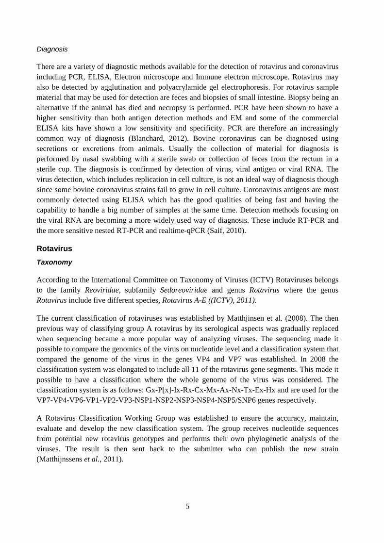
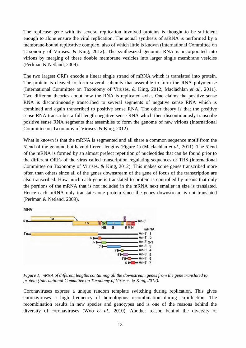
What is known is that the mRNA is segmented and all share a common sequence motif from the 5´end of the genome but have different lengths (Figure 1) (Maclachlan et al., 2011). The 5´end of the mRNA is formed by an almost prefect repetition of nucleotides that can be found prior to the different ORFs of the virus called transcription regulating sequences or TRS (International Committee on Taxonomy of Viruses. & King, 2012). This makes some genes transcribed more often than others since all of the genes downstream of the gene of focus of the transcription are also transcribed. How much each gene is translated to protein is controlled by means that only the portions of the mRNA that is not included in the mRNA next smaller in size is translated. Hence each mRNA only translates one protein since the genes downstream is not translated (Perlman & Netland, 2009).
Figure 1, mRNA of different lengths containing all the downstream genes from the gene translated to protein (International Committee on Taxonomy of Viruses. & King, 2012).
Coronaviruses express a unique random template switching during replication. This gives coronaviruses a high frequency of homologous recombination during co-infection. The recombination results in new species and genotypes and is one of the reasons behind the diversity of coronaviruses (Woo et al., 2010). Another reason behind the diversity of
14
coronaviruses is the relatively high error rate of the RNA polymerase, which may have effect on the host range. Studies have shown that the main difference between Bat-SCoVs4
Graham & Baric, 2010
and SARSCoVs are point mutations that have arisen from RNA polymerase errors (
).
Prevalence in Brazil and other parts of the world
Stipp et al. (2009) collected 282 samples of feces from both diarrheic (n=221) and normal feces (n=61) calves in 2004 to assess the prevalence of bovine coronavirus in Brazilian beef and dairy cattle herds. The study found bovine coronavirus to be presents in 19,0 % of the diarrheic samples and 3,3% in the normal feces samples. The calves were divided into different age groups: 1-15 days, 16-30, 31-45 and 46-60. The age group 16-30 days of age showed the highest percentage of positive samples (29 %) but at least one or more positive samples were found in each group (Stipp et al., 2009). Several other studies around the world have been done to estimate the prevalence of coronavirus in the feces of diarrheic calves. In Australia between 2007 and 2008 the prevalence was estimated at 21,6 % (Izzo et al., 2011). In Switzerland the frequency of samples positive for coronavirus between 2005 and 2006 was 8,8 % (Uhde et al., 2008). In Dutch dairy calves in 2007 7,4 % of diarrheic samples contained coronavirus (Bartels et al., 2010). In Swedish calves during a study in 2002 to 2005 the prevalence of coronavirus positive diarrheic fecal samples was 8 % (De Verdier, 2006).
Molecular epidemiology
When doing epidemiological studies on Coronavirus there are several different genes one may focus on. Gonzáles et al. (2003) focused on the S-, E-, M-, N-, Polymerase- and Helicase-genes when revising the then current taxonomy of Coronaviridae, with the S-gene the most diverse and numerous at the time. The S-gene have been shown to be a good gene for epidemiological studies because of the diversity and that no homolog of this gene have been found in other viruses, except for very distant homologs in toroviruses (Gonzalez et al., 2003). Further studies of epidemiology based on the S-gene have since been done in several countries.
In Sweden and Denmark Liu et al. (2006) showed the relatedness between Swedish and Danish samples of coronaviruses collected from calves with respiratory symptoms and/or diarrhea based on phylogenetic analysis of the S-gene. They managed to show that some strains of the virus circulated in the herds for a long time while in other herds samples from different time-periods clustered into different groups providing evidence of an introduction of a new virus into the herd (Liu et al., 2006).
Studies in South Korea showed that coronaviruses sampled during 2004 and 2005 was highly genetically related and clustered within the same groups as previously collected coronavirus samples with varying clinical symptoms. All Korean samples, although having different clinical symptoms like winter dysentery, respiratory symptoms and calf diarrhea, clustered into groups much differentiated from the Canadian and American reference strains used, showing that the
4 Bat SARS Coronaviruses
15
viruses circulating in South Korea had evolved in a different evolutionary pathway than the reference strains (Park et al., 2007).
These studies show the possibility to use the S-gene for analysis of molecular epidemiology, to determine relatedness between strains and see how the genome of the virus changes through time at a given place. Bidokhti et al. (2012) also showed the possibility to use the S-gene divergence as a tool for tracing the transmission of bovine coronavirus by phylogenetic analysis. Although it’s better to use the entire S-gene when doing phylogenetic studies, using only partial S-gene is still sufficient enough as a phylogenetic marker (Martinez et al., 2012). Interest of the coronaviruses has increased significantly after the SARS outbreak in 2003. The interest have resulted in an increased number of coronaviruses being discovered and sequenced giving a much bigger database over viral strains (Woo et al., 2010).
MATERIALS AND METHODS
Lab description and equipment:
All laboratory work was done in LABMAS (Laboratório de Biologia Molecular Aplicada e Serologia) at Faculdade de Medicina Veterinária e Zootecnia, University of Sao Paulo, Sao Paulo, Brazil. All rounds of thermocycling were performed in Veriti™ (Applied Biosystems™) except for the PCR prior to sequencing which was performed in an Eppendorf™.
Sample collection and preparation
For this study two batches of samples were used. Batch 1 consisted of 48 samples of feces from calves at a dairy farm approximately 250 km from São Paulo. Samples were collected from calves of 1 week to 1 month of age presenting both normal feces and diarrhea. After collection feces samples were put in isopor boxes with cooling blocks in for transport to the lab. There they were put in refrigerator until sample preparation started the next day.
Feces suspensions were prepared in 1,5 ml Eppendorf tubes by mixing feces and DEPC water (ultra-pure water previously treated with 0,1% diethyl-pyrocarbonate) w/v 50%. Suspensions were thereafter centrifuged at 12000x for 5 minutes at 4°C. 250µl of the supernatants was transferred to new 1,5 ml Eppendorf tube for RNA extraction.
Batch two consisted of 22 previously collected samples, with its RNA already extracted and stored at the lab. This batch had already been used in studies on rotavirus and coronavirus but none had yet been genotyped and sequenced.
RNA extraction
RNA extraction was carried out using TRIzol Reagent (Invitrogen, Carlsbad, CA) according to the manufactures instructions but with some steps changed in preference of the staff in the lab. Instead of incubating the tubes in room temperature for 10 minutes after adding isopropanol we put them in the freezer at -20°C for 15 minutes. 20µl suspensions of extracted RNA were made with DEPC water.
16
750µl of TRIzol Reagent were added to earlier collected supernatants. The samples were briefly vortexed and left to incubate in room temperature for 5 minutes. 200 µl of pure chloroform were added to the samples and thereafter incubated for 10 minutes in 4°C. The tubes were centrifuged at 12000x for 15 minutes at 4°C. The centrifugation separated the sample into three phases and 400 µl of the top one, which contains RNA, was transferred to a new 1,5 ml Eppendorf tube already containing 500 µl of pure isopropanol. The new tube was shaken vigorously by hand and thereafter put in a freezer at -20°C for 15 min. Then samples were centrifuged again at 12000x for 15 minutes at 4°C.
The RNA formed a pellet in the bottom of the tube. The aqueous phase was removed from the tube by pouring it out and excess fluid was removed by turning the tube upside-down on paper briefly. 750µl of 75% ethanol was added to wash the pellet and thereafter the samples were centrifuged at 12000x for 5 min at 4°C. The ethanol was poured off and excess fluid removed by putting tubes on a heating block at 57°C for 5 minutes or until the fluid was visibly evaporated. RNA pellets were thereafter resuspended with 20 µl of DEPC water mixed by passing the fluid up and down the pipette.
Reverse Transcription
cDNA synthesis was carried out with SuperScript™ III Reverse Transcriptase (Invitrogen, Carlsbad, CA). 7 µl of RNA suspension was transferred to 200 µl Eppendorf tubes and denatured at 95°C for 5 minutes. 4 µl Buffer 5 X, 2 µl dNTP 10µM, 2 µl DTT, 1 µl SuperScript™ III Reverse Transcriptase, 1 µl Random Primers, and 3 µl DEPC water was then added to each tube to a total of 20 µl. Samples were put in a thermocycler (Veriti) at 37°C for 60 minutes, 70° for 15 minutes and then 4°C until sample collection. Tubes were then stored in a freezer at -20°C to the next day.
Multiplex semi-nested Reverse Transcription Polymerase Chain Reaction
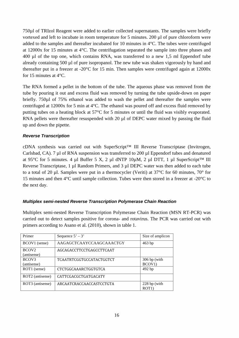
Multiplex semi-nested Reverse Transcription Polymerase Chain Reaction (MSN RT-PCR) was carried out to detect samples positive for corona- and rotavirus. The PCR was carried out with primers according to Asano et al. (2010), shown in table 1.
Primer Sequence 5’ – 3’ Size of amplicon
BCOV1 (sense) AAGAGCTCAAYCCAAGCAAACTGY 463 bp
BCOV2 (antisense)
AGCAGACCTTCCTGAGCCTTCAAT
BCOV3 (antisense)
TCAATRTCGGTGCCATACTGGTCT 306 bp (with BCOV1)
ROT1 (sense) CTCTGGCAAARCTGGTGTCA 492 bp
ROT2 (antisense) CATTCGACGCTGATGACATY
ROT3 (antisense) ARCAATCRACCAACCASTCCTGTA 228 bp (with ROT1)
17
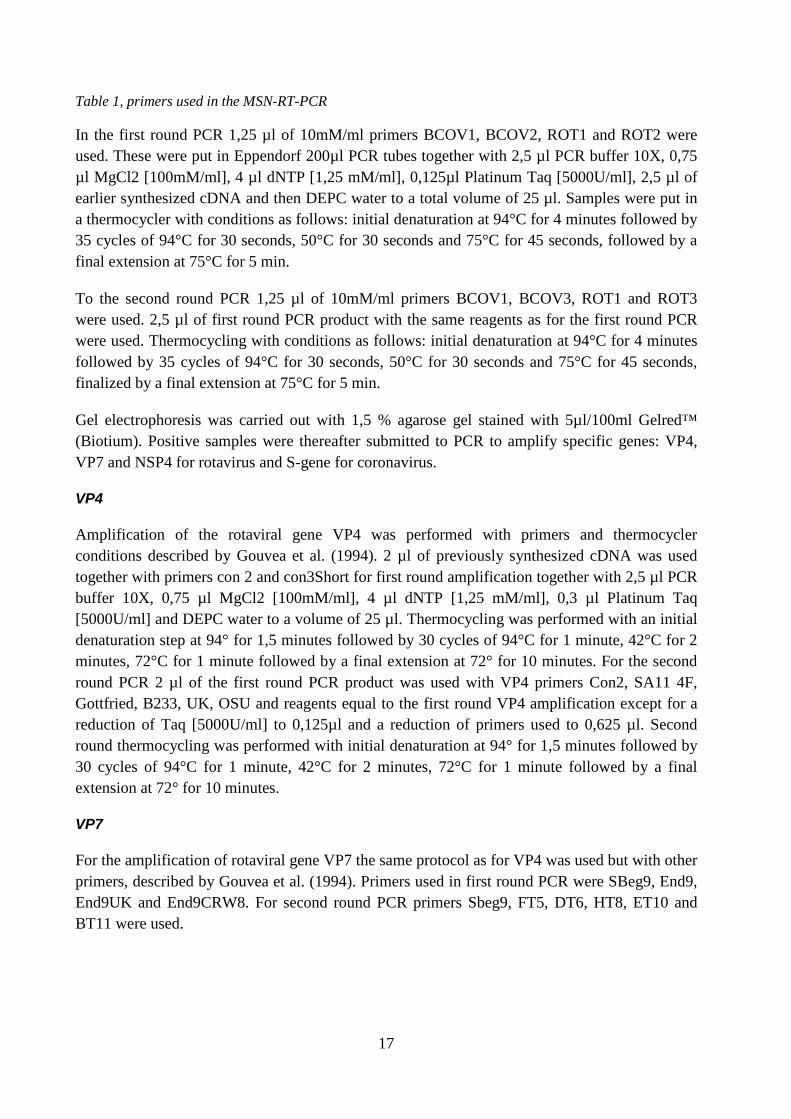
Table 1, primers used in the MSN-RT-PCR
In the first round PCR 1,25 µl of 10mM/ml primers BCOV1, BCOV2, ROT1 and ROT2 were used. These were put in Eppendorf 200µl PCR tubes together with 2,5 µl PCR buffer 10X, 0,75 µl MgCl2 [100mM/ml], 4 µl dNTP [1,25 mM/ml], 0,125µl Platinum Taq [5000U/ml], 2,5 µl of earlier synthesized cDNA and then DEPC water to a total volume of 25 µl. Samples were put in a thermocycler with conditions as follows: initial denaturation at 94°C for 4 minutes followed by 35 cycles of 94°C for 30 seconds, 50°C for 30 seconds and 75°C for 45 seconds, followed by a final extension at 75°C for 5 min.
To the second round PCR 1,25 µl of 10mM/ml primers BCOV1, BCOV3, ROT1 and ROT3 were used. 2,5 µl of first round PCR product with the same reagents as for the first round PCR were used. Thermocycling with conditions as follows: initial denaturation at 94°C for 4 minutes followed by 35 cycles of 94°C for 30 seconds, 50°C for 30 seconds and 75°C for 45 seconds, finalized by a final extension at 75°C for 5 min.
Gel electrophoresis was carried out with 1,5 % agarose gel stained with 5µl/100ml Gelred™ (Biotium). Positive samples were thereafter submitted to PCR to amplify specific genes: VP4, VP7 and NSP4 for rotavirus and S-gene for coronavirus.
VP4
Amplification of the rotaviral gene VP4 was performed with primers and thermocycler conditions described by Gouvea et al. (1994). 2 µl of previously synthesized cDNA was used together with primers con 2 and con3Short for first round amplification together with 2,5 µl PCR buffer 10X, 0,75 µl MgCl2 [100mM/ml], 4 µl dNTP [1,25 mM/ml], 0,3 µl Platinum Taq [5000U/ml] and DEPC water to a volume of 25 µl. Thermocycling was performed with an initial denaturation step at 94° for 1,5 minutes followed by 30 cycles of 94°C for 1 minute, 42°C for 2 minutes, 72°C for 1 minute followed by a final extension at 72° for 10 minutes. For the second round PCR 2 µl of the first round PCR product was used with VP4 primers Con2, SA11 4F, Gottfried, B233, UK, OSU and reagents equal to the first round VP4 amplification except for a reduction of Taq [5000U/ml] to 0,125µl and a reduction of primers used to 0,625 µl. Second round thermocycling was performed with initial denaturation at 94° for 1,5 minutes followed by 30 cycles of 94°C for 1 minute, 42°C for 2 minutes, 72°C for 1 minute followed by a final extension at 72° for 10 minutes.
VP7
For the amplification of rotaviral gene VP7 the same protocol as for VP4 was used but with other primers, described by Gouvea et al. (1994). Primers used in first round PCR were SBeg9, End9, End9UK and End9CRW8. For second round PCR primers Sbeg9, FT5, DT6, HT8, ET10 and BT11 were used.
18
NSP4
For amplification of rotaviral gene NSP4 primers were used in accordance of Lee et al. (2000). Primers used were 10Beg16 and 10End722. 2,5µl of previously synthesized cDNA was put in tubes with 2,5 µl PCR buffer 10X, 0,75 µl MgCl2 [100mM/ml], 4 µl dNTP [1,25 mM/ml], 0,125 µl Platinum Taq [5000U/ml] and DEPC water to a volume of 25 µl. The samples were put in a thermocycler at with the following temperatures: Initial denaturation at 94°C for 2 minutes followed by 35 cycles of 95°C for 45 seconds, 49°C for 30 seconds, 72°C for 1,5 minutes and last final extension at 72°C for 10 minutes.
S-gene
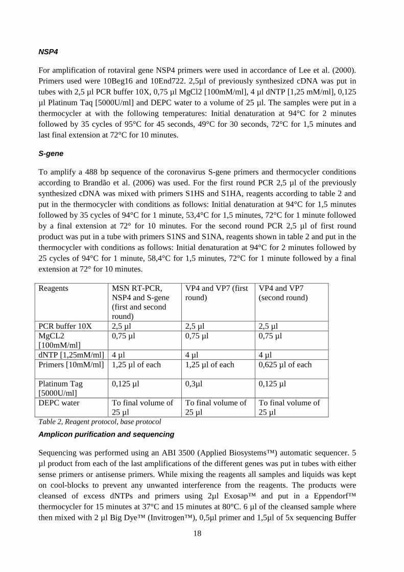
To amplify a 488 bp sequence of the coronavirus S-gene primers and thermocycler conditions according to Brandão et al. (2006) was used. For the first round PCR 2,5 µl of the previously synthesized cDNA was mixed with primers S1HS and S1HA, reagents according to table 2 and put in the thermocycler with conditions as follows: Initial denaturation at 94°C for 1,5 minutes followed by 35 cycles of 94°C for 1 minute, 53,4°C for 1,5 minutes, 72°C for 1 minute followed by a final extension at 72° for 10 minutes. For the second round PCR 2,5 µl of first round product was put in a tube with primers S1NS and S1NA, reagents shown in table 2 and put in the thermocycler with conditions as follows: Initial denaturation at 94°C for 2 minutes followed by 25 cycles of 94°C for 1 minute, 58,4°C for 1,5 minutes, 72°C for 1 minute followed by a final extension at 72° for 10 minutes.
Reagents MSN RT-PCR, NSP4 and S-gene (first and second round)
VP4 and VP7 (first round)
VP4 and VP7 (second round)
PCR buffer 10X 2,5 µl 2,5 µl 2,5 µl MgCL2 [100mM/ml]
0,75 µl 0,75 µl 0,75 µl
dNTP [1,25mM/ml] 4 µl 4 µl 4 µl Primers [10mM/ml] 1,25 µl of each 1,25 µl of each
0,625 µl of each
Platinum Tag [5000U/ml]
0,125 µl 0,3µl 0,125 µl
DEPC water To final volume of 25 µl
To final volume of 25 µl
To final volume of 25 µl
Table 2, Reagent protocol, base protocol
Amplicon purification and sequencing
Sequencing was performed using an ABI 3500 (Applied Biosystems™) automatic sequencer. 5 µl product from each of the last amplifications of the different genes was put in tubes with either sense primers or antisense primers. While mixing the reagents all samples and liquids was kept on cool-blocks to prevent any unwanted interference from the reagents. The products were cleansed of excess dNTPs and primers using 2µl Exosap™ and put in a Eppendorf™ thermocycler for 15 minutes at 37°C and 15 minutes at 80°C. 6 µl of the cleansed sample where then mixed with 2 µl Big Dye™ (Invitrogen™), 0,5µl primer and 1,5µl of 5x sequencing Buffer
19
(Invitrogen™). Here too the reagents and samples were kept on cool-blocks during mixing. Samples were put in a Eppendorf™ thermocycler with an sequencing protocol and later on cleansed using Big Dye Terminator™ (Invitrogen™). The samples were mixed with the Big Dye terminator according to the manufacturer and put on a mixing board for 30 minutes, after which the plate were put in a centrifuge. Samples were then given to the lab technician who performed the sequencing.
Sequence editing and building of phylogenetic trees
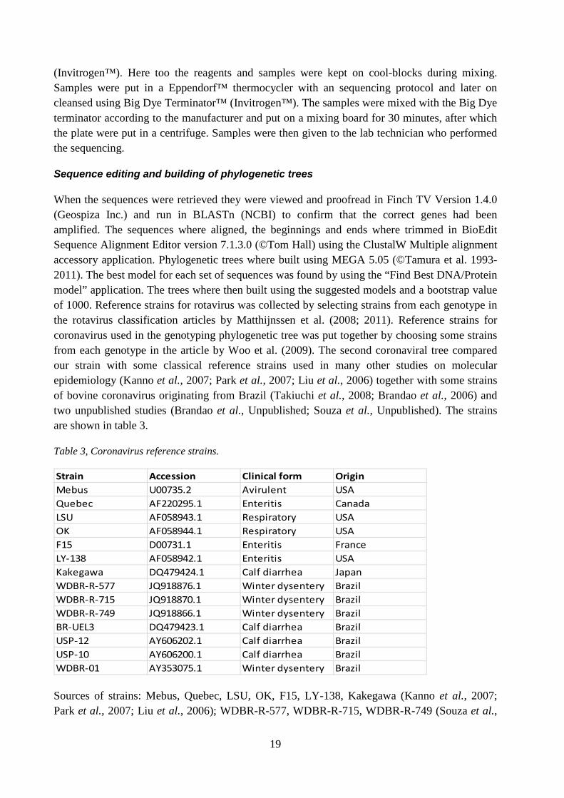
When the sequences were retrieved they were viewed and proofread in Finch TV Version 1.4.0 (Geospiza Inc.) and run in BLASTn (NCBI) to confirm that the correct genes had been amplified. The sequences where aligned, the beginnings and ends where trimmed in BioEdit Sequence Alignment Editor version 7.1.3.0 (©Tom Hall) using the ClustalW Multiple alignment accessory application. Phylogenetic trees where built using MEGA 5.05 (©Tamura et al. 1993-2011). The best model for each set of sequences was found by using the “Find Best DNA/Protein model” application. The trees where then built using the suggested models and a bootstrap value of 1000. Reference strains for rotavirus was collected by selecting strains from each genotype in the rotavirus classification articles by Matthijnssen et al. (2008; 2011). Reference strains for coronavirus used in the genotyping phylogenetic tree was put together by choosing some strains from each genotype in the article by Woo et al. (2009). The second coronaviral tree compared our strain with some classical reference strains used in many other studies on molecular epidemiology (Kanno et al., 2007; Park et al., 2007; Liu et al., 2006) together with some strains of bovine coronavirus originating from Brazil (Takiuchi et al., 2008; Brandao et al., 2006) and two unpublished studies (Brandao et al., Unpublished; Souza et al., Unpublished). The strains are shown in table 3.
Table 3, Coronavirus reference strains.
Strain Accession Clinical form OriginMebus U00735.2 Avirulent USAQuebec AF220295.1 Enteritis CanadaLSU AF058943.1 Respiratory USAOK AF058944.1 Respiratory USAF15 D00731.1 Enteritis FranceLY-138 AF058942.1 Enteritis USAKakegawa DQ479424.1 Calf diarrhea JapanWDBR-R-577 JQ918876.1 Winter dysentery BrazilWDBR-R-715 JQ918870.1 Winter dysentery BrazilWDBR-R-749 JQ918866.1 Winter dysentery BrazilBR-UEL3 DQ479423.1 Calf diarrhea BrazilUSP-12 AY606202.1 Calf diarrhea BrazilUSP-10 AY606200.1 Calf diarrhea BrazilWDBR-01 AY353075.1 Winter dysentery Brazil
Sources of strains: Mebus, Quebec, LSU, OK, F15, LY-138, Kakegawa (Kanno et al., 2007; Park et al., 2007; Liu et al., 2006); WDBR-R-577, WDBR-R-715, WDBR-R-749 (Souza et al.,
20
Unpublished); BR-UEL3 (Takiuchi et al., 2008); USP-12, USP-10 (Brandao et al., 2006) and WDBR-01 (Brandao et al., Unpublished).
RESULTS
MSN RT-PCR
For batch one MSN RT-PCR showed positive samples for rotavirus with amplicons of expected size (228 bp) in 12 of the 48 samples. Amplicons of the expected size for Coronavirus (306 bp) was detected in 4 of the 48 samples. Three samples were found positive for both rotavirus and coronavirus. Some of the regarded positive samples only had faint bands on the electrophoresis gel but were still submitted to continued evaluation of specific genes in hope that the specific primers would amplify sequences with more quantity.
In batch two 14 of the 20 samples showed amplification of segments matching that of rotavirus and two matching coronavirus. None of these samples displayed dual infection with both viruses.
Rotavirus batch one
There were a lot of difficulties in the attempts to amplify the genes VP4, VP7 and NSP4 in the first batch. Several different protocols with different primers were used but none gave satisfying results. Often amplification was achieved but the amplicons where of the wrong sizes for their respective protocols and/or there were amplicons of several different sizes in the same sample.
Rotavirus batch two
In batch two we had better, but still not good, results of genotyping the samples. The protocol used here were the same base protocol that did not work for batch one.
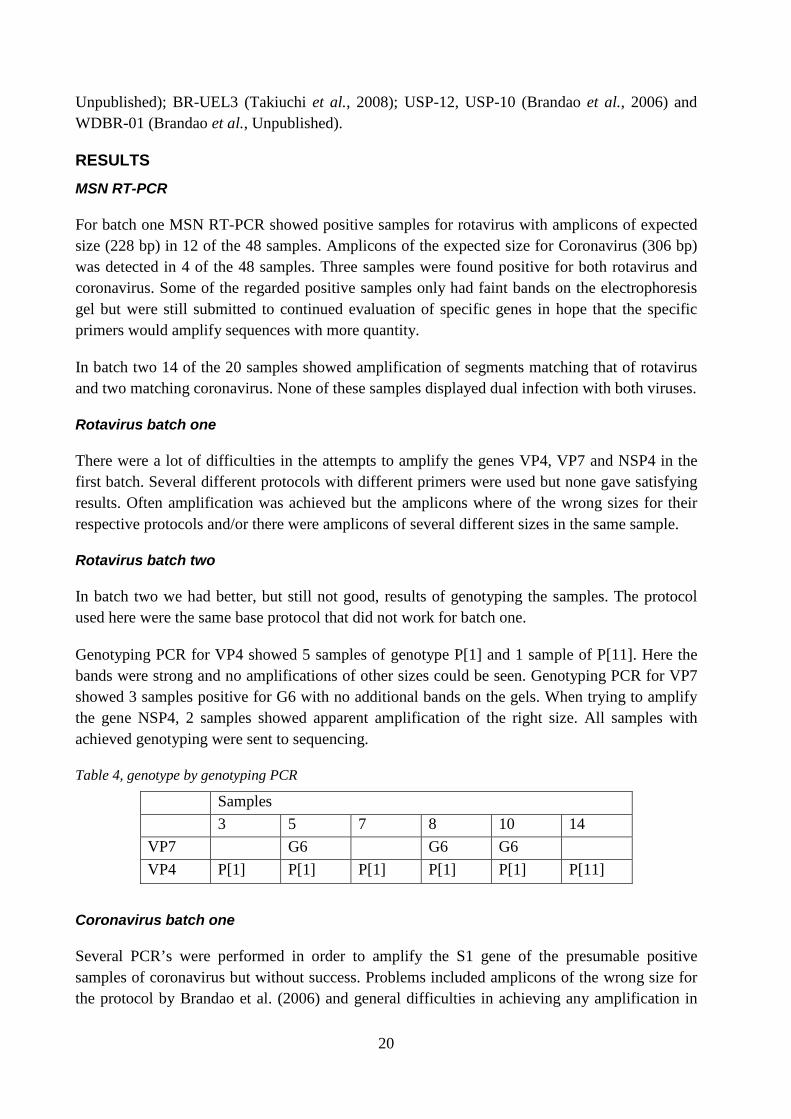
Genotyping PCR for VP4 showed 5 samples of genotype P[1] and 1 sample of P[11]. Here the bands were strong and no amplifications of other sizes could be seen. Genotyping PCR for VP7 showed 3 samples positive for G6 with no additional bands on the gels. When trying to amplify the gene NSP4, 2 samples showed apparent amplification of the right size. All samples with achieved genotyping were sent to sequencing.
Table 4, genotype by genotyping PCR
Samples 3 5 7 8 10 14 VP7 G6 G6 G6 VP4 P[1] P[1] P[1] P[1] P[1] P[11]
Coronavirus batch one
Several PCR’s were performed in order to amplify the S1 gene of the presumable positive samples of coronavirus but without success. Problems included amplicons of the wrong size for the protocol by Brandao et al. (2006) and general difficulties in achieving any amplification in
21
the samples at all. When changing protocol and trying a nested PCR using primers 2BP/4BM no amlifications were seen at all. The primerpair 2BP/4BM was designed for avian coronavirus but binds to a moderatly conservative region that excist among all species of coronavirus (Culver et al., 2008).
Coronavirus batch two
In batch two, one of the two samples showed amplification of the predicted size (885 bp and 488 bp). This sample was then submitted to sequencing.
Sequencing results
Batch one
An attempt was made to sequence some samples where amplifications of the right size for VP7 were achieved even though there were more amplifications of different sizes in the samples. The sequencing gave results only for the sense primer Beg9. The sequence did not match anything when run in BLASTn except that its 24 last nucleotides were identical with the end of some VP7 genes.
Batch two
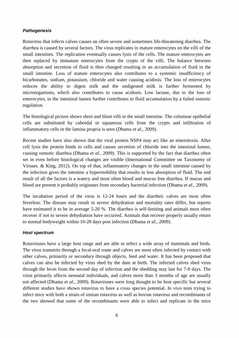
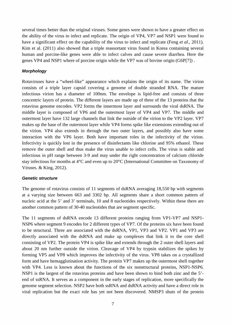
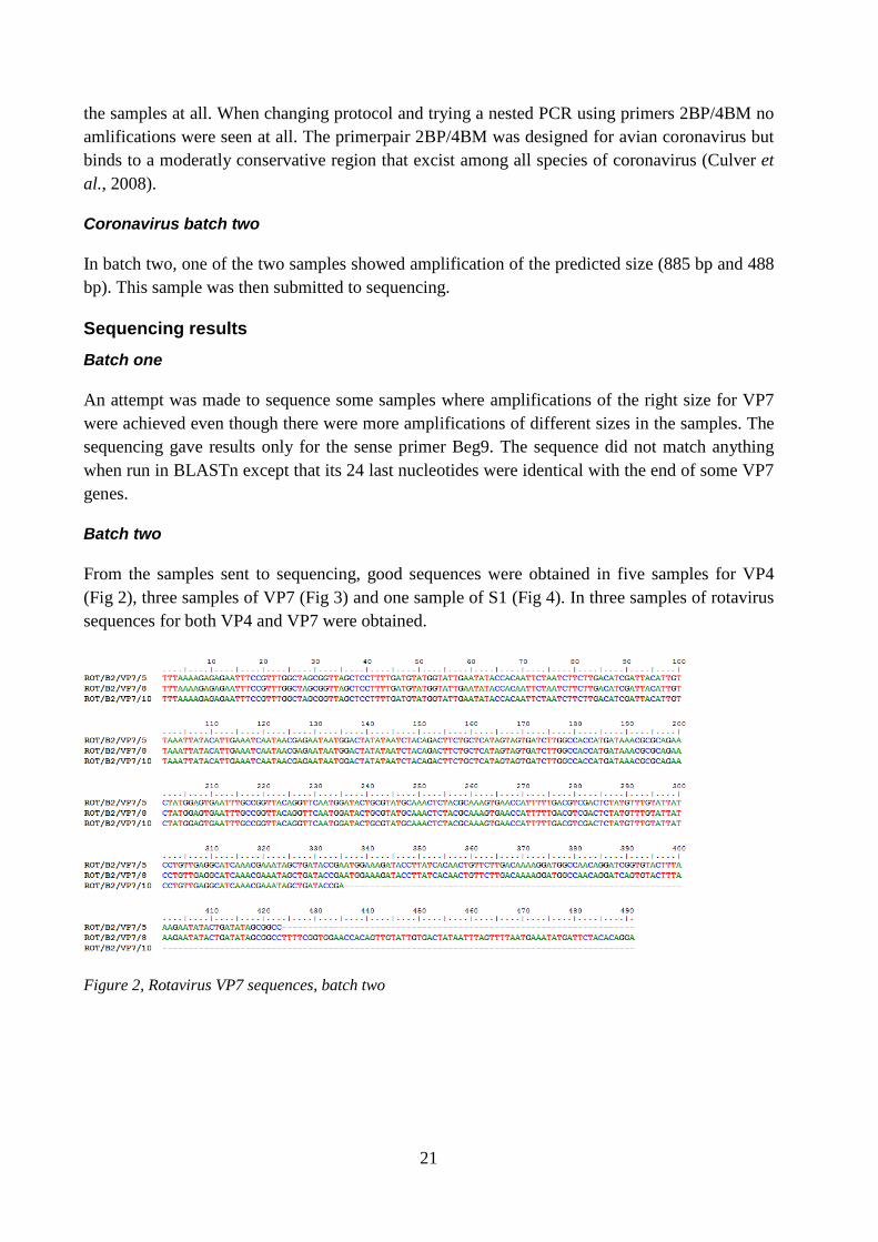
From the samples sent to sequencing, good sequences were obtained in five samples for VP4 (Fig 2), three samples of VP7 (Fig 3) and one sample of S1 (Fig 4). In three samples of rotavirus sequences for both VP4 and VP7 were obtained.
Figure 2, Rotavirus VP7 sequences, batch two
22
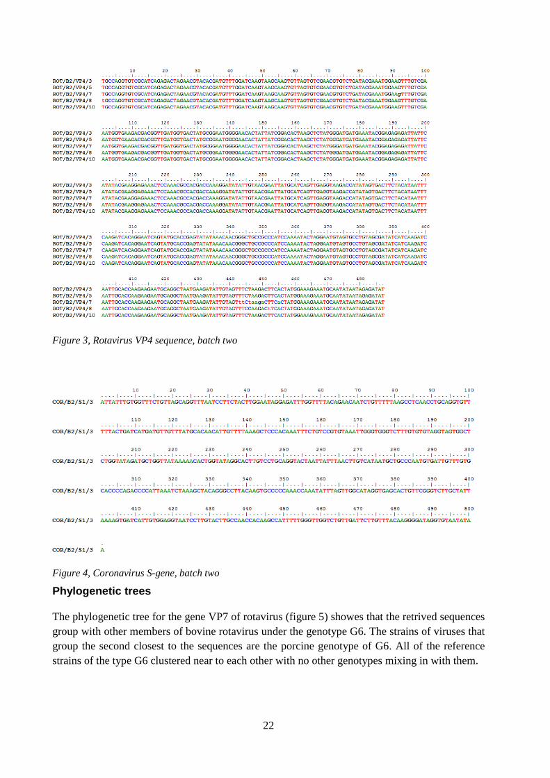
Figure 3, Rotavirus VP4 sequence, batch two
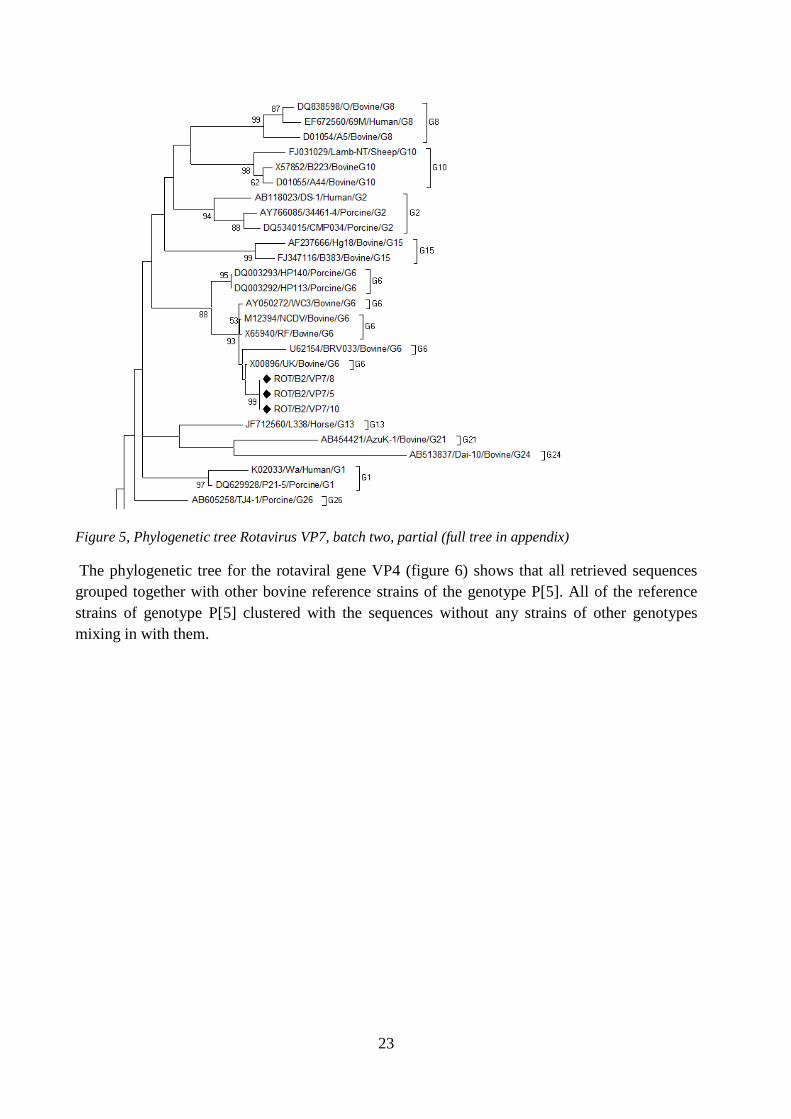
Figure 4, Coronavirus S-gene, batch two
Phylogenetic trees
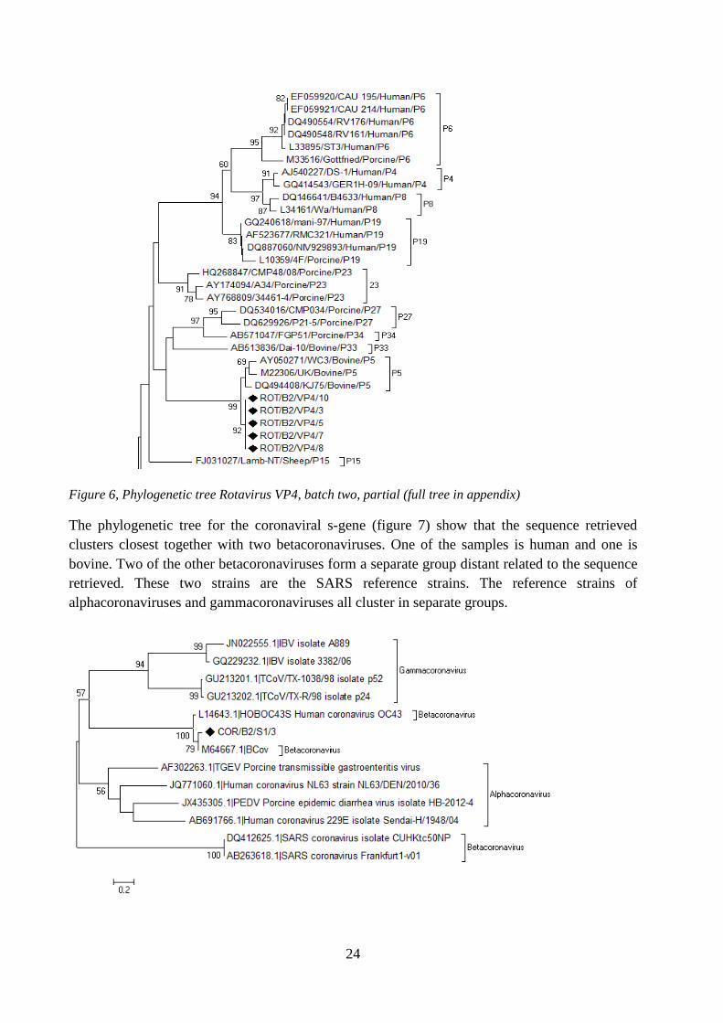
The phylogenetic tree for the gene VP7 of rotavirus (figure 5) showes that the retrived sequences group with other members of bovine rotavirus under the genotype G6. The strains of viruses that group the second closest to the sequences are the porcine genotype of G6. All of the reference strains of the type G6 clustered near to each other with no other genotypes mixing in with them.
23
Figure 5, Phylogenetic tree Rotavirus VP7, batch two, partial (full tree in appendix)
The phylogenetic tree for the rotaviral gene VP4 (figure 6) shows that all retrieved sequences grouped together with other bovine reference strains of the genotype P[5]. All of the reference strains of genotype P[5] clustered with the sequences without any strains of other genotypes mixing in with them.
24
Figure 6, Phylogenetic tree Rotavirus VP4, batch two, partial (full tree in appendix)
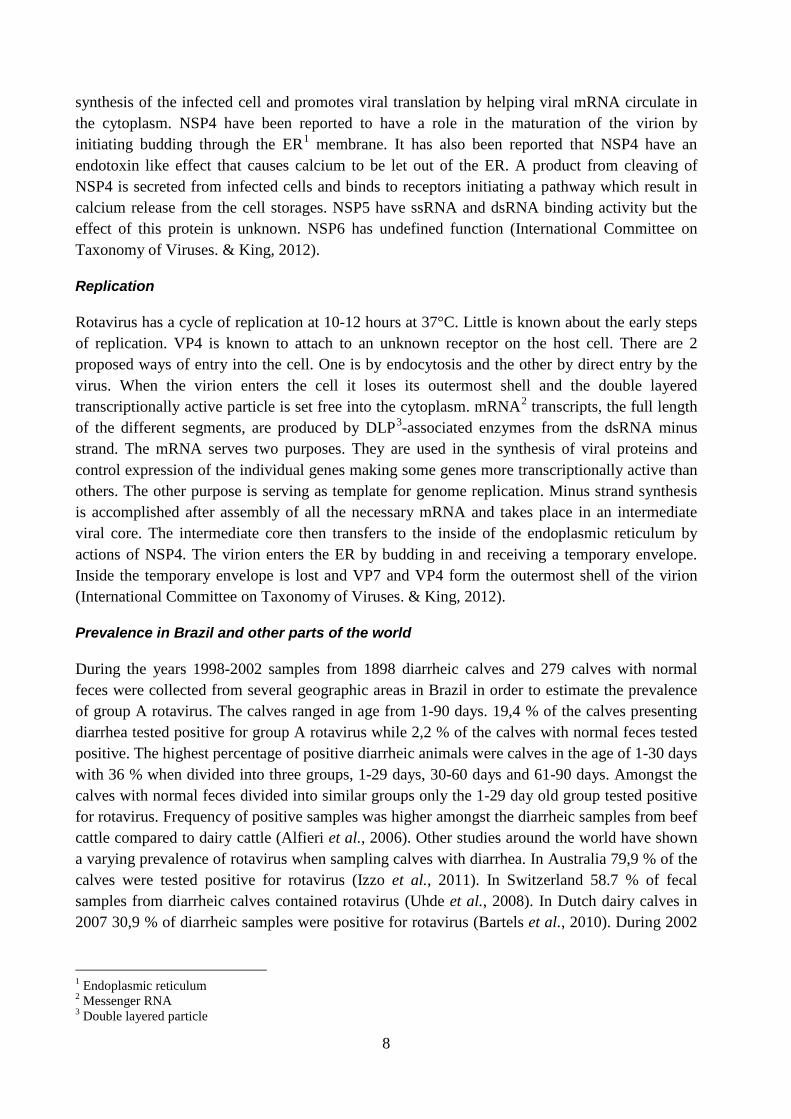
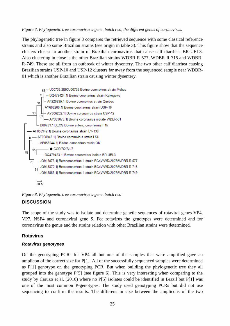
The phylogenetic tree for the coronaviral s-gene (figure 7) show that the sequence retrieved clusters closest together with two betacoronaviruses. One of the samples is human and one is bovine. Two of the other betacoronaviruses form a separate group distant related to the sequence retrieved. These two strains are the SARS reference strains. The reference strains of alphacoronaviruses and gammacoronaviruses all cluster in separate groups.
25
Figure 7, Phylogenetic tree coronavirus s-gene, batch two, the different genus of coronavirus.
The phylogenetic tree in figure 8 compares the retrieved sequence with some classical reference strains and also some Brazilian strains (see origin in table 3). This figure show that the sequence clusters closest to another strain of Brazilian coronavirus that cause calf diarrhea, BR-UEL3. Also clustering in close is the other Brazilian strains WDBR-R-577, WDBR-R-715 and WDBR-R-749. These are all from an outbreak of winter dysentery. The two other calf diarrhea causing Brazilian strains USP-10 and USP-12 clusters far away from the sequenced sample near WDBR-01 which is another Brazilian strain causing winter dysentery.
Figure 8, Phylogenetic tree coronavirus s-gene, batch two
DISCUSSION
The scope of the study was to isolate and determine genetic sequences of rotaviral genes VP4, VP7, NSP4 and coronaviral gene S. For rotavirus the genotypes were determined and for coronavirus the genus and the strains relation with other Brazilian strains were determined.
Rotavirus Rotavirus genotypes
On the genotyping PCRs for VP4 all but one of the samples that were amplified gave an amplicon of the correct size for P[1]. All of the successfully sequenced samples were determined as P[1] genotype on the genotyping PCR. But when building the phylogenetic tree they all grouped into the genotype P[5] (see figure 6). This is very interesting when comparing to the study by Caruzo et al. (2010) where no P[5] isolates could be identified in Brazil but P[1] was one of the most common P-genotypes. The study used genotyping PCRs but did not use sequencing to confirm the results. The differens in size between the amplicons of the two
26
genotypes is small, 622 versus 555 nucleotides for P[1] and P[5] respectively (Gouvea et al., 1994). The small differens between the genotyping products make it easy to mistake one for another. This shows the need for sequencing and phylogenetic analyses to be able to get accurate results when genotyping rotaviruses. The sequences retrieved from VP7 all grouped in with the genotype G6, closest to other bovine strains, when building the phylogenetic tree (see figure 5), which is in accordance with the genotyping PCR. The genotype G6 is previously described as the most common genotype circulating in Brazil (Caruzo et al., 2010).
One sample was genotyped as P[11] but it was not possible to sequence. Since all of the samples in batch two were from the same farm it would have been interesting to see if it really was P[11] or if the genotyping PCR was incorrect. P[11] is an usual genotype circulating all over the world (Midgley et al., 2012; Caruzo et al., 2010; Monini et al., 2008; Reidy et al., 2006). But if the genotyping as P[11] was correct that would mean that there were two different strains of rotavirus circulating at the farm at the same time.
In the first batch no genotypes were successfully determined but two sequences of VP7 were still retrieved. When run in BLASTn they did not match any known rotavirus VP7 sequences except for that they were achieved using VP7 primer Beg9 and had its 24 last nucleotides identical with some VP7 genes of genotype G3. There are two possibilities to how this could have happened. Either something has been amplified that was not actually rotavirus or a gene from a strain of rotavirus that has not yet been sequenced was amplified. Both possibilities have things talking for and against them. If something that is not a rotavirus has been amplified a gene in some other microorganism that have the same start (primer binding site) and ending as some strains of rotavirus gene VP7 has been found. It seems however unlikely that in a sample tested positive for rotavirus something randomly that has its resemblance to rotavirus in such a way would be able to amplify. On the other hand it also seems unlikely today, with such a vast amount of VP7 genotypes to compare with, that if it was a previously unknown rotavirus found here. If the sequence actually is rotavirus it would mean that a completely new genotype has been discovered (see tree in appendix 3).
Rotavirus amplification problems
During this project there were difficulties with poor results when doing the genotyping PCRs. In the first batch it was not possible to determine the genotypes with accuracy and in the second batch only three G-genotypes and six P-genotypes could be determined out of 14 samples positive for rotavirus on the MSN-RT-PCR. Numerous different ways to amplify the genes in the first batch were tried. Then PAGE was used to see if there was rotavirus in the samples. The PAGE also turned up negative for all the samples which begs the question if there were any rotavirus in the samples to begin with? A possibility would be that there were low viral amounts that could not be detected by PAGE but were sufficient enough for the MSN-RT-PCR. In the study where the MSN-RT-PCR was developed they saw that the PCR where able to detect rotavirus in samples that PAGE was not (Asano et al., 2010). Problems with genotyping have also been seen in other studies. Reidy et al. (2006) managed to genotype the G-genotype in 97% of the samples in their study while they only managed to P-genotype 58%. The difficulties in genotyping could be because a long time have gone since the primers for genotyping was made,
27
in this case from 1994, and the evolution of rotaviruses with the diversity they have achieved may likely have altered the primer binding sites. An updating of the primers would be advisable.
Coronavirus
Coronavirus genus
The sample of coronavirus where the gene S1 was retrieved grouped together with the other bovine coronavirus and with the human coronavirus OC-43 in the group Betacoronavirus as expected (see figure 7).
The strain of coronavirus retrieved from sequencing grouped together with two other strains of Betacoronaviruses but two other members of the group formed a separate branch, the SARS coronaviruses. This can be explained by the fact that SARS is thought to be an early split-off from the group containing Bovine and Human coronavirus OC-43 (Woo et al., 2009). In ICTV SARS and Betacoronavirus 1, which contain bovine coronavirus and human coronavirus OC-43, have been defined as different species but both belong to the genus Betacoronaviruses ((ICTV), 2011).
Relations to other Brazilian strains
The phylogenetic tree with the classical reference strains and Brazilian strains (see figure 8) grouped the sequenced sample together with some of the other bovine coronavirus strains from Brazil. The closest related sample is another Brazilian strain of calf diarrhea causing coronavirus, BR-UEL3. The two other Brazilian calf diarrhea causing samples USP-10 and USP-12 clusters on a separate branch and with a nucleotide identity of 91,6 % and 90,7 % respectively. Compared to the retrieved sequence it seems that they have evolved in two different evolutionary pathways. Both the strains USP-10 and USP-12 have a 18 nucleotide deletion previously described by Brandao et al. (2006) which the retrieved strain and BR-UEL3 did not have.
Interestingly the sequence retrieved seems more related to strains of winter dysentery: WDBR-R-577, WDBR-R-715 and WDBR-R-749 than the strains USP-10 and USP-12, which like the sequenced sample, originate from calf diarrhea. USP-10 and USP12 also grouped together with a strain causing winter dysentery in Brazil, WDBR-01. This supports the theory that one strain of rotavirus may be able to cause different clinical symptoms in different animals. That strains close to each other or that one strain can cause different clinical symptoms have been shown before (Bidokhti et al., 2012).
Coronavirus amplification problems
As with rotavirus there were difficulties in amplifying the S-gene in coronavirus in the first batch. When the PCR was performed amplification with clear and strong bands on the gel electrophoresis was achieved but the problem was that the bands were of the wrong size and sequencing on the samples did not give good sequences. Since trouble amplifying both rotavirus and coronavirus in the first batch were present there is a possibility that there was some PCR interfering substances in the samples. Since the same protocol worked to some extent on one sample in batch two it speaks for that the protocol itself should be sufficient. It is however not
28
possible to say anything about the effectiveness of the protocol in batch two either since there were only two samples.
CONCLUSION
In this study the immense diversity of rotavirus and coronavirus has been affirmed and there is evidence of several different strains of these viruses circulating in Brazil. Continued research on the molecular epidemiology of both rotavirus and coronavirus in Brazil is important in order to determine which strains are present, where they originate from and to be able to detect if new strains starts circulating. This knowledge is necessary to enable use of vaccines effective for the strains present. The viruses ability to change makes it important to have continuous surveillance so that new strains can be identified before they cause a lot of problems.
During this study troubles with the protocol available for genotyping of bovine rotavirus by Gouvea et al (1994a and 1994b) were experienced. The protocol worked only to some extent and was not efficient in genotyping samples of rotavirus, showing the importance of continuous updating of the laboratory methods used to genotype rotavirus. Since genotyping PCRs sometimes give inaccurate results one could also opt for it being more effective to sequence and determine genotypes by phylogeny directly, and only use the genotyping PCRs when sequencing is non-successful.
ACKNOWLEDGEMENT
There are some people and organizations that I would like to thank for allowing me to do this project and assisting me in the work I’ve done. I would like to thank Professor Fabio Gregori and LABMAS (Laboratório de Biologia Molecular Aplicada e Serologia) for taking me in and allowing me to do this project. I would like to thank SIDA, the fund of Gulli Stråhlfeldts and Veterinärmedicinska fakultetens stipendiesamfond for their financial support of this project, making it possible to do this study in Brazil. I would like to thank Sheila Oliveira de Souza Silva, MSc, research assistant, for help with the sequencing. I would like to thank my Swedish supervisor Professor Mikael Berg (Department of Biomedical Science and Veterinary Public Health, SLU). I would like to thank all of the students and workers at LABMAS for their kindness and help. Most of all I would like to thank PhD student Nara Thiers Cacciatori Galleti for her tireless work and advice, helping me through the whole project.
29
REFERENCES
(ICTV), I.C.O.T.O.V. [ONLINE] AVAILABLE FROM: HTTP://WWW.ICTVONLINE.ORG/VIRUSTAXONOMY.ASP?VERSION=2011.
AGRICULTURA, M.D. [ONLINE] AVAILABLE FROM: HTTP://WWW.AGRICULTURA.GOV.BR/ANIMAL/ESPECIES/BOVINOS-E-BUBALINOS. [ACCESSED 12-05].
ALFIERI, A.A., PARAZZI, M.E., TAKIUCHI, E., MEDICI, K.C. & ALFIERI, A.F. (2006). FREQUENCY OF GROUP A ROTAVIRUS IN DIARRHOEIC CALVES IN BRAZILIAN CATTLE HERDS, 1998-2002. TROP ANIM HEALTH PROD 38(7-8), 521-6.
ASANO, K.M., DE SOUZA, S.P., DE BARROS, I.N., AYRES, G.R., SILVA, S.O., RICHTZENHAIN, L.J. & BRANDAO, P.E. (2010). MULTIPLEX SEMI-NESTED RT-PCR WITH EXOGENOUS INTERNAL CONTROL FOR SIMULTANEOUS DETECTION OF BOVINE CORONAVIRUS AND GROUP A ROTAVIRUS. J VIROL METHODS 169(2), 375-9.
BARTELS, C.J., HOLZHAUER, M., JORRITSMA, R., SWART, W.A. & LAM, T.J. (2010). PREVALENCE, PREDICTION AND RISK FACTORS OF ENTEROPATHOGENS IN NORMAL AND NON-NORMAL FAECES OF YOUNG DUTCH DAIRY CALVES. PREV VET MED 93(2-3), 162-9.
BIDOKHTI, M.R., TRAVEN, M., OHLSON, A., BAULE, C., HAKHVERDYAN, M., BELAK, S., LIU, L. & ALENIUS, S. (2012). TRACING THE TRANSMISSION OF BOVINE CORONAVIRUS INFECTIONS IN CATTLE HERDS BASED ON S GENE DIVERSITY. VET J 193(2), 386-90.
BLANCHARD, P.C. (2012). DIAGNOSTICS OF DAIRY AND BEEF CATTLE DIARRHEA. VET CLIN NORTH AM FOOD ANIM PRACT 28(3), 443-64.
BRANDAO, P.E., GREGORI, F., RICHTZENHAIN, L.J., ROSALES, C.A., VILLARREAL, L.Y. & JEREZ, J.A. (2006). MOLECULAR ANALYSIS OF BRAZILIAN STRAINS OF BOVINE CORONAVIRUS (BCOV) REVEALS A DELETION WITHIN THE HYPERVARIABLE REGION OF THE S1 SUBUNIT OF THE SPIKE GLYCOPROTEIN ALSO FOUND IN HUMAN CORONAVIRUS OC43. ARCH VIROL 151(9), 1735-48.
BRANDAO, P.E., VILLARREAL, L.Y., GREGORI, F. & JEREZ, J.A. (UNPUBLISHED). GENETIC DIVERSITY OF WINTER DYSENTERY BOVINE CORONAVIRUS (BCOV).
CARUZO, T.A., BRITO, W.M., MUNFORD, V. & RACZ, M.L. (2010). MOLECULAR CHARACTERIZATION OF G AND P-TYPES BOVINE ROTAVIRUS STRAINS FROM GOIAS, BRAZIL: HIGH FREQUENCY OF MIXED P-TYPE INFECTIONS. MEM INST OSWALDO CRUZ 105(8), 1040-3.
CULVER, F.A., BRITTON, P. & CAVANAGH, D. (2008). RT-PCR DETECTION OF AVIAN CORONAVIRUSES OF GALLIFORM BIRDS (CHICKEN, TURKEY, PHEASANT) AND IN A PARROT. METHODS MOL BIOL 454, 35-42.
DE VERDIER, K. (2006). INFEKTIONSPANORAMAT VID DIARREÉR HOS SVENSKA KALVAR. SVENSK VETERINÄRTIDNING (8-9), 29-32.
DHAMA, K., CHAUHAN, R.S., MAHENDRAN, M. & MALIK, S.V. (2009). ROTAVIRUS DIARRHEA IN BOVINES AND OTHER DOMESTIC ANIMALS. VET RES COMMUN 33(1), 1-23.
DONOVAN, G.A., DOHOO, I.R., MONTGOMERY, D.M. & BENNETT, F.L. (1998). CALF AND DISEASE FACTORS AFFECTING GROWTH IN FEMALE HOLSTEIN CALVES IN FLORIDA, USA. PREV VET MED 33(1-4), 1-10.
FAO TOP EXPORTS- MEAT- CATTLEBONELESS (BEEF & VEAL). [ONLINE] AVAILABLE FROM: HTTP://FAOSTAT.FAO.ORG/SITE/342/DEFAULT.ASPX. [ACCESSED 12-05].
30
FENG, N., SEN, A., WOLF, M., VO, P., HOSHINO, Y. & GREENBERG, H.B. (2011). ROLES OF VP4 AND NSP1 IN DETERMINING THE DISTINCTIVE REPLICATION CAPACITIES OF SIMIAN ROTAVIRUS RRV AND BOVINE ROTAVIRUS UK IN THE MOUSE BILIARY TRACT. J VIROL 85(6), 2686-94.
GARAICOECHEA, L., BOK, K., JONES, L.R., COMBESSIES, G., ODEON, A., FERNANDEZ, F. & PARRENO, V. (2006). MOLECULAR CHARACTERIZATION OF BOVINE ROTAVIRUS CIRCULATING IN BEEF AND DAIRY HERDS IN ARGENTINA DURING A 10-YEAR PERIOD (1994-2003). VET MICROBIOL 118(1-2), 1-11.
GAY, C.C., HODGSON, J.C., LOFSTEDT, J. & BOLIN, S.R. DIARRHEA IN NEONATAL RUMINANTS: INTESTINAL DISEASES IN RUMINANTS: MERCK VETERINARY MANUAL. [ONLINE] AVAILABLE FROM: HTTP://WWW.MERCKMANUALS.COM/VET/PRINT/DIGESTIVE_SYSTEM/INTESTINAL_DISEASES_IN_RUMINANTS/DIARRHEA_IN_NEONATAL_RUMINANTS.HTML. [ACCESSED 2012-12-05].
GONZALEZ, J.M., GOMEZ-PUERTAS, P., CAVANAGH, D., GORBALENYA, A.E. & ENJUANES, L. (2003). A COMPARATIVE SEQUENCE ANALYSIS TO REVISE THE CURRENT TAXONOMY OF THE FAMILY CORONAVIRIDAE. ARCH VIROL 148(11), 2207-35.
GOUVEA, V., SANTOS, N. & TIMENETSKY MDO, C. (1994). VP4 TYPING OF BOVINE AND PORCINE GROUP A ROTAVIRUSES BY PCR. J CLIN MICROBIOL 32(5), 1333-7.
GRAHAM, R.L. & BARIC, R.S. (2010). RECOMBINATION, RESERVOIRS, AND THE MODULAR SPIKE: MECHANISMS OF CORONAVIRUS CROSS-SPECIES TRANSMISSION. J VIROL 84(7), 3134-46.
INTERNATIONAL COMMITTEE ON TAXONOMY OF VIRUSES. & KING, A.M.Q. (2012). VIRUS TAXONOMY : CLASSIFICATION AND NOMENCLATURE OF VIRUSES : NINTH REPORT OF THE INTERNATIONAL COMMITTEE ON TAXONOMY OF VIRUSES. LONDON: ACADEMIC PRESS. (IMMUNOLOGY AND MICROBIOLOGY 2011. ISBN 9780123846846 (ALK. PAPER).
IZZO, M.M., KIRKLAND, P.D., MOHLER, V.L., PERKINS, N.R., GUNN, A.A. & HOUSE, J.K. (2011). PREVALENCE OF MAJOR ENTERIC PATHOGENS IN AUSTRALIAN DAIRY CALVES WITH DIARRHOEA. AUST VET J 89(5), 167-73.
KANNO, T., HATAMA, S., ISHIHARA, R. & UCHIDA, I. (2007). MOLECULAR ANALYSIS OF THE S GLYCOPROTEIN GENE OF BOVINE CORONAVIRUSES ISOLATED IN JAPAN FROM 1999 TO 2006. J GEN VIROL 88(PT 4), 1218-24.
KIM, H.J., PARK, J.G., MATTHIJNSSENS, J., LEE, J.H., BAE, Y.C., ALFAJARO, M.M., PARK, S.I., KANG, M.I. & CHO, K.O. (2011). INTESTINAL AND EXTRA-INTESTINAL PATHOGENICITY OF A BOVINE REASSORTANT ROTAVIRUS IN CALVES AND PIGLETS. VET MICROBIOL 152(3-4), 291-303.
LIU, L., HAGGLUND, S., HAKHVERDYAN, M., ALENIUS, S., LARSEN, L.E. & BELAK, S. (2006). MOLECULAR EPIDEMIOLOGY OF BOVINE CORONAVIRUS ON THE BASIS OF COMPARATIVE ANALYSES OF THE S GENE. J CLIN MICROBIOL 44(3), 957-60.
LORENZ, I., FAGAN, J. & MORE, S.J. (2011). CALF HEALTH FROM BIRTH TO WEANING. II. MANAGEMENT OF DIARRHOEA IN PRE-WEANED CALVES. IR VET J 64(1), 9.
MACLACHLAN, N.J., DUBOVI, E.J. & FENNER, F. (2011). FENNER'S VETERINARY VIROLOGY. 4TH. ED. AMSTERDAM ; BOSTON: ELSEVIER/AP. ISBN 9780123751584.
MALIK, Y.S., SHARMA, K., VAID, N., CHAKRAVARTI, S., CHANDRASHEKAR, K.M., BASERA, S.S., SINGH, R., MINAKSHI, PRASAD, G., GULATI, B.R., BHILEGAONKAR, K.N. & PANDEY, A.B. (2012). FREQUENCY OF GROUP A ROTAVIRUS WITH MIXED G AND P GENOTYPES IN BOVINES: PREDOMINANCE OF G3 GENOTYPE AND ITS EMERGENCE IN COMBINATION WITH G8/G10 TYPES. JOURNAL OF VETERINARY SCIENCE 13(3), 271.
31
MARTINEZ, N., BRANDAO, P.E., DE SOUZA, S.P., BARRERA, M., SANTANA, N., DE ARCE, H.D. & PEREZ, L.J. (2012). MOLECULAR AND PHYLOGENETIC ANALYSIS OF BOVINE CORONAVIRUS BASED ON THE SPIKE GLYCOPROTEIN GENE. INFECT GENET EVOL 12(8), 1870-8.
MATTHIJNSSENS, J., CIARLET, M., MCDONALD, S.M., ATTOUI, H., BANYAI, K., BRISTER, J.R., BUESA, J., ESONA, M.D., ESTES, M.K., GENTSCH, J.R., ITURRIZA-GOMARA, M., JOHNE, R., KIRKWOOD, C.D., MARTELLA, V., MERTENS, P.P., NAKAGOMI, O., PARRENO, V., RAHMAN, M., RUGGERI, F.M., SAIF, L.J., SANTOS, N., STEYER, A., TANIGUCHI, K., PATTON, J.T., DESSELBERGER, U. & VAN RANST, M. (2011). UNIFORMITY OF ROTAVIRUS STRAIN NOMENCLATURE PROPOSED BY THE ROTAVIRUS CLASSIFICATION WORKING GROUP (RCWG). ARCH VIROL 156(8), 1397-413.
MIDGLEY, S.E., BANYAI, K., BUESA, J., HALAIHEL, N., HJULSAGER, C.K., JAKAB, F., KAPLON, J., LARSEN, L.E., MONINI, M., POLJSAK-PRIJATELJ, M., POTHIER, P., RUGGERI, F.M., STEYER, A., KOOPMANS, M. & BOTTIGER, B. (2012). DIVERSITY AND ZOONOTIC POTENTIAL OF ROTAVIRUSES IN SWINE AND CATTLE ACROSS EUROPE. VET MICROBIOL 156(3-4), 238-45.
MOHD NOR, N., STEENEVELD, W., MOURITS, M.C. & HOGEVEEN, H. (2012). ESTIMATING THE COSTS OF REARING YOUNG DAIRY CATTLE IN THE NETHERLANDS USING A SIMULATION MODEL THAT ACCOUNTS FOR UNCERTAINTY RELATED TO DISEASES. PREV VET MED 106(3-4), 214-24.
MONINI, M., CAPPUCCINI, F., BATTISTA, P., FALCONE, E., LAVAZZA, A. & RUGGERI, F.M. (2008). MOLECULAR CHARACTERIZATION OF BOVINE ROTAVIRUS STRAINS CIRCULATING IN NORTHERN ITALY, 2003-2005. VET MICROBIOL 129(3-4), 384-9.
PARK, S.J., LIM, G.K., PARK, S.I., KIM, H.H., KOH, H.B. & CHO, K.O. (2007). DETECTION AND MOLECULAR CHARACTERIZATION OF CALF DIARRHOEA BOVINE CORONAVIRUSES CIRCULATING IN SOUTH KOREA DURING 2004-2005. ZOONOSES PUBLIC HEALTH 54(6-7), 223-30.
PERLMAN, S. & NETLAND, J. (2009). CORONAVIRUSES POST-SARS: UPDATE ON REPLICATION AND PATHOGENESIS. NAT REV MICROBIOL 7(6), 439-50.
RAZZAQUE, M.A., BEDAIR, M., ABBAS, S. & AL-MUTAWA, T. (2009). ECONOMIC IMPACT OF CALF MORTALITY ON DAIRY FARMS IN KUWAIT. PAKISTAN VETERINARY JOURNAL 29(3), 97-101.
REIDY, N., LENNON, G., FANNING, S., POWER, E. & O'SHEA, H. (2006). MOLECULAR CHARACTERISATION AND ANALYSIS OF BOVINE ROTAVIRUS STRAINS CIRCULATING IN IRELAND 2002-2004. VET MICROBIOL 117(2-4), 242-7.
SAIF, L.J. (2010). BOVINE RESPIRATORY CORONAVIRUS. VET CLIN NORTH AM FOOD ANIM PRACT 26(2), 349-64.
SOUZA, S.P., ASANO, K.M., RICHTZENHAIN, L.J. & BRANDAO, P.E. (UNPUBLISHED). PARTIAL SPIKE AND HEMAGGLUTININ-ESTERASE GENES OF BOVINE CORONAVIRUS (BCOV) EVIDENCE A UNIQUE LINEAGE DURING AND OUTBREAK OF RESPIRATORY AND ENTERIC DISEASE IN BRAZIL.
STIPP, D.T., BARRY, A.F., ALFIERI, A.F., TAKIUCHI, E., AMUDE, A.M. & ALFIERI, A.A. (2009). FREQUENCY OF BCOV DETECTION BY A SEMI-NESTED PCR ASSAY IN FAECES OF CALVES FROM BRAZILIAN CATTLE HERDS. TROP ANIM HEALTH PROD 41(7), 1563-7.
TAKIUCHI, E., ALFIERI, A.F. & ALFIERI, A.A. (2008). MOLECULAR ANALYSIS OF THE BOVINE CORONAVIRUS S1 GENE BY DIRECT SEQUENCING OF DIARRHEIC FECAL SPECIMENS. BRAZ J MED BIOL RES 41(4), 277-82.
UHDE, F.L., KAUFMANN, T., SAGER, H., ALBINI, S., ZANONI, R., SCHELLING, E. & MEYLAN, M. (2008). PREVALENCE OF FOUR ENTEROPATHOGENS IN THE FAECES OF YOUNG DIARRHOEIC DAIRY CALVES IN SWITZERLAND. VET REC 163(12), 362-6.
32
WOO, P.C., HUANG, Y., LAU, S.K. & YUEN, K.Y. (2010). CORONAVIRUS GENOMICS AND BIOINFORMATICS ANALYSIS. VIRUSES 2(8), 1804-20.
WOO, P.C., LAU, S.K., HUANG, Y. & YUEN, K.Y. (2009). CORONAVIRUS DIVERSITY, PHYLOGENY AND INTERSPECIES JUMPING. EXP BIOL MED (MAYWOOD) 234(10), 1117-27.
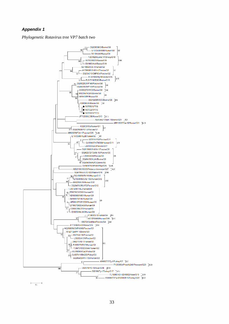
33
Appendix 1
Phylogenetic Rotavirus tree VP7 batch two
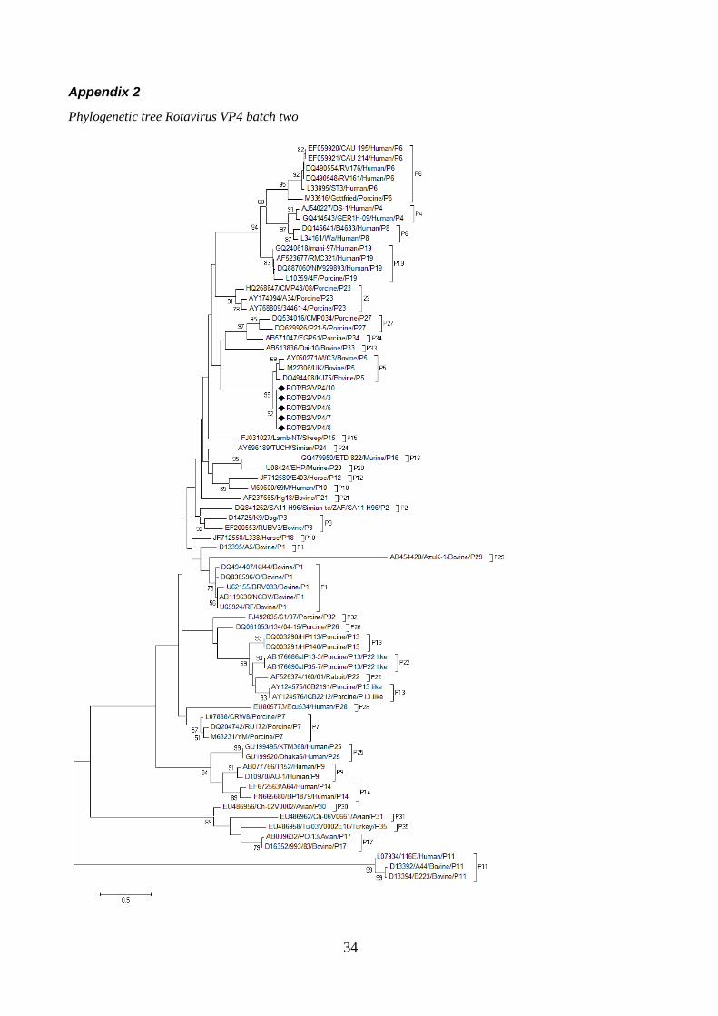
34
Appendix 2
Phylogenetic tree Rotavirus VP4 batch two
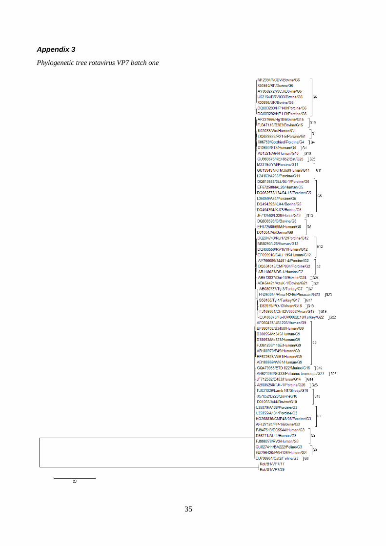
35
Appendix 3
Phylogenetic tree rotavirus VP7 batch one