Embed Size (px)
Citation preview
Chapter 7
Diagnosis and Management of Orbital Cellulitis
Imtiaz A. Chaudhry, Waleed Al-Rashed,Osama Al-Sheikh and Yonca O. Arat
Additional information is available at the end of the chapter
http://dx.doi.org/10.5772/52719
1. Introduction
Orbital cellulites is an uncommon infectious process in which patient may present withpain, reduced visual acuity, compromised ocular motility and significant proptosis. [1]- [3]In the modern era of relatively early access to the health care facilities, complete loss of vi‐sion from orbital cellulitis is rare. In the vast majority of cases, a history of upper respirato‐ry tract infection prior to the onset is very common especially in children. [4], [5] Chandleret al, [6] for simplicity has classified the disease into 5 categories and emphasized the pos‐sibility of fatal outcome due to the extension of the abscess to cavernous sinus in the formof thrombosis and intracranial spread. In addition to the loss of vision, orbital cellulitis canbe associated with a number of other serious complications that may include intracranialcomplications in the form of cavernous sinus thrombosis, meningitis, frontal abscess andeven death. Historically, since the wide spread use of effective antibiotics, the serious com‐plications of orbital cellulitis have become much less frequent. In the past, loss of visionwas a relatively more common outcome of orbital cellulitis. [2] In the recent years, onlyfew case reports of loss of vision following orbital cellulitis has been reported in the litera‐ture. For example, Connel et al, [3] reported case of a 69-year-old man who presented withno light perception vision, proptosis and significant ophthalmoplegia. In their case, despiteemergent drainage of the abscess and systemic antibiotics, no improvement in vision wasnoted despite the return of the full ocular motility and disappearance of proptosis. Connelet al, [3] postulated Streptococcal-related ischemic necrosis of the optic nerve as a possiblemechanism of loss of vision in their patient. In one of the recent survey of 52 patients treat‐ed for orbital cellulitis, over 35% had decreased vision and on their last follow-up, only 4%had decreased visual acuity. [1] Our own experience in treating 218 patients with orbitalcomplications of cellulitis revealed that visual acuity improved in 16.1% and worsened in6.2%, including 4.3% that sustained complete loss of vision. [8] We attributed the perma‐
© 2013 Chaudhry et al.; licensee InTech. This is an open access article distributed under the terms of theCreative Commons Attribution License (http://creativecommons.org/licenses/by/3.0), which permitsunrestricted use, distribution, and reproduction in any medium, provided the original work is properly cited.
nent loss of vision to the delay in diagnosis and intervention. Further, there were 9 cases ofintracranial orbital abscess extension that required either extended treatment with system‐ic antibiotics alone or in combination with neurosurgical intervention. [3]
2. Patient presentation
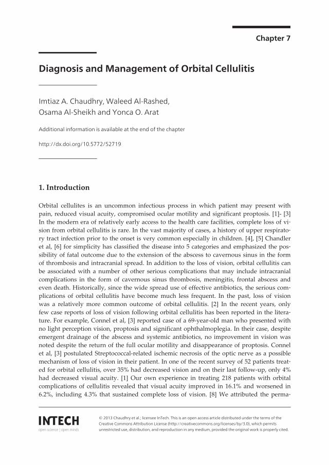
Patients with orbital cellulitis may present with signs of eyelid swelling, conjunctival chemosis,diplopia and proptosis which may not be prominent in cases of preseptal cellulitis. [1], [8], [9]These patients may present with corneal infections resulting from exposure keratopathy dueto their inability to close their eyes. Many of these patients come with local symptoms in theform of eyelid edema, redness, chemosis, decreased ocular motility and proptosis (Figure 1).Patients having superficial signs of swelling (preseptal cellulitis) should be differentiated fromdeeper infection resulting in orbital cellulitis, in which case, signs and symptoms resultingfrom inflammation may be helpful. [9] In particular, external ophthalmoloplegia, proptosisand decreased visual acuity are associated with orbital cellulitis rather than preseptal cellulitis.[8], [9] Temperature greater than 37.5℃ and leukocytosis resulting in fever may be moreprominent feature of the pediatric orbital cellulitis. [4], [5] In children, external ophthalmo‐plegia and proptosis may be the most common features, while decreased visual acuity andchemosis may be less frequent signs in both the pediatric as well as in the adult patients. Incases of the optic nerve involvement, disc edema or neuritis with rapidly progressing atrophyresulting in blindness may occur. Mechanical pressure on the optic nerve and possiblycompression of the central retinal and other feeding arteries results in optic nerve atrophy. [10]Also orbital inflammation itself may spread directly into the substance of the optic nervecausing small necrotic areas or abscesses. [2] Compression of the feeding vessels as well asinflammation may result in the infarction of the optic nerve, infarction of the sclera, choroidsas well as the retina. Inflammation may result in septic uveitis, iridocyclitis or choroiditis witha cloudiness of the vitreous, including septic pan ophthalmitis. A less common complicationof orbital cellulitis is glaucoma that can cause decreased vision, reduced visual field or evenenlarged blind spot on presentation. On occasion, one may not find any fundus abnormalities.Among our patients presenting to a tertiary eye care center in the developing country,presenting signs of 218 patients with orbital cellulitis included, eyelid swelling in 71.5%,proptosis in 68.3%, motility restriction in 59.2%, pain in 52.3%, and decreased visual acuity in14.2% of cases. [8]
3. Differential diagnosis
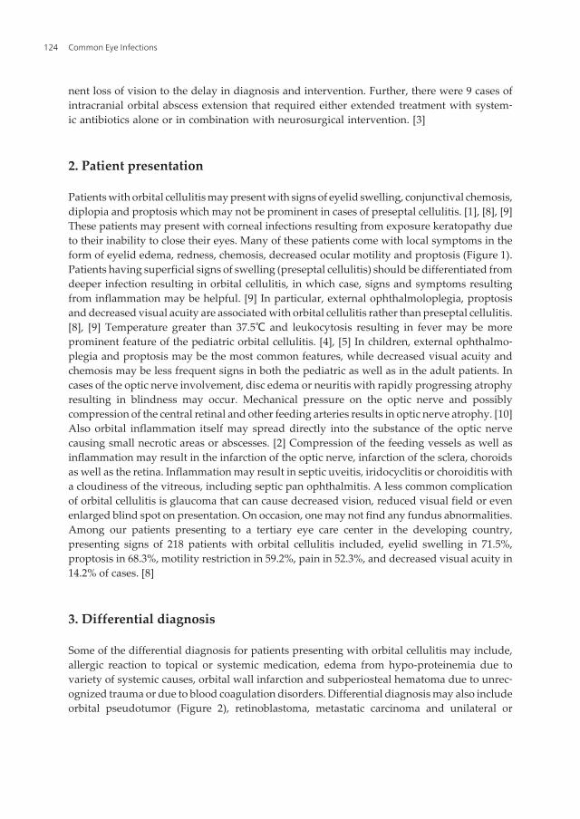
Some of the differential diagnosis for patients presenting with orbital cellulitis may include,allergic reaction to topical or systemic medication, edema from hypo-proteinemia due tovariety of systemic causes, orbital wall infarction and subperiosteal hematoma due to unrec‐ognized trauma or due to blood coagulation disorders. Differential diagnosis may also includeorbital pseudotumor (Figure 2), retinoblastoma, metastatic carcinoma and unilateral or
Common Eye Infections124
bilateral exophthalmos secondary to thyroid related orbitopathy. [11] In all cases, carefulhistory, thorough physical examination along with carefully selected imaging studies mayhelp in differentiating orbital cellulitis from other causes of proptosis.
Figure 2. A 25-year-old male with bilateral eyelid swelling, proptosis and painful diplopia was found to have evidenceof bilateral orbital pseudotumor and treated with systemic corticosteroids after imaging studies failed to show evi‐dence of any infectious cause of his symptoms.
4. Most common predisposing factors for orbital cellulitis
In the most reported series, the most common predisposing factor for orbital cellulitis is sinusdisease, especially in children. [1], [4], [8] Usually, the infection originates from sinusitis. It canoriginate from face or eyelids after a recent or past trauma, dental abscess or from a distantsource by hematogenous spread. [1], [8], [11-13] For simplification purposes, Chandler et al,[6] grouped complication of sinus inflammation into 5 classes. In the group 1, eyelids may beswollen alongwith presence of orbital content edema (preseptal cellulitis). Swelling may reflectimpedance of drainage through ethmoidal vessels. Group II reflects evidence of orbitalcellulitis in which inflammatory cells diffusely infiltrate orbital tissues. In Group II, the eyelids
Figure 1. A child with left sided eyelid erythema, swelling and proptosis following a bout of upper respiratory infec‐tion.
Diagnosis and Management of Orbital Cellulitishttp://dx.doi.org/10.5772/52719
125
may be swollen along with conjunctival chemosis as well as some degree of proptosis. Visualloss may be present in Group II patients. Purulent material may be collecting as subperiostealabscess between the periorbita and the bony walls of the orbit in Group III. These patients mayhave significant conjunctival chemosis, eyelid edema, along with tenderness in the involvedareas with variable degree of proptosis, and decreased ocular motility. The abscess may beanywhere in the vicinity of the orbit. Patients in group IV (orbital abscess), may present withtheir abscess being inside or outside the muscle cone following untreated orbital cellulitis.These patients may have significantly more pain, proptosis, decreased ocular motility andvariable degree of severe visual loss. Patients in group V may present with bilateral eyelidedema along with involvement of third, fifth and sixth cranial nerves which is thought to bedue to the extension of the infectious process into the cavernous sinus with formation ofthrombosis. These patients may have nausea, vomiting along with signs of nervous systeminvolvement which could also be due to septicemia. Signs of proptosis, eyelid edema, opticneuritis, frozen globe, decreased supra-orbital nerve conduction may be hallmarks of orbitalapex syndrome which is thought to be due to the sinusitis in the area of the superior orbitalfissure and optic foramen. [14]
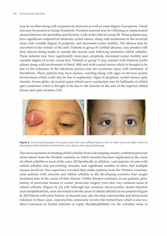
Figure 3. External photographs of a young male child who suffered trauma over his right brow area after which hedeveloped orbital cellulitis and formation of an abscess that required drainage.
Our own experience in treating orbital cellulitis from a developing country confirmed previousobservations from the Western countries in which sinusitis has been implicated as the causeof orbital cellulitis in most of the cases. [8] Specifically in children, vast majority of cases withorbital cellulitis had pre-existing sinusitis, and significant number of them had multiplesinuses involved. Our experience revealed that unlike patients from the Western countries,most patients with sinusitis and orbital cellulitis in the developing countries had soughttreatment later in the course of their disease. Unlike Western countries, in our patients, priorhistory of periocular trauma or ocular/ periocular surgery were also very common cause oforbital cellulitis. (Figure 3). [1], [10] Although less common, dacryocystitis, dental infectionand endophthalmitis, were also found to be the cause of orbital cellulitis in our patients (Figure4). [8] Patients with prior history of sinusitis may also develop osteomyelitis and intracranialinfection. In these cases, osteomyelitis, commonly involve the frontal bone which is due to adirect extension of frontal infection or septic thrombophlebitis via the valveless sinus of
Common Eye Infections126
Breschet. [15] Less common cause of osteomyelitis results from the ethmoidal sinusitis becausefrom this location, infection can rapidly spread through the thin lamina papyracea into theorbit or maxilla, where arterial anastomoses are sufficient to prevent necrosis due to septicthrombosis of a single artery. Although meningitis may be the most common intracranialcomplication of sinus disease, epidural, subdural and brain parenchymal abscess can alsodevelop. [15]
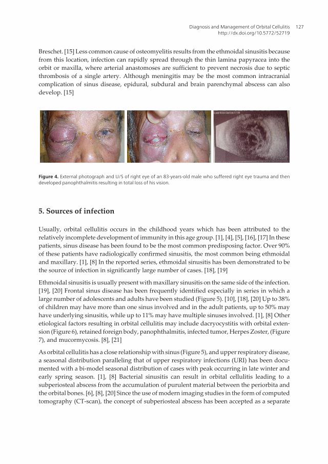
Figure 4. External photograph and U/S of right eye of an 83-years-old male who suffered right eye trauma and thendeveloped panophthalmitis resulting in total loss of his vision.
5. Sources of infection
Usually, orbital cellulitis occurs in the childhood years which has been attributed to therelatively incomplete development of immunity in this age group. [1], [4], [5], [16], [17] In thesepatients, sinus disease has been found to be the most common predisposing factor. Over 90%of these patients have radiologically confirmed sinusitis, the most common being ethmoidaland maxillary. [1], [8] In the reported series, ethmoidal sinusitis has been demonstrated to bethe source of infection in significantly large number of cases. [18], [19]
Ethmoidal sinusitis is usually present with maxillary sinusitis on the same side of the infection.[19], [20] Frontal sinus disease has been frequently identified especially in series in which alarge number of adolescents and adults have been studied (Figure 5). [10], [18], [20] Up to 38%of children may have more than one sinus involved and in the adult patients, up to 50% mayhave underlying sinusitis, while up to 11% may have multiple sinuses involved. [1], [8] Otheretiological factors resulting in orbital cellulitis may include dacryocystitis with orbital exten‐sion (Figure 6), retained foreign body, panophthalmitis, infected tumor, Herpes Zoster, (Figure7), and mucormycosis. [8], [21]
As orbital cellulitis has a close relationship with sinus (Figure 5), and upper respiratory disease,a seasonal distribution paralleling that of upper respiratory infections (URI) has been docu‐mented with a bi-model seasonal distribution of cases with peak occurring in late winter andearly spring season. [1], [8] Bacterial sinusitis can result in orbital cellulitis leading to asubperiosteal abscess from the accumulation of purulent material between the periorbita andthe orbital bones. [6], [8], [20] Since the use of modern imaging studies in the form of computedtomography (CT-scan), the concept of subperiosteal abscess has been accepted as a separate
Diagnosis and Management of Orbital Cellulitishttp://dx.doi.org/10.5772/52719
127
entity. [20], [22] Because of the reports of rapidly progressive visual and intracranial compli‐cations from subperiosteal abscess, some clinicians argue for the prompt surgical drainage ofthe abscess and paranasal sinuses when a subperiosteal abscess is first diagnosed by a CT-scan.[15], [20], [22-26] Among our survey of 218 patients who required treatment of their orbitalcellulitis, imaging studies revealed that sinus disease was the most cause in 39.4%, trauma in19.7%, endophthalmitis in 13.3%, (Figure 8), orbital implants in 8.2%, dacryocystitis in 4.6%,retained orbital foreign body in 3.2%, dental infection in 2.7%, and scleral buckle in 2.3%.8 Ahistory of sinusitis and recent trauma was the cause of orbital cellulitis in 4.1%, and intraocularor orbital tumors were the cause in another 4% of patients.
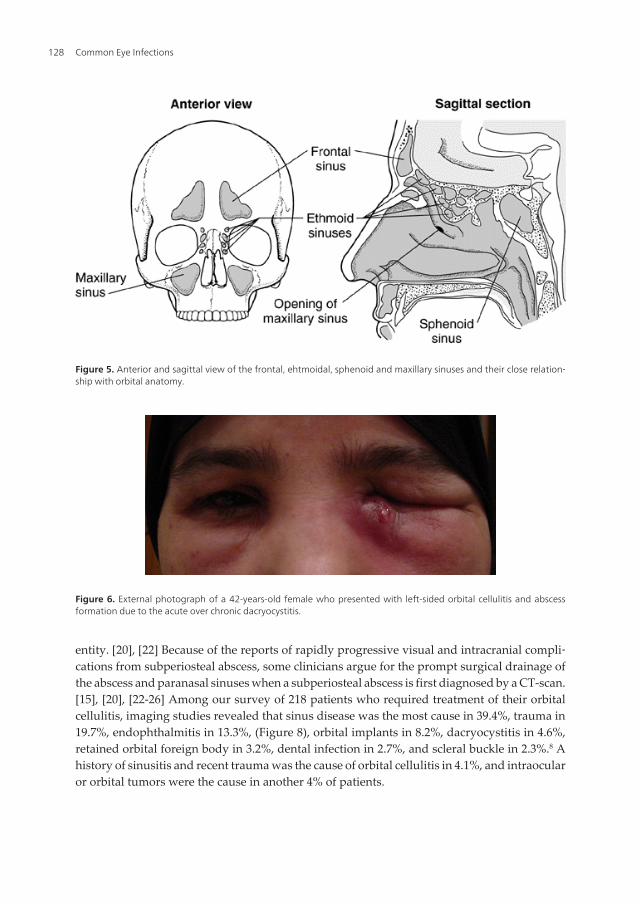
Figure 5. Anterior and sagittal view of the frontal, ehtmoidal, sphenoid and maxillary sinuses and their close relation‐ship with orbital anatomy.
Figure 6. External photograph of a 42-years-old female who presented with left-sided orbital cellulitis and abscessformation due to the acute over chronic dacryocystitis.
Common Eye Infections128

In diabetic and immune-compromised patients one has to rule out fungal infection as the causeof orbital cellulitis, the most common being Mucormycosis and Aspergillosis. While infectionwith Mucormycosis has no climatic or age restriction, Aspergillosis usually occurs in hot andhumid climates in patients older than 20 years of age. Although predisposing factors forAspergillosis are unclear multiple risk factors for Mucormycosis have been proposed, amongthem diabetic ketoacidosis is the most common. The course of onset for Mucromycosisinfection is rapid (usually 1-7 days) as compared with slow infection due to Aspergillosis whichcan take a month to a year. Otolaryngologic findings in patients with Mucormycosis mayinclude nasal and palatal necrosis along with paranasal sinusitis. In Aspergillosis, one mayfind evidence of chronic fibrosis and non-necrotizing granulomatous reaction of the involvedstructures. In cases of Mucromycosis infections there is evidence of ischemic necrosis alongwith thrombosed arteries
Figure 8. External photographs and U/S of the right eye of a 73-years-old male who developed panophthalmitis aftercataract surgery.
Figure 7. External photograph of a 19-years-old male who presented with 3 day history of left-sided facial erythema,swelling, conjunctival chemosis, proptosis and eruptive skin lesions. A diagnosis of Herpes Zoster Ophthalmicus wasmade and patient was treated for acute Zoster infection and its complications.
Diagnosis and Management of Orbital Cellulitishttp://dx.doi.org/10.5772/52719
129
6. Epidemiology of orbital cellulitis
Orbital complications of sinusitis have been reported to range anywhere between 0.5 to 3.9%.[1], [17], [27] However, the incidence of abscess formation vary considerably from 0-25% inthe reported series. [15] No cases of abscess formation was reported in the published seriesfrom the Children’s Memorial Hospital in Chicago including 87 patients with orbital cellulitisand from Children’s Hospital in Pittsburgh including 104 orbital celulitis cases. [28], [29] Onthe other hand, a larger study of 6,770 patients from the Hospital for Sick Children in Torontorevealed that 2.3% developed orbital complications; of which 10.7% had abscess formation.[19] Another study reported 20.8% incidence of abscess formation among the 158 patientsadmitted for orbital cellulitis. There was a 20.8% incidence of abscess formation. [30] Amongother series which has reported orbital complications of sinus disease, the incidence of abscessformation had varied from 6.25 to 20 % to as high as 78.6%. [18], [31], [32] One may attributedifferences among these studies due to the inclusion criteria, age group and the severity of thecomplications studied by these authors. The incidence of major complications followingsinusitis may be low, however such complications may be associated with considerablemorbidity and mortality. [8], [15] According to the published report, in the pre-antibiotic era,orbital cellulitis resulted in death from meningitis in 17% of cases and blindness in 20%. [6]However, in the antibiotic era, incidence of menengitis was reported as 1.9% in patients withorbital cellulitis, despite prompt treatment with systemic antibiotics. [33]
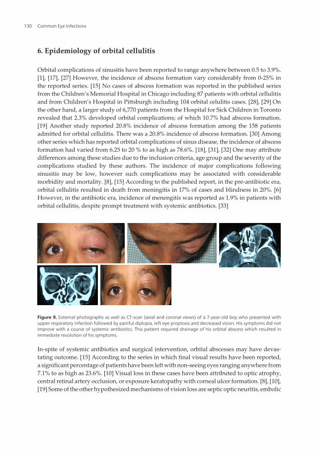
Figure 9. External photographs as well as CT-scan (axial and coronal views) of a 7-year-old boy who presented withupper respiratory infection followed by painful diplopia, left eye proptosis and decreased vision. His symptoms did notimprove with a course of systemic antibiotics. This patient required drainage of his orbital abscess which resulted inimmediate resolution of his symptoms.
In-spite of systemic antibiotics and surgical intervention, orbital abscesses may have devas‐tating outcome. [15] According to the series in which final visual results have been reported,a significant percentage of patients have been left with non-seeing eyes ranging anywhere from7.1% to as high as 23.6%. [10] Visual loss in these cases have been attributed to optic atrophy,central retinal artery occlusion, or exposure keratopathy with corneal ulcer formation. [8], [10],[19] Some of the other hypothesized mechanisms of vision loss are septic optic neuritis, embolic
Common Eye Infections130
or thrombotic lesions in the vascular supply of the optic nerve, choroid or retina. It has beenpostulated that delayed medical and surgical intervention may produce unacceptable visualoutcome. [19], [20], [22]- [25] Among our 218 patients with diagnosis of orbital cellulitis, therewere 116 cases of radiologically confirmed subperiosteal abscess, (Figure 9), 87% of themrequired drainage, and the remaining 13% were observed closely until their resolution whilethose patients were being treated with systemic antibiotics. [8] Thirty-nine eyes (17.8%) hadendophthalmitis causing orbital cellulitis which required evisceration (9.6%) or enucleation(8.2%). Seven orbits required exenteration and 6 infected orbital implants had to be removed.Six patients had dacryocystitis that required a dacryocystorhinostomy to treat orbital cellulitisin addition to the administration of systemic antibiotics. [8]
7. Investigative studies
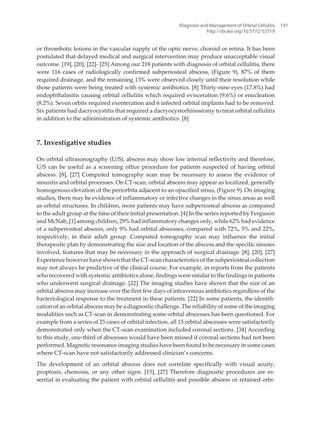
On orbital ultrasonography (U/S), abscess may show low internal reflectivity and therefore,U/S can be useful as a screening office procedure for patients suspected of having orbitalabscess. [8], [27] Computed tomography scan may be necessary to assess the evidence ofsinusitis and orbital processes. On CT-scan, orbital abscess may appear as localized, generallyhomogenous elevation of the periorbita adjacent to an opacified sinus, (Figure 9). On imagingstudies, there may be evidence of inflammatory or infective changes in the sinus areas as wellas orbital structures. In children, more patients may have subperiosteal abscess as comparedto the adult group at the time of their initial presentation. [4] In the series reported by Fergusonand McNab, [1] among children, 29% had inflammatory changes only, while 62% had evidenceof a subperiosteal abscess, only 9% had orbital abscesses, compared with 72%, 5% and 22%,respectively, in their adult group. Computed tomography scan may influence the initialtherapeutic plan by demonstrating the size and location of the abscess and the specific sinusesinvolved, features that may be necessary in the approach of surgical drainage. [8], [20], [27]Experience however have shown that the CT-scan characteristics of the subperiosteal collectionmay not always be predictive of the clinical course. For example, in reports from the patientswho recovered with systemic antibiotics alone, findings were similar to the findings in patientswho underwent surgical drainage. [22] The imaging studies have shown that the size of anorbital abscess may increase over the first few days of intravenous antibiotics regardless of thebacteriological response to the treatment in these patients. [22] In some patients, the identifi‐cation of an orbital abscess may be a diagnostic challenge. The reliability of some of the imagingmodalities such as CT-scan in demonstrating some orbital abscesses has been questioned. Forexample from a series of 25 cases of orbital infection, all 15 orbital abscesses were satisfactorilydemonstrated only when the CT-scan examination included coronal sections. [34] Accordingto this study, one-third of abscesses would have been missed if coronal sections had not beenperformed. Magnetic resonance imaging studies have been found to be necessary in some caseswhere CT-scan have not satisfactorily addressed clinician’s concerns.
The development of an orbital abscess does not correlate specifically with visual acuity,proptosis, chemosis, or any other signs. [15], [27] Therefore diagnostic procedures are es‐sential in evaluating the patient with orbital cellulitis and possible abscess or retained orbi‐
Diagnosis and Management of Orbital Cellulitishttp://dx.doi.org/10.5772/52719
131
tal foreign bodies. Although sinus X-ray may demonstrate an air-fluid level when presentin an abscess cavity, gas-free abscesses may not be readily visible. [15] Ultrasound may de‐tect an abscess of the anterior orbit or the medial wall with 90% accuracy, [25] although anacute abscess may be poorly delineated. Currently, the investigative procedure of choice todiagnose an orbital infection is the CT-scan, although MRI can be utilized when there is acontra-indications for CT-scan. [8], [27], [35] By CT-scan, orbital walls, extraocular muscles,optic nerve, intraconal area and adipose tissue can be seen clearly. An orbital abscess canbe seen as a homogenous, a ring-like, or a heterogenous mass. In these studies, the site oforigin, orbital or subperiosteal, and extent of abscess are readily visible. [8], [22] When ad‐ministered, contrast-media can enhance the surrounding wall of an abscess. Computed to‐mography scan will not differentiate between preseptal cellulitis and eyelid edema but willdifferentiate between preseptal and orbital cellulitis. [15] Beside foreign bodies, sinus dis‐ease and intracranial complications may also be visible on the CT-scan. [8] Our experiencehas shown that CT-scan may be the most comprehensive source of information about orbi‐tal infections and the most sensitive means of monitoring resolving orbital or intracraniallesions. [8], [27] Computed tomography scan is indicated in all patients with periorbital in‐flammation in whom proptosis, ophthalmoplegia, or a decrease in visual acuity develop, inwhom a foreign body or an abscess is suspected, severe eyelid edema prevents an ade‐quate examination, or surgery is contemplated. [8], [15], [20], [22], [35] In our study of the218 patients with orbital cellulitis, diagnosis was made clinically and confirmed by CT-scans or U/S in 90.4% and 36.2% orbits, respectively. [8] Orbital abscesses were identifiedin 53.2% of orbits. In all cases of orbital cellulitis, there was evidence of inflammatory orinfective changes of the orbital structures. Abscess location was found to be medial in 35%,superior in 33%, intraconal in 13%, superomedial in 6%, inferomedial in 6% and lateral in2% of orbits. [8]
8. Bacteriology of orbital infection
In the reported series, the bacteriology of orbital abscesses has received little attention. In seriesin which the contents of the abscess cavity have been cultured, a wide range of organisms havebeen reported. [8], [36] Most commonly reported bacterial species from the abscesses of theorbit and periorbital area include Staphylococcus aureus, Staphylococcus epidermidis,Streptococci, Diphtheroids, Haemophilus influenzae, Escherichia Coli, multiple species ofaerobes and anaerobes. There was no growth in up to 25% of abscesses. [15], [17] Microbio‐logical results from Ferguson and McNab, [1] series varied, with differences in the rate oftesting between the pediatric age group and the older age group. In their series, some formsof cultures were performed in 93% of their patients. Fifty percent of their patiens had bloodcultures none of which yielded positive results. According to their study, cultures taken fromabscesses were more likely to yield positive results. The authors noted that there was nocorrelation between cultures taken from conjunctival swab and the etiological organismsrecovered from the abscesses of those patients with positive cultures. In their study, Staphy‐lococcus aureus was the most common micro-organism recovered. In their pediatric age group
Common Eye Infections132
various species of Streptococcus predominated. Among their pediatric patients, 4 patients hadanaerobic Streptococcus isolates, two had mixed anaerobes and one had Clostridium bifer‐mentans. In Ferguson and McNab’s, [1] series, orbital cellulitis due to anaerobes was muchless common in adults, with only one case of mixed anaerobes identified. In their series, only5 adults and 4 pediatric patients had multiple organisms isolated from the abscesses. Nopathogenic organisms were isolated from their 6 adult and 15 pediatric patients in whom thecultures were performed. [1] Although in the past, H influenza was a major pathogenic bacteriaresponsible for orbital cellulitis in the pediatric age group, [8], [15] in the series reported byFerguson and McNab,1 no cases of H. influenza were identified in the pediatric age group andonly one case was found in an adult patient. This observation has been attributed to the generalimmunization of children with H. influenza type B vaccine since the early 1990s. [1], [27]Schramm et al,5 reported 32 cases of orbital abscesses, the predominant microorganisms beingStaphylococci, Streptococci and Bacteroids species.
The role of anaerobes, not usually considered pathogens in the sinus disease is unclear,although considerable number of cultures in adult patients have yielded anaerobes. [8], [20],[23] In general, patients during their first decade of life may have infection caused by a singleaerobic pathogen which may be responsive to the medical therapy alone. On the other hand,patients older than 15 years of age may have complex infections caused by multiple aerobicand anaerobic organisms that may be slow to clear despite medical and surgical intervention.[23] The virulence of pathogens and responsiveness to anti-microbial agents appear to be age-related. [20], [37] With enlarging of the size of the sinus cavities, the ostia gets narrow creatingoptimal condition for anaerobic bacterial growth. As the person ages, there is a trend towardsappearance of more complex infections. In mixed infections, aerobes utilize oxygen whichencourage growth of more anaerobic microorganisms. On the other hand anaerobes produceB-Lactamase which makes antibiotics less effective. Harris, [20] reviewed microbiology resultsof 37 patients with orbital abscesses in which one-third were younger than 9 years, 58% wereculture negative and the rest had single aerobic pathogen. From his series, 16 patients betweenages 9-14 years showed transition towards more complex infections. Among these, 9 patientswhich were older than 15 years had positive cultures despite being on systemic antibiotics for3 days. In Harris’s, [20] study, older group had more often polymicrobial infections andanaerobes were found in all cases. According to our study the most common microorganismsisolated from the drained abscesses were the most common microorganisms isolated from thedrained abscesses were Staphylococci and Streptococci species; less common organismsincluded Propionibacterium acnes, Haemophilus influenzae, Bacillus, and fungi. [8]
9. Medical management
Medical management depends on the patient’s appearance, ability to take oral medications,compliance and clinical progression of the disease. Patients presenting with signs andsymptoms of eyelid edema, diplopia, reduced visual acuity, abnormal light reflexes, ophthal‐moplegia and proptosis need admission (Figure 10). Further, if a patient appears toxic and eyeexam is difficult to be completely performed, along with signs of CNS involvement as evident
Diagnosis and Management of Orbital Cellulitishttp://dx.doi.org/10.5772/52719
133
by lethargy, vomiting, seizures, headache or cranial nerve deficit, admission is needed forfurther evaluation and proper treatment. Intravenous antibiotics are usually started once thediagnosis of orbital cellulitis is suspected, broad-spectrum antibiotics that cover most grampositive and gram negative bacteria are considered initially. The recommendations forantibiotics are usually based on the microorganisms most frequently suspected from abscesses;Staphylococcus aureus, Staphylococcus epidermidis, Streptococci, and Hemophilus species.[15] Empiric antibiotics should cover methicillin-resistant Staph.aureus if suspected. [38], [39]One should suspect mixed infections including aerobic and anaerobic species in the abscesses.[20] Warm compresses over the involved area may help to improve the softening of the tissuesto bring in more blood circulation in the area where blood supply is already abundant. If noimprovement occurs in 24-48 hours of systemic antibiotics, one may consider InfectiousDisease, Ear, Nose and Throat and/or Neurosurgery consultations. [27]
Historically, cultures from the conjunctiva, nose and throat are usually not representative ofthe pathogens cultured from the abscesses and blood cultures may frequently be negative andare not usually helpful. [15] Most patients in the reported series, had received a combinationof a third-generation cephalosporin and flucloxacillin. [1], [8] According to those reports, mostpatients had received oral antibiotics on discharge for varying periods of time. [40] Forexample, all patients in the Ferguson and McNab’s [1], series had received intravenousantibiotic treatment during their admission and most of these patients had received multidrugtherapy with up to 5 different antibiotics at some point. In these cases treatment regimens wereempirically based and instituted prior to the identification of responsible organisms. [1] In ourexperience, most of our patients also had multiple antibiotic regimens administered duringtheir stay in the hospital and most of them were discharged on at least one antibiotic therapy.[8] In our study of 218 patients having orbital cellulitis and abscess, all patients receivedsystemic antibiotic treatment, and in all patients, treatment regimens were empirically basedand were instituted before the identification of any responsible organisms. [8] In our study,the most common antibiotic regimen included cephalosporins in 90%, and aminoglycosidesin 66% with a combination of other antibiotics. These antibiotics included flucloxacillin in 15%,vancomycin in 13%, ampicillin in 6%, metronidazole in 4%, and penicillin in 3% of patients.In our study, most patients received oral antibiotics on their discharge for varying periods,ranging from 3 days to 3 weeks. [8]
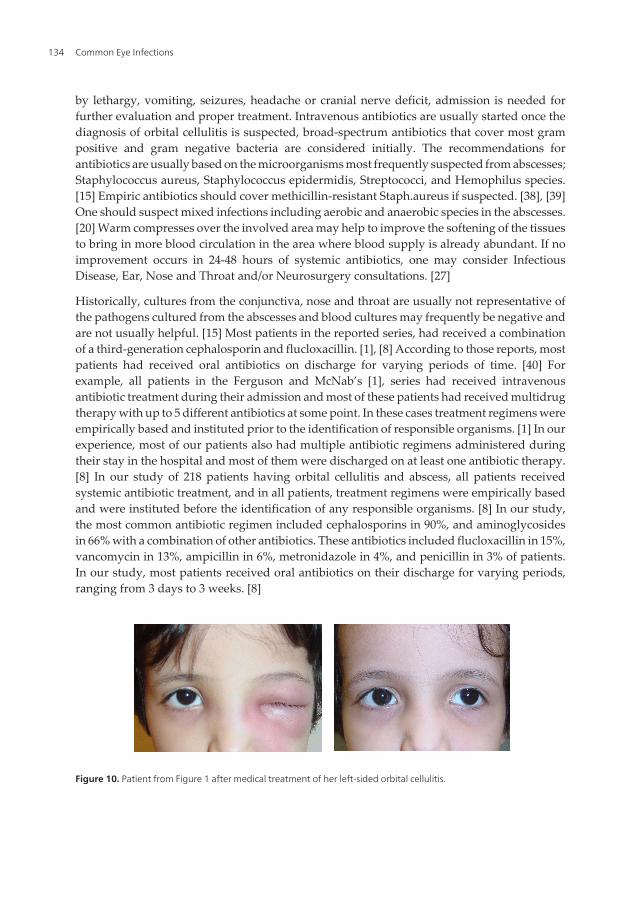
Figure 10. Patient from Figure 1 after medical treatment of her left-sided orbital cellulitis.
Common Eye Infections134
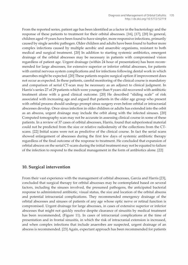
From the reported series, patient age has been identified as a factor in the bacteriology and theresponse of these patients to treatment for their orbital abscesses. [16], [17], [20] In general,children aged <9 years have been found to have simpler, more responsive infections, primarilycaused by single aerobic pathogen. Older children and adults have been found to harbor morecomplex infections caused by multiple aerobic and anaerobic organisms, resistant to bothmedical and surgical treatment. [20] In addition to starting systemic antibiotics, emergentdrainage of the orbital abscesses may be necessary in patients with compromised visionregardless of patient age. Urgent drainage (within 24 hour of presentation) has been recom‐mended for large abscesses, for extensive superior or inferior orbital abscesses, for patientswith central nervous system complications and for infections following dental work in whichanaerobes might be expected. [20] These patients require surgical option if improvement doesnot occur as expected. In these patients, careful monitoring of the clinical course is mandatoryand comparison of serial CT-scan may be necessary as an adjunct to clinical judgment. InHarris’s series 27 of 29 patients which were younger than 9 years old recovered with antibiotictreatment alone with a good clinical outcome. [20] He described “sliding scale” of riskassociated with increasing age and argued that patients in the older age group who presentwith orbital process should undergo prompt sinus surgery even before orbital or intracranialabscesses develop. Once sinus infection in older children or adults has extended into the orbitas an abscess, urgent drainage may include the orbit along with the infected sinuses. [20]Computed tomography scan may not be accurate in assessing clinical course in some of thesepatients. In a review of 37 cases of orbital abscesses, Harris, found that subperiosteal materialcould not be predicted from the size or relative radiodensity of the collections from the CT-scans. [22] Initial scans were not as predictive of the clinical course. In fact the serial scansshowed enlargement of abscesses during the first few days of systemic antibiotic therapyregardless of the final outcome of the response to treatment. He concluded that expansion oforbital abscess on the serial CT-scans during the initial treatment may not be equated to failureof the infection to respond to the medical management in the form of antibiotics alone. [22]
10. Surgical intervention
From their vast experience with the management of orbital abscesses, Garcia and Harris [23],concluded that surgical therapy for orbital abscesses may be contemplated based on severalfactors, including the sinuses involved, the presumed pathogens, the anticipated bacterialresponse to administered antibiotic, visual status, the size and location of the orbital abscessand potential intracranial complications. They recommended emergency drainage of theorbital abscesses and sinuses of patients of any age whose optic nerve or retinal function iscompromised. Urgent drainage for large abscesses, in cases of extensive superior or inferiorabscesses that might not quickly resolve despite clearance of sinusitis by medical treatmenthas been recommended, (Figure 11). In cases of intracranial complications at the time ofpresentation and in frontal sinusitis, in which the risk of intracranial extension is increased,and when complex infections that include anaerobes are suspected, urgent drainage of anabscess is recommended. [23] Again, expectant approach has been recommended for patients
Diagnosis and Management of Orbital Cellulitishttp://dx.doi.org/10.5772/52719
135

younger than 9 years of age in whom simple infections may be suspected. Surgical option maystill be exercised if clinical improvement does not occur in a timely manner and if relativeafferent pupillary defect develops at any time. Further, surgical option should be consideredin cases of fever not abating within 36 hours of systemic antibiotic treatment suggesting thatthe infection may not be responding to the choice of antibiotics being administered. Surgeryshould also be considered when there has been deterioration of vision despite 48 hours ofappropriate antibiotic therapy and no improvement despite 72 hours of such treatment.Usually, CT-scan improvement should be expected to lag behind the clinical picture. In fact,the CT findings may worsen during the first few days of hospitalization despite successfultreatment with antibiotics alone. [23]
Figure 11. External photograph of an 8-year-old boy who required drainage of his right orbital abscess after failing 3days of by systemic antibiotic treatment. CT-scan (axial and saggital cuts) showed an evidence of maxillary sinusitisand an abscess formation in the superior orbit.
In majority of the cases, surgical intervention is indicated for significant underlying sinusdisease, orbital or subperiosteal abscess, or both in the children, (Figure 11). [1], [5], [8], [17]For older patients, sinus surgery remains the most common surgical intervention. Recentliterature suggests that the volume of subperiosteal abscess seems to be the most importantcriterion in determining medical versus surgical management; the volumes of abscessesneeding surgery appears to be larger than the volumes of abscesses not needing surgery. Ingeneral, volumes of <1,250 mm may not need surgical intervention. [41], [42] There may be anargument regarding early drainage of an orbital abscess to prevent complications whereasearly surgical intervention has the possibility of seeding the infection. [20] For practicalpurposes, Harris, has outlined a useful approach in the management of an orbital abscess. [20]He emphasizes on the emergent drainage for patients of any age whose visual function maybe compromised. Also for the patients in whom a large orbital abscess causes discomfort,presence of superior or inferior orbital abscess, evidence of intracranial extension, involvementof frontal sinuses, and a known dental source of the infection in patients older than 9 years,urgent drainage usually within 24 hours has been recommended. [20] Wait and see approachmay be indicated for patients younger than 9 years of age having medial subperiosteal abscessof modest size, for patients having no visual compromise and in those having no intracranialor frontal sinus involvement. In these patients, careful evaluation and close monitoring of theiroptic nerve function and the level of consciousness and mental state are necessary. Whenindicated, one may consider making an incision approximately 2-inch down to the periosteumat the inner quadrant of the orbit to drain these orbital abscesses. [27] Patients with suspectedfungal orbital cellulitis (especially Mucormycosis), need to be treated with intravenous
Common Eye Infections136
Amphotericin B and predisposing factors such as diabetes, acidosis and other medicalconditions need to be addressed. Wide excision along with debridement of the necrotic tissueis desired. If necessary, a drain may be inserted and tissues may not need to be sutured andmay be left for granulation. One may consider removal of drain when no further drainageoccurs. In some cases, endoscopic approach may be utilized and has been found to be effectivefor the treatment subperiosteal abscess as a result of sinus infection. Some of the advantagesof endoscopic surgical drainage may be the avoidance of external ethmoidectomy andassociated external facial scar and an early drainage of the affected sinuses and subperostealabscess at the same time. [27], [43] In our study [8], among the 116 radiologically confirmedorbital abscesses, 87% required drainage, and the remaining 13% required close observationuntil their resolution while on systemic antibiotics. Thirty-nine eyes (17.8%) had endophthal‐mitis causing orbital cellulitis and required evisceration or enucleation. Seven orbits requiredexenteration and 6 infected orbital implants had to be removed. Other 6 patients had dacryo‐cystitis that required a dacryocystorhinostomy to treat orbital cellulitis in addition to theadministration of systemic antibiotics. Combined endoscopic sinus surgery with transnasalorbital abscess drainage was carried out in some of our patients with sinusitis and orbitalabscess, especially in the medial orbit. [8]
11. Complications of orbital cellulitis
Although less common, major complications related to orbital cellulitis and abscess can occur.Even after the successful treatment of such infections, permanent visual loss or loss of functionof the vital structures may remain. Ferguson [1] reported no visual function loss among theirpatients after resolution of their infections. Only one of their patients from the pediatric agegroup had proptosis on follow-up; one had ophthalmoplegia and one had recollection of theabscess. One of their adult patients developed presumed meningitis and another adult patientrequired enucleation. In rare circumstances, the microorganism may cause necrotizing eyeliddisease often referred as necrotizing fasciitis. [3], [44]- [46] This may progress to systemicmanifestations including the potentially fatal toxic streptococcus syndrome, characterized bymulti-organ failure. [44], [46] These complications can occur in the absence of antecedent healthproblems or history of trauma. [3], [45], [46] The virulence of this organism is related to theproduction of M proteins and exotoxins A and B. [47] These proteins act as super-antigens invitro and mediate tissue necrosis by causing massive release of cytokines such as tumornecrosis factor and interleukins.
12. Visual loss in orbital cellulitis
Permanent loss of vision has been noted as a complication of orbital infection and up to onefifth of patients with orbital inflammation had blindness in the pre-antibiotic era. [2] Now, al‐though permanent loss of vision resulting from orbital inflammation is unusual it can still oc‐cur, (Figure 12). [8], [25], [26] Patt [26] reported 38 patients with orbital cellulitis and resultant
Diagnosis and Management of Orbital Cellulitishttp://dx.doi.org/10.5772/52719
137
permanent vision loss one of which progressed to no light perception vision. Loss of visionwith orbital inflammation may result from optic neuritis as a reaction to adjacent or nearby in‐fection, ischemia due to thrombophlebitis along valveless orbital veins, or compressive/pres‐sure ischemia possibly resulting in central artery/occlusion, (Figure 12). [22], [26] Permanentirreversible visual loss may occur in cases with orbital and subperiosteal abscess despite earlyintervention. In a survey of 46 cases with confirmed diagnosis of orbital and subperiosteal ab‐scess in which visual results were reported, permanent loss of vision occurred in 15% of thecases. [48] Blindness was attributed to the central retinal artery occlusion in 4, optic atrophy in2 of hese patients. Permanent visual loss in orbital cellulitis probably has a vascular cause,whereas partial vision loss that respond to antibiotic therapy and drainage procedures may bedue to inflammatory infiltrates or presence of compressive optic neuropathy. [21] It is be‐lieved that the confinement of the optic nerve in the orbital apex area and within its bony ca‐nal along with its proximity to the posterior ethmoid and sphenoid sinuses may furtherhighlight the importance of the these factors in the exacerbation of posterior orbital celluliltis.Physcians need to be aware that patients with sinusitis and associated orbital cellulitis may beat risk for developing severe vision deficit requiring timely intervention. In a review of 148 pa‐tients with orbital abscess from 13 series reported by Hornblass [15], 3 patients had evidence ofno light perception vision.
Clinical examination by itself may not exactly delineate the nature of orbital inflammatoryprocesses, clinicians may have to rely on imaging studies to select potential surgical candi‐dates. Despite availability of modern CT-Scan and MRI studies, the physician stil needs to re‐ly on the clinical progression of the inflammation based on vision, pupillary fuction, andassessment of ocular motility. Patt and Manning [26], reported 4 patients with vision loss dueto orbital cellulitis and in each of these cases had CT-scan readings of “no definite abscess”contributing to the delay in diagnosis of orbital abscess, with a resultant delay in surgicaldrainage.
Ethmoidal sinuses are separated from the orbital cavity by the lamina papyracea and anteriorand posterior ethmoidal foramina serve as additional connections that may allow infection togain access from ethmoidal air cells to the orbital cavity, (Figure 5). Periorbita in this area isloosely attached to bone and may be elevated by a purulent collection, resulting in subperios‐teal abscess. Acute visual loss due to sinusitis may either be secondary to complications of or‐bital cellulitis or may be seen as a part of orbital apex syndrome. orbital cellulitis or as a part ofthe orbital apex syndrome. [27] Two cases of acute visual loss have been reported by El-Sayedand Muhaimeid [49], as a complication of orbital cellulitis due to sinusitis. One of these pa‐tients had dramatic improvement in vision from hand motion to normal vision after systemicantibiotic treatment of pansinusitis and associated orbital cellulitis. The second patient, (a 10-year old girl), achieved normal visual acuity from no light perception after only surgical inter‐vention by exploration of sphenoid and ethmoid sinuses along-with intravenous antibioticadministration. Three cases of sphenoethmoiditis with minimal signs of orbital inflammationcausing permanent loss of vision have been reported by Slavin and Glaser. [48] These authorssuggested the use of term “posterior orbital cellulitis” for such cases and defined it as a clini‐cal syndrome in which early severe visual loss overshadows or precedes accompanying in‐flammatory orbital signs. Acute blindness may also result from orbital infarction syndrome.
Common Eye Infections138
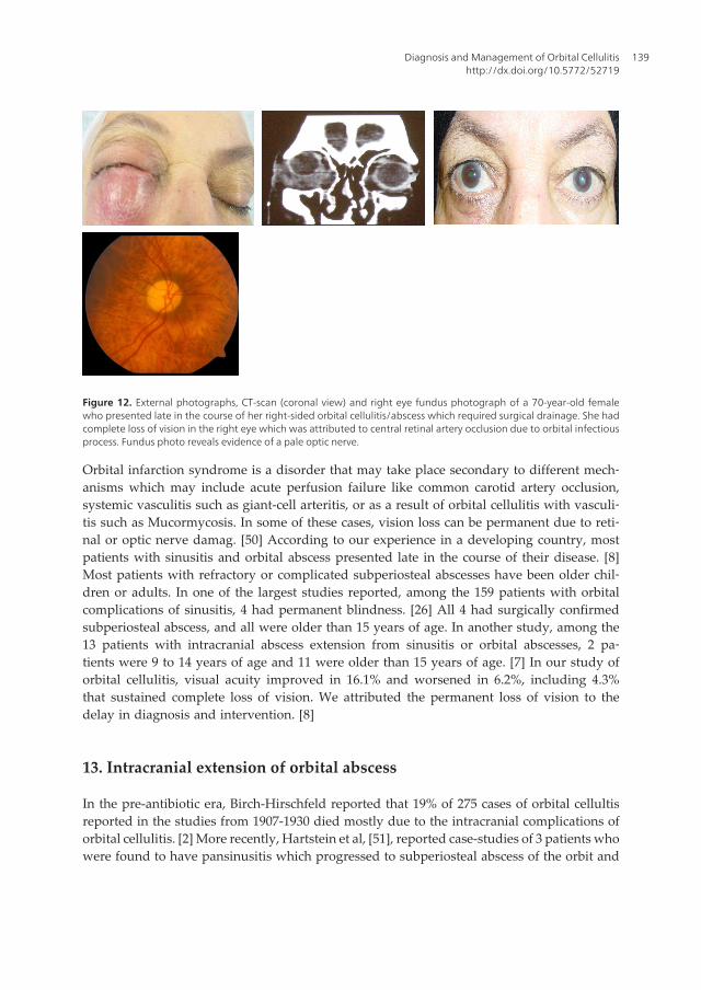
Figure 12. External photographs, CT-scan (coronal view) and right eye fundus photograph of a 70-year-old femalewho presented late in the course of her right-sided orbital cellulitis/abscess which required surgical drainage. She hadcomplete loss of vision in the right eye which was attributed to central retinal artery occlusion due to orbital infectiousprocess. Fundus photo reveals evidence of a pale optic nerve.
Orbital infarction syndrome is a disorder that may take place secondary to different mech‐anisms which may include acute perfusion failure like common carotid artery occlusion,systemic vasculitis such as giant-cell arteritis, or as a result of orbital cellulitis with vasculi‐tis such as Mucormycosis. In some of these cases, vision loss can be permanent due to reti‐nal or optic nerve damag. [50] According to our experience in a developing country, mostpatients with sinusitis and orbital abscess presented late in the course of their disease. [8]Most patients with refractory or complicated subperiosteal abscesses have been older chil‐dren or adults. In one of the largest studies reported, among the 159 patients with orbitalcomplications of sinusitis, 4 had permanent blindness. [26] All 4 had surgically confirmedsubperiosteal abscess, and all were older than 15 years of age. In another study, among the13 patients with intracranial abscess extension from sinusitis or orbital abscesses, 2 pa‐tients were 9 to 14 years of age and 11 were older than 15 years of age. [7] In our study oforbital cellulitis, visual acuity improved in 16.1% and worsened in 6.2%, including 4.3%that sustained complete loss of vision. We attributed the permanent loss of vision to thedelay in diagnosis and intervention. [8]
13. Intracranial extension of orbital abscess
In the pre-antibiotic era, Birch-Hirschfeld reported that 19% of 275 cases of orbital cellultisreported in the studies from 1907-1930 died mostly due to the intracranial complications oforbital cellulitis. [2] More recently, Hartstein et al, [51], reported case-studies of 3 patients whowere found to have pansinusitis which progressed to subperiosteal abscess of the orbit and
Diagnosis and Management of Orbital Cellulitishttp://dx.doi.org/10.5772/52719
139
subsequent intracranial extension. All 3 patients had been treated with systemic antibioticsand surgical drainage of the orbital abscesses as well as sinuses. Two of the 3 patients requiredsurgical drainage of their intracranial abscesses.
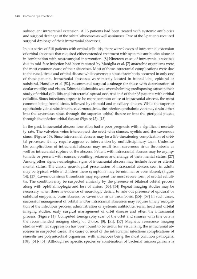
In our series of 218 patients with orbital cellulitis, there were 9 cases of intracranial extensionof orbital abscesses that required either extended treatment with systemic antibiotics alone orin combination with neurosurgical intervention. [8] Nineteen cases of intracranial abscessesdue to mid-face infection had been reported by Maniglia et al, [7] anaerobic organisms werethe most common cause of their abscesses. Most of these intracranial complications were dueto the nasal, sinus and orbital disease while cavernous sinus thrombosis occurred in only oneof these patients. Intracranial abscesses were mostly located in frontal lobe, epidural orsubdural. Handler et al [52], recommend surgical drainage for those with deterioration ofocular motility and vision. Ethmoidal sinusitis was overwhelming predisposing cause in theirstudy of orbital cellulitis and intracranial spread occurred in 6 of their 65 patients with orbitalcellulitis. Sinus infections appear to be more common cause of intracranial abscess, the mostcommon being frontal sinus, followed by ethmoid and maxillary sinuses. While the superiorophthalmic vein drains into the cavernous sinus, the inferior ophthalmic vein may drain eitherinto the cavernous sinus through the superior orbital fissure or into the pterigoid plexusthrough the inferior orbital fissure (Figure 13). [15]
In the past, intracranial abscess formation had a poor prognosis with a significant mortali‐ty rate. The valveless veins interconnect the orbit with sinuses, eyelids and the cavernoussinus, (Figure 13). Since intracranial abscess may be a life-threatening complication of orbi‐tal processes, it may require aggressive intervention by multidisciplinary team. Undesira‐ble complications of intracranial abscess may result from cavernous sinus thrombosis aswell as intracranial rupture of the abscess. Patient with intracranial abscess may be asymp‐tomatic or present with nausea, vomiting, seizures and change of their mental status. [27]Among other signs, neurological signs of intracranial abscess may include fever or alteredmental status. The classic neurological presentation of intracranial abscess seen in adultsmay be typical, while in children these symptoms may be minimal or even absent, (Figure14). [27] Cavernous sinus thrombosis may represent the most severe form of orbital celluli‐tis. The condition may be suspected clinically by the presence of bilateral orbital processalong with ophthalmoplegia and loss of vision. [53], [54] Repeat imaging studies may benecessary when there is evidence of neurologic deficit, to rule out presence of epidural orsubdural empyema, brain abscess, or cavernous sinus thrombosis. [55]- [57] In such cases,successful management of orbital and/or intracranial abscesses may require timely recogni‐tion of the infectious process, administration of systemic antibiotics, serial head and orbitalimaging studies, early surgical management of orbit disease and often the intracranialprocess, (Figure 14). Computed tomography scan of the orbit and sinuses with fine cuts isthe recommended imaging study of choice. [8], [51], [57] Magnetic resonance imagingstudies with fat suppression has been found to be useful for visualizing the intracranial ab‐scesses in suspected cases. The cause of most of the intracranial infectious complications ofsinusitis are polymicrobial organisms, with anaerobes being the most common pathogens.[38], [51]- [54] Although no specific species or combination of bacterial microorganisms is
Common Eye Infections140
found to be predominant; Streptococcus, Staphylococcus, Bacteriodes, and Fusobacteriumspecies are frequently encountered. Hartstein et al. reported 3 cases of intracranial abscessall of which had evidence of polymicrobial infection with no predominance of any oneparticular organism. [51] Initial treatment of such patients requires broad-spectrum antibi‐otics including beta-lactamase resistant antibiotics that have good anaerobic coverage, aswell as good central nervous system penetration. [20], [23], [27], [51] Routine follow-upimaging studies may be indicated based on the clinical examination. Proper managementof these patients may require a multidisciplinary team that includes an orbital surgeon,otolaryngologists, neurosurgeon, and an infectious disease specialist.
Figure 13. Lateral view of the schematic drawing showing extensive venous drainage of the facial structures alongwith orbital veins and their direct connections with cavernous sinus.
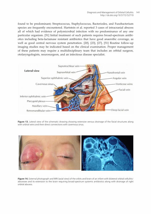
Figure 14. External photograph and MRI (axial view) of the orbits and brain of an infant with bilateral orbital cellulitis/abscesses and its extension to the brain requiring broad-spectrum systemic antibiotics along with drainage of rightorbital abscess.
Diagnosis and Management of Orbital Cellulitishttp://dx.doi.org/10.5772/52719
141
Author details
Imtiaz A. Chaudhry1, Waleed Al-Rashed2, Osama Al-Sheikh3 and Yonca O. Arat4
1 Houston Oculoplastics Associates, Memorial Herman Medical Plaza, Texas Medical Cen‐ter, Houston, Texas, USA
2 Al Imam Mohammad Ibn Saud Islamic University, Faculty of Medicine, Riyadh, SaudiArabia, Saudi Arabia
3 Oculoplastic and Orbit Division, King Khaled Eye Specialist Hospital, Riyadh, Saudi Ara‐bia
4 Department of Ophthalmology, University of Wisconsin-Madison, Wisconsin, USA
References
[1] Ferguson MP, McNab AA. Current treatment and outcome in orbital cellulitis. AustN.Z. J Ophthalmol. 1999; 27: 375-9.
[2] Duke-Elder S, MacFaul PA. The ocular adnexa: part 2. Lacrimal orbital and para orbi‐tal diseases. In: Duke-Elder S, ed. System of Ophthalmology. Vol. 13. London: HenryKimpton; 1974:859–89.
[3] Connel B, Kamal Z, McNab AA. Fulminant orbital cellulits with complete loss of vi‐sion. Clin Exp Ophthalmol 2001; 29: 260-1.
[4] Meara DJ. Sinonasal disease and orbital cellulitis in children. Oral Maxillofac SurgClin North Am. 2012;24:487-96.
[5] Bedwell J, Bauman NM. Management of pediatric orbital cellulitis and abscess. CurrOpin Otolaryngol Head Neck Surg. 2011;19:467-73.
[6] Chandler JR, Langenbrunner DJ, Stevens ER. The pathogenesis of orbital complica‐tions in acute sinusitis. Laryngoscope. 1970;80:1414 –28.
[7] Maniglia AJ, Goodwin WJ, Arnold JE, Gonz E. Intracranial abscess secondary to na‐sal, sinus and orbital infections in adults and children. Arch Otolaryngol Head NeckSurg 1989; 115: 1424-9.
[8] Chaudhry IA, Shamsi FA, Elzaridi E, Al-Rashed W. Al-Amri AM, Al-Anezi F, AratYO, Holck DEE. Outcome of treated orbital cellulitis from a tertiary eye care center inthe Middle East. Ophthalmology 2007;114:345-54.
Common Eye Infections142
[9] Chaudhry IA, Shamsi FA, Elzaridi E, Al-Rashed W, Al-Amri A, Arat YO. Inpatientpreseptal cellulitis: experience from a tertiary eye care centre Br. J. Ophthalmol.2008;92;1337-41.
[10] Jarrett WH, Gutman FA. Ocular complications of infection in the paranasal sinuses.Arch Ophthalmol, 1969; 81: 683-8.
[11] Fezza J, Chaudhry IA, Kwon YH, Grannum E, Sinard J, Wolfley DE. Orbital melano‐ma presenting as orbital cellulities: A clinicopathologic report. Ophthal Plastic Re‐constr Surg 1998;14:286-9.
[12] Chaudhry IA, Shamsi FA, Morales J. Orbital cellulitis following implantation ofaqueous drainage devices for glaucoma. Eur J Ophthalmol, 2007;17:136-140.
[13] Chaudhry IA. Herpes Zoster Presenting with Orbital Cellulitis, Proptosis, and Oph‐thalmoplegia Middle East J Ophthalmol. 2006;13:167-9.
[14] Krouschnabel EF. Orbital apex syndrome due to sinus infection. The Laryngoscope,1974: 84: 353-71.
[15] Hornblass A, Herschorn BJ, Stern K, Grimes C. Orbital abscess. Surv Ophthalmol.1984; 29: 169-78.
[16] Hauser, A and Fogarasi, S. Periorbital and Orbital Cellulitis. Pediatrics in Review.2010;31:242-9.
[17] Seltz LB, Smith J, Durairaj VD, Enzenauer R, Todd J. Microbiology and antibioticmanagement of orbital cellulitis. Pediatrics. 2011;127:566-72.
[18] Morgan PR, Morrison WV. Complications of frontal and ethmoid sinusitis. The Lar‐yngoscope, 1980; 90: 661-6.
[19] Fearon B, Edmonds B, Bird R. Orbital-facial complication of sinusitis in children. TheLaryngoscope, 1979; 86: 947-53.
[20] Harris GJ. Subperiosteal abscess of the orbit: age as a factor in the bacteriology andresponse to treatment. Ophthalmology, 1994; 101: 585-95.
[21] Coşkun M, Ilhan Ö, Keskin U, Ayintap E, Tuzcu E, Semiz H, Öksüz H. Central reti‐nal artery occlusion secondary to orbital cellulitis and abscess following dacryocysti‐tis. Eur J Ophthalmol. 2011;21:649-52.
[22] Harris GJ. Subperiosteal abscess of the orbit: computed tomography and the clinicalcourse. Ophth Plast Reconstr Surg. 1996; 12: 1-8.
[23] Garcia GJ, Harris GJ. Criteria from nonsurgical management of subperiosteal abscessof the orbit: analysis of outcomes 1988-1998. Ophthalmology 2000; 107: 1454-8.
[24] Harris GJ. Subperiosteal abscess of the orbit: older children and adults require ag‐gressive treatment: Editorial. Ophth Plast Reconstr Surg. 2001; 17: 395-7.
Diagnosis and Management of Orbital Cellulitishttp://dx.doi.org/10.5772/52719
143
[25] Schramm VL, Myres EN, Kennerdell JS. Orbital complications of acute sinusitis:Evaluation, management and outcome. ORL Digest. 1979; 86: 221-30.
[26] Patt BS, Manning SC. Blindness resulting from orbital complications of sinusitis. Oto‐laryngol. Head Neck Surg 1991; 104: 789-95.
[27] Chaudhry IA, Al-Rashed W, Arat YO. The hot orbit: Orbital cellulitis. Middle EastAfr J Ophthalmol 2012;19:34-42.
[28] Gellady AM, Shulman ST, Ayoub EM. Periorbital and orbital cellulitis in Children.Pediatrics, 1978; 61: 272-7.
[29] Watters EC, Waller PH. Acute orbital cellulitis. Arch Ophthalmol 1976; 94: 785.
[30] Weiss A, Friendly D, Eglin K. Bacterial periorbital and orbital cellulitis in childhood.Ophthalmology, 1983; 90: 195-204.
[31] Welsh LW, Welsh JJ. Orbital complications of sinus disease. The Laryngoscope, 1974;84: 848-56.
[32] Giletto JB, Scherr SA, Mikaelian DO. Orbital complications of acute sinusitis in chil‐dren. Trans Pa Acad Ophthalmol Otolaryngol, 1980; 34: 60.
[33] Smith AT, Spencer JT. Orbital complications resulting from lesions of sinuses, AnnOtology, Rhinology, and Laryngdology, 1948; 57: 5.
[34] Langham – Brown JJ, Rhys-Williams S. Computed tomography of acute orbital infec‐tion: the importance of coronal sections. Clin Radiol. 1989; 40: 471-4.
[35] Hilal SK. Computed tomography of the orbit. Ophthalmology, 1979; 86: 864-9.
[36] Bagheri A, Tavakoli M, Aletaha M, Salour H, Ghaderpanah M. Orbital and preseptalcellulitis: a 10-year survey of hospitalized patients in a tertiary eye hospital in Iran.Int Ophthalmol. 2012;32:361-7.
[37] Donahue SP, Schwartz G. Preseptal and orbital cellulitis in childhood: A changingmicrobiologic spectrum. Ophthalmology, 1998: 105: 585-95.
[38] Mathias MT, Horsley MB, Mawn LA, Laquis SJ, Cahill KV, Foster J, Amato MM, Du‐rairaj VD. Atypical presentations of orbital cellulitis caused by methicillin-resistantStaphylococcus aureus. Ophthalmology. 2012;119:1238-43.
[39] Kobayashi D, Givner LB, Yeatts RP, Anthony EY, Shetty AK. Infantile orbital celluli‐tis secondary to community-associated methicillin-resistant Staphylococcus aureus. JAAPOS. 2011;15:208-10.
[40] Emmett Hurley P, Harris GJ. Subperiosteal abscess of the orbit: duration of intrave‐nous antibiotic therapy in nonsurgical cases. Ophthal Plast Reconstr Surg.2012;28:22-6.
Common Eye Infections144
[41] Todman MS, Enzer YR. Medical management versus surgical intervention of pedia‐tric orbital cellulitis: the importance of subperiosteal abscess volume as a new criteri‐on. Ophthal Plast Reconstr Surg. 2011;27:255-9.
[42] Gavriel H, Yeheskeli E, Aviram E, Yehoshua L, Eviatar E. Dimension of subperiostealorbital abscess as an indication for surgical management in children. OtolaryngolHead Neck Surg. 2011;145:823-7.
[43] Bhargava D, Saukhla D, Ganesan A, Chand P. Endoscopic sinus surgery fro orbitalsubperiosteal abscess secondaryto sinusitis. Rhinology 2001; 39: 151-5.
[44] Ingraham HJ, Ryan ME, Burns JT. Streptococcal preseptal cellulitis complicated bythe toxic streptococcus syndrome. Ophthalmology 1994; 102: 1223-6.
[45] Shayegani A, MacFarlane D. Kazim M. Streptococcal gangrene of the eyelids and or‐bit. Am J Ophthalmol 1995; 120: 784-92.
[46] Marshall DH, Jordan DR, Gilberg SM. Periocular necrotizing fasciitis: a review of fivecases. Ophthalmology 1996; 104: 1857-62.
[47] Meyer MA. Streptococcal toxic shock syndrome complicating preseptal cellulitis. AmJ Ophthalmol 1996; 123: 841-3.
[48] Slavin ML, Glaser J. Acute severe irreversible visual loss with sphenoethmoiditis -‘posterior’ orbital cellulitis. Arch Ophthalmol. 1987; 105: 345-8.
[49] El-Sayed Y, Al-Muhaimeid H. Acute visual loss in association with sinusitis. J LaryngOtol. 1993; 107:840-2.
[50] Borruat FX, Bogousslavasky J, Uffer S, Klainguti G, Schatz NJ. Orbital infarction syn‐drome. Ophthalmology, 1993; 100:562-8.
[51] Hartstein ME, Steinvurzel MD, Choen CP. Intracranial abscess as a complication ofsubperiosteal abscess of the orbit. Ophth plast Reconstr Surg. 2001; 17: 398-403.
[52] Handler LC, Davey IC, Hill JC, Lauryssen C. The acute orbit: differentiation of orbitalcellulitis from subperiosteal abscess by computerized tomography. Neuroradiology,1991; 33: 15-18.
[53] Giannoni CM, Stewart MG, Alford EL. Intracranial complications of sinusitis. Lar‐yngoscope 1997; 107: 863-7.
[54] Brook I. Bacteriology of intracranial abscess in children. J Neurosurg 1981; 54: 484-8.
[55] Weber AL, Mikuli D. Inflammatory disorders of the periorbital sinuses and theircomplications. Radiol Clin North Am 1987; 25: 615-30.
[56] Towbin R, Han B, Kaufmann R, Burke M. Postseptal cellulitis: CT in diagnosis andmanagement. Radiology 1986; 158: 735-7.
Diagnosis and Management of Orbital Cellulitishttp://dx.doi.org/10.5772/52719
145

[57] Harr DL, Quencer RM, Abrams GW. Computed tomography and ultrasound in theevaluation of orbital infection and pseudotumor. Radiology 1982; 152: 395.
[58] Brook I, Frazier EH. Microbiology of subperiosteal orbital abscess and associatedmaxillary sinusitis. Laryngoscope 1996; 106: 1010-3.
Common Eye Infections146


![Recurrent Sinusitis and Periorbital Cellulitis Secondary ... · Orbital complications of sinusitis are commoner in children than adults and have favorable prognosis [2]. Orbital complications](https://img.pdfslide.us/doc/110x75/5f6cbfd225e3ef0aaa5a247a/recurrent-sinusitis-and-periorbital-cellulitis-secondary-orbital-complications.jpg)