Embed Size (px)
Citation preview
REVIEW
Diagnosis and management of Neuro-Behcet’s disease:international consensus recommendations
Seema Kalra • Alan Silman • Gulsen Akman-Demir • Saeed Bohlega • Afshin Borhani-Haghighi •
Cris S. Constantinescu • Habib Houman • Alfred Mahr • Carlos Salvarani •
Petros P. Sfikakis • Aksel Siva • Adnan Al-Araji
Received: 29 November 2013 / Accepted: 4 December 2013 / Published online: 24 December 2013
� The Author(s) 2013. This article is published with open access at Springerlink.com
Abstract Neuro-Behcet’s disease (NBD) is one of the
more serious manifestations of Behcet’s disease (BD),
which is a relapsing inflammatory multisystem disease
with an interesting epidemiology. Though NBD is rela-
tively uncommon, being potentially treatable, neurologists
need to consider it in the differential diagnosis of inflam-
matory, infective, or demyelinating CNS disorders. Evi-
dence-based information on key issues of NBD diagnosis
and management is scarce, and planning for such studies is
challenging. We therefore initiated this project to develop
expert consensus recommendations that might be helpful to
neurologists and other clinicians, created through an
extensive literature review and wide consultations with an
international advisory panel, followed by a Delphi exer-
cise. We agreed on consensus criteria for the diagnosis of
NBD with two levels of certainty in addition to recom-
mendations on when to consider NBD in a neurological
patient, and on the use of various paraclinical tests. The
management recommendations included treatment of the
parenchymal NBD and cerebral venous thrombosis, the use
of disease modifying therapies, prognostic factors, outcome
measures, and headache in BD. Future studies are needed
to validate the proposed criteria and provide evidence-
based treatments.
Keywords Neuro-Behcet’s disease � Behcet’s disease �Delphi method � Consensus � Diagnosis � Management
On behalf of the International Neuro Behcet’s Advisory Group.
S. Kalra � A. Al-Araji (&)
Neurology Research Department, University Hospital of North
Staffordshire, Stoke-on-Trent, England, UK
e-mail: [email protected]
S. Kalra
Keele University, Keele, Staffordshire, UK
A. Silman
Central Manchester University Hospital, Manchester, UK
G. Akman-Demir
Neurology Department, Bilim University,
Istanbul, Turkey
S. Bohlega
Department of Neurosciences, Saudi Neurology Society,
King Faisal Specialist Hospital and Research Centre,
Riyadh, Saudi Arabia
A. Borhani-Haghighi
Health Policy Research Center and Department of Neurology,
Shiraz University of Medical Sciences, Shiraz, Iran
C. S. Constantinescu
Academic Division of Clinical Neurology, Queen’s Medical
Centre, University of Nottingham, Nottingham, UK
H. Houman
Department of Internal Medicine, Faculty of Medicine of Tunis,
Hospital La Rabta, University el Manar 2 Tunis, Tunis, Tunisia
A. Mahr
Department of Internal Medicine, Hospital Saint-Louis,
University Paris 7, Rene Diderot, Paris, France
C. Salvarani
Rheumatology Unit, Department of Internal Medicine, Azienda
Ospedaliera, IRCCS di Reggio Emilia, Viale Risorgimento n 80,
42123 Reggio Emilia, Italy
P. P. Sfikakis
First Department of Propedeutic Internal Medicine, Athens
University Medical School, Athens, Greece
A. Siva
Neurology Department, Istanbul University, Istanbul, Turkey
123
J Neurol (2014) 261:1662–1676
DOI 10.1007/s00415-013-7209-3
Introduction
Hulusi Behcet, a Turkish dermatologist, described the triad
of recurrent oral and genital ulcers with uveitis in 1937 [1];
the disease is commonly referred to as Behcet’s disease
(BD) and is recognized as a multisystem inflammatory
disorder of unknown aetiology [2]. Interestingly, BD is
more prevalent along the ancient Silk Road, including
countries in the Far East, the Middle East, and the Medi-
terranean basin [3]. It has been reported, however, from
most countries across the globe [4]. Neuro-BD (NBD)
refers to the neurological manifestations of the disease.
The various systemic features of BD and its commonly
accepted diagnostic criteria, in addition to the description
of the various epidemiological and clinical features of
NBD have been described in previous publications [3–5].
In recent years, paraclinical diagnostic tests and an
increasing range of immunomodulatory treatments are
available for NBD patients. Practice guidelines are needed
to improve the diagnostic process, improve quality of care,
encourage sensible use of resources, and ensure a balanced
consideration of potentially harmful medications. Because
NBD is relatively uncommon, studies providing high-
quality evidence are very limited. Published studies include
mainly personal experiences or single-centre approaches.
Our aim was to reach expert consensus recommenda-
tions on the key issues of the diagnosis and management of
NBD. As BD is a systemic disease, we felt that wide
consultation with an international, multidisciplinary panel
was essential to identify the key issues (scope of the rec-
ommendations) before reaching consensus through a Del-
phi exercise amongst a group with a majority of
neurologists with special interest in BD.
The Delphi method has been widely used in healthcare
and especially in developing clinical practice guidelines
where rigorous data are lacking. It includes repeated rounds
of communications and voting amongst a panel of experts.
The outcome represents the collective opinion of the panel
[6, 7].
In this article, we aim to address the key issues in
diagnosis and management of NBD. We will present a brief
summary of the relevant background literature on each
topic of interest, followed by the list of agreed consensus
recommendations.
Methods
Panel selection
The project was initiated by invitations to a wide range of
experts with academic and clinical experience in the field
of BD across the globe, who were mostly members of the
International Society for Behcet’s Disease. Fifty-two
experts from 22 countries accepted the invitation, including
a voluntary patients’ group representative. They included
22 neurologists, 11 internists, and 19 other specialists,
including 13 rheumatologists, two ophthalmologists, one
dermatologist, one immunologist and one paediatrician.
The panel was structured into three groups: a project
organising committee (POC), a Neuro-Behcet’s advisory
group and a Neuro-Behcet’s consensus group. The POC
consisted of four members, AA-A, the convenor of the
project, SK, the researcher and bibliographer, AS, an aca-
demic clinical epidemiologist, and GAD, an experienced
neuro-Behcet’s expert. The advisory group consisted of all
52 panel members who participated till the second round of
the Delphi process. The consensus group consisted of 12
members (majority neurologists: seven) chosen from the
advisory group based on their active contribution in the
consensus process, specialty, and publications records.
They continued the further steps of the Delphi exercise and
are the authors of this paper. The project methodology was
discussed, amended, and agreed upon by all participating
members before starting the consensus process.
Search strategy and selection criteria
Literature was searched on Cochrane, Medline, and Em-
base databases using the key search terms ‘‘Behcet *’’,
‘‘Neuro Behcet *’’, and ‘‘triple-symptom complex’’ for
entries from 1948 until April 2011. Titles and abstracts of
published articles were reviewed. The search was limited to
human studies only, published in the English language. The
articles addressing diagnosis or treatment were reviewed,
which could be case reports, case series, observations,
comparison studies, interventional studies, or reviews.
These were supplemented by reference lists from the
authors’ own collections. Full texts of relevant articles
were reviewed and the final reference list was generated on
the basis of relevance to the scope of this consensus. The
search was updated every 3 months until April 2013. Draft
recommendations were generated with their level of evi-
dence; levels I–IV were used to grade the articles [8].
The Delphi consensus process
We used a four-round Delphi consensus process through
email communications. In the first round, we defined
the scope of the project and established the need for the
diagnostic criteria for NBD and achieved agreement on the
disease terminology. The scope consisted of the key issues
in the diagnosis and management of NBD that were
accepted to be covered in this consensus. This was defined
after an extensive literature search, and a list was sent out
to the advisory group and amended after the feedback. This
J Neurol (2014) 261:1662–1676 1663
123
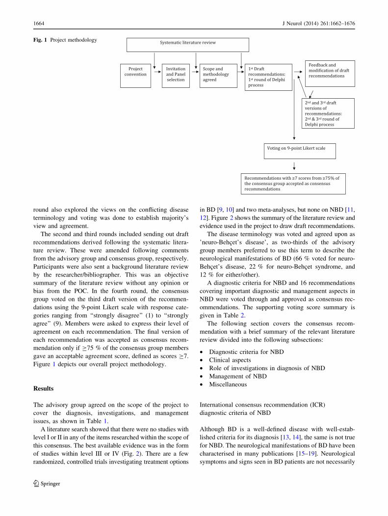
round also explored the views on the conflicting disease
terminology and voting was done to establish majority’s
view and agreement.
The second and third rounds included sending out draft
recommendations derived following the systematic litera-
ture review. These were amended following comments
from the advisory group and consensus group, respectively.
Participants were also sent a background literature review
by the researcher/bibliographer. This was an objective
summary of the literature review without any opinion or
bias from the POC. In the fourth round, the consensus
group voted on the third draft version of the recommen-
dations using the 9-point Likert scale with response cate-
gories ranging from ‘‘strongly disagree’’ (1) to ‘‘strongly
agree’’ (9). Members were asked to express their level of
agreement on each recommendation. The final version of
each recommendation was accepted as consensus recom-
mendation only if C75 % of the consensus group members
gave an acceptable agreement score, defined as scores C7.
Figure 1 depicts our overall project methodology.
Results
The advisory group agreed on the scope of the project to
cover the diagnosis, investigations, and management
issues, as shown in Table 1.
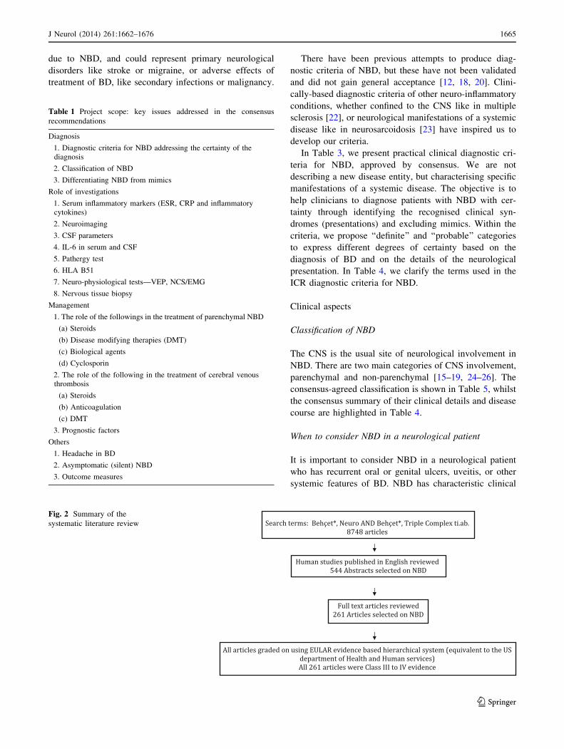
A literature search showed that there were no studies with
level I or II in any of the items researched within the scope of
this consensus. The best available evidence was in the form
of studies within level III or IV (Fig. 2). There are a few
randomized, controlled trials investigating treatment options
in BD [9, 10] and two meta-analyses, but none on NBD [11,
12]. Figure 2 shows the summary of the literature review and
evidence used in the project to draw draft recommendations.
The disease terminology was voted and agreed upon as
’neuro-Behcet’s disease’, as two-thirds of the advisory
group members preferred to use this term to describe the
neurological manifestations of BD (66 % voted for neuro-
Behcet’s disease, 22 % for neuro-Behcet syndrome, and
12 % for either/other).
A diagnostic criteria for NBD and 16 recommendations
covering important diagnostic and management aspects in
NBD were voted through and approved as consensus rec-
ommendations. The supporting voting score summary is
given in Table 2.
The following section covers the consensus recom-
mendation with a brief summary of the relevant literature
review divided into the following subsections:
• Diagnostic criteria for NBD
• Clinical aspects
• Role of investigations in diagnosis of NBD
• Management of NBD
• Miscellaneous
International consensus recommendation (ICR)
diagnostic criteria of NBD
Although BD is a well-defined disease with well-estab-
lished criteria for its diagnosis [13, 14], the same is not true
for NBD. The neurological manifestations of BD have been
characterised in many publications [15–19]. Neurological
symptoms and signs seen in BD patients are not necessarily
Fig. 1 Project methodology
1664 J Neurol (2014) 261:1662–1676
123
due to NBD, and could represent primary neurological
disorders like stroke or migraine, or adverse effects of
treatment of BD, like secondary infections or malignancy.
There have been previous attempts to produce diag-
nostic criteria of NBD, but these have not been validated
and did not gain general acceptance [12, 18, 20]. Clini-
cally-based diagnostic criteria of other neuro-inflammatory
conditions, whether confined to the CNS like in multiple
sclerosis [22], or neurological manifestations of a systemic
disease like in neurosarcoidosis [23] have inspired us to
develop our criteria.
In Table 3, we present practical clinical diagnostic cri-
teria for NBD, approved by consensus. We are not
describing a new disease entity, but characterising specific
manifestations of a systemic disease. The objective is to
help clinicians to diagnose patients with NBD with cer-
tainty through identifying the recognised clinical syn-
dromes (presentations) and excluding mimics. Within the
criteria, we propose ‘‘definite’’ and ‘‘probable’’ categories
to express different degrees of certainty based on the
diagnosis of BD and on the details of the neurological
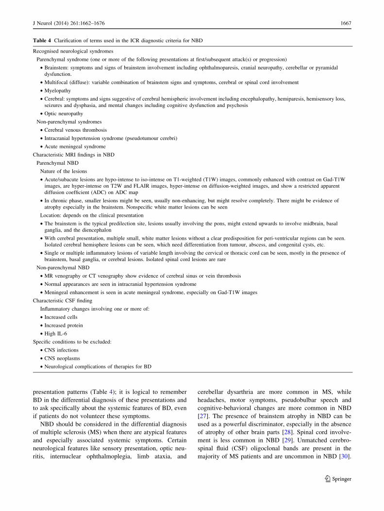
presentation. In Table 4, we clarify the terms used in the
ICR diagnostic criteria for NBD.
Clinical aspects
Classification of NBD
The CNS is the usual site of neurological involvement in
NBD. There are two main categories of CNS involvement,
parenchymal and non-parenchymal [15–19, 24–26]. The
consensus-agreed classification is shown in Table 5, whilst
the consensus summary of their clinical details and disease
course are highlighted in Table 4.
When to consider NBD in a neurological patient
It is important to consider NBD in a neurological patient
who has recurrent oral or genital ulcers, uveitis, or other
systemic features of BD. NBD has characteristic clinical
Fig. 2 Summary of the
systematic literature review
Table 1 Project scope: key issues addressed in the consensus
recommendations
Diagnosis
1. Diagnostic criteria for NBD addressing the certainty of thediagnosis
2. Classification of NBD
3. Differentiating NBD from mimics
Role of investigations
1. Serum inflammatory markers (ESR, CRP and inflammatorycytokines)
2. Neuroimaging
3. CSF parameters
4. IL-6 in serum and CSF
5. Pathergy test
6. HLA B51
7. Neuro-physiological tests—VEP, NCS/EMG
8. Nervous tissue biopsy
Management
1. The role of the followings in the treatment of parenchymal NBD
(a) Steroids
(b) Disease modifying therapies (DMT)
(c) Biological agents
(d) Cyclosporin
2. The role of the following in the treatment of cerebral venousthrombosis
(a) Steroids
(b) Anticoagulation
(c) DMT
3. Prognostic factors
Others
1. Headache in BD
2. Asymptomatic (silent) NBD
3. Outcome measures
J Neurol (2014) 261:1662–1676 1665
123
Table 2 Voting score summary
Recommendation Score range Mode Median
Recommendation: ICR diagnostic criteria for NBD 7–9 8 8
Recommendation 1: classification of NBD: 1a 5–9 9 9
Onset presentation of NBD subtypes: 1b 7–9 Bimodal 8 and 9 8
Course of subtypes: 1c 7–9 9 9
Recommendation 2: when to consider NBD: 2a 7–9 9 9
How to differentiate: 2b 4–9 Bimodal 8 and 9 8
Recommendation 3: role of inflammatory markers 8–9 9 9
Recommendation 4: role of neuroimaging 5–9 9 9
Recommendation 5: role of CSF: 5a 8–9 9 9
Expected findings in CSF: 5b 6–9 9 9
Recommendation 6: role of IL-6 cytokine 5–9 9 8
Recommendation 7: role of pathergy test 6–9 9 8
Recommendation 8: role of HLA-B51 5–9 9 9
Recommendation 9: role of neurophysiology 6–9 9 9
Recommendation 10: role of nervous tissue biopsy 6–9 9 9
Recommendation 11: management of parenchymal NBD: 11a 8–9 9 9
Role of steroids in parenchymal NBD: 11b 6–9 Bimodal 8 and 9 8
Role of disease modification treatment: 11c 4–9 9 9
Type of disease modification treatment: 11d 4–9 9 8
Role of biological agents: 11e 4–9 9 9
Role of cyclosporin: 11f 4–9 9 9
Recommendation 12: Management of CVT: role of steroids: 12a 7–9 9 9
Role of anticoagulation: 12b 7–9 Bimodal 7 and 9 8
Role of disease modification treatment: 12c 7–9 9 9
Recommendation 13: prognostic factors 13a 6–9 9 9
Poor prognostic factors and treatment: 13b 7–9 9 9
Recommendation 14: headache in BD: 14a 5–9 9 8
Headache at the time of flare ups: 14b 7–9 Bimodal 8 and 9 8
When to investigate: 14c 8–9 9 9
Recommendation 15: asymptomatic (Silent) NBD 7–9 9 9
Recommendation 16: outcome measures 16a 7–9 9 8
Validation of outcome measure: 16b 5–9 9 9
Table 3 International consensus recommendation (ICR) criteria for NBD diagnosis
Definite NBD meeting all of the following three criteria
1. Satisfy the ISGa criteria for BD
2. Neurological syndromeb (with objective neurological signs) recognised to be caused by BD and supported by relevant
and characteristicc abnormalities seen on either or both:
a. Neuroimaging
b. CSF
3. No better explanation for the neurological findings
Probable NBD meeting one of the following two criteria in the absence of a better explanation for the neurological findings:
1. Neurological syndrome as in definite NBD, with systemic BD features but not satisfying the ISG criteria
2. A non-characteristic neurological syndrome occurring in the context of ISG criteria-supported BD
a ISG International Study Group Criteria 1990 or any other accepted current or future criteriab The recognised syndromes and the ^characteristic findings on investigations are described in Table 4 and in the text
1666 J Neurol (2014) 261:1662–1676
123
presentation patterns (Table 4); it is logical to remember
BD in the differential diagnosis of these presentations and
to ask specifically about the systemic features of BD, even
if patients do not volunteer these symptoms.
NBD should be considered in the differential diagnosis
of multiple sclerosis (MS) when there are atypical features
and especially associated systemic symptoms. Certain
neurological features like sensory presentation, optic neu-
ritis, internuclear ophthalmoplegia, limb ataxia, and
cerebellar dysarthria are more common in MS, while
headaches, motor symptoms, pseudobulbar speech and
cognitive-behavioral changes are more common in NBD
[27]. The presence of brainstem atrophy in NBD can be
used as a powerful discriminator, especially in the absence
of atrophy of other brain parts [28]. Spinal cord involve-
ment is less common in NBD [29]. Unmatched cerebro-
spinal fluid (CSF) oligoclonal bands are present in the
majority of MS patients and are uncommon in NBD [30].
Table 4 Clarification of terms used in the ICR diagnostic criteria for NBD
Recognised neurological syndromes
Parenchymal syndrome (one or more of the following presentations at first/subsequent attack(s) or progression)
• Brainstem: symptoms and signs of brainstem involvement including ophthalmoparesis, cranial neuropathy, cerebellar or pyramidal
dysfunction.
• Multifocal (diffuse): variable combination of brainstem signs and symptoms, cerebral or spinal cord involvement
• Myelopathy
• Cerebral: symptoms and signs suggestive of cerebral hemispheric involvement including encephalopathy, hemiparesis, hemisensory loss,
seizures and dysphasia, and mental changes including cognitive dysfunction and psychosis
• Optic neuropathy
Non-parenchymal syndromes
• Cerebral venous thrombosis
• Intracranial hypertension syndrome (pseudotumour cerebri)
• Acute meningeal syndrome
Characteristic MRI findings in NBD
Parenchymal NBD
Nature of the lesions
• Acute/subacute lesions are hypo-intense to iso-intense on T1-weighted (T1W) images, commonly enhanced with contrast on Gad-T1W
images, are hyper-intense on T2W and FLAIR images, hyper-intense on diffusion-weighted images, and show a restricted apparent
diffusion coefficient (ADC) on ADC map
• In chronic phase, smaller lesions might be seen, usually non-enhancing, but might resolve completely. There might be evidence of
atrophy especially in the brainstem. Nonspecific white matter lesions can be seen
Location: depends on the clinical presentation
• The brainstem is the typical predilection site, lesions usually involving the pons, might extend upwards to involve midbrain, basal
ganglia, and the diencephalon
• With cerebral presentation, multiple small, white matter lesions without a clear predisposition for peri-ventricular regions can be seen.
Isolated cerebral hemisphere lesions can be seen, which need differentiation from tumour, abscess, and congenital cysts, etc.
• Single or multiple inflammatory lesions of variable length involving the cervical or thoracic cord can be seen, mostly in the presence of
brainstem, basal ganglia, or cerebral lesions. Isolated spinal cord lesions are rare
Non-parenchymal NBD
• MR venography or CT venography show evidence of cerebral sinus or vein thrombosis
• Normal appearances are seen in intracranial hypertension syndrome
• Meningeal enhancement is seen in acute meningeal syndrome, especially on Gad-T1W images
Characteristic CSF finding
Inflammatory changes involving one or more of:
• Increased cells
• Increased protein
• High IL-6
Specific conditions to be excluded:
• CNS infections
• CNS neoplasms
• Neurological complications of therapies for BD
J Neurol (2014) 261:1662–1676 1667
123
CSF shows more cells in parenchymal NBD and neutro-
phils might predominate, while cells are usually scarce in
MS and lymphocytes predominate [31].
Other systemic inflammatory disorders, especially those
that might present with uveo-meningitic syndromes
including sarcoidosis, systemic lupus erythematosus, and
primary Sjogren’s syndrome, are important differentials;
occasionally primary CNS lymphoma can present with
uveal involvement and a diencephalic lesion. The differ-
entiation requires identification of characteristic clinical
patterns in addition to the serological markers and other
paraclinical tests [32]; interestingly, the peripheral nervous
system is more often involved in non-BD inflammatory
diseases.
Acute parenchymal NBD might simulate arterial stroke,
which is uncommon in BD patients and when encountered,
is more often due to atherosclerosis rather than inflamma-
tion (Table 6).
Role of investigations in the diagnosis of NBD
Serum inflammatory markers
Although raised ESR and other serum inflammatory
markers have been found to be associated with disease
activity in BD [10], no definite identifiable pattern has been
recognised to be linked with NBD activity. Few studies
have reported concurrent appearance or worsening of sys-
temic features and non-specific constitutional symptoms at
the neurological presentation, whilst one study reported
only modest elevations in inflammatory markers in less
than a quarter of NBD patients [18, 19, 33].
Neuroimaging
Neuroimaging has a significant role in the diagnosis of NBD;
MRI is the gold-standard neuro-imaging modality. MRI
abnormalities have been well-described in NBD [34–41]. The
consensus characteristics of MRI lesions are listed in Table 4.
MRI is extremely useful in differentiating NBD from its
mimics. The brainstem–thalamic–basal ganglia lesions, in
the proper clinical context can strongly support the diag-
nosis of acute/subacute parenchymal NBD, and on occa-
sions can raise this possibility even when the systemic
features of BD are scarce [28]. Chronic parenchymal NBD
lesions are iso-intense, smaller, and at times difficult to
differentiate from lesions seen in multiple sclerosis.
In general, multiple sclerosis lesions are predominantly
periventricular, with infrequent involvement of the basal
ganglia, internal capsule, and the peripheral part of the
pons, whilst chronic parenchymal NBD lesions are pre-
dominantly subcortical. Brainstem atrophy in association
with subcortical lesions points toward NBD [28]. In neuro-
Lupus, though sub-cortical white matter lesions are seen,
basal ganglia or brainstem involvement is uncommon [31].
CSF
Cerebrospinal fluid constituents are altered in around
70–80 % of patients with parenchymal NBD [14–17]. CSF
Table 6 Recommendations on the diagnosis of NBD
Recommendation 1
(a) There are two main subtypes of NBD: parenchymal, an
inflammatory meningo-encephalitic process, and non-
parenchymal, which occurs secondary to vascular involvement.
These differ by clinical, laboratory, neuro-radiological,
pathological, and prognostic characteristics
(b) Parenchymal NBD usually presents with a sub-acute onset of
brainstem syndrome with or without other features, cerebral
hemispheric or spinal cord syndrome, and features will include
pyramidal weakness, behavioural changes, headaches,
ophthalmoplegia and sphincter changes. Non-parenchymal NBD
commonly presents with headache and visual features secondary
to intracranial hypertension, usually due to cerebral venous
thrombosis. It can also present as an acute stroke related to arterial
thrombosis, dissection, or aneurysm, although this is uncommon
(c) Parenchymal NBD usually follows a relapsing-remitting
pattern or a primary/secondary progressive course. Non-
parenchymal disease can be monophasic, but recurrences may
occur. A mixed parenchymal and non-parenchymal disease
presentation can occur
Recommendation 2
(a) We recommend considering NBD in the differential
diagnosis of multiple sclerosis, stroke affecting the young,
intracranial hypertension, meningo-encephalitis, and myelitis
(b) NBD can be differentiated from its mimics by a combination
of characteristic clinical and paraclinical neurological findings in
addition to the associated systemic features
Table 5 Consensus classification of neuro-Behcet’s disease
Central nervous system
Parenchymal
• Multifocal/diffuse
• Brainstem
• Spinal cord
• Cerebral
• Asymptomatic (silent)
• Optic neuropathy
Non-parenchymal
• Cerebral venous thrombosis: intracranial hypertension
• Intracranial aneurysm
• Cervical extracranial aneurysm/dissection
• Acute meningeal syndrome
Peripheral nervous system (relation to BD uncertain)
• Peripheral neuropathy and mononeuritis multiplex
• Myopathy and myositis
Mixed parenchymal and non-parenchymal disease
1668 J Neurol (2014) 261:1662–1676
123
protein is modestly raised in most cases, and oligoclonal
bands are usually absent [19, 30, 33]. The CSF cell count is
raised in 60–80 % of parenchymal NBD cases (range
0–400 9 10 cells/L) and there could be CSF neutrophilia,
lymphocytosis, or mixed cellularity [15, 16, 19]. CSF
glucose is usually normal in NBD and low levels point
toward CNS infections [15].
Patients with CVT or intracranial hypertension without
CVT (pseudotumour cerebri) have normal CSF constitu-
ents, but usually high CSF opening pressure.
IL-6 cytokine
Serum IL-6 levels have been reported to correlate with BD
disease activity, although this finding has not been con-
sistently reproduced [42, 43].
Raised CSF levels of IL-6 have been seen in patients
with acute parenchymal NBD [43–49]. A smaller rise in
IL-6 levels has also been reported in a proportion of pro-
gressive parenchymal NBD [21, 43]. Raised CSF IL-6
levels are usually associated with raised CSF cell count and
protein, and these three parameters have been associated
with disease activity and outcome over 3 years. Occa-
sionally elevated CSF IL-6 levels were reported in the
presence of normal CSF cells and protein [21, 43].
Japanese studies have shown reductions in CSF IL-6
levels in response to various treatments, but it is difficult to
draw a clear conclusion, as these studies involved small
numbers of patients [21, 44, 46, 47].
Collectively, these data indicate that IL-6 is not a reli-
able biomarker of NBD or BD, and the absence of IL-6
should not be viewed as the absence of disease activity. In
addition, little is known about the normal expected levels
of IL-6 in the CSF.
Pathergy test
The pathergy test is one of the major criteria in BD diag-
nosis [14]. Pathergy reaction is a non-specific hyperreac-
tivity of the skin to trauma, such as a needle prick. A
positive test is defined as a papule or pustule that typically
appears 24–48 h after an intradermal injection of the skin
with a 20-gauge needle. Pathergy positivity is highly sug-
gestive but not pathognomonic of BD.
False positive tests can be seen in pyoderma gangreno-
sum, Sweet syndrome, inflammatory bowel diseases,
familial Mediterranean fever, acute myeloid leukemia, and
interferon alpha treatment [48, 49]. Pathergy test sensitivity
has significant geographical variation. While 60–70 % of
Turkish and Japanese BD patients have a positive test, it is
uncommon in Northern European and North American BD
patients [49, 50].
HLA-B51
BD is associated with the major histocompatibility com-
plex HLA-B5 allele and, more specifically, with HLA-B51
[11].
HLA-B51/B5 prevalence varies across the globe, being
higher in Asian, Middle Eastern, and Southern European
populations, and lower in Northern Europe and North
America [11]. Overall, the HLA B5 genotype is seen in
40–65 % of patients diagnosed with BD, and in 10–20 %
of healthy individuals of ethnically-matched control
populations [11]. The relatively modest sensitivity and
specificity of HLA-B51/B5 imply that HLA class 1
genotyping has only a limited value as diagnostic test. In
addition, the prevalence of HLA-B51/B5 among subjects
with NBD is not dissimilar to that found in patients with
BD without neurological involvement [12]. Therefore,
testing for HLA-B51/B5 does not appear to provide a
substantial aid for the diagnosis of NBD. Conflicting
findings have been reported as to whether or not
HLA-B51/B5 status may predict a more severe BD course
[51, 52].
Neurophysiology tests
Neurophysiological testing may be useful if peripheral
nervous system involvement or optic nerve involvement is
suspected. Although neurophysiology tests can detect
central or peripheral nervous system involvement, MRI
remains the gold standard for CNS involvement. Occa-
sionally, EEG can be useful in the differential diagnosis
from acute viral encephalitis [5]. Visual-evoked potential
(VEP) can detect optic nerve involvement, however, fre-
quent uveal involvement in BD might hamper the useful-
ness of VEPs [53].
A number of studies have reported abnormal findings on
neurophysiological testing in the absence of clinical signs
or symptoms [54–57]. These include asymptomatic
abnormalities on nerve conduction studies, electromyog-
raphy, and motor-, sensory- or brainstem-evoked poten-
tials. The significance of these abnormalities is uncertain,
and caution should be exercised before these are taken as
evidence of central and/or peripheral nervous system
involvement. The diagnosis of NBD should not be made
solely on the basis of these abnormalities.
Nervous tissue biopsy
The pathologic findings of CNS involvement in BD are not
pathognomonic, but are well-described in the literature
[58–60]. The basic pathology in the acute/subacute
parenchymal presentation is a perivasculitis characterised
by perivascular infiltration with lymphocytes, neutrophils,
J Neurol (2014) 261:1662–1676 1669
123
and rarely, eosinophils with or without signs of necrosis.
In later stages, inflammatory infiltration is less prominent,
and axonal loss and gliosis predominate [58–60].
The clinical presentation, neuroimaging, and CSF find-
ings are usually sufficient to secure a diagnosis without the
need for a tissue diagnosis. Tumour-like presentation,
though uncommon, has been reported in the literature [61,
62]. Careful history taking in such patients commonly
reveals the systemic symptoms of BD, and help in the early
diagnosis. Occasionally, a tissue diagnosis is needed after
all other diagnostic avenues have been used (Table 7).
Management of NBD
Treatment of parenchymal NBD
There have been no controlled or comparative trials on the
treatment of any aspect of NBD [63]. Most neurologists
with experience in the management of NBD treat a relapse
or acute presentation with daily 1 g IV methylprednisolone
infusions, followed by a slowly tapering course of oral
steroids in parallel to treatment given for other CNS
neuroinflammatory relapses like neuro-Lupus and neuro-
sarcoidosis [22, 64]. It is important to avoid an abrupt
cessation of therapy to avoid early relapse. The dose and
duration of the initial IV treatment and the subsequent oral
therapy vary between different centres [6, 63].
Retrospective studies have shown that two-thirds of
patients with brainstem lesions or cerebral lesions make
good recovery in response to the courses of steroids, but the
other third have recurrence of relapses or progressive
course [15–18].
The timing of the start of disease modifying therapies
(DMT) is not always straightforward. The rationale is to
help in controlling the inflammatory process, to prevent or
reduce the frequency of further neurological relapses, to
reduce steroid exposure, and possibly to control the other
systemic features of this multisystem disease.
Azathioprine was reported to prevent inflammation of
the second eye after the first ocular episode in BD [9, 10].
Because of its relatively predictable and low side effects
profile, azathioprine is commonly used as a first DMT in
many centres for the serious manifestations of BD,
including NBD. There are other publications reporting
success with alternative DMTs for NBD, including myco-
phenolate mofetil [65], methotrexate [66, 67], chlorambucil
[68], and cyclophosphamide [69].
Infliximab was reported to be effective in treating
refractory ocular and NBD, and in achieving favourable
outcome [70–72], with continued benefit in follow-up
studies over 1- and 4-year periods [73, 74]. Adalimumab
has been reported as an effective alternative to infliximab
[75, 76]. There are case reports supporting the use of eta-
nercept [77], Tocilizumab [78] and interferon alpha [79,
80]. To date, experience with infliximab is considerably
larger compared to other anti-TNF agents [81].
Table 7 Recommendations on the role of investigations in diagnosis
of NBD
Recommendation 3
ESR, CRP, and inflammatory cytokines are non-specific markers
of inflammation; these might be elevated at the neurological
presentation, but are of limited value in the differential diagnosis
of NBD
Recommendation 4
We recommend considering MRI study including contrast and
MRV in suspected NBD. This commonly demonstrates
characteristic features especially in acute/sub-acute parenchymal
involvement and can confirm CVT. The distinct MRI findings
are helpful in the differentiation from the other CNS
inflammatory disorders
Recommendation 5
(a) We recommend CSF examination in suspected NBD, as it has
a supportive role in the diagnosis, in addition to looking for
mimics and especially CNS infections
(b) Parenchymal NBD is usually associated with CSF pleocytosis
(either neutrophilic or lymphocytic, but rarely as florid as seen in
bacterial meningitis), and/or raised protein. Oligoclonal bands are
frequently absent. A completely normal CSF does not exclude
parenchymal NBD. Non-parenchymal NBD is associated with
elevated CSF pressure only. The role of CSF abnormalities in
prognosis and monitoring of the disease needs further research
Recommendation 6
Raised CSF IL-6 is an indicator of ongoing disease activity in
NBD, usually in association with raised CSF constituents. While
we recommend considering CSF IL-6 for disease monitoring,
especially in the absence of other raised inflammatory CSF
constituents, its use in monitoring therapeutic response needs
further research
Recommendation 7
The pathergy test is simple and has a well-established role in BD
diagnosis. We recommend pathergy testing in suspected NBD,
since a positive result, especially with other systemic BD
features, would contribute significantly toward NBD diagnosis.
A negative test, however, will not exclude NBD
Recommendation 8
BD is associated with the HLA-B5 allele and, more specifically,
with HLA-B51. It is not clear if HLA-B51/B5 testing has a role
in the diagnosis or prognosis of BD or NBD
Recommendation 9
Neurophysiologic tests are not routinely recommended for NBD.
These may be useful if peripheral nervous system or optic nerve
involvement is suspected. Asymptomatic neurophysiological
findings are of doubtful clinical significance. The diagnosis of
NBD should be avoided when solely based on asymptomatic
neurophysiological findings
Recommendation 10
Nervous tissue biopsy can occasionally be useful in the diagnosis
of NBD. It is usually not recommended as a part of the
diagnostic process. As it is an invasive procedure, we
recommend considering it when all other diagnostic avenues
have been exhausted, especially for tumour-like presentation
1670 J Neurol (2014) 261:1662–1676
123
Cyclosporin is effective in the treatment of ocular BD,
but has been linked with higher risk of NBD development
[10, 82–85].
Treatment of cerebral venous thrombosis (CVT)
CVT is a characteristic pattern of NBD presentation.
Anticoagulation is the standard treatment of systemic
venous thrombosis and CVT of any aetiology [86]. On
the other hand, the usage of anticoagulants in CVT due
to NBD is controversial and a matter of debate between
experts [87]. There are no high-quality data to support
the contradictory opinions. The rationale for the differ-
ence is that the advocates for avoiding anticoagulants
believe that CVT in BD is due to an inflammatory
process and that the thrombus formed is tightly adherent
to the vessel wall [88], which necessitate the use of anti-
inflammatory agents only. Moreover, the possibility of
bleeding after the rupture of a coexisting aneurysm
anywhere in the body may have detrimental conse-
quences [10, 89]. The supporters of anticoagulants argue
that they will consider the use of anti-inflammatory
medications to combat the presumed inflammatory aeti-
ology, but they prefer to use anticoagulants, after
excluding systemic aneurysms, at least to reduce the risk
of further extension of the clot in the cerebral venous
system. Our consensus group was split almost equally on
both sides of the argument.
The duration of anticoagulant use varies, but is usually
around 3–6 months in uncomplicated cases [90]. The
duration will be probably for life if clear evidence for an
underlying pro-thrombotic status is found.
Prognostic factors
Two major retrospective case series on NBD [16, 17] and
another study with some prospective data [18] have con-
sistently reported brainstem or spinal cord presentation,
frequent relapses, early disease progression, and high CSF
pleocytosis as poor prognostic factors. Disability and
dependent status at initial presentation, a primary or sec-
ondary progressive course, relapse during steroid dose
tapering, fever, meningeal signs, and bladder involvement
showed a possible association with poor outcome, as
defined by poor survival and dependant status [18].
Factors such as gender, presence of other systemic
manifestations of BD, and age at onset did not have any
influence [18].
Although there are no data from RCTs, with the avail-
able evidence from personal experiences, the early use of
DMT might be considered where one or more poor prog-
nostic factors are encountered. Other relevant factors also
need to be considered in this decision (Table 8).
Miscellaneous
Headache
The literature on headache in BD was reviewed in the
previous Lancet Neurology paper [5], which summarised
all relevant, major, published case series.
Headache is the most common neurological symptom in
patients with BD. Most of these headaches are due to pri-
mary headache disorders, commonly migraine and tension-
type headaches [91]. Aykutlu et al. [92] have shown that
Table 8 Recommendations on the management of NBD
Recommendation 11
(a) There is no level I evidence on the treatment options of NBD.
The following recommendations are mainly based on
observational data
(b) For acute/sub-acute parenchymal NBD attack, a course of
corticosteroids is recommended, preferably IV methyl
prednisolone for 3–10 days followed by a maintenance oral
corticosteroid for a few months (up to 6 months)
(c) We recommend considering a disease modifying therapy
(DMT) after a significant parenchymal relapse depending on
severity, response to steroid, previous neurological relapses,
disease course, and other associated systemic BD features
(d) Azathioprine is recommended as a first-line DMT;
alternatives include mycophenolate mofetil, methotrexate, and
cyclophosphamide
(e) We recommend considering a biological agent, including
TNF-alpha-blockers (infliximab, adalimumab, etanercept) or
interferon alpha, when first=line therapies are ineffective or
intolerable and when the disease is relapsing or showing
aggressive neurological or systemic features
(f) We recommend caution in using cyclosporin in BD patients
because of the potential association with neurological
complications. It should be avoided in patients with a history of
NBD and the medication should be stopped when BD patients
develop neurological features suggestive of parenchymal CNS
involvement
Recommendation 12
(a) For CVT in BD, we recommend the use of corticosteroid for
a limited period for the acute/sub-acute presentation
(b) There is no convincing evidence to use or withhold the use of
anticoagulants, which is a standard treatment of CVT of any
aetiology. If anticoagulation is to be started, caution should be
taken to rule-out a systemic aneurysm
(c) We recommend considering a DMT, especially if there is a
previous history of CVT, active systemic disease, or a history of
associated parenchymal NBD
Recommendation 13
(a) Poor prognostic features of NBD include brainstem or
myelopathy presentation, frequent relapses, early disease
progression, and presence of CSF pleocytosis in parenchymal
NBD
(b) We recommend early consideration of a disease modifying
treatment when one or more poor prognostic features are
encountered
J Neurol (2014) 261:1662–1676 1671
123
the characteristics of the primary headaches in BD patients
were not different from those seen in the general popula-
tion presented to headache clinics. Only about 10 % of BD
patients with headaches are due to direct neurological
involvement [5]. These are usually seen in association with
other neurological features. Headaches have been reported
at the time of flare up of systemic BD features in the
absence of CNS involvement [91–93]. Recognition of the
different types of headaches in BD patients might reduce
the unnecessary expenditure and risk of specialist
investigations.
Asymptomatic (silent) NBD
Asymptomatic abnormalities on neurological examination,
neuroimaging, neuropsychology or neurophysiology test-
ing, which are referred to as asymptomatic NBD, have
been reported [5, 26]. The significance of these findings is
not clear. In a retrospective comparative study, four of 22
BD patients with no specific neurological symptoms but
abnormal findings on MRI or neuropsychology testing
developed NBD attacks after a mean follow-up of about
13 years [57]. The outcome was milder compared to a
group of 55 symptomatic NBD patients.
Outcome measures
There is no validated scale for measuring disability in
NBD. The following three potential scales could be con-
sidered. Two of these require a neurologist’s input with
special training.
The modified Rankin scale is internationally accepted
and well-validated for the measurement of disability in
cerebro-vascular diseases [94–97]. This scale measures
overall functional ability and does not focus on individual
functional system/domain like cognition. It is easy to per-
form and can be used without special training. The scores
range from 0 to 6.
Expanded disability status scale (EDSS) is validated and
is the most widely utilized assessment tool in MS [98].
Some studies have used EDSS to measure neurological
disability in NBD, but it has not been validated. The
complexity and the technical skills required for its use
make it difficult to be adopted by non-neurologists in
routine practice.
Neuro-Behcet’s disability score (NBDS) has been pro-
posed for parenchymal-NBD patients to quantify disabili-
ties [99]. This comprises scores for motor and cognitive
status. NBDS is the arithmetic sum of both scores and
ranges from 0 to 8, with 8 being death due to NBD.
Although it appears to be more thorough than the Rankin
scale, in the absence of validation this more complex score
would be difficult to interpret and extrapolate (Table 9).
Comments and conclusions
We present recommendations for the key issues in the
diagnosis and management of NBD, which are intended for
the use of practicing clinicians.
Recommendations are a way to support effective clinical
practice. While uncertainties, especially in dealing with
uncommon conditions, are likely to persist, recommenda-
tions can aid clinicians in determining the best options for a
particular patient.
The strengths of our consensus recommendations
include an extensive literature review and the use of the
best available evidence, wide-scale consultations with
international experts, involvement of a patient group rep-
resentative, emphasis on issues that are of particular
interest to clinical practice, and setting a high level for
accepting a consensus recommendation.
The limitations include lack of high-grade, evidence-
based data on all of the issues covered by this consensus,
and reliance on level III and IV evidences and experts’
opinions to reach the consensus. There are inherent limi-
tations to the consensus process and the Delphi method.
The proposed diagnostic criteria for NBD include two
levels of certainty, but with strict requirements including
objective neurological signs to reduce false positive
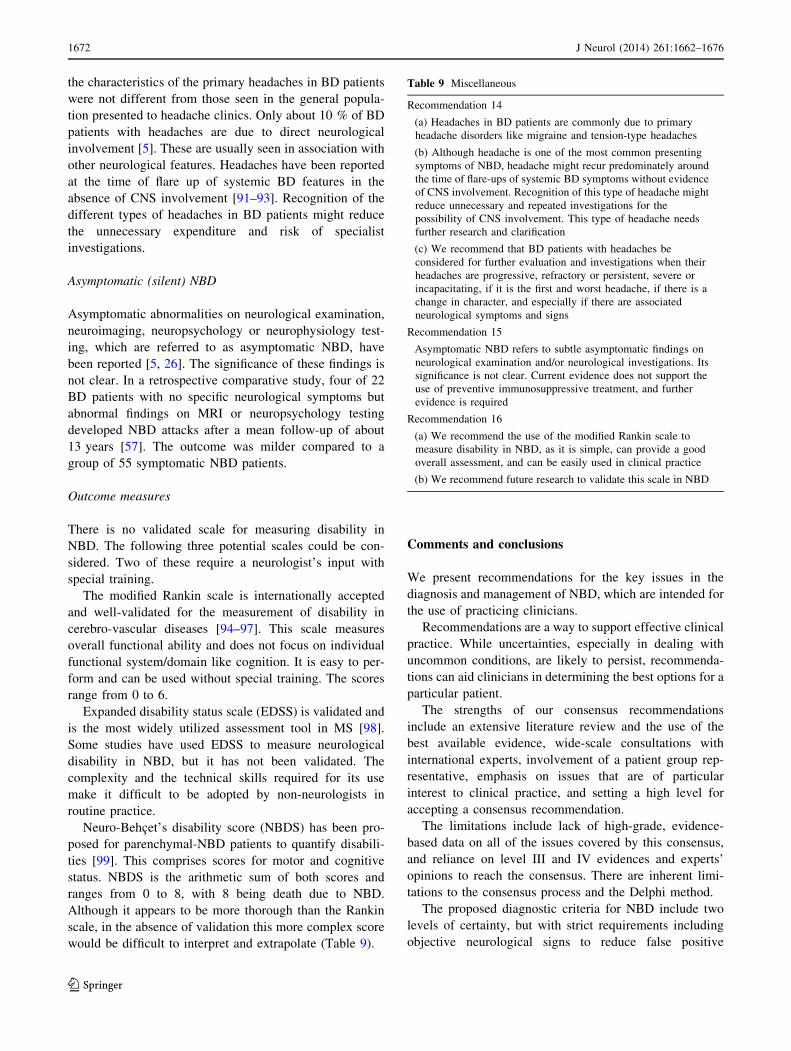
Table 9 Miscellaneous
Recommendation 14
(a) Headaches in BD patients are commonly due to primary
headache disorders like migraine and tension-type headaches
(b) Although headache is one of the most common presenting
symptoms of NBD, headache might recur predominately around
the time of flare-ups of systemic BD symptoms without evidence
of CNS involvement. Recognition of this type of headache might
reduce unnecessary and repeated investigations for the
possibility of CNS involvement. This type of headache needs
further research and clarification
(c) We recommend that BD patients with headaches be
considered for further evaluation and investigations when their
headaches are progressive, refractory or persistent, severe or
incapacitating, if it is the first and worst headache, if there is a
change in character, and especially if there are associated
neurological symptoms and signs
Recommendation 15
Asymptomatic NBD refers to subtle asymptomatic findings on
neurological examination and/or neurological investigations. Its
significance is not clear. Current evidence does not support the
use of preventive immunosuppressive treatment, and further
evidence is required
Recommendation 16
(a) We recommend the use of the modified Rankin scale to
measure disability in NBD, as it is simple, can provide a good
overall assessment, and can be easily used in clinical practice
(b) We recommend future research to validate this scale in NBD
1672 J Neurol (2014) 261:1662–1676
123
diagnosis and improve accuracy. It only uses investiga-
tions, which have well-established supportive roles in the
diagnosis. Its use is facilitated by the clear explanations for
the terms used. We would like to emphasise that it is a
clinical and not pathological criteria. It would require some
neurological expertise to characterise the neurological
syndromes. It might not help in the diagnosis of difficult
and controversial neurological presentations of NBD and
caution needs to be practiced, as its accuracy, sensitivity
and specificity are unknown while validation is needed.
The use of anticoagulants in Behcet’s venous thrombo-
sis and CVT is an important topic that needs priority in
future research. International cooperation is needed to
establish future studies on the best treatment options for
NBD patients. These recommendations need to be updated
in the future, pending further evidence.
Acknowledgments We acknowledge Professor Clive Hawkins, Dr.
Simon Ellis and Dr. Brendan Davies, consultant neurologists at the
University Hospital North Staffordshire, for their feedback on the
diagnostic criteria of NBD.
Conflicts of interest All authors declare that they have no conflicts
of interest.
Open Access This article is distributed under the terms of the
Creative Commons Attribution License which permits any use, dis-
tribution, and reproduction in any medium, provided the original
author(s) and the source are credited.
Advisory group members
In addition to the authors of this paper, the following
subjects were members of the advisory group: Canada—
Prof Simon Carette (Medicine); Egypt—Prof Samir Helmy
Assaad-Khalil (Internal Medicine), Prof Sahar N Saleem
(Neuro-radiology); France—Prof Loic Guillevin (Internal
Medicine), Prof Isabelle Kone-paut (Paediatrics); Ger-
many—Dr. Andreas Altenburg (Dermatology), Dr. Peter
Berlit (Neurology), Dr. Thomas Stache (Neurology);
Greece—Prof Panagiota Boura (Internal Medicine and
Clinical Immunology); Israel—Prof Eldad Ben-Chetrit
(Rheumatology); Italy—Dr. Loredana La Mantia (Neurol-
ogy, Neuropathology), Dr. Ignazio Olivieri (Rheumatol-
ogy); Japan—Prof Shunsei Hirohata (Rheumatology and
Internal Medicine); Jordan—Dr. Wafa Madanat (Rheu-
matology); Libya—Prof Khaled Elmuntaser (Internal
Medicine and Rheumatology); Netherlands—Dr. Jan
VanLaar (Internist, Immunologist and Oncologist);
Qatar—Dr. Thurayya Arayssi (Rheumatology); Republic
of Ireland—Dr. Mary Keogan (Immunology); Spain—Dr.
Norberto Ortego-Centeno (Internal Medicine), Dr. Alex
Olive (Rheumatology), Dr. Sergio Martınez-Yelamos
(Neurology), Dr. Cristina Ramo-Tello (Neurology),
Dr. Roser Solans (Internal Medicine and Autoimmune
Diseases); Switzerland—Dr. Oliver Findling (Neurology),
Prof Marcel Arnold (Neurology); Turkey—Prof Ayse Al-
tintas (Neurology), Dr. Murat Kurtuncu (Neurology), Prof
Salih Pay (Rheumatology and Internal Medicine); UAE—
Dr. Sarmed Al Fahad (Neurology); United Kingdom—Dr.
John Bamford (Neurology), Prof Dorian O Haskard
(Rheumatology), Dr. Desmond Kidd (Neurology), Mrs.
Janet Mather (Patient Representative), Prof Philip I. Mur-
ray (Ophthalmology), Prof Neil Scolding (Neurology), Prof
Miles Stanford (Ophthalmology); United States—Dr.
Kenneth T. Calamia (Rheumatology), Dr. Luis R. Espinoza
(Rheumatology), Dr. Nadera J. Sweiss (Medicine), Dr.
Nagagopal Venna (Neurology).
References
1. Behcet H (1937) Uber residivierende, aphtose durch ein Virus
verursachtes Geschwure am Mund, am Auge und an der Genit-
alien. Derm Wschr 105:1152–1157
2. Hatemi G, Yazici Y, Yazici H (2013) Behcet’s syndrome. Rheum
Dis Clin North Am 39:245–261
3. Verity DH, Marr JE, Ohno S (1999) Behcet’s disease, the Silk
Road and HLA-B51: historical and geographical perspectives.
Tissue Antigens 54:213–220
4. Khairallah M, Accorinti M, Muccioli C, Kahloun R, Kempen JH
et al (2012) Epidemiology of Behcet disease. Ocul Immunol In-
flamm 20:324–335
5. Al-Araji A, Kidd DP (2009) Neuro-Behcet’s disease: epidemi-
ology, clinical characteristics, and management. Lancet Neurol
8:192–204
6. Powell C (2003) The Delphi technique: myths and realities. J Adv
Nurs 41:376–382
7. Strober BE, Clay Cather J, Cohen D et al (2012) A Delphi con-
sensus approach to challenging case scenarios in moderate-to-
severe psoriasis: Part 2. Dermatol Ther (Heidelb) 2:2. Epub 2012
Mar 30
8. Sackett D, Rosenberg W, Muir Gray J, Haynes R, Richardson W
(1996) Evidence based medicine: what it is and what it isn’t. Br
Med J 312:71–77
9. Yazici H, Fresko I, Yurdakul S (2007) Behcet’s syndrome: dis-
ease manifestations, management, and advances in treatment. Nat
Clin Pract Rheumatol 3:148–155
10. Hatemi G, Silman A, Bang D et al (2008) Management of Beh-
cet’s disease: a systematic literature review for the EULAR
evidence based recommendations for the management of Beh-
cet’s disease. Ann Rheum Dis 67:1656–1662
11. Menthon M, LaValley MP, Maldini C, Guillevin L, Mahr A
(2009) HLA-B51/B5 and the risk of Behcet disease: a systematic
review and meta-analysis of case control genetic association
studies. Arthritis Rheum 61:1287–1296
12. Maldini C, Lavalley MP, Cheminant M, de Menthon M, Mahr A
(2012) Relationships of HLA-B51 or B5 genotype with Behcet’s
disease clinical characteristics: systematic review and meta-analy-
ses of observational studies. Rheumatol (Oxford) 51:887–900
13. International Study Group for Behcet’s Disease (1990) Criteria
for diagnosis of Behcet’s disease. Lancet 335:1078–1080
14. International Team for the Revision of the International Criteria
for Behcet’s Disease (ITR-ICBD), Davatchi F, Assaad-Khalil S
J Neurol (2014) 261:1662–1676 1673
123
et al (2013) The international criteria for Behcet’s disease
(ICBD): a collaborative study of 27 countries on the sensitivity
and specificity of the new criteria. J Eur Acad Dermatol Vene-
reol. doi:10.1111/jdv.12107
15. Al-Fahad S, Al-Araji A (1999) Neuro-Behcet’s disease in Iraq: a
study of 40 patients. J Neurol Sci 170:105–111
16. Akman-Demir G, Serdaroglu P, Tasci B (1999) Clinical patterns of
neurological involvement in Behcet’s disease: evaluation of 200
patients. The Neuro-Behcet Study Group. Brain 122:2171–2182
17. Kidd D, Steuer A, Denman AM, Rudge P (1999) Neurological
complications of Behcet’s syndrome. Brain 122:2183–2194
18. Siva A, Kantarci OH, Saip S et al (2001) Behcet’s disease:
diagnostic and prognostic aspects of neurological involvement.
J Neurol 248:95–103
19. Al-Araji A, Sharquie K, Al-Rawi Z (2003) Prevalence and pat-
terns of neurological involvement in Behcet’s disease: a
prospective study from Iraq. J Neurol Neurosurg Psychiatry
74:608–613
20. Hentati F, Fredj M, Gharbi N, Hamida M (1993) Clinical and
biological aspects of neuro-Behcet’s in Tunisia. In: Wechsler B,
Godeau P (eds) Behcet’s disease. Elsevier, Amsterdam,
pp 415–418
21. Hirohata S, Kikuchi H, Sawada T et al (2012) Clinical charac-
teristics of neuro-Behcet’s disease in Japan: a multicenter retro-
spective analysis. Mod Rheumatol 22:405–413
22. McDonald WI, Compston A, Edan G et al (2001) Recommended
diagnostic criteria for multiple sclerosis: guidelines from the
international panel on the diagnosis of multiple sclerosis. Ann
Neurol 50:121–127
23. Zajicek JP, Scolding NJ, Foster O et al (1998) Central ner-
vous system sarcoidosis—diagnosis and management. QJM
92:103–117
24. Bohlega S, AlKawi MZ, Omer S et al (1993) Neuro-Behcet’s:
clinical syndromes and prognosis. In: Wechsler B, Godeau P
(eds) Behcet’s disease. Elsevier, Amsterdam, pp 429–433
25. Borhani-Haghighi A, Pourmand R, Nikseresht AR (2005) Neuro-
Behcet disease. A review. Neurologist 11:80–89
26. Siva A, Saip S (2009) The spectrum of nervous system
involvement in Behcet’s syndrome and its differential diagnosis.
J Neurol 56:513–529
27. Motomura S, Tabira T, Kuriowa Y et al (1998) A clinical com-
parative study of multiple sclerosis and neuro-Behcet syndrome.
J Neurol Neurosurg Psychiatry 43:210–213
28. Coban O, Bahar S, Akman-Demir G et al (1999) Masked
assessment of MRI findings: is it possible to differentiate neuro-
Behcet’s disease from other central nervous system diseases?
Neuroradiology 41:255–260
29. Yesilot N, Mutlu M, Gungor O, Baykal B, Serdaroglu P, Akman-
Demir G (2007) Clinical characteristics and course of spinal cord
involvement in Behcet’s disease. Eur J Neurol 14:729–737
30. Saruhan-Direskeneli G, Yentur SP, Mutlu M et al (2013) Intra-
thecal oligoclonal IgG bands are infrequently found in neuro-
Behcet’s disease. Clin Exp Rheumatol 31(3 Suppl 77):25–27
31. McLean BN, Miller D, Thompson EJ (1995) Oligoclonal banding
of IgG in CSF, blood-brain barrier function, and MRI findings in
patients with sarcoidosis, systemic lupus erythematosus, and
Behcet’s disease involving the nervous system. J Neurol Neuro-
surg Psychiatry 58:548–554
32. Reske D, Petereit HF, Heiss WD (2005) Difficulties in the dif-
ferentiation of chronic inflammatory diseases of the central ner-
vous system–value of cerebrospinal fluid analysis and
immunological abnormalities in the diagnosis. Acta Neurol Scand
112:207–213
33. Joseph FG, Scolding NJ (2007) Neuro- Behcet’s disease in
Caucasians: a study of 22 patients. Euro J Neurol 14:174–180
34. Al Kawi MZ, Bohlega S, Banna M (1997) MRI findings in neuro-
Behcet’s disease. Neurology 41:405–408
35. Wechsler B, Dell’lsola B, Vidailhet M et al (1993) MRI in 31
patients with Behcet’s disease and neurological involvement:
prospective study with clinical correlation. J Neurol Neurosurg
Psychiatry 56:793–798
36. Coban O, Bahar S, Akman-Demir G et al (1996) A controlled
study of reliability and validity of MRI findings in neuro-Behcet’s
disease. Neuroradiology 38:312–316
37. Kocer N, Islak C, Siva A, Saip S, Akman C, Kantarci O, Ham-
uryudan V (1999) CNS involvement in neuro-Behcet syndrome:
an MR study. Am J Neuroradiol 20:1015–1024
38. Lee SH, Yoon PH, Park SJ, Kim DI (2001) MRI findings in
neuro-Behcet’s disease. Clin Radiol 56:485–494
39. Akman-Demir G, Bahar S, Coban O, Tasci B, Serdaroglu P
(2003) Cranial MRI in Behcet’s disease: 134 examinations of 98
patients. Neuroradiology 45:851–859
40. Borhani-Haghighi A, Sarhadi S, Farahangiz S (2011) MRI find-
ings of neuro-Behcet’s disease. Clin Rheumatol 30:765–770
41. Albayram S, Saip S, Hasiloglu ZI et al (2011) Evaluation of
parenchymal neuro-behcet disease by using susceptibility-
weighted imaging. Am J Neuroradiol 32:1050–1055
42. Yamakawa Y, Sugita Y, Nagatani T et al (1996) Interleukin-6(IL-6) in patients with Behcet’s disease. J Dermatol Sci
11:189–195
43. Akman-Demir G, Erdem Tuzun, Sema Icoz et al (2008) Inter-
leukin-6 in neuro-Behcet’s disease: association with disease
subsets and long-term outcome. Cytokine 44:373–376
44. Hirohata S, Isshi K, Oguchi H et al (1997) Cerebrospinal fluid
interleukin-6 in progressive neuro-Behcet’s syndrome. Clin
Immunol Immunopathol 82:12–17
45. Saruhan-Direskeneli G, Yentur SP, Akman-Demir G, Isik N,
Serdaroglu P (2003) Cytokines and chemokines in neuro-Beh-
cet’s disease compared to multiple sclerosis and other neuro-
logical diseases. J Neuroimmunol 145:127–134
46. Fujikawa K, Aratake K, Kawakami A et al (2007) Successful
treatment of refractory neuro-Behcet’s disease with infliximab: a
case report to show its efficacy by magnetic resonance imaging,
transcranial magnetic stimulation and cytokine profile. Ann
Rheumat Dis 66:136–137
47. Kikuchi H, Aramaki K, Hirohata S (2008) Effect of infliximab in
progressive neuro-Behcet’s syndrome. J Neurol Sci 272:99–105
48. Budak-Alpdogan T, Demircay, Alpdogan O et al (1998) Skin
hyperreactivity of Behcet’s patients (pathergy reaction) is also
positive in interferon alpha-treated chronic myeloid leukaemia
patients, indicating similarly altered neutrophil functions in both
disorders. Br J Rheumatol 37:1148–1151
49. Hatemi I, Hatemi G, Celik AF et al (2008) Frequency of pathergy
phenomenon and other features of Behcet’s syndrome among
patients with inflammatory bowel disease. Clin Exp Rheumatol
26(4 Suppl 50):S91–S95
50. Yazici H, Chamberlain MA, Tuzun Y et al (1984) A comparative
study of the pathergy among Turkish and British patients with
Behcet’s disease. Ann Rheumc Dis 43:74–75
51. Gul A, Uyar FA, Inanc M et al (2001) Lack of association of
HLA-B*51 with a severe disease course in Behcet’s disease.
Rheumatology 40:668–672
52. Chang HK, Kim JU, Cheon KS, Chung HR, Lee KW, Lee IH
(2001) HLA-B51 and its allelic types in association with Behcet’s
disease and recurrent aphthous stomatitis in Korea. Clin Exp
Rheumatol 19(5 Suppl 24):S31–S35
53. Turker H, Terzi M, Bayrak O, Cengiz N, Onar M, Us O (2008)
Visual evoked potentials in differential diagnosis of multiple
sclerosis and neurobehcet’s disease. Tohoku J Exp Med
216:109–116
1674 J Neurol (2014) 261:1662–1676
123
54. Kececi H (2005) Behcet’s disease without neurological mani-
festations. Tohoku J Exp Med 207:1
55. Ozisik HI, Karlidag R, Hazneci E, Kizkin S, Ozcan C (2005)
Cognitive event-related potential and neuropsychological find-
ings in Behcet’s disease without neurological manifestations.
Tohoku J Exp Med 206:15–22
56. Anlar O, Akdeniz N, Tombul T, Calka O, Bilgili SG (2006)
Visual evoked potential findings in Behcet’s disease without
neurological manifestations. Int J Neurosci 116:281–287
57. Yesilot N, Shehu M, Oktem-Tanor O, Serdaroglu P, Akman-
Demir G (2006) Silent neurological involvement in Behcet’s
disease. Clin Exp Rheumatol 24(5 Suppl 42):S65–S70
58. Inaba G (1981) Clinical features of Neuro-Behcet’s syndrome. In:
Inaba G (ed) Behcet’s Disease. University of Tokyo Press,
Tokyo, pp 235–246
59. Hayashi H, Fukuda Y, Kuwabara N (1981) Pathological studies
on Neuro-Behcet’s disease. In: Inaba G (ed) Behcet’s disease.
University of Tokyo Press, Tokyo, pp 197–211
60. Hirohata S (2008) Histopathology of central nervous system
lesions in Behcet’s disease. J Neurol Sci 267:41–47
61. Matsuo K, Yamada K, Nakajima K, Nakagawa M (2005) Neuro-
Behcet disease mimicking brain tumor. Am J Neuroradiol
26:650–653
62. Appenzeller S, de Castro R, Queiroz L de S et al (2006) Brain
tumor-like lesion in Behcet disease. Rheumatol Int 26:577–580
63. Akman-Demir G, Saip S, Siva A (2011) Behcet’s Disease. Curr
Treat Options Neurol 13:290–310
64. Bertsias GK, Ioannidis JP, Aringer M, Bollen E, Bombardieri S,
Bruce IN (2010) EULAR recommendations for the management
of systemic lupus erythematosus with neuropsychiatric manifes-
tations: report of a task force of the EULAR standing committee
for clinical affairs. Ann Rheum Dis 69:2074–2082
65. Shugaiv E, Tuzun E, Mutlu M, Kiyat-Atamer A, Kurtuncu M,
Akman-Demir G (2011) Mycophenolate mofetil as a novel
immunosuppressant in the treatment of neuro-Behcet’s disease
with parenchymal involvement: presentation of four cases. Clin
Exp Rheumatol 29(4 Suppl 67):S64–S67
66. Hirohata S, Suda H, Hashimoto T (1998) Low-dose weekly
methotrexate for progressive neuropsychiatric manifestations in
Behcet’s disease. J Neurol Sci 159:181–185
67. Kikuchi H, Aramaki K, Hirohata S (2003) Low dose MTX for
progressive neuro-Behcet’s disease. A follow-up study for
4 years. Adv Exp Med Biol 528:575–578
68. O’Duffy JD, Robertson DM, Goldstein NP (1984) Chlorambucil
in the treatment of uveitis and meningoencephalitis of Behcet’s
disease. Am J Med 76:75–84
69. Ait Ben Haddou EH, Imounan F, Regragui W et al (2012)
Neurological manifestations of Behcet’s disease: evaluation of
40 patients treated by cyclophosphamide. Rev Neurol (Paris)
168:344–349
70. Sarwar H, McGrath H Jr, Espinoza LR (2005) Successful treat-
ment of long-standing neuro-Behcet’s disease with infliximab.
J Rheumatol 32:181–183
71. Pipitone N, Olivieri I, Padula A et al (2008) Infliximab for the
treatment of Neuro-Behcet’s disease: a case series and review of
the literature. Arthritis Rheum 59:285–290
72. Al-Araji A, Siva A, Saip S et al (2010) Treatment of Neuro-
Behcet’s disease with infliximab. An international multi-centre
case-series of 18 patients. Clin Exp Rheumatol 28 (Suppl 60):
S119
73. Fasano A, D’Agostino M, Caldarola G, Feliciani C, De Simone C
(2011) Infliximab monotherapy in neuro-Behcet’s disease: four
year follow-up in a long-standing case resistant to conventional
therapies. J Neuroimmunol 239:105–107
74. Giardina A, Ferrante A, Ciccia F, Vadala M, Giardina E, Triolo G
(2011) One year study of efficacy and safety of infliximab in the
treatment of patients with ocular and neurological Behcet’s dis-
ease refractory to standard immunosuppressive drugs. Rheumatol
Int 31:33–37
75. Leccese P, D’Angelo S, Angela P, Coniglio G, Olivieri I (2010)
Switching to adalimumab is effective in a case of neuro-Behcet’s
disease refractory to infliximab. Clin Exp Rheumatol 28(4 Suppl
60):S102
76. Olivieri I, Leccese P, D’Angelo S et al (2011) Efficacy of ada-
limumab in patients with Behcet’s disease unsuccessfully treated
with infliximab. Clin Exp Rheumatol 29(4 Suppl 67):S54–S57
77. Alty JE, Monaghan TM, Bamford JM (2007) A patient with
neuro-Behcet’s disease is successfully treated with etanercept:
further evidence for the value of TNF alpha blockade. Clin
Neurol Neurosurg 109:279–281
78. Shapiro LS, Farrell J, Borhani-Haghighi A (2012) Tocilizumab
treatment for neuro-Behcet’s disease, the first report. Clin Neurol
Neurosurg 114:297–298
79. Nichols JC, Ince A, Akduman L, Mann ES (2001) Interferon-
a2a treatment of neuro-Behcet disease. J Neuroophthalmol
21:109–111
80. Monastirli A, Chroni E, Georgiou S et al (2010) Interferon-atreatment for acute myelitis and intestinal involvement in severe
Behcet’s disease. QJM 103:787–790
81. Arida A, Fragiadaki K, Giavri E, Sfikakis PP (2011) Anti-TNF
agents for Behcet’s disease: analysis of published data on 369
patients. Semin Arthritis Rheum 41:61–70
82. Kotake S, Higashi K, Yoshikawa K, Sasamoto Y, Okamoto T,
Matsuda H (1999) Central nervous system symptoms in patients
with Behcet’s Behcet disease receiving ciclosporin therapy.
Ophthalmology 106:586–589
83. Kato Y, Numaga J, Kato S et al (2001) Central nervous system
in a population of Behcet’s disease patients with refractory
uveitis treated with cyclosporine A. Clin Exp Ophthalmol
29:335–336
84. Kotter I, Gunaydin I, Batra M et al (2006) CNS involvement
occurs more frequently in patients with Behcet’s disease under
cyclosporin A than under other medications–results of a retro-
spective analysis of 117 cases. Clin Rheumatol 25:482–486
85. Akman-Demir G, Ayranci O, Kurtuncu M, Vanli EN, Mutlu M,
Tugal-Tutkun I (2008) Ciclosporin for Behcet’s uveitis: is it
associated with an increased risk of neurological involvement?
Clin Exp Rheumatol 26:S84–S90
86. Bousser MG, Ferro JM (2007) Cerebral venous thrombosis: an
update. Lancet Neurol 6:162–170
87. Tayer-Shifman OE, Seyahi E, Nowatzky J, Ben-Chetrit E (2012)
Major vessel thrombosis in Behcet’s disease: the dilemma of
anticoagulant therapy—the approach of rheumatologists from
different countries. Clin Exp Rheumatol 30:735–740
88. Seyahi E, Yurdakul S (2011) Behcet’s syndrome and thrombosis.
Mediterr J Hematol Infect Dis 3(1):e2011026. doi:10.4084/
MJHID.2011.026
89. Calamia KT, Schirmer M, Melikoglu M (2011) Major vessel
involvement in Behcet’s disease: an update. Curr Opin Rheu-
matol 23:24–31
90. Aguiar de Sousa D, Mestre T, Ferro JM (2011) Cerebral venous
thrombosis in Behcet’s disease: a systematic review. J Neurol
258:719–727
91. Borhani-Haghighi A, Aflaki E, Ketabchi L (2008) The prevalence
and characteristics of different types of headache in patients with
Behcet’s disease, a case-control study. Headache 48:424–429
92. Aykutlu E, Baykan B, Akman-Demir G, Topcular B, Ertas M
(2006) Headache in Behcet’s disease. Cephalalgia 26:180–186
93. Saip S, Siva A, Altintas A et al (2005) Headache in Behcet’s
syndrome headache 45:911–919
94. Rankin J (1957) Cerebral vascular accidents in patients over the
age of 60. Scott Med J 2:200–215
J Neurol (2014) 261:1662–1676 1675
123
95. Bonita R, Beaglehole R (1988) Modification of Rankin scale:
recovery of motor function after stroke. Stroke 19:1497–1500
96. Wilson JL, Hareendran A, Grant M et al (2002) Improving the
assessment of outcomes in stroke: use of a structured interview to
assign grades on the modified Rankin scale. Stroke 33:2243–2246
97. Wilson JL, Hareendran A, Hendry A et al (2005) Reliability of
the modified Rankin scale across multiple raters: benefits of a
structured interview. Stroke 36:777–781
98. Kurtzke JF (1983) Rating neurologic impairment in multiple
sclerosis: an expanded disability status scale (EDSS). Neurology
33:1444–1452
99. Kurtuncu M, Tuzun E, Mutlu M, Pehlivan M, Serdaroglu P,
Akman-Demir G (2008) Clinical patterns and course of neuro-
Behcet’s disease: analysis of 354 patients comparing cases pre-
sented before and after 1990. Clin Exp Rheumatol 26(Suppl.
50):S1–S47
1676 J Neurol (2014) 261:1662–1676
123