Embed Size (px)
Citation preview

Diagnosis and Management of Common Perinatal Urologic
ProblemsDr. Jonathan VB Riddell MD FRCP ABU
(Dipl.)Assistant Professor – Pediatric Urology
CME Disclosure
• I have no conflicts of interest or relevant financial relationships with any commercial entities.
CHU Ste. Justine – Montreal QuebecHopital Mere‐Enfant
Brief Overview
• Fetal urologic abnormalities• Evolution into perinatal period• Implications for provider• Unrecognized hypospadias at time of circumcision

Impact of Technology (US)‐Kidneys, bladder and amniotic fluid can be visualized as early as 12 weeks‐Renal dysplasia, duplication, hydronephrosis and hydroureter can be detected‐Bladder cycling, megacystis and trabeculation‐Urologic anomalies can be isolated or exist with syndromes‐Definitive diagnosis usually still made neonatally
Prenatal Urinary Tract Dilation
• Occurs in 1‐2% of pregnancies• 80,000 children per year in USA• Reflects spectrum of etiologies from physiologic (majority) to others with significant morbidity even mortality
Controversies in Prenatal Urology
• Evidence co‐relating severity of prenatal UT dilation with postnatal pathology lacking
• No uniformity on how to define, classify and grade UT dilation both pre‐ and postnatal
• UT dilation is dynamic; hydration, bladder volume and pt position all play a role
• Uropathies represent a spectrum

SFU System


The good

Isolated UT dilation – Implications for Provider
• Arrange ultrasound after 48h• If unilateral pathology or bilateral low risk one offers antibiotic prophylaxis and VCUG
• Arrange non‐urgent referral to pediatric urologist/nephrologist (if not involved) within 1 month
Top 3 for unilateral pathology
• Uretero‐pelvic junction obstruction• Vesico‐ureteral reflux• Megaureter
• Children with prenatal UT dilation have rate 12‐38% VUR
• If dilation persists postnatally, rate >40%• Poor correlation between severity of dilation and grade of reflux
Do low risk patients need VCUG?
• Guidelines: discretion of clinician• Circumcision is more effective than CAP• CAP most effective in dilating reflux• Is family willing to give CAP?• Is family reliable for surveillance?• Education for urine cultures

Unilateral High Risk
• Often UPJ syndrome or primary nonrefluxingmegaureter
• VCUG helpful to R/O reflux – sets FU to monitor function as opposed to UTI
• Over 2/3 of former SFU 3/4 managed conservatively improve or remain stable over months to years
Operative indications in <6 months of age
• Renal insufficiency• Recurrent febrile UTIs or sepsis• Bilateral severe disease with thinning and renal scan evidence of obstruction or progression

The bad… Exceptions to guidelines
• the grading system is designed to be used in cases of isolated UT dilation and not to be applied to unique situations or anomalous kidneys such as solitary, ectopic, multicysticdysplastic kidneys (MCDK) or other cystic diseases of the kidney.
• * certain situations (eg. Posterior urethral valves, bilateral severe hydronephrosis) may require more expedient follow up
• In these cases preservation of renal function becomes paramount
Implications to provider
• Earlier urologic involvement (preferably prenatally)
• Ultrasound can be done <48h• Consider serial serum Cr• Consider antimicrobial prophylaxis
The ugly…
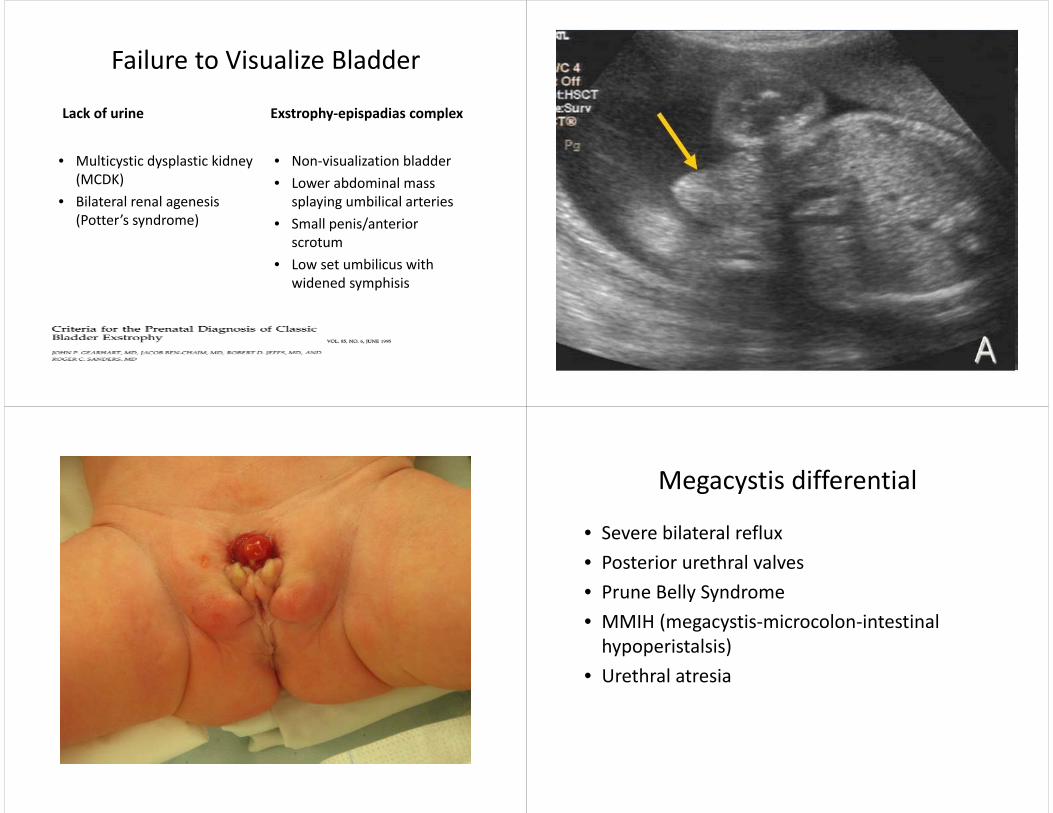
Failure to Visualize Bladder
Lack of urine
• Multicystic dysplastic kidney (MCDK)
• Bilateral renal agenesis (Potter’s syndrome)
Exstrophy‐epispadias complex
• Non‐visualization bladder • Lower abdominal mass
splaying umbilical arteries• Small penis/anterior
scrotum• Low set umbilicus with
widened symphisis
Exstrophy picture/US Megacystis differential
• Severe bilateral reflux• Posterior urethral valves• Prune Belly Syndrome• MMIH (megacystis‐microcolon‐intestinalhypoperistalsis)
• Urethral atresia

Approach to Megacystis
• Sex of infant• Renal parenchyma – degree of dysplasia• Degree of hydronephrosis/obstruction• Serial evaluation of amniotic fluid• Presence of other fetal anomalies
Fetal Ultrasound Criteria for MegacystisDifferential
Severe VUR Prune Belly PosteriorUrethral Valves
MMIH Urethral Atresia
Keyhole sign ‐ ‐ + ‐ ‐
Bladder wall Thin Thin Trabeculatedwith dilated prox. urethra
Thin Symmetricaland cystic
AF Normal Oligo Normal ‐> oligo
Normal ‐> polyhydramnios
Severe oligo ‐> anhydramnios
Other US findings
Ureteraltortuosity
Pulmonary hypoplasia
Renal dysplasia/VURD
Colon not visible; proximaldilation
Small chest/hugeabdomen
Patho‐physiology
Yo‐yo voiding
Abnormalsmooth muscle
Sail‐like valves at UG membrane
Decrease in ganglion cells
Survive if fistula develops
In Utero Intervention for Posterior Urethral Valves
• Holmes et. al (UCSF 2001) reviewed 36 cases of fetal intervention (open surgery) for suspected valves
• 43% mortality rate• Long‐term outcomes did not improve for postnatally confirmed PUV pts
• And only 14/36 actually had PUV!
Vesico‐Aminotic Shunts
• PLUTO trial (2013) randomized 31 singleton pregnancies with LUTO to shunt vs. conservative management
• Only 20% enrolment reached; 2/3 pts had PUV and 1/3 urethral atresia
• Eight from VAS group survived to 28 days vs. four in conservative group (p=0.27 ITT, p=0.03 based on treatment received)

Shunt complications
• SROM after shunt insertion (3/15)• Dislodged (3/15)• Blocked (1/15)• Other series observe bowel injury, errant placement, entero‐colitis,chorio‐amnionitis, urinary ascites
• Many infants delivered prematurely with respiratory complications
Ultrasound Obstet Gynecol 2015; 45: 452–458Fetal intervention for severe lower urinary tract obstruction: a multicenter case–control study
comparing fetal cystoscopy with vesicoamniotic shunting R. RUANO*†‡, N. SANANES§¶, H. SANGI‐HAGHPEYKAR†‡, S. HERNANDEZ‐RUANO**,
R. MOOG††, F. BECMEUR††, A. ZALOSZYC‡‡, A. M. GIRON§§, B. MORIN§ and R. FAVRE*
• 111 fetuses with severe LUTO; fetal cystoscopy (34) vesicoamniotic shunting (16) and no fetal intervention in 61.
• Conclusion Fetal cystoscopy and vesicoamniotic shunt‐ ingimprove the 6‐month survival rate in cases of severe LUTO. However, only fetal cystoscopy may prevent impairment of renal function in fetuses with posterior urethral valves.
Take home message
• Multidisciplinary consensus going forward relies heavily on AP diameter of renal pelvis to stratify risk
• Low risk patients can be managed without VCUG, antibiotics or invasive studies
• Intermediate and high risk cases benefit from a multidisciplinary approach
• A huge academic opportunity awaits…

Discovery of MMIP
• Mega‐meatus intact prepuce• Pass 5Fr catheter to protect urethra• If too close/thin apply vaseline gauze • Stitch only to control bleeding• Stitching prepuce can lead to pathologic phimosis