Embed Size (px)
Citation preview
In Practice ● AP R I L 2005208
PREVALENCE AND MANIFESTATION
Idiopathic (primary) epilepsy is less common in catsthan in dogs (see table on the right). Idiopathic epilepsiesin dogs are generally genetic, but there is little evidenceof this in the cat. It is probable that a considerable proportion of the idiopathic feline group actually haveacquired epilepsy, but no obvious structural lesions areseen on magnetic resonance imaging (MRI).
Tonic-clonic generalised seizures are the most com-mon type of seizure seen in dogs, but are less oftenobserved in the cat. Complex partial seizures, duringwhich animals may remain in sternal recumbency or can be very active (eg, running and climbing), are typicalin cats. Urination and defecation may be seen. Someseizures last only a few seconds (eg, face twitching or ‘blank’ staring). Generally, such cats will have multi-ple episodes a day. In theauthor’s experience, manyepileptic cats have clusterseizures as their firstseizure event. While this isa poor prognostic sign indogs, this does not appearto be the case in cats andgood seizure control maybe achieved easily.
APPROACH TODIAGNOSIS
The causes of seizures aretraditionally divided intointracranial and extracranial(see box on the right). Intra-
CO
MP
AN
ION
A
NIM
AL
P
RA
CT
ICE
SEIZURES are the one of the most common presentations in the neurological feline patient and can be a daunting prospect for the veterinary clinician. The list of possible differential diagnoses is huge anddemands a careful and systematic diagnostic approach. This article steers the practitioner through thework-up and provides guidance on the provision and monitoring of antiepileptic drug therapy in cats.
Diagnosis and control
of epilepsy in the cat CLARE RUSBRIDGE
Clare Rusbridgegraduated fromGlasgow in 1991 andcompleted a smallanimal internship at the University ofPennsylvania, USA.After a year in smallanimal practice inCambridgeshire, she joined the RoyalVeterinary College(RVC) to undertake a BSAVA/Petsaversresidency inneurology andsubsequently spent a year at the RVC as a staff clinician inneurology. Shecurrently runs a smallanimal neurologyreferral service at theStone Lion VeterinaryCentre in London. Sheis an RCVS specialistin veterinaryneurology and isboard-certified by theEuropean College ofVeterinary Neurology.
Two-year-old male domestic shorthairedcat with idiopathic epilepsy, whichpresented in status epilepticus. After aloading dose of phenobarbital (15 mg/kgintramuscularly divided over a 30-minuteperiod), the patient was sedated withpropofol which had been mixed withsodium chloride and administered via aninfusion pump; 1·5 ml/hour propofol over24 hours was required to control theseizures. The infusion was then graduallywithdrawn over the following 48 hours
cranial causes may be further subdivided into idiopathic(ie, primary/genetic) and acquired (ie, secondary/sympto-matic/cryptogenic). For prognostic purposes, it is useful todivide acquired epilepsy into static and progressive braindisease.
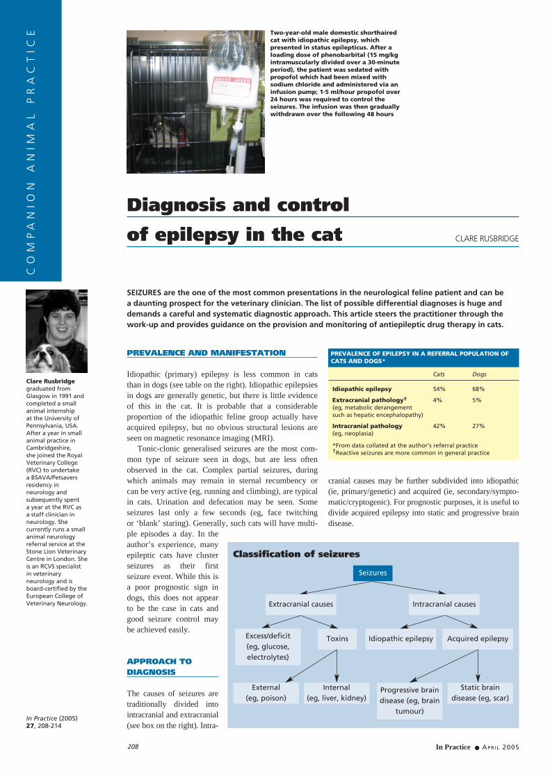
Cats Dogs
Idiopathic epilepsy 54% 68%
Extracranial pathology† 4% 5%(eg, metabolic derangement such as hepatic encephalopathy)
Intracranial pathology 42% 27%(eg, neoplasia)
*From data collated at the author’s referral practice†Reactive seizures are more common in general practice
PREVALENCE OF EPILEPSY IN A REFERRAL POPULATION OFCATS AND DOGS*
In Practice (2005)27, 208-214
Classification of seizures
Excess/deficit(eg, glucose, electrolytes)
Acquired epilepsyIdiopathic epilepsyToxins
External (eg, poison)
Internal (eg, liver, kidney)
Progressive braindisease (eg, brain
tumour)
Static brain disease (eg, scar)
Extracranial causes Intracranial causes
Seizures

In Practice ● AP R I L 2005 209
Easily confused non-epileptic conditions
Myoclonus Myoclonus is an occasional diagnosis in cats and is characterised by a brief, repetitive contraction of a muscle/muscle group. Occasionally, myoclonus can be induced by a repeatable stimulus (eg, sudden movement in the visual field) or may be seen at certain times (most commonly as the animal falls asleep).
Mutilation Syndromes leading to mutilation are rarely the result of seizures although someof these conditions may respond to anticonvulsant drugs. A common example isa (presumed) orofacial pain syndrome, which causes the cat to paw at, and muti-late, its mouth/tongue. This is especially seen in Burmese cats.
Syncope Syncope is very rare in cats.
Type Conditions
D Degenerative Lysosomal storage disease
A Anomalous Hydrocephalus, lissencephaly
M Metabolic Hepatic encephalopathy, hypoglycaemia, hypocalcaemia, uraemia, hypertriglyceridaemia, hypernatraemia, polycythaemia vera (primary erythrocytosis), post-metabolic acquired epilepsy, hypoxia, hyperosmolarity, mitochondrial encephalomyelopathy
N Neoplastic Primary and secondary brain tumoursNutritional Thiamine (vitamin B1) deficiency (raw or overcooked fish diet, anorexia)
I Inflammatory Granulomatous meningoencephalomyelitis, eosinophilic meningoencephalomyelitis, feline polioencephalomyelitis
Infectious Feline infectious peritonitis, toxoplasmosis, feline immunodeficiency virus infection, Cryptococcus infection, post-infection acquired epilepsy, abscess
Idiopathic Epilepsy where all diagnostic tests such as haematology, cerebrospinal fluid analysis, viral titres and magnetic resonance imaging are normal
Iatrogenic Post-surgical acquired epilepsy
T Traumatic Trauma to the cerebrum or diencephalon, post-traumatic epilepsyToxic Lead, organoarsenicals, organomercurials, organophosphates, chlorinated
hydrocarbons, bromethalin, pyrethrins, metaldehyde, strychnine, metronidazole
V Vascular Cerebral ischaemic necrosis (feline ischaemic encephalopathy)
DIFFERENTIAL DIAGNOSIS OF SEIZURES IN CATS
The list of differential diagnoses for seizures is hugeand, as with most aspects of neurology, it is easiest to sub-categorise them using the DAMNIT-V system (see table,above right). This list can be daunting and, hence, thework-up of an epileptic patient requires a systematicapproach to narrow down the likely cause of the seizures.
HISTORY Good history taking is of paramount importance, as seizurescan be easily confused with other causes of collapse ormovement disorders (see box on the right). It is very helpfulto have a video recording of the suspected seizure.
Histopathology section from the medulla of a three-year-old female cat which presented with seizures. The cat had a progressive gait abnormality characteristic of cerebellardisease and was deaf. The nerve cells (arrows) are full of a storage material which was autofluorescent underultraviolet light. This is characteristic of ceroid lipofuscinosis.Haematoxylin and eosin stain. Magnification x 400. Picture, Dr Caroline Hahn, Neuromuscular Laboratory, University of Edinburgh
Nine-month-old cat with hepatic encephalopathy, showingstunting and copper-coloured irises, which are typical of cats with congenital portosystemic shunts. Serumbiochemistry revealed low urea and low-normal albuminlevels and raised resting bile acid values
Transverse T2-weighted MRI scan from an 11-month-old cat which presented with a three-week history of seizures. Theimage shows high signal intensity, which is characteristic of oedema, within the caudate lobes (arrow). There is also a high signal area within the periaqueductal grey matter.This appearance on MRI is typical of a mitochondrialencephalopathy (ie, a mitochondrial enzyme defect).Urinalysis revealed a citric aciduria. The cat was givensupplements containing the mitochondrial cofactors, L-carnitine and coenzyme Q, together with thiamine andriboflavin. The seizures ceased and the animal’s demeanourimproved markedly – it became more active, and started to play and go outside

In Practice ● AP R I L 2005210
The timing and nature of a seizure may provide cluesabout its aetiology. For example, partial seizures suggesta focal lesion. It can be difficult to distinguish partial sec-ondary generalised seizures from primary generalisedones – in such cases, ask the owner if the seizure starts in one body part or is asymmetrical (eg, only in one sideof the face). Establish if the animal is normal betweenseizures. Abnormal behaviour in the interictal period,such as lethargy, aimless wandering, and inappropriateurination and defecation, implies an intracranial patholo-gy. Finally, explore the possibility of a past medical his-tory (eg, head trauma) or a previous diagnosis of systemicillness such as toxoplasmosis or diabetes.
NEUROLOGICAL EXAMINATIONA neurological examination is invaluable, yet its impor-tance is often underestimated. The aim is to answer threemain questions, discussed below.
Is the animal normal?Cats with idiopathic epilepsy will have normal results onneurological examination (except in the postictal period),while cats with progressive brain disease generally pro-vide abnormal findings. The clinician should be aware ofrostral lesions (eg, in the frontal lobe), as postural reac-tions are often normal. Disease of the frontal lobe ischaracterised by behavioural changes (eg, lethargy, aim-less wandering and loss of normal greeting behaviour).In the case of progressive disease (eg, a slow growingneoplasm), motor and sensory deficits will develop withtime, so it is important to repeat the neurological exami-nation after a few weeks, especially if other diagnostictests such as MRI are not available.
If there are deficits, can these be related to disease of the forebrain?In the absence of metabolic disease, seizures indicatedisease of the cerebrum or diencephalon and any of thefollowing deficits would be suggestive of an intracranialpathology:■ Behavioural changes;■ Depression/stupor/coma;■ Circling (towards the side of the lesion);■ Postural deficits (contralateral to the lesion);■ Visual deficits (contralateral to the lesion, normalpupillary light responses).
The side and location of a pathology can be estab-lished. Asymmetrical forebrain disease is most likely tohave a neoplastic aetiology.
Is there multifocal disease?The neurological examination should be used to deter-mine whether there are any deficits relating to pathologiesof more than one area of the nervous system. For exam-ple, vestibular signs suggesting brainstem disease implyeither an inflammatory process (eg, feline infectious peri-tonitis [FIP]) or a multifocal tumour (eg, lymphoma).
CLINICAL EXAMINATIONA general clinical examination is important. For instance,central nervous system infections are rarely confined tothis area and there will be systemic signs such as retinalchanges in cases of FIP and toxoplasmosis. Toxoplasmo-sis is also associated with myositis, pneumonia andhepatic disease.
RULE OUT EXTRACRANIAL DISEASE Full haematological and biochemical analysis should becarried out to rule out metabolic causes of seizures, themost common of which are listed in the table on page209. In addition, dynamic bile acid testing (ie, before andtwo hours after feeding) is appropriate to rule out hepaticencephalopathy.
Transverse T1-weighted gadolinium-enhanced MRI scan of the brain showing a meningioma in a 14-year-old maledomestic shorthaired cat which presented with seizures. Thetumour is seen as an enhanced (white) area with contrast.Note the thickened bone (black) adjacent to the tumour(hyperostosis) and the spread of the tumour along the meninges (arrow)
(above) Transverse T1-weighted gadolinium-enhanced MRIscan of the brain of a one-year-old domestic shorthaired cat with feline infectious peritonitis. There is mild ventriculardilation, which is occurring secondarily to obstruction of cerebrospinal fluid pathways. There is meningealenhancement suggesting an inflammatory or neoplasticinfiltrate (arrow). Cerebrospinal fluid analysis revealed aneutrophilic pleocytosis and a high anti-FCoV antibody titre.(below) Histopathological sample showing the cerebralcortex. There is massive inflammatory cell influx (basophiliccells, arrow) in the meninges, which explains why the areawas enhanced on MRI. Haematoxylin and eosin. Magnification x 20

In Practice ● AP R I L 2005 211
■ A serum immunoglobulin M (IgM) titre for Toxo-plasma species implies recent exposure to the parasiteand is more useful than a single IgG antibody titre; ■ An antibody titre for Cryptococcus species is useless,as this fungus stimulates little immune response. Themost appropriate test for this type of infection is a cryp-tococcal antigen (CRAG) titre.
The clinical history, neurological findings, facilitiesavailable and the owner’s wishes/financial circumstanceswill all determine whether any or some of these tests arecarried out.
Advanced diagnostic imagingFollowing a neurological examination, MRI or computedtomography (CT) are particularly helpful for evaluatingthe epileptic patient. Both techniques allow the structureof the brain to be assessed, with the resulting infor-mation presented as a series of ‘slices’. Disease process-es can be identified by alterations in the symmetry of the brain, differences in intensity and their ability to be enhanced by contrast media. MRI has some advantages over CT in that it is multiplanar, providessuperior soft tissue contrast and involves no ionisingradiation.
Biochemical analysis is also useful for providing evi-dence of multisystemic disease. For example, it is veryunusual for toxoplasmosis not to result in myositis (ele-vated creatine kinase) or hepatitis (elevated liver enzymesor bile acids). In addition, baseline parameters can beestablished for future monitoring and for assessing apatient’s suitability for receiving antiepileptic drugs.
INVESTIGATION OF INTRACRANIAL DISEASEInfectious disease Screening for possible infectious disease is often advis-able in cats with neurological disease (see table above).However, with the exception of feline leukaemia virus(FeLV) and feline immunodeficiency virus (FIV), this isusually only useful if there is some evidence of infec-tious disease following the clinical examination or otherdiagnostic tests. The clinician should also consider howhelpful certain tests are. For example:■ An anti-feline coronavirus (FCoV) antibody titre inthe serum is of limited diagnostic value, but a positiveanti-FCoV antibody titre in the cerebrospinal fluid ishighly suggestive of FIP;
Cerebrospinal fluid analysis
Cerebrospinal fluid is normally clear and colourless,with a cell count of <6 cells/ml and a protein level of <0·3 g/litre. The normal cell population consists ofmonocytes, lymphocytes and, very rarely, neutrophils.The principles of obtaining cerebrospinal fluid and its analysis were discussed in an earlier article (seeRusbridge 1997). Infectious diseases that causeseizures and result in cerebrospinal fluid changes arelisted in the table above.
Electroencephalography
Electroencephalography provides a graphical record-ing of the electrical activity of the brain and in thepast has been used to indicate the presence andrough location of an underlying pathology. Diseasessuch as encephalitis and hydrocephalus have specificwave patterns, while focal epilepsy is characterisedby sporadic abnormal waveforms called ‘spikes’ overthe area from which the seizures originate. Nowa-days, few veterinary neurologists use electroen-cephalography because of its poor sensitivity andspecificity in domestic animals.
Disease Common neurological signs Useful diagnostic tests Frequency of condition
Feline infectious Cerebellar disease Haematology, biochemistry Commonperitonitis (FIP) Vestibular disease Cerebrospinal fluid analysis (neutrophilic
Seizures pleocytosis, cell count >100 cells/mm3, protein >2 g/litre) Positive cerebrospinal fluid anti-FCoV titre MRI/CT (hydrocephalus, hydromyelia, meningitis, ependymitis, choroiditis)Ocular examination
Feline immunodeficiency Behavioural changes, ataxia, Cerebrospinal fluid analysis Occasionalvirus (FIV) encephalopathy visual deficits, seizures, (mild mononuclear pleocytosis)
polyneuropathy Cerebrospinal fluid/serum FIV titreElectroencephalographyMRI
Toxoplasmosis Altered mental state Cerebrospinal fluid analysis RareSeizures (mixed pleocytosis)Systemic signs Serum biochemistryPolyneuropathy Radiography
Bronchoalveolar lavageAntibody titresElectromyography Electroneurography Ocular examination
Cryptococcus infection Seizures, cerebellar and Cerebrospinal fluid analysis Rarevestibular disease, with (mixed pleocytosis)or without systemic signs Demonstration of organisms by
Indian ink stainCRAG titre (serum/cerebrospinal fluid)MRI/CT
FCoV Feline coronavirus, MRI Magnetic resonance imaging, CT Computed tomography, CRAG Cryptococcal antigen
INFECTIOUS DISEASES CAUSING SEIZURES IN THE CAT
In Practice ● AP R I L 2005212
TREATMENT
If a definitive cause of epilepsy in a cat can be deter-mined, treatment should be aimed at removing this cause.Otherwise, antiepileptic drug therapy should be initiated.
WHEN SHOULD THERAPY BE STARTED?While there is no precise answer to this question, gener-ally treatment is initiated if:■ Seizures are more frequent than every 12 weeks;■ There are clusters of seizures/status epilepticus;■ Seizures last longer than five minutes;■ Seizure frequency is obviously increasing.
Repeated seizures damage the brain and lead to thephenomenon of ‘kindling’ (ie, make further seizuresmore likely). In the author’s opinion, it is better to initiatetherapy promptly and subsequently withdraw the anti-epileptic drugs if they later prove to be unnecessary.
CHOICE OF ANTIEPILEPTIC DRUGSThere are no antiepileptic drugs licensed for use in thecat (see table below). Clients should be made aware of
this and an appropriate disclaimer should be obtainedfrom owners before treatment is instituted.
In cats, phenobarbital is the typical antiepileptic drugof choice (see table below). Phenobarbital solution(Epiphen solution; Vétoquinol) can make dosing easier.The alternative is to use generic 15 mg phenobarbitaltablets, which may need to be divided for administration.
Diazepam can be very effective, but is more common-ly associated with idiosyncratic hepatic necrosis than
Drug Advantages Disadvantages Dose rate
Phenobarbital Effective in the majority Initial sedation 1 to 3 mg/kg every 12 hours of cases Polyuria/polydipsia
Appetite stimulationTwice daily dosingMetabolised by the liver
Diazepam Unlike dogs, tolerance does Sedation 0·2 to 2 mg/kg twice or three times dailynot develop in cats Appetite stimulation (can be used once daily in combination Effective in ~80% of cases Idiosyncratic hepatic failure with phenobarbital)Can be used in combination Potential for abuse (by owners)with phenobarbital Serum concentration monitoring
may be difficult
Potassium Once daily dosing Eosinophilic bronchoalveolitis 30 to 40 mg/kg every 24 hoursbromide Used with/without Bitter taste
phenobarbital Takes a long time to achieve Not metabolised by the liver steady state Polyuria/polydipsia and Affected by dietary saltsedation less than with Polyuria/polydipsia and sedationphenobarbital May cause vomiting
NB None of these drugs are licensed for use in the cat
PRIMARY ANTIEPILEPTIC DRUGS FOR USE IN THE CAT
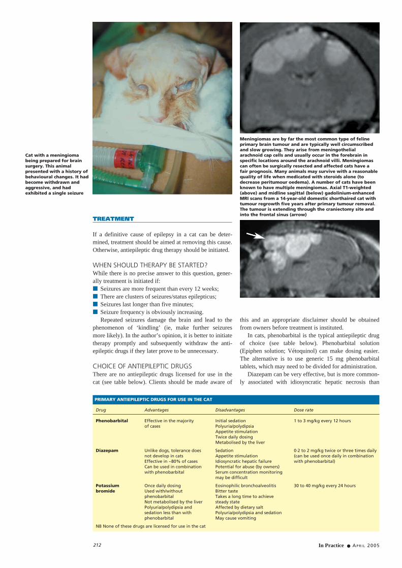
Cat with a meningiomabeing prepared for brainsurgery. This animalpresented with a history ofbehavioural changes. It hadbecome withdrawn andaggressive, and hadexhibited a single seizure
Meningiomas are by far the most common type of felineprimary brain tumour and are typically well circumscribedand slow growing. They arise from meningothelialarachnoid cap cells and usually occur in the forebrain inspecific locations around the arachnoid villi. Meningiomascan often be surgically resected and affected cats have a fair prognosis. Many animals may survive with a reasonablequality of life when medicated with steroids alone (todecrease peritumour oedema). A number of cats have beenknown to have multiple meningiomas. Axial T1-weighted(above) and midline sagittal (below) gadolinium-enhancedMRI scans from a 14-year-old domestic shorthaired cat withtumour regrowth five years after primary tumour removal.The tumour is extending through the craniectomy site andinto the frontal sinus (arrow)
In Practice ● AP R I L 2005 213
phenobarbital. Potassium bromide used as a single dailydose is an effective anticonvulsant, but causes eosino-philic bronchoalveolitis in about 50 per cent of cases andcan be difficult to administer because of its bitter taste. Itis usually used in combination with phenobarbital ordiazepam in cats. The eosinophilic bronchoalveolitis ischaracterised by a cough, and is usually reversible onremoval of the drug.
MONITORING
SEIZURE DIARYThe veterinary surgeon should advise the owner to keepa seizure diary, which should be brought to all consulta-tions. A simple chart indicating the frequency of seizuresis particularly useful, as it allows progress to be assessedquickly. Other notes, such as what time of day theseizure occurred, the length and severity of the seizure,and details about the pre- and postictal periods, can alsobe helpful. For example, an animal consistently havingseizures when tablets are due to be given suggests that a‘trough’ concentration of the drug may be inadequate.
SERUM ANTIEPILEPTIC DRUGCONCENTRATIONSMonitoring the serum concentrations of antiepileptic drugs(see table, below right) enables:■ The lowest effective dose to be used;■ Dosing to be accurately adjusted;■ Possible toxicosis to be avoided;■ Better seizure control.
Serum concentrations should be measured:■ After initiating new drug therapy;■ After dosage changes;■ If breakdown in control occurs;■ Every six to 12 months.
LIVER FUNCTION TESTSPotentially, antiepileptic drug therapy may damage theliver in two ways:■ CHRONIC DISEASE, characterised by hepatic cirrhosisdue to a persistently high dose of antiepileptic drugssuch as phenobarbital or phenytoin, which cause anongoing sublethal injury. There have been no reports ofchronic hepatic failure occurring secondarily to the useof antiepileptic drugs in cats, but this is more likely to bedue to low case numbers, rather than low susceptibility;■ ACUTE INJURY, characterised by intrahepatic cholestasis.This is often classed as an idiosyncratic reaction. It has beendescribed in cats following the administration of diazepamand typically occurs within two weeks of starting therapy.
Veterinary surgeons and owners are often very con-cerned about potential liver failure. In reality, this is rare,especially if the following guidelines are followed:■ Check liver enzymes in cats showing signs of anorex-ia, lethargy, marked polyuria/polydipsia or vomiting,especially within two weeks of initiating therapy;■ Monitor liver enzymes and function (ie, bile acids,albumin, etc) every six to 12 months;■ Do not exceed the therapeutic range and avoid pro-longed administration of doses high within the therapeu-tic range (phenobarbital >30 µg/ml or >130 µmol/litre);■ Avoid administering phenobarbital at doses greaterthan 6 mg/kg twice daily;■ Avoid combination therapy (if possible).
OPTIONS FOR THE UNCONTROLLEDEPILEPTIC CAT
IS THE DOSE ADEQUATE?Check the serum concentration. One of the most com-mon reasons for inadequate control is that the dose wasreduced because of unacceptable sedation. Phenobarbi-tal-induced sedation should wear off in a couple ofweeks and it is important to make the client aware ofthis. If the sedation lasts for longer and is truly unaccept-able, build up the dose slowly to the desired level and re-evaluate the serum concentration.
IS THE OWNER DOSING THE CAT CORRECTLYAND IS THE ANIMAL RECEIVING THE TABLET?Veterinary surgeons should instruct owners that pheno-barbital/diazepam should be dosed every 12 hours, asopposed to twice per day. Uneven dosing can lead toinadequate trough serum concentrations and seizures.The independent lifestyle of cats can sometimes makeregular dosing difficult and, as is widely known, it canbe very difficult to administer tablets to some cats.
ADD ANOTHER ANTICONVULSANT In cats, diazepam and/or potassium bromide can be usedin combination with phenobarbital. The author prefers touse phenobarbital initially and then add diazepam as the second agent. Often very small doses of diazepam(eg, 1 mg, once daily in the evening) can make a signifi-cant difference. Hepatic function should be routinelymonitored as both drugs are metabolised by a similarroute. Potassium bromide can also be used in combina-
Drug Therapeutic range Sampling Notes
Phenobarbital 15 to 45 µg/ml 7 to 14 days Most cats require at least 20 µg/ml (40 to 160 µmol/litre) after first dose (100 µmol/litre) for seizure control
The author recommends avoiding prolonged administration of >30 µg/ml
Diazepam 0·5 µg/ml (human) 14 days after Measured serum concentration first dose includes diazepam and its
metabolites as an equivalent of diazepam
Potassium 800 to 2500 mg/litre 12 to 16 weeks Takes a long time to achieve bromide (15 to 20 mmol/litre) after first dose steady state
Phenytoin 1 to 8 mg/litre (dog) 14 days after Frequent monitoring is required in Therapeutic range first dose the catfor cats has not Long half-life means drug can been established accumulate in cats
ANTIEPILEPTIC DRUG SERUM CONCENTRATIONS
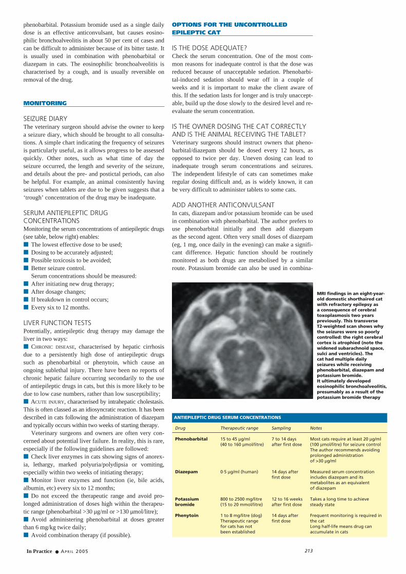
MRI findings in an eight-year-old domestic shorthaired catwith refractory epilepsy as a consequence of cerebraltoxoplasmosis two yearspreviously. This transverse T2-weighted scan shows whythe seizures were so poorlycontrolled: the right cerebralcortex is atrophied (note thewidened subarachnoid space,sulci and ventricles). The cat had multiple daily seizures while receivingphenobarbital, diazepam andpotassium bromide. It ultimately developedeosinophilic bronchoalveolitis,presumably as a result of thepotassium bromide therapy

tion with phenobarbital, but clinicians should bear inmind the risk of eosinophilic bronchoalveolitis.
Although not licensed for use in the cat, propento-fylline (Vivitonin; Intervet) may have some antiepilepticfunction and may be useful in difficult cases. It can alsobe used in addition to existing drugs. A suitable dose is 5 mg/kg twice daily, which must be administered on anempty stomach.
Taurine is an inhibitory amino acid and has someanticonvulsant properties. It has been suggested that taurine supplementation may reduce the frequency ofseizures in epileptic cats (van Gelder and others 1977),but this has yet to be proven.
INCREASE CURRENT THERAPY TO KEEP SERUM CONCENTRATIONS HIGH WITHIN THE THERAPEUTIC RANGEUnacceptable side effects of sedation and pelvic limbataxia usually prevent significant increases in dosage.
CHANGE TO A DIFFERENT ANTIEPILEPTIC DRUGPhenytoin has a half-life of 40 hours in the cat. There-fore, it can accumulate in an animal and can be toxic.Unlike phenobarbital it cannot be excreted unchanged asit has two phenyl rings rather than one, and must behydroxylated and conjugated for excretion, which is aslow process in the cat. However, in cases of refractoryepilepsy, phenytoin may be useful at a dose rate of 1·5mg/kg/day (use phenytoin elixir). Frequent monitoring
of the serum concentration is strongly advisable anddrugs metabolised by the same route (ie, phenobarbitaland diazepam) should be withdrawn. Phenytoin may alsobe useful in cats where the administration of tabletsevery 12 hours is particularly difficult. An overdose ofphenytoin may result in sialosis, frequent vomiting andweight loss.
The author currently uses levetiracetam (Keppra; UCBPharma) at a dose rate of 10 to 20 mg/kg twice daily in: cases of refractory epilepsy; in animals with adverseeffects following the administration of phenobarbitalalone; or in cats with liver compromise. Levetiracetam isa novel antiepileptic drug, which has been shown to beone of the best tolerated antiepileptic drugs in human trials. It causes limited induction of cytochrome P450enzymes (ie, is less challenging to the liver). The authoruses levetiracetam either as a monotherapy or in combina-tion with phenobarbital and/or propentofylline. However,it is too early as yet to determine the effectiveness andsafety of this drug for use in the cat.
ReferencesRUSBRIDGE, C. (1997) Collectionand interpretation ofcerebrospinal fluid in cats anddogs. In Practice 19, 322-331VAN GELDER, N. M., KOYAMA,I. & JASPER, H. H. (1977) Taurinetreatment of spontaneouschronic epilepsy in a cat.Epilepsia 18, 45-54