Embed Size (px)
Citation preview
1
When you or a family member experience severe gastrointestinal symptoms that do not respond to over-the-counter treatments or re-solve on their own, you know that you need to get help quickly. While patients will often see their primary care provider first, sometimes it is necessary to see a specialist in gastrointestinal disease or a gastroenterologist to aid in making a diagnosis and initiating proper treatment. If there is a reasonable suspicion of inflammatory bowel diseases (IBD), such as Crohn’s disease or ulcerative colitis, it is best to seek out a gas-troenterologist who specializes in treating IBD.
If your health plan does not provide access to a gastroenterologist, find a primary care provider with the most experience in diagnosing gastro-intestinal (GI) illness. These health care provid-ers can refer you for the tests and procedures discussed in this brochure, which will be the basis for making your diagnosis, finding the best therapies, and managing your condition.
Finding out if you have IBD may require many tests. This brochure explains which tests you may need to undergo to make a clear diagno-sis as well as to monitor the ongoing status of your Crohn’s disease or ulcerative colitis. Although it is not possible to cover every di-agnostic test in a brochure, the most common tests have been included. If you have a ques-tion about a test not mentioned here, contact the Crohn’s & Colitis Foundation for more information at www.crohnscolitisfoundation.org or 888-694-8872 (MY-GUT-PAIN).
What’s InsideDo you have IBD? ...................................................... 2
Crohn’s disease or ulcerative colitis? ................. 2Could it be something else? .................................. 3
The diagnostic process ........................................... 4Patient history and physical exam ...................... 4Genes and family history ......................................... 4Blood and stool tests ................................................ 5Markers of inflammation ......................................... 6Ruling out other diseases ....................................... 6Specialized blood tests ............................................ 6Tests for optimizing therapy ................................. 7Endoscopic procedures ........................................... 7The role of biopsy and the surgical pathologist..................................................10
Radiology scans or diagnostic imaging ...........11X-rays .............................................................................. 11Barium contrast studies ........................................ 12Cross-sectional imaging ........................................ 12Magnetic resonance imaging (MRI) .................. 16Ultrasound ................................................................... 16Role of diagnostic imaging ....................................17Radiation risks .............................................................17
Monitoring your health ..........................................18Scheduling tests ....................................................... 19Questions for your doctor or nurse ................. 19Colon cancer ..............................................................20Chromoendoscopy ................................................... 21Other cancers ............................................................. 21Immunizations ........................................................... 22
Special considerations .........................................22When the patient is under 18 ............................. 22Health insurance....................................................... 24Support and resources .......................................... 24
The future of diagnostics ....................................25
Glossary ......................................................................25
The Crohn’s & Colitis Foundation provides information for educational purposes only, which is current as of the print date. We encourage you to review this educational mate-rial with your health care professional as this information should not replace the recommendations and advice of your doctor. The Foundation does not provide medical or other health care opinions or services. The inclusion of another organization’s resources or referral to another organization does not represent an endorsement of a particular individual, group, company, or product.
2 3
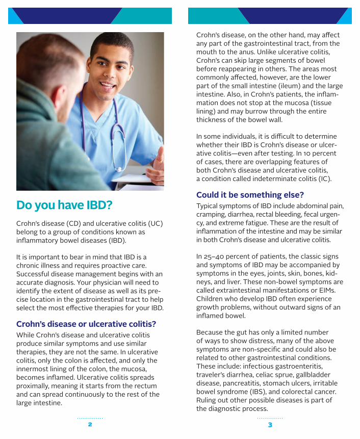
Crohn’s disease, on the other hand, may affect any part of the gastrointestinal tract, from the mouth to the anus. Unlike ulcerative colitis, Crohn’s can skip large segments of bowel before reappearing in others. The areas most commonly affected, however, are the lower part of the small intestine (ileum) and the large intestine. Also, in Crohn’s patients, the inflam-mation does not stop at the mucosa (tissue lining) and may burrow through the entire thickness of the bowel wall.
In some individuals, it is difficult to determine whether their IBD is Crohn’s disease or ulcer-ative colitis—even after testing. In 10 percent of cases, there are overlapping features of both Crohn’s disease and ulcerative colitis, a condition called indeterminate colitis (IC).
Could it be something else?Typical symptoms of IBD include abdominal pain, cramping, diarrhea, rectal bleeding, fecal urgen-cy, and extreme fatigue. These are the result of inflammation of the intestine and may be similar in both Crohn’s disease and ulcerative colitis.
In 25–40 percent of patients, the classic signs and symptoms of IBD may be accompanied by symptoms in the eyes, joints, skin, bones, kid-neys, and liver. These non-bowel symptoms are called extraintestinal manifestations or EIMs. Children who develop IBD often experience growth problems, without outward signs of an inflamed bowel.
Because the gut has only a limited number of ways to show distress, many of the above symptoms are non-specific and could also be related to other gastrointestinal conditions. These include: infectious gastroenteritis, traveler’s diarrhea, celiac sprue, gallbladder disease, pancreatitis, stomach ulcers, irritable bowel syndrome (IBS), and colorectal cancer. Ruling out other possible diseases is part of the diagnostic process.
Do you have IBD?Crohn’s disease (CD) and ulcerative colitis (UC) belong to a group of conditions known as inflammatory bowel diseases (IBD).
It is important to bear in mind that IBD is a chronic illness and requires proactive care. Successful disease management begins with an accurate diagnosis. Your physician will need to identify the extent of disease as well as its pre-cise location in the gastrointestinal tract to help select the most effective therapies for your IBD.
Crohn’s disease or ulcerative colitis? While Crohn’s disease and ulcerative colitis produce similar symptoms and use similar therapies, they are not the same. In ulcerative colitis, only the colon is affected, and only the innermost lining of the colon, the mucosa, becomes inflamed. Ulcerative colitis spreads proximally, meaning it starts from the rectum and can spread continuously to the rest of the large intestine.
4 5
risk factor for developing Crohn’s disease or ulcerative colitis, although most patients with IBD do not have a family history of IBD.
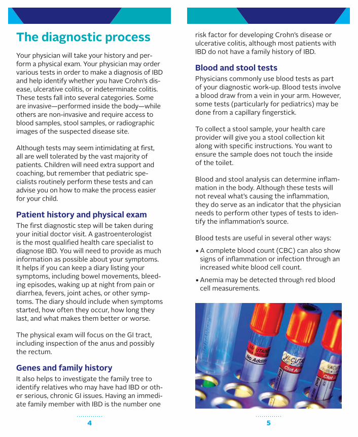
Blood and stool testsPhysicians commonly use blood tests as part of your diagnostic work-up. Blood tests involve a blood draw from a vein in your arm. However, some tests (particularly for pediatrics) may be done from a capillary fingerstick.
To collect a stool sample, your health care provider will give you a stool collection kit along with specific instructions. You want to ensure the sample does not touch the inside of the toilet.
Blood and stool analysis can determine inflam-mation in the body. Although these tests will not reveal what’s causing the inflammation, they do serve as an indicator that the physician needs to perform other types of tests to iden-tify the inflammation’s source.
Blood tests are useful in several other ways:
• A complete blood count (CBC) can also show signs of inflammation or infection through an increased white blood cell count.
• Anemia may be detected through red blood cell measurements.
The diagnostic processYour physician will take your history and per-form a physical exam. Your physician may order various tests in order to make a diagnosis of IBD and help identify whether you have Crohn’s dis-ease, ulcerative colitis, or indeterminate colitis. These tests fall into several categories. Some are invasive—performed inside the body—while others are non-invasive and require access to blood samples, stool samples, or radiographic images of the suspected disease site.
Although tests may seem intimidating at first, all are well tolerated by the vast majority of patients. Children will need extra support and coaching, but remember that pediatric spe-cialists routinely perform these tests and can advise you on how to make the process easier for your child.
Patient history and physical examThe first diagnostic step will be taken during your initial doctor visit. A gastroenterologist is the most qualified health care specialist to diagnose IBD. You will need to provide as much information as possible about your symptoms. It helps if you can keep a diary listing your symptoms, including bowel movements, bleed-ing episodes, waking up at night from pain or diarrhea, fevers, joint aches, or other symp-toms. The diary should include when symptoms started, how often they occur, how long they last, and what makes them better or worse.
The physical exam will focus on the GI tract, including inspection of the anus and possibly the rectum.
Genes and family historyIt also helps to investigate the family tree to identify relatives who may have had IBD or oth-er serious, chronic GI issues. Having an immedi-ate family member with IBD is the number one
6 7
OmpC. However, these tests are NOT to be used for the diagnosis as these biomarkers are present in all patients, and may be present in patients without IBD. They may, however, be helpful in differentiating between Crohn’s disease and ulcerative colitis when there is uncertainty at time of diagnosis.
Tests for optimizing therapyThiopurine methyltransferase (TPMT) testing may be ordered when physicians are consider-ing the use of mercaptopurine or azathioprine for patients. Testing can help to determine whether you would be an appropriate candi-date for these medications and what the opti-mal starting dose would be for each person.
Immunosuppressive drugs, such as anti-TNF and interleukin inhibitors (also known as bio-logic therapy) can cause reactivation of a latent (silent) tuberculosis or hepatitis B infection; all patients need to be screened for these diseas-es before starting such treatment.
For more information on IBD treatments, down-load the Foundation’s Understanding IBD Med-ications and Side Effects brochure by visiting www.crohnscolitisfoundation.org.
Endoscopic proceduresEndoscopy is a procedure that lets your doctor look inside your body. It uses an instrument called an endoscope, or “scope” for short. Scopes have a tiny camera attached to a long, thin, flexible tube that allows your physician to see images of your intestine magnified on a screen. Endoscopy helps your physician to see if inflammation is present, where it is located, assess its severity, and obtain biopsies to con-firm the diagnosis. Endoscopy is also vital for monitoring your therapy—healing of the lining of the intestine is a sign that your medication is effective.
• Liver and kidney function can be assessed. The liver and kidneys may be affected by IBD or the medications used to treat the disease.
• An electrolyte panel is important to check for dehydration and side effects of medications.
• They can help a doctor predict how well you may respond to a particular medication mov-ing forward.
Blood tests are part of both the initial work-up and ongoing follow-up and monitoring of your condition. They usually do not require any special preparation. See the Routine Blood and Stool Tests table on pages 14 and 15.
Markers of inflammationProteins found in blood and stool, also called biomarkers, may be useful tests for detecting inflammation. Stool biomarkers include calpro-tectin and lactoferrin. Blood biomarkers include C-reactive protein (CRP) and erythrocyte sedi-mentation rate (ESR). Research has shown that these biomarkers are useful in predicting IBD activity, but they are also present in other GI dis-eases. These blood and stool tests may be more helpful for guiding invasive testing, detecting flares, and optimizing medical therapies than for diagnosing IBD. The use of some biomarkers is relatively new, and they are not used by all phy-sicians nor covered by all insurance policies.
Ruling out other diseasesGastrointestinal infections with similar symp-toms may be identified by testing small stool samples. These tests may look for C. difficile, E. coli, campylobacter, yersinia, salmonella, shigella, and other infections.
Specialized blood testsSpecialized tests include serology tests for biomarkers that researchers have associated with IBD, such as pANCA, ASCA, CBir1, and
8 9
For a colonoscopy, you should expect to:
• Receive restricted diet instructions and follow them.
• Drink a bowel preparation (prescribed by your physician).
• Dedicate the night before your test to con-firm the bowel purging process.
• Wear loose, comfortable clothing to your procedure.
• Have a friend or family member drop you off and pick you up after the procedure.
Colonoscopies are generally very safe proce-dures, but there is an extremely small risk of bowel perforation during the exam, which may be increased if you have IBD. You may want to discuss the risk with the physician performing the test.
Many patients ask about the usefulness of a less invasive “virtual colonoscopy,” or CT colonography. Although these radiology-based tests are an exciting new development, they are not recommended for suspected IBD, where biopsies and direct viewing of the colon and small bowel are required.
Sigmoidoscopy: an endoscopic evaluation of the lower one-half to one-third of the colon. In patients with ulcerative colitis, inflammation begins in the rectum. Therefore, a sigmoidos-copy can be a good diagnostic test to confirm the disease and to monitor your response to therapy. It can be performed without sedation, because it is a very short procedure and is asso-ciated with less discomfort than a colonoscopy. The preparation for this procedure is less com-plex than a colonoscopy, usually requiring only one or two enemas the day of the procedure.
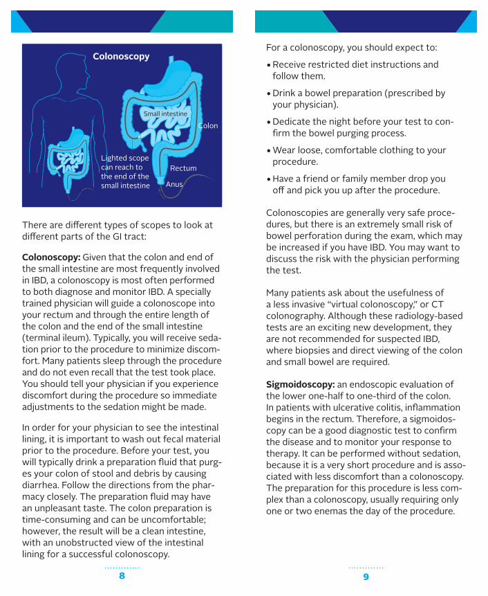
There are different types of scopes to look at different parts of the GI tract:
Colonoscopy: Given that the colon and end of the small intestine are most frequently involved in IBD, a colonoscopy is most often performed to both diagnose and monitor IBD. A specially trained physician will guide a colonoscope into your rectum and through the entire length of the colon and the end of the small intestine (terminal ileum). Typically, you will receive seda-tion prior to the procedure to minimize discom-fort. Many patients sleep through the procedure and do not even recall that the test took place. You should tell your physician if you experience discomfort during the procedure so immediate adjustments to the sedation might be made.
In order for your physician to see the intestinal lining, it is important to wash out fecal material prior to the procedure. Before your test, you will typically drink a preparation fluid that purg-es your colon of stool and debris by causing diarrhea. Follow the directions from the phar-macy closely. The preparation fluid may have an unpleasant taste. The colon preparation is time-consuming and can be uncomfortable; however, the result will be a clean intestine, with an unobstructed view of the intestinal lining for a successful colonoscopy.
Lighted scope can reach to the end of the small intestine
ColonSmall intestine
Rectum
Anus
Colonoscopy
10 11
disease is ulcerative colitis or Crohn’s disease. Results from evaluation of biopsies can take as long as one week. Pathologists also look for the presence of any abnormal cells such as precancerous or cancerous cells in the inflamed area of the intestine.
Radiology scans or diagnostic imaging Traditional upper endoscopy and colonoscopy will not be able to evaluate about two-thirds of the small intestine. Therefore, in addition to capsule endoscopy, radiologic exams or diagnostic imaging are performed to evaluate these segments of intestines as well as to eval-uate areas outside the bowel.
The Imaging Tests chart on pages 14 and 15 discusses the areas of interest in the intestine as well as the radiology and endoscopy tests that you may undergo.
X-raysX-rays are the oldest way of imaging the inside of the body. X-rays are cost-effective and useful for detection of blockages in the small or large intestine. Patients with Crohn’s disease, for ex-ample, can have inflammation and/or scarring of the small bowel that narrows the intestine and prevents the easy passage of stool and air. This is called a small bowel obstruction. The large bowel can also become blocked and dilated. Rarely, people with ulcerative colitis can develop a widening of the large bowel called toxic megacolon. These are serious compli-cations that can be seen on a plain X-ray. No preparation or contrast material is required with an X-ray. The test exposes you to a small amount of radiation.
Esophagogastroduodenoscopy (EGD) or upper endoscopy: an endoscopic evaluation of the upper end of the digestive system that physicians use to evaluate a wide variety of symptoms, including, but not limited to: upper abdominal pain, nausea, vomiting, and difficulty swallowing. An endoscopy typically requires fasting after midnight until the test. A longer upper endoscope, called an enteroscope, can be used to look for inflammation further into the small bowel. A standard enteroscopy can typically evaluate the first one-third of the small bowel.
Video capsule endoscopy (VCE): a procedure that allows your physician to view areas of the bowel that cannot be reached with traditional scopes. This involves swallowing a capsule that is equipped with a camera—a “pill camera” (PillCam,® Endo Capsule®). As it travels through the intestines, the capsule automatically takes pictures. The images are wirelessly sent to a receiver worn by the patient. The capsule is expelled during a bowel movement, usually within a day.
Endoscopic ultrasound (EUS): a technique that uses an ultrasound probe attached to an endoscope to obtain deep images of the gut below the surface. With IBD, physicians use EUS most often to look at fistulas in the rectal area. Fistulas are abnormal connections from the intestine to another part of the intestine, another organ of the body, or the surface of the skin.
The role of biopsy and the surgical pathologistA pathologist is a physician who will examine biopsy tissue, obtained under endoscopy or through surgery, under the microscope for specific features that help make the diagnosis of IBD. In addition, the pathologist may iden-tify findings that can determine whether the
12 13
techniques are extremely helpful in diagnosing and managing IBD. These imaging studies can evaluate the entire thickness of the bowel wall and can detect inflammation and complications such as fistulas, abscesses, and obstruction.
Computed tomography (CT scan), also known as a CAT scan, takes simultaneous X-rays from several different angles to reconstruct a realis-tic image of the internal organs. It may involve a contrast material delivered orally, rectally, or intravenously to improve the quality of the test. During the test, you will be on a special ta-ble that advances through the scanner to take images at each level of your abdomen. Newer scanners have an open design to minimize claustrophobia. A CT scan of the abdomen takes 5 to 20 minutes to complete. The CT scan is used to rule out complications of IBD, such as intra-abdominal abscesses, strictures, small bowel obstructions or blockages, fistulas, and bowel perforation.
CT enterography (CTE) is a variation of a CT scan. During this exam, a special oral and/or intravenous contrast agent is given to better outline the intestines. In addition, CTE recon-structs images in 3-D to better visualize the small bowel in relation to other organs. The physician may suggest this exam to identify areas of inflamed bowel and more subtle ob-structions or blockages.
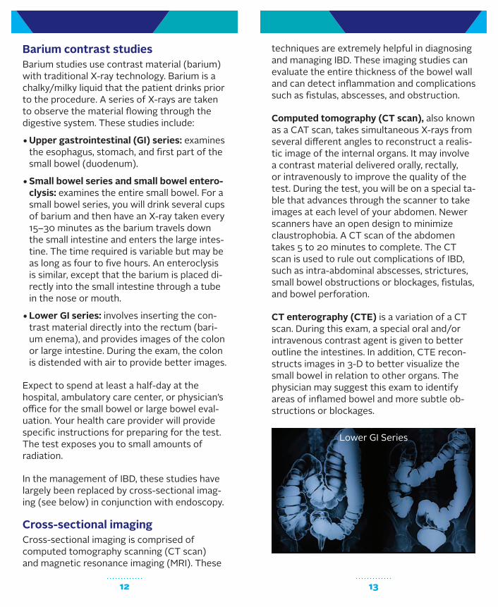
Barium contrast studiesBarium studies use contrast material (barium) with traditional X-ray technology. Barium is a chalky/milky liquid that the patient drinks prior to the procedure. A series of X-rays are taken to observe the material flowing through the digestive system. These studies include:
• Upper gastrointestinal (GI) series: examines the esophagus, stomach, and first part of the small bowel (duodenum).
• Small bowel series and small bowel entero-clysis: examines the entire small bowel. For a small bowel series, you will drink several cups of barium and then have an X-ray taken every 15–30 minutes as the barium travels down the small intestine and enters the large intes-tine. The time required is variable but may be as long as four to five hours. An enteroclysis is similar, except that the barium is placed di-rectly into the small intestine through a tube in the nose or mouth.
• Lower GI series: involves inserting the con-trast material directly into the rectum (bari-um enema), and provides images of the colon or large intestine. During the exam, the colon is distended with air to provide better images.
Expect to spend at least a half-day at the hospital, ambulatory care center, or physician’s office for the small bowel or large bowel eval-uation. Your health care provider will provide specific instructions for preparing for the test. The test exposes you to small amounts of radiation.
In the management of IBD, these studies have largely been replaced by cross-sectional imag-ing (see below) in conjunction with endoscopy.
Cross-sectional imagingCross-sectional imaging is comprised of computed tomography scanning (CT scan) and magnetic resonance imaging (MRI). These
Lower GI Series
14 15
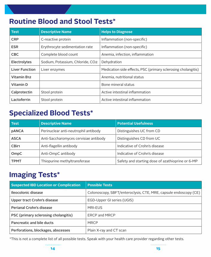
Routine Blood and Stool Tests*Test Descriptive Name Helps to Diagnose
CRP C-reactive protein Inflammation (non-specific)
ESR Erythrocyte sedimentation rate Inflammation (non-specific)
CBC Complete blood count Anemia, infection, inflammation
Electrolytes Sodium, Potassium, Chloride, CO2 Dehydration
Liver Function Liver enzymes Medication side effects, PSC (primary sclerosing cholangitis)
Vitamin B12 Anemia, nutritional status
Vitamin D Bone mineral status
Calprotectin Stool protein Active intestinal inflammation
Lactoferrin Stool protein Active intestinal inflammation
*This is not a complete list of all possible tests. Speak with your health care provider regarding other tests.
Specialized Blood Tests*Test Descriptive Name Potential Usefulness
pANCA Perinuclear anti-neutrophil antibody Distinguishes UC from CD
ASCA Anti-Saccharomyces cervisiae antibody Distinguishes CD from UC
CBir1 Anti-flagellin antibody Indicative of Crohn’s disease
OmpC Anti-OmpC antibody Indicative of Crohn’s disease
TPMT Thiopurine methyltransferase Safety and starting dose of azathioprine or 6-MP
Imaging Tests*Suspected IBD Location or Complication Possible Tests
Ileocolonic disease Colonoscopy, SBFT/enteroclysis, CTE, MRE, capsule endoscopy (CE)
Upper tract Crohn’s disease EGD-Upper GI series (UGIS)
Perianal Crohn’s disease MRI-EUS
PSC (primary sclerosing cholangitis) ERCP and MRCP
Pancreatic and bile ducts MRCP
Perforations, blockages, abscesses Plain X-ray and CT scan
16 17
bladder, and those in the pelvic area. Currently, endoscopic ultrasound and MRI are both used to diagnose perianal Crohn’s disease. Physi-cians in the U.S. do not typically use ultrasound to evaluate the small bowel; however, in Eu-rope, they use ultrasound more often to assess for blockages in the small bowel. Ultrasound emits no radiation, and relies on the shad-ows cast by inaudible sound waves. Although ultrasounds do not usually require preparation (other than not eating for a few hours before the test), you should check with your physician.
Role of diagnostic imagingAs is the case with laboratory tests, diagnostic imaging may also play multiple roles in treating and managing IBD. Not only will the radiology scans help to determine if you have Crohn’s disease, but they will also reveal the extent and severity of the inflammatory process and assess complications of disease such as an obstruction, fistula, or abscess. This information will allow your physician to recommend the best course of therapy. For more information on medication options, review the Foundation’s Understanding IBD Medications and Side Effects brochure on www.crohnscolitisfoundation.org.
Even after diagnosis, imaging studies may be used to determine how well you are responding to therapy and confirming that your disease is in remission. This is called “monitoring your IBD,” and it is a critical part of getting and staying well.
Radiation risksThere is research that indicates radiation as a risk factor for cancer. It is clear that health- related radiological scans contribute the most to radiation exposure for the majority of pa-tients. CT scans currently generate the largest amount of radiation among the types of scans discussed in this brochure. Despite the radi-ation exposure associated with CT, it is still a very useful test for diagnosing IBD and its com-
These tests expose you to radiation. You may discuss with your physician whether imaging alternatives, such as MRI, are more appropriate for you. (See Radiation Risks, page 17, for more information.)
Be aware that some patients are allergic to the contrast agent in intravenous form. Let the technician know if you think you have an allergy. Patients with kidney disease, diabetes, or dehydration are at increased risk for kidney side effects from the intravenous contrast material.
Magnetic resonance imaging (MRI)Magnetic resonance imaging is a useful way to view the abdominal organs and the intesti-nal tract. It can also identify inflammation and complications such as fistulae and obstruc-tion. The machine moves along the body using magnetic waves to take pictures, which gen-erate a two- or three-dimensional image that can be viewed as a series of cross-sections. One of the advantages of MRI is that it does not involve radiation exposure. However, it is generally more expensive and also takes longer to perform. Patients who are claustrophobic may have more difficulty with MRI since it is more confining. Tell your physician if you have concerns about enclosed spaces.
Magnetic resonance enterography (MRE) can give a detailed picture of the small and large intestine, similar to CT enterography.
Magnetic resonance cholangiopancreatogra-phy (MRCP) is a specialized MRI exam that pro-vides a view of the bile ducts, gall bladder, liver, and pancreas. This test is helpful in diagnosing primary sclerosing cholangitis (PSC) in IBD patients, a condition characterized by severe inflammation and scarring in the bile ducts.
UltrasoundUltrasound technology is used to study many organs in the abdomen, typically the liver, gall-
18 19
• Stool markers and cultures—identifies inflammation and infectious complications
Scheduling tests There is usually no specific order of tests, but rather a need to have all the information generated in a reasonable period of time. If you are ill and undergoing an initial evaluation, or experiencing a serious flare, the timing is certainly more pressing than for a routine, elective, monitoring procedure, which you may schedule months in advance. The physi-cian may want the results as soon as possible. A flexible schedule will be helpful in making yourself available for testing. Remember, there are lead times built into obtaining results of biopsies and beginning treatment. At times, diagnostic tests may be clustered together for your convenience.
You will want to work with your employer to take advantage of available sick days to cover testing requirements. If you already have utilized available sick time, you will have to consider short-term disability or family leave. Remember that this is a serious health issue and you will want to get your disease in remission as soon as possible so you can return to work.
If the patient is your child, or college student, make sure to speak with your child’s teacher, guidance counselor, or college admissions offi-cer to discuss any necessary school accommo-dations that may be required so that ongoing diagnostic testing may take place.
Questions for your doctor or nurse1. What is the purpose of the test? What will
happen if we get a positive result?
2. Do I need to fast or prepare otherwise?
3. How long will it take?
4. Will the test expose me to radiation?
plications. However, other exams, such as MRI and ultrasound, are being used increasingly to decrease radiation exposure for patients.
You and your physician will discuss the risks and benefits of all your decisions. There are no risk- free options, however, the absolute risk associ-ated with radiation from imaging is much lower than the risk of having poorly controlled IBD be-cause of inadequate monitoring of your disease.
If you think you may be pregnant, inform your physician, as it is important to avoid all tests that can expose your fetus to radiation.
Monitoring your healthWith proper treatment and monitoring, you will maximize your chances for good health and decrease the likelihood of missing signs of additional disease.
Once you are diagnosed and begin treatment, there may be regular follow-up appointments to monitor your disease, watch for signs of a flare, address any changes in symptoms, and identify possible side effects of treatment. During these follow-up appointments, your physician may order several tests. Tests that your physician may order on a regular basis will include the following:
• Complete blood count—identifies anemia, infection, and inflammation, and monitors certain medications
• ESR (erythrocyte sedimentation rate)—identifies inflammation
• C-reactive protein—identifies inflammation
• Liver enzymes—screens for liver complications
• Electrolytes—checks for dehydration and medication side effects
20 21
specimens will be taken throughout the colon, looking for cancer or precancerous changes called dysplasia. A surveillance colonoscopy is generally recommended eight to ten years after diagnosis of ulcerative colitis or Crohn’s disease involving the colon and every one to two years thereafter. Patients who have PSC should begin surveillance colonoscopy at the time of diagnosis and then annually.
ChromoendoscopyWhen used for dysplasia surveillance, your doctor may recommend a colonoscopy with chromoendoscopy. During chromoendosco-py, a blue liquid dye is sprayed over the colon surface during the colonoscopy procedure. The blue dye collects and pools in the crevices of the colon lining, drawing your doctor’s eye to slight or subtle changes in the lining of your intestine, which may be precancerous areas. Chromoendoscopy cannot be used if the colon is not clean, and it is not very effective if the colon is inflamed. Therefore, an excellent bow-el preparation and control of inflammation are very important. It is common to see blue stool, and occasionally blue urine, for a short time following this procedure.
Guidelines for surveillance change over time, so you should ask your doctor about what is new in the detection of colon cancer.
Other cancersPatients with IBD are also at increased risk for other types of cancer outside of the intestinal track. Again, these risks are low but include skin cancers, smoking-related cancers, and lympho-ma. The increased risk observed may be related to the immune suppression medications that are used to treat many patients with IBD. You should discuss the best surveillance strategies for these diseases with your doctor as guide-lines change and new data become available.
5. Can I go alone or must I have a companion?
6. When will I learn the results? Who will be giving them to me? May I have a hard copy for my records?
7. Will we be repeating this test or procedure? How often?
8. Will health insurance cover the cost of this test, and if so, how frequently?
To help keep track of your test information, please reference the Diagnostic Test Log at the back of this brochure. We suggest you keep it somewhere handy so you can access it easily. The log also serves as a convenient reference for when you meet or speak with a health care provider.
Colon cancerPatients with long-standing ulcerative colitis or Crohn’s disease involving the colon are at increased risk of developing colon cancer. However, the magnitude of this risk is cur-rently a subject of debate. The risk increases with the duration of disease, the extent of the colon involved, and the degree of inflammation present. Other risk factors include the diagno-sis of colon cancer in a first-degree relative or the presence of primary sclerosing cholangitis (PSC), a disease that damages and blocks bile ducts inside and outside the liver.
Despite the increased risks, only a small per-centage of patients with IBD develop colon cancer. With proper treatment and monitoring of your IBD, you should be able to maximize your chances of good health over the long term and not miss signs of additional disease.
Surveillance colonoscopy is currently the recommended strategy for detecting pre- cancerous changes or cancer in patients with IBD. During the colonoscopy, multiple biopsy

22 23
therapy. As a result of these tests, the pediatric gastroenterologist may make changes to your child’s medications.
Blood tests require only about two teaspoons of blood from a child and most children do well with blood draws. If a child is anxious, formal relaxation techniques can be taught and anes-thetic creams can numb his or her arm.
Recurrent diagnostic imaging should be mini-mized to reduce lifetime exposure to radiation. MRIs are becoming a more common choice, because they do not involve radiation, but this technology is evolving and may not be available in every location. Also, an MRI is more costly. Stool tests for lactoferrin and calprotectin may help identify patients who need additional diagnostic testing.
Another concern in pediatric IBD may be the use of endoscopy. As with adults, colonoscopy plays a central role in diagnosing children. Children receive general anesthesia rather than con-scious sedation, as in adults. Complications are extremely rare, especially when performed in a specialized setting like a pediatric IBD center.
Keep in mind that your physician will only order diagnostic tests if the clinical picture leads him or her to believe that IBD is a possibility—one you cannot afford to overlook. In addition, the risk of not knowing that your child has IBD or inadequate monitoring of IBD is far greater than the risk from diagnostic testing.
Your child’s physician may also direct you to counseling and support to help your child through what could be a challenging time. The Crohn’s & Colitis Foundation has child-specific literature available to help you teach your child about diagnosing and living with IBD. Available reading material for kids may even help them get over the fears of blood testing and proce-
ImmunizationsLike everyone, patients with IBD should strive to keep their immunizations (vaccinations) up to date. This is especially true for IBD patients who take immune suppression treatments. Be sure to discuss this with your doctor on an annual basis.
Special considerations When the patient is under 18Although IBD most typically appears in young adulthood, there are increasing numbers of cases in patients under 18 years of age. Chil-dren are not miniature adults and the process of diagnosing and treating IBD must be tailored to their biology and anatomy. You will need the advice of a pediatric gastroenterologist, a sub-specialist in the field who treats IBD in kids.
Symptoms of concern in children include:
• Abdominal pain
• Diarrhea
• Failure to gain weight or grow
• Fatigue
• Fever
• Rectal bleeding
• Relapsing gastrointestinal illness over several months
• Weight loss
A pediatric gastroenterologist will order the diagnostic tests and procedures that are the safest and most appropriate for your child. They will also discuss all treatment goals with you. As discussed in this brochure, your child may require ongoing laboratory tests, endos-copy, and diagnostic imaging to monitor their disease and look for complications of medical
24 25
through email ([email protected]), online live chat, social media, postal mail, or fax. The IBD Help Center is available Monday through Friday, 9:00 a.m. to 5:00 p.m. EST.
The future of diagnosticsWe look forward to a future when diagnostic testing will provide better guidance for choos-ing therapies and telling us whether a patient will have a mild or more serious disease course.
Scientists are currently studying biological indicators of disease activity (biomarkers), such as specific genetic changes, levels of inflamma-tion-related molecules in blood circulation, and products of deleterious bacterial species in the gut and in the stool, to establish which of these biomarkers can be used to accurately forecast onset and progression of IBD in general and specifically among family members. Through the Founda tion’s research initiatives, it is our plan to accelerate research focused on identification and validation of the biomarkers of inflamma-tion and on developing sensing technologies to detect these biomarkers. By achieving these goals, we will advance towards better IBD diagnostics and better monitoring of disease progression and response to therapies.
Visit our website, www.crohnscolitisfoundation.org, to learn more and see how you can get involved in supporting our research initiatives.
GlossaryAnti-OmpC (outer membrane protein C): the antibody to a specific protein on the outer membrane, recently identified as a significant biomarker. New data shows that anti-OmpC levels are high among members of families that have a history of both Crohn’s disease and ulcerative colitis.
dures. Contact the Foundation’s Irwin M. and Suzanne R. Rosenthal IBD Resource Center (IBD Help Center) at888-694-8872 (MY-GUT-PAIN) or [email protected] for free copies of this literature.
Health insuranceIt is crucial to evaluate your coverage when you face the prospect of IBD. Levels of coverage vary and you may want to make changes to your health insurance going forward.
High-deductible health plans may require you to absorb much of the cost of the initial diag-nostic work-up, including endoscopy, radiology, and laboratory work. It may be more cost-ef-fective to pay higher premiums and reduce out-of-pocket costs if you need ongoing tests and procedures. Costs of procedures vary by location and sometimes insurers require prior approval before you undergo a test. This is particularly true when a test of the same type is repeated within a specific time frame. You will need to speak with your health insurance provider about the provisions of your plan.
If you are uninsured and cannot cover the cost of insurance premiums, you can look at resources available in your state for the un-insured and consider enrolling in a plan that permits access to the basic diagnostic require-ments and treatments for IBD.
Support and resourcesThe Crohn’s & Colitis Foundation’s Irwin M. and Suzanne R. Rosenthal IBD Resource Center (IBD Help Center) is a free service designed to provide you with disease-specific information, guidance, and support. It is staffed by caring information specialists who provide education on IBD management, available treatment op-tions, and coping strategies as well as referrals to helpful resources and programs. The IBD Help Center can be reached toll-free by phone at 888-MY-GUT-PAIN (888-694-8872) or
26 27
Electrolytes: laboratory test panel including serum sodium, potassium, chloride, and carbon dioxide that may indicate dehydration and other complications or medication side effects.
ERCP (endoscopic retrograde cholangiopan-creatography): a type of endoscopy that utilizes X-ray to diagnose a liver disease called primary sclerosing cholangitis (PSC).
ESR (erythrocyte sedimentation rate): a laboratory blood test for non-specific inflammation.
Granuloma: a collection of cells in the intes-tinal lining, visible under the microscope, that indicates the body’s attempt to get rid of a foreign material; sometimes seen in Crohn’s disease, but not always present.
Gut: the intestine or bowel.
Hemoglobin and hematocrit: measurements of red blood cell number and volume, found in the CBC, useful in determining anemia.
Lactoferrin: a stool test for intestinal inflam-mation that aids in predicting active IBD.
MRCP (magnetic resonance cholangiopan-creatography): a type of MRI that allows the physician to see images of the bile ducts, which are similar to ERCP images.
MRI (magnetic resonance imaging): an imag-ing test that uses a magnetic field and pulses of radio wave energy to make pictures of organs and structures within the body.
p-ANCA (perinuclear anti-neutrophil cyto-plasmic antibodies): a serology test that may aid in diagnosing ulcerative colitis, distinguish-ing it from Crohn’s disease, and predicting disease course.
ASCA (anti-saccharomyces cerevesiae): a serology test useful in distinguishing Crohn’s disease from ulcerative colitis and predicting disease course.
Biomarkers: proteins in the body that may be measured by laboratory tests to assist in diagnosis and management of disease.
Biopsy: a tissue sample provided to a patholo-gist to help diagnose and classify disease.
Calprotectin: a stool test for intestinal inflam-mation that aids in predicting active disease.
CBC (complete blood count): a laboratory blood test that helps to detect anemia, infection, and inflammation.
CBiR1 (anti-flagellin): this antibody may be a marker of Crohn’s disease complicated by fis-tulas, perforations, or other serious problems.
CRP (C-reactive protein): a laboratory test that indicates non-specific inflammation in the body.
CT (computed tomography): an imaging test that uses X-rays to make detailed pictures of structures within the body.
CTE (computed tomography enterography): a variation of the CT scan where the patient swallows special contrast agents to give a sharp outline of the intestines in the X-rays.
DEXA (bone densitometry scan): an X-ray that assesses the thickness of bones and risk for osteoporosis (thin bones) and fractures.
EIM (extraintestinal manifestations): signs and symptoms outside of the gastrointestinal tract associated with IBD.
28
PPD (purified protein derivative): tuberculo-sis (TB) skin test, advised for all patients taking biologic therapies, to assess the presence of latent and active TB disease.
Radiographic: Relating to the process that depends on X-rays.
Small bowel enteroclysis: an imaging test that evaluates the small intestine by infusing barium and air through a tube inserted into the small intestine via the nose.
Serology: a blood test to identify antibodies (proteins) that may have developed in re-sponse to an infection, other foreign proteins, or one’s own proteins.
SBFT/SBS (small bowel follow-through/small bowel series): an imaging test that eval-uates the small intestine, involving swallowing barium, after which serial X-rays are taken.
US (ultrasound): an imaging test in which high-frequency sound waves, not heard by the human ear, are transmitted through body tissues using a transducer, relaying information to a computer for display.
Toxic megacolon: an acute condition where the colon is dilated or enlarged, a complication associated with ulcerative colitis.
TPMT (thiopurine methyltransferase): a labo-ratory blood test for the activity of an enzyme that helps in breaking down the medications azathioprine and 6-MP, helping to establish proper dosing of these medications.
Virtual colonoscopy (also known as CT enterography): a less invasive, new version of colonoscopy, done without sedation and using X-rays and computer-based, virtual-reality technology to produce 3-D images of the lining of the colon. Virtual colonoscopy is not currently used to diagnose or monitor IBD.
About the Crohn’s & Colitis FoundationEstablished in 1967, the Crohn’s & Colitis Foundation is a nonprofit, volunteer-driven organization dedicated to finding the cures for Crohn’s disease and ulcerative colitis, and im-proving the quality of life of children and adults affected by these diseases.
Since our founding, the Foundation has re-mained at the forefront of research in Crohn’s disease and ulcerative colitis. Today, we fund cutting-edge studies at major medical institu-tions, nurture investigators at the early stages of their careers, and finance underdeveloped areas of research.
In addition, the Crohn’s & Colitis Foundation provides a comprehensive series of education programs, printed and online resources, sup-port services, and advocacy programs to mem-bers of the IBD community, including patients and caregivers.
We can help! Contact us at: 888-MY-GUT-PAIN(888-694-8872) [email protected]
Credits:Reviewers: Tauseef Ali, MD, and Emmanuelle Williams, MD
Contributors: Hollie Barnett and Kira Stegman
Design & Layout: Rubicon Design Associates
The Crohn’s & Colitis Foundation is a nonprofit organiza-tion that relies on the generosity of private contributions to advance its mission to cure Crohn’s disease and ulcer-ative colitis, and to improve the quality of life of children and adults affected by these diseases.
www.crohnscolitisfoundation.org
6/2018
733 Third Avenue Suite 510New York, NY 10017800-932-2423
This educational material is supported by the Maxine and Jack Zarrow Family Foundation. Additional support is provided through the Crohn’s & Colitis Foundation’s annual giving program and donors.