Embed Size (px)
Citation preview
1
Diacylglycerol acyltransferase 2 (DGAT2) links glucose utilization to fatty acid
oxidation in the brown adipocytes
Zehra Irshad, Federica Dimitri, Mark Christian and Victor A Zammit1
Translational and Experimental Medicine
Division of Biomedical Sciences
Warwick Medical School, CV4 7AL, UK
1Corresponding author: Victor A Zammit
Email: [email protected]
Fax: +44 (0)2476522798
Short title: DGAT2 enables BAT thermogenesis from glucose
by guest, on May 22, 2018
ww
w.jlr.org
Dow
nloaded from
2
Abbreviations:
Astat: Atglistatin
Etmox: Etomoxir
DGAT1-iB: DGAT1-specific inhibitor (cis-4-{3-fluoro-4-[({5-[(4-fluorophenyl)amino]-
1,3,4-oxadiazol-2-yl}; carbonyl)-amino]phenoxy} cyclohexane carboxylic acid)
DGAT2-iC: DGAT2-selective inhibitor (N-(4,5-dihydronapththol[1.2-d]thiazol-2-yl)-2-
(3,4-dimethoxyphenyl)acetamide)
DAGT2-iJ: DGAT2- specific inhibitor (3-bromo-4-[2-fluoro-4-({4-oxo-2-[(2-pyridin-2-
ylethyl)amino]1,3-thiazol-5(4H)-ylidene} methyl)phenoxy] benzonitrile)
Key words
From JLR list:
Adipocytes; DGAT; Fatty acid/Metabolism; Lipolysis; Adipose tissue
Additional key words:
Thermogenesis; de novo lipogenesis; β3-agonist; substrate channelling; brown adipose
tissue.
by guest, on May 22, 2018
ww
w.jlr.org
Dow
nloaded from
3
SUMMARY
Brown adipose tissue uptake of glucose and fatty acids is very high during non-
shivering thermogenesis. Adrenergic stimulation markedly increases glucose uptake, de
novo lipogenesis and FA oxidation simultaneously. The mechanism that enables this
concerted response has hitherto been unknown. Here we find that in a primary brown
adipocytes and brown adipocyte-derived cell line (IMBAT-1) acute inhibition and longer-
term knockdown of DGAT2 links the increased de novo synthesis of fatty acids from
glucose to a pool of TG which is simultaneously hydrolysed, providing FA for
mitochondrial oxidation. DGAT1 does not contribute to this pathway, but uses
exogenous FA and glycerol to synthesise a functionally distinct pool of TG to which
DGAT2 also contributes to it. The DGAT2-dependent channelling of 14C from glucose into
TG and CO2 was reproduced in β3-agonist-stimulated primary brown adipocytes.
Knockdown of DGAT2 in IMBAT-1 affected the mRNA levels of several genes important
in FA activation and esterification. Therefore, in β3-agonist activated brown adipocytes,
DGAT2 specifically enables channelling of de novo synthesised FA into a rapidly mobilised
pool of TG which is simultaneously hydrolysed to provide substrates for mitochondrial
fatty acid oxidation.
by guest, on May 22, 2018
ww
w.jlr.org
Dow
nloaded from
4
INTRODUCTION
The diacylglycerol acytransferases, DGAT1 and DGAT2 catalyse the last, dedicated
step of triacylglercerol (TG) synthesis. Although they catalyse the same reaction (with
minor differences in substrate preferences) and are co-expressed in all the cell types in
which they occur, they are functionally non-redundant. In particular, in the liver, DGAT2
is specialised for the formation of TG from de novo synthesised fatty acids and newly
formed diglycerides (DG) thus acting upstream of DGAT1 in the de novo synthesis of TG
(1). This renders DGAT2 essentially rate-limiting for the de novo formation of TG in the
liver (2), and is consistent with the observation that the liver of mice lacking a key
enzyme of the glycerol-3-P pathway (Gpat 1-/-) is depleted of TG, and is unable to esterify
de novo synthesised FA to TG (3). Although DGAT2 also participates in the maintenance
of the TG pool(s) in lipid droplets through the lipolysis-reesterification cycling that occurs
between TG and DG (4), the esterification between preformed FA and partial glycerides
is primarily performed by DGAT1 in HepG2 cells (1) and in murine liver (5). This is
consistent with the observations that Dgat 1-/- and Dgat 2-/- mice have very different
phenotypes, with Dgat 1-/- animals having a metabolically favourable phenotype
(including lower plasma and tissue TG) (6), whereas Dgat 2-/- animals dying within
several hours after birth, and being devoid of TG (lipopenic) (7). This specialisation of
hepatic DGAT2 for the utilization of de novo synthesised fatty acids (FA) is readily
rationalised in view of the role of the liver in integrating glycaemia, triglyceridaemia and
hepatic TG content, and accommodating large fluxes of glucose metabolism, de novo
lipogenesis (DNL) and TG synthesis and secretion (2). However, the wider applicability
of this functional specialization of DGAT2 to other tissues and conditions remains to be
determined (8,9).
Brown adipose tissue (BAT) is another tissue that, like the liver, clears large
amounts of both glucose and FA (either non-esterified or as products of lipoprotein lipase
action on triglyceride-rich lipoproteins) from the circulation (10). The very high rates of
uptake have been suggested to play a role in the regulation of glycaemia and
triglyceridaemia, respectively (11-13). During cold exposure, BAT has the highest rate of
glucose uptake and lipogenesis when compared to white adipose tissue (WAT) and the
liver (14). BAT glucose metabolism is independent of insulin but, during β-adrenergic
by guest, on May 22, 2018
ww
w.jlr.org
Dow
nloaded from
5
stimulation, is stimulated by an increase in GLUT1 expression (via cAMP) and
translocation to the plasma membrane, mediated through mTORC2 (15,16). Although
glucose makes a relatively minor direct contribution (<20%) towards thermogenesis
(through glycolysis and oxidation of pyruvate) (17,18), the contribution through
oxidation of FA synthesised de novo from glucose and other lipogenic substrates (e.g.
lactate (19)) could be considerably larger (13) . Indeed, when non-shivering
thermogenesis is maximally stimulated, BAT can account for 33% of whole-body
lipogenesis, and newly synthesised FAs make a significant contribution towards the
thermogenic capacity of BAT in adult rodents (14). The very substantial lipogenic
capacity of BAT (20) and its activation in vivo upon cold-exposure of animals (21,22) are
accompanied by increased nuclear expression of SREBP1 and elevated gene expression
of lipogenic and FA-elongation enzymes when mice are maintained at sub-
thermoneutral temperatures (23).
Therefore, in view of the very high lipogenic potential of BAT (10,24,25), the
contribution of de novo synthesised FA towards BAT thermogenesis may be substantial
(13,26). FA, whether provided exogenously or synthesised de novo from glucose, activate
UCP1 (by over-riding the inhibitory action of purine nucleotides on the protein), and
provide the ultimate substrate for uncoupled respiration in brown adipocytes (27).
Glucose is also required to form glycerol-3-P for the (re)synthesis of TG from FA.
Therefore, like the liver, BAT has to integrate large fluxes of glucose and fatty acid
metabolism, and may be a tissue in which the distinctive functions of DGAT1 and DGAT2
may be important in directing different FA pools towards specific pathways during
adrenergic stimulation, when glucose uptake, de novo lipogenesis (DNL) and TG lipolysis
are all coordinately activated (21,22,28). Expression profiling of mouse tissues, indicates
that DGAT2 is expressed in BAT judging by the high level of its mRNA expressed in the
tissue (29) and that its expression is increased (more than that of DGAT1) in BAT of cold-
acclimated rodents (30). Therefore, in the present study, we have investigated the roles
of DGAT1 and DGAT2 in the metabolism of glucose and de novo synthesised FA, compared
to that of exogenous fatty acids in a brown adipocyte cell line (IMBAT-1) and in mouse
primary brown adipocytes. We identified a specific role for DGAT2 in linking glucose
uptake and DNL to the formation of TG which acts directlyde novo as a source of FA for
oxidation, independently from the uptake and metabolism of extracellular FA.
by guest, on May 22, 2018
ww
w.jlr.org
Dow
nloaded from
6
MATERIALS AND METHODS
Preparation and culture of primary brown adipocytes
All animal maintenance and usage was made in accordance with the requirements
of the Animal Welfare and Ethical Review Body of the University of Warwick. Black
c57b16 mice were used. Primary brown adipocytes were prepared from the stromal
fraction resulting from the collagenase digestion of inter-scapular brown adipose tissue
obtained from 4-week old mice. The stromal fraction was suspended in DMEM10, filtered
through a 40μm strainer, and cells plated in T25 flasks. The cells were allowed to expand
until 80-90% confluent with regular replacement of media. After trypsinization and
resuspension in DMEM10 medium 5 x 10⁴cells/well of 12-well plate were then plated
and allowed to reach 100% confluence. Differentiation was induced by addition of
medium containing insulin (1μg/ml), IBMX (250uM), triiodothyronine, T3 (1nM),
indomethacin (30μM), rosiglitazone (2μM) and dexamethasone (0.5μM). After 96h,
during which the medium was replenished once, the cells were switched to maintenance
medium (DMEM10 medium supplemented with insulin, T3 and rosiglitazone only). After
a further 48h, the cells were switched to a medium which contained the same
concentrations of insulin and T3, but from which rosiglitazone was omitted. After a
further 48h the medium was changed to DMEM10, and the cells were used for
experimental treatments.
Generation, differentiation and culture of IMBAT-1 cells
The immortalised brown adipocyte cell line (IMBAT-1) was generated as
described previously (31). Briefly, preadipocytes isolated from murine interscapular
brown adipose tissue were immortalized by retroviral-mediated expression of
temperature-sensitive SV40 large T-antigen H-2kb-tsA58. Cells were cultured in DMEM-
F12 medium (20mM D glucose) supplemented with 10% foetal bovine serum (FBS), 1%
L-glutamine, 1% Penicillin/streptomycin, Amphotericin B and 50μg/ml of G418 at 33°C
in 5% CO2 and 95% air- water saturated atmosphere. Unless specified otherwise, cells
were passaged and harvested after treatment with 0.25% trypsin and 0.02% EDTA. For
by guest, on May 22, 2018
ww
w.jlr.org
Dow
nloaded from
7
differentiation to mature adipocytes, pre-adipocytes were plated onto gelatine-coated
plates and cultured until confluent. Induction medium containing 7.5mM glucose, 1μg/ml
insulin, 250nM dexamethasone, 0.5mM IBMX, 1nM triidothyronine (T3) and 125μM
Indomethacin was added for 48 hours at 37°C. Cells were then maintained in 7.5mM
glucose medium containing 1nM T3 and 1μg insulin /ml for 5 days at 37°C. Unless
otherwise stated, all experiments were performed on day 7 of differentiation or after a
further 72h treatment with siRNA. The experimental incubation medium was DMEM-F12
medium containing 7.5mM D-glucose, 10% fetal bovine serum, 1% L-glutamine, 1%
penicillin/streptomycin, 1mM L-carnitine, 0.75mM oleate with 0.25% BSA and 0.75mM
glycerol.
SiRNA-mediated knockdown of DGAT1 or DGAT2
Transfections were carried out on the day 7 of differentiation using Smartpool
siRNA, designed by the manufacturers for DGAT1 and DGAT2, and transfection reagent
lipofectamine RNAiMax, following the manufacturer’s instructions. Differentiated
adipocytes were dissociated with trypsin/EDTA followed by addition of DMEM/F12 with
10% FBS. Mixtures of transfection reagents, OptiMEM medium, 10μM siRNA-DGAT1 or
10μM siRNA-DGAT2 or 10μM siRNA-control were incubated at room temperature for 25
min. They were then added to the dissociated cells along with antibiotic-free DMEM-F12
medium (7.5mM glucose). Control cells were treated with scrambled siRNA. Media were
changed after 24 hours and cells were cultured for further 48 hours at 37°C in
maintenance medium. Preliminary experiments established that optimal knockdowns
were achieved after 72h of siRNA treatment.
Measurement of the incorporation of 14C-glucose label into CO2 and cellular TG
IMBAT-1 or primary brown adipocytes cells plated in gelatine-coated 6-well
plates containing 7.5mM glucose medium were incubated for 2h in the presence or
absence of the β3-adrenergic agonist CL (10μM) before the addition of label to start the
measurements. Preliminary experiments established the incorporation of [U-14C]-
glucose into labelled products was linear for 2h and this period was used routinely when
[U-14C]-glucose incorporation was studied. Where indicated, oleate or palmitate (0.75mM
plus 0.25% albumin) and glycerol (0.75mM) were added to the incubations. When
incorporation of label from 1-[14C]-oleate, U-[14C]-palmitate or 2-[3H]-glycerol was
by guest, on May 22, 2018
ww
w.jlr.org
Dow
nloaded from
8
measured, the labelling period was 1h to ensure linearity of incorporation into TG. In one
series of experiments labelling with exogenous 1-[14C]-oleate was extended to 8h to
monitor the release of 14CO2. When effects of DGAT1 or DGAT2 inhibition were studied,
the appropriate inhibitors were added 30 min before the start of the incubations with the
label. Where indicated, etomoxir (80 µM) was added 30 min before the start of the
experimental period by addition U-14C-glucose label. Lipolysis was inhibited by addition
of tetrahydrolipstatin (THL, 200μM) or Atglistatin (10μM) at the same time as the
addition of CL i.e. 2 hours before addition of U-14C-glucose label.
Measurement of 14CO2 labelling
Formation of 14CO2 after incubation of cells with labelled substrates for the
required period of time was measured by transferring 1 ml of the incubation medium to
a 20-ml glass conical flask containing a 0.5-ml Fisher centre well holding filter paper to
which 400μl of benzethonium hydroxide had been added (32). The flask was stoppered
and 1.0 ml of 1 M H2SO4 was injected through the rubber stopper. The flasks were shaken
for 60 min at 37°C to allow the liberated 14CO2 to be absorbed by the benzethonium
hydroxide, after which the radioactivity associated with the contents of the centre-well
was quantified using a liquid scintillation counter.
Measurement of incorporation of label into TG
At the end of the incubation period, the cells were washed with cold PBS, and total
lipids were extracted from them using a chloroform/methanol mixture (2:1 v/v) (33).
The chloroform layer was aspirated into a glass tube and dried under a stream of N2 gas.
The dried material was re-solubilized in chloroform (500μl) and the entire volume was
applied onto a TLC plate coated with Silica Gel 60 for separation of the radioactive
triglyceride product, using hexane/diethyl ether/formic acid (70:30:1, v/v/v) as the
mobile phase. A TG-standard (tripalmitin) was used to identify the position of the TG
band visualized using iodine vapour. The radioactivity associated with each band was
quantified after scraping into scintillation vials, addition of scintillant (Ultima GoldTM,
Perkin Elmer) and measuring the associated radioactivity using a scintillation counter.
Saponification of the total lipids was performed as described in (34) with minor
modifications. The cellular total lipid extract were dried and dissolved in 0.75ml of 30%
KOH and heated at 70°C for 10 min. An equal volume of 95% ethanol was then added and
by guest, on May 22, 2018
ww
w.jlr.org
Dow
nloaded from
9
the mixture was heated at 70 °C for 2 hours. After cooling, the aqueous fraction was
acidified with 3M HCl and extracted thrice with light petroleum. The organic fraction was
evaporated, re-dissolved in 1ml of light petroleum, transferred to scintillation vial and
quantified as the TG-acyl fraction. The remaining aqueous fraction contained the TG-
glyceryl fraction; 0.5ml aliquot of this sample was taken and measured for radioactivity
using scintillation counter.
Real time PCR quantitation of mRNA expression
Total RNA was extracted from IMBAT-1 cells using TRIzol reagent. cDNA was
prepared using reverse transcription. Briefly RNA (1ug) was mixed with oligodT (1µl) in
a final volume of 12 µl by adding RNAse free water. Samples were heated at 70°C for 5
min before chilling on ice. Subsequently, 8µl of mixture containing RNAse inhibitor (10
U/µl), dNTPs (10 mM), Bioscript reverse transcriptase and RNAse free water were added
to each sample. Samples were heated at 40 °C for 60 min and the reaction was stopped
by heating to 70 °C for 10min. The cDNA formed was mixed with 180µl of nuclease-free
water and stored at -20°C. Samples were thawed only once before quantification. Reverse
transcription (RT-PCR) was performed using SYBR green dye and expression for all the
samples (n ≥ 3) was calculated by using the DCt method, incorporating the efficiencies of
each primer pair. The variances of input cDNA were normalised against the levels of three
housekeeping genes; L19, B-actin and 36B4. Melting curve analysis confirmed
amplification specificity. The primers used are detailed in Supplemental Table S1.
Statistical analyses
Differences between means for independent groups of data were analysed by ANOVA
followed by the post hoc Tukey test.
Materials
Dulbecco’s Modified Eagle’s Medium DMEM10, DMEMF12, etomoxir sodium salt,
CL, L-carnitine, benzethonium hydroxide, light petroleum (bp: 40-60 °C), glyceryl
tripalmitate, sodium oleate, insulin, IBMX, Indomethacin, and 3,3’5-triiodo-L-thyamine
were purchased from Sigma-Aldrich. TLC-pre-coated plates, 10ml conical flasks with
centre wells, lipofectamine, and RNAiMax were purchased from Fischer scientific.
Rosiglitazone was from Cayman Chemical. Radiolabelled [U-14C]-glucose (specific
by guest, on May 22, 2018
ww
w.jlr.org
Dow
nloaded from
10
activity 250-360mCi/mmol), [1-14C] Oleic acid (specific activity: 40-60mCi/mmol) and
[2-3H] glycerol (specific activity 0.5-1.0 Ci/mmol) was purchased from Perkin Elmer LAS
(UK). ON-TARGETplus Mouse Dgat1 smartpool siRNA, ON-TARGETplus Mouse Dgat2
smartpool siRNA and ON-TARGETplus non-targeting siRNA were purchased from GE
Healthcare UK. Of the three inhibitors of DGAT activity used, DGAT1iB (cis-4-{3-fluoro-4-
[({5-[(4-fluorophenyl)amino]-1,3,4-oxadiazol-2-yl}carbonyl)-amino]phenoxy}
cyclohexane carboxylic acid) and DGAT2-iC (N-(4,5-dihydronapththol[1.2-d]thiazol-2-
yl)-2-(3,4-dimethoxyphenyl)acetamide) used were obtained from Astra Zeneca and have
been used previously as DGAT1 and DGT2 inhibitors, respectively (33). A third
inhibitor, DGAT2-iJ (3-bromo-4-[2-fluoro-4-({4-oxo-2-[(2-pyridin-2-ylethyl)amino]1,3-
thiazol-5(4H)-ylidene}methyl)phenoxy] benzonitrile) was obtained from Janssen
Research and Development UK, and has been used previously as a specific DGAT2
inhibitor (5). All primers were purchased from Sigma-Aldrich.
RESULTS
A. Experiments using the brown adipocyte-derived cell line IMBAT-1
In view of the large amount of preliminary work that was needed to establish the
conditions under which the metabolic fluxes of interest could be measured, and the
concentrations of inhibitors required, we used a well-established brown adipocyte-
derived cell line, IMBAT-1. This cell line has been well characterised, and has been shown
to express all the proteins that are characteristic of brown adipocytes, especially UCP1
(35). Their use also allowed us to verify the effects observed with specific DGAT1 and
DGAT2 inhibitors through the efficient knockdown of the two proteins in these cells using
siRNA.
The main findings were validated using primary brown adipocytes (see below).
A.1 DGAT2 mRNA expression is preferentially induced during differentiation
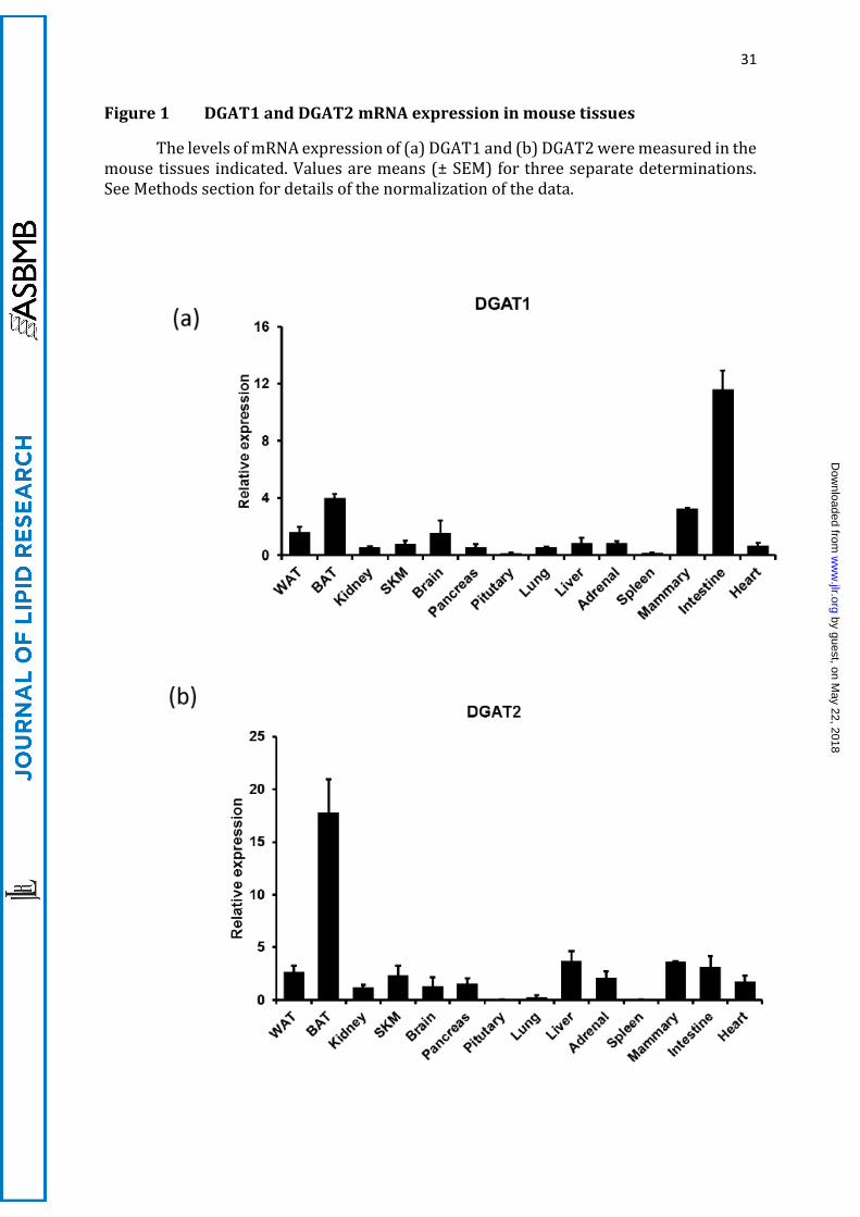
We investigated the expression of DGATs across a panel of murine tissues. DGAT2
mRNA was most highly expressed in BAT, and present at much lower levels in WAT, liver,
intestine and mammary gland (Figure 1). DGAT1 mRNA was most highly expressed in
the intestine, followed by BAT, white adipose tissue and mammary gland. We then
by guest, on May 22, 2018
ww
w.jlr.org
Dow
nloaded from
11
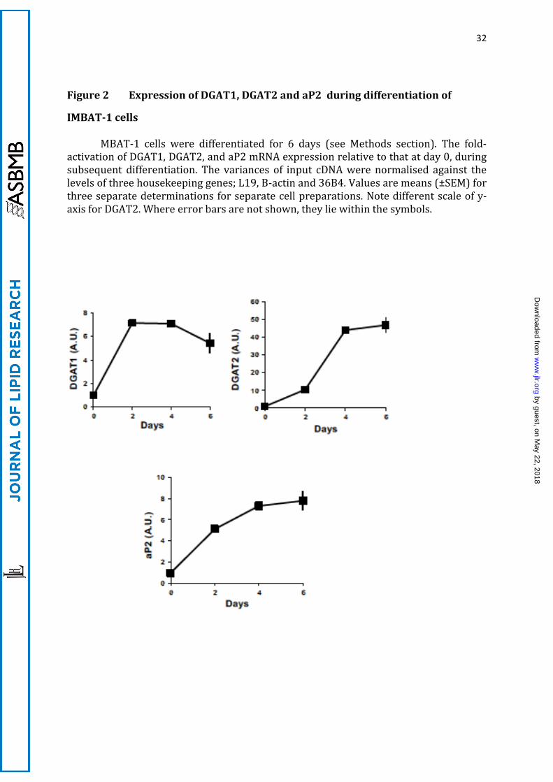
studied the profile of mRNA expression for both DGATs, compared with the
differentiation marker aP2 during brown adipocyte differentiation. All three mRNAs
were induced at day 2 of differentiation, but whereas DGAT1 mRNA was induced
moderately (7-fold) during the first days of differentiation, levelling off thereafter and
declining on day 6, DGAT2 mRNA was induced 50-fold and continued to be induced
throughout the period of differentiation (Figure 2). This time course of the expression
of the two genes is the opposite to that previously described during the differentiation of
white adipocytes, in which an initial peak in DGAT2 expression (during initial
differentiation with glucose as the main substrate) is overtaken by a sustained expression
of DGAT1 mRNA during the latter stages of differentiation (35).
A.2 β3-adrenergic stimulation increases incorporation of de novo synthesised
FA into TG and CO2
We next measured the rates of labelling from U-14C-glucose into TG and CO2 in IMBAT-
1 cells, and ascertained that they were linear during the 2h-period of the incubations (not
shown). Stimulation of IMBAT-1 cells with the β3-agonist CL316324 (CL) for 2h before
the start of incubations (by addition of U-14C-glucose label) resulted in a significant
increase in the incorporation of 14C-label from glucose into CO2 (Figure 3a). The rates of
CL-stimulated glucose incorporation were 22.8 ±1.3 and 50.6 ±1.0 pmol/h/106cells for
TG-acyl and TG-glyceryl moieties, respectively. The incorporation into CO2 was 10.6± 1.4
pmol/h/106cells, although this flux is not directly comparable to the previous two, as (i)
14CO2 is generated both during lipogenesis and FA oxidation, and (ii) 14CO2 derived from
the oxidation of de novo synthesised FA will have been diluted by unlabelled acyl moieties
within the pre-existing TG pool. From the observation that 106 cells yield approximately
10mg wet weight of cellular material, it is calculated that these rates are of the same order
of magnitude as the values reported for glucose uptake (~7.2 µm/h/g) and de novo
lipogenesis rates (~4.8 µm/h/g) in cold-acclimated rats in vivo (14,30).
Etomoxir (an inhibitor of CPT1 which controls FA entry into mitochondria (36))
inhibited CO2 labelling marginally under basal conditions (Figure 3) suggesting that, as
expected, in the absence of CL-stimulated TG lipolysis, CO2 labelling from glucose was
due mostly to that generated in the course of lipogenesis. The increase in CO2 labelling
induced by CL was totally prevented by etomoxir (Figure 3a). As a result, it could be
by guest, on May 22, 2018
ww
w.jlr.org
Dow
nloaded from
12
calculated that CL increased the rate of 14CO2 labelling due to CPT 1-dependent oxidation
of de novo synthesised FA by approximately 4-fold (Fig 3a).
Stimulation of the cells with CL also activated incorporation of label from U-14C-
glucose into both parts of the TG molecule (glyceryl and acyl, Figures 3b and 3c,
respectively) although differentially. Stimulation of U-14C-glucose incorporation into the
acyl moieties of the TG molecule was consistently significantly higher (200%) than that
into the glyceryl moiety of TG (50%) indicating that, whereas increased glucose uptake
by the cells by CL may have been a common contributor towards the increased labelling
of TG from glucose (25), de novo lipogenesis was stimulated independently of, and to a
higher extent than, the pathway leading from glucose to triose phosphates (the last
common intermediates of glycerol-3-P and FA formation). Etomoxir did not affect
incorporation of glucose into TG-acyl or TG-glyceryl moieties, confirming that its action
is specific to the inhibition of FA oxidation.
A.3 CL-stimulation of glucose incorporation into TG-FA and their oxidation is
ATGL-dependent
To test the possible role of TG synthesis and hydrolysis in the provision of glucose-
derived FA for oxidation, we tested the effects of inhibition of TG hydrolysis, using
Atglistatin (a specific inhibitor of adipose triglyceride lipase, ATGL) and the non-specific
lipase inhibitor tetrahydrolipstatin (THL). Both inhibitors prevented all the effects of CL
treatment on CO2 and TG labelling from U-14C-glucose without affecting basal rates
(Figure 4). CL-stimulation of the incorporation of glucose into both acyl and glyceryl
moieties of TG was totally prevented by ATGL inhibition by Atglistatin. This indicated
that a metabolite generated by TG lipolysis activates one or more steps in the pathway
leading from glucose to TG synthesis, and occurs independently of the interruption of the
provision of FA for oxidation as a result of the inhibition of TG hydrolysis. This positive
feedback on TG synthesis supports previous observations that TG lipolysis generates
signalling molecules that affect lipogenic fluxes (37-39).
A.4 Oxidation of de novo synthesised FA is DGAT2-dependent
A.4.1 Inhibitor studies
by guest, on May 22, 2018
ww
w.jlr.org
Dow
nloaded from

13
In view of the preferential activation of the esterification of de novo synthesised FA
into TAG labelled from glucose, we studied the possibility that DGAT1 and DGAT2 may
have different roles in the esterification of newly synthesised FA into TG after β3-
stimulation of IMBAT-1 cells. We used three compounds which have previously been
well-characterised as specific inhibitors of the two DGATs. Compound DGAT1-iB was
previously used (1,33,40,41) as a highly specific inhibitor of DGAT1; Compound DGAT2-
iC (see methods) was used in (1) as a selective inhibitor of DGAT2 at lower
concentrations; and compound DGAT2-iJ was developed and used as a highly specific
inhibitor of DGAT2 ((5); see Methods). As these compounds have been developed as
inhibitors of the human enzymes, we performed dose-response studies to ascertain that
they were effective inhibitors of TG synthesis in IMBAT-1 cells at concentrations that
were not deleterious to cell viability. The concentrations required for each compound to
inhibit incorporation of U-14C-glucose into the glyceryl or acyl moieties of TG (see
Supplemental Figure S1) were similar to those found to be effective in HepG2 cells and
on the recombinant human enzymes (1,5,33). In the same experiments, we quantified
cell viability, as judged by MTT mitochondrial viability assays, at the end of the 2h
incubations. Thereafter, the effects of DGAT1 or DGAT2 inhibition were investigated in
detail, using single concentrations of each compound (see Legends to Figures) that gave
significant (but less than maximal) inhibition of the parameters studied, but with the
retention of full cell viability.
Both the DGAT2 inhibitors tested (DGAT2-iC and DGAT2-iJ) resulted in a strong
inhibition of the CL-stimulated incorporation of 14C-glucose into CO2 and TG-acyl groups
(see Figure 5 a, b). By contrast, inhibition of DGAT1 (with DGAT1-iB ) did not affect either
incorporation of label into 14CO2 (Fig 5a) or into TG-acyl groups (Fig 5b). These
observations suggest that DGAT2 activity is specialised for the esterification of de novo
synthesised fatty acids into TG and subsequent mitochondrial oxidation of FA products
of TG lipolysis. The validity of this conclusion is strengthened by the fact that the two
inhibitors of DGAT2 used are structurally unrelated. Both DGAT1 and DGAT2 inhibition
decreased significantly the glucose incorporation into the glyceride part of TG (Fig 5c),
indicating that glycerol-3-P newly synthesised from glucose is incorporated into DG pools
that are used by either DGAT1 or DGAT2 as substrates.
Importantly, the above observations were not altered qualitatively when
experiments were conducted in the presence of added exogenous glycerol (0.75mM) and
by guest, on May 22, 2018
ww
w.jlr.org
Dow
nloaded from
14
oleate (0.75mM in the presence of 0.25% albumin) to the medium (Figure 5d-f); only
DGAT2 inhibition resulted in the loss of labelling from U-14C-glucose into TG-acyl
moieties and CO2 (Figure 5d, e). Therefore, addition of exogenous oleate (or palmitate,
not shown) did not affect the ability of DGAT2 inhibition specifically to affect the
incorporation of de novo synthesised FA into TG (compare Figure 5b and 5e) or the
formation of CO2 after CL treatment. These observations indicate that de novo
synthesised FA are compartmentalised rapidly into a pool of TG which is not accessible
to exogenously added FA. Addition of exogenous glycerol and oleate appeared to increase
the effect of either DAGT1 or DGAT2 inhibition on glucose incorporation into TG-glyceryl,
but did not affect the relative importance of the two enzymes in this process (compare
Figure 5c and f).
A.4.ii DGAT1- and DGAT2-knockdown studies
To exclude the possibility that non-specific effects of enzyme inhibitors were
compromising the validity of our observations, we investigated the longer-term effects of
the knockdown of the expression of either protein using individually targeted siRNA
treatment of IMBAT-1 cells. The effects of 72h siRNA treatment on mRNA expression of
the DGAT1 and DGAT2 are shown in Figure 6. A scrambled sequence was used in control
cells. Note that reliable quantification of protein expression was not possible using a
range of commercially available antibodies (not shown). Similarly, DGAT activities could
not be reliably quantified by available DGAT assays using the amounts of material
available. However, the fact that both inhibition and knockdown studies resulted in
identical observations (see below) suggests that the large decreases in mRNA were
accompanied by similar losses in enzyme activities and protein expression.
The data in Figure 7a show that knockdown of DGAT2 had the same major
inhibitory effect on the incorporation of label from U-14C-glucose into CO2 both under
basal and CL-stimulated conditions as shown by the inhibitors, confirming that de novo
synthesised FA need to be incorporated into TG by DGAT2 before they can undergo
mitochondrial oxidation. Knockdown of DGAT1 had no effect at all (Figure 7a).
Therefore, these data are identical qualitatively and quantitatively to those obtained
using the respective DGAT1 and DGAT2 inhibitors (Figure 5). Knockdown of DGAT2 also
had a large inhibitory effect on the incorporation of glucose into total TG (Figure 7b).
Saponification of the lipid fraction confirmed the preferential stimulus by CL of glucose
by guest, on May 22, 2018
ww
w.jlr.org
Dow
nloaded from
15
incorporation into the acyl moieties of TG, and showed that DGAT2 knockdown
prevented the CL effects on the incorporation of label from U-14C-glucose into both TG-
acyl and TG-glyceryl moieties under both basal and CL-stimulated conditions (Figure 7e,
f). Therefore, effects of longer-term DGAT2 down-regulation with siRNA treatment on
the formation of TG-acyl moieties, and their subsequent use for 14CO2 were identical to
those observed after short-term inhibition (Figure 5). By contrast, there was no effect
on glucose incorporation onto TG-acyl when DGAT1 was knocked down (Figure 7d), and
much smaller effects than those achieved by DGAT2 knockdown on incorporation of
glucose into TG-glyceryl (Figure 7c). These observations confirm that DGAT2 is
specialised for the channelling of de novo synthesised fatty acids towards oxidation (to
CO2) initially through their incorporation into a distinct pool of TG, followed by lipolysis
(Figures 7c, 7e)
A.5 DGAT2 knockdown attenuates the CL-mediated induction of UCP1 mRNA
expression
In view of the observed close association between DGAT2 down-regulation and
metabolism of de novo synthesised FA, we investigated whether long-term down-
regulation of DGAT2 or DGAT1 is accompanied by changes of lipogenic gene expression.
We measured the mRNA expression (before and after CL treatment) of UCP1 and of
several genes which are involved in the pathways leading from glucose to TG synthesis
and CO2 formation (Figure 8). Incubation of the cells with CL for 2h increased the
expression of UCP1 mRNA five-fold in IMBAT-1 cells (as observed previously in BAT in
vivo (42)); DGAT2 knockdown resulted in the halving of this increase whereas DGAT1
knockdown had no effect (Figure 8). GPAT4 mRNA expression after CL-treatment was
decreased specifically by DGAT2 knockdown. None of the other genes tested (including
Elvol3, not shown) showed significant differences in mRNA expression levels after CL-
stimulation or knockdown of either DGAT compared to control (scrambled) siRNA-
treated cells.
Therefore, knockdown of DGAT2 moderately but specifically affected the expression
of genes that are central to FA esterification (GPAT4), and uncoupling of mitochondrial
FA oxidation (UCP1) (3,43,44) indicating that it may play a central role in the
maintenance of BAT function.
by guest, on May 22, 2018
ww
w.jlr.org
Dow
nloaded from
16
A.6 Direct oxidation of exogenously added oleate is very low
We next compared the roles of DGAT1 and DGAT2 in determining the metabolism of
exogenously added, preformed fatty acids. We performed a series of experiments in
which the normal concentration of unlabelled glucose was accompanied by glycerol (0.75
mM) and oleate (0.75 mM in the presence of 0.25% albumin). We performed parallel
experiments in which we labelled either the glycerol (2-3H-glycerol) or oleate (1-14C-
oleate). We also used U-14C-palmitate to verify that the effects observed with oleate could
be replicated using a saturated FA; the results with palmitate (not shown) were identical
to those obtained with oleate.
Contrary to the incorporation of de novo synthesised FA and glycerol-3-P derived
from U-14C-glucose, the labelling of TG from either exogenous glycerol or oleate were not
stimulated by CL, showing that the effects of the β3-agonist were specific to the
stimulation of glucose metabolism and FA derived from it. Although exogenous 1-14C-
oleate was very rapidly incorporated into TG (75 ± 9 nmol oleate/h/106 cells), no
significant 14C-label above background was recovered in oxidation products (either CO2
or acid-soluble metabolites, ASM). Thus, although incubation with 1-14C-oleate (and U-
14C-palmitate) achieved the same degree of overall cellular TG labelling as that achieved
by U-14C-glucose, only minimal 14C-label was recovered in CO2 or ASM after exposure to
1-14C-oleate even when incubations were extended to 8h after addition of label to
eliminate the possibility of a time-lag (not shown). There are two conclusions from these
observations. Firstly, that exogenous FA are not oxidised directly to CO2 to any significant
extent, although they are rapidly incorporated into TG. Secondly, that the pool of TG into
which exogenous oleate is esterified is distinct from that into which glucose-derived de
novo synthesised FAs are esterified by DGAT2, since the latter do give rise to 14CO2
linearly over a 2h incubation period. This is consistent with the observation in A.4, above,
that addition of exogenous oleate did not alter the pattern of fluxes of U-[14C]-glucose into
TG-acyl or TG-glyceryl moieties. The simplest explanation for these combined
observations is that exogenous (pre-formed) oleate is incorporated into a separate, large
pool of TG in which the specific activity of 1-14C-oleate is highly diluted, resulting in
negligible labelling of the CO2 produced from the oxidation of its constituent FA after
lipolysis. De novo synthesised fatty acids appear to have been incorporated into a TG
pool in which a much higher specific activity of the acyl groups was achieved. Therefore,
by guest, on May 22, 2018
ww
w.jlr.org
Dow
nloaded from
17
this pool must be considerably smaller, and must turn over rapidly to give the linear and
immediate CO2 formation observed.
These inferences were supported by the effects on exogenous 1-14C-oleate
incorporation into TG after the inhibition of either DGAT1 (using DGAT1-iB) or DGAT2
(using DGAT2-iJ). When added individually, each inhibitor affected only marginally the
incorporation of exogenous 1-14C-oleate into TG. However, incubation of cells
simultaneously with both DGAT1-iB and DGAT2-iJ resulted in complete inhibition of
labelling of TG from exogenously added 1-14C-oleate (Figure 9a). Therefore, the
esterification of FA derived from exogenous, pre-formed oleate (or palmitate, not shown)
can be catalysed redundantly by either DGAT1 or DGAT2. Only the combined inhibition
of the two enzymes decreased esterification of exogenously added oleate into TG
significantly (Figure 9a). This was entirely different from the ability of DGAT2 down-
regulation specifically to inhibit TG-acyl labelling from glucose.
Incorporation of 2-[3H]-glycerol into TG was maximally decreased by inhibition
of DGAT1 (Figure 9b). Although DGAT2 inhibition also moderately decreased 2-3H-
glycerol incorporation into TG, this effect was much smaller than that of DGAT1
inhibition, and combined DGAT2 and DGAT1 inhibition did not increase that achieved by
DGAT1 inhibition alone. Therefore, exogenous glycerol is used for the synthesis of DG
which serves preferentially as a substrate for DGAT1 (Figure 9b), whereas glucose-
derived glycerol-3-P is used preferentially for the synthesis of DG used by DGAT2 (Figure
7e).
B. Experiments using mouse primary brown adipocytes
Having established the experimental conditions for the use of DGAT1 and DGAT2
inhibitors, we wanted to validate the salient aspects of our observations using mouse
primary brown adipocytes. This enabled us to ascertain that the same conclusions are
applicable in a cell system closer to the physiological condition.
B.1 CL stimulates etomoxir-sensitive glucose incorporation into CO2
When primary brown adipocytes were treated with CL, the rate of CO2 formation
from U-14C-glucose was increased by 5.5-fold (Fig 10a). This was prevented by etomoxir,
indicating that the glucose had to be converted into FA before it was oxidised. This
by guest, on May 22, 2018
ww
w.jlr.org
Dow
nloaded from
18
inference was confirmed by the demonstration that incubation with TOFA (an inhibitor
of lipogenesis at the ATP-citrate lyase step) also inhibited the etomoxir-sensitive
incorporation of glucose into CO2 to the same extent (Figure 10a). Although this scale of
activation was similar to the 4-fold stimulation by CL of etomoxir-sensitive conversion
of glucose into CO2 observed in IMBAT-1 cells (see above), it occurred from a much lower
background (control) of CO2 production in control primary cells. The absolute rates of
incorporation of U-14C-glucose into CO2, TG-acyl and TG-glyceryl were 15.1 ± 1.1, 10.0 ±
1.5 and 10.0 ± 2.1 nmol/h/106 cells, respectively. Therefore, primary brown adipocytes
had very similar rates of glucose incorporation as IMBAT-1 cells (see above) but were
more oxidative, suggesting that the mobilization of the DGAT2-dependent TG pool is
even more rapidly mobilised in primary cells.
B.2 Inhibition of DGAT2, but not DGAT1, prevents glucose conversion into CO2
The CL-induced increase in CO2 production from glucose was entirely prevented by
inhibition of DGAT2 (Figure 10a), confirming that, as in IMBAT-1 cells, the FA
synthesised from glucose had to be incorporated into TG before they could be oxidised
(subsequent to lipolysis). By contrast, inhibition of DGAT1 had no effect on the etomoxir-
sensitive conversion of glucose into CO2. Therefore, the data obtained with primary
brown adipocytes fully confirmed the evidence for specialised role of DGAT2 in
channelling de novo synthesised FAs towards oxidation in brown adipocytes. The effect
of DGAT2 was mimicked by inhibition of CPT1 with etomoxir and by the inhibition of de
novo lipogenesis by TOFA (Figure 10a) confirming that DGAT2 action lies on a pathway
linking DNL to FA oxidation in brown adipocytes.
B.3 Inhibition of DGAT2 but not of DAGT1 prevents incorporation of U-14C-glucose
into TG
Glucose incorporation into TG-acyl and TG-glyceryl moieties were both prevented
by DGAT2 inhibition in primary brown adipocytes (Figure 10 b, c). Moreover, this effect
was mimicked by the inhibition of de novo FA synthesis with TOFA (an inhibitor of de
novo lipogenesis) indicating that DGAT2 utilises newly synthesised diglycerides and FA
to form a distinct pool of TG. Inhibition of DGAT1 had no effect at all on incorporation of
labelled glucose into TG-acyl groups (as observed in IMBAT-1 cells). Although there was
a tendency for the inhibition of DGAT1 to affect incorporation into TG-glyceryl groups the
by guest, on May 22, 2018
ww
w.jlr.org
Dow
nloaded from
19
effect was less than that shown by DGAT2 inhibition (as in IMABT-1 cells), and did not
reach statistical significance (Figure 10 c).
Discussion
Glucose is one of the two major substrates used by BAT. The high rate of glucose
uptake during cold exposure acts as a ‘glucose sink’, and has been suggested to be able to
improve insulin sensitivity physiologically during cold exposure (9,45) and potentially
to form the basis for pharmacological strategies aimed at increasing glucose utilization
by BAT to regulate blood glucose in obesity and diabetes (12,45,46). However, BAT
bioenergetics are centred around the uncoupled oxidation of FA as the source of
thermogenic capacity. Glucose-derived pyruvate makes only a minor direct contribution
towards mitochondrial electron transport chain activity, although there is a strong
relationship between FA oxidation and glucose uptake in BAT (47). The simultaneous
activation of glucose transport, de novo fatty acid synthesis and fatty acid oxidation (FAO)
in brown adipocytes by β-adrenergic stimulation (14,48,49) is consistent with the
observations of increased expression of enzymes involved in FA-synthesis (e.g. FSN), FA-
desaturation (SCD1) and FA–elongation (e.g. Elvol3, Elvol6) after cold-exposure or CL-
treatment in vivo (13,23,37). However, the rationale for the oxidation of newly
synthesised FA has previously been questioned and considered paradoxical (9,10). It has
been proposed that the newly synthesised FA are not oxidised immediately, but are
stored in LDs in anticipation of subsequent thermogenic activation (10,24,30).
The present study has addressed this apparent paradox by showing that, in the
brown adipocytes (primary cells and IMBAT-1 cell line), glucose-derived FAs are
specifically channelled into a rapidly mobilised pool of TG, which is distinct from the bulk
TG pool into which exogenous, preformed FA are esterified. This is evidenced by the
DGAT2-dependence of the labelling of CO2 derived from de novo synthesised FA, but not
that derived from exogenous oleate, in spite of equivalent overall labelling of total cellular
TG by either substrate. The ability of TOFA (an inhibitor of FA synthesis) to replicate the
effects of DGAT2 inhibition supports the concept that channelling of de novo synthesised
FA towards oxidation occurs via DGAT2-mediated TG synthesis. This segregation of a
glucose-derived, DGAT2-dependent synthesis of a distinct pool of TG enables the cell to
oxidise de novo synthesised FA directly, and independently from preformed FA derived
exogenously. Therefore, DGAT2 acts as the link between increased glucose utilization
by guest, on May 22, 2018
ww
w.jlr.org
Dow
nloaded from
20
and uncoupled mitochondrial FA oxidation, as part of a concerted response of this
pathway to adrenergic stimulation in brown adipocytes (see Figure 11). Previous
observations in vivo had found evidence for distinct pools of TG being used to channel
FAs towards oxidation or esterification (50,51) and for the dependence of the
stereospecific distribution of de novo synthesised and exogenous FA in cellular TG
(52,53). The current observations on the specialised role of DGAT2 in brown adipocytes
provide a mechanism through which these distinct TG pools could be achieved, if the
concept can be extended to other cell types.
When we tested this concept in primary brown adipocytes, we found that in this
cellular model too DGAT2 (but not DGAT1) inhibition totally prevented incorporation of
glucose into TG acyl and TG-glyceryl moieties, and the formation of CO2 from de novo
synthesised FA. Indeed, because primary brown adipocytes are more oxidative than
IMBAT-1 cells, the role of DGAT2 in mediating the channelling of glucose carbons towards
FA oxidation was even more pronounced. Moreover, we demonstrated that the effect of
DGAT2 inhibition on these parameters could be mimicked by inhibition of de novo
lipogenesis with TOFA, thus confirming the close link between DGAT2 action and
utilization of de novo synthesised FAs for TG synthesis and subsequent oxidation.
The specialised role of DGAT2 in esterifying de novo synthesised FAs is similar to
that described originally in HepG2 cells (1,2). More recently, urokinase-type
Plasminogen Activator (uPA) has been shown to stimulate TG synthesis from acetate, in
parallel with increased de novo lipogenesis (DNL) and the specific induction of the
expression of DGAT2 (54). These observations suggest that this specialised function of
DGAT2 may be a ubiquitous function of this enzyme in different cell types, with metabolic
outcomes depending on tissue function, e.g. linking glycaemia to triglyceridaemia in the
liver (2), and the channelling of glucose-derived FA towards rapid oxidation in BAT
(present data).
The ability of ATGL inhibition to prevent the CL-mediated stimulation of CO2
formation from de novo synthesised FA derived from glucose would be expected to result
from the interruption of FA supply by inhibition of TG lipolysis. However, in addition,
ATGL inhibition also prevented the stimulation of glucose incorporation into TG (both
glyceride and acyl moieties) indicating that a product of lipolysis activates glucose
metabolism by the cells through a positive feedback mechanism (Figure 11). Inhibition
of TG lipolysis is suggested to have interrupted this feedback activation of one or more
by guest, on May 22, 2018
ww
w.jlr.org
Dow
nloaded from
21
steps leading from glucose to the synthesis of glycerol-3-P, de novo FA synthesis and
esterification into TG. This is consistent with previous observations that in Atgl -/- mice
BAT shows diminished glucose uptake (55), and that ATGL expression is essential for β3-
activation of DNL in BAT in vivo (37,55). Our data suggest that DNL is specifically
stimulated to a greater extent than incorporation of glucose-derived glyceryl moieties
into TG; therefore, positive feedback activation of DNL may occur partly independently
of the stimulation of triose-phosphate formation. The importance of ATGL in the
provision of ligands for the positive feedback-activation of PPARα target gene activation
is well-established (56).
A proportion of the CO2 labelling from U-14C-glucose arises during lipogenesis
and the direct oxidation of the resulting acetyl-CoA through the TCA cycle; this is known
to make only a minor contribution towards thermogenesis (19). We distinguished
between this direct oxidation of glucose and the CO2 derived from FA oxidation by using
etomoxir. This inhibitor of CPT1 decreased CO2 labelling only moderately in the absence
of CL, but totally prevented the stimulation (4-fold in IMBAT-1 cells and 5.5-fold in
primary adipocytes) mediated by CL, indicating that the increased formation of 14CO2
from glucose after CL stimulation was entirely due to increased oxidation of de novo
synthesised FA.. Importantly, the CL-mediated and etomoxir-sensitive stimulation of CO2
labelling from glucose was specifically prevented by down-regulation of DGAT2, but not
of DGAT1. This indicated that (i) DNL and TG synthesis precede the formation of CO2,
and (ii) that DGAT2 is specifically involved in this pathway. Such sensitivity to DGAT2
down-regulation reinforces the conclusion that CO2 production does not occur directly
from the de novo synthesised FAs, but only subsequent to TG formation. Oxidation of
exogenously added fatty acids is deduced to have been similarly indirect in IMBAT-1 cells
because detection of labelled CO2 or acid-soluble metabolite (ASM) formation was very
low when cells were incubated with 1-14C-oleate or U-14C-palmitate, in spite of their very
rapid labelling of cellular TG. This was also the case in primary cells (not shown). This
can be explained if the added 14C-oleate was esterified into a large, pre-existing pool of
TG within which its specific activity was highly diluted. This indirect route for FA
oxidation has been described for several tissues and cell types (57-60).
It is well-established that the activation of ATGL by β3-adrenergic agonism
promotes TG turnover by simultaneously promoting glucose uptake and DNL, including
the induction of proteins involved in DNL (37), thus providing substrates for DGAT2.
by guest, on May 22, 2018
ww
w.jlr.org
Dow
nloaded from
22
Because of the importance of long-chain acyl-CoA synthase 1 (ACSL1) in channelling FA
(whether supplied exogenously or synthesised de novo) towards mitochondrial β-
oxidation and of GPAT4 in diverting FA towards TG synthesis (27,61), we considered
whether the very low rates of labelling of 14CO2 from exogenously added 1-14C-oleate to
generate directly could have been due to a low expression of ACSL1 from IMBAT-1 cells.
This possibility was excluded because we detected high levels of expression of ACSL1
mRNA (Figure 8). In addition, adequate expression of ACSL1 can also be inferred from
the observation that de novo synthesised FA were oxidised rapidly to CO2 simultaneously
with TG labelling from glucose in a DGAT2-dependent manner. GPAT4 expression was
at least partly dependent on continued DGAT2 expression in IMBAT-1 cells (Figure 8b),
and may play a major role in ensuring that both endogenously derived and exogenously
added FA are esterified to TG prior to oxidation.
In IMBAT-1 cells, DGAT1 and DGAT2 act redundantly for the incorporation of
glucose-derived glycerol-3-P into TG, although less so after longer-term knockdown.
However, exogenous glycerol is used preferentially for the formation of DG utilised by
DGAT1, indicating that the two enzymes also have differential access to DG synthesised
from exogenous or endogenously synthesised glycerol-3-P. This compartmentalization
is different from the observations previously made on two liver systems (HepG2 cells (1)
and murine liver (5)) in which DGAT2 is specialised for the incorporation of exogenous
glycerol into TG. This difference in the handling of glycerol-3-P formed from exogenous
glycerol may reflect the differences between the metabolism of TG in liver and BAT.
Thus, lipolysis in BAT goes to completion (to glycerol and FA) whereas it is mostly
restricted to the formation of DG and monoglyceride (MG) in the liver, e.g. in rat liver in
vivo (62,63) and in primary rat hepatocytes (64). Therefore, in BAT, the lipolysis-
reesterification cycling occurs between TG and FA, whereas in the liver cycling occurs
primarily between DG and TG, or MG and DG (64) without the release of glycerol. In the
liver, DG is mostly used for the resynthesis of TG either for maintenance of cytosolic TG
stores in lipid droplets, or incorporated into VLDL (1,2,65). The specialization of specific
enzyme isoforms for the channelling of FA is increasingly being recognised (see (61,66)).
In summary, all our data on primary brown adipocytes and a brown adipocyte
derived cell line indicate that the DGAT2-dependent pool of TG is distinct from that into
which exogenous, preformed FA are esterified. As a result, newly synthesised FA are
made immediately available for mitochondrial oxidation in a DGAT2-dependent manner.
by guest, on May 22, 2018
ww
w.jlr.org
Dow
nloaded from
23
By contrast, exogenous FA are esterified into a separate, larger, pre-existing pool of TG.
Assuming that this pool of TG is itself mobilised during β-adrenergic stimulation, this
indicates that exogenous FA and de novo synthesised FA are esterified into separate TG
pools before being oxidised. However, whereas DGAT2 is exclusively responsible for the
formation of the TG pool specific for de novo synthesised FA, it also participates, together
with DGAT1, in the (re)esterification of exogenous FA into the larger pool of cellular TG.
These data suggest that within the multilocular structure of BAT lipid droplets, a sub-
population of droplets, originate specifically as a result of DGAT2 activity, enriched in de
novo synthesised FA, and preferentially mobilised to enable glucose to contribute
immediately towards thermogenesis. This is consistent with the previous observation
that, within a given cell, individual lipid droplets have differential access to the TG
biosynthetic machinery (67).
The very high level of expression of DGAT2 mRNA in the tissue in vivo, and its large
fold-induction and persistence during IMBAT-1 cell differentiation are indicative of the
importance of DGAT2 expression for BAT function. In this context, the 4-fold induction of
UCP1 in IMBAT-1 cells upon CL-stimulation is markedly blunted after knockdown of
DGAT2 expression, suggesting that DGAT2 expression is important for the maintenance
of the brown adipocyte phenotype. It is noteworthy that Dgat2-/- mice do not survive
beyond 12h after birth. This has been ascribed to defects in skin lipid synthesis, and the
associated dehydration (7). Our data raise the possibility that they may also be unable to
thermoregulate normally. It is well-established that deficient thermogenesis results in
increased mortality in neonatal rodents (68) and that prevention of glucose uptake by
BAT in vivo by down-regulation of mTORC2 results in hypothermia (16). Although it is
likely that the contribution that glucose makes towards heat production in BAT is not
fully developed in the neonate (owing to the lower rates of DNL in the tissue before
weaning (14)) it would become more important as the pups switch to a high
carbohydrate diet upon weaning (at 3 weeks of age) such that glucose- and DGAT2-
dependent non-shivering thermogenesis might become critical for survival post-
weaning.
by guest, on May 22, 2018
ww
w.jlr.org
Dow
nloaded from
24
ACKNOWLEDGEMENTS
This work was supported by a Medical Research Council UK grant to VAZ. The authors
thank Jensen (Johnson & Johnson) and AstraZeneca for the provision of inhibitor
compounds.
REFERENCES
1. Wurie, H. R., L. Buckett, L., and V. A. Zammit. 2012. Diacylglycerol acyltransferase 2 acts upstream of diacylglycerol acyltransferase 1 and utilizes nascent diglycerides and de novo synthesized fatty acids in HepG2 cells. FEBS J. 279: 3033-3047.
2. Zammit, V. 2013. Hepatic Triglyceride Synthesis and Secretion:DGAT2 as the Link
between Glycaemia and Triglyceridaemia. Biochem. J. 451: 1 - 12. 3. Wendel, A. A., D. Cooper, O. R. Ilkayeva, D. Muoio, and R. A. Coleman. 2013.
Glycerol-3-phosphate acyltransferase (GPAT)-1, but not GPAT4, incorporates newly synthesized fatty acids into triacylglycerol and diminishes fatty acid oxidation. J. Biol. Chem. 288: 27299-27306.
4. Eichmann, T. O., M. Kumari, J. Haas, R. V. Farese Jr., R. Zimmermann, A. Lass, and
R. Zechner. 2012. Studies on the substrate and stereo/regioselectivity of adipose triglyceride lipase, hormone-sensitive lipase, and diacylglycerol-O-acyltransferases. J. Biol. Chem. 287: 41446-41457.
5. Qi, J., W. Lang, J. G. Geisler, P. Wang, I. Petrounia, S. Mai, C. Smith, H. Askari, G.
T.mStruble, R. Williams, S. Bhanot, B. P. Monia, S. Bayoumy, E. Grant,G. W. Caldwell, M. J. Todd, Y. Liang, M.D. Gaul, K. T. Demarest, and M.A. Connelly. 2012. The use of stable isotope-labeled glycerol and oleic acid to differentiate the hepatic functions of DGAT1 and -2. J. Lipid Res. 53: 1106-1116.
6. Chen, H. C., S. J. Smith, Z. Ladha, D. R. Jensen, L. D. Ferreira, L. K. Pulawa, J. G.
McGuire, R. E. Pitas, R. H., Eckel, and R.V. Farese. 2002. Increased insulin and leptin sensitivity in mice lacking acyl CoA:diacylglycerol acyltransferase 1. J. Clin. Invest. 109: 1049-1055.
7. Stone, S., H.M. Myers, S. M. Watkins, B. E. Brown, K. R. Feingold, P. M. Elias, and R.
V. Farese. 2004. Lipopenia and skin barrier abnormalities in DGAT2-deficient mice. J. Biol. Chem. 279: 11767-11776.
8. Li, C., L. Li, J. Lian, R. Watts, R. Nelson, B. Goodwin, and R. Lehner. 2015. Roles of
Acyl-CoA:Diacylglycerol Acyltransferases 1 and 2 in Triacylglycerol Synthesis and Secretion in Primary Hepatocytes. Arterioscler. Thromb. Vasc. Biol . 35: 1080-1091.
by guest, on May 22, 2018
ww
w.jlr.org
Dow
nloaded from
25
9. Townsend, K. L., and Y. H. Tseng. 2014. Brown fat fuel utilization and
thermogenesis. Trends Endocrinol. Metab. 25: 168-177. 10. Cannon, B., and J. Nedergaard. 2004. Brown adipose tissue: function and
physiological significance. Physiol. Rev. 84: 277-359. 11. Villarroya, F., and A. Vidal-Puig. 2013. Beyond the sympathetic tone: the new
brown fat activators. Cell Metab. 17: 638-643. 12. Whittle, A. J., M. Lopez, and A. Vidal-Puig. 2011. Using brown adipose tissue to treat
obesity - the central issue. Trends Mol. Med. 17: 405-411. 13. Hankir, M. K., M. A. Cowley, and W. K. Fenske. 2016. A BAT-Centric Approach to
the Treatment of Diabetes: Turn on the Brain. Cell Metab. 24: 31-40. 14. Trayhurn, P. 1981. Fatty acid synthesis in mouse brown adipose tissue. The
influence of environmental temperature on the proportion of whole-body fatty acid synthesis in brown adipose tissue and the liver. Biochim. Biophys. Acta 664: 549-560.
15. Olsen, J. M., M. Sato, O. S. Dallner, A. L. Sandstrom, D. F. Pisani, J. C. Chambard, E. Z.
Amri, D. S. Hutchinson, and T. Bengtsson. 2014. Glucose uptake in brown fat cells is dependent on mTOR complex 2-promoted GLUT1 translocation. J. Cell. Biol. 207: 365-374.
16. Albert, V., K. Svensson, M. Shimobayashi, M. Colombi, S. Munoz, V. Jimenez, C.
Handschin, F. Bosch, and M. N. Hall. 2016. mTORC2 sustains thermogenesis via Akt-induced glucose uptake and glycolysis in brown adipose tissue. EMBO Mol. Med. 8: 232-246.
17. Saggerson, E. D., T. W. McAllister, and H. S. Baht. 1988. Lipogenesis in rat brown
adipocytes. Effects of insulin and noradrenaline, contributions from glucose and lactate as precursors and comparisons with white adipocytes. Biochem. J. 251: 701-709.
18. Ma, S. W., and D. O. Foster. 1986. Uptake of glucose and release of fatty acids and
glycerol by rat brown adipose tissue in vivo. Can. J. Physiol. Pharmacol. 64: 609-614.
19. Brito, M. N., N. A. Brito, S. R. Brito, M. A. Moura, N. H. Kawashita, I. C. Kettelhut, and
R. Migliorini. 1999. Brown adipose tissue triacylglycerol synthesis in rats adapted to a high-protein, carbohydrate-free diet. Am. J. Physiol. 276: R1003-1009.
20. Cooney, G. J., and E. A. Newsholme. 1982. The maximum capacity of glycolysis in
brown adipose tissue and its relationship to control of the blood glucose concentration. FEBS Lett. 148: 198-200.
by guest, on May 22, 2018
ww
w.jlr.org
Dow
nloaded from
26
21. Minokoshi, Y., M. Saito, and T. Shimazu. 1988. Sympathetic activation of lipid synthesis in brown adipose tissue in the rat. J. Physiol. 398: 361-370.
22. Yu, X. X., D. A. Lewin, W., Forrest, and S. Adams. 2002. Cold elicits the simultaneous
induction of fatty acid synthesis and beta-oxidation in murine brown adipose tissue: prediction from differential gene expression and confirmation in vivo. FASEB J. 16: 155-168.
23. Tan, Chong Y., S. Virtue, G. Bidault, M. Dale, R. Hagen, J. L. Griffin, and A. Vidal-Puig.
2015. Brown Adipose Tissue Thermogenic Capacity Is Regulated by Elovl6. Cell Reports 13: 2039-2047.
24. Festuccia, W. T., P. G. Blanchard, V. Turcotte, M. Laplante, M. Sariahmetoglu, D. N.
Brindley, D. Richard, and Y. Deshaies. 2009. The PPARgamma agonist rosiglitazone enhances rat brown adipose tissue lipogenesis from glucose without altering glucose uptake. Am. J. Physiol. Regul. Integr. Comp. Physiol. 296: R1327-1335.
25. Orava, J., P. Nuutila, M. E. Lidell, V. Oikonen, T. Noponen, T. Viljanen, M. Scheinin,
M. Taittonen, T. Niemi, S. Enerback, and K. Virtanen. 2011. Different metabolic responses of human brown adipose tissue to activation by cold and insulin. Cell Metab. 14: 272-279.
26. Isler, D., H. P. Hill, and M. Meier. 1987. Glucose metabolism in isolated brown
adipocytes under beta-adrenergic stimulation. Quantitative contribution of glucose to total thermogenesis. Biochem. J. 245: 789-793.
27. Ellis, J. M., L. O. Li, P. C. Wu, T. R. Koves, O. Ilkayeva, R. D. Stevens, S. Watkins, D. M.
Muoio, and R. Coleman. 2010. Adipose acyl-CoA synthetase-1 directs fatty acids toward beta-oxidation and is required for cold thermogenesis. Cell Metab. 12: 53-64.
28. Moura, M. A., W. T. Festuccia, N. H. Kawashita, M. A. Garofalo, S. R. Brito, I. C.
Kettelhut, and R. Migliorini. 2005. Brown adipose tissue glyceroneogenesis is activated in rats exposed to cold. Pflugers Arch. 449: 463-469.
29. Wu, C., C. Orozco, J., Boyer, M. Leglise, J. Goodale, S. Batalov, C. L. Hodge, J. Haase,
J. Janes, J. W. Huss 3rd, and A. Su. 2009. BioGPS: an extensible and customizable portal for querying and organizing gene annotation resources. Genome Biol. 10: R130.
30. Labbe, S. M., A. Caron, I. Bakan, M. Laplante, M., Carpentier, A. C., Lecomte, R., and
D. Richard. 2015. In vivo measurement of energy substrate contribution to cold-induced brown adipose tissue thermogenesis. FASEB J. 29: 2046-2058.
31. Rosell, M., M. Kaforou, A. Frontini, A. Okolo, Y. W. Chan, E. Nikolopoulou, S.
Millership, M. E. Fenech, D. MacIntyre, J. O. Turner, J. D. Moore, E. Blackburn, W. T. Gullick, S. Cinti, G. Montana, M. G. Parker, and M. Christian. 2014 Brown and white adipose tissues: intrinsic differences in gene expression and response to cold exposure in mice. Am. J. Physiol. Endocrinol. Metab. 306: E945-964.
by guest, on May 22, 2018
ww
w.jlr.org
Dow
nloaded from
27
32. Hulver, M. W., J. R. Berggren, R. N. Cortright, R. W. Dudek, R. P. Thompson, W. J.
Pories, K. G. MacDonald, G. W. Cline, G. I. Shulman, G. L. Dohm, and J. Houmard. 2003. Skeletal muscle lipid metabolism with obesity. Am. J. Physiol. Endocrinol. Metab. 284: E741-747.
33. Wurie, H. R., L. Buckett, and V. A. Zammit. 2011. Evidence that diacylglycerol
acyltransferase 1 (DGAT1) has dual membrane topology in the endoplasmic reticulum of HepG2 cells. J. Biol. Chem. 286: 36238-36247.
34. Saggerson, E. D., and A. L. Greenbaum. 1970. The regulation of triglyceride
synthesis and fatty acid synthesis in rat epididymal adipose tissue. Biochem. J. 119: 193-219.
35. Payne, V. A., W. S. Au, S. L. Gray, E. D. Nora, S. M. Rahman, R.Sanders, R. Hadaschik,
J. E. Friedman, S. O'Rahilly, S., and J. J. Rochford. 2007. Sequential regulation of diacylglycerol acyltransferase 2 expression by CAAT/enhancer-binding protein beta (C/EBPbeta) and C/EBPalpha during adipogenesis. J. Biol. Chem. 282: 21005-21014.
36. Zammit, V. A. 1999. The malonyl-CoA-long-chain acyl-CoA axis in the maintenance
of mammalian cell function. Biochem. J. 343: 505-515. 37. Mottillo, E. P., P. Balasubramanian, Y. H. Lee, C. Weng, E. E. Kershaw, E. E., and J.
Granneman. 2014. Coupling of lipolysis and de novo lipogenesis in brown, beige, and white adipose tissues during chronic beta3-adrenergic receptor activation. J. Lipid Res. 55: 2276-2286.
38. Haemmerle, G., T. Moustafa, G. Woelkart, S. Buttner, A. Schmidt, A., T. van de
Weijer, M. Hesselink, D. aeger, P. C. Kienesberger, K. Zierler, R. Schreiber, T. Eichmann, D. Kolb, P. Kotzbeck, M. Schweiger, M. Kumari, S. Eder, G. Schoiswohl, N. Wongsiriroj, N. M. Pollak, P. F. Radner, K. Preiss-Landl, T. Kolbe, T. Rulicke, B. Pieske, M. Trauner, A. Lass, R. Zimmermann, G. Hoefler, S. Cinti, E. E. Kershaw, P. Schrauwen, F. Madeo, B. Mayer, and R. Zechner. 2011. ATGL-mediated fat catabolism regulates cardiac mitochondrial function via PPAR-alpha and PGC-1. Nat. Med. 17: 1076-1085.
39. Tang, T., M. J. Abbott, M. Ahmadian, A. B. Lopes, Y. Wang, and H. Sul. 2013.
Desnutrin/ATGL activates PPARdelta to promote mitochondrial function for insulin secretion in islet beta cells. Cell Metab. 18: 883-895.
40. Camus, G., E. Herker, A. A. Modi, J. T. Haas, H. R. Ramage, R. V. Farese, Jr., and M.
Ott. 2013. Diacylglycerol acyltransferase-1 localizes hepatitis C virus NS5A protein to lipid droplets and enhances NS5A interaction with the viral capsid core. J Biol Chem 288: 9915-9923.
41. Herker, E., C. Harris, C. Hernandez, A. Carpentier, K. Kaehlcke, A. R. Rosenberg, R.
V. Farese, R. V., Jr., and M. Ott. 2010. Efficient hepatitis C virus particle formation requires diacylglycerol acyltransferase-1. Nat. Med. 16: 1295-1298.
by guest, on May 22, 2018
ww
w.jlr.org
Dow
nloaded from
28
42. Mottillo, E. P., A. E. Bloch, T. Leff, and J. Granneman. 2012.Lipolytic products
activate peroxisome proliferator-activated receptor (PPAR) alpha and delta in brown adipocytes to match fatty acid oxidation with supply. J. Biol. Chem. 287: 25038-25048.
43. Tan, C. Y., S. Virtue, G. Bidault, M. Dale, R. Hagen, J. L. Griffin, and A. Vidal-Puig.
2015. Brown Adipose Tissue Thermogenic Capacity Is Regulated by Elovl6. Cell Rep. 13: 2039-2047.
44. Yamashita, A., Y. Hayashi, N. Matsumoto, Y. Nemoto-Sasaki, S. Oka, T. Tanikawa,
and T. Sugiura. 2014. Glycerophosphate/Acylglycerophosphate acyltransferases. Biology (Basel) 3: 801-830.
45. Stanford, K. I., R. J. Middelbeek, K. L. Townsend, D. An, E. B. Nygaard,K. M. Hitchcox,
K. R. Markan, K. Nakano, M. F. Hirshman, Y. H. Tseng, and L. Goodyear. 2013. Brown adipose tissue regulates glucose homeostasis and insulin sensitivity. J. Clin. Invest. 123: 215-223.
46. Bartelt, A., O. T. Bruns, R. Reimer, H. Hohenberg, H. Ittrich, K. Peldschus, M. G. Kaul,
U. I. Tromsdorf, H. Weller, C. Waurisch, A. Eychmuller, P. L. Gordts, F. Rinninger, K. Bruegelmann, B. Freund, P. Nielsen, M. Merkel, M., and J. Heeren. 2011. Brown adipose tissue activity controls triglyceride clearance. Nat. Med. 17: 200-205.
47. Marette, A., and L. Bukowiecki. 1991. Noradrenaline stimulates glucose transport
in rat brown adipocytes by activating thermogenesis. Evidence that fatty acid activation of mitochondrial respiration enhances glucose transport. Biochem. J. 27: 119-124.
48. Yu, Y. H., Y. Zhang, P. Oelkers, S. L. Sturley, D. J. Rader, and H. Ginsberg. 2002.
Posttranscriptional control of the expression and function of diacylglycerol acyltransferase-1 in mouse adipocytes. J. Biol. Chem. 277: 50876-50884.
49. Knight, B. L., A. Hebbachi, D. Hauton, A. M. Brown, D. Wiggins, D. D. Patel, and G.
F. Gibbons. 2005. A role for PPARalpha in the control of SREBP activity and lipid synthesis in the liver. Biochem. J. 389: 413-421.
50. Banke, N. H., A. R. Wende, T. C. Leone, J. M. O'Donnell, E. D. Abel, D. P. Kelly, and E.
Lewandowski. 2010. Preferential oxidation of triacylglyceride-derived fatty acids in heart is augmented by the nuclear receptor PPARalpha. Circ. Res. 107: 233-241.
51. Bu, S. Y., and D. G. Mashek. 2010. Hepatic long-chain acyl-CoA synthetase 5
mediates fatty acid channeling between anabolic and catabolic pathways. J.. Lipid Res. 51: 3270-3280.
52. Henderson, R. J., W. W. Christie, and J. H. Moore. 1979. Positional distribution of
exogenous and endogenous fatty acids in triacylglycerols formed by rat adipocytes in vitro. Biochim. Biophys. Acta 574: 8-17.
by guest, on May 22, 2018
ww
w.jlr.org
Dow
nloaded from
29
53. Henderson, R. J., W. W. Christie, and J. H. Moore. 1979. Esterification of exogenous and endogenous fatty acids by rat adipocytes in vitro. Biochim. Biophys. Acta 573: 12-22.
54. Paland, N., A. Gamliel-Lazarovich, R. Coleman, and B. Fuhrman, B. 2014. Urokinase-
type plasminogen activator (uPA) stimulates triglyceride synthesis in Huh7 hepatoma cells via p38-dependent upregulation of DGAT2. Atherosclerosis 237: 200-207.
55. Hoy, A. J., C. R. Bruce, S. M. Turpin, A. J. Morris, M. A. Febbraio, and M. Watt. 2011.
Adipose triglyceride lipase-null mice are resistant to high-fat diet-induced insulin resistance despite reduced energy expenditure and ectopic lipid accumulation. Endocrinology 152: 48-58.
56. Ahmadian, M., M. J. Abbott, T. Tang, C. S. Hudak, Y. Kim, M. Bruss, M. K. Hellerstein,
H. Y. Lee, V. T. Samuel, G. I. Shulman, Y. Wang, R. E. Duncan, C. Kang, and H. Sul. 2011. Desnutrin/ATGL is regulated by AMPK and is required for a brown adipose phenotype. Cell Metab. 13: 739-748.
57. Badin, P. M., D. Langin, and C. Moro, C. 2013. Dynamics of skeletal muscle lipid
pools. Trends Endocrinol. Metab. 24: 607-615. 58. Guo, Z., B. Burguera, and M. Jensen. 2000. Kinetics of intramuscular triglyceride
fatty acids in exercising humans. J. Appl. Physiol. 89: 2057-2064. 59. Kanaley, J. A., S. Shadid, M. T. Sheehan, Z. Guo, and M. Jensen. 2009. Relationship
between plasma free fatty acid, intramyocellular triglycerides and long-chain acylcarnitines in resting humans. J. Physiol. 587: 5939-5950.
60. Kanaley, J. A., S. Shadid, M. T. Sheehan, Z. Guo, and M. Jensen. 2013.
Hyperinsulinemia and skeletal muscle fatty acid trafficking. Am. J. Physiol. Endocrinol. Metab. 305: E540-548.
61. Cooper, D. E., T. Grevengoed, E. L. Klett, and R. A. Coleman. 2015. Glycerol-3-
phosphate Acyltransferase Isoform-4 (GPAT4) Limits Oxidation of Exogenous Fatty Acids in Brown Adipocytes. J. Biol. Chem. 290: 15112-15120.
62. Yang, L. Y., A. Kuksis, J. J. Myher, and G. Steiner. 1995. Origin of triacylglycerol
moiety of plasma very low density lipoproteins in the rat: structural studies. J. Lipid Res. 36: 125-136.
63. Yang, L. Y., A. Kuksis, J. J. Myher, and G. Steiner. 1996.Contribution of de novo fatty
acid synthesis to very low density lipoprotein triacylglycerols: evidence from mass isotopomer distribution analysis of fatty acids synthesized from [2H6]ethanol. J. Lipid Res. 37: 262-274.
64. Lankester, D., A. Brown, and V. Zammit. 1998. Use of cytosolic triacylglycerol
hydrolysis products and of exogenous fatty cid for the synthesis of triacylglycerol secreted by cultured hepatocytes. J. Lipid Res. 32: 1635-1645.
by guest, on May 22, 2018
ww
w.jlr.org
Dow
nloaded from
30
65. Zammit, V. A., L. K. Buckett, A. V. Turnbull, H. Wurie, and A. Proven. 2008.
Diacylglycerol acyltransferases: Potential roles as pharmacological targets. Pharmacol. Ther. 118: 295-302.
66. Ong, K. T., M. T. Mashek, S. Y. Bu, A. S. Greenberg, and D. G. Mashek. 2011. Adipose
triglyceride lipase is a major hepatic lipase that regulates triacylglycerol turnover and fatty acid signaling and partitioning. Hepatology 53: 116-126
67. Kuerschner, L., C. Moessinger, and C. Thiele. 2008. Imaging of lipid biosynthesis:
how a neutral lipid enters lipid droplets. Traffic 9: 338-352. 68. Adams, B. A., S. L. Gray, E. R. Isaac, A. C. Bianco, A. J. Vidal-Puig, A., and N. Sherwood.
2008. Feeding and metabolism in mice lacking pituitary adenylate cyclase-activating polypeptide. Endocrinology 149: 1571-1580.
by guest, on May 22, 2018
ww
w.jlr.org
Dow
nloaded from
31
Figure 1 DGAT1 and DGAT2 mRNA expression in mouse tissues
The levels of mRNA expression of (a) DGAT1 and (b) DGAT2 were measured in the mouse tissues indicated. Values are means (± SEM) for three separate determinations. See Methods section for details of the normalization of the data.
by guest, on May 22, 2018
ww
w.jlr.org
Dow
nloaded from
32
Figure 2 Expression of DGAT1, DGAT2 and aP2 during differentiation of
IMBAT-1 cells
MBAT-1 cells were differentiated for 6 days (see Methods section). The fold-activation of DGAT1, DGAT2, and aP2 mRNA expression relative to that at day 0, during subsequent differentiation. The variances of input cDNA were normalised against the levels of three housekeeping genes; L19, B-actin and 36B4. Values are means (±SEM) for three separate determinations for separate cell preparations. Note different scale of y-axis for DGAT2. Where error bars are not shown, they lie within the symbols.
by guest, on May 22, 2018
ww
w.jlr.org
Dow
nloaded from
33
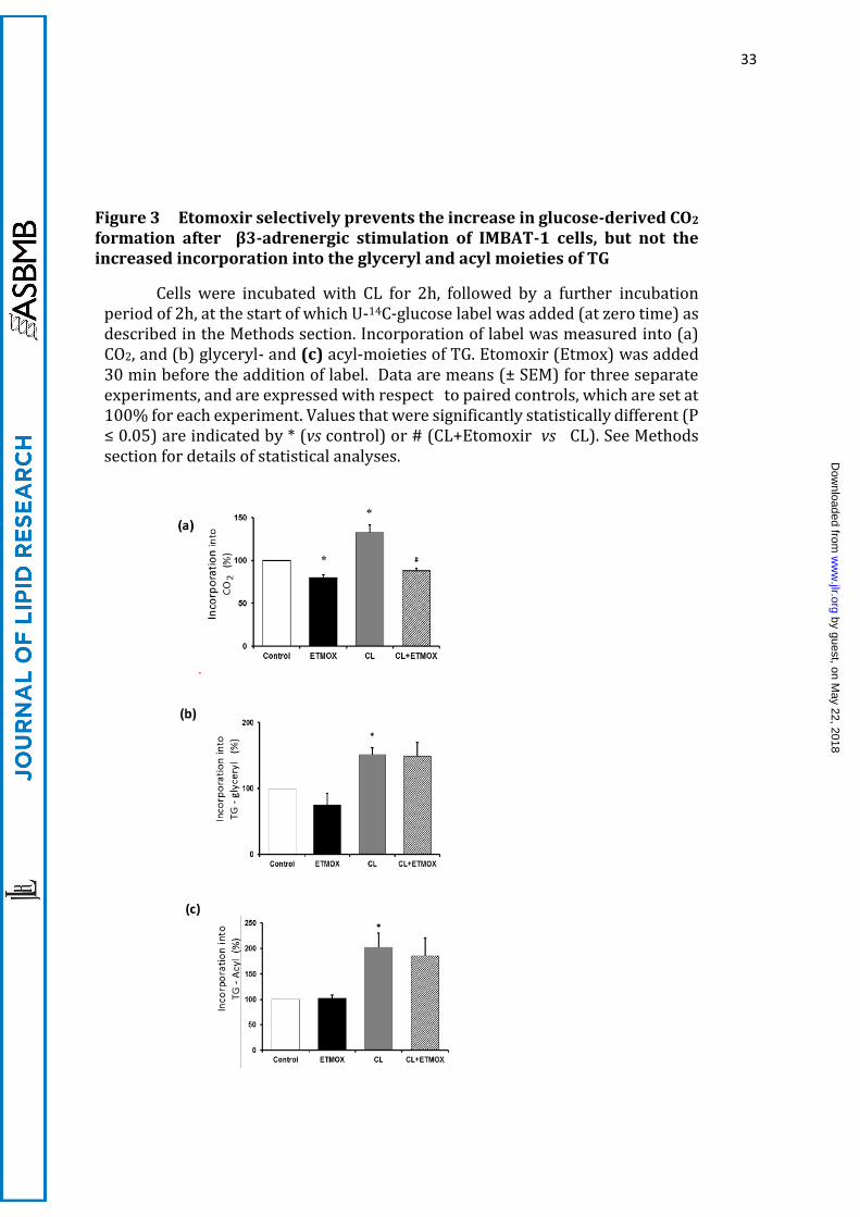
Figure 3 Etomoxir selectively prevents the increase in glucose-derived CO2 formation after β3-adrenergic stimulation of IMBAT-1 cells, but not the increased incorporation into the glyceryl and acyl moieties of TG
Cells were incubated with CL for 2h, followed by a further incubation period of 2h, at the start of which U-14C-glucose label was added (at zero time) as described in the Methods section. Incorporation of label was measured into (a) CO2, and (b) glyceryl- and (c) acyl-moieties of TG. Etomoxir (Etmox) was added 30 min before the addition of label. Data are means (± SEM) for three separate experiments, and are expressed with respect to paired controls, which are set at 100% for each experiment. Values that were significantly statistically different (P ≤ 0.05) are indicated by * (vs control) or # (CL+Etomoxir vs CL). See Methods section for details of statistical analyses.
by guest, on May 22, 2018
ww
w.jlr.org
Dow
nloaded from
34
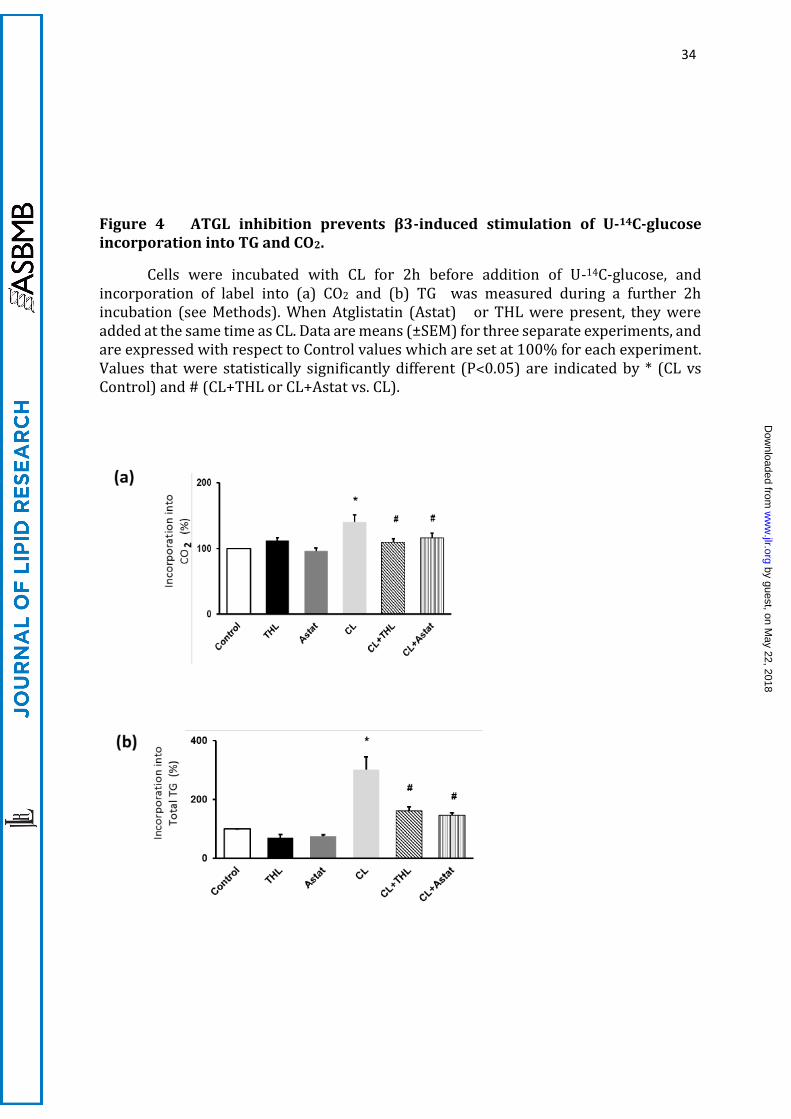
Figure 4 ATGL inhibition prevents β3-induced stimulation of U-14C-glucose incorporation into TG and CO2.
Cells were incubated with CL for 2h before addition of U-14C-glucose, and incorporation of label into (a) CO2 and (b) TG was measured during a further 2h incubation (see Methods). When Atglistatin (Astat) or THL were present, they were added at the same time as CL. Data are means (±SEM) for three separate experiments, and are expressed with respect to Control values which are set at 100% for each experiment. Values that were statistically significantly different (P˂0.05) are indicated by * (CL vs Control) and # (CL+THL or CL+Astat vs. CL).
by guest, on May 22, 2018
ww
w.jlr.org
Dow
nloaded from
35
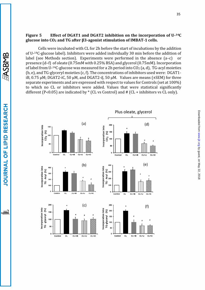
Figure 5 Effect of DGAT1 and DGAT2 inhibition on the incorporation of U-14C glucose into CO2 and TG after β3-agonist stimulation of IMBAT-1 cells.
Cells were incubated with CL for 2h before the start of incubations by the addition of U-14C-glucose label). Inhibitors were added individually 30 min before the addition of label (see Methods section). Experiments were performed in the absence (a–c) or presence (d–f) of oleate (0.75mM with 0.25% BSA) and glycerol (0.75mM). Incorporation of label from U-14C-glucose was measured for a 2h period into CO2 (a, d), TG-acyl moieties (b, e), and TG-glyceryl moieties (c, f). The concentrations of inhibitors used were: DGAT1-iB, 0.75 µM; DGAT2-iC, 50 µM, and DGAT2-iJ, 50 µM. Values are means (±SEM) for three separate experiments and are expressed with respect to values for Controls (set at 100%) to which no CL or inhibitors were added. Values that were statistical significantly different (P˂0.05) are indicated by * (CL vs Control) and # (CL + inhibitors vs CL only).
by guest, on May 22, 2018
ww
w.jlr.org
Dow
nloaded from
36
Figure 6 Effects of siRNA-mediated DGAT1- or DGAT2-knockdown on the respective levels of mRNA expression of the two enzymes
Differentiated cells were treated for 72h with either control (scrambled, SC) siRNA or siRNA targeted towards DGAT1 (SiD1) or DGAT2 (SiD2) and the level of (a) DGAT1 and (b) DGAT2 mRNA expression was measured. Values (n=3) are means (±SEM) for a representative experiment. Values that were statistically significantly different (P˂0.05) from SC are indicated by an asterisk.
by guest, on May 22, 2018
ww
w.jlr.org
Dow
nloaded from
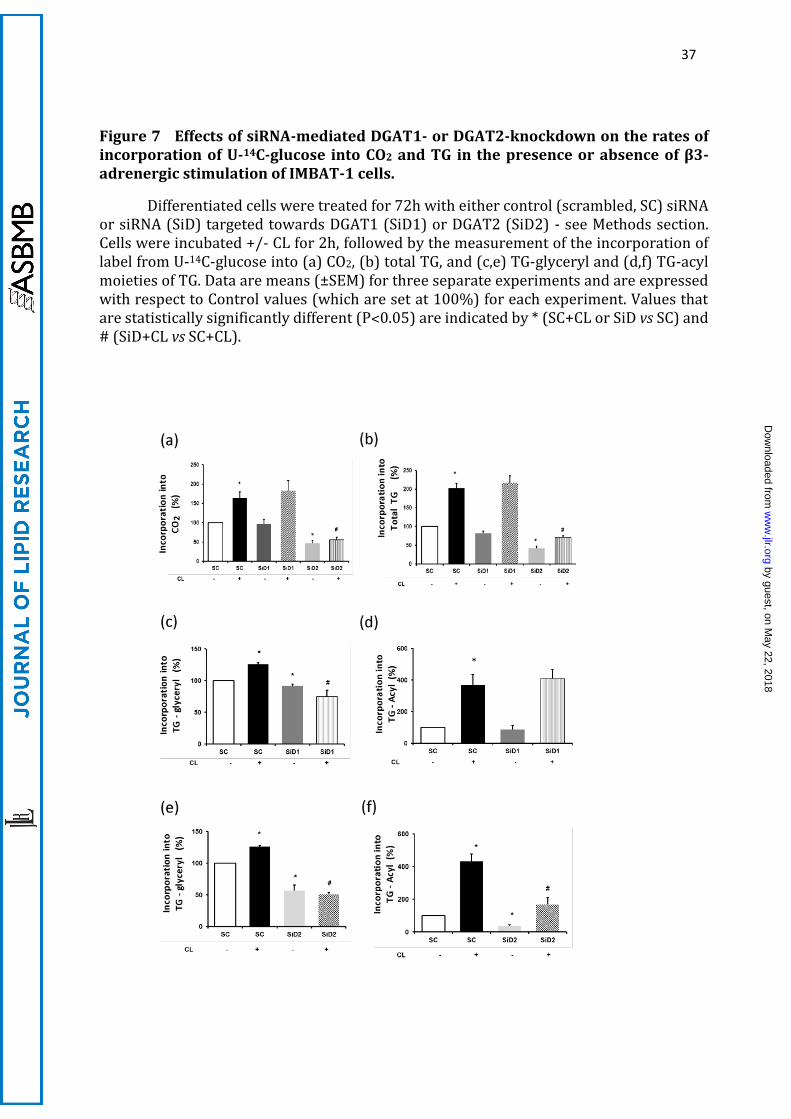
37
Figure 7 Effects of siRNA-mediated DGAT1- or DGAT2-knockdown on the rates of incorporation of U-14C-glucose into CO2 and TG in the presence or absence of β3-adrenergic stimulation of IMBAT-1 cells.
Differentiated cells were treated for 72h with either control (scrambled, SC) siRNA or siRNA (SiD) targeted towards DGAT1 (SiD1) or DGAT2 (SiD2) - see Methods section. Cells were incubated +/- CL for 2h, followed by the measurement of the incorporation of label from U-14C-glucose into (a) CO2, (b) total TG, and (c,e) TG-glyceryl and (d,f) TG-acyl moieties of TG. Data are means (±SEM) for three separate experiments and are expressed with respect to Control values (which are set at 100%) for each experiment. Values that are statistically significantly different (P˂0.05) are indicated by * (SC+CL or SiD vs SC) and # (SiD+CL vs SC+CL).
by guest, on May 22, 2018
ww
w.jlr.org
Dow
nloaded from
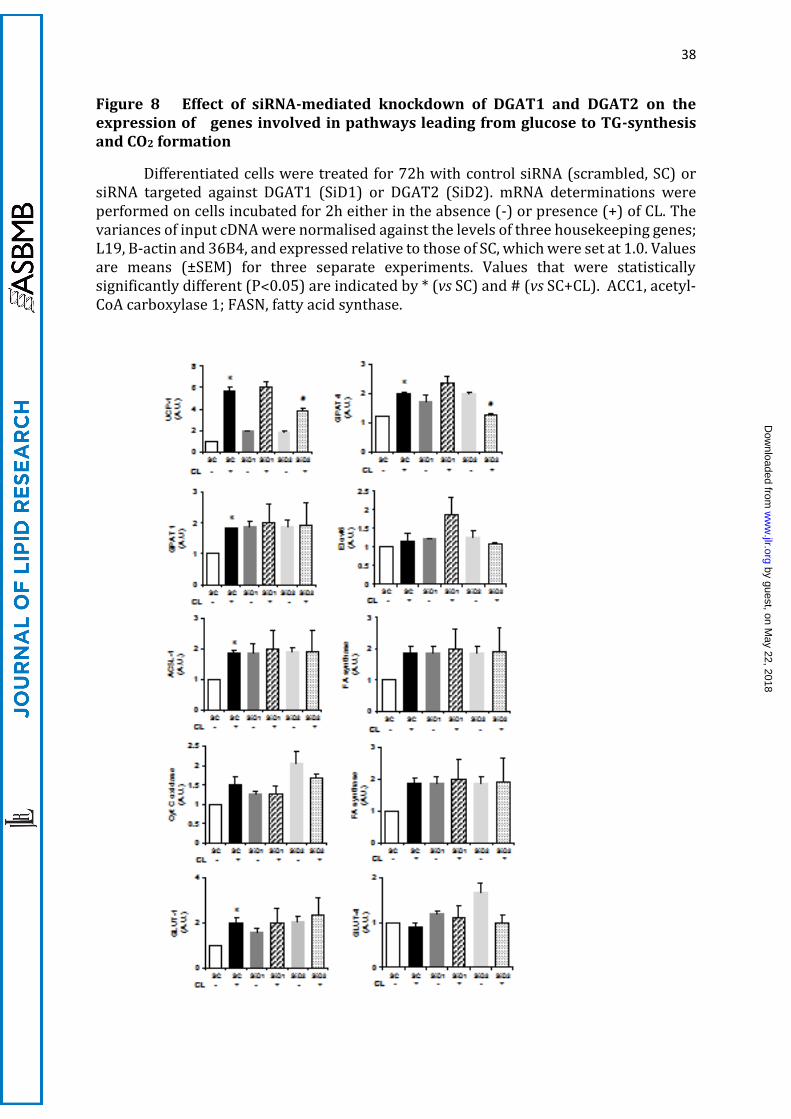
38
Figure 8 Effect of siRNA-mediated knockdown of DGAT1 and DGAT2 on the expression of genes involved in pathways leading from glucose to TG-synthesis and CO2 formation
Differentiated cells were treated for 72h with control siRNA (scrambled, SC) or siRNA targeted against DGAT1 (SiD1) or DGAT2 (SiD2). mRNA determinations were performed on cells incubated for 2h either in the absence (-) or presence (+) of CL. The variances of input cDNA were normalised against the levels of three housekeeping genes; L19, B-actin and 36B4, and expressed relative to those of SC, which were set at 1.0. Values are means (±SEM) for three separate experiments. Values that were statistically significantly different (P˂0.05) are indicated by * (vs SC) and # (vs SC+CL). ACC1, acetyl-CoA carboxylase 1; FASN, fatty acid synthase.
by guest, on May 22, 2018
ww
w.jlr.org
Dow
nloaded from
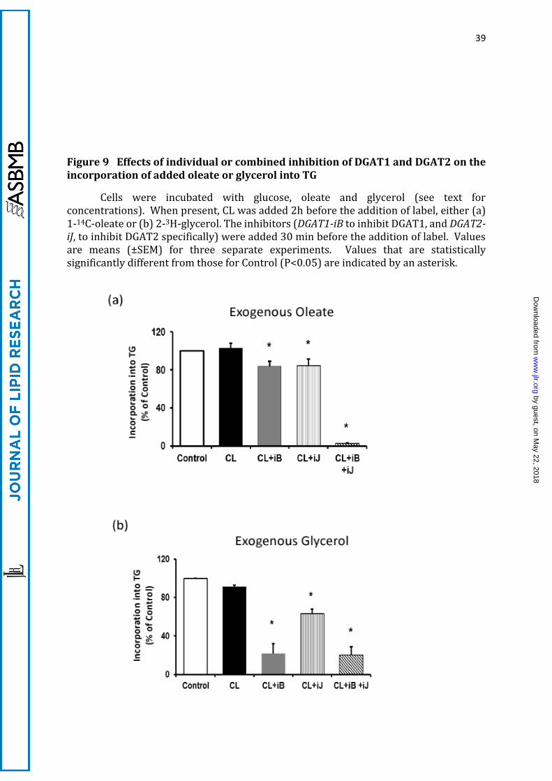
39
Figure 9 Effects of individual or combined inhibition of DGAT1 and DGAT2 on the incorporation of added oleate or glycerol into TG
Cells were incubated with glucose, oleate and glycerol (see text for concentrations). When present, CL was added 2h before the addition of label, either (a) 1-14C-oleate or (b) 2-3H-glycerol. The inhibitors (DGAT1-iB to inhibit DGAT1, and DGAT2-iJ, to inhibit DGAT2 specifically) were added 30 min before the addition of label. Values are means (±SEM) for three separate experiments. Values that are statistically significantly different from those for Control (P<0.05) are indicated by an asterisk.
by guest, on May 22, 2018
ww
w.jlr.org
Dow
nloaded from
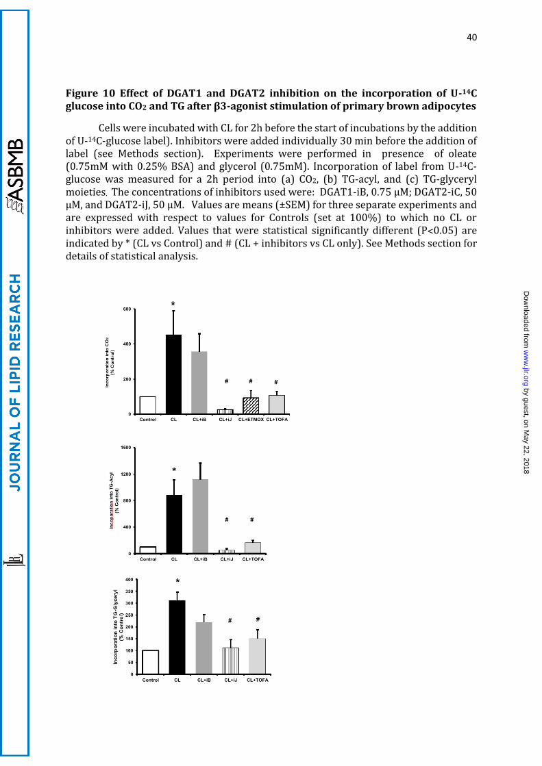
40
Figure 10 Effect of DGAT1 and DGAT2 inhibition on the incorporation of U-14C glucose into CO2 and TG after β3-agonist stimulation of primary brown adipocytes
Cells were incubated with CL for 2h before the start of incubations by the addition of U-14C-glucose label). Inhibitors were added individually 30 min before the addition of label (see Methods section). Experiments were performed in presence of oleate (0.75mM with 0.25% BSA) and glycerol (0.75mM). Incorporation of label from U-14C-glucose was measured for a 2h period into (a) CO2, (b) TG-acyl, and (c) TG-glyceryl moieties. The concentrations of inhibitors used were: DGAT1-iB, 0.75 µM; DGAT2-iC, 50 µM, and DGAT2-iJ, 50 µM. Values are means (±SEM) for three separate experiments and are expressed with respect to values for Controls (set at 100%) to which no CL or inhibitors were added. Values that were statistical significantly different (P˂0.05) are indicated by * (CL vs Control) and # (CL + inhibitors vs CL only). See Methods section for details of statistical analysis.
by guest, on May 22, 2018
ww
w.jlr.org
Dow
nloaded from
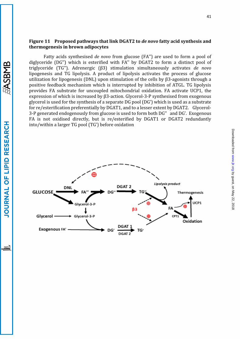
41
Figure 11 Proposed pathways that link DGAT2 to de novo fatty acid synthesis and thermogenesis in brown adipocytes
Fatty acids synthesised de novo from glucose (FA’’) are used to form a pool of diglyceride (DG’’) which is esterified with FA’’ by DGAT2 to form a distinct pool of triglyceride (TG’’). Adrenergic (β3) stimulation simultaneously activates de novo lipogenesis and TG lipolysis. A product of lipolysis activates the process of glucose utilization for lipogenesis (DNL) upon stimulation of the cells by β3-agonists through a positive feedback mechanism which is interrupted by inhibition of ATGL. TG lipolysis provides FA substrate for uncoupled mitochondrial oxidation. FA activate UCP1, the expression of which is increased by β3-action. Glycerol-3-P synthesised from exogenous glycerol is used for the synthesis of a separate DG pool (DG’) which is used as a substrate for re/esterification preferentially by DGAT1, and to a lesser extent by DGAT2. Glycerol-3-P generated endogenously from glucose is used to form both DG’’ and DG’. Exogenous FA is not oxidised directly, but is re/esterified by DGAT1 or DGAT2 redundantly into/within a larger TG pool (TG’) before oxidation
by guest, on May 22, 2018
ww
w.jlr.org
Dow
nloaded from