Embed Size (px)
Citation preview

RESEARCH Open Access
Diacylated lipopeptide from Mycoplasma synoviaemediates TLR15 induced innate immuneresponsesIrena Oven1, Katarina Resman Rus1, Daliborka Dušanić1, Dušan Benčina1, Calvin L Keeler Jr2 and Mojca Narat1*
Abstract
Avian-specific toll like receptor 15 (TLR15) is functionally equivalent to a group of TLR2 family proteins that themammalian innate immune system utilizes to recognize a broad spectrum of microbe-associated molecularpatterns, including bacterial lipoproteins. In this study we examined the role of chicken TLR2 family members in theinnate immune response to the avian pathogenic bacterium, Mycoplasma synoviae. We found that Mycoplasmasynoviae, and specifically the N-terminal diacylated lipopeptide (MDLP) representing the amino-terminal portion ofits mature haemagglutinin protein, significantly induces the expression of TLR15, but not TLR1 and TLR2 in chickenmacrophages and chondrocytes. TLR15 activation is specific and depends on diacylation of the lipopeptide.Activation of TLR15 after stimulation with Mycoplasma synoviae and MDLP triggers an increase in the expression oftranscription factor nuclear factor kappa B and nitric oxide production. Moreover, transfection of avian macrophagecells with small interfering RNA reduces the expression of TLR15 after stimulation with MDLP. This leads todecreased activation of the innate immune response, as measured by nitric oxide production. Additionally,pretreatment of cells with neutralizing anti-TLR15 antibody results in a notable attenuation of MDLP-driven releaseof nitric oxide. This positive correlation may constitute a mechanism for stimulating the innate immune responseagainst avian mycoplasmas in chicken cells via TLR15.
IntroductionMycoplasmas are the smallest self-replicating organisms,and are distinguished from other bacteria by their smallsize and total lack of a cell wall. As obligate parasitesthey usually exhibit strict host and tissue specificity.Mycoplasmas have been shown to interact with thehost’s immune system on many levels, which includesmodulating the host immune system and stimulating aninflammatory response. These abilities enable mycoplas-mas to establish a chronic, persistent infection (reviewedin [1]). In poultry the most pathogenic species are Myco-plasma synoviae and Mycoplasma gallisepticum. Myco-plasma synoviae most frequently colonizes the upperrespiratory tract, causing subclinical infections, althoughthis condition can also lead to the development of sys-temic infection and/or infectious synovitis in chickensand turkeys [2,3]. In the absence of a cell wall, the
majority of the mycoplasma surface antigens are lipopro-teins. In the avian pathogens Mycoplasma gallisepticumand Mycoplasma synoviae an abundantly expressedvariable lipoprotein haemagglutinin (VlhA) is believedto play a major role in pathogenesis of the disease bymediating adherence and immune evasion [4]. VlhA ispost-translationaly cleaved into 2 proteins, the aminoterminal lipoprotein portion MSPB and the more anti-genically variable C terminal haemagglutinin MSPA. Inphenotypically distinct Mycoplasma synoviae popula-tions truncated forms of MSPB (tMSPB) also occur[3,5,6]. Both MSPB and tMSPB contain an amino ter-minal proline rich region [5], which has been shown toinduce strong local and systemic antibody responses ininfectious synovitis [3] and the production of proin-flammatory cytokines and other effector molecules [7],although the mechanisms underlying this response arestill not clear. Other Mycoplasma lipoproteins andlipopeptides have also been found to be subject tosimilar post-translational modifications. One of these
* Correspondence: [email protected] of Animal Science, Biotechnical Faculty, University of Ljubljana,Groblje 3, SI-1230 Domžale, SloveniaFull list of author information is available at the end of the article
VETERINARY RESEARCH
© 2013 Oven et al.; licensee BioMed Central Ltd. This is an open access article distributed under the terms of the CreativeCommons Attribution License (http://creativecommons.org/licenses/by/2.0), which permits unrestricted use, distribution, andreproduction in any medium, provided the original work is properly cited.
Oven et al. Veterinary Research 2013, 44:99http://www.veterinaryresearch.org/content/44/1/99
is the macrophage stimulatory lipopeptide MALP-2 fromMycoplasma fermentas, which is derived from a 43 kDasurface lipoprotein that is post-translationally cleavedinto the mature 14-residue long N-terminal lipopeptideMALP-2 [8].Bacterial lipoproteins, including mycoplasmal lipopro-
teins, belong to a group of pathogen associated molecularpatterns (PAMPs) which interact with Toll like receptors(TLRs) present on host cells [9]. In humans, TLR1, TLR2and TLR6 are implicated in the recognition of bacterial li-poproteins, where TLR2 dimerizes with either TLR1 orTLR6 to enhance the recognition of lipoproteins and elicitthe cellular cytokine response through the activation of nu-clear factor kappa B (NF-κB ) and the mitogen activatedprotein kinase signaling cascade [10].In birds, 10 avian TLRs have been described so far, of
which TLR2a, TLR2b, TLR3, TLR4, TLR5, and TLR7 areorthologs to those found in humans. Additionally, avianTLR21, which is absent from mammalian genome, is thefunctional homologue of mammalian TLR9, recognizingCpG DNA. Besides TLR2a and TLR2b, members of theavian TLR2 group also include two TLR1-like proteins(TLR1La and TLR1Lb), and TLR15, which appears to beunique to avian and reptile species [11]. Orthologs of mam-malian TLR6 and TLR10 have not been found in birds(reviewed in [12]). Several studies report that both types ofchTLR1L can interact with both types of chTLR2 in differ-ent combinations in response to bacterial lipoproteins, al-though the findings of these studies are somewhatcontradictory [13-15]. TLR15 has so far been identified onlyin avian and reptile species [11]. Its induction was first ob-served in the cecum of chickens infected with Salmonellaenterica [16]. Later studies have also reported the inductionof chTLR15 after stimulation of chicken fibroblasts and het-erophils by heat-killed Gram-negative and Gram-positivebacteria, but not by any of the other tested TLR agonist[17-20]. A more recent study reported the induction ofTLR15 mRNA expression after stimulation with CpG-oligonucleotide (CpG-ODN), tripalmitoylated lipopeptide(PAM3CSK4) and lipopolysaccharide (LPS) [21], whereasanother study suggested a novel mechanism of activation,where TLR15 is activated through its cleavage by microbialproteases [22]. A third recent study showed that yeast ly-sates can induce the TLR15-dependent activation of NF-κBexpression, however, the exact agonist was not identified[11]. Nevertheless, the fact that TLR15 induction appearsto be unique to the avian species and is molecularly distinctfrom other known TLRs, suggests a specific and uniquerole in defense against avian pathogens [18].In this study we report a novel ligand for TLR15, a
diacylated lipopeptide derived from Mycoplasmasynoviae, a common chicken pathogen. We used a syn-thetic diacylated lipopetide (MDLP) based on the N-terminal sequence of MSPB, and its diacyl modification
was chosen by anology to other Mycoplasma-derivedlipopeptides (eg. MALP-2, FSL-1). Its non acylated pep-tide analog (NAP) was also synthesized. We found thatMDLP was capable of inducing TLR15 expression, whichled to NF-κB activation and nitric oxide production.
Materials and methodsReagents and chemicalsUnless otherwise noted, reagents and chemicals werepurchased from Sigma–Aldrich Corp., St. Louis, USA.
Mycoplasma synoviae cultureMycoplasma synoviae strains WVU 1853 and ULB 01/P4 were grown at 37 °C on modified Frey’s mediumcontaining 12% porcine serum (Life Technologies Inc.,Gaithersburg, USA) and 0.1 g of NAD per liter of brothmedium (Merck & Co. Inc., Whitehouse Station, USA),but without addition of thallium acetate [23].
MSPB lipoprotein isolation and lipopetide / peptidedeterminationMSPB lipoprotein was isolated from Mycoplasma synoviaestrain ULB 01/P4 as previously described [7]. The aminoacid sequence of the N-terminal region of MSPB proteinsof type strain WVU1853 and strain ULB 01/P4 were pre-dicted previously [5] from the 5′-end of the vlhA gene se-quence. The proposed N-terminal amino acid sequence(CGDQTPAPEPTPGNPNTDNPQNPN) was the same inboth strains. Based on this sequence, the 14 amino acidNAP peptide (CGDQTPAPEPTPGN) was synthesized, aswell as the corresponding lipopeptide, MDLP, containingan S-(2,3-bispalmitoyloxypropyl) N-terminal modification(Pam2CGDQTPAPEPTPGN) (both EMC microcollectionsGmbH, Tuebingen, Germany), which mimics the putativediacyl lipid moiety found in mycoplasma lipoproteins [8].
Cell culturesThe chicken macrophage cell line HD11 was cultured inRPMI medium, supplemented with 8% fetal bovine serum(FBS) and 2% chicken serum, at 37 °C in a 5% CO2 atmos-phere. Monocyte-derived macrophages (MDM) were pre-pared by Histopaque®-1077 density gradient centrifugationof chicken blood as described previously [7,24] and platedin RPMI 1640 medium, supplemented with 10% FBS(Hyclone, USA), 100 U/mL penicillin, and 100 U/mLstreptomycin, at 41 °C in a 5% CO2 atmosphere.Primary chicken chondrocytes (CCH) were isolated as
previously described [25]. CCH were cultivated up to sixpassages in DMEM medium supplemented with 7.5%FBS and 2.5% chicken serum. Cells were incubated at37 °C in a 5% CO2 atmosphere.
Oven et al. Veterinary Research 2013, 44:99 Page 2 of 11http://www.veterinaryresearch.org/content/44/1/99

Macrophage exposure to Mycoplasma synoviae,Pam2CGDQTPAPEPTPGN lipopeptide (MDLP) andCGDQTPAPEPTPGN peptide (NAP)The number of Mycoplasma synoviae cells per macro-phage (HD11) was adjusted using the colony formingunits (CFU) technique, as previously described [24].HD11 cells were exposed to bacteria by replacing theirgrowth medium with medium containing bacteria at amultiplicity of approximately 100 viable Mycoplasmasynoviae cells per macrophage. HD11, CCH and MDMcells were exposed to 1 μM of MDLP or 1 μM NAP.Non-exposed cells were used as negative controls. Cellcultures exposed to different agents were incubated at37 °C (for HD11 and CCH) or 41 °C (for MDM) and 5%CO2 before harvesting cells for RNA. At 1, 6, or 24 h ofexposure, growth medium was aspirated from plates,RLT lysis buffer (Qiagen Corp., Valencia, USA) wasadded and cells were harvested with a rubber policeman.
Quantitative real-time RT-PCRTo confirm and validate gene expression changes quan-titative real-time RT-PCR (RT-qPCR) was performed on:chTLR15, chTLR1, chTLR2, iNOS and NF-κB usingGAPDH as a housekeeping control gene (Table 1). RT-qPCR primers were designed using PrimerQuestSM (In-tegrated DNA Technologies, Leuven, Belgium) andchecked for specificity in silico with PrimerBlast (NCBI).RNA was isolated from cells using the RNAeasy mini kitfollowing on- column DNase-I digestion in accordancewith the manufacturer’s protocol (Qiagen). cDNA wasreverse transcribed using the High-Capacity cDNA Re-verse Transcription Kit (Applied Biosystems, Foster City,USA). RT-qPCR assays were performed using the 2×FastStart Universal SYBR Green Master Mix (Rox)(Roche Diagnostics GmbH, Mannheim, Germany).Quantitative PCR was performed for each sample intriplicate on an Mx3000p QPCR System (Agilent Tech-nologies – Stratagene, Santa Clara, USA). The three-stepamplification procedure was performed in a 20 μL reac-tion volume containing 300 nM of each primer and150 pg of cDNA. Reaction conditions were set to 10 minat 95 °C (first segment, one cycle), 15 s at 95 °C and
1 min at the Tm of the specific primer pair (secondsegment, 40 cycles) followed by one cycle with 15 s at95 °C, 30 s at the designated Tm and 15 s at 95 °C (dis-sociation curve segment). Data were analyzed usingMxPro 4.0 Software (Agilent Technologies). Gene ex-pression values of non-infected or non-treated cells wereused for gene expression calibration. Appropriate con-trols (no template and no reverse transcription control)were also performed in each run. At least three inde-pendent experiments were performed to collect RNA forRT-qPCR. Relative gene expression was assayed in eachexperiment and experimental condition separately. Nor-malized relative quantities were calculated using the effi-ciency corrected 2-(ΔΔCt) method [26]. The effect ofintra-assay variation on the statistical significance of theresults was reduced by log transformation of normalizedrelative quantities, mean centering and autoscaling bythe method of Willems et al. [27].
Western blotting1 × 106 HD11 cells were cultured in RPMI containing8% FBS and 2% chicken serum overnight and were thenstimulated with 1 μM MDLP or NAP for 24 h. Celllysates were prepared in RIPA buffer. Protein contentwas determined and equal amounts of lysates were frac-tionated by 10% SDS-PAGE and electrotransferred topolyvinylidene difluoride membranes. Rabbit polyclonalanti-TLR15 antibody (Imgenex, San Diego, USA) andanti -actin 2Q1055 antibody (Abcam, Cambridge, UK)were used for the detection of proteins. Detection wasby an enhanced chemiluminescence system (Amersham,Freiburg, Germany).
Nitric oxide assayNitric oxide (NO), induced in HD11 cells by DLP, wasestimated, with some modifications, as previously de-scribed [7]. Briefly, HD11 cells were seeded into 24-wellplates (2 × 10⁵ cells / well) and NAP or MDLP wasadded to a final concentration of 0.5, 1 or 1.5 μM. As apositive control HD11 cells were incubated in the pres-ence of 0.1 μg/mL E. coli O127:B8 LPS. Cell culture su-pernatants were sampled after 6 and 24 h of incubation
Table 1 Oligonucleotides used as primers in RT-qPCR analysis of gene expression in chicken cells.
Gene symbol RefSeq mRNA number Forward primer Reverse primer
GAPDH NM_204305.1 ATCGTCAAGGCTGAGAACGGG ATCTGCCCATTTGATGTTGCT
TLR15 NM_001037835.1 AACCTGGTGCATTTGAGAACCTGC TTTCAGGTGAGGTGCAAGACCAGA
TLR1 NM_001007488.3 AGCTGTGTCAGCATGAGAGGAACT AGTTGGGCGACAACACAAAGATGG
TLR2NM_001161650.1 AGAACGACTCCAACTGGGTGGAAA AGAGCGTCTTGTGGCTCTTCTCAA
NM_204278.1
iNOS NM_204961.1 GCATTCTTATTGGCCCAGGA CATAGAGACGCTGCTGCCAG
NF-κB NM_205134.1 AGGACTTAAAATGGCAGGAGA GCTGTTCGTAGTGGTAAGTCT
Oven et al. Veterinary Research 2013, 44:99 Page 3 of 11http://www.veterinaryresearch.org/content/44/1/99

at 37 °C, in 5% CO2. Control supernatants were sampledafter 24 h. Quantification of nitric oxide was performedusing the Griess Reagent System (Promega GmbH,Mannheim, Germany), according to the manufacturer’sinstructions.
RNA silencingSilencing of TLR15 was performed using the ScreeningDsiRNA TriFECTa Kit (cat.# GGC.RNAI.N001037835.12,Integrated DNA Technologies). A universal negative con-trol siRNA (NC siRNA) not homologous to any knowntranscript in the vertebrate transcriptome, was used tonormalize relative gene inhibition of the target gene. Apositive control siRNA against HPRT-1 was used to assesstransfection and knockdown efficiency. Twenty-four h be-fore transfection, 8 × 10⁴ HD11 cells / well were seededonto a 24-well plate to reach 40-50% confluence. Thesecells were transfected with a cocktail of three TLR15siRNAs (150 nM total) or a nonsense negative control (NCsiRNA), using the chemical transfection reagent X-tremeGENE siRNA Transfection reagent (Roche). Transfectionwas performed according to the manufacturer’s instruc-tions. Briefly, siRNAs were diluted in 50 μL Opti-MEMmedium and mixed gently. Four μL of X-tremeGENE wasdiluted in 50 μL Opti-MEM medium and combined withthe diluted RNAi duplex, incubated for 20 min at roomtemperature, then added to each well containing cells togive a final volume of 500 μL and a final siRNA concentra-tion of 150 nM. The conditions for siRNA transfectionwere optimized by adjusting different siRNA concentrations(10, 50, and 150 nM) and different transfection methods(reverse and forward) using Tye 563™ Fluorescent Control(Integrated DNA Technologies). Transfection efficiencywas evaluated under a fluorescent microscope. Eight hoursafter transfection, HD11 cells were treated with 1 μMMDLP or NAP for 12 h, and RNA isolated from cells orNO was measured in the supernatant as described above.
Antibody neutralization assayHD11 cells (5 × 10⁴ cells / well) were seeded onto 96-well culture plates and pre-treated with 2.5 μL of rabbitpolyclonal anti-TLR15 antibodies (Imgenex), 2.5 μL ofnormal rabbit serum (1:1 in glycerol) or PBS for 1 h at37 °C in 5% CO2. Following the 1 h incubation, 1 μMMDLP or NAP were applied in the continuing presenceof neutralizing antibodies, and the cells were further in-cubated overnight in the same conditions. NO from cellsupernatants was determined as described above.
Statistical analysisStatistical analysis was performed for selected experi-ments to determine the confidence limit at which twomeasurements were statistically different. A Student t-test (p < 0.05) was applied to each dataset in SigmaPlot
program, and p-values were obtained and are reportedin the figure legends.
ResultsExpression of TLR15, TLR1 and TLR2 genes after infectionof HD11 cells with Mycoplasma synoviaeWe first sought to determine which chicken TLR2 fam-ily members are involved in the immune response toMycoplasma synoviae infection of macrophages. There-fore, we analyzed the expression of TLR15, TLR1 andTLR2 genes after infection of HD11 macrophages withlive Mycoplasma synoviae for 1, 6 or 24 h. For the ana-lysis of TLR1 and TLR2 mRNA expression we usedprimers that recognized both forms of each chTLR re-ceptors. As shown in Figure 1A, infection of HD11 cellswith Mycoplasma synoviae significantly induces the ex-pression of TLR15 (approximately 8-fold, p < 0.001) after1 h and 6 h, whereas after 24 h the expression of TLR15was not up-regulated compared with non-infected cells.Interestingly, the expression of TLR1 and TLR2 was notup-regulated; moreover, they were significantly down-regulated after 6 h and 24 h (p < 0.001) (Figure 1A).It was previously shown that the N-terminal lipoprotein
fraction of VlhA is responsible for the induction of a strongimmune response [7]. Based on the N-terminal sequence ofthe MSPB protein, and by analogy to MALP-2 and FSL-1,we synthesized the Pam2CGDQTPAPEPTPGN lipopeptide,termed MDLP, and its non-acylated peptide analog, NAP.We analyzed the transcription levels of the TLR15, TLR1and TLR2 genes in HD11 cells after treating them with ei-ther 1 μM MDLP or NAP for 1 h, 6 h or 24 h. As shown inFigure 1B MDLP significantly induces the expression ofTLR15 compared with non-treated cells and NAP-treatedcells. The expression of TLR15 was highest after 1 h of ex-posure (13-fold compared with non-treated cells,p < 0.001). The expression of TLR15 was not up-regulatedwhen the cells were treated with the non-modified peptide(Figure 1B, NAP). The expression of TLR15 was still up-regulated after 6 h and 24 h (6.5-fold and 5-fold, respect-ively, p < 0.001). Interestingly, as with the Mycoplasmasynoviae infection experiment, the mRNA expressions ofTLR1 and TLR2 were significantly down-regulated after thetreatment of the cells with MDLP (p < 0.01), whereas whentreated with NAP, the expression remained the same com-pared to non-treated cells (Figure 1B, TLR1 and TLR2,respectively).Since HD11 cells are a virally transformed macrophage
cell line, we wanted to confirm that this cell line is a suit-able model for our experiments by analyzing the expressionof TLR15, TLR1 and TLR2 genes in primary macrophages.Therefore, we isolated monocyte derived macrophagesfrom chicken blood and treated them with 1 μM of MDLPor NAP for 1 h, 6 h or 24 h. At each time point expressionof the TLR15, TLR1 and TLR2 genes was analyzed. As
Oven et al. Veterinary Research 2013, 44:99 Page 4 of 11http://www.veterinaryresearch.org/content/44/1/99

shown in Figure 1C MDLP significantly induces the expres-sion of TLR15 compared with non-treated cells and NAP-treated cells. The expression of TLR15 was highest after 1 hof exposure (4.6-fold compared with non-treated cells,p = 0.003). The expression of TLR15 was not up-regulatedwhen the cells were treated with the non-modified peptide(Figure 1C, NAP). At longer treatment times (6 h and 24 h)the expression of TLR15 was still induced although thelevel was not statistically significant (2.6-fold and 1.5-fold,p = 0.025 and p = 0.139, respectively).
To confirm that the expression of TLR15 was inducednot only at the mRNA level, but also at the protein level,we performed a western blot analysis in HD11 cells after24 h of exposure to MDLP or NAP. Changes in proteinlevels were consistent with the mRNA data, showingthat NAP did not affect the expression of TLR15 pro-tein, whereas treatment with MDLP induced up-regulation of TLR15 protein expression (Figure 1D).Mycoplasma synoviae has been shown to interact with
non-immune cells, and is often isolated from the joints
Figure 1 Mycoplasma synoviae and its diacylated lipopeptide induce expression of TLR15 in chicken macrophages. A) Expression ofTLR15, TLR1 and TLR2 mRNA after infection of HD11 cells with Mycoplasma synoviae. HD11 cells were infected with live Mycoplasma synoviae (MS)for 1 h, 6 h or 24 h and mRNA expression was analyzed by RT-qPCR. NC represents non-infected cells. Bars show mean ± S.E. (n = 4); ***, p < 0.001compared to non-infected cells. B) Expression of TLR15, TLR1 and TLR2 mRNA after treatment of HD11 cells with diacylated lipopeptide or its non-acylated analog. HD11 cells were treated with 1 μM non-acylated peptide (NAP) or diacylated lipopeptide (MDLP) for 1 h, 6 h or 24 h and mRNAexpression was analyzed by RT-qPCR. NC represents non-treated cells. Bars show mean ± S.E. (n = 4); **, p < 0.01 and ***, p < 0.001 compared toNAP-treated cells at each time point for selected gene. C) Expression of TLR15, TLR1 and TLR2 mRNA after treatment of MDM cells with diacylatedlipopeptide or its non-acylated analog. MDM cells were treated with 1 μM non-acylated peptide (NAP) or diacylated lipopeptide (MDLP) for 1 h,6 h or 24 h and mRNA expression was analyzed by RT-qPCR. NC represents non-treated cells. Bars show mean ± S.E. (n = 3); *, p < 0.05 and **,p < 0.01 compared to NAP-treated cells at each time point for selected gene. D) Expression levels of TLR15 and β-actin, as determined byWestern blotting. HD11 cells were treated with NAP or MDLP for 24 h and TLR15 protein levels were determined. Levels of endogenous β-actinwere determined by Western blotting to validate input for each sample.
Oven et al. Veterinary Research 2013, 44:99 Page 5 of 11http://www.veterinaryresearch.org/content/44/1/99

of chickens showing signs of infectious synovitis. To in-vestigate if Mycoplasma synoviae can modify the expres-sion of chicken TLR2-family genes in non-immune cells,we infected primary chicken chondrocyte cells (CCH)with Mycoplasma synoviae for 1 h, 6 h and 24 h, and an-alyzed the expression of TLR15, TLR1 and TLR2 genes.In contrast to the response in the immune cells, the ex-pression of TLR15 was the highest after 24 h of infection(11-fold, p < 0.001), whereas TLR1 and TLR2 expressionremained unchanged or was even slightly down-regulated, correlating to the results observed in chickenmacrophages (Figure 2A). We found similar resultswhen we treated CCH cells with MDLP or NAP. MDLPinduced TLR15 expression in CCH after 1 h (Figure 2B,6-fold, p = 0.005), and the expression of the TLR15 wasfurther increased after 6 h and 24 h (Figure 2B, 12-foldand 17-fold, p < 0.001 and p = 0.002, respectively). NAPdid not change the expression of any of the analyzedTLRs compared with non-infected cells (Figure 2B).
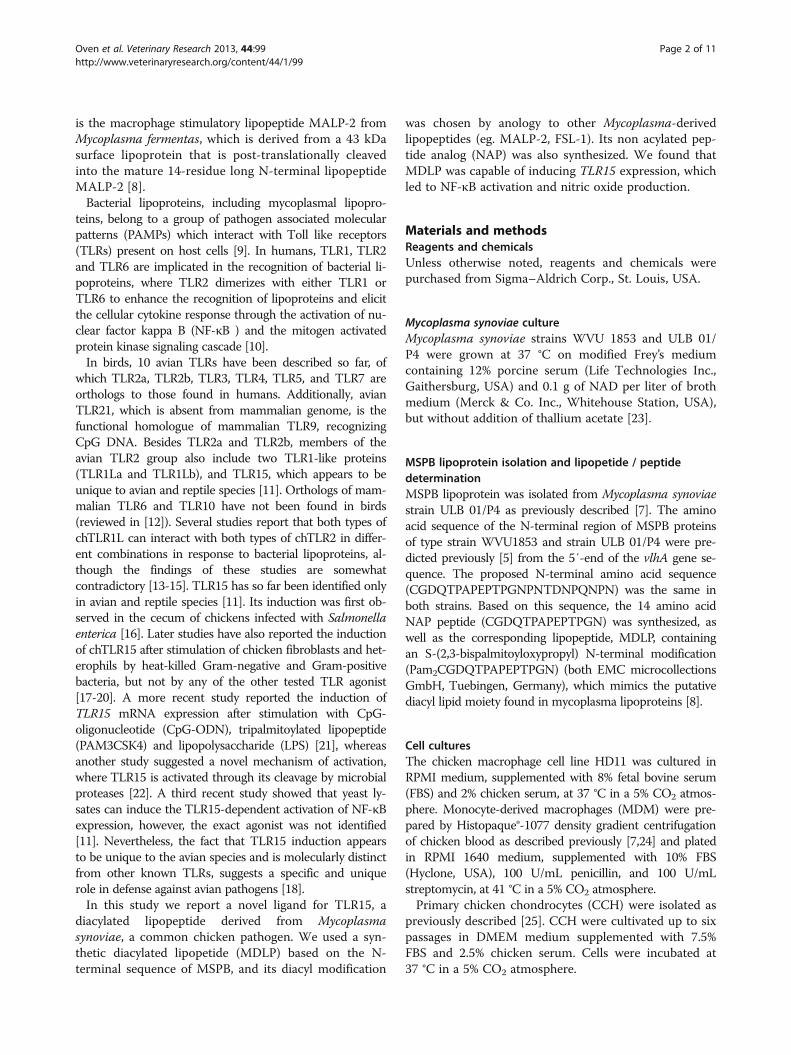
Increased TLR15 expression is associated with increasedNF-κB, iNOS message and nitric oxide productionTo further investigate the mechanism of the TLR15 me-diated innate immune response, we measured NF-κBmRNA expression following infection of HD11 cells withviable Mycoplasma synoviae or treatment with MDLPcompared to non-infected or NAP-treated cells as con-trols. Both viable Mycoplasma synoviae cells and MDLP
induced elevated levels of NF-κB transcripts in HD11cells after 1 h and 6 h (2.3- and 3.6-fold, p < 0.01 forMycoplasma synoviae, and 3- and 3.4-fold, p < 0.001 forMDLP, respectively), whereas non-stimulated or NAPstimulated cells did not induce NF-κB transcription(Figure 3). The expression of NF-κB mRNA wasinhibited after 24 h (Figure 3).Increased NF-κB mRNA expression also corresponded
to an increased innate immune response, as monitoredby changes in iNOS mRNA level and its associated NOproduction as a functional measure of TLR15 activation.HD11 cells were infected with Mycoplasma synoviae ortreated with 1 μM MDLP or NAP for 1 h, 6 h and 24 h,and iNOS mRNA levels were measured by RT-qPCR.The infection of cells with Mycoplasma synoviae ortreatment with MDLP resulted in a significant increasein iNOS mRNA expression after 6 h and 24 h(Figure 4A). The treatment of cells with NAP did not in-duce iNOS expression at any time point (Figure 4A). Wenext investigated if elevated iNOS mRNA levels correlatewith the production of NO in HD11 cell culture super-natants after stimulation of the cells with increasingamount of MDLP or NAP. LPS was used as a positivecontrol. As shown in Figure 4B, treatment of cells withMDLP or LPS indeed resulted in NO production after6 h and 24 h, whereas NAP did not stimulate NO pro-duction. In addition, MDLP induced NO at similar levelscompared to LPS (Figure 4B). MDLP was also able to
Figure 2 Mycoplasma synoviae and its diacylated lipopeptide induce expression of TLR15 in primary chicken chondrocytes. A)Expression of TLR15, TLR1 and TLR2 mRNA after infection of cells with Mycoplasma synoviae. Primary chicken chondrocytes were infected with liveMycoplasma synoviae (MS) for 1 h, 6 h or 24 h and mRNA expression was analyzed by RT-qPCR. NC represents non-infected cells. Bars showmean ± S.E. (n = 4); ***, p < 0.001 compared to non-infected cells. B) Expression of TLR15, TLR1 and TLR2 mRNA after treatment of cells withdiacylated lipopeptide or its non-acylated analog. Primary chicken chondrocytes were treated with 1 μM non-acylated peptide (NAP) ordiacylated lipopeptide (MDLP) for 1 h, 6 h or 24 h and mRNA expression was analyzed by RT-qPCR. NC represents non-treated cells. Bars showmean ± S.E. (n = 4); **, p < 0.01 and ***, p < 0.001 compared to NAP-treated cells at each time point for selected gene.
Oven et al. Veterinary Research 2013, 44:99 Page 6 of 11http://www.veterinaryresearch.org/content/44/1/99
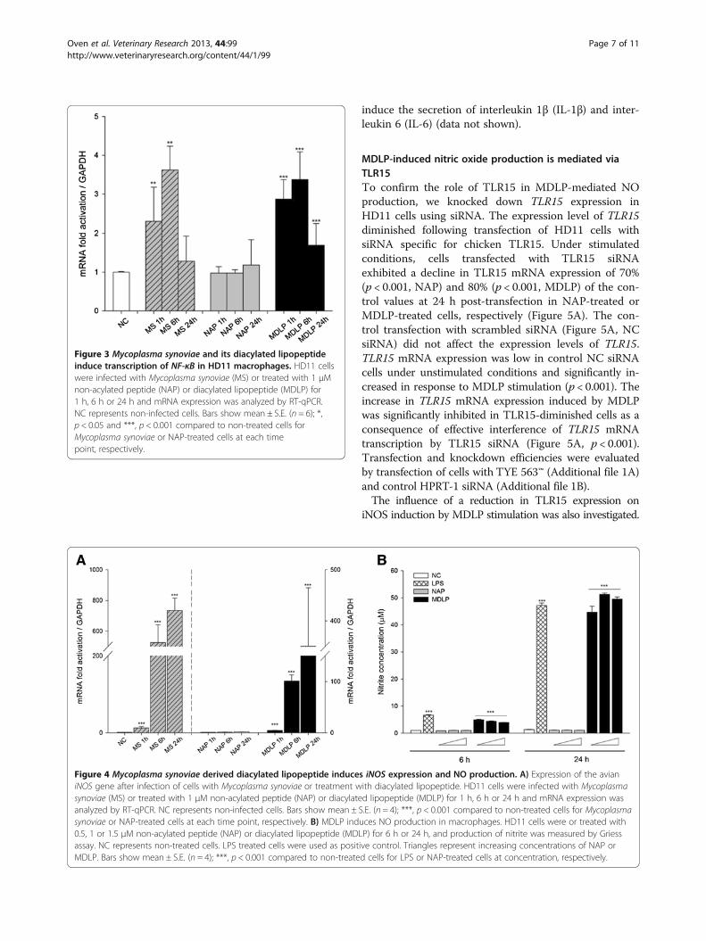
induce the secretion of interleukin 1β (IL-1β) and inter-leukin 6 (IL-6) (data not shown).
MDLP-induced nitric oxide production is mediated viaTLR15To confirm the role of TLR15 in MDLP-mediated NOproduction, we knocked down TLR15 expression inHD11 cells using siRNA. The expression level of TLR15diminished following transfection of HD11 cells withsiRNA specific for chicken TLR15. Under stimulatedconditions, cells transfected with TLR15 siRNAexhibited a decline in TLR15 mRNA expression of 70%(p < 0.001, NAP) and 80% (p < 0.001, MDLP) of the con-trol values at 24 h post-transfection in NAP-treated orMDLP-treated cells, respectively (Figure 5A). The con-trol transfection with scrambled siRNA (Figure 5A, NCsiRNA) did not affect the expression levels of TLR15.TLR15 mRNA expression was low in control NC siRNAcells under unstimulated conditions and significantly in-creased in response to MDLP stimulation (p < 0.001). Theincrease in TLR15 mRNA expression induced by MDLPwas significantly inhibited in TLR15-diminished cells as aconsequence of effective interference of TLR15 mRNAtranscription by TLR15 siRNA (Figure 5A, p < 0.001).Transfection and knockdown efficiencies were evaluatedby transfection of cells with TYE 563™ (Additional file 1A)and control HPRT-1 siRNA (Additional file 1B).The influence of a reduction in TLR15 expression on
iNOS induction by MDLP stimulation was also investigated.
Figure 3 Mycoplasma synoviae and its diacylated lipopeptideinduce transcription of NF-κB in HD11 macrophages. HD11 cellswere infected with Mycoplasma synoviae (MS) or treated with 1 μMnon-acylated peptide (NAP) or diacylated lipopeptide (MDLP) for1 h, 6 h or 24 h and mRNA expression was analyzed by RT-qPCR.NC represents non-infected cells. Bars show mean ± S.E. (n = 6); *,p < 0.05 and ***, p < 0.001 compared to non-treated cells forMycoplasma synoviae or NAP-treated cells at each timepoint, respectively.
Figure 4 Mycoplasma synoviae derived diacylated lipopeptide induces iNOS expression and NO production. A) Expression of the avianiNOS gene after infection of cells with Mycoplasma synoviae or treatment with diacylated lipopeptide. HD11 cells were infected with Mycoplasmasynoviae (MS) or treated with 1 μM non-acylated peptide (NAP) or diacylated lipopeptide (MDLP) for 1 h, 6 h or 24 h and mRNA expression wasanalyzed by RT-qPCR. NC represents non-infected cells. Bars show mean ± S.E. (n = 4); ***, p < 0.001 compared to non-treated cells for Mycoplasmasynoviae or NAP-treated cells at each time point, respectively. B) MDLP induces NO production in macrophages. HD11 cells were or treated with0.5, 1 or 1.5 μM non-acylated peptide (NAP) or diacylated lipopeptide (MDLP) for 6 h or 24 h, and production of nitrite was measured by Griessassay. NC represents non-treated cells. LPS treated cells were used as positive control. Triangles represent increasing concentrations of NAP orMDLP. Bars show mean ± S.E. (n = 4); ***, p < 0.001 compared to non-treated cells for LPS or NAP-treated cells at concentration, respectively.
Oven et al. Veterinary Research 2013, 44:99 Page 7 of 11http://www.veterinaryresearch.org/content/44/1/99

NC siRNA cells responded to treatment with MDLP with asignificant increase in iNOS expression (Figure 5A, iNOS,p < 0.001). As anticipated, iNOS induction by MDLP stimu-lation was significantly inhibited in TLR15-deficient cells(p= 0.001, Figure 5A, iNOS). Furthermore, this data wascorroborated by observed changes in NO production. Fol-lowing treatment with DLP, NO levels in cell supernatantswere markedly increased in NC siRNA cells, but NO pro-duction was significantly inhibited in TLR15-diminishedcells (p= 0.003, Figure 5B). This data further suggests thatTLR15 may be essential for iNOS induction and NO pro-duction by MDLP stimulation.
MDLP-induced NO production is inhibited by blockingTLR15To further asses the role of TLR15 in MDLP-induced in-nate immune responses in chicken macrophages, we in-vestigated the effect of blocking TLR15 with anti-TLR15antibodies on NO production (Figure 6). Pre-incubationof HD11 cells with neutralizing rabbit polyclonal anti-TLR15 antibody for 1 h, followed by stimulation withMDLP or NAP, resulted in almost complete inhibition ofNO production. Normal rabbit serum showed signifi-cantly lower inhibitory effect in comparison to TLR15antiserum (p = 0.001).
DiscussionIn mammalian cells, lipopeptides mediate an innate im-mune response through interactions with TLR1, TLR2 andTLR6. Chickens lack TLR6 but possess two types of TLR1and two types of TLR2 receptors that are most likely the re-sult of gene duplication [28,29], and an additional avianspecific receptor, TLR15. Therefore, this study was designedto address if TLR15 is involved in the recognition of Myco-plasma synoviae lipoprotein MSPB. We found that thisavian pathogen, and specifically the N-terminal diacylatedlipopeptide portion of MSPB (MDLP), activates the expres-sion of TLR15 in chicken macrophages and primarychicken chondrocytes. The activation of TLR15 leads toNF-κB activation and NO production. The specificity ofthis interaction was confirmed by the inhibition of iNOSexpression and NO production through the depletion ofTLR15 by siRNA. Moreover, when cells were pretreatedwith neutralizing anti-TLR15 antibody, MDLP-mediatedNO production was inhibited. We conclude that byinteracting with TLR15 MDLP stimulates an innate im-mune response in chicken cells.Previous studies by Lavrič et al. showed the induction
of proinflamatory cytokines and chemokines, as well asiNOS, in Mycoplasma synoviae infected chicken macro-phages [24]. Moreover, a second study showed that
Figure 5 Silencing of TLR15 decreases iNOS gene expression and NO production in MDLP stimulated HD11 macrophages. A) Silencingof TLR15 decreases iNOS gene expression. HD11 cells were transfected with siRNA specific for TLR15 (TLR15 siRNA) or scrambled negative controlsiRNA (NC siRNA) for 8 h and then stimulated with NAP or MDLP for additional 12 h. mRNA expression for iNOS and TLR15 was determined byRT-qPCR. Bars show mean ± S.E. (n = 4); **, p < 0.01 and ***, p < 0.001. B) Silencing of TLR15 decreases MDLP-induced NO production. HD11 cellswere transfected with TLR15 siRNA or NC siRNA for 8 h and then stimulated with NAP or MDLP for additional 12 h. After, production of nitritewas measured in cell culture supernatants by Griess assay. Bars show mean ± S.E. (n = 4); ** p < 0.01.
Oven et al. Veterinary Research 2013, 44:99 Page 8 of 11http://www.veterinaryresearch.org/content/44/1/99

lipoprotein MSPB and its amino-terminal part tMSPBwere responsible for a strong immune response inchicken macrophages with production of NO, IL-6 andIL-1β [7], similarly to what has been shown for othermycoplasma lipopeptides, such as MALP-2 and FSL-1[8,30]. The N-terminal amino acid sequence of WVU1853 strain tMSPB is CDGQTPAPEXT [7], in which twoacyl chains are bound to a cysteine [8,31]. The study byLavrič et al. also suggested that the induction ofproinflamatory cytokines as well as iNOS could be afunctional consequence of TLR pathway inductionthrough Mycoplasma synoviae PAMPs.In mammals, mycoplasma lipopeptides MALP-2 and
FSL-1 initiate innate immune response through the TLR2/TLR6 mediated signaling pathway. However, in chickens,the signaling pathway involved in Mycoplasma synoviaemediated immune response has not yet been determined.We have therefore evaluated the involvement of chickenTLR2 subfamily members in the Mycoplasma synoviae andMSPB-derived lipopeptide, MDLP, mediated immune re-sponse. Our results show that MDLP was able to rapidly in-duce the expression of TLR15, whereas expression of theother two chTLR2 family members, TLR1 and TLR2, wasdown-regulated in response to MDLP. This result was spe-cific for the lipid moiety since NAP did not induce anychanges in the expression of chTLR2 family members.
In general, the binding of a TLR to its appropriate lig-and initiates a specific signaling cascade, ultimatelyresulting in the activation of transcription factor NF-κB,and the expression of innate immune response genes,such as proinflamatory cytokines and iNOS. This path-way is mostly conserved in avian species [28]. In thepresent study, the innate immune response to MDLP inHD11 cells was monitored by the induction of iNOSand its associated NO production. We show that stimu-lation of chicken macrophages with Mycoplasmasynoviae or MDLP leads to the activation of NF-κB andiNOS, which in turn leads to the production of nitricoxide. The levels of NF-κB and iNOS expression weretime dependent, with NF-κB peaking at 6 h, followed byiNOS at 24 h. Participation of TLR15 in MDLP stimula-tion was corroborated by reducing TLR15 transcriptionby RNA silencing. iNOS expression and NO productionwere significantly inhibited in TLR15-diminished cellscompared with control NC siRNA cells. Moreover, theMDLP-induced NO response in macrophages was abro-gated by blocking antibodies specific for TLR15.Induction of iNOS, and the resulting increase in NO
production, not only has a positive role in combating in-fectious disease but also contributes to a number ofautoimmune and inflammatory disorders. Mycoplasmasynoviae has been associated with infectious synovitis injoints of naturally and experimentally infected chickens[3,23]. A recent study by Dušanić et al. [32] has shownthat infection of primary chicken chondrocytes withMycoplasma synoviae induces NF-κB expression andNO production. Therefore, we investigated the involve-ment of the chTLR2 subfamily in the recognition ofMycoplasma synoviae and its diacylated lipopeptide inchicken chondrocytes. Our results showed that Myco-plasma synoviae infection induced expression of TLR15mRNA in chicken chodrocytes after 24 h. The expres-sion of TLR1 remained unchanged, whereas the expres-sion of TLR2 was significantly downregulated. We alsoshowed upregulation of TLR15 after 6 h and 24 h ofstimulation by MDLP, but not NAP, whereas the ex-pressions of TLR1 and TLR2 remained unchanged. Theexpression of TLR2 has also been shown in humanchondrocytes, where activation of the TLR2 pathwayled to the production of NO, resulting in an inflamma-tory response [33]. In mice, the expression of TLR2resulted in experimentally induced arthritis [34]. Takentogether these findings suggest that an innate immuneresponse, including MDLP-mediated activation of chickenchondrocytes via the TLR15 pathway, has the potential tocontribute to joint inflammation in infectious synovitis.TLR15 induction was first reported in chicken cecum
following Salmonella infection [16]. A later study hasreported the up-regulation of TLR15 expression afterstimulation of cells with live and heat-killed Gram
Figure 6 Preincubation of HD11 macrophages with anti-TLR15antibody decreases MDLP-stimulated NO response. HD11 cellswere preincubated with rabbit polyclonal anti-TLR15 antibody,normal rabbit serum (rabbit IgG) or phosphate buffer (PBS) for 1 hand then stimulated with NAP or MDLP for 24 h. Nitriteconcentration in cell culture supernatants was measured by Griessassay. PBS-pretreated cells were used as control. Bars show mean ±S.E. (n = 3); ***, p < 0.001.
Oven et al. Veterinary Research 2013, 44:99 Page 9 of 11http://www.veterinaryresearch.org/content/44/1/99
negative and Gram positive bacteria, commonly isolatedfrom chickens, but not with equine specific pathogenRhodococcus equi, therefore suggesting that TLR15may respond specifically to avian pathogens [18]. Intri-guingly, a recent study showed the ability of a yeast lys-ate to activate TLR15-dependent NF-kB pathways inHEK293 cells or stimulate IL-1b mRNA upregulationin chicken macrophages, which was abrogated by heatinactivation or pre-exposure of the lysate to PMSF[11]. Moreover, various TLR agonists have also beenevaluated, including PAM3CSK and FSL-1 [18,21,22].Interestingly, studies by Nerren et al. [18] and Ciraciet al. [21] showed an increase in TLR15 expression fol-lowing stimulation with PAM3CSK. It has been reportedpreviously that not only the acylation of lipopeptides,but also the peptide backbone structure is important inrecognition of lipopeptides by TLR2 in mammals [35,36].Our study differs from others in the use of avian specificMycoplasma derived lipopeptide sequence to stimulatethe expression of an avian specific receptor.In conclusion, this is the first study to report the in-
duction of TLR15 expression and activation of TLR15-mediated immune responses after stimulation with adiacylated lipopeptide derived from the avian pathogenMycoplasma synoviae. These findings demonstrateMDLP as a possible ligand for TLR15. However, they donot exclude other possible ligands for TLR15. Mamma-lian TLR2 has a broad range of ligand specificities, andis capable of recognizing a broad repertoire of PAMPs,including several associated with Gram-positive bacteria,mycobacteria, protozoan parasites, yeast, as well as mi-crobial lipoproteins, glycoproteins, glycolipids and non-enteric LPS [9]. TLR2 has been reported to recognizethese ligands alone or in cooperation with other TLR2family members – TLR1, TLR6 and TLR10. Furtherstudies are needed to determine, whether TLR15 acts ashomodimer or as heterodimer with other chTLRs inresponse to various ligands.
Additional file
Additional file 1: Estimation of siRNA transfection and knock-downefficiency in HD11 cells. A) Transfection of cells with TYE 563™ asobserved under fluorescent microscope. HD11 cells were transfected with150 nM TYE 563™ oligo and its expression was observed underflourescent microscope. Transfected cells are shown in red. B) Knockdown efficiency in HD11 cells transfected with HPRT-1 siRNA asmeasured by RT-qPCR. HD11 cells were transfected with 150 nM negativecontrol (NC siRNA) or siRNA specific for HPRT-1 (HPRT-1 siRNA) for 24 hand mRNA expression of HPRT-1 gene was measured by RT-qPCR. Barsshow mean ± S.E. (n = 3); ***, p < 0.001.
AbbreviationsTLR: Toll like receptor; MDLP: Mycoplasma synoviae-derived diacylatedlipopeptide; NAP: Non-acylated peptide; CCH: Chicken chondocyte cells;LPS: Lipopolysaccharide; PAMP: Pathogen associated molecular pattern;CpG-ODN: CpG-oligonucleotide; PAM3CSK: Tripalmitoylated lipopeptide;
NO: Nitric oxide; iNOS: Inducible nitric oxide synthase; HPRT-1:Hypoxanthine phosphoribosyltransferase 1; NF-κB: Nuclear factor kappa B;GAPDH: Glyceraldehyde 3-phosphate dehydrogenase; RT-qPCR: Quantitativereal time polymerase chain reaction; siRNA: Small interfering ribonucleic acid;IL: interleukin.
Competing interestsThe authors declare that they have no competing interests.
Authors’ contributionsIO conceived the study, carried out the isolation and infection of cells,RNA isolation and subsequent gene expression analysis, siRNA and NOconcentration assays, performed the statistical analysis and drafted themanuscript. KR participated in infection of cells, RNA isolation andsubsequent gene expression analysis. DB carried out Mycoplasmasynoviae cultivation and CFU determination and participated inmanuscript editing. DD advised IO on RT-qPCR and performed isolationof CCH. CK participated in designing the study and manuscript editing.MN participated in designing the study, drafting the manuscript andcoordination between authors. All authors read and approved the finalmanuscript.
AcknowledgementsWe thank Ana Jakopič and Nika Debeljak for technical assistance. This workwas supported by grants J4-2020 and P4-0220 from the Slovenian ResearchAgency, Republic of Slovenia.
Author details1Department of Animal Science, Biotechnical Faculty, University of Ljubljana,Groblje 3, SI-1230 Domžale, Slovenia. 2Department of Animal and FoodSciences, College of Agriculture and Natural Resources, University ofDelaware, 040 Townsend Hall, Newark, DE 19717-1303, USA.
Received: 25 September 2012 Accepted: 3 October 2013Published: 17 October 2013
References1. Razin S, Yogev D, Naot Y: Molecular biology and pathogenicity of
mycoplasmas. Microbiol Mol Biol Rev 1998, 62:1094–1156.2. Lockaby SB, Hoerr FJ, Lauerman LH, Kleven SH: Pathogenicity of
Mycoplasma synoviae in broiler chickens. Vet Pathol 1998, 35:178–190.3. Narat M, Bencina D, Kleven SH, Habe F: The hemagglutination-positive
phenotype of Mycoplasma synoviae induces experimental infectioussynovitis in chickens more frequently than does the hemagglutination-negative phenotype. Infect Immun 1998, 66:6004–6009.
4. Noormohammadi AH: Role of phenotypic diversity in pathogenesis ofavian mycoplasmosis. Avian Pathol 2007, 36:439–444.
5. Bencina D, Drobnic-Valic M, Horvat S, Narat M, Kleven SH, Dovc P:Molecular basis of the length variation in the N-terminal part ofMycoplasma synoviae hemagglutinin. FEMS Microbiol Lett 2001,203:115–123.
6. Noormohammadi AH, Markham PF, Whithear KG, Walker ID, Gurevich VA,Ley DH, Browning GF: Mycoplasma synoviae has two distinct phasevariable major membrane antigens, one of which is a putativehemagglutinin. Infect Immun 1997, 65:2542–2547.
7. Lavric M, Bencina D, Kothlow S, Kaspers B, Narat M: Mycoplasma synoviaelipoprotein MSPB, the N-terminal part of VlhA haemagglutinin, inducessecretion of nitric oxide, IL-6 and IL-1beta in chicken macrophages.Vet Microbiol 2007, 121:278–287.
8. Muhlradt PF, Kiess M, Meyer H, Sussmuth R, Jung G: Isolation, structureelucidation, and synthesis of a macrophage stimulatory lipopeptide fromMycoplasma fermentans acting at picomolar concentration. J Exp Med1997, 185:1951–1958.
9. Qureshi S, Medzhitov R: Toll-like receptors and their role in experimentalmodels of microbial infection. Genes Immun 2003, 4:87–94.
10. Kawai T, Akira S: Signaling to NF-kappaB by Toll-like receptors. Trends MolMed 2007, 13:460–469.
11. Boyd AC, Peroval MY, Hammond JA, Prickett MD, Young JR, Smith AL:TLR15 is unique to avian and reptilian lineages and recognizes a yeast-derived agonist. J Immunol 2012, 189:4930–4938.
Oven et al. Veterinary Research 2013, 44:99 Page 10 of 11http://www.veterinaryresearch.org/content/44/1/99
12. Brownlie R, Allan B: Avian toll-like receptors. Cell Tissue Res 2011,343:121–130.
13. Fukui A, Inoue N, Matsumoto M, Nomura M, Yamada K, Matsuda Y,Toyoshima K, Seya T: Molecular cloning and functional characterization ofchicken toll-like receptors. A single chicken toll covers multiplemolecular patterns. J Biol Chem 2001, 276:47143–47149.
14. Higuchi M, Matsuo A, Shingai M, Shida K, Ishii A, Funami K, Suzuki Y,Oshiumi H, Matsumoto M, Seya T: Combinational recognition of bacteriallipoproteins and peptidoglycan by chicken Toll-like receptor 2 subfamily.Dev Comp Immunol 2008, 32:147–155.
15. Keestra AM, de Zoete MR, van Aubel RA, van Putten JP: The centralleucine-rich repeat region of chicken TLR16 dictates unique ligandspecificity and species-specific interaction with TLR2. J Immunol 2007,178:7110–7119.
16. Higgs R, Cormican P, Cahalane S, Allan B, Lloyd AT, Meade K, James T, LynnDJ, Babiuk LA, O’Farrelly C: Induction of a novel chicken Toll-like receptorfollowing Salmonella enterica serovar Typhimurium infection.Infect Immun 2006, 74:1692–1698.
17. Lu Y, Sarson AJ, Gong J, Zhou H, Zhu W, Kang Z, Yu H, Sharif S, Han Y:Expression profiles of genes in Toll-like receptor-mediated signaling ofbroilers infected with Clostridium perfringens. Clin Vaccine Immunol 2009,16:1639–1647.
18. Nerren JR, He H, Genovese K, Kogut MH: Expression of the avian-specifictoll-like receptor 15 in chicken heterophils is mediated bygram-negative and gram-positive bacteria, but not TLR agonists.Vet Immunol Immunopathol 2010, 136:151–156.
19. Nerren JR, Swaggerty CL, MacKinnon KM, Genovese KJ, He H, Pevzner I,Kogut MH: Differential mRNA expression of the avian-specific toll-likereceptor 15 between heterophils from Salmonella-susceptibleand -resistant chickens. Immunogenetics 2009, 61:71–77.
20. Shaughnessy RG, Meade KG, Cahalane S, Allan B, Reiman C, Callanan JJ,O’Farrelly C: Innate immune gene expression differentiates the earlyavian intestinal response between Salmonella and Campylobacter.Vet Immunol Immunopathol 2009, 132:191–198.
21. Ciraci C, Lamont SJ: Avian-specific TLRs and downstream effectorresponses to CpG-induction in chicken macrophages. Dev Comp Immunol2011, 35:392–398.
22. de Zoete MR, Bouwman LI, Keestra AM, van Putten JPM: Cleavage andactivation of a Toll-like receptor by microbial proteases. Proc Natl AcadSci USA 2011, 108:4968–4973.
23. Kleven SH: Mycoplasma synoviae infection. In Diseases of Poultry. 11th
edition. Edited by Saif YM, Barnes HJ, Glisson JR, Fadly AM, McDougald LR,Swayne DE. Ames: Iowa: State University Press; 2003:756–766.
24. Lavric M, Maughan MN, Bliss TW, Dohms JE, Bencina D, Keeler CL Jr,Narat M: Gene expression modulation in chicken macrophages exposedto Mycoplasma synoviae or Escherichia coli. Vet Microbiol 2008,126:111–121.
25. Dusanic D, Bercic RL, Cizelj I, Salmic S, Narat M, Bencina D: Mycoplasmasynoviae invades non-phagocytic chicken cells in vitro. Vet Microbiol2009, 138:114–119.
26. Pfaffl MW: A new mathematical model for relative quantification inreal-time RT-PCR. Nucleic Acids Res 2001, 29:e45.
27. Willems E, Leyns L, Vandesompele J: Standardization of real-time PCRgene expression data from independent biological replicates.Anal Biochem 2008, 379:127–129.
28. Cormican P, Lloyd AT, Downing T, Connell SJ, Bradley D, O’Farrelly C: Theavian Toll-Like receptor pathway–subtle differences amidst generalconformity. Dev Comp Immunol 2009, 33:967–973.
29. Temperley ND, Berlin S, Paton IR, Griffin DK, Burt DW: Evolution of thechicken Toll-like receptor gene family: a story of gene gain and geneloss. BMC Genomics 2008, 9:62.
30. Into T, Dohkan J, Inomata M, Nakashima M, Shibata K, Matsushita K:Synthesis and characterization of a dipalmitoylated lipopeptide derivedfrom paralogous lipoproteins of Mycoplasma pneumoniae.Infect Immun 2007, 75:2253–2259.
31. Shibata K, Hasebe A, Into T, Yamada M, Watanabe T: The N-terminallipopeptide of a 44-kDa membrane-bound lipoprotein of Mycoplasmasalivarium is responsible for the expression of intercellular adhesionmolecule-1 on the cell surface of normal human gingival fibroblasts.J Immunol 2000, 165:6538–6544.
32. Dusanic D, Bencina D, Oven I, Cizelj I, Bencina M, Narat M: Mycoplasmasynoviae induces upregulation of apoptotic genes, secretion of nitricoxide and appearance of an apoptotic phenotype in infected chickenchondrocytes. Vet Res 2012, 43:7.
33. Liu-Bryan R, Pritzker K, Firestein GS, Terkeltaub R: TLR2 signaling inchondrocytes drives calcium pyrophosphate dihydrate and monosodiumurate crystal-induced nitric oxide generation. J Immunol 2005,174:5016–5023.
34. Joosten LA, Koenders MI, Smeets RL, Heuvelmans-Jacobs M, Helsen MM,Takeda K, Akira S, Lubberts E, van de Loo FA, van den Berg WB: Toll-likereceptor 2 pathway drives streptococcal cell wall-induced jointinflammation: critical role of myeloid differentiation factor 88.J Immunol 2003, 171:6145–6153.
35. Buwitt-Beckmann U, Heine H, Wiesmuller KH, Jung G, Brock R, Ulmer AJ:Lipopeptide structure determines TLR2 dependent cell activation level.FEBS J 2005, 272:6354–6364.
36. Kang JY, Nan X, Jin MS, Youn SJ, Ryu YH, Mah S, Han SH, Lee H, Paik SG,Lee JO: Recognition of lipopeptide patterns by Toll-like receptor2-Toll-like receptor 6 heterodimer. Immunity 2009, 31:873–884.
doi:10.1186/1297-9716-44-99Cite this article as: Oven et al.: Diacylated lipopeptide from Mycoplasmasynoviae mediates TLR15 induced innate immune responses. VeterinaryResearch 2013 44:99.
Submit your next manuscript to BioMed Centraland take full advantage of:
• Convenient online submission
• Thorough peer review
• No space constraints or color figure charges
• Immediate publication on acceptance
• Inclusion in PubMed, CAS, Scopus and Google Scholar
• Research which is freely available for redistribution
Submit your manuscript at www.biomedcentral.com/submit
Oven et al. Veterinary Research 2013, 44:99 Page 11 of 11http://www.veterinaryresearch.org/content/44/1/99