Embed Size (px)
Citation preview
Diabetic Foot Study Group of the EASD
15th Scientific Meeting
28 - 30 September 2018Berlin · Germany
Programme and Abstracts

DFSG SecretariatNordre Fasanvej 113, 2nd floor2000 FrederiksbergDenmarkTel.: +45 70 20 03 05
Index
DFSG Executive Committee 4
DFSG Membership 5
Programme 6
Continued Medical Education (CME) 18
Oral abstracts: Paul Brand Award and Prize Orals 20
Oral abstracts 26
Poster Abstracts 74
General information 188
Social Events 191
Morning Run 192
Industry Sponsored Satellite Symposia 193
Sponsor and exhibitor information 194
Authors 198
3

DFSG Executive Committee
DFSG EXECUTIVE COMMITTEE 2018
Chairman Klaus Kirketerp-MøllerDenmark
Scientific Secretary José Luis Lázaro-Martínez, Spain
Roberto AnichiniItaly
Vice Chairman Nikolaos PapanasGreece
Enrico BroccoItaly
Raju AhluwaliaUnited Kingdom
Treasurer Maureen Bates United Kingdom
Anna TrochaGermany
Frances GameUnited Kingdom
4

DFSG membershipDiabetologists, orthopaedic and vascular surgeons, podiatrists, specialist nurses and other medical specialists with an interest in caring for diabetic patients with foot problems form the main body of Members of the Diabetic Foot Study Group.
How does one become a member?
One must have an abstract accepted for oral or poster presentation at a DFSG Scientific Meeting. One must present this abstract in person, either as first author, or co-author at the same meeting.
Only after successful presentation can one apply to the DFSG secretariat, [email protected] within 2 months after the conference or onsite at the conference to become a member of the DFSG.
● DFSG Members do not pay a yearly membership fee. They can register for DFSG Scientific Meeting at a reduced rate.
● DFSG Members are entitled to participate in the Scientific and Business Meetings of the Group, to vote and to elect the Executive Committee.
● DFSG Members have to attend at least one out of every three Scientific Meetings following each other or else they forfeit their membership.
● Please note that membership of EASD does not mean automatic membership of DFSG.
5
Time No. Title Speaker
12.00 Registration desk opens
14:00-14:15 WelcomePlenary room Rubin
DFSG Chairman Klaus Kirketerp-Møller, DK
14.15-15.30 Oral Presentations: Surgery and PADPlenary room Rubin
Chairs: Klaus Kirketerp-Møller, Nikolaos Papanas
OP01 Relationship between diabetic retinopathy and lower-extremity artery disease in subjects with type 2 diabetes
Kamel Mohammedi, France
OP02 Arterial disease below the ankle in the diabetic foot: The final fontier
Chris Manu, United Kingdom
OP03 Ultrasound versus sharp wound debridement in healing of recalcitrant neuropathic diabetic foot ulcers: clinical and pathological study
Fady Azmy Kyrillos, Egypt
OP04 An Update on Percutaneous Needle Tenotomy Treatment of Patients with Diabetes, Hammer, Mallet and Claw Toe Deformities
Jonas Hedegaard Andersen, Denmark
OP05 Outcome of endovascular first approach in diabetic asian patients with lower limb ischaemia
Alok Tiwari, United Kingdom
15.35-16.30 Oral Presentations: DiagnosticsPlenary room Rubin
Chairs: Maureen Bates, Edward Jude
OP06 Reliability of a novel thermal imaging system for temperature assessment of feet
Wegin Tang, United Kingdom
OP07 Validation of a smartphone-based infrared thermal camera and its possibilities with 3D thermal mapping of diabetic foot ulcer detection.
R.F.M. van Doremalen, Netherlands
OP08 Does infrared thermography in addition to standard care reduce ulcer recurrence rate: data from a multi-centre single blind placebo controlled clinical trial
Nina Petrova, United Kingdom
OP09 Prognostic role of procalcitonin in patients with critical limb ischemia and diabetic foot infection
Marco Meloni, Italy
16.30-17.00 Coffee breakExhibition area
17.00-18.00 Industry SymposiumPlenary room Rubin
Due to CME regulations no industry names or logos are allowed in the programme. Detailed programme of this session is available on page 193.
18.00-19.10 Oral Presentations: Biomechanics, offloading and recurrencePlenary room Rubin
Chairs: Ernst Chantelau, Stephan Morbach
OP10 Correlation between decreased first metatar-sophalangeal joint mobility in weigh bearing position and increased hallux plantar pressure
Mateo López Moral, Spain
OP11 Patients with diabetes mellitus with and without neuropathy exhibit disturbed midfoot energetics during walking
Giovanni Matricali, Belgium
OP12 The Importance of Evaluating Functional Hallux Limits in Patients at High-Risk of Hallux Ulceration
Raúl Molines Barroso, Spain
OP13 What kind of footwear do people at risk due to diabetic neuropathy wear?
Johana Venerová, Czech Republic
OP14 Ulcer Location and Probability of New Ulcers During Remission
Dirk Hochlenert, Germany
19.10-20.00 Welcome Reception Exhibition areaIncluded in the registration fee. Please note that the event is not a dinner.
Friday 28 September 2018
6
Time No. Title Speaker
07.00 Diabetes Run/Walk, 4 km, open to all participants. No pre-registration necessary.Meet in the lobby of Vienna House Andel’sFor more details see page 192
09:00-10:30 Oral Presentations: Infection and AmputationPlenary room Rubin
Chairs: Enrico Brocco, Edward Jude
OP15 Lower-Extremity Amputation and Mortality in Diabetic and Non-Diabetic Patients with Necrotizing Fasciitis
Junho Ahn, United States
OP16 Diabetic foot osteomyelitis treatment: An audit of success rates in differing circumstances
Hannah Bond, United Kingdom
OP17 WBC-SPECT/CT to assess diabetic foot osteomyelitis remission: contribution of a Composite Severity Index
Julien Vouillarmet, France
OP18 The role of inflammatory markers on the time to healing in diabetic foot osteomyelitis treated by surgery or antibiotics
Aroa Tardáguila García, Spain
OP19 The use of locally delivered Highly Purified Calcium Sulphate impregnated with antibiotics in the management of the diabetic foot with osteomyelitis
Rajesh Jogia, United Kingdom
OP20 Osteomyelitis Sequestrectomy and application of an antibiotic-eluting bone substitute to avoid minor amputation and preserve mechanical stability in the diabetic foot
Cristian Nicoletti, Italy
OP21 Factors Predicting Re-amputation After Transmetatarsal Amputation in Patients with Diabetes Mellitus
Katherine Raspovic, United States
10.30-11.15 Coffee breakExhibition area
11.15-12:15 Industry SymposiumPlenary room Rubin
Due to CME regulations no industry names or logos are allowed in the programme. Detailed programme of this session is available on page 193.
12.20-12.40 Paul Brand Award presentationPlenary room Rubin
Chairs: Klaus Kirketerp-Møller, Sicco Bus
Paul Brand Award Oral Novel plantar pressure-sensing smart insoles reduce foot ulcer incidence in 'high risk' diabetic patients: a longitudinal study
Caroline Abbott, United Kingdom
12.40-13.40 Lunch breakExhibition area
Saturday 29 September 2018
Pro
gra
mm
eO
ral
Ab
stra
cts
Pos
ter
Ab
stra
cts
Info
rmat
ion
Sp
onso
r/E
xhib
itor
Au
thor
s
7
Time No. Title Speaker
13.40-14.40 Poster discussion I, 5 parallel sessions
13.40-14.40 Session A: Top ten postersPoster room Onyx
Chair: Anna Trocha
P001 External fixation in the management of infected Charcot foot, 6-month follow-up results
Veronika Woskova, Czech Republic
P002 Does the establishment of multidisciplinary diabetic foot team influence diabetes-related amputations?
Ieva Baikstiene, Lithuania
P003 Large-scale Retrospective Cohort Study of Post-operative Complications Following Ankle Fracture Surgery in Patients with Diabetes Mellitus
George Liu, United States
P004 Impact of foot infection on the outcomes of cell therapy in diabetic patients with no-option critical limb ischemia
Michal Dubsky, Czech Republic
P005 Incidence and clinical outcomes of new onset diabetic foot ulceration after transplantation
Angelica Sharma, United Kingdom
P006 Outcome of deep heel lesions in diabetic patients: the real world
Roberto da Ros, Italy
P007 Presence, Characterisation and Clinical impact of Anaemia in Diabetic Foot Ulceration: A cross sectional study with longitudinal follow up of DFU outcomes in a tertiary care setting
Matthew Anson, United Kingdom
P008 More than 50% of active, high-risk diabetic foot patients are unaware of their foot risk status and why they are referred to a multidisciplinary foot team
Daina Walton, United Kingdom
P009 There is nothing 'minor' about minor diabetic foot amputation
Joanne Casey, United Kingdom
P010 The foot and the kidney: Is there a relation between chronic kidney disease and diabetic foot ulcer, and does chronic kidney disease affect the outcome of the ulcer?
Catya Franssen, Belgium
13.40-14.40 Session B: Wound healing and CharcotPoster room Onyx
Chair: Oleg Udovichenko
P011 Diabetes mellitus and Charcot neuro-osteoarthropathy (CNA): retrospective analysis and identification of predictive factors
Elisabetta Iacopi, Italy
P012 The associated mortality on presentation with an acute Charcot foot
Erika Vainieri, United Kingdom
P013 A Retrospective Study of Patients with Charcot Neuropathic Osteopathy
Lena Soender Snogdal, Denmark
P014 The Charcot foot and mortality from 2000 to 2016 Susanne Engberg, Denmark
P015 Recombinant Type 1 Human Collagen from Tobacco Plant is Safe and Effective in Promoting and Sustaining Wound Repair in Diabetic Foot (DF) Post-surgical Lesions
Elisabetta Iacopi, Italy
P016 Healing Chronic Diabetic Foot Ulcers with Cyclical Pressurized Topical Wound Oxygen Therapy: Expanded results from the TWO2 multi-national, multi-center, randomized, double blinded, placebo controlled trial
Michael Edmonds, United Kingdom
P017 Use of Larval debridement therapy in the management of multiple infected diabetic foot ulcers – preventing amputation
Edward Jude, United Kingdom
Saturday 29 September 2018
8
Time No. Title Speaker
P018 Cold Atmospheric Pressure Plasma as a Novel Treatment Modality in Diabetic Foot Ulcers: a Pilot Study
Rimke Lagrand, Netherlands
P019 Autologous Mononuclear versus Mesenchymal Stem cells in healing of recalcitrant neuropathic diabetic foot ulcers
Ahmed Albehairy, Egypt
P020 Impact of combined treatment with patch application and aerosol mixture of bio-protective and regenerative biochemical agents (kaolin,sodium hyaluronate,silicon and titanium dioxide nanocry-stals), versus patch monotherapy, in the prevention and treatment of the diabetic foot ulcer
Eleni Matopoulou, Greece
13.40-14.40 Session C: Amputation and surgeryPoster room Onyx
Chair: Alexandra Jirkovská
P021 Risk factors associated with unplanned above knee amputation after transtibial amputation during the perioperative period
Dane Wukich, United States
P022 Drastic reduction in lower limb amputation rates in a European island state
Ruth Scicluna, Malta
P023 Predictors of further intervention after toe amputation in diabetic patients
Jamal Hoballah, Lebanon
P024 Long-term prognosis of patients with diabetic foot disease in Tanzania: amputation, sepsis and death over the course of two decades
Zulfiqarali G. Abbas, Tanzania
P025 Chopart amputation with subtalar joint arthrodesis at the same time are the effective method of prevention of foot deformity
Yuta Terabe, Japan
P026 Mortality Rates in Diabetics undergoing major lower limb amputation, in a small island state
Francesca Theuma, Malta
P028 Reconstruction of severe atherosclerotic and obstructive diabetic feet using thoracodorsal artery perforator flaps with long vascular pedicle
Sang Wha Kim, Korea, Rep. of South
P029 Analysis of post-surgical complications after metatarsal head resection performing by plantar or dorsal approach
Esther Garcia Morales, Spain
P030 Reconstruction of diabetic lower leg and foot soft tissue defects using thoracodorsal artery perforator chimeric flaps
Youn Hwan Kim, Korea, Rep. of South
13.40-14.40 Session D: Epidemiology and infectionPoster room Onyx
Chair: Luigi Uccioli
P031 Characteristics, management and outcome of patients with diabetic foot hospitalized in a tertiary referral hospital
Stavroula-Panagiota Lontou, Greece
P032 Prevalence of foot pathology among the patients of Out-Patient Diabetes Clinic Ljubljana
Vilma Urbancic-Rovan, Slovenia
P033 A Prompt Surgical Management of Necrotizing Fasciitis in Diabetic Foot (DF) Patients Saves Limbs and Lives
Chiara Goretti, Italy
P034 Local delivery of antibiotics via highly purified calcium sulphate beads as an adjunct to surgical debridement in the acute management of diabetic foot osteomyelitis
Michael Pierides, United Kingdom
P035 Severe Diabetic Foot Infection and Osteomyelitis can be Successfully Treated with Outpatient Parenteral Antimicrobial Therapy
Satyan Rajbhandari, United Kingdom
Pro
gra
mm
eO
ral
Ab
stra
cts
Pos
ter
Ab
stra
cts
Info
rmat
ion
Sp
onso
r/E
xhib
itor
Au
thor
s
9
Time No. Title Speaker
P036 Infections among patients with a diabetic foot attack in UZ Leuven: antibiotic strategy and responsible germs
Toon Vissers, Belgium
P037 Review of Incidence of Adverse Effects in Patients Prescribed Linezolid for Diabetic Foot Infections
Julie Fosbrook, United Kingdom
P039 A retrospectieve study to review the incidence of Clostridium Difficile in diabetic foot ulcer patients who have been prescribed Co-amoxiclav Clindamycin Cephalosporins and Ciprofloxacin
Krishna Gohil, United Kingdom
P040 First Consult at Diabetic Foot Unit: What (bacteria) brings you in today?
Diana Duarte, Portugal
P041 The changing bacteriology of diabetic minor amputation
Samuel Galea, Malta
13.40-14.40 Session E: OrganisationPoster room Onyx
Chair: Roberto Anichini
P042 Rick classes and their treatment in everyday team work
Fabrizia Toscanella, Italy
P044 How to successfully identify multidisciplinary units for prevention and diabetic foot care in France ?
Julien Vouillarmet, France
P045 Development of MyFootCare: a smartphone application to actively engage people in their diabetic foot ulcer self-care
Jaap van Netten, Netherlands
P046 Does integrated foot care really matter? Matilde Monteiro-Soares, Portugal
P047 Assessment of the effectiveness of a specialised Diabetes Foot Clinic in South India -A 5 year observational study
Vijay Viswanathan, India
P049 AID concept for multidisciplinary treatment of diabetic foot ulcers
Shinobu Ayabe, Japan
P050 The Integral Role of the Diabetic Foot Clinical Nurse Specialist in the Multidisciplinary Foot Team
Ian Alenjandro, United Kingdom
P051 Does attendance to a diabetic foot clinic result in improved glycaemic control with or without direct focus on glycaemic control?
Alexandros-Leonidas Liarakos, United Kingdom
P052 Adoption of IWGDF Guidance on Prevention and Management of Foot Problems in Diabetes for Iranian Version
Neda Mehrdad, Iran
14.45-15.45 Industry SymposiumPlenary room Rubin
Due to CME regulations no industry names or logos are allowed in the programme. Detailed programme of this session is available on page 193.
15:45-16:15 Coffee breakExhibition area
16:15-17:15 Invited talk: Rapid fire questions to past chairmenPlenary room Rubin
Chair: Klaus Kirketerp-Møller
Ralf Lobmann, GermanyEdward Jude, United KingdomMichael Edmonds, United KingdomStephan Morbach, Germany Klaus Kirketerp-Møller, Denmark
Saturday 29 September 2018
10
Time No. Title Speaker
17:15-18:00 Oral Award PresentationsPlenary room Rubin
Chairs: Anna Trocha, José Luis Lázaro Martínez
Prize Oral 1
Bone marrow oedema of the navicular, intermediate cuneiform and 5th metatarsal is a significant predictor of sagittal plane deformity in acute Charcot osteoarthropathy
Maximilian de Sancha, United Kingdom
Prize Oral 2
The use of autologous leucocyte, platelet and fibrin patches* in the management of hard-to-heal diabetic foot ulcers: a multicentre, multinational, observer-blinded, randomised controlled trial
Frances Game, United Kingdom
Prize Oral 3
Data linkage and geospatial mapping exposes inequalities in outcomes for diabetic foot disease in Glasgow
Joanne Hurst, United Kingdom
18:05-18:45 Business Meeting and AssemblyPlenary room RubinFor members of DFSG only
19:30-24:00 Conference DinnerThe dinner is not included in the registration fee.Address: Brauhaus Lemke am Alex, Karl-Liebknecht-Str. 13, 10178 Berlin. For bus schedule, please see General Information page 191
Pro
gra
mm
eO
ral
Ab
stra
cts
Pos
ter
Ab
stra
cts
Info
rmat
ion
Sp
onso
r/E
xhib
itor
Au
thor
s
11
Time No. Title Speaker
08:00-09:00 Poster discussion II, 5 parallel sessions
08:00-09:00 Session F: InfectionPoster room Onyx
Chair: Raju Ahluwalia
P053 The Benefits of the Wound Assessement Tool by Photographic Images in the Early Diagnosis in Diabetic Foot Infection
Irene Sanz, Spain
P054 Bedside transcutaneous bone biopsy for diagno-sing diabetic foot osteomyelitis: a feasibility study
Olga-Anna Kosmopoulou, Belgium
P055 Candida Albicans osteomyelitis in patient with type 2 diabetes
Vasiliki Mamakou, Greece
P056 Radiological and clinical outcomes in the medium-term of the use of an antibiotic bone substitute in the diabetic foot
Christine Whisstock, Italy
P057 Evaluation: Local delivery of antibiotics in elective surgery to cure chronic diabetic foot osteomyelitis and its value to antibiotic stewardship
Paula Grannon, United Kingdom
P058 Multidrug resistant bacteria increase risk of minor amputations and delay of postsurgical wound's healing in diabetic foot patients
Tatiana Zelenina, Russian Federation
P059 Temocillin: a useful addition to the antibiotic armoury for diabetic foot infection?
Naomi Fleming, United Kingdom
P060 Outcome of diabetic foot ulcer with osteomyelitis in Egypt
Ahmed Albehairy, Egypt
P061 Management of severe infection of diabetic foot in a low resource environment
Adalberto Silva, Portugal
P062 A retrospective study to determine how common Teicoplanin-induced Thrombocyctopaenia really is, in patients with diabetic foot disease
Jacqueline Mildred, United Kingdom
08:00-09:00 Session G: Neuropathy and diagnosticsPoster room Onyx
Chair: Nina Petrova
P063 Peripheral Neuropathy in patients with Sarcopenia and type 2 diabetes mellitus
Julia Onuchina, Russian Federation
P064 Vibratip: Evaluation of two examination protocols against two thresholds of clinical polyneuropathy in type 2 diabetes mellitus
Nikolaos Papanas, Greece
P065 Baseline Vibration Perception Threshold does not Affect Response to First Line Treatment in Painful Diabetic Neuropathy
Yassine Noui, United Kingdom
P066 Early diagnosis of diabetic neuropathy using a quantitative sensory testing device (A pilot study)
Francisco Javier Alvaro Afonso, Spain
P067 Application of the frequency rhythmic electrical modulation system (FREMS-therapy) in treatment of neuropathic pain in patients with diabetic neuropathy
Ekaterina Zaitseva, Russian Federation
P068 Evaluation of the relation between glycated albumin and peripheral diabetic neuropathy
Mona Mohamed Abdelsalam, Egypt
P070 Impact of the serum level of trace elements on symptoms of peripheral neuropathy in type 2 diabetes
Rania Bahriz, Egypt
P071 Transcutaneous oxygen pressure - a suitable parameter for assessing the effect of autologous cell therapy in patients with ischemic diabetic foot
Andrea Nemcova, Czech Republic
Sunday 30 September 2018
12

Time No. Title Speaker
P072 Role of thermal imaging for diagnostic assessment of acute charcot foot. A case series
David Coppini, United Kingdom
P073 The use of thermography for the detection of diabetic foot complications
Cynthia Formosa, Malta
08:00-09:00 Session H: Offloading and OrganisationPoster room Onyx
Chair: Anne Rasmussen
P074 Assessment of patients' needs and prototype development regarding custom-made diabetic footwear for home use
Tessa Busch-Westbroek, Netherlands
P075 Maximizing compliance to mid-term offloading in outpatients with recurrent diabetic foot ulcers: tolerability and efficacy of orthotic insoles
Giovanni Boschetti, Italy
P076 Attitudes and attributes of women and men using therapeutic shoes
John Alnemo, Sweden
P077 Flexor tenotomy in the treatment of toe ulcers: a feasibility study
Stokman Liesbeth, Belgium
P078 An innovative sealed therapeutic shoe to off-load and heal diabetic forefoot ulcers
Gustav Jarl, Sweden
P079 Real Life Experience of VACOped Boots in the Management of Diabetic Foot Ulcers
Wee Teck Lim, United Kingdom
P080 The effect of percutaneous flexor tenotomy on healing and prevention of foot ulcers in patient with claw toe deformity
Luuk Smeets, Netherlands
P082 Examination of diabetic foot by podiatrists in primary sector
Alima Ashraf, Denmark
P084 Telemedicine and home-monitoring applications for the diabetic foot: a systematic review
Wouter aan de Stegge, Netherlands
08:00-09:00 Session I: Adherence and Co-morbiditiesPoster room Onyx
Chair: Edward Jude
P085 A Systematic Review to determine the effectiveness of Motivational Interviewing as an intervention to improve adherence behaviours for the prevention of diabetic foot ulceration
Jodi Binning, United Kingdom
P086 People with diabetes are interested in education on foot health self-management
Jarmila Jirkovska, Czech Republic
P087 Diabetic Foot Self Care Knowledge and Practice in Women with Diabetes
Mohammad Reza Amini, Iran
P088 Physical activity and its relationship to the psychological status in patients with the diabetic foot
Eliška Vrátná, Czech Republic
P089 The risk of deep vein thrombosis in patients seen in our multi-disciplinary diabetic foot clinic
Shailesh Gohil, United Kingdom
P090 The occurence of Obstructive Sleep Apnea Syndrome in patients with diabetic foot and it´s possible association with limb ischemia and wound healing
Vladimira Fejfarova, Czech Republic
P091 Diabetic foot and cutaneous T-cell lymphoma: a clinical case
Liliana Fonseca, Portugal
P092 Diabetic neuropathy is a risk factor of chronic venous insufficiency
Pavlina Pithova, Czech Republic
P093 Independent correlations between the presence of retinopathy and kidney disease in diabetes and measures of both metabolic control and neuropathy
Dragan Tesic, Serbia
Pro
gra
mm
eO
ral
Ab
stra
cts
Pos
ter
Ab
stra
cts
Info
rmat
ion
Sp
onso
r/E
xhib
itor
Au
thor
s
13
Time No. Title Speaker
P094 Factors Contributing To Increased Hospital Length Of Stay For Diabetic Foot Patients
Chris Manu, United Kingdom
08:00-09:00 Session J: PAD, Outcome and miscellaneousPoster room Onyx
Chair: Maureen Bates
P095 Ulcers of the ankle are part of the DFS Anna Katharina Trocha, Germany
P097 Mechanisms of wound healing in rats with streptozotocin-induced diabetes mellitus
Anna Gorbacheva, Russian Federation
P099 6-years results of the treatment of diabetic foot ulcers
Vadim Bregovskiy, Russian Federation
P100 Study of Diabetic Foot at Ain-Shams University Hospitals: Risk Categorization and Predictors
Yara Eid, Egypt
P101 Recurrent ulcer versus single foot ulcer: is there any difference between patients and outcomes of the treatment?
Anastasia Demina, Russian Federation
P102 Patient and health care professional's perspectives on what is the most appropriate clinical outcome for patients at risk of reulceration
Katie Gray, United Kingdom
P104 The need for lower limb revascularization or amputation in diabetic foot infections: a cohort study
Tiago Santos, Portugal
P105 Reliability and usefulness of classic physical signs of lower extremity ischemia in general diabetic population
Maria Bahteeva, Russian Federation
P106 Calf muscle electrostimulation effects vascular perfusion and walking capacity in type 2 diabetes patients with intermittent claudication
Alfred Gatt, Malta
09:00-10:45 Oral Presentations: MiscellaneousPlenary room Rubin
Chairs: Ralf Lobmann, Frances Game
OP22 Risk of hospitalization for cardiovascular events or chronic kidney disease stratified by gender in patients with or without diabetic foot syndrome
Elisabetta Salutini, Italy
OP23 Heel ulcers differ largely for risk factors, treatment and outcome
Lena Rösgen, Germany
OP24 Differences between genders in outcomes of diabetic foot ulcer at one year follow-up
Cesare Miranda, Italy
OP25 The presence of chronic kidney disease in patients with diabetes foot disease is an independent risk factor for early mortality following major amputations - A retrospective study
Victoria Milbourn, United Kingdom
OP26 Risk of Foot Ulcer Development in Patients with Diabetes - Relation to Isokinetic Muscle Strength, Sensory Function and Clinical Findings
Niels Ejskjaer, Denmark
OP27 Efficacy of sucrose-octasulfate dressing in neuro-ischaemic DFU considering factors influencing wound closure rate; a post-hoc analysis of the EXPLORER RCT
Ralf Lobmann, Germany
OP28 Non-neuronal control of proliferation and migration of keratinocytes on site of ulceration
Ekaterina Artemova, Russian Federation
10:45-11:25 Coffee breakExhibition area
Sunday 30 September 2018
14
Time No. Title Speaker
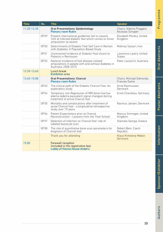
11.25-12.35 Oral Presentations: EpidemiologyPlenary room Rubin
Chairs: Alberto Piaggesi, Nicolaas Schaper
OP29 Present international guidelines fail to classify 76% of infected diabetic feet which comes to minor amputation as severe
Elizabeth Pendry, United Kingdom
OP30 Determinants of Diabetic Foot Self Care in Women with Diabetes: A Population-Based Study
Mahnaz Sanjari, Iran
OP31 Uncensored Incidence of Diabetic Foot Ulcers to Patients in Remission
Lawrence Lavery, United States
OP32 National incidence of foot disease-related amputations in people with and without diabetes in Australia, 2008-2015
Peter Lazzarini, Australia
12:35-13.45 Lunch breakExhibition area
13.45-15.00 Oral Presentations: CharcotPlenary room Rubin
Chairs: Michael Edmonds, Frances Game
OP33 The clinical path of the Diabetic Charcot Foot. An exploratory study
Anne Rasmussen, Denmark
OP34 Temporary non-Regression of MRI bone marrow edema (edema equivalent signal changes) during treatment of active Charcot foot
Ernst Chantelau, Germany
OP35 Mortality and complications after treatment of acute Charcot foot - a longitudinal retrospective study over 19 years
Rasmus Jansen, Denmark
OP36 Patient Expectations prior to Charcot Reconstruction – Lessons from the 'Foot School'
Marcus Simmgen, United Kingdom
OP37 Detection of infection on Charcot foot: role of labeled leucocyte scan
Stamata Georga, Greece
OP38 The role of quantitative bone scan parameters for diagnosis of Charcot foot
Robert Bem, Czech Republic
Thank you for attending Klaus Kirketerp-Møller, Denmark
15.00 Farewell reception (included in the registration fee).Lobby of Vienna House Andel’s
Pro
gra
mm
eO
ral
Ab
stra
cts
Pos
ter
Ab
stra
cts
Info
rmat
ion
Sp
onso
r/E
xhib
itor
Au
thor
s
15

1616

17
Pro
gra
mm
eO
ral
Ab
stra
cts
Pos
ter
Ab
stra
cts
Info
rmat
ion
Sp
onso
r/E
xhib
itor
Au
thor
s
AbstractsGeneral Information
Industry symposiaSponsor and exhibitor information
17
18
Prize Oral
EUROPEAN CME CREDITS
The DFSG 2018 has been accredited 11 European CME credits (ECMEC®s) by the European Accreditation Council for Continuing Medical Education (EACCME®).
To receive the CME credits, please sign the attendance sheet at the registration desk each day after 14.00. The CME certificates will be sent by e-mail after the conference.
GERMAN CME CREDITS
The DFSG 2018 has been accredited 12 CME credits by Ärztekammer Berlin.
To receive the CME credits, please sign and add your Barcode or EFN number to the German CME attendance sheet at the registration desk each day.
CME Credits
18

19
Pro
gra
mm
eO
ral
Ab
stra
cts
Pos
ter
Ab
stra
cts
Info
rmat
ion
Sp
onso
r/E
xhib
itor
Au
thor
s
Prize OralsOral abstracts
Poster Abstracts
19
20
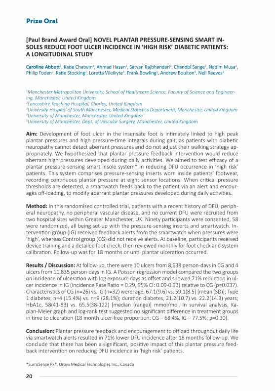
Prize Oral
[Paul Brand Award Oral] NOVEL PLANTAR PRESSURE-SENSING SMART IN-SOLES REDUCE FOOT ULCER INCIDENCE IN ‘HIGH RISK’ DIABETIC PATIENTS: A LONGITUDINAL STUDY
Caroline Abbott1, Katie Chatwin1, Ahmad Hasan1, Satyan Rajbhandari2, Chandbi Sange2, Nadim Musa2, Philip Foden3, Katie Stocking3, Loretta Vileikyte4, Frank Bowling5, Andrew Boulton4, Neil Reeves1
1Manchester Metropolitan University, School of Healthcare Science, Faculty of Science and Engineer-ing, Manchester, United Kingdom2Lancashire Teaching Hospital, Chorley, United Kingdom3University Hospital of South Manchester, Medical Statistics Department, Manchester, United Kingdom4University of Manchester, Manchester, United Kingdom5University of Manchester, Dept. of Vascular Surgery, Manchester, United Kingdom
Aim: Development of foot ulcer in the insensate foot is intimately linked to high peak plantar pressures and high pressure-time integrals during gait, as patients with diabetic neuropathy cannot detect aberrant pressures and do not adjust their walking strategy ap-propriately. We hypothesized that plantar pressure feedback intervention would reduce aberrant high pressures developed during daily activities. We aimed to test efficacy of a plantar pressure-sensing smart insole system* in reducing DFU occurrence in ‘high risk’ patients. This system comprises pressure-sensing inserts worn inside patients’ footwear, recording continuous plantar pressure at eight sensor locations. When critical pressure thresholds are detected, a smartwatch feeds back to the patient via an alert and encour-ages off-loading, to modify aberrant plantar pressures developed during daily activities.
Method: In this randomised controlled trial, patients with a recent history of DFU, periph-eral neuropathy, no peripheral vascular disease, and no current DFU were recruited from two hospital sites within Greater Manchester, UK. Ninety participants were consented, 58 were randomized, all being set-up with the pressure-sensing inserts and smartwatch. In-tervention group (IG) received feedback alerts from the smartwatch when pressures were ‘high’, whereas Control group (CG) did not receive alerts. At baseline, participants received device training and a detailed foot check, then reviewed monthly for foot check and system calibration. Follow-up was for 18 months or until plantar ulceration occurred.
Results / Discussion: At follow-up, there were 10 ulcers from 8,638 person-days in CG and 4 ulcers from 11,835 person-days in IG. A Poisson regression model compared the two groups on incidence of ulceration with log exposure days as offset and showed 71% reduction in ul-cer incidence in IG (Incidence Rate Ratio = 0.29, 95% CI: 0.09-0.93) relative to CG (p=0.037). Characteristics of CG (n=26) vs. IG (n=32) were: age, 67.1(9.6) vs. 59.1(8.5) [mean (SD)]; Type 1 diabetes, n=4 (15.4%) vs. n=9 (28.1%); duration diabetes, 21.2(10.7) vs. 22.2(14.3) years; HbA1c, 58(41-83) vs. 65.5(38-122) [median (range)] mmol/mol. In survival analysis, Ka-plan-Meier graph and log-rank test suggested no significant difference in treatment groups in time to ulceration (18 month ulcer-free proportion: CG – 68.4%, IG – 77.5%; p=0.30).
Conclusion: Plantar pressure feedback and encouragement to offload throughout daily life via smartwatch alerts resulted in 71% lower DFU incidence after 18 months follow-up. We conclude that there has been a significant, positive impact of this plantar pressure feed-back intervention on reducing DFU incidence in ‘high risk’ patients.
*SurroSense Rx®, Orpyx Medical Technologies Inc., Canada
21
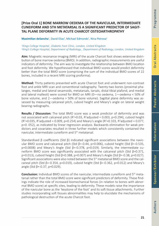
[Prize Oral 1] BONE MARROW OEDEMA OF THE NAVICULAR, INTERMEDIATE CUNEIFORM AND 5TH METATARSAL IS A SIGNIFICANT PREDICTOR OF SAGIT-TAL PLANE DEFORMITY IN ACUTE CHARCOT OSTEOARTHROPATHY
Maximilian deSancha1, David Elias2, Michael Edmonds1, Nina Petrova1
1Kings College Hospital , Diabetic Foot Clinic, London, United Kingdom2King’s College Hospital, Department of Radiology , Department of Radiology, London, United Kingdom
Aim: Magnetic resonance imaging (MRI) of the acute Charcot foot shows extensive distri-bution of bone marrow oedema (BMO). In addition, radiographic measurements are useful indicators of deformity. The aim was to investigate the relationship between BMO location and foot deformity. We hypothesised that individual BMO scores would predict deformity better than the total BMO score (comprising the sum of the individual BMO scores of 22 bones, included in a recent MRI scoring proforma).
Method: Thirty patients presented with acute Charcot foot and underwent non-contrast foot and ankle MRI scan and conventional radiography. Twenty-two bones (proximal pha-langes, medial and lateral sesamoids, metatarsals, tarsals, distal tibial plafond, and medial and lateral malleoli) were scored for BMO on MRI (0—no oedema, 1—oedema < 50% of bone volume, and 2—oedema > 50% of bone volume). Sagittal plane deformity was as-sessed by measuring calcaneal pitch, cuboid height and Meary’s angle on lateral weight bearing radiographs.
Results / Discussion: The total BMO score was a weak predictor of deformity and was not associated with calcaneal pitch (R2=0.03, R2adjusted = 0.003, p=0.294), cuboid height (R2=0.035, R2adjusted = 0.009, p=0.254) and Meary’s angle (R2=0.103, R2adjusted = 0.077, p=0. 052), as indicated by linear regression analysis. Backwards elimination for weak pre-dictors and covariates resulted in three further models which consistently contained the navicular, intermediate cuneiform and 5th metatarsal.
Standardised β coefficients (Std β) indicated significant associations between the navic-ular BMO score and calcaneal pitch (Std β=-0.44, p=0.006), cuboid height (Std β=-0.535, p=0.0008) and Meary’s Angle (Std β=-0.378, p=0.019). Similarly, the intermediate cu-neiform BMO score was significantly associated with the calcaneal pitch (Std β=0.371, p=0.013), cuboid height (Std β=0.388, p=0.007) and Meary’s Angle (Std β=-0.38, p=0.027). Significant associations were also noted between the 5th metatarsal BMO score and the cal-caneal pitch (Std β=-0.354, p=0.019), cuboid height (Std β=-0.362, p=0.012) and Meary’s angle (Std β=-0.37, p=0.029).
Conclusion: Individual BMO scores of the navicular, intermediate cuneiform and 5th meta-tarsal rather than the total BMO score were significant predictors of deformity. These find-ings indicate the role of increased biomechanical forces (in relation to bones with abnor-mal BMO score) at specific sites, leading to deformity. These models raise the importance of the navicular bone as the ‘keystone of the foot’ and its soft tissue attachments. Further studies incorporating soft tissues abnormalities may help to elucidate the mechanisms of pathological destruction of the acute Charcot foot.
Pro
gra
mm
eO
ral
Ab
stra
cts
Pos
ter
Ab
stra
cts
Info
rmat
ion
Sp
onso
r/E
xhib
itor
Au
thor
s
22
Prize Oral
[Prize Oral 2] THE USE OF AUTOLOGOUS LEUCOCYTE, PLATELET AND FIBRIN PATCH-ES IN THE MANAGEMENT OF HARD-TO-HEAL DIABETIC FOOT ULCERS: A MULTI-CENTRE, MULTINATIONAL, OBSERVER-BLINDED, RANDOMISED CONTROLLED TRIAL
Frances Game1, William Jeffcoate2, Tarnow Lise3, Jacobsen Judith4, Whitham Diane5, Harrison Elea-nor5, Ellender Sharon5, Magnus Löndahl6
1Derby Teaching Hospitals NHS Foundation Trust, Derby, United Kingdom2Nottingham City Hospital, Foot Ulcer Trials Unit, Diabetes and Endocrinology, Nottingham, United Kingdom3Holbaek Sygehus, Holbaek, Denmark4Statcon Aps, Denmark5University of Nottingham, United Kingdom6Clinical Sciences, Lund University, Dept. of Endocrinology, Endocrinology, Lund, Sweden
Aim: The autologous leucocyte, platelet and fibrin patches* uses bedside centrifugation without additional reagents to generate a disc comprising autologous platelet-rich fibrin and leucocytes which is applied to the surface of the wound. The aim of the study was to test the effectiveness of the autologous leucocyte, platelet and fibrin patches* on the healing of hard-to-heal foot ulcers in people with diabetes.
Method: 595 people with diabetes and a foot ulcer consented to participate. After a 4 week run-in-period those with a reduction in ulcer area of < 50% were randomised to either pre-specified good standard care alone or care supplemented by weekly application of the autologous leucocyte, platelet and fibrin patches*. The primary outcome was per-centage of ulcers healed within 20 weeks, defined as complete epithelialisation confirmed by an observer blind to randomisation group and maintained for four weeks.
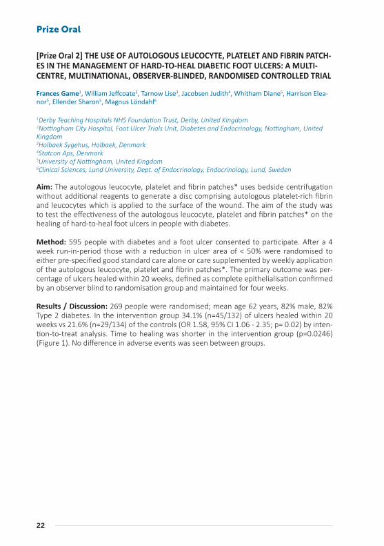
Results / Discussion: 269 people were randomised; mean age 62 years, 82% male, 82% Type 2 diabetes. In the intervention group 34.1% (n=45/132) of ulcers healed within 20 weeks vs 21.6% (n=29/134) of the controls (OR 1.58, 95% CI 1.06 - 2.35; p= 0.02) by inten-tion-to-treat analysis. Time to healing was shorter in the intervention group (p=0.0246) (Figure 1). No difference in adverse events was seen between groups.
23
Conclusion: The use of the autologous leucocyte, platelet and fibrin patches* is associated with significant enhancement of healing of hard-to heal foot ulcers in people with diabetes.
*LeucoPatch®, now being marketed by Reapplix under the name 3C Patch®
Pro
gra
mm
eO
ral
Ab
stra
cts
Pos
ter
Ab
stra
cts
Info
rmat
ion
Sp
onso
r/E
xhib
itor
Au
thor
s
24
Prize Oral
[Prize Oral 3] DATA LINKAGE AND GEOSPATIAL MAPPING EXPOSES INEQUALI-TIES IN OUTCOMES FOR DIABETIC FOOT DISEASE IN GLASGOW
Joanne Hurst1, Lesley Gibson2, Ruth Barn1, David Wylie3, Brian Kennon4, Sicco Bus5, James Wood-burn1
1Glasgow Caledonian University, School Health and Life Sciences, Glasgow, United Kingdom2University of Edinburugh, School of Engineering , Edinburugh, United Kingdom3NHS Greater Glasgow and Clyde, Podiatry Department, Glasgow, United Kingdom4Queen Elizabeth University Hospital, Department of Diabetes, Glasgow, United Kingdom5Academisch Medisch Centrum Universiteit van Amsterdam, Amsterdam, Netherlands
Aim: The aim of this study was to identify trends and inequalities in outcomes for diabetic foot disease in the geographical boundary of a large Scottish health board using data link-age and geospatial mapping.
Method: 112,231 people with diabetes were extracted from the Scottish Care Information – Diabetes Collaboration (SCI-diabetes) clinical repository and anonymously linked to the National Records Scotland (NRS) and the Scottish Morbidity record (SMR01) to identify death, amputation and ulceration outcome events between 2002-2016. Geospatial map-ping software (ArcGIS Desktop 10.4 Geostatistical Analyst) was used to map these out-comes across 1460 data zones within the NHS Greater Glasgow and Clyde boundary using the Scottish Index of Multiple Deprivation (SIMD) 2016 map. Getis-Ord Gi* cluster analysis was used to spatially identify statistically significant ‘hot spots’ for each outcome map. The relationship between SIMD quintiles and the frequency of observed outcome was investi-gated using chi-squared analysis.
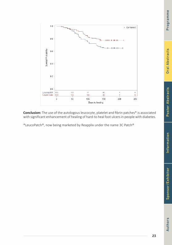
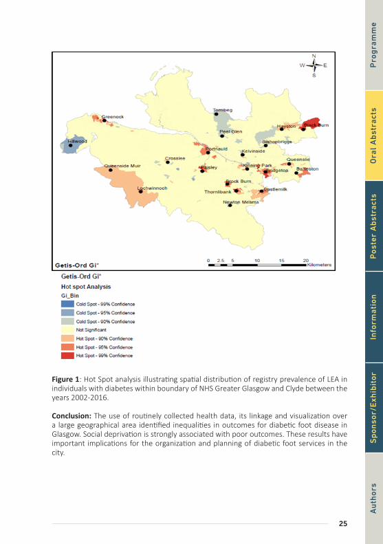
Results / Discussion: Over a 14-year period, foot ulceration was observed in 6935 regis-try patients. Lower extremity amputations (LEA) were identified in 1507 patients. 3804 deaths were recorded in patients with a past history of foot ulceration or LEA. Geospatial mapping identified statistically significant hot/cold spot clusters for all outcomes across the geographical boundary of the health board (Figure 1). Significant hot spot clusters were associated with higher levels of deprivation, whilst cold spots were found in lower lev-els of deprivation for ulceration (P<0.001); LEA (P<0.001); death preceded by ulceration (P<0.009); death preceded by LEA (P=0.002); and all-cause death (P<0.001).
25
Figure 1: Hot Spot analysis illustrating spatial distribution of registry prevalence of LEA in individuals with diabetes within boundary of NHS Greater Glasgow and Clyde between the years 2002-2016.
Conclusion: The use of routinely collected health data, its linkage and visualization over a large geographical area identified inequalities in outcomes for diabetic foot disease in Glasgow. Social deprivation is strongly associated with poor outcomes. These results have important implications for the organization and planning of diabetic foot services in the city.
Pro
gra
mm
eO
ral
Ab
stra
cts
Pos
ter
Ab
stra
cts
Info
rmat
ion
Sp
onso
r/E
xhib
itor
Au
thor
s
26
Oral Abstracts
[OP01] RELATIONSHIP BETWEEN DIABETIC RETINOPATHY AND LOWER-EX-TREMITY ARTERY DISEASE IN SUBJECTS WITH TYPE 2 DIABETES
Kamel Mohammedi1, Ninon Foussard2, Marie Azard3, Vincent Rigalleau4, Samy Hadjadj5
1Hôpital Haut-Lévêque, Département D’endocrinologie, Diabétologie, Nutrition, Université de Bor-deaux, Faculté de Médecine, Centre de Recherche Inserm - Université de Bordeaux U1219 “Bordeaux Population Health”, Bordeaux, France2Hôpital Haut-Lévêque, Département D’endocrinologie, Diabétologie, Nutrition, Bordeaux, France3Chu de Bordeaux, Service D’ophtalmologie, Bordeaux, France4Hopital Haut-Leveque, Département de Nutrition et Diabetologie, Pessac, France5Chu Poiters, & Endocrinology, Dept. of Diabetology, Poitiers, France
Aim: Previous studies linked diabetic retinopathy to lower-extremity artery disease (LEAD), but there remains some uncertainty as whether this association is mediated by traditional risk factors. Herein, we sought to evaluate: (i) the association between diabetic retinopathy stages and the risk of LEAD; and (ii) the influence of blood pressure and diabetic parame-ters in such association in the French SURDIAGENE (SURvie, DIAbete de type 2 et GENEti-que) type 2 diabetes cohort.
Method: Diabetic retinopathy was staged at baseline as absent, non-proliferative, pre-proliferative, or proliferative. Diabetic kidney disease (DKD) was defined as urinary albumin-to-creatinine ratio≥30 mg/g and/or estimated glomerular filtration rate<60 ml/min/1.73 m2. Major LEAD was defined as the first occurrence during follow-up of trans-metatarsal amputation with peripheral revascularization, transtibial/transfemoral ampu-tation, or requirement of peripheral revascularization. Cox regression models were fitted to estimate hazard ratios (HR) and 95%CI for major LEAD during follow-up by diabetic reti-nopathy stages at baseline, after adjustment for key confounding covariates. Analyses were performed in the whole cohort and in subgroups (below and above the median) by base-line duration of diabetes, HbA1c or systolic blood pressure (SBP).
Results / Discussion: We investigated 1468 participants (men: 58%, mean±SD age: 65±11 years). The median (IQR) duration of diabetes, HbA1c and SBP were at baseline at 13 (6, 21) years, 7.5% (6.7, 8.6) and 130 (120, 141) mmHg, respectively. Non-proliferative, pre-proliferative, and proliferative retinopathy existed at baseline in 431 (29%), 107 (7.2%), and 101 (7%) participants, respectively, and DKD was present in 525 (36%) individuals. During a median of 5.5 years of follow-up, major LEAD occurred in 105 (7%) participants. The 6-year incidences of major LEAD were 3.2%, 11.3%, 8.6% and 15.6% in participants with absent, non-proliferative, pre-proliferative, and proliferative retinopathy, respectively (Log-rank p<0.0001). The risk of major LEAD was higher in participants with non-prolifer-ative (HR 2.37, 95%CI [1.45-3.89]), pre-proliferative (2.93 [1.35-6.36]) or proliferative ret-inopathy (4.51 [2.00-10.14]), compared with those with no retinopathy at baseline (trend p<0.0001). These findings were independent to baseline DKD, which was also associated with major LEAD (2.27 [1.45-3.55], p<0.0001). Comparable results were observed across duration of diabetes (p for heterogeneity=0.19) or HbA1c subgroups (p for heterogene-ity=0.15). However, the magnitude of retinopathy-LEAD association was stronger in par-ticipants with higher SBP levels and attenuated in those with lower ones (p for heteroge-neity=0.05).
27
Conclusion: The risk of major LEAD increased proportionally with diabetic retinopathy worsening. This association seems to be potentially influenced by hypertension rather than diabetic parameters.
Pro
gra
mm
eA
uth
ors
Ora
l A
bst
ract
sP
oste
r A
bst
ract
sIn
form
atio
nS
pon
sor/
Exh
ibit
or
28
Oral Abstracts
[OP02] ARTERIAL DISEASE BELOW THE ANKLE IN THE DIABETIC FOOT: THE FINAL FONTIER
Chris Manu1, Daina Walton2, Timothy Jemmott3, Nina Petrova4, Hisham Rashid5, Kirsty Winkley6, Michael Edmonds3
1King’s College Hospital, London, United Kingdom2Diabetic Foot Clinic, King’s College Hospital, Diabetes UK Clinical Champion 2017-2019, London, United Kingdom3Diabetic Foot Clinic, King’s College Hospital, London, United Kingdom4Diabetic Foot Clinic, King’s College Hospital NHS Foundation Trust, London, United Kingdom5King’s College Hospital, Vascular Department, London, United Kingdom6King’s College London, United Kingdom
Aim: Recommendations from most guidelines suggests that peripheral arterial disease (PAD) is unlikely when Ankle Brachial Pressure Index (ABPI) is normal, that is between 0.9 - 1.3. The aim of this study was to evaluate the presence or absence of distal arterial disease below the ankle in limbs with normal ABPI between 0.9 - 1.3, as indicated by Toe Brachial Pressure Index (TBPI), Transcutaneous Oxygen (TcPO2) and the associated clinical impact.
Method: The ABPI, TBPI and forefoot TcPO2 were measured in both limbs of consecutive patients attending our outpatient clinic with diabetic foot ulceration. We used TBPI and forefoot TcPO2 to diagnose the presence of arterial disease below the ankle, compared to measurements of ABPI per limb. We also assessed clinical outcome on a patient level, with regards to subsequent amputation and mortality.
Results / Discussion: Measurements were taken in 154 patients, of which there were 121 limbs with a presumed absence of PAD as indicated by ABPI between 0.9 - 1.3. Within these limbs with normal ABPI range, 57% (69 limbs) had a low TBPI of <0.7, indicative of distal disease below the ankle (Group 1). The remaining 52 limbs (Group 2), had both ABPI and TBPI in the normal range. Absolute ankle pressures were similar in both groups, 159±32mmHg vs 159±25mmHg in Group 1 and Group 2 respectively, [p=0.478]. Howev-er, the forefoot TcPO2 was significantly lower in Group 1, 48±15mmHg vs 54±12mmHg in Group 2, [p=0.010], as was their absolute toe pressure, 72±21mmHg vs 112±19mmHg respectively, [p=0.001]. There were 43 patients in Group 1 and 21 patients in Group 2. More patients in Group 1 underwent minor amputation over the subsequent year; 26% vs 5%, [p=0.0455]. Over the subsequent 18months 2/43(5%) in Group 1 underwent a major amputation but none in Group 2. There was also a higher 2 year mortality in Group 1 pa-tients, 14% vs 5% mortality in Group 2, but did not meet statistical significance, [p=0.267].
Conclusion: A normal ABPI does not exclude PAD below the ankle in patients with diabe-tes. Over 50% of patients with normal ABPI between 0.9-1.3 have distal arterial disease in the foot which is associated with significant morbidity and mortality.
29
[OP03] ULTRASOUND VERSUS SHARP WOUND DEBRIDEMENT IN HEALING OF RECALCITRANT NEUROPATHIC DIABETIC FOOT ULCERS: CLINICAL AND PATHO-LOGICAL STUDY
Fady Azmy Kyrillos1, Ahmed Albehairy1, Mohamed Roshdi1, Wagdi Elkashef2, Manal Tarshoby1
1Diabetes and Endocrinology Department - Mansoura University, Mansoura, Egypt2Pathology Department, Mansoura University, Mansoura, Egypt
Aim: To compare clinical outcome, pathological and immuno-histochemical effect of low frequency ultrasound (LFU) wound debridement versus sharp debridement on recalcitrant neuropathic diabetic foot ulcers.
Method: 21 diabetic patients of matched age and sex with recalcitrant neuropathic foot ulcers (duration ≥ 6 months with standard therapy, sharp debridement and proper off-loading), were recruited from Mansoura Diabetic Foot Clinic (Specialized Medical Hospital- Mansoura university). Only grade 1A and 2A ulcer (University of Texas) were included in the study. After written consent was taken, all patients continued on same ulcer manage-ment with randomization into 2 groups according to method of debridement: Sharp group; continued using scalpel (11 patients) and Ultrasound (US) group; using LFU (12 patients). Patients received 1 debridement session every 2 weeks for 2 months. Tissue biopsies were taken from the base and edge of ulcers at the first session and after 2 months of debride-ment. Clinical outcome was assessed by reduction of ulcers surface area after 2 months. Pathological parameters for healing were assessed blindly by the pathologist. Pathological scoring included cellularity (fibroblast, fibrocyte and macrophage), vascular proliferation, type of collagen, inflammatory cells and fibrosis. Immunoreactivity of Matrix metallopro-teinase-1 (MMP-1) was also assessed.
Results / Discussion: Greater reduction in ulcers surface area in US group (43%) versus sharp group (24.24%) (p=0.001).Improvement in total ulcer pathology score was evident after each type of debridement with more improvement in US group versus sharp group (23.21% vs.6.67%, respectively) (p=0.004). Significant increase in cellularity in base and edge of the ulcers, vascular proliferation of ulcer base and inflammation of the ulcer edge after 2 months of US debridement (p=0.04, 0.03, 0.04, 0.03 respectively), while sharp debridement decreased the cellularity in the base of ulcers (p=0.04) with no significant change in other pathological parameters. MMP-1 expression decreased significantly in both base and edge of ulcers treated by sharp debridement (p=0.03, 0.02 respectively), while increased significantly in the base of ulcers after US debridement (p=0.037).
Conclusion: LFU debridement is superior to sharp debridement regarding healing of re-calcitrant neuropathic diabetic foot ulcers. In contrast to sharp debridement, LFU debride-ment increases expression of MMP-1, cellularity, vascular proliferation and inflammation in the ulcers improving the total pathology score and indicating better opportunity for ulcer healing.
Pro
gra
mm
eA
uth
ors
Ora
l A
bst
ract
sP
oste
r A
bst
ract
sIn
form
atio
nS
pon
sor/
Exh
ibit
or
30
Oral Abstracts
[OP04] AN UPDATE ON PERCUTANEOUS NEEDLE TENOTOMY TREATMENT OF PATIENTS WITH DIABETES, HAMMER, MALLET AND CLAW TOE DEFORMITIES
Jonas Hedegaard Andersen1, Anne Rasmussen2, Klaus Kirketerp-Møller2, Susanne Engberg2
1Hilleroed Hospital, Steno Diabetes Center Copenhagen, Denmark2Bispebjerg Hospital, Steno Diabetes Center Copenhagen, Denmark
Aim:The aim of the study was to examine the effectiveness of minimally invasive flexor needle tenotomy, to prevent and heal toe ulcers.
At DFSG 2016 we presented data from 42 patients. We now submit the updated study of 109 patients and a mean follow-up of 68.7 weeks(Q1-Q3=38.9-99.1). The prolonged follow up and increased patient number allows for more robust conclusions on the effects and potential side effects.
Method:Patients treated between February 2015 and April 2017 that underwent needle tenotomy of the flexor tendons of the toes. The surgical procedure was performed with a needle, under local anesthetics. The needle was introduced through the skin immediately proximal to the web level, corresponding to the placement of the flexor tendons.
Results / Discussion: 109 patients were treated, 35(32%) patients had at least one toe with an ulcer and 74(68%) patients had impending ulcerations on treated toes. Average age was 65.5 years(±11), 73(66.97%) were males, 72 had type II diabetes(66.1%) average duration was 23.2 years(±14.1), BMI 30.9 kg/m2(±5.3) and HbA1c 63.3mmol/mol(±14.5).
In the group with ulcers all surgical incisions healed uneventfully, the average time to healing was 4.4 days(Q1-Q3=2-7), the average time of ulcers before intervention was 6.1 weeks(±6.2), time to ulcer healing after incision was 26.8 days (±35.8 days). There were no serious adverse events e.g. infections or amputations in the follow-up period, 15 pa-tients(42.9%) had transfer complications, and 4(11.4%) complained of transient pain under involved toes, and 1(1.2%) complained of altered balance.
In the group without ulcers all incisions healed uneventfully, the average time to healing was 4.5 days(Q1-Q3=2-7). There were no serious adverse events e.g. infections or am-putations in the follow-up period, 15 patients(20.3%) had transfer complications, 3(4.1%) received re-tenotomies due to insufficient primary procedure, 9(12.2%) complained of transient pain under involved toes, and 1(1.2%) complained of altered balance.
Conclusion: The larger number of patients and longer follow-up supports the conclusion that needle tenotomy is a safe and effective procedure for treating claw, mallet and hammertoe defor-mities in diabetic patients. This procedure should be offered all patients at-risk of ulcers due to a hammer, mallet or claw toe. The procedure can result in transfer ulcers if not performed on all toes of one foot at same primary intervention. The follow-up period is still relatively short, and further investigation is needed.
31
[OP05] OUTCOME OF ENDOVASCULAR FIRST APPROACH IN DIABETIC ASIAN PATIENTS WITH LOWER LIMB ISCHAEMIA
Alok Tiwari1, Min Qi Chen2, Vikram Vijayan2, Harvinder Raj Singh Sidhu2
1Department of Vascular Surgery, University Hospitals Birmingham, Birmingham, United Kingdom2Department of Surgery, Singapore
Aim: An endovascular-first approach is being increasingly utilised worldwide for lower limb sal-vage. There is currently very little in the published literature regarding this approach and of the burden of disease in a multi-ethnic group of patients from Asian countries.
Method: All patients presenting to a single institution as an emergency with critical limb ischaemia and tissue-loss undergoing angioplasty were identified from hospital database for 2016. Patient demographics and the anatomical distribution of disease were retrospectively ana-lysed. Primary outcome was the number of lower limb arteries successfully revascularised as well as the 30 day and 12 month amputation-free survival.
Results: 138 limbs (108 patients, 63% male) underwent endovascular first approach intervention in 2016. The ethnic distribution of this population was 53% Chinese, 33% Malay and 14% In-dian. 80% of patients were diabetic, 43% had diagnosed ischaemic heart disease, 84% had hypertension and 21% has end stage renal failure requiring dialysis (mainly haemodialysis). The mean number of arteries affected was 3.8. The majority of patients had infra-popliteal disease with the Anterior Tibial (87%) and Posterior Tibial (83%) arteries most commonly affected. Iliac artery disease was only seen in 6.5% of limbs. The mean number of arteries revascularised at the primary operation was 2.9. 18 patients died within 12 months. The estimated amputation-free survival at 30 days was 88% and 75% at 12 months.
Conclusion: Multi-ethnic Asian patients, presenting with critical limb ischaemia and tissue-loss, have significant multilevel peripheral arterial disease which can be safely and successfully man-aged with an endovascular-first approach. There exists an enormous burden of disease in these patients, requiring multiple vessel recanalizations to affect limb salvage. Despite this, there remains a significant risk of limb-loss and mortality in such patients, primarily due to late presentation but also due to underlying cardiovascular and renal disease.
Pro
gra
mm
eA
uth
ors
Ora
l A
bst
ract
sP
oste
r A
bst
ract
sIn
form
atio
nS
pon
sor/
Exh
ibit
or
32
Oral Abstracts
[OP06] RELIABILITY OF A NOVEL THERMAL IMAGING SYSTEM FOR TEMPERA-TURE ASSESSMENT OF FEET
Wegin Tang1, NL Petrova1, A Whittam2, A MacDonald3, S Ainarkar4, AN Donaldson1, J Bevans4, J Allen3, P Plassmann5, B Kluwe6, F Ring6, L Rogers2, R Simpson2, G Machin2, ME Edmonds1
1Diabetic Foot Clinic, King’s College Hospital NHS Foundation Trust, London, United Kingdom2Temperature and Humidity, National Physical Laboratory, London; United Kingdom3Microvascular Diagnostics, Northern Medical Physics and Clinical Engineering, Newcastle Upon Tyne Hospitals, United Kingdom4Community Podiatry Department, Pennine Acute Hospitals NHS Trust, Manchester5Photometrix Imaging Ltd, Pontypridd, Wales, United Kingdom6Department of Computing, University of South Wales, Pontypridd
Aim: Thermal imaging is a useful modality for identifying preulcerative lesions (“hot spots”) in diabetic foot patients. Despite its recognised potential, at present, there is no readily available instrument for routine podiatric assessment of patients at risk. To address this need, a novel thermal imaging system was recently developed. The aim of this study was to assess the reliability of this device for temperature assessment of healthy feet.
Method: Plantar skin foot temperatures were measured with the novel thermal imaging device (Diabetic Foot Ulcer Prevention System (DFUPS), constructed by Photometrix Imaging Ltd) and also with a hand-held infrared spot thermometer* after 20 minutes of barefoot resting with legs supported and extended in 105 subjects (52 males and 53 females; age range 18 to 69 years) as part of a multicentre clinical trial (NCT02317835). The temperature differences between the right and left foot at five regions of interest, including 1st and 4th toes, 1st, 3rd and 5th metatarsal heads were calculated. The intra-instrument agreement (three repeated measures) and the inter-instru-ment agreement (hand-held-thermometer and thermal imaging device) were quantified using intra-class correlation coefficients (ICCs) and their 95% confidence intervals (CI).
Results / Discussion: Both devices showed almost perfect agreement in replication by instrument. The intra-instrument ICCs for the thermal imaging device at all five regions ranged from 0.95 to 0.97 and the intra-instrument ICCs for the hand-held-thermometer ranged from 0.94 to 0.97. There was substantial to perfect inter-instrument agreement between the hand-held thermometer and the thermal imaging device and the ICCs at all five regions ranged between 0.94 and 0.97.
Conclusion: The newly developed thermal imaging device showed very good agreement in repeated temperature assessments at defined regions as well as substantial to perfect agreement in temperature assessment with the hand-held infrared thermometer. In addition to the reported non-inferior performance in temperature assessment, the thermal imaging device holds the potential to provide an instantaneous thermal image of all sites of the feet (plantar, dorsal, lateral and medial views). The National Physical Laboratory has implemented a step change in quantitative thermal imaging reducing uncertainty from 2 °C to 0.2 °C (k = 2), to deliver equivalence to current clinical thermometry devices and enable the robust use of DFUPS as a clinical tool. The proposed thermal imaging device can become a useful instru-ment in the diabetic foot clinic, to identify patients at risk of diabetic foot ulcer.
*Thermofocus® 01500A3, Tecnimed, Italy
33
[OP07] VALIDATION OF A SMARTPHONE-BASED INFRARED THERMAL CAMERA AND ITS POSSIBILITIES WITH 3D THERMAL MAPPING OF DIABETIC FOOT UL-CER DETECTION
R.F.M. van Doremalen1, Jaap van Netten2, J.G. van Baal3, M.M.R. Vollenbroek3, F. van der Heijden3
1Zorggroep Twente Hospital, University of Twente, University of Twente, Almelo, Netherlands2Queensland University of Technology, Faculty of Health, Department of Surgery, Ziekenhuis Groep Twente, Almelo, the Netherlands. Department of Rehabilitation, Academic Medical Center, University of Amsterdam, Amsterdam Movement Sciences, the Netherlands3Zorggroep Twente Hospital, Netherlands
Aim: Infrared thermal imaging (IR) is not routinely implemented for early detection of dia-betic foot ulcers (DFU), despite accurately measuring the relevant skin temperature. This is primarily because most IR cameras are unwieldy and expensive. However, low-cost, smart-phone-based IR cameras are now available. Secondly, evaluation of IR images is labor-in-tensive, due to complex background differentiation, left-right comparison, and comparison over time, and it is limited to the plantar side. This might be solved by creating 3D thermal images of the feet, to allow automatic evaluation beyond plantar. We aim to validate a smartphone-based IR camera against a high-resolution camera and to explore the possibil-ities of 3D thermal mapping.
Method: We included 32 patients with a current or recently healed DFU. A plantar IR im-age was acquired of both feet with the smartphone-based system*, and with a high-reso-lution thermal imaging camera*2 within a controlled environment as gold standard. From eight patients, 3D thermal images were acquired with a 3D-camera*3 aligned with three smartphone-based cameras*.
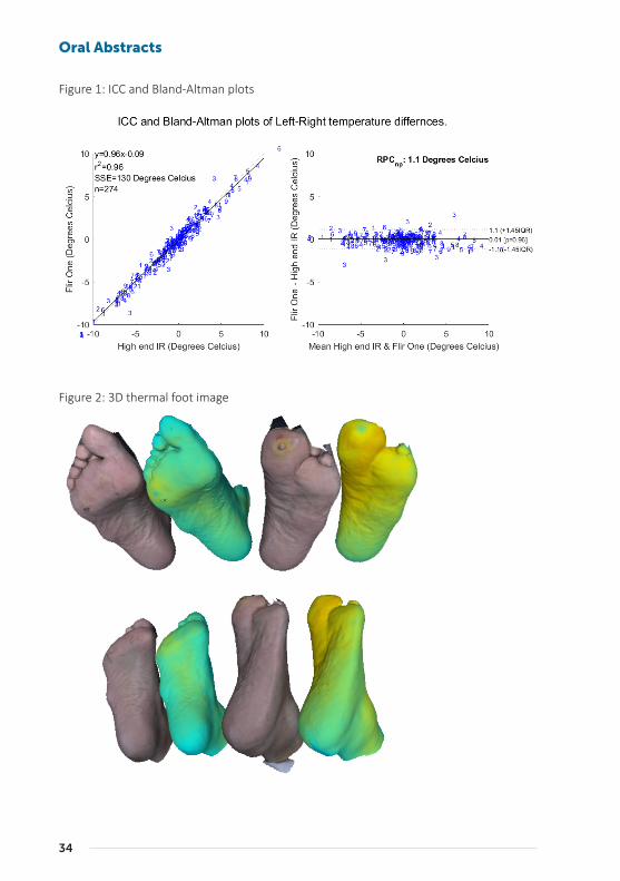
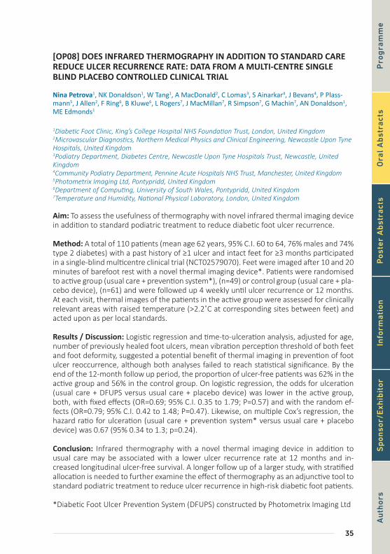
Results / Discussion: Intra-class correlation (r2=0.96) and Bland-Altman plot proved al-most perfect agreement (figure 1). The clinical relevant outcome (>2.2oC difference detec-tion) gave 90% sensitivity and 95% specificity. The first-ever 3D thermal images in people with DFU were successfully created and allow intensive analysis (figure 2).
Conclusion: A smartphone-based IR camera has excellent validity for thermal foot assess-ment in people with diabetes. The addition of 3D may prove useful, but is still in develop-ment.
*FLIR –One, FLIR Systems, Wilsonville, US
*2 FLIR -SC305
*3 Vectra-XT 3D-camera, Canfield scientific, Parsippany, US
Pro
gra
mm
eA
uth
ors
Ora
l A
bst
ract
sP
oste
r A
bst
ract
sIn
form
atio
nS
pon
sor/
Exh
ibit
or
34
Oral Abstracts
Figure 1: ICC and Bland-Altman plots
Figure 2: 3D thermal foot image
35
[OP08] DOES INFRARED THERMOGRAPHY IN ADDITION TO STANDARD CARE REDUCE ULCER RECURRENCE RATE: DATA FROM A MULTI-CENTRE SINGLE BLIND PLACEBO CONTROLLED CLINICAL TRIAL
Nina Petrova1, NK Donaldson1, W Tang1, A MacDonald2, C Lomas3, S Ainarkar4, J Bevans4, P Plass-mann5, J Allen2, F Ring6, B Kluwe6, L Rogers7, J MacMillan7, R Simpson7, G Machin7, AN Donaldson1, ME Edmonds1
1Diabetic Foot Clinic, King’s College Hospital NHS Foundation Trust, London, United Kingdom2Microvascular Diagnostics, Northern Medical Physics and Clinical Engineering, Newcastle Upon Tyne Hospitals, United Kingdom3Podiatry Department, Diabetes Centre, Newcastle Upon Tyne Hospitals Trust, Newcastle, United Kingdom4Community Podiatry Department, Pennine Acute Hospitals NHS Trust, Manchester, United Kingdom5Photometrix Imaging Ltd, Pontypridd, United Kingdom6Department of Computing, University of South Wales, Pontypridd, United Kingdom7Temperature and Humidity, National Physical Laboratory, London, United Kingdom
Aim: To assess the usefulness of thermography with novel infrared thermal imaging device in addition to standard podiatric treatment to reduce diabetic foot ulcer recurrence.
Method: A total of 110 patients (mean age 62 years, 95% C.I. 60 to 64, 76% males and 74% type 2 diabetes) with a past history of ≥1 ulcer and intact feet for ≥3 months participated in a single-blind multicentre clinical trial (NCT02579070). Feet were imaged after 10 and 20 minutes of barefoot rest with a novel thermal imaging device*. Patients were randomised to active group (usual care + prevention system*), (n=49) or control group (usual care + pla-cebo device), (n=61) and were followed up 4 weekly until ulcer recurrence or 12 months. At each visit, thermal images of the patients in the active group were assessed for clinically relevant areas with raised temperature (>2.2˚C at corresponding sites between feet) and acted upon as per local standards.
Results / Discussion: Logistic regression and time-to-ulceration analysis, adjusted for age, number of previously healed foot ulcers, mean vibration perception threshold of both feet and foot deformity, suggested a potential benefit of thermal imaging in prevention of foot ulcer reoccurrence, although both analyses failed to reach statistical significance. By the end of the 12-month follow up period, the proportion of ulcer-free patients was 62% in the active group and 56% in the control group. On logistic regression, the odds for ulceration (usual care + DFUPS versus usual care + placebo device) was lower in the active group, both, with fixed effects (OR=0.69; 95% C.I. 0.35 to 1.79; P=0.57) and with the random ef-fects (OR=0.79; 95% C.I. 0.42 to 1.48; P=0.47). Likewise, on multiple Cox’s regression, the hazard ratio for ulceration (usual care + prevention system* versus usual care + placebo device) was 0.67 (95% 0.34 to 1.3; p=0.24).
Conclusion: Infrared thermography with a novel thermal imaging device in addition to usual care may be associated with a lower ulcer recurrence rate at 12 months and in-creased longitudinal ulcer-free survival. A longer follow up of a larger study, with stratified allocation is needed to further examine the effect of thermography as an adjunctive tool to standard podiatric treatment to reduce ulcer recurrence in high-risk diabetic foot patients.
*Diabetic Foot Ulcer Prevention System (DFUPS) constructed by Photometrix Imaging Ltd
Pro
gra
mm
eA
uth
ors
Ora
l A
bst
ract
sP
oste
r A
bst
ract
sIn
form
atio
nS
pon
sor/
Exh
ibit
or
36
Oral Abstracts
[OP09] PROGNOSTIC ROLE OF PROCALCITONIN IN PATIENTS WITH CRITICAL LIMB ISCHEMIA AND DIABETIC FOOT INFECTION
Marco Meloni1, Valentina Izzo1, Laura Giurato1, Luigi Uccioli1
1University of Tor Vergata, Rome, Italy
Aim: Procalcitonin (PCT) is considered a reliable marker for severe infection and sepsis. The aim of this study is to evaluate the prognostic role of PCT in hospital patients with critical limb ischemia (CLI) and DFI.
Method: Consecutive in hospital patients with CLI and DFI (moderate-severe infection according to Infectious Disease Society of America Classification) have been included. All patients have been treated by a pre-set limb salvage protocol including revascularization, aggressive debridement, antibiotic therapy and off-loading. Demographic data, comorbidi-ties and inflammatory markers are evaluated. Recovery of DFI has been considered in case of recovery of clinical signs of infection and normalization of inflammatory markers (white blood cells, c-reactive protein, PCT). According to the positive or negative values of PCT (cut off 0.5 ng/ml), patients have been respectively divided in 2 groups: PCT+ (study group) and PTC- (control group). Hospital outcomes expressed as limb salvage (discharge with pre-served limb), major amputation (above the ankle), death, duration of hospitalization (days) and duration of foot infection (days) are reported and compared between the two groups.
Results / Discussion: Ninty-six patients have been included. The mean age was 67,9±11,3 years, 77,2% males, 90,2% had type 2 diabetes, the mean diabetes duration was 21,2 years with a mean HbA1c of 67±16 mmol/mol, 43.2% on end stage renal disease, 73.9% with ischemic heart disease. 23/96 (23,9%) patients showed high values of PCT while 73/96 (76,1%) normal values. PCT+ patients reported higher rate of major amputation (26.1% vs 1.3% p=0.0001), lower rate of limb salvage (47,8% vs 87.6% p=0.0002), higher rate of death (26.1% vs 10.9% p=0-004), longer hospitalization (27.3 vs 12.5 days p=0.002) and longer duration of foot infection during hospitalization (16.4 vs 9.3 days p=0.006) in com-parison to PCT- patients. Dialyzed patients showed higher levels of PCT than patients with normal renal function (12.5 vs 2.9% p=0.0001). PCT was an independent predictor of death [4.7 (CI 1.3-6.9) p=0.002], major amputation [3.1 (CI 1.5-5-5) p=0.0006] and duration of foot infection [1.9 (CI 1.5-4.3) p=0.03]. White blood cells, C-reactive protein, erythrocyte sedimentation rate and the grade of infection (moderate or severe) did not influence the outcomes.
Conclusion: PCT may be a useful marker to stratify the severity of infection in diabetic patients with CLI and moderate/severe DFI.
37
[OP10] CORRELATION BETWEEN DECREASED FIRST METATARSOPHALANGE-AL JOINT MOBILITY IN WEIGH BEARING POSITION AND INCREASED HALLUX PLANTAR PRESSURE
Mateo López Moral1, José Luis Lázaro Martínez2, Yolanda García Álvarez1, Aroa Tardáguila García1, Jose Luis Garcia Klepzig3, Raúl Molines Barroso1
1Diabetic Foot Unit, Complutense University of Madrid, Madrid, Spain2Complutense University Madrid, Diabetic Foot Unit, Madrid, Spain3Hospital Clínico San Carlos, Madrid, Spain
Aim: To analyze ranges of joint mobility in the first metatarsophalangeal joint and the asso-ciation between the increase of hallux plantar pressure.
Method: Cross-sectional study was performed in a diabetic foot unit between December 2017 and February 2018. Fourteen patients (27 feet) with diabetic neuropathy and no his-tory of ulcer were included. Measurement of goniometry was conducted with a validated software tool* for the measurement of joint goniometry. Photography of the lateral view of the foot was used to draw the angles in the first metatarsophalangeal joint (1st MPJ). Range of 1st MPJ in weigh bearing and non-weigh bearing position were calculated. Max-imum plantar pressures and integral pressure/time in the hallux were registered by Foot Scan 7.x Gait Interface (Rsscan International, Olen, Belgium) The clinician who calculated the goniometric measures was blinded from the results of plantar pressure and the posi-tion of the patients in the photographs.
Pearson coefficient correlation was used to evaluate the relationship between plantar pressure and goniometric measures. The strength of the difference of effect size was cal-culated by correlation coefficient (r).
Results / Discussion: Patients showed a mean age of 69.21 ± 6.05 years. All patients had type 2 diabetes mellitus with a mean evolution of 23.64 ± 17.57 years. Eight patients (57.1%) had history of retinopathy and 4 (28.6%) nephropathy. The mean of body mass index was 28.83 ± 3.66 kg/m2. There is a negative correlation between a lower mobility of the 1st MPJ in weigh bearing position and elevated hallux plantar pressure (p=0.003; r= -0.567) and higher hallux integral pressure/time (p=0.011; r= -0.499). On the other hand, there was no correlation between range of motion of the 1st MPJ in non – weigh bearing position and plantar pressure or integral pressure/time in the hallux (p=0.428; r= -0.166; p=0.594 r=-0.112, respectively). The assessment of the 1st MPJ in weigh bearing position in-creases the association with elevated level of plantar pressures, we recommend including this measure in biomechanic exploration of patients at high risk of hallux ulceration. The increase of plantar pressure under the 1st MPJ may be related with the risk of diabetic foot ulcer. Further studies are necessaries to demonstrate this relationship.
Conclusion: The assessment of the goniometry in weigh bearing position of the 1st MPJ is correlated with the increase of plantar pressure under the 1st MPJ.
*Autocad®, Autodesk for Microsoft Windows
Pro
gra
mm
eA
uth
ors
Ora
l A
bst
ract
sP
oste
r A
bst
ract
sIn
form
atio
nS
pon
sor/
Exh
ibit
or
38
Oral Abstracts
[OP11] PATIENTS WITH DIABETES MELLITUS WITH AND WITHOUT NEUROPA-THY EXHIBIT DISTURBED MIDFOOT ENERGETICS DURING WALKING
Giovanni Matricali1, Filip Staes2, Frank Nobels3, Jozef Tits4, Philip Roosen5, Herman Bruyninckx2, Dirk Desmet2, Sander Wuite6, Kevin Deschamps2
1Uz Leuven, Ku Leuven, Leuven, Belgium2Ku Leuven, Belgium3Onze-Lieve-Vrouwziekenhuis Aalst, Dept of Endocrinology, Diabetes Liga, Aalst, Belgium4Ziekenhuis Oost Limburg, Belgium5U Gent, Belgium6Uz Leuven, Leuven, Belgium
Aim: People with diabetes mellitus (DM) with and without peripheral neuropathy (PNP) exhibit elevated forefoot plantar pressures and reduced ankle power generation during walking. This study aims to compare the power and work in foot segments of patients with DM with and without PNP, and healthy controls.
Method: Foot kinetics of 13 diabetic patients with PNP (DMn), 13 diabetic patients without PNP (DM) and 13 non-diabetic persons (CTRL) were measured using an integrated mea-surement set-up including a force-pressure platform and a 3D-motion analysis system. In this age-, sex- and walking speed matched comparative study, differences in multi-segment foot kinetics were quantified using the IOR-4Segment-model 1. Rearfoot, Chopart, Lisfranc and Hallux positive and peak power absorption and generation, positive and negative work and negative work ratios were calculated and compared. Data normality was checked with the Shapiro–Wilk test. One-way repeated measures ANOVA was used to assess differences between three groups.
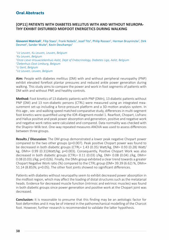
Results / Discussion: The DM group demonstrated a lower peak negative Chopart power compared to the two other groups (p=0.007). Peak positive Chopart power was found to be decreased in both diabetic groups (CTRL= 1.43 (0.35) Watt/kg, DM= 0.93 (0.28) Watt/kg, DMn= 0.99 (0.31)Watt/kg; p=0.003). Consequently, Positive Chopart Work was also decreased in both diabetic groups (CTRL= 0.11 (0.03) J/kg, DM= 0.08 (0.04) J/kg, DMn= 0.08 (0.03) J/kg; p=0.026). Finally, the DMn group exhibited a clear trend towards a greater Chopart Negative Work ratio (%) compared to the CTRL group (DM= 39.39 (6.62) %, DMn= 51.14 (8.85)%; p=0.05). The other foot joints showed no significant differences.
Patients with diabetes without neuropathy seem to exhibit decreased power absorption in the midfoot region, which may affect the loading of distal structures such as the metatarsal heads. Evidence for decreased muscle function (intrinsic and extrinsic muscles) was found in both diabetic groups since power generation and positive work at the Chopart joint was decreased.
Conclusion: It is reasonable to presume that this finding may be an aetiologic factor for foot deformities and it may be of interest in the pathomechanical modelling of the Charcot foot. However, further research is recommended to validate the latter hypothesis.
39
[OP12] THE IMPORTANCE OF EVALUATING FUNCTIONAL HALLUX LIMITS IN PATIENTS AT HIGH-RISK OF HALLUX ULCERATION
Raúl Molines Barroso1, José Luis Lázaro Martínez1, Mateo López-Moral1, Francisco Javier Alvaro Afonso1, Irene Sanz1, Esther Garcia Morales1
1Diabetic Foot Unit. Complutense University Clinic., Instituto de Investigación Sanitaria del Hospital Clínico San Carlos, Madrid, Spain
Aim: To evaluate the association between the risk of hallux reulceration and functional hallux limitus, together with other biomechanical variables in patients with diabetes and history of neuropathic ulcer.
Method: A prospective study conducted between January 2012 and December 2017 was performed in a diabetic foot unit to assess risk factors associated with hallux reulceration. Fifty-six consecutive patients with diabetic neuropathy and with previous forefoot ulcer were included. Standardized weightbearing radiographs and standardized biomechanical measurements were obtained. Functional hallux limitus was defined as the limitation of first MPJ in weight-bearing position in the absence of limitation of the range of first MPJ in resting postion. Participants were follow-up monthly. Ulceration during the follow-up period was the main outcome measure assessed in the study. Those who developed a new ulcer were classified in two groups: hallux/other location. Cox survival model of propor-tional hazards over hallux ulceration-free survival time was calculated with variables with P<0.2 in the univariate.
Results / Discussion: Patients were followed-up prospectively for a median time period of 28.52 IR[14.19 - 64.37] months. Twenty-nine patients (51.8%) suffered a new ulceration (9 in the hallux, 6 in the toes, 13 beneath the metatarsals, and 1 in the heel) and showed a median reulceration -free survival time of 18.79 IR[5.97-29.93] months. A shorter duration of DM (p=0.038; Cohen’s d=0.385), a higher body mass index (p=0.006; Cohen’s d=0.542), a higher foot posture index (p=0.042; Cohen’s d=0.354), a functional hallux limitus (p=0.003; r=0.551), and a lower first metatarsal inclination in Rx (p=0.045; Cohen’s d=0.355) were associated with hallux ulceration in the univariate analysis. Functional hallux limitus was the only variable associated with the hallux ulceration-free survival time in the multivariate Cox model (p=0.046; CI[1.030–24.536]; HR5.026). Hallux amputation can have devastating effects on foot biomechanics and increases the risk of new ulcers, and lower extremity amputation. Research on the risk factors of hallux ulceration can help to avoid hallux am-putation and its consequences. To date, relationship between range of motion of first MPJ and hallux ulceration had not been demonstrated in patients with diabetes followed-up prospectively. According to our results, we recommended to evaluate the dorsiflexion of the hallux in the weight-bearing position and to identify functional hallux limitus in the biomechanical screening of patients at high risk of ulceration.
Conclusion: The presence of functional hallux limitus increases the probability of suffering hallux ulceration in patients with diabetic neuropathy and history of ulcer.
Pro
gra
mm
eA
uth
ors
Ora
l A
bst
ract
sP
oste
r A
bst
ract
sIn
form
atio
nS
pon
sor/
Exh
ibit
or
40
Oral Abstracts
[OP13] WHAT KIND OF FOOTWEAR DO PEOPLE AT RISK DUE TO DIABETIC NEUROPATHY WEAR?
Johana Venerová1, Katerina Herdova1, Jarmila Jirkovska2, Monika Nyvltova2, Libuse Fialova2, Sva-topluk Solar2, Miroslav Zavoral2
1Medical Department of the First Faculty of Medicine and Military University Hospital, Diabetes Cen-ter, Charles University , Prague 6, Czech Republic2Medical Department of the First Faculty of Medicine and Military University Hospital, Charles Univer-sity, Diabetes Center, Prague 6, Czech Republic
Aim: The purchase and wearing of appropriate and well-fitting footwear is the basics of foot ulcer prevention in patients with diabetes. The importance of both grows with level of risk of foot ulceration. With regards to loss of protective sensation, selection of well-fitting footwear for a person with diabetic neuropathy is difficult and requires measuring of feet. Patients at risk are educated about footwear at regular check-ups in diabetic and podiatric clinics. The aim of our study was to determine which footwear patients at risk of foot ulceration with peripheral neuropathy and no previous foot ulcer or amputation (Category 1 IWGDF Risk Classification System 2015) use and how they purchase it.
Method: In March 2018, we carried out a research through an anonymous questionnaire in our diabetic clinic. 46 consecutive patients educated regarding footwear with proven peripheral neuropathy [73% of males, most frequent age group 71-80 years (38.7%), sec-ondary (39%) and higher education (36.7%), 69.3% of pensioners] completed the ques-tionnaire. Obtained data were statistically processed.
Results / Discussion: 48.9% of the patients use pre-fabricated or custom-made medical grade footwear. 28.6% use off –the – shelf leisure footwear, and 22.5% use pre-fabricated general-ly healthy footwear not directly intended for diabetics and thus not fulfilling all the required parameters. 77.5% of patients buy footwear without measuring the size of their feet. 14.2% of the patients purchased the preventive footwear but for different reasons did not use it. Reasons for not buying the preventive footwear: 1/ Feeling they did not need it. 2/ Foot-wear for diabetics does not comply with a notion of comfortable healthy footwear. 3/ They did not like the footwear design.
Reasons they do not use the already purchased preventive footwear: 1/ Diabetic footwear is not comfortable. 2/ Broad shoe tip. 3/ Stiff outsole. 4/Footwear was too heavy. 5/ Not comfortable when driving a car.
Conclusion: The results show that notwithstanding the education, patients underestimate the connection between the loss of protective sensation and the risk of injury due to in-appropriate footwear. They indicate a discrepancy between a general idea of comfortable and healthy shoes and footwear specially designed for diabetics. It seems useful to com-plement the education with a leaflet describing the parts of the shoe and their importance for protection of feet at risk, and with an advise how to individually verify the right size of shoe by taking out the insole and stepping on it with full weight.
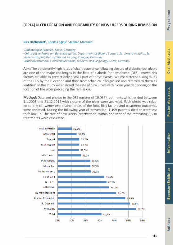
41
[OP14] ULCER LOCATION AND PROBABILITY OF NEW ULCERS DURING REMISSION
Dirk Hochlenert1, Gerald Engels2, Stephan Morbach3
1Diabetologist Practice, Koeln, Germany2Chirurgische Praxis am Bayenthalgürtel, Department of Wound Surgery, St. Vinzenz Hospital, St. Vinzenz-Hosptal, Dep. of Wound Surgery, Cologne, Germany3Marienkrankenhaus, Internal Medicine, Diabetes and Angiology, Soest, Germany
Aim: The persistently high rates of ulcer recurrence following closure of diabetic foot ulcers are one of the major challenges in the field of diabetic foot syndrome (DFS). Known risk factors are able to predict only a small part of these events. We characterised subgroups of the DFS by their location and their biomechanical background and referred to them as ‘entities’. In this study we analysed the rate of new ulcers within one year depending on the location of the ulcer preceding the remission.
Method: Data and photos in the DFS register of 10,037 treatments which ended between 1.1.2005 and 31.12.2012 with closure of the ulcer were analysed. Each photo was relat-ed to one of twenty-two distinct areas of the foot. Risk factors and treatment outcomes were analysed. During the following year of prevention, 1,499 patients died or were lost to follow up. The rate of new ulcers (reactivation) within one year of the remaining 8,538 treatments were calculated.
Pro
gra
mm
eA
uth
ors
Ora
l A
bst
ract
sP
oste
r A
bst
ract
sIn
form
atio
nS
pon
sor/
Exh
ibit
or
42
Oral Abstracts
Results / Discussion: The reactivation rates show marked differences. The lowest rates occur after rhagades at the fore- and midfoot (29%), malleoli centrally (30%) and after interdigital ulcers at the IP joints (31.7%). The highest rates follow ulcers below the first metatarsal head (MTH, 54.5%), below 2nd - 5th MTH (51.3%), at the interphalangeal joint of the hallux plantar (H. rigidus) (50.5%) and ulcers in scars after amputations (48.9%).
Conclusion: The ‘entity’ of an ulcer represents a relevant and easy to detect risk factor for new diabetic foot ulcers. Entities with higher reactivation rates are typically related to bio-mechanical problems difficult to correct without surgery. It was not easy to access surgery in the period analysed. Compensation using shoes depends on the adherence to therapy. This might be interpreted as demonstration of the need to find better solutions for biome-chanical problems which could be to resolve them by surgery or to increase adherence to conservative compensation of the biomechanical problems.
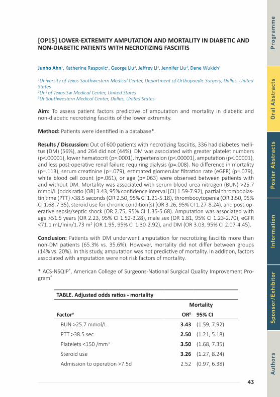
43
[OP15] LOWER-EXTREMITY AMPUTATION AND MORTALITY IN DIABETIC AND NON-DIABETIC PATIENTS WITH NECROTIZING FASCIITIS
Junho Ahn1, Katherine Raspovic2, George Liu3, Jeffrey Li3, Jennifer Liu3, Dane Wukich1
1University of Texas Southwestern Medical Center, Department of Orthopaedic Surgery, Dallas, United States2Uni of Texas Sw Medical Center, United States3Ut Southwestern Medical Center, Dallas, United States
Aim: To assess patient factors predictive of amputation and mortality in diabetic and non-diabetic necrotizing fasciitis of the lower extremity.
Method: Patients were identified in a database*.
Results / Discussion: Out of 600 patients with necrotizing fasciitis, 336 had diabetes melli-tus (DM) (56%), and 264 did not (44%). DM was associated with greater platelet numbers (p<.00001), lower hematocrit (p=.0001), hypertension (p<.00001), amputation (p<.00001), and less post-operative renal failure requiring dialysis (p=.008). No difference in mortality (p=.113), serum creatinine (p=.079), estimated glomerular filtration rate (eGFR) (p=.079), white blood cell count (p=.061), or age (p=.063) were observed between patients with and without DM. Mortality was associated with serum blood urea nitrogen (BUN) >25.7 mmol/L (odds ratio [OR] 3.43, 95% confidence interval [CI] 1.59-7.92), partial thromboplas-tin time (PTT) >38.5 seconds (OR 2.50, 95% CI 1.21-5.18), thrombocytopenia (OR 3.50, 95% CI 1.68-7.35), steroid use for chronic condition(s) (OR 3.26, 95% CI 1.27-8.24), and post-op-erative sepsis/septic shock (OR 2.75, 95% CI 1.35-5.68). Amputation was associated with age >51.5 years (OR 2.23, 95% CI 1.52-3.28), male sex (OR 1.81, 95% CI 1.23-2.70), eGFR <71.1 mL/min/1.73 m2 (OR 1.95, 95% CI 1.30-2.92), and DM (OR 3.03, 95% CI 2.07-4.45).
Conclusion: Patients with DM underwent amputation for necrotizing fasciitis more than non-DM patients (65.3% vs. 35.6%). However, mortality did not differ between groups (14% vs. 20%). In this study, amputation was not predictive of mortality. In addition, factors associated with amputation were not risk factors of mortality.
* ACS-NSQIP®, American College of Surgeons-National Surgical Quality Improvement Pro-gram®
TABLE. Adjusted odds ratios - mortality
Mortality
Factora ORb 95% CI
BUN >25.7 mmol/L 3.43 (1.59, 7.92)
PTT >38.5 sec 2.50 (1.21, 5.18)
Platelets <150 /mm3 3.50 (1.68, 7.35)
Steroid use 3.26 (1.27, 8.24)
Admission to operation >7.5d 2.52 (0.97, 6.38)
Pro
gra
mm
eA
uth
ors
Ora
l A
bst
ract
sP
oste
r A
bst
ract
sIn
form
atio
nS
pon
sor/
Exh
ibit
or
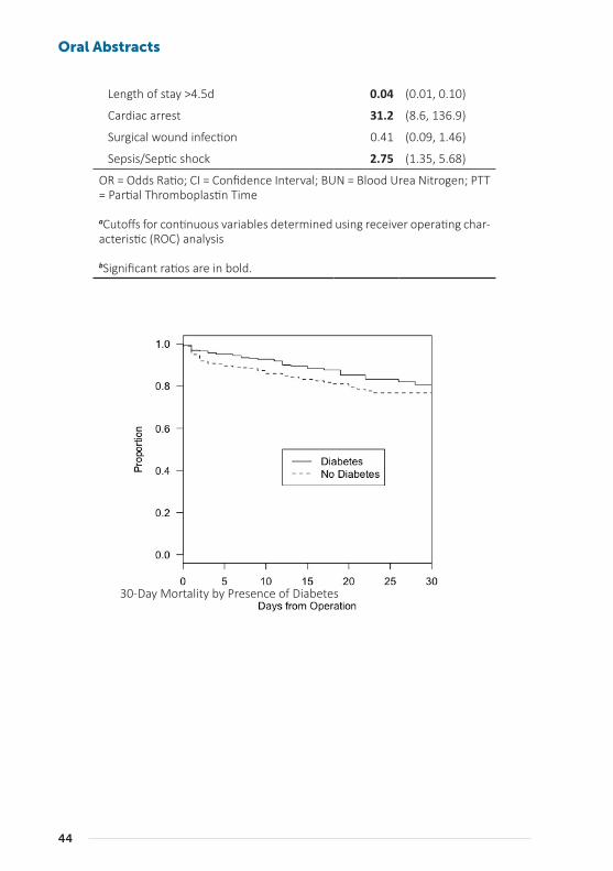
44
Oral Abstracts
Length of stay >4.5d 0.04 (0.01, 0.10)
Cardiac arrest 31.2 (8.6, 136.9)
Surgical wound infection 0.41 (0.09, 1.46)
Sepsis/Septic shock 2.75 (1.35, 5.68)
OR = Odds Ratio; CI = Confidence Interval; BUN = Blood Urea Nitrogen; PTT = Partial Thromboplastin Time
aCutoffs for continuous variables determined using receiver operating char-acteristic (ROC) analysis
bSignificant ratios are in bold.
30-Day Mortality by Presence of Diabetes
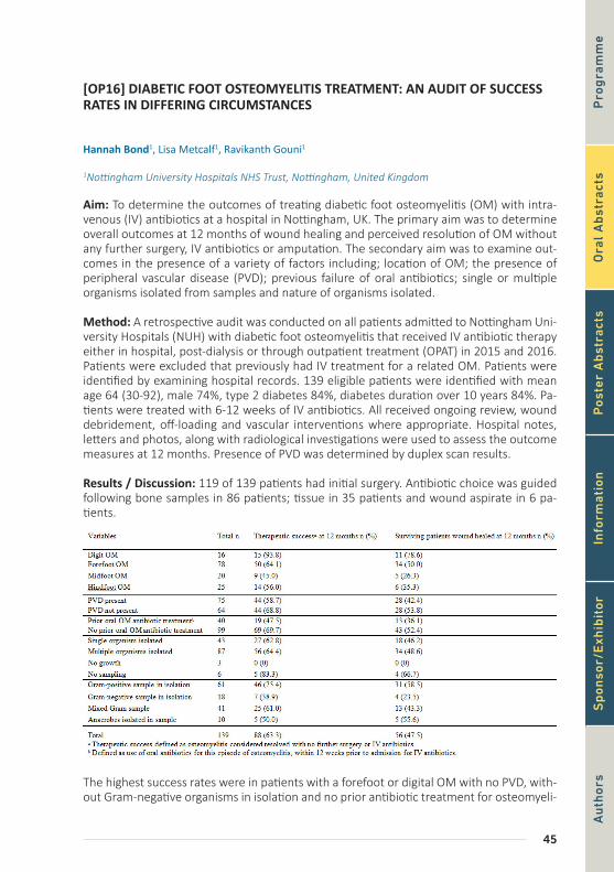
45
[OP16] DIABETIC FOOT OSTEOMYELITIS TREATMENT: AN AUDIT OF SUCCESS RATES IN DIFFERING CIRCUMSTANCES
Hannah Bond1, Lisa Metcalf1, Ravikanth Gouni1
1Nottingham University Hospitals NHS Trust, Nottingham, United Kingdom
Aim: To determine the outcomes of treating diabetic foot osteomyelitis (OM) with intra-venous (IV) antibiotics at a hospital in Nottingham, UK. The primary aim was to determine overall outcomes at 12 months of wound healing and perceived resolution of OM without any further surgery, IV antibiotics or amputation. The secondary aim was to examine out-comes in the presence of a variety of factors including; location of OM; the presence of peripheral vascular disease (PVD); previous failure of oral antibiotics; single or multiple organisms isolated from samples and nature of organisms isolated.
Method: A retrospective audit was conducted on all patients admitted to Nottingham Uni-versity Hospitals (NUH) with diabetic foot osteomyelitis that received IV antibiotic therapy either in hospital, post-dialysis or through outpatient treatment (OPAT) in 2015 and 2016. Patients were excluded that previously had IV treatment for a related OM. Patients were identified by examining hospital records. 139 eligible patients were identified with mean age 64 (30-92), male 74%, type 2 diabetes 84%, diabetes duration over 10 years 84%. Pa-tients were treated with 6-12 weeks of IV antibiotics. All received ongoing review, wound debridement, off-loading and vascular interventions where appropriate. Hospital notes, letters and photos, along with radiological investigations were used to assess the outcome measures at 12 months. Presence of PVD was determined by duplex scan results.
Results / Discussion: 119 of 139 patients had initial surgery. Antibiotic choice was guided following bone samples in 86 patients; tissue in 35 patients and wound aspirate in 6 pa-tients.
The highest success rates were in patients with a forefoot or digital OM with no PVD, with-out Gram-negative organisms in isolation and no prior antibiotic treatment for osteomyeli-
Pro
gra
mm
eA
uth
ors
Ora
l A
bst
ract
sP
oste
r A
bst
ract
sIn
form
atio
nS
pon
sor/
Exh
ibit
or
46
Oral Abstracts
tis; in this group there was a therapeutic success rate of 92.3% (26 patients). The patients with the poorest outcomes had PVD, Gram-negative organisms present and had received previous oral treatment for OM and had a success rate of 41.2% (17 patients).
Conclusion: The success rates of treatment for diabetic foot osteomyelitis varied substan-tially depending on the situation. It was expected that patients with hindfoot osteomyeli-tis or PVD would have worse outcomes, but the presence of Gram-negative bacteria and previous failure on oral antibiotics also appears to be an indicator of a reduced chance of successful treatment. This audit will be used to give patients more specific advice on their prognosis based on their individual circumstances.
47
[OP17] WBC-SPECT/CT TO ASSESS DIABETIC FOOT OSTEOMYELITIS REMIS-SION: CONTRIBUTION OF A COMPOSITE SEVERITY INDEX
Julien Vouillarmet1, Morelec Isabelle1, Moret Myriam3, Michon Paul1, Tordo Jeremy1, Dubreuil Julien1
1Centre Hospitalier Lyon Sud, Pierre Bénite, France3Groupement Hospitalier Lyon Est, France
Aim: Diabetic foot osteomyelitis (DFO) is a frequent complication of diabetes, and an im-portant risk factor of amputation. We previously published that White Blood Cell (WB-C)-SPECT/CT could be a useful tool to assess DFO remission and guide the duration of antibiotic treatment. The aim of the current study is to evaluate the performance of WBC-SPECT/CT to diagnose DFO remission using a standardized hybrid image–based scoring sys-tem, Composite Severity Index (CSI).
Method: All WBC-SPECT/CT performed for patients with DFO at the end of antibiotic treat-ment in a single nuclear medicine department were retrospectively read by 2 nuclear phy-sicians (1 senior and 1 junior), blind to clinical results. Exams included planar acquisitions at 2h and 20h post-injection and a WBC-SPECT/CT at 20h. Classic visual assessment was performed and with CSI to classify images as follow: intensity of uptake, from 0 to 3 at 2 hours and 20 hours after injection on planar acquisitions (CSIi2h and CSIi20h) and stage of bone erosion on SPECT/CT at 20h, from 0 to 3 (CSIs). CSIi2h, CSIi20h, and CSIs were sum to define a score. Comparison between performances of composite score index and visual assessment was realized. Successful treatment of DFO was defined by absence of DFO relapse in the same site and absence of surgical bone resection/amputation within 1 year.
Results / Discussion: Fifty-eight patients with 60 DFO were included. Eight were excluded of analysis due to absence of follow-up or absence of SPECT/CT. Of the 52 DFO included in the analysis, 9 (17.6%) showed a relapse during follow-up. Sensitivity, specificity, posi-tive predictive value, negative predictive value and accuracy of WBC-SPECT/CT to predict relapse of DFO in the same area with visual assessment were lower for junior physician (respectively at 60%, 80.4%, 40%, 89.9%, and 75%) than for senior physician (respectively 90%, 83.3%, 56.3%, 97.2%, and 84.6%) with moderate inter-rater agreement (Kappa at 0.587). Performances with the use of CSI were significantly better only for junior physician with good inter-rater agreement (kappa at 0.76).
Conclusion: We show in this study that classic interpretation of WBC-SPECT/CT with visu-al assessment to assess DFO remission needs experience and supports that CSI could be useful for junior nuclear physician to discriminate residual infections and inflammatory post-treatment uptake and to normalize interpretation of WBC-SPECT/CT.
Pro
gra
mm
eA
uth
ors
Ora
l A
bst
ract
sP
oste
r A
bst
ract
sIn
form
atio
nS
pon
sor/
Exh
ibit
or
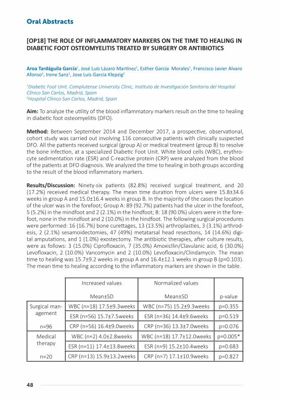
48
Oral Abstracts
[OP18] THE ROLE OF INFLAMMATORY MARKERS ON THE TIME TO HEALING IN DIABETIC FOOT OSTEOMYELITIS TREATED BY SURGERY OR ANTIBIOTICS
Aroa Tardáguila García1, José Luis Lázaro Martínez1, Esther Garcia Morales1, Francisco Javier Alvaro Afonso1, Irene Sanz1, Jose Luis Garcia Klepzig2
1Diabetic Foot Unit. Complutense University Clinic, Instituto de Investigación Sanitaria del Hospital Clínico San Carlos, Madrid, Spain2Hospital Clínico San Carlos, Madrid, Spain
Aim: To analyze the utility of the blood inflammatory markers result on the time to healing in diabetic foot osteomyelitis (DFO).
Method: Between September 2014 and December 2017, a prospective, observational, cohort study was carried out involving 116 consecutive patients with clinically suspected DFO. All the patients received surgical (group A) or medical treatment (group B) to resolve the bone infection, at a specialized Diabetic Foot Unit. White blood cells (WBC), erythro-cyte sedimentation rate (ESR) and C-reactive protein (CRP) were analyzed from the blood of the patients at DFO diagnosis. We analyzed the time to healing in both groups according to the result of the blood inflammatory markers.
Results/Discussion: Ninety-six patients (82.8%) received surgical treatment, and 20 (17.2%) received medical therapy. The mean time duration from ulcers were 15.8±34.6 weeks in group A and 15.0±16.4 weeks in group B. In the majority of the cases the location of the ulcer was in the forefoot; Group A: 89 (92.7%) patients had the ulcer in the forefoot, 5 (5.2%) in the mindfoot and 2 (2.1%) in the hindfoot; B: 18 (90.0%) ulcers were in the fore-foot, none in the mindfoot and 2 (10.0%) in the hindfoot. The following surgical procedures were performed: 16 (16.7%) bone curettages, 13 (13.5%) arthroplasties, 3 (3.1%) arthrod-esis, 2 (2.1%) sesamoidectomies, 47 (49%) metatarsal head resections, 14 (14.6%) digi-tal amputations, and 1 (1.0%) exostectomy. The antibiotic therapies, after culture results, were as follows: 3 (15.0%) Ciprofloxacin, 7 (35.0%) Amoxicllin/Clavulanic acid, 6 (30.0%) Levofloxacin, 2 (10.0%) Vancomycin and 2 (10.0%) Levofloxacin/Clindamycin. The mean time to healing was 15.7±9.2 weeks in group A and 16.4±12.1 weeks in group B (p=0.103). The mean time to healing according to the inflammatory markers are shown in the table.
Increased values
Mean±SD
Normalized values
Mean±SD p-value
Surgical man-agement
n=96
WBC (n=18) 17.5±9.3weeks WBC (n=75) 15.2±9.3weeks p=0.355
ESR (n=56) 15.7±7.5weeks ESR (n=36) 14.4±9.6weeks p=0.519
CRP (n=56) 16.4±9.0weeks CRP (n=36) 13.3±7.0weeks p=0.076
Medical therapy
n=20
WBC (n=2) 4.0±2.8weeks WBC (n=18) 17.7±12.0weeks p=0.005*
ESR (n=11) 17.4±13.8weeks ESR (n=9) 15.2±10.4weeks p=0.683
CRP (n=13) 15.9±13.2weeks CRP (n=7) 17.1±10.9weeks p=0.827
49
*Differences were assumed significant at p<0.05 for a confidence interval of 95%.
Conclusion: The inflammatory markers have not utility on the prognosis of the time to healing in patients with DFO regardless of the treatment administered. In addition, the medical therapy manages well bone infections if we prescribe the antibiotics according to the result of bone culture.
Pro
gra
mm
eA
uth
ors
Ora
l A
bst
ract
sP
oste
r A
bst
ract
sIn
form
atio
nS
pon
sor/
Exh
ibit
or
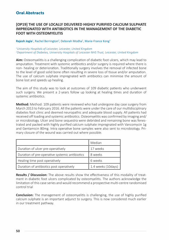
50
Oral Abstracts
[OP19] THE USE OF LOCALLY DELIVERED HIGHLY PURIFIED CALCIUM SULPHATE IMPREGNATED WITH ANTIBIOTICS IN THE MANAGEMENT OF THE DIABETIC FOOT WITH OSTEOMYELITIS
Rajesh Jogia1, Rachel Berrington2, Deborah Modha2, Marie-France Kong1
1University Hospitals of Leicester, Leicester, United Kingdom2Department of Diabetes, University Hospitals of Leicester NHS Trust, Leicester, United Kingdom
Aim: Osteomyelitis is a challenging complication of diabetic foot ulcers, which may lead to amputation. Treatment with systemic antibiotics and/or surgery is required where there is non- healing or deterioration. Traditionally surgery involves the removal of infected bone to the level of good solid bone often resulting in severe loss of tissue and/or amputation. The use of calcium sulphate impregnated with antibiotics can minimise the amount of bone lost and speeds up healing.
The aim of this study was to look at outcomes of 109 diabetic patients who underwent such surgery. We present a 2-years follow up looking at healing times and duration of systemic antibiotics.
Method: Method: 109 patients were reviewed who had undergone day case surgery from March 2013 to February 2016. All the patients were under the care of our multidisciplinary diabetes foot clinic and deemed neuropathic and adequate blood supply. All patients had received off loading and systemic antibiotics. Osteomyelitis was confirmed by imaging and/or microbiology. Ulcer and bone sequestra were debrided and remaining bone was fenes-trated and packed with highly purified calcium sulphate impregnated with Vancomycin 1g and Gentamicin 80mg. Intra operative bone samples were also sent to microbiology. Pri-mary closure of the wound was carried out where possible.
Median
Duration of ulcer pre-operatively 17 weeks
Duration of pre-operative systemic antibiotics 8 weeks
Healing time post operatively 6 weeks
Duration of antibiotics post operatively 1.4 weeks (10days)
Results / Discussion: The above results show the effectiveness of this modality of treat-ment in diabetic foot ulcers complicated by osteomyelitis. The authors acknowledge the limitation of this case series and would recommend a prospective multi-centre randomised control trial
Conclusion: The management of osteomyelitis is challenging, the use of highly purified calcium sulphate is an important adjunct to surgery. This is now considered much earlier in our treatment pathway.
51
[OP20] OSTEOMYELITIS SEQUESTRECTOMY AND APPLICATION OF AN ANTIBI-OTIC-ELUTING BONE SUBSTITUTE TO AVOID MINOR AMPUTATION AND PRE-SERVE MECHANICAL STABILITY IN THE DIABETIC FOOT
Cristian Nicoletti1, Emanuele Nasole2, Luca Spazzapan2, Elena Solagna2
1Director Diabetic Foot Unit, Pederzoli Hospital, Peschiera del Garda, Italy2Diabetic Foot Unit, Pederzoli Hospital, Peschiera del Garda, Italy
Aim: To evaluate the effectiveness of an antibiotic eluting injectable synthetic bone graft substitute in the treatment of diabetic foot osteomyelitis (OM).
Method: Consecutive type 2-diabetic patients affected by OM of first metatarsal head, midfoot and/or hindfoot who presented from October 2016 to March 2017 were included. All patients underwent standard care, bone biopsy, systemic antibiotic therapy according to antibiogram, OM sequestrectomy and implantation of a synthetic bone graft eluting either gentamicin or vancomycin, according to antibiogram. All patients had negative vas-cular assessment for critical limb ischemia (CLI) and were assigned our standard offloading protocol. Patients were considered as “healed” if they had first-intention closure and/or did not undergo any other surgical procedures to heal.
Results / Discussion: A total of 12 patients were included: 8 with 1st-metatarsal head OM, 2 with cuboid and 2 with heel OM. Nine patients received the gentamicin-eluting bone substitute and 3 the vancomycin-eluting product. Six patients (50%) healed after the treat-ment. Three patients (25%) failed to heal due to CLI recurrence. The remaining 3 patients (25%) failed to heal probably due to low compliance to the offloading protocol. One patient (with heel OM and ESRD) underwent Lower Extremity Amputation (LEA).
Conclusion: OM sequestrectomy with the use of the antibiotic-eluting synthetic bone graft substitutes was effective in healing diabetic patients with OM of the foot, thereby avoiding minor amputations, with consequences for biomechanical stability, and reducing the need for long-term antibiotic therapy.
Pro
gra
mm
eA
uth
ors
Ora
l A
bst
ract
sP
oste
r A
bst
ract
sIn
form
atio
nS
pon
sor/
Exh
ibit
or
52
Oral Abstracts
[OP21] FACTORS PREDICTING RE-AMPUTATION AFTER TRANSMETATARSAL AMPUTATION IN PATIENTS WITH DIABETES MELLITUS
Katherine Raspovic1, Dane Wukich1, Junho Ahn1, George Liu1
1University of Texas Southwestern Medical Center, United States
Aim: To assess factors predictive of re-amputation in patients with diabetes mellitus (DM) after transmetarsal amputations (TMA) in the perioperative period.
Method: Patients were identified in a database* between 2011 and 2015. Patient factors were compared between successful and failed TMA groups using univariate analyses and Benjamini-Hochberg method of p-value adjustment for multiple comparisons. Indepen-dent risk factors were determined by multivariate regression. Amputations that spared the ankle joint were considered minor amputations, while those that sacrificed the ankle joint were considered major amputations
Results / Discussion: Of 1,339 patients undergoing TMA, 89 (6.6%) underwent re-ampu-tation (14 minor and 75 major) in the 30-day perioperative period. Through univariate analysis, no differences in age (p=0.603), body-mass index (BMI) (p=0.205), race (p=0.997), pre-operative function (p=0.475), or smoking in the past year (p=0.303) were observed between patients with and without successful TMAs. In the multivariate analysis, predic-tors of re-amputation were pre-operative transfusion (adjusted odds ratio [OR] 2.45, 95% confidence interval [CI] 1.04-5.24), insulin use (adjusted OR 2.25, 95% CI 1.07-5.54), serum blood urea nitrogen >20 mg/dL (adjusted OR 1.96, 95% CI 1.15-3.45), end-stage renal dis-ease (ESRD) (adjusted OR 2.05, 95% CI 1.06-3.90), congestive heart failure (adjusted OR 0.13, 95% CI 0.01-0.60) and post-operative wound complications such as dehiscence and infection (adjusted OR 3.27, 95% CI 1.33-7.27).
Conclusion: Patients with DM with the risk factors described in this study are at higher risk of failing initial TMA and requiring re-amputation. 5.6% of diabetic patients required conversion of a TMA to an above ankle amputation during the first 30 days.
* ACS-NSQIP®, American College of Surgeons-National Surgical Quality Improvement Program®
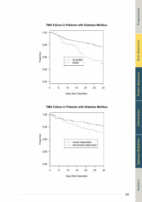
53
Pro
gra
mm
eA
uth
ors
Ora
l A
bst
ract
sP
oste
r A
bst
ract
sIn
form
atio
nS
pon
sor/
Exh
ibit
or
54
Oral Abstracts
[OP22] RISK OF HOSPITALIZATION FOR CARDIOVASCULAR EVENTS OR CHRON-IC KIDNEY DISEASE STRATIFIED BY GENDER IN PATIENTS WITH OR WITHOUT DIABETIC FOOT SYNDROME
Elisabetta Salutini1, Anna Tedeschi1, Secondina Viti1, Piergiorgio Francia2, Laura Policardo3, Paolo Francesconi3, Giuseppe Seghieri3, Alessandra De Bellis1, Roberto Anichini1
1Diabetes Unit and Diabetic Foot Unit, San Jacopo Hospital, Pistoia, Italy2University of Florence, Department of Clinical and Experimental Medicine, Florence, Italy3Ars Toscana, Florence, Italy
Aim: Diabetic foot syndrome (DFS) is associated with an increased risk of hospitalizations for acute atherosclerotic cardio-vascular diseases (ASCVD) or for chronic kidney disease (CKD). If there is a gender difference in the excess risk for ASCVD or CKD remains yet to be fully ascertained. In the present study we evaluate the risk of future hospitalization for ASCVD and CKD in a population of diabetic people stratified by gender.
Methods: Diabetic patients living in Tuscany on January 1th 2011 were identified by a val-idated algorithm from administrative databases and were divided into two groups: those with or without DFS (DFS+ and DFS-) based on hospitalizations for DFS during the period 2011-2016 (foot ulcers, infections, major or minor amputations, foot gangrene or Charcot, identified by ICD-9-CM codes). Patients were also stratified by gender.
The DFS- associated excess risk for first hospitalization for ASCVD or CKD, during the period 2011-2016, was assessed separately in males and females by Cox regression analysis, after adjusting for age and previous co-morbidities.
Results: 241,844 diabetic patients were included in the analysis (M: 49.5%). The adjusted risk for DFS was about two-fold higher among males (OR:2.02; 95%CI:1.90-2.14; p<0.0001) while the adjusted DFS-associated excess risk for next ASCVD was greater among females (HR:1.65; 95%CI: 1.47-1.89; p<0.0001) than among males (HR:1.46; 95%CI:1.35-1.57; p<0.0001). The excess risk for CKD hospitalization linked to DFS was higher among females (HR: 2.96; 95%CI: 2.28-3.78; p<0.0001) than among males (HR: 2.50; 95%CI: 2.13-2.92; p<0.0001).
Conclusion: In our population the risk for DFS was greater in men while the adjusted DFS-associated excess risk of future hospitalization for ASCVD or CKD were higher among women.
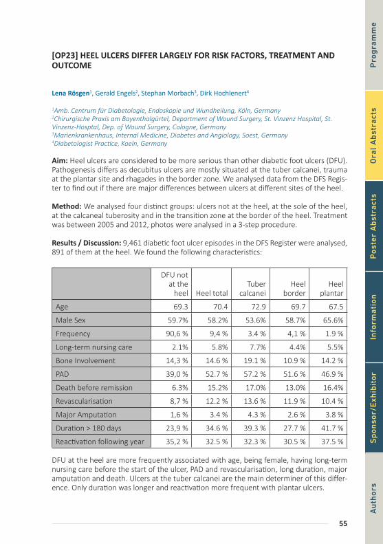
55
[OP23] HEEL ULCERS DIFFER LARGELY FOR RISK FACTORS, TREATMENT AND OUTCOME
Lena Rösgen1, Gerald Engels2, Stephan Morbach3, Dirk Hochlenert4
1Amb. Centrum für Diabetologie, Endoskopie und Wundheilung, Köln, Germany2Chirurgische Praxis am Bayenthalgürtel, Department of Wound Surgery, St. Vinzenz Hospital, St. Vinzenz-Hosptal, Dep. of Wound Surgery, Cologne, Germany3Marienkrankenhaus, Internal Medicine, Diabetes and Angiology, Soest, Germany4Diabetologist Practice, Koeln, Germany
Aim: Heel ulcers are considered to be more serious than other diabetic foot ulcers (DFU). Pathogenesis differs as decubitus ulcers are mostly situated at the tuber calcanei, trauma at the plantar site and rhagades in the border zone. We analysed data from the DFS Regis-ter to find out if there are major differences between ulcers at different sites of the heel.
Method: We analysed four distinct groups: ulcers not at the heel, at the sole of the heel, at the calcaneal tuberosity and in the transition zone at the border of the heel. Treatment was between 2005 and 2012, photos were analysed in a 3-step procedure.
Results / Discussion: 9,461 diabetic foot ulcer episodes in the DFS Register were analysed, 891 of them at the heel. We found the following characteristics:
DFU not at the
heel Heel totalTuber
calcaneiHeel
borderHeel
plantar
Age 69.3 70.4 72.9 69.7 67.5
Male Sex 59.7% 58.2% 53.6% 58.7% 65.6%
Frequency 90,6 % 9,4 % 3.4 % 4,1 % 1.9 %
Long-term nursing care 2.1% 5.8% 7.7% 4.4% 5.5%
Bone Involvement 14,3 % 14.6 % 19.1 % 10.9 % 14.2 %
PAD 39,0 % 52.7 % 57.2 % 51.6 % 46.9 %
Death before remission 6.3% 15.2% 17.0% 13.0% 16.4%
Revascularisation 8,7 % 12.2 % 13.6 % 11.9 % 10.4 %
Major Amputation 1,6 % 3.4 % 4.3 % 2.6 % 3.8 %
Duration > 180 days 23,9 % 34.6 % 39.3 % 27.7 % 41.7 %
Reactivation following year 35,2 % 32.5 % 32.3 % 30.5 % 37.5 %
DFU at the heel are more frequently associated with age, being female, having long-term nursing care before the start of the ulcer, PAD and revascularisation, long duration, major amputation and death. Ulcers at the tuber calcanei are the main determiner of this differ-ence. Only duration was longer and reactivation more frequent with plantar ulcers.
Pro
gra
mm
eA
uth
ors
Ora
l A
bst
ract
sP
oste
r A
bst
ract
sIn
form
atio
nS
pon
sor/
Exh
ibit
or
56
Oral Abstracts
Conclusion: Heel ulcers need longer and more intense treatment. Nevertheless, the out-come is worse than average ulcers at other sites. The longer duration might at least partial-ly explain the high rate of death before wound closure. Amongst heel ulcers, ulcers at the borders contributed less to these negative characteristics, the ones at the tuber calcanei most and plantar ulcers somewhere in between. Considering this diversity and the com-pletely different pathogenesis heel ulcers at these three different sites should be seen as distinct entities.
57
[OP24] DIFFERENCES BETWEEN GENDERS IN OUTCOMES OF DIABETIC FOOT ULCER AT ONE YEAR FOLLOW-UP
Cesare Miranda1, Giorgio Zanette1, Matteo Cassin2, Riccardo Neri2, Umberto Grandis2, Eugenio Cervesato2, Andrea Da Porto3, Roberto Da Ros4
1Clinic of Diabetology, Pordenone Hospital, Pordenone, Italy2Clinic of Cardiology, Pordenone Hospital, Pordenone, Italy3Internal Medicine, Tolmezzo , Italy4Clinic of Diabetology, Monfalcone Hospital, Monfalcone, Italy
Aim: Diabetic foot ulcer is a major health problem which affects 15% to 25% of patients with diabetes at some point in their life. While racial/ethnic differences in diabetes are well documented, less attention has been given to differences in outcomes of diabetic foot ulcer by gender. Aim of the study was to identify differences between genders in outcomes of diabetic foot ulcer at one year follow-up .
Method: We have retrospectively evaluated 172 patients with diabetic foot ulcer between January 2016 and December 2016. For the study period 155 patients with diabetic foot ulcer were included while 17 patients were excluded because lost to follow-up.We divided population in two groups based on gender. After one year of follow-up the following out-comes were evaluated: healing, minor amputation, major amputation, revascularisation, death.
Results / Discussion: The majority of patients were male (n.106, 68%). The mean age was longer in women (77.6± 9.75 vs 71,6± 10.3 years p<0.001).Women and men had similar diabetes duration (17.9±8.0 vs. 19.0±11.2 years) while women had worse metabolic con-trol (HbA1c 7.92%±1.94 vs 7.66%± 1.36) and higher prevalence of chronic kidney disease (48.9% vs 33.9%).Men were more common ex smokers (52.8% vs 14.2% p<0001) , they had higher prevalence of ischaemic heart disease (34.9% vs 18.3% p<0.03). Both groups pre-sented a similar rate of neuropathy and peripheral arterial disease.The outcomes for wom-en and men were respectively: healing ( 91.8% vs 77.3% p<0.02)minor amputations(16,1 % vs 10,2 %) major amputations (2% vs 1,88 %) revascularisation (22.4% vs 15%) death (2.0% vs 9.4%). At the multivariate regression model of all predictors found at univariate analysis only sex male resulted an independent predictor of non-healing even after correction for age and HbA1c levels. Presence of Ischaemia and of Infection at baseline were associated (p < 0,019 and < 0,0001 respectevly) with worse outcome (healing) in Males but no in Females.
Conclusion: Data from this study show that women with diabetic foot ulcer were older but have better rate of healing, while men had higher rate of ex smokers , higher prevalence of ischemic heart disease and a higher risk of amputations and death. Sex Male were in-dependently associated with poor outcome (Healing) at one year. The first explanation it’s probably linked to a more frequent smoking habit.With the limitation of little study sample the negative effect of ischaemia and infection to outcome appear to be more pronounced in Man.
Pro
gra
mm
eA
uth
ors
Ora
l A
bst
ract
sP
oste
r A
bst
ract
sIn
form
atio
nS
pon
sor/
Exh
ibit
or
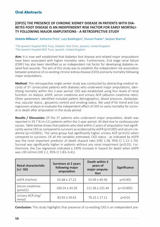
58
Oral Abstracts
[OP25] THE PRESENCE OF CHRONIC KIDNEY DISEASE IN PATIENTS WITH DIA-BETES FOOT DISEASE IS AN INDEPENDENT RISK FACTOR FOR EARLY MORTALI-TY FOLLOWING MAJOR AMPUTATIONS - A RETROSPECTIVE STUDY
Victoria Milbourn1, Katharine Pinto2, Lucy Boddington2, Duncan Fowler2, Sanjeev Sharma2
1The Ipswich Hospital NHS Trust, Diabetic Foot Clinic, Ipswich, United Kingdom2The Ipswich Hospital NHS Trust, Ipswich, United Kingdom
Aim: It is now well established that diabetes foot disease and related major amputations have been associated with higher mortality rates. Furthermore, End stage renal failure (ESRF) has also been identified as an independent risk factor for developing diabetes-re-lated foot wounds. The aim of this study was to establish the independent risk association between presence of co-existing chronic kidney disease (CKD) and early mortality following major amputations.
Method: This retrospective single center study was conducted by abstracting medical re-cords of 37 consecutive patients with diabetes who underwent major amputations, iden-tifying mortality within this 2-year period. CKD was established using four levels of renal function: on dialysis, eGFR, serum creatinine and urinary ACR (albumin creatinine ratio). Other parameters identified included patient demographics, blood pressure, dyslipidae-mia, vascular status , glycaemic control and smoking status. We used X2 for trend and Cox regression analysis to evaluate the independent effect of CKD on early mortality for surviv-al or death after amputation in the study period.
Results / Discussion: Of the 37 patients who underwent major amputation, death was reported in 29.7 % (n=11) patients within the 2-year period. All died due to cardiovascular causes. Table below shows that patients who died within 2 years of amputation had signifi-cantly worse CKD as compared to survivors as evidenced by eGFR (p=0.005) and serum cre-atinine (p=<0.0001). The same group had significantly higher urinary ACR (p=0.01) when compared to survivors. Of all the variables estimated, CKD status - as indicated by eGFR was the most important predictor of death (hazard ratio [HR] 1.58, 95% CI 1.11–1.70). Survival was significantly higher in patients without any renal impairment (p=0.01). Fur-thermore, the Cox regression indicated a 145% increase in hazard for death when eGFR was <30 ml/min (HR 2.2, 95% CI 1.83–3.41).
Renal characteristic (+/- SD)
Survivors at 2 years following major
amputation
Death within 2 years of
major amputa-tion
Significance
eGFR (ml/min) 65.68 ± 27.22 55.09 ± 40.36 p=0.005
Serum creatinine (µmol/L) 100.24 ± 43.29 211.36 ± 225.44 p=<0.0001
Urinary ACR (mg/mmol) 83.93 ± 29.63 76.15 ± 17.11 p=0.01
Conclusion: This study highlights that presence of co-existing CKD is an independent pre-
59
dictor of early mortality in diabetes subjects following major amputation, indicating that this risk increases significantly as CKD worsens. Based on our outcomes, we hypothesize that aggressive management of CKD might improve early mortality following major ampu-tations. Larger studies will be needed to further explore this hypothesis.
Pro
gra
mm
eA
uth
ors
Ora
l A
bst
ract
sP
oste
r A
bst
ract
sIn
form
atio
nS
pon
sor/
Exh
ibit
or
60
Oral Abstracts
[OP26] RISK OF FOOT ULCER DEVELOPMENT IN PATIENTS WITH DIABETES - RELATION TO ISOKINETIC MUSCLE STRENGTH, SENSORY FUNCTION AND CLIN-ICAL FINDINGS
Niels Ejskjaer1, Anne Sofie Bruno Pedersen2, Laerke Walther Junggreen Have2, Henning Andersen2, Christer Zoylner Swan3
1Aalborg University Hospital, Department of Clinical Medicine, Aalborg University, Aalborg, Denmark2Aarhus University Hospital, Clinical Neurology Research Group, Aarhus, Denmark3Aarhus University Hospital, Department of Otorhinolaryngology and Head and Neck Surgery, Aarhus, Denmark
Aim: To investigate whether reduced muscle strength in the lower extremities in diabetic patients is associated to the development of diabetic foot ulcer (DFU).
Method: We conducted a retrospective cohort study on 95 diabetic patients who partici-pated in studies on diabetic polyneuropathy (DPN) and motor function 12-16 years earlier. Isokinetic muscle strength at the ankle and knee, neurological impairment scores (NIS), vibration perception thresholds (VPT), and demographic data were obtained from the ini-tial studies. Patient files were systematically reviewed, and information on DFU occurrence and macrovascular disease (MVD) acquired.
Results / Discussion: Twenty-six patients developed DFU. A temporal relationship was found for development of DFU among patients with reduced strength at both the ankle and knee (all P<0.05). Univariate analyses showed a relationship between DFU and re-duced strength for ankle dorsal flexion (P<0.001), ankle plantar flexion (P<0.005), knee extension (P<0.001), and knee flexion (P<0.005). DFU was related to NIS (P<0.001) and MVD (P<0.05) in both univariate and multivariate regression analyses. After adjustment for MVD, all strength measures were related to DFU. When adjusting for NIS, a trend was only found for ankle dorsal flexion (P=0.08).
Conclusion: In DPN, muscle weakness at the ankle and knee contributes to development of foot ulcers.
61
[OP27] EFFICACY OF SUCROSE-OCTASULFATE DRESSING IN NEURO-ISCHAEMIC DFU CONSIDERING FACTORS INFLUENCING WOUND CLOSURE RATE; A POST-HOC ANALYSIS OF THE EXPLORER RCT
Ralf Lobmann1, Michael Edmonds2, José Luis Lázaro-Martínez3, Jacues Martini4, Alberto Piaggesi5
1Klinikum Stuttgart - Krankenhaus Bad Cannstatt, Klinik für Endokrinologie, Diabetologie und Geri-atrie, Stuttgart, Germany2Diabetic Foot Clinic, King´s College Hospital, London, United Kingdom3Diabetic Foot Uni, University Podiatry Clinic, Complutense University of Madrid, Spain4Dept. of Endocrinology, Rangueil University Hospital, Toulouse, France5Dept. of Endocrinology and Metabolism, University of Pisa, Pisa, Italy
Aim: According to most recent guidelines, no treatment added to optimal Standard of care (SOC) including efficient off-loading has shown any clear benefit in the management of diabetic foot ulcer (DFU). Efficacy of a sucrose octa-sulfate wound dressing (TLC-NOSF dressing) versus a neutral dressing (TLC) in addition to the same standard of care, in pa-tients presenting with a neuro-ischaemic DFU, was then assessed in neuro-ischaemic DFU, through a European RCT.
Method: This double-blind RCT was conducted in 43 centres in patients presenting with a non-infected DFU (grade IC/ IIC, Texas Classification), and a surface area > 1cm² . The pri-mary outcome was the wound closure rate by week 20 in the ITT population (binary logistic analysis). Secondary outcomes included time to closure and adverse events occurrence (infection, notably).
Results / Discussion: A total of 240 patients were randomised and received either the treatment dressing (n=124) or the control dressing (n=114). At Week 20, wound closure occurred in 34 patients (30%) in the control group and in 60 patients (48%) in the treat-ment group (adjusted odds ratio 2·60 [95% CI 1·43 to 4·73], p=0·002).
Post-hoc analysis were undertaken, considering parameters that may influence the tissue repair process (wound duration, wound area, wound location, vascular status, patients’ characteristics…), always showing favourable outcomes for the sucrose-octasulfate dress-ing, whatever the characteristics of the treated wound.
Conclusion: Sucrose octasulfate dressing and good standard of care is significantly more effective than neutral dressing, in the management of neuro-ischaemic DFUs, and specifi-cally when treatment is initiated early in the wound evolution.
Pro
gra
mm
eA
uth
ors
Ora
l A
bst
ract
sP
oste
r A
bst
ract
sIn
form
atio
nS
pon
sor/
Exh
ibit
or
62
Oral Abstracts
[OP28] NON-NEURONAL CONTROL OF PROLIFERATION AND MIGRATION OF KERATINOCYTES ON SITE OF ULCERATION
Ekaterina Artemova1, Zera Abdulvapova2, Evgeniy Ivanov3, Anna Gorbacheva1, Alla Tokmakova2, Svetlana Gavrilova3, Gagik Galstyan1
1Endocrinology Research Center, Moscow, Russian Federation2Endocrinology Research Centre, Diabetic Foot Department, Moscow, Russian Federation3Lomonosov Moscow State University, Faculty of Fundamental Medicine, Russian Federation
Aim: To assess proliferation and migration of keratinocytes at the nonhealing edges of neu-ropathic wounds, identify key enzymes for the synthesis of catecholamines in keratinocytes
Method: 25 patients with DM, with neuropathic ulcers (duration about 12 months) and 5 patients without diabetes with decubitus were enrolled. DF patients were underwent to standard treatment including debridement, atraumatic dressing, offloading with remov-able total contact cast, antibacterial therapy if it needs. Severity of peripheral neuropathy was assessed according to the NDS scales; was evaluated. Histological (hematoxylin and eosin) and immunohistochemical (Ki 67, α7nAChR, keratin К10, tyrosine hydroxylase) ex-amination of wound edge were done during treatment (0, 10, 24 days).
Results / Discussion: All patients have severe neuropathy according to NDSm(>8). The average size of DF ulcers before and on 10th day of treatment was of 5.56 cm2 and 4,29 cm2, respectively (p<0,004). Neuropathic ulcers were characterized by hyperproliferative epidermis. Mitotically active keratinocytes reside throughout the suprabasal layers. Ki-67 expressed all layers of the epidermis, but a greater staining density was detected in the basal layer. The density of a7nAChR-positive cells increased from 0 to 24 days. Skin samples taken from patients on the 0th and 10th day of therapy were characterized by a low density for tyrosine hydroxylase, in contrast to samples taken on the 24th day. There was a low expression of K10 keratin differentiation markers before the beginning of therapy. There was formed a hyperproliferative epidermis, the cells of which lost the ability for terminal differentiation, the process of cornification was disturbed.
Conclusion: All layers of the epidermis of wound edge actively proliferated at condition-ally separated stages of the wound process, which led to a pathological thickening of the epidermis, despite regular debridement. An increase in the expression of receptors for α7nAChR indicates a low migration potential of keratinocytes of the edge of neuropathic wounds. Due to the presence of severe neuropathy, the pathological pattern in the epider-mis can be caused by a deficiency of nerve regulatory influences (the amount of media-tor released by damaged nervous structures) and inadequate amounts of catecholamines synthesized in skin cells, which led to the impaired migration and differentiation. The data obtained can serve as a basis for the development of local therapy for wound defects to enhance the migration of keratinocytes. It is necessary to search for signaling pathways that block excessive proliferation of epidermal cells that form a pathologically thickened epidermis.
63
[OP29] PRESENT INTERNATIONAL GUIDELINES FAIL TO CLASSIFY 76% OF IN-FECTED DIABETIC FEET WHICH COMES TO MINOR AMPUTATION AS SEVERE
Elizabeth Pendry1, Joanne Casey1, Jennifer Tremlett1, Nina Petrova1, Prash Vas1, Chris Manu2, Mi-chael Edmonds1
1King’s College Hospital, Diabetic Foot Clinic, London, United Kingdom2King’s College Hospital, London, United Kingdom
Aim: We have previously reported that patients with foot infections severe enough to need admission to hospital and treatment with intravenous antibiotics do not show the classi-cal inflammatory signs of infection. We now report that only 26% of patients with infec-tion severe enough to need minor amputation show two or more inflammatory signs of Temperature >38°C or <36°C, Heart rate >90 beats/minute, Respiratory rate >20 breaths/minute, White blood cell (WBC) count >12,000 or <4,000/mm3 which, would classify there infection as severe according to International Guidelines
Method: We studied 50 patients who presented with infection and needed minor am-putation. Clinical parameters (body temperature, pulse rate and respiratory rate) were recorded on the day of admission. Blood samples were also collected on admission and inflammatory markers were measured. Data are mean + standard deviation.
Results / Discussion: The mean plasma C-reactive protein on admission was significantly raised at 111±89mg/l, (reference range (rr) below 5mg/l). However, the mean body tem-perature was only 37 ± 0.69 °C. The mean pulse rate was 81±18 beats/min and the mean respiratory was 17 ±1breaths/min. The mean WBC count was 10.91± 4.17 109L,) rr 4.0 to 11.0 109L) and the mean neutrophil count was 8.59 ±3.89 109L, (rr2.2-6.3 109L).
With reference to guideline criteria, only 4/50 patients had a body temperature >38°C and 1/50 patients had a body temperature <36°C; 17/50 patients had a pulse rate> 90 beats/min and 0/50 patients had a respiratory rate >20 breaths/min, 15/50 patients had WBC >12.000mm3 and 1/50 had WBC <4000/mm3. Overall, only 12/50 patients fulfilled the current definition of severe foot infection.
Conclusion: There is concern that diabetic foot infections which are serious enough to require minor amputation may not be classified as severe according to present guidelines and may not receive the urgency which they deserve. It is important to revisit diabetic foot infection guidelines.
Pro
gra
mm
eA
uth
ors
Ora
l A
bst
ract
sP
oste
r A
bst
ract
sIn
form
atio
nS
pon
sor/
Exh
ibit
or
64
Oral Abstracts
[OP30] DETERMINANTS OF DIABETIC FOOT SELF CARE IN WOMEN WITH DIA-BETES: A POPULATION-BASED STUDY
Mahnaz Sanjari1, Maryam Aalaa2, Mohammad Reza Amini1, Neda Mehrdad2, Hossein Adibi2, Mosta-fa Qorbani2, Hamidreza Aghaei Meybodi2
1Diabetes Research Center, Endocrinology and Metabolism Clinical Sciences Institute, Tehran Universi-ty of Medical Sciences, Tehran, Iran2Endocrinology & Metabolism Research Institute, Tehran University of Medical Sciences, Tehran, Iran
Aim: The aim of this study is to determine of Diabetic Foot Self-Care determinants in Ira-nian women with diabetes.
Method: In this cross-sectional study, 457 women with type 2 diabetes were recruited using random multistage cluster sampling. The data was completed by demographic ques-tionnaire and Diabetic Foot Self Care Questionnaire (DFSQ). This study is investigating de-mographic and lifestyle factors (age, gender, BMI, marital, educational and social economic status), general health status (HbA1c level, life satisfaction, self-rated health, quality of Life (DQOL), physical activity (IPAQ), depression (Beck) and Social Capital Questionnaire (SC-IQ)) and Diabetic Foot Self Care Questionnaire (Personal Self-care, Podiatric Care and Footwear and Socks). The descriptive statistic and adjusted logistic regression models were used to assess the associations between Diabetic Foot Self Care and other determinants.
Results / Discussion: The mean age and duration of diabetes were 51.8± 7.7 and 6.8± 5.9 years, respectively. The mean total Diabetic Foot Self Care was 60.38 (SD: 9.9). Also, the mean of Personal Self-care, Podiatric Care and Footwear and Socks were (M:24.87, SD:7.00), (M:18.02, SD: 2.11), (M: 17.52, SD: 3.95) respectively. The covariant logistic re-gression models were adjusted by age, education, job status, and social economic status. In linear regression analysis, the results were not shown any relations between determinants such as HbA1c level, life satisfaction, self-rated health, quality of Life, physical activity, de-pression and Social Capital Questionnaire.
By linear regression analyses, the best determinants of low Diabetic Foot self Care was depression. The result showed that the women with depression, had lower score, in total total Diabetic Foot Self- care (ß:-2.142, SE: 0.944, P: 0.033), and also had low scores in Podi-atric Care (ß: -0.772, SE:0.197, P: 0.000), But there was any statistical correlation with Per-sonal Self-care (ß: -0.612, SE:0.675, P:0.365) and Footwear and Socks (ß:-0.654, SE: 0.372, P: 0.080). The occurrence of acceptable diabetic foot self care in patient with depression was 33% lower than in normal patients (Odd:0.67)
Conclusion: Depression is common and associated with multiple adverse outcomes in pa-tient with diabetes. The results of this study suggest that the presence of depression may determine low diabetic foot self care of women with diabetes. This initial finding permits subsequent experimental investigations to identify strategies that can be valuable to im-prove diabetic foot control.

65
[OP31] UNCENSORED INCIDENCE OF DIABETIC FOOT ULCERS TO PATIENTS IN REMISSION
Lawrence Lavery1, Jonathan Bloom2, Brian Petersen2, Gary Rothenberg3
1University of Texas Southwestern Medical Center, Department of Plastic Surgery, Dallas, United States2Podimetrics, Inc., Somerville, United States3University of Michigan School of Medicine, Ann Arbor, United States
Aim: Diabetic foot ulcers (DFU) are known to be associated with increased morbidity, mor-tality, and resource utilization. Patients with history of DFU are among those at highest risk, with several prospective studies reporting annual incidence between 20% and 40% during remission. However, nearly all of these studies report DFU-free survival and right-censor outcomes by (1) disenrolling patients upon initial observation of DFU occurrence, and (2) characterizing multiple distinct DFU observed on the same date as a single occurrence. Re-porting uncensored incidence, which includes multiple outcomes to a given patient, may more accurately reflect the true burden of DFU. We hypothesized that the uncensored incidence is meaningfully larger than the censored incidence.
Method: A recent multi-center investigation (NCT02647346) recorded all DFU occurring in a cohort of 129 participants in remission from a prior DFU. Participants were followed for 34 weeks or until withdrawing consent. We modeled DFU occurrences as a nonhomo-geneous Poisson point process over time indexed from the participant entering remission. From this, we estimated the instantaneous incidence through Savitzky-Golay smoothing of the counting process and numerical differentiation. We compared the time-dependent censored and uncensored incidence curves qualitatively and assessed the aggregate differ-ence between the censored and uncensored incidence qualitatively by testing the ratios of DFU/participant for significance.
Results / Discussion: At least one DFU occurred to 37 participants, resulting in a censored ratio of 0.29 DFU/participant (37/129). The uncensored ratio was found to be 0.41 DFU/participant (53/129) given a total of 53 DFU during the study. The 16 additional DFU in the uncensored ratio occurred to eleven participants. Six of these participants presented with multiple DFU on the same date. Although the observed difference of 0.12 DFU/participant is not statistically-significant at the alpha=0.05 level (p=0.06), this study was not a priori powered to detect this difference. Both the censored and uncensored instantaneous inci-dence curves are unimodal with annualized peak rates of 0.42 DFU/participant/year and 0.71 DFU/participant/year during month four of remission. The censored and uncensored instantaneous incidences asymptote to the same baseline value of approximately 0.03 DFU/participant/year after month 18 of remission.
Conclusion: These data suggest a clinically-meaningful component of incidence that is un-derreported in the literature and potentially underappreciated by researchers and practi-tioners. Better characterizing DFU incidence for those in remission may enable improved allocation of resources, organization of care, and communication of prognosis, possibly resulting in reduced DFU-related morbidity, mortality, and resource utilization.
Pro
gra
mm
eA
uth
ors
Ora
l A
bst
ract
sP
oste
r A
bst
ract
sIn
form
atio
nS
pon
sor/
Exh
ibit
or
66
Oral Abstracts
[OP32] NATIONAL INCIDENCE OF FOOT DISEASE-RELATED AMPUTATIONS IN PEOPLE WITH AND WITHOUT DIABETES IN AUSTRALIA, 2008-2015
Peter Lazzarini1, Wenxiam Leow2, Jaap van Netten3
1School of Public Health and Social Work, Queensland University of Technology, Brisbane, Australia2School of Clinical Sciences, Queensland University of Technology, Brisbane, Australia3Queensland University of Technology, Australia. Faculty of Health, Department of Surgery, Ziekenhuis Groep Twente, Almelo, the Netherlands. Department of Rehabilitation, Academic Medical Center, University of Amsterdam, Amsterdam Movement Sciences, the Netherlands.
Aim: To investigate the national annual incidence of amputations caused by foot disease in people with and without diabetes for the first time.
Method: We retrospectively analyzed the Australian National Hospital Morbidity Database for all inpatients in Australia admitted between 2008-to-2015 with foot disease (diagnosed foot ulcers or foot infections) that underwent an amputation procedure (minor (below ankle) or major (above ankle)). We identified those with diagnosed diabetes. Incidence rates for these foot disease-related amputations (FDAs) were expressed per 100,000 per-son-years using the Australian resident population for each year as the denominator.
Results / Discussion: Overall, 33,123 FDAs occurred in Australia between 2008-to-2015; 80% had diabetes, 79% were minor amputations, 72% male, 60% were >65 years old. Fig-ure 1 displays the annual incidence for all, diabetes and without diabetes FDAs. All minor FDAs increased by 32% (12.4-to-16.5 per 100,000 person-years); in those with diabetes by 43% (9.0-to-12.8) and without diabetes by 6% (3.5-to-3.7) (all: p<0.01). All major FDAs decreased by 22% (4.6-to-3.6); in those with diabetes by 21% (3.7-to-2.9) and without diabetes by 26% (1.0-to-0.7) (all: p<0.01).
Conclusion: These findings show a decreasing national incidence of major amputations at the expense of increasing minor amputations. Similar findings have previously been found in diabetes populations across the world; however, to our knowledge, this is the first time this has been found for amputations specifically caused by foot disease in populations with and without diabetes. This study provides important new information on a national ampu-tation burden that is preventable with evidence-based foot disease care.
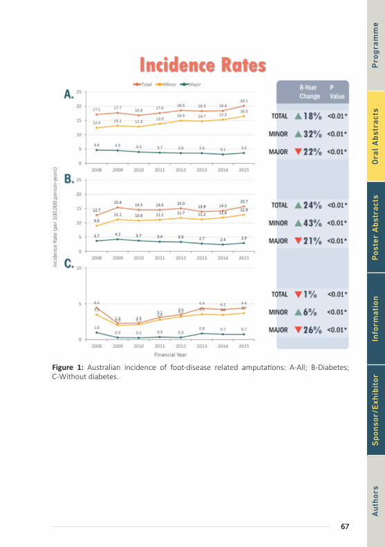
67
Figure 1: Australian incidence of foot-disease related amputations: A-All; B-Diabetes; C-Without diabetes.
Pro
gra
mm
eA
uth
ors
Ora
l A
bst
ract
sP
oste
r A
bst
ract
sIn
form
atio
nS
pon
sor/
Exh
ibit
or
68
Oral Abstracts
[OP33] THE CLINICAL PATH OF THE DIABETIC CHARCOT FOOT. AN EXPLORATO-RY STUDY
Anne Rasmussen1, Klaus Kirketerp-Møller2, Henrik Ullits Andersen1, Susanne Engberg1
1Steno Diabetes Center Copenhagen, Gentofte, Denmark2Bispebjerg Hospital, Steno Diabetes Center Copenhagen, Denmark
Introduction / Aim: Charcot foot in diabetes patients is a rare complication with an in-cidence of approximately 0.3%/year. It is costly both for the patient and the health care system. The aim of this study was to look at the clinical progress of the Charcot foot.
Method: A retrospective cohort of patients with type 1 (T1D) and type 2 (T2D) diabe-tes seen in the multidisciplinary foot clinic in our Centre from 2003 to 2016. All data was collected from our electronic patient record selecting patients with the diagnosis code DM14.6 (Neuropathic arthropathy). Data on the treatment and examination were extract-ed individually.
Results: 114 patients with Charcot foot were identified. Of these 97 (85%) had an active Charcot. The mean age of the patients at debut of Charcot was 55±12 years (range: 28-83 years), 56% were males, the diabetes duration was 25±15 years, 55% had T1D, HbA1c was 72±22 mmol/mol, 86% had neuropathy (vibration threshold >25 V), 59% were non-smok-ers and 50 (44%) lived alone. All patients with active Charcot foot were treated with off-loading (cast or sandals with rocker bottom) as soon as the diagnosis was suspected. The duration from start of symptoms to diagnosis was 10 weeks (range: 1-52). The period of treatment in 82% was 7.5 months (median) (range: 2-36 months). The localization of the affection was in the middle foot in 89 (78%) patients, and 46 (40%) had bony prominences in planta at the end of the treatment. Four (3%) patients had amputation below the knee in the follow up period due to an infected foot ulcer. All patients were prescribed special footwear for prevention of future foot complications. Only 22 (19%) patients returned to their previous occupation after the diagnosis of Charcot.
Conclusion: The Charcot patients were dysregulated, had a long delay from symptoms to diagnosis and a long treatment period. The localization was mostly in the middle foot, as expected. Patients typically live alone and only few went back to their previous occupation after the diagnosis. We hope that this study will lead to a better, equal and systematic treatment for this group of vulnerable patients.
69
[OP34] TEMPORARY NON-REGRESSION OF MRI BONE MARROW EDEMA (EDE-MA EQUIVALENT SIGNAL CHANGES) DURING TREATMENT OF ACTIVE CHAR-COT FOOT
Ernst Chantelau1, Brigitte Zweck2
1Practice of Endocrinology and Diabetology, Düsseldorf, Germany2Heinrich-Heine- University Medical School, Düsseldorf, Germany
Aim: Edema equivalent signal changes (EESC) in MRI of bone (socalled bone marrow ede-ma) represent microvessel neo-angiogenesis. Sequential MR imaging during total contact cast treatment (TCCT) of active Charcot foot has revealed regression of EESC in the foot, which was steady in 2/3 of the cases, and discontinuous in about 1/3 of cases. Conditions related to temporary non-regression of EESC (NREESC) were assessed.
Method: Patient charts review, observational study of 30 MRI follow-studies (FUS) in 22 cases.
Results / Discussion: Early versus late NREESC was observed in the first FUS performed after 12 weeks versus >24 weeks of TCCT . Early NREESC in 17/30 FUS was associated with (n=6) or without (n=9) clinical foot inflammation, from undue load-bearing (n=6), or physi-ologic fracture callus formation (n=9). 2 FUS seemed unrelated to either feature. EESC was decreased after a further 12 weeks of TCCT in every subsequent FUS.
Late NREESC in 13/30 FUS was associated with clinical foot inflammation relapse ( from premature loading-bearing activity n=6), or without inflammation relapse (from callus hy-perplasia, n=3). In 1 FUS, EESC appeared stagnant compared to < 1 month before.
6/30 FUS (2 with early and 4 with late NREESC) displayed spotty EESC developing else-where in the foot skeleton (migrating EESC), possibly due to bone injuries from improper/unphysiologic load-distribution inside a biomechanically improper TCC.
Conclusion: When interpreting NREESC inflammation-related and callus-related neoangio-genesis need to be differentiated, both of which may cause similar EESC in bone. Hence, additional methods may be required, e.g. monitoring of bone alkaline phosphatase as indi-cator of osteoblast activity (callus formation).
Pro
gra
mm
eA
uth
ors
Ora
l A
bst
ract
sP
oste
r A
bst
ract
sIn
form
atio
nS
pon
sor/
Exh
ibit
or
70
Oral Abstracts
[OP35] MORTALITY AND COMPLICATIONS AFTER TREATMENT OF ACUTE CHARCOT FOOT - A LONGITUDINAL RETROSPECTIVE STUDY OVER 19 YEARS
Rasmus Jansen1, Bo Jørgensen2, Per Holstein2, Klaus Kirketerp-Møller3, Ole Lander Svendsen1
1Bispebjerg Hospital, Dept. of Endocrinology, Copenhagen NV, Denmark2Bispebjerg Hospital, Copenhagen Wound Healing Center, Copenhagen NV, Denmark3Steno Diabetes Center Copenhagen, Copenhagen Wound Healing Center, Bispebjerg University Hos-pital, Copenhagen Wound Healing Center, Copenhagen, Denmark
Aim: Charcot foot is a rare but disabling complication to diabetic neuropathy, and can cause permanent, limb-threatening deformities. Treatment consists of long-term off-load-ing, assistance with wound healing and surgical intervention in the stable phase, all to prevent further damage to the foot.
The objective of this study was to investigate a population of patients admitted with a Charcot foot, their treatment and resolution on a case-by-case basis. We wanted to assess the consequences of an acute Charcot foot and its complications.
Method: The study was conducted a retrospective study of all patients admitted to the Co-penhagen Wound Healing Center between 1996-2015 under the diagnosis of Charcot foot (DM14.6) and diabetes mellitus type 1 or 2 (DE10.X and DE11.X). Both physical records, electronic records and online databases were used. Results were then compared to annual reports from the Danish Diabetes Register.
Results / Discussion: A total of 392 patients were identified, of which 173 were included. There were 26% with type 1 diabetes (initial HbA1c 81.7 ±21.4 mmol/mol) and 74% with type 2 diabetes (initial HbA1c
66.5 ±20.3 mmol/mol). Primary off-loading was with a removable walker in 95% of the cases, with an average off-loading time of 8.3 months. The 5-year mortality was 14%, and a mean survival time from onset of 12.7 years. Main causes of death were cardiovascular and nephrological.
There was an association between lack of compliance and occurrence of foot complica-tions, as well as between having a Charcot foot and leaving the workforce.
Conclusion: More patients had T1DM and had a higher HbA1c and blood pressure than the overall population of diabetes patients in the region. A total of 67% developed com-plications such as ulcers, while non-compliant patients did significantly worse than those being compliant. However, the 5-year mortality was low, 14%, and comparable to diabetes patients without Charcot foot.
71
[OP36] PATIENT EXPECTATIONS PRIOR TO CHARCOT RECONSTRUCTION – LES-SONS FROM THE ‘FOOT SCHOOL’
Marcus Simmgen1, Raju Ahluwalia1, Venu Kavarthapu1, Ines Reichert1
1King’s College Hospital, London, United Kingdom
Aim: This study aimed to find out patient priorities ahead of major reconstructive surgery for Charcot foot disease.
Surgical outcomes are better in patients that are actively engaged in the decision-making process. Complex stabilizing surgery for Charcot neuro-arthropathy is an increasingly rec-ognized therapeutic option for misaligned, unstable or fractured feet. While it can offer a last resort to avoid a major lower extremity amputation, the post-operative period is challenging, long and places high demands on patient co-operation. Understanding patient expectations in detail could improve healthcare professionals’ interaction with their pa-tients and subsequently enhance patients’ engagement during the peri-operative process.
Method: A day-long ‘Foot School’ programme has been developed at our institution for small groups of patients awaiting reconstructive orthopaedic surgery with internal fixation. All aspects of the multidisciplinary involvement are being addressed and generous time allowed for patients to pose questions. During the day we administered a questionnaire to determine patients’ expectations and wishes for surgery.
Results / Discussion: 17 patients from three ‘Foot Schools’ completed the questionnaire. The group comprised of 8 female and 9 male patients and their average age was 60 ± 7 years. 76.4% of patients (n=13/17) had Diabetes mellitus type 2. The mean duration of Diabetes was 19.9 years (range 4 – 44 years). 64.7% of patients (n=11) had midfoot Charcot neuro-arthropathy, 11.8% (n=2) had hindfoot involvement and the remainder had combined disease.
In their ranking of importance, 76.4% listed ‘keeping the foot’ as their top priority (n=13/17). In the combined 2nd – 5th rankings (n=68), functional aspects dominated: the ‘ability to stand and walk’ with ‘improved balance and gait’ comprised 25% of responses (n=17/68). This was followed by other aspects of quality of life: ‘no more casts’, ‘no more ulcers’ and ‘a pain-free foot’ were given in 22% of replies (n=15/68). Several patients ex-pressed their hopes in changes to day-to-day activities: ‘independence from carers’, ‘play-ing with grandchildren’ and ‘return to work’ made up 16.2% of responses (n=11/68).
Conclusion: In this patient population we found an overwhelming priority to avoid an am-putation of the foot. That desire was present even though most patients had already been on a long journey of deteriorating foot health and previous orthopaedic surgery. There was a broad range of expectations and the emphasis varied between individual patients. It is important to be aware of these findings when engaging patients during the planning of a major orthopaedic procedure and its post-operative challenges.
Pro
gra
mm
eA
uth
ors
Ora
l A
bst
ract
sP
oste
r A
bst
ract
sIn
form
atio
nS
pon
sor/
Exh
ibit
or
72
Oral Abstracts
[OP37] DETECTION OF INFECTION ON CHARCOT FOOT: ROLE OF LABELED LEU-COCYTE SCAN
Stamata Georga1, Christos Manes2, Triantafillos Didangelos3, Dimitrios Skoutas2, Georgios Arsos1
13rd Dept of Nuclear Medicine, Aristotle University Medical School, Papageorgiou General Hospital, Thessaloniki, Greece2Diabetes Center, Papageorgiou Hospital, Thessaloniki, Greece3Aristotele University, Medical School, Diabetes Center, 1st Propedeutic Dept of Internal Medicine, Ahepa Hospital, Thessaloniki, Greece
Aim: Diagnosing infection on Charcot foot remains challenging despite multimodality im-aging. The study is aiming to explore the role of 99mTc-HMPAO labeled leucocyte scan (LS), alone or combined with SPECT/CT scan and 99mTc-nanocolloid bone marrow scan (BMS) in diagnosing infection on Charcot foot.
Methods: Eighty Charcot feet, in 72 patients, with clinical suspicion of diabetic foot osteo-myelitis (DFO) were investigated by planar LS. Fifty Charcot feet had concomitant plantar ulcers. Thirty one Charcot feet were further investigated by SPECT/CT scan. In 18 Charcot feet BMS was also performed to interpret abnormal leucocyte bone uptake. LS images were interpreted alone and in conjunction with SPECT/CT scans and were compatible with DFO when persistent increased leucocyte bone uptake was observed. Increased leucocyte bone uptake without corresponding BMS uptake confirmed the diagnosis of DFO. Final diagnosis was based on clinical and radiological/scintigraphic follow-up or surgical results.
Results: Among the 80 Charcot feet investigated, final diagnosis was DFO in 21, acute Char-cot arthropathy (CA) in 20, soft tissue infection in 24 and no infection in 15. LS showed fo-cally intense bone uptake in 18/21 and focally mild bone uptake in 3/21 cases of DFO. Con-versely, LS showed mild diffuse or no leucocyte bone uptake in 16/20 cases of acute CA and in all uninfected Charcot feet, a pattern clearly distinguishable from that of DFO. Sensitivity, specificity, accuracy, positive (PPV) and negative predictive value (NPV) of planar LS for DFO on Charcot foot were 90.5%, 86.4 %, 87.5 %, 70.4% and 96.2% respectively. SPECT/CT enabled exact assessment of the extent of infection, especially in cases with concomitant ulcers. SPECT/CT addition highly improved sensitivity and NPV to 100%, but only slightly specificity from 86.4% to 88.5% and accuracy from 87.5% to 90.3%. BMS addition further improved specificity and accuracy of LS (to 91.7% and 94.4%, respectively) by reducing false positive LS results caused by leukocyte accumulation at sites of active bone marrow.
Conclusion: LS is a reliable imaging modality for diagnosing infection on Charcot foot. SPECT/CT addition improved the diagnostic performance of LS for DFO especially in cases with concomitant ulcers. With a NPV of 100%, LS-SPECT/CT is a valuable method to exclude bone infection on Charcot foot, and it could serve as a screening test in cases with clinical suspicion of infection on Charcot foot. In cases of increased leucocyte uptake on Charcot foot, BMS addition is helpful in discriminating DFO from acute CA.
73
[OP38] THE ROLE OF QUANTITATIVE BONE SCAN PARAMETERS FOR DIAGNO-SIS OF CHARCOT FOOT
Robert Bem1, Marie Buncová2, Michal Dubsky1, Vladimíra Fejfarová1, Andrea Němcová1, Veronika Woskova1
1Diabetes Centre, Institute for Clinical and Experimental Medicine, Prague, Czech Republic2Radioisotope Division, Institute for Clinical and Experimental Medicine, Czech Republic
Aim: Early morphological diagnosis and evaluation of disease activity play an important role in the management of Charcot foot (CF). Quantitative bone scan (QBS) effectively con-nected evaluation of bone morphology with activity of CF. The aim of our study was to assess the role of quantitative bone scan parameters – blood flow velocity (BFV) and bone isotope uptake parameters in the diagnosis of active CF.
Method: Forty-seven patients with unilateral active CF (39 men, 31 patients with type 2 di-abetes) underwent QBS during the study period 2013-2018. Patient with bilateral CF, active foot ulcer and approved osteomyelitis were excluded from the study. QBS was performed following intravenous injection of 740 MBq of technetium-99m methylenediphosphonate. Quantitative parameters – BFV (speed of isotope flow from aortic bifurcation to the ankle); the ratio of bone uptake of whole affected foot and unaffected foot (WFR), the ratio of bone uptake of the most affected area of affected foot and the identical area of contralat-eral foot (MAR) and the ratio of bone uptake of the most affected area of affected foot and the tibia diaphysis (FTR) were assessed.
Results / Discussion: BFV was significantly increased in affected foot in comperison with unaffected foot (11.3±4.9 vs. 5.1±4.9 cm/s; p<0.03). Significant correlations between the ratio BFV of the affected and unaffected foot and WFR, MAR, FTR (p<0.0001; <0.001 and <0.04) were seen. In addition, there were significant correlations between perfusion pa-rameters and all bone uptake parameters in patients with type 1 diabetes, but not signifi-cant in patients with type 2 diabetes.
Conclusion: Our study suggests that QBS parameters, especially BFV, should be used for the diagnosis of active CF. The difference between type 1 and type 2 diabetes patients in correlation of perfusion and bone uptake parameters may give evidence for various patho-genesis of CF.
Supported by project for Development of Research Organization 00023001 (IKEM, Prague, Czech Republic) – Institutional support.
Pro
gra
mm
eA
uth
ors
Ora
l A
bst
ract
sP
oste
r A
bst
ract
sIn
form
atio
nS
pon
sor/
Exh
ibit
or
74
Poster Abstracts
[P001] EXTERNAL FIXATION IN THE MANAGEMENT OF INFECTED CHARCOT FOOT, 6-MONTH FOLLOW-UP RESULTS
Veronika Woskova1, Alexandra Jirkovská1, Robert Bem1, Michal Dubský1, Vladimíra Fejfarová1, Kamil Navrátil2, Andrea Němcová1, Karol Sutoris2
1Diabetes Centre, Institute for Clinical and Experimental Medicine, Prague, Czech Republic2Clinic of Transplant Surgery, Institute for Clinical and Experimental Medicine, Prague, Czech Republic
Aim: Charcot neuroosteoarthropathy (CNO) is a serious diabetic complication leading to bone and joint destruction, deformities, ulceration and, frequently, to concomitant infec-tion. The cornerstone of treatment is immobilization and complete off-loading. In some patients, deformities and bone prominences preclude use of an accommodation device and different types of fixators can be an alternative to conservative treatment. Use of these devices in patients with ulcer and/or deep infection, especially osteomyelitis (OM) is controversial. The aim of our study was to evaluate six-month outcomes in patients with ulcerated infected CNO with severe deformity and/or instability treated by external fixation (EF) as part of comprehensive management.
Method: A total of 21 patients with CNO and infected foot ulcer (mean age 55±13 years, 15 male, 11 with Type-2 DM) who underwent surgery using an EF device between Jan 2015 and Sept 2017 were included in the study. All patients presented with deformity and/or foot in-stability without critical limb ischemia (mean TcPO2 54±14 mmHg). Foot infection was iden-tified by clinical signs, imaging techniques and microbiology results. A short-term (<3 month) history of severe to moderate infection prior EF application was documented in 12 (57%) patients. Perioperative culture-guided parenteral ATB was administrated and followed by oral ATB during ambulation, if required. Postoperative off-loading was performed using a wheel-chair until construct removal. Patients were followed-up for six months in terms of perioper-ative complications, EF-related complications, ulcer healing and ability to bear weight.
Results / Discussion: Of the 21 infected CNO patients, 20 completed the 6-month follow-up period. One patient died of a cardiovascular event one week after EF removal, wound un-healed. Mean time to construct removal was 10.7±3.7 weeks. Additional hospital admission was required by 3(15%) patients (2 pin tract infection, 1 revascularization after EF removal). Premature extraction was needed in 2 (10%) patients (1 due to force-related wire damage, 1 due to calcaneal PTI). After six months of follow-up, no additional foot surgery was required, wound healing was achieved in 18 (90%); of these, 14 (78%) were switched to weight bearing using individual or prefabricated orthoses or individual footwear.
Conclusion: Our study demonstrated the safety and short-term effect of external fixation as part of comprehensive therapy in selected patients with infected CNO. An experienced multidisciplinary foot clinic team, long-term culture-guided ATB therapy to control infec-tion and strict adherence to scheduled regular visits during ambulation are all prerequi-sites for successful treatment.
Supported by MH CZ - DRO (Institute for Clinical and Experimental Medicine – IKEM, IN 00023001)
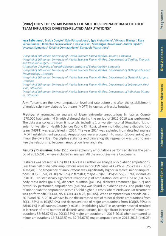
75
[P002] DOES THE ESTABLISHMENT OF MULTIDISCIPLINARY DIABETIC FOOT TEAM INFLUENCE DIABETES-RELATED AMPUTATIONS?
Ieva Baikstiene1, Evalda Danyte1, Egle Patkauskiene1, Egle Kreivaitiene1, Viktoras Sliauzys2, Rasa Verkauskiene1, Rimantas Zalinkevicius3, Linas Velicka2, Mindaugas Stravinskas4, Andrei Pijadin4, Vytautas Kymantas5, Kristina Cerniauskiene6, Danguole Vaznaisiene7
1Hospital of Lithuanian University of Health Sciences Kauno Klinikos, Kaunas, Lithuania2Hospital of Lithuanian University of Health Sciences Kauno Klinikos, Department of Cardiac, Thoracic and Vascular Surgery, Lithuania3Lithuanian University of Health Sciences Institute of Endocrinology, Lithuania4Hospital of Lithuanian University of Health Sciences Kauno Klinikos, Department of Orthopaedics and Traumatology, Lithuania5Hospital of Lithuanian University of Health Sciences Kauno Klinikos, Department of General Surgery, Lithuania6Hospital of Lithuanian University of Health Sciences Kauno Klinikos, Department of Laboratory Med-icine, Lithuania7Hospital of Lithuanian University of Health Sciences Kauno Klinikos, Department of Infectious Diseas-es, Lithuania
Aim: To compare the lower amputation level and rate before and after the establishment of multidisciplinary diabetic foot team (MDFT) in Kaunas university hospital.
Method: A retrospective analysis of lower extremity amputations in Kaunas County (570,000 habitants, ~4 % with diabetes) during the period of 2012-2016 was performed. The data was collected from 6 hospitals, including 1 university hospital (Hospital of Lithu-anian University of Health Sciences Kauno Klinikos), where multidisciplinary diabetic foot team (MDFT) was established in 2014. The year 2014 was excluded from detailed analysis (MDFT establishment process). Amputations were grouped into major (above ankle) and minor (below ankle). Descriptive statistics and binary logistic regression was used to ana-lyze the relationship between amputation level and rate.
Results / Discussion: Total 1511 lower-extremity amputations performed during the peri-od of 2012-2016 where included in analysis. All the amputees were Caucasians.
Diabetes was present in 455(30.11 %) cases. Further we analyze only diabetic amputations. Less than half of diabetic amputations were minor(199 cases -43.74% vs. 256 cases - 56.26 % major). The frequency of amputations was significantly higher in males: minor amputa-tions 109(73.15%) vs. 40(26.85%) in females; major - 89(61.81%) vs. 55(38.19%) in females (p<0.05). No statistically significant relationship of amputation level with HbA1c (p=0.59), body mass index (p=0.69), diabetes duration (p=0.35), diabetes treatment (p=0.57) and previously performed amputations (p=0.96) was found in diabetic cases. The probability of minor diabetic amputation was ~3.5 fold higher in cases where endovascular treatment was performed(OR=3.44, 95 % CI=1.43-8.26, p<0.05). When compared two periods 2012-2013 and 2015-2016 we have found the increased rate of minor diabetic amputations from 50(31.65%) to 103(53.9%) and decreased rate of major amputations from 108(68.35%) to 88(46.1%) in all Kaunas County (p<0.05). Establishing MDFT in university hospital resulted in increase of total number of diabetic amputations, but significant increase of minor am-putations 58(66.67%) vs. 29(33.33%) major amputations in 2015-2016 when compared to minor amputations 16(33.33%) vs. 32(66.67%) major amputations in 2012-2013 (p<0.05)
Pro
gra
mm
eA
uth
ors
Ora
l A
bst
ract
sP
oste
r A
bst
ract
sIn
form
atio
nS
pon
sor/
Exh
ibit
or
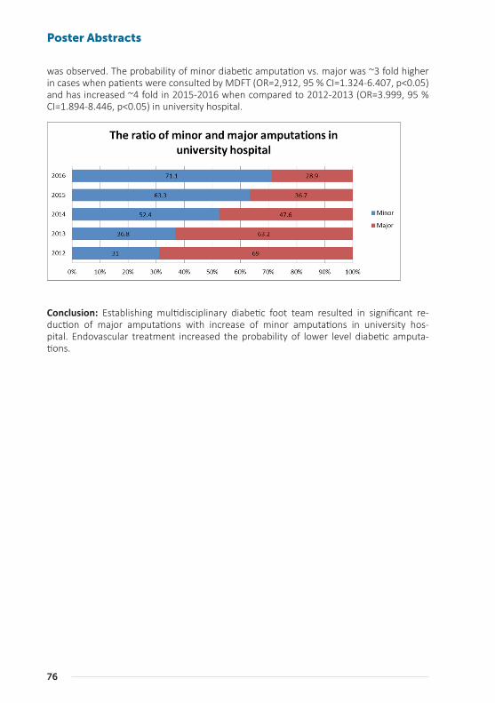
76
Poster Abstracts
was observed. The probability of minor diabetic amputation vs. major was ~3 fold higher in cases when patients were consulted by MDFT (OR=2,912, 95 % CI=1.324-6.407, p<0.05) and has increased ~4 fold in 2015-2016 when compared to 2012-2013 (OR=3.999, 95 % CI=1.894-8.446, p<0.05) in university hospital.
Conclusion: Establishing multidisciplinary diabetic foot team resulted in significant re-duction of major amputations with increase of minor amputations in university hos-pital. Endovascular treatment increased the probability of lower level diabetic amputa-tions.
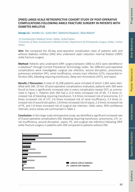
77
[P003] LARGE-SCALE RETROSPECTIVE COHORT STUDY OF POST-OPERATIVE COMPLICATIONS FOLLOWING ANKLE FRACTURE SURGERY IN PATIENTS WITH DIABETES MELLITUS
George Liu1, Jennifer Liu1, Junho Ahn2, Katherine Raspovic1, Dane Wukich2
1Ut Southwestern Medical Center, Dallas, United States2University of Texas Southwestern Medical Center, Department of Orthopaedic Surgery, Dallas, United States
Aim: We compared the 30-day post-operative complication rates of patients with and without diabetes mellitus (DM) who underwent open reduction internal fixation (ORIF) ankle fracture surgery.
Method: Patients who underwent ORIF surgery between 2006 to 2015 were identified in a database* through Current Procedural Terminology codes. Ten different post-operative complications were investigated: surgical site infection, wound disruption, pneumonia, pulmonary embolism (PE), renal insufficiency, urinary tract infection (UTI), myocardial in-farction (MI), bleeding requiring transfusion, deep vein thrombosis (DVT), and sepsis.
Results / Discussion: A total of 16,298 patients were included of which 1,962 were iden-tified with DM. Of the 10 post-operative complications evaluated, patients with DM were found to have a significantly increased rate in every complication except DVT, as summa-rized in Figure 1. Patients with DM had a 12.6 times increased risk of MI, 7.3 times in-creased risk of bleeding requiring transfusion, 5.4 times increased risk of pneumonia, 3.7 times increased risk of UTI, 3.6 times increased risk of renal insufficiency, 3.3 times in-creased risk of wound disruption, 2.8 times increased risk of sepsis, 2.3 times increased risk of PE, and 1.9 times increased risk of surgical site infection. Odds ratios, 95% confidence intervals, and p-values are summarized in Table 1.
Conclusion: In this large-scale retrospective study, we identified a significant increased rate of 9 post-operative complications (MI, bleeding requiring transfusion, pneumonia, UTI, re-nal insufficiency, wound disruption, sepsis, PE, and surgical site infection) following ORIF ankle fracture surgery in patients with DM compared to patients without DM.
0 1 2 3 4
Myocardial Infarction
Renal Insufficiency
Sepsis
Pneumonia
Wound Disruption
Pulmonary Embolism
Deep Vein Thrombosis
Bleeding Requiring Transfusion
Urinary Tract Infection
Surgical Site Infection
patients without diabetespatients with diabetes
% of patients with complication
Figure 1 P
rog
ram
me
Au
thor
sO
ral
Ab
stra
cts
Pos
ter
Ab
stra
cts
Info
rmat
ion
Sp
onso
r/E
xhib
itor
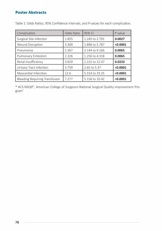
78
Poster Abstracts
Table 1: Odds Ratios, 95% Confidence Intervals, and P-values for each complication.
Complication Odds Ratio 95% CI P value
Surgical Site Infection 1.855 1.245 to 2.791 0.0027
Wound Disruption 3.309 1.896 to 5.787 <0.0001
Pneumonia 5.367 3.144 to 9.166 0.0065
Pulmonary Embolism 2.326 1.256 to 4.318 0.0065
Renal Insufficiency 3.659 1.222 to 12.47 0.0233
Urinary Tract Infection 3.759 2.65 to 5.37 <0.0001
Myocardial Infarction 12.6 5.314 to 33.25 <0.0001
Bleeding Requiring Transfusion 7.277 5.156 to 10.42 <0.0001
* ACS-NSQIP®, American College of Surgeons-National Surgical Quality Improvement Pro-gram®
79
[P004] IMPACT OF FOOT INFECTION ON THE OUTCOMES OF CELL THERAPY IN DIABETIC PATIENTS WITH NO-OPTION CRITICAL LIMB ISCHEMIA
Michal Dubsky1, Anna Pysna1, Andrea Němcová1, Alexandra Jirkovská1, Robert Bem1, Vladimíra Fejfarová1, Veronika Woskova1, Karol Sutoris2
1Diabetes Centre, Institute for Clinical and Experimental Medicine, Prague, Czech Republic2Clinic of Transplant Surgery, Institute for Clinical and Experimental Medicine, Prague, Czech Republic
Aim: Therapeutic vasculogenesis by autologous cell therapy is an alternative treatment method for diabetic patients with no-option critical limb ischemia (NO-CLI). Diabetic foot infection (DFI) might be one of the main causes of major amputation in patients with dia-betic foot disease, especially in those with NO-CLI. The aim of our study was to assess the impact of DFI on the outcomes of cell therapy in patients with NO-CLI and diabetes.
Method: Eighty-nine patients with CLI persisting after unsuccessful standard revascular-ization treated by cell therapy in our foot clinic over 9 years were included in the study and followed-up 24 months after the procedure. DFI factors were assessed at baseline and included: mild or moderate clinical signs of infection (WIfI up to 2), CRP, presence of osteo-myelitis on X-ray, positive probe-to-bone test, multi-resistant pathogens (Staphylococcus MRSA, Klebsiella ESBL, Stenotrophomonas, Alcaligenes) and pathogens resistant to oral antibiotic treatment. All DFI factors were compared between amputated and non-ampu-tated patients. The exclusion criterion was the presence of severe infection at baseline.
Results / Discussion: Of 89 patients treated by cell therapy 13 (14.6%) died without a causal link with cell treatment during the follow-up, major amputation was necessary to perform in 22/76 (28.9%) of surviving patients. Mild or moderate clinical signs of DFI were present in 16/22 (72.7%) of amputated patients and in 8/54 (14.8%) of non-amputated patients (p < 0.001). Osteomyelitis on X-ray (45.5 vs. 20.4%) and the presence of multire-sistant pathogens (27.3 vs. 7.4%) were significantly more prevalent in patients after major amputation compared to the limb salvage group (p = 0.048 and p = 0.032, respectively). Mean levels of CRP were significantly higher in the amputated group (29.3 vs. 10.5 mg/l, p = 0.02), other baseline DFI factors were without a significant difference.
Conclusion: Our study proved that even mild and moderate DFI had a negative impact on the outcome of cell therapy of CLI and confirmed the need of comprehensive therapy of diabetic patients with most the severe neuroischemic ulcers. Therapeutic vasculogenesis in patients with infection, especially with resistant pathogens, osteomyelitis and higher CRP, should be always considered.
Supported by Ministry of Health of the Czech Republic, grant no. 16-27262A, by project for Development of Research Organization 00023001 (IKEM, Prague, Czech Republic) – Insti-tutional support.
Pro
gra
mm
eA
uth
ors
Ora
l A
bst
ract
sP
oste
r A
bst
ract
sIn
form
atio
nS
pon
sor/
Exh
ibit
or
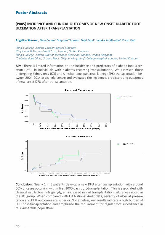
80
Poster Abstracts
[P005] INCIDENCE AND CLINICAL OUTCOMES OF NEW ONSET DIABETIC FOOT ULCERATION AFTER TRANSPLANTATION
Angelica Sharma1, Siew Cohen2, Stephen Thomas2, Tejal Patel2, Janaka Karalliedde3, Prash Vas4
1King’s College London, London, United Kingdom2Guy’s and St Thomas’ NHS Trust, London, United Kingdom3King’s College London, Unit of Metabolic Medicine, London, United Kingdom4Diabetes Foot Clinic, Ground Floor, Cheyne Wing, King’s College Hospital, London, United Kingdom
Aim: There is limited information on the incidence and predictors of diabetic foot ulcer-ation (DFU) in individuals with diabetes receiving transplantation. We assessed those undergoing kidney only (KO) and simultaneous pancreas-kidney (SPK) transplantation be-tween 2004-2014 at a single-centre and evaluated the incidence, predictors and outcomes of new-onset DFU after transplantation.
Method: Overall, we identified 243 (60% male) patients undergoing a KO (n=90) and SPK (n=143) transplantation. Median (range) follow up was 6 (3-13) years. Electronic records and podiatry notes were reviewed.
Results / Discussion: The average age at transplantation was 49(±12) years. The duration of diabetes was 20.4(±11.2) years and 52% had T1DM. Overall 38/243 developed new on-set DFU, giving an overall incidence of 16% during the follow-up period. In the SPK group, those who developed DFU were more likely to have history of peripheral vascular disease (PVD) [37.5% vs. 4%, p<0.05]. In the KO group, they were more likely to have T1DM than T2DM (29% vs. 10%), PVD [32 % vs. 8%], higher pre-transplantation HbA1c (7.5 ±1.2% vs. 6.8±1.4%) and higher serum creatinine (809±243µmol/l vs. 660±202µmol/l)–all p<0.05. The median time from transplantation to the onset of first DFU was 500 days in the SPK group while it was 1000 days in the KO group (figure). 11/38 cases (29%) occurred within the first year of transplantation. The Site, Ischemia, Neuropathy, Bacterial Infection, and Depth (SINBAD) score was 2(±1.4). The median duration of healing was 6(3-14) weeks. All individuals healed their first DFU but 7/38 (18%) required a minor amputation to heal. There was no significant difference in transplant failure between those with and without DFU in the SPK group (31% vs 23%); but a twofold increased risk of transplant failure was noted in the KO group (50% vs. 23%, p=0.02). The mortality rate for the overall group was 22% (n=9). 16 individuals (39%) developed further recurrence of foot ulceration after the first episode post-transplantation.
Conclusion: Nearly 1 in 6 patients develop a new DFU after transplantation with around 50% of cases occurring within first 1000 days post-transplantation. This is associated with classical risk factors. Intriguingly, an increased risk of transplantation failure was noted in the KO group. When compared with UK National Audit data, severity of ulcer at presen-tation and DFU outcomes are superior. Nonetheless, our results indicate a high burden of DFU post-transplantation and emphasise the requirement for regular foot surveillance in this vulnerable population.
81
[P006] OUTCOME OF DEEP HEEL LESIONS IN DIABETIC PATIENTS: THE REAL WORLD
Roberto da Ros1, Roberta Assaloni1, Barbara Brunato1, Enrica Barro1, Silvana Carlucci1, Cesare Mi-randa2
1Diabetes Center, Monfalcone, Italy2Diabetes Center, Aas5, Pordenone, Italy
Aim: Surgical treatment of deep heel lesions represents a challenge in the management of diabetic foot with high risk of failure and of major amputation. These lesions are frequent in people with multiple comorbidity and frail situation that limit surgical conservative pos-sibilities. Aim of the study: to evaluate outcome of deep heel lesions with osteomyelitis, relapse after healing, walking ability and survival in diabetic patients
Method: Interrogation of surgical database between January 2008 and December 2017, founded 71 patients with surgical treatment of calcaneus osteomyelitis in diabetic patients. 99% of patients have type 2 diabetes, mean age was 72±11 years (mean ± SD), 82% were male, long history of diabetes 20 ± 12 years and quite good metabolic control (HbA1c 7.5% ± 1,5) were founded. 13(18%) patients presented chronic renal damage and 12 (17%) end stage renal failure, ischaemic heart disease was present in 29 (41%) patients.
Results/Discussion: The 71 patients had very advanced and complicated wounds, lesions were at heel level, all Texas grade 3: 12 B (with infection), 7 C (with ischaemia), 52 D (with infection and ischaemia). 59(83%) patients underwent revascularization (4 by pass, 55 angioplasty), 41 (58%) targeted to peronal or tibial posterior artery. Outcome: 37 (52%) patients healed, 9 (13%) underwent below the knee or syme amputation, 14 (20%) pa-tients died with lesions, 11 (15%) are still ulcerated. Mean healing time was 243±114 days. Surgical treatment consisted: surgical removal of the lesion in 9 (13%) patients, surgical removal of lesion + partial calcanectomy in 62 (87%). In 29 (41%) patients we used nega-tive pressure and dermal substitute or skin graft to obtain healing. Mean follow up was 3.3 ± 1.6 years. Relapse rate was 11%. Failure of treatment and early death were associated with walking disability (p<0.01), presence of renal impairment or cardiovascular disease (p<0.05), not targeted or successful revascularization (p= 0.01). Age, diabetes duration, metabolic control, lesions of contralateral leg were not significantly associated with out-come.
Conclusion: Data from this study confirm that deep heel lesions are a critical event for di-abetic patients with low healing rate, high incidence of major amputation and early death and walking disability in particular when they occur in frail patients. In absence of renal failure, cardiovascular disease and walking disability healing of the lesion is possible with a long time to heal but a low incidence of relapse and long survival.
Pro
gra
mm
eA
uth
ors
Ora
l A
bst
ract
sP
oste
r A
bst
ract
sIn
form
atio
nS
pon
sor/
Exh
ibit
or
82
Poster Abstracts
[P007] PRESENCE, CHARACTERISATION AND CLINICAL IMPACT OF ANAEMIA IN DIABETIC FOOT ULCERATION: A CROSS SECTIONAL STUDY WITH LONGITUDI-NAL FOLLOW UP OF DFU OUTCOMES IN A TERTIARY CARE SETTING
Matthew Anson1, Janaka Karalliedde2, Ian Alejandro3, Maureen Bates3, Chris Manu1, Michael Ed-monds3, Ian Macdougall1, Prash Vas3
1King’s College Hospital, London, United Kingdom2King’s College London, Unit of Metabolic Medicine, London, United Kingdom3King’s College Hospital, Diabetic Foot Clinic, London, United Kingdom
Aim: Anaemia is a commonly understood to be prevalent in diabetic foot ulceration (DFU). However, the degree in which it and iron homeostasis, influence DFU outcomes, is poorly understood and seldom researched. Aims are to assess the prevalence of anaemia and functional iron deficiency (FID, low iron indices but normal haemoglobin) in patients at-tending a tertiary diabetic foot clinic, and determine whether they are predictive of a poor DFU outcome.
Method: A cross sectional study with a prospective, observational intent was undertaken between November 2017 and February 2018. Patients were stratified into how they were assessed on visitation, into New (DFU-N, n=48) or Follow up (DFU-FU, n=31) groups. They were then subsequently classified into an anaemia, FID or normal subgroups. The DFU-N cohort was followed up for a period of 6 weeks, and prognosis classified as favourable (healing or improvement in DFU size) or unfavourable (static, worsening DFU, amputation or death).
Results / Discussion: There was no significant difference in age (66±15 v 63±12 years), gender (males 73% v 84%) HbA1C (8.3±2.2 v 9.5±2.9, p=0.11) or DFU severity (p=0.73) between the two groups. EGFR was lower in the DFU-FU group (72±20 v 57±21 ml/min, p=0.007) and there was a trend for the DFU-FU to have a longer duration of diabetes (16±11 v 22±15 years, p=0.053). Prevalence of anaemia and FID in the DFU-N group was 40% and 40% respectively; in the DFU-FU it was 55% and 32% respectively. For the whole cohort, haemoglobin value correlated with CRP (rho –0.282, p=0.13), eGFR (0.282, p=0.01) and serum albumin (0.393, p<0.0001). Serum iron correlated with CRP (-0.591, p<0.0001), White cell count (WCC) (-0.359, p=0.001), albumin (0.233, p=0.02). In the DFU-N cohort who were followed up, presence of anaemia and FID were both predictors of an unfa-vourable ulcer prognosis at 6 weeks, with an unfavourable outcome in 82% of those with anaemia and 67% of FID compared to only 14% in those with normal haematinics (p<0.05).
Conclusion: A very high prevalence of Anaemia and FID was noted in patients with DFU. This was associated with poor DFU outcome, even at 6 weeks. Taken together, along with the association with CRP, WCC and albumin, our findings are suggestive that inflammation of any degree may initiate the pathway to anaemia development. Studies looking at larger, more diverse cohorts with assessment of DFU outcomes over 12, 24 and 52-weeks are required to confirm our early findings.
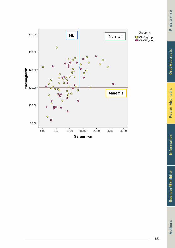
83
Pro
gra
mm
eA
uth
ors
Ora
l A
bst
ract
sP
oste
r A
bst
ract
sIn
form
atio
nS
pon
sor/
Exh
ibit
or
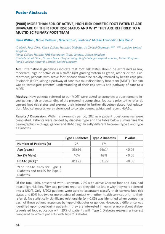
84
Poster Abstracts
[P008] MORE THAN 50% OF ACTIVE, HIGH-RISK DIABETIC FOOT PATIENTS ARE UNAWARE OF THEIR FOOT RISK STATUS AND WHY THEY ARE REFERRED TO A MULTIDISCIPLINARY FOOT TEAM
Daina Walton1, Nicole Mottolini2, Nina Petrova1, Prash Vas3, Michael Edmonds1, Chris Manu4
1Diabetic Foot Clinic, King’s College Hospital, Diabetes UK Clinical Champion 2017 - 2019, London, United Kingdom2Kings College Hospital NHS Foundation Trust, London, United Kingdom3Diabetes Foot Clinic, Ground Floor, Cheyne Wing, King’s College Hospital, London, United Kingdom4King’s College Hospital, London, United Kingdom
Aim: International guidelines indicate that foot risk status should be expressed as low, moderate, high or active or in a traffic light grading system as green, amber or red. Fur-thermore, patients with active foot disease should be rapidly referred by health care pro-fessionals (HCPs) along a pathway of care to a multidisciplinary foot team (MDfT). Our aim was to investigate patients’ understanding of their risk status and pathway of care to a MDfT.
Method: New patients referred to our MDfT were asked to complete a questionnaire in-vestigating their understanding of the presenting complaints, foot care prior to the referral, current foot risk status and express their interest in further diabetes-related foot educa-tion. Medical records were referenced to collate demographics and recent HbA1c.
Results / Discussion: Within a six-month period, 202 new patient questionnaires were completed. Patients were divided by diabetes type and the table below summarises the demographics with age, gender and HbA1c significantly different between Type 1 and Type 1 Diabetes.
Type 1 Diabetes Type 2 Diabetes P value
Number of Patients (n) 28 174 -
Age (years) 53±16 66±14 <0.05
Sex (% Male) 46% 68% <0.05
HbA1c (IFCC)* 81±22 62±22 <0.05
*For HbA1c n=26 for Type 1 Diabetes and n=165 for Type 2 Diabetes.
Of the total, 46% presented with ulceration, 22% with active Charcot foot and 33% had intact high-risk feet. Fifty-two percent reported they did not know why they were referred into a MDfT. Only 8/202 patients were able to accurately classify their current foot risk status and 60% had two or more points of contact with other health services prior to their referral. No statistically significant relationship (p > 0.05) was identified when comparing each of these patient responses by type of diabetes or gender. However, a difference was identified upon questioning patients if they are interested in learning more about diabe-tes-related foot education with 29% of patients with Type 1 Diabetes expressing interest compared to 70% of patients with Type 2 Diabetes.
85
Conclusion: Despite escalation of care to the MDfT, more than 50% of high-risk diabetic foot patients are unaware of their foot risk status and why they are referred. Additionally, patients expressed a variable interest in learning about diabetes-related foot complica-tions. These findings identify communication and educational barriers between patients and HCPs which may ultimately lead to delayed presentation and impact clinical outcomes.
Pro
gra
mm
eA
uth
ors
Ora
l A
bst
ract
sP
oste
r A
bst
ract
sIn
form
atio
nS
pon
sor/
Exh
ibit
or
86
Poster Abstracts
[P009] THERE IS NOTHING ‘MINOR’ ABOUT MINOR DIABETIC FOOT AMPUTA-TION
Joanne Casey1, Mike Edmonds1, Jennifer Tremlett1, Elizabeth Pendry1, Prash Vas1, Chris Manu2
1Kings College Hospital, Diabetic Foot Clinic, London, United Kingdom2King’s College Hospital, London, United Kingdom
Aim: Minor amputation is often considered to have less impact than major amputation and can be overlooked as to its severity. The aim of this study was to analyse the possible impact of minor amputation on diabetic foot patients.
Method: A retrospective analysis of in-patient minor amputations was undertaken over a 12 month period, January to December 2016. Data was extracted from electronic patient records and clinical coding for minor amputation and diabetes. We analysed the patients in two groups, according to whether they were alive or dead at 2 years as well as comparing base line co-morbidities on admission (estimated glomerular filtration rate (eGFR), C-reac-tive protein (CRP)).
Results / Discussion: There were 74 patients admitted to hospital who underwent minor amputation over the 12 month period. Of these 74 patients 91% had Type 2 diabetes and 72% were male. The average length of stay (LOS) was 29± 22days, 4x higher than the nation-al average hospital LOS for other diabetes conditions. There was 22% mortality at one year post minor amputation and 31% mortality over 2 years. Group 1 consisted of 23/74 (31%) who had died within 2 years of their procedure date. Group 2 consisted of 51/74 (69%) who had survived past 2 years. Only the mean eGFR on admission was significantly lower in group 1 compared to group 2, 49±7mL/min vs 65± 6mL/min, p=0.005. When Group 1 was compared to Group 2 males were 78% vs 69% respectively, p=0.400, mean age was 71±1yr vs 66±2yr, p=0.060 and CRP on admission was100±58mg/L vs 106±82mg/L, p-value 0.400.
Conclusion: Minor amputation is associated with significant mortality in patients with dia-betic foot ulceration. This increased risk of mortality is associated with low eGFR on admis-sion to hospital. Diabetic patients who undergo a minor amputation and have a low eGFR are a vulnerable group of patients and need special attention from the multidisciplinary foot team.
87
[P010] THE FOOT AND THE KIDNEY: IS THERE A RELATION BETWEEN CHRONIC KIDNEY DISEASE AND DIABETIC FOOT ULCER, AND DOES CHRONIC KIDNEY DISEASE AFFECT THE OUTCOME OF THE ULCER?
Catya Franssen1, Eveline Devriendt1, Eveline Dirinck1, Amaryllis Van Craenenbroeck2, Saskia Van Bouwel3, Frida Peiffer1, Jeroen Hendriks4, Paul Van Schil4, Christophe De Block1, Patrick Lauwers4
1Antwerp University Hospital, Department of Endocrinology, Edegem, Belgium2Antwerp University Hospital, Department of Nephrology, Edegem, Belgium3Antwerp University Hospital, Department of Orthopedic Surgery, Edegem, Belgium4Antwerp University Hospital, Department of Thoracic and Vascular Surgery, Edegem, Belgium
Introduction: Many patients worldwide suffer from diabetes mellitus. This is a chronic dis-ease, related to macrovascular (cardiovascular disease) and microvascular (retinopathy, neuropathy, nephropathy) complications. One of the most invalidating complications of diabetes is the diabetic foot ulcer (DFU).
Aims: The aim of this study is to provide an overview of the existing literature on a possible association between chronic kidney disease and a diabetic foot ulcer, and to evaluate if renal failure is associated with a worse outcome in patients with a DFU.
Methods: Studies were identified by searching a biomedical database*, using following keywords in multiple combinations: ‘kidney failure’, ‘renal failure’, ‘renal insufficiency’, ‘chronic kidney disease’, ‘creatinine clearance’, ‘diabetic nephropathy’, ‘microalbuminuria’, ‘proteinuria’, ‘diabetic foot’, ‘diabetic foot ulcer’, ‘ulcer’ and ‘diabetic wound’. Studies pub-lished between 2000 and 2017, reporting on the relationship between renal impairment and diabetic foot ulcer, were selected. Studies with a population of at least 100 patients were included.
Results: A total of 9273 articles was found, of which 18 were included. Eleven papers were retrospective cohort and cross-sectional studies; two were prospective observational stud-ies, and five were review articles. Patient numbers ranged from 147 to 90 617. An indepen-dent association between DFU and reduced eGFR could not be demonstrated in one pro-spective observational study. However, nine of the included studies described an increased incidence and prevalence of diabetic foot ulcer in patients with diabetic nephropathy, and ten studies described worse outcomes of diabetic foot ulcer with lower healing rates, more amputations and lower survival rates in patients with renal impairment. This negative influ-ence was even more evident in patients on dialysis (2 cohort studies, 2 review papers). One study highlighted the hypothesis that presence of a chronic DFU might induce a decline in renal function.
Conclusion: The majority of papers included in current literature review pointed towards an association between renal impairment and diabetic foot ulcer, and an association be-tween renal impairment and a worse clinical outcome. These results must however be interpreted with caution given the heterogeneity of the studies. More research on this topic is necessary to ascertain an association between chronic kidney disease and diabetic foot ulcer.
* PubMed
Pro
gra
mm
eA
uth
ors
Ora
l A
bst
ract
sP
oste
r A
bst
ract
sIn
form
atio
nS
pon
sor/
Exh
ibit
or
88
Poster Abstracts
[P011] DIABETES MELLITUS AND CHARCOT NEURO-OSTEOARTHROPATHY (CNA): RETROSPECTIVE ANALYSIS AND IDENTIFICATION OF PREDICTIVE FAC-TORS
Elisabetta Iacopi1, Matteo Geluardi1, Chiara Goretti1, Nicola Riitano1, Letizia Pieruzzi1, Alberto Piag-gesi2
1Pisa University Hospital, Medicine Department, Diabetic Foot Section, Pisa, Italy2Azienda Ospedaliero-Universitaria Pisana, Sezione Dipartimentale Piede Diabetico - Dipartimento DI Area Medica, Pisa, Italy
Aim: We retrospectively analysed the cohort of CNA patients followed in our clinic and the correlations between clinical and demographic characteristics and the evolution of the disease to identify predictive factors of severity and clinical outcomes.
Method: We retrospectively searched in our databases for all patients with a diagnosis of CNA between 2000 and 2017. We analysed both inpatient and outpatient clinic records and we traced all patients which were submitted to a structured telephone interview. The items were limb salvage with or without minor or major amputation, number of surgical procedures performed and mortality.
Results / Discussion: The diagnosis of CNA was supposed in 567 and confirmed in 436 pts (male/female % 64/37; mean age 48.6±11.9 yrs; type of diabetes (1-2) 25/75, BMI (Kg/m2) 29.7 ± 7.3). All patients showed high prevalences of comorbidities, in particular ischemic cardiopathy (24.7%), diabetic retinopathy (31.5%) and peripheral artery disease (62.2%). One out of three had bilateral CNA. The onset modality was pain in 64%, oedema in 72% and the occurrence of a lesion in 47% of them. During the follow up of 89.1±76.4 months (7-188) 43% of patients underwent to a minor amputation and 4.7% of them to a metatarsal stabilization. In the same period 8.7% of patients required a major amputation and 14.8% of them died. Multivariate Cox regression was performed both in terms of ma-jor amputation and death. Amputation was predicted by peripheral artery disease, smoke habits and acute onset modalities while mortality was predicted by male sex, renal failure, peripheral artery disease and acute onset modalities.
Conclusion: Despite its high prevalence CNA is often little known and misdiagnosed. Its impact on limb salvage and survival rate posed it as a relevant diabetes complication. The comorbidities parameters which acts on diabetes exert a role also on CNA patients and on their clinical evolution.
89
[P012] THE ASSOCIATED MORTALITY ON PRESENTATION WITH AN ACUTE CHARCOT FOOT
Erika Vainieri1, Jody Lucas1, Timothy Jemmott2, Prash Vas2, Michael Edmonds2, Chris Manu1
1King’s College Hospital, London, United Kingdom2Diabetic Foot Clinic, King’s College Hospital, London, United Kingdom
Aim: The association of increased mortality with diabetic foot ulceration is well reported in the literature. But less so is the mortality post presentation with an acute Charcot foot. The aim was to evaluate the mortality after presentation with an acute Charcot foot with and without foot ulceration within an outpatient setting.
Method: A retrospective analysis of consecutive patient who presented to our foot clinic over 24 months (January 2015 to December 2016). Data was extracted from electronic patient records to compare mortality depending on 3 clinical presentations. Group A: pa-tients presenting with foot ulceration only; Group B patients presenting with a confirmed acute Charcot foot but with an intact foot; and Group C patients presenting with both an acute Charcot foot and foot ulceration.
Results / Discussion: There were 116 patients in Group A, with foot ulceration but no signs of neither chronic nor acute Charcot foot, the mean age of 67±12 years. Group A had a 6 months mortality of 5%. Group B consisted of 36 patients, mean age 56±14 years and 56% Male. There was a one year mortality of 3% in Group B with active Charcot foot only. There were 15 patient s in Group C, mean age 60±14 years and 62% male. Group C had a one year mortality of 6%. There was no significant difference in patient’s age or gender at pre-sentation between Group B and C. However, the Charcot only, Group B were significantly younger than the ulceration only Group A, 56yrs vs 67yrs, p=0.001.
Conclusion: It is important to recognise the associated mortality on presentation with an acute Charcot foot which can be doubled with the concurrent presentation with new foot ulceration
Pro
gra
mm
eA
uth
ors
Ora
l A
bst
ract
sP
oste
r A
bst
ract
sIn
form
atio
nS
pon
sor/
Exh
ibit
or
90
Poster Abstracts
[P013] A RETROSPECTIVE STUDY OF PATIENTS WITH CHARCOT NEUROPATHIC OSTEOPATHY
Lena Soender Snogdal1, Ellen Hamborg-Petersen2, Knud Yderstræde3, Johnny Frøkjær2
1Center of Wound Healing, Dept. of Plasticsurgery, Odense University Hospital, Dept. of Endocrinolo-gy/Steno Diabetes Centre Odense, Odense University Hospital, Odense, Denmark2University Centre of Wound Healing, Dept. of Plastic Surgery, Dept. of Orthopaedic Surgery, Odense University Hospital, Odense, Denmark3University Centre of Wound Healing, Dept. of Plastic Surgery, Steno Diabetes Centre Odense, Dept. of Endocrinology, Dk - Odense C, Denmark
Background: Charcot is localised to bone and joints. Often unilateral affection of middle-, backfoot or ancle. Patients with Charcot are 40-60 years of age in average. Charcot has a high risk of affecting work status. It is related to periferal neuropathia. Charcot is a condi-tion with change in bone, often leading to bone destruction in bone, instability, foot ulcers and high risk of later amputation.
Aim: To review characteristics, management and outcome of the out-clinic patients at Uni-versity Centre of Wound Healing, Odense University Hospital during 2009-2015.
Method: Clinically details are retrospectively collected from medical records. We have ac-cess to the following data:
●● Age, gender, smoking, alcohol●● Debut of symptoms, affected site in foot, foot ulcers, deformity●● Diabetes: duration, HbA1c, neuropathy●● Other comorbidities: cardiovascular events, nefropathy, dialysis, claudicatio,
rethinopaty●● Treatment: off-loading, casting, surgery
Results/Discussion: Our preliminary results are following:●● N=56 patients with Charcot (51 diabetic patients/7 non-diabetic patients)●● Gender: 40 males/16 women●● Age: 56 years●● Smoking: 6 patients ●● Alcohol consumption: 6 patients●● Diabetes type: 10 Type 1 Diabetes Mellitus, 39 Type 2 Diabetes Mellitus;
7 unknown/non-diabetics●● Diabetes duration at debut of Charcot: 25 y in Type 1 Diabetes Mellitus;
12 y in Type 2 Diabetes Mellitus●● Casting/X-lite: 51 patients
Conclusion: Our out-clinic patients with Charcot Arthropathy are similar to the patients previously described in literature. Thus, we hope to gain knowledge from this quite frail group of patients to improve treatment, and maybe even avoid the condition in the future. In the future we are planning a prospective study of all Charcot patients at our clinic.
91
References:
1. Munson ME, Wrobel JS, Holmes CM, Hanauer DA. Data mining for identifying novel associations and temporal relationships with Charcot foot. Journal of diabetes research. 2014;2014:214353.2. Boulton AJ. Diabetic neuropathy and foot complications. Handbook of clinical neurolo-gy. 2014;126:97-107.3. Jeffcoate W. The causes of the Charcot syndrome. Clinics in podiatric medicine and surgery. 2008;25(1):29-42, vi.4. Bariteau JT, Tenenbaum S, Rabinovich A, Brodsky JW. Charcot arthropathy of the foot and ankle in patients with idiopathic neuropathy. Foot & ankle international. 2014;35(10):996-1001.5. Petrova NL, Edmonds ME. Charcot neuro-osteoarthropathy-current standards. Diabe-tes/metabolism research and reviews. 2008;24 Suppl 1:S58-61.6. Ward JD. The diabetic leg. Diabetologia. 1982;22(3):141-7.7. Rogers LC, Frykberg RG, Armstrong DG, Boulton AJ, Edmonds M, Van GH, et al. The Charcot foot in diabetes. Journal of the American Podiatric Medical Association. 2011;101(5):437-46.
Pro
gra
mm
eA
uth
ors
Ora
l A
bst
ract
sP
oste
r A
bst
ract
sIn
form
atio
nS
pon
sor/
Exh
ibit
or
92
Poster Abstracts
[P014] THE CHARCOT FOOT AND MORTALITY FROM 2000 TO 2016
Susanne Engberg1, Klaus Kirketerp-Møller2, Henrik Ullits Andersen1, Anne Rasmussen1
1Steno Diabetes Center Copenhagen, Gentofte, Denmark2Steno Diabetes Center Copenhagen, Copenhagen Wound Healing Center, Bispebjerg University Hos-pital, Copenhagen Wound Healing Center, Copenhagen, Denmark
Aim: The Charcot foot is a relatively rare but serious foot complication in patients with diabetes and neuropathy. The aim was to study the mortality and compare baseline char-acteristics at diagnosis between Charcot patients who died or survived during follow-up.
Method: A retrospective cohort study including all patients with a Charcot diagnosis at our centre from 2000 to 2016. Poisson regression analyses were done.
Results / Discussion: A total of 164 patients had the diagnosis Charcot (DM146) and 52 (31.1%) died during follow-up. The patients that died had lived for 1973 days (range: 219-4895) i.e. 5.4 years. Patients were followed for 1122 years in total, median 6.0 years/pa-tient (Q1-Q3=3.2-10.0). The mortality rate was 4.6/100 person-years-at-risk. Patients who died were significantly older (61±10 vs. 54±11 years), had less often type 1 diabetes (40 (27-55) vs. 60% (50-70)), were less often non-smokers (35 (22-49) vs. 52% (42-61)), had a lower hemoglobin (mmol/L)(7.9±1.2 vs. 8.3±0.9), a higher creatinine (µmol/L)(114 [88-161] vs. 85 [69-109] ) and were more often treated with antitrombotic agents (71 (57-83) vs. 53% (43-62)) compared to survivors. Rate ratios for death were insignificantly different among smokers and non-smokers, among patients with type 1 and type 2 diabetes, among patients with a diabetes duration below or above 10 years and among patients with HbA1c above or below 60 mmol/mol - both in univariate analyses and after adjustment for age and gender.
Conclusion: The mortality rate among patients with Charcot feet was 4.6%/person-years-at-risk and was not affected by smoking, diabetes type, diabetes duration or HbA1c-level.
93
[P015] RECOMBINANT TYPE 1 HUMAN COLLAGEN FROM TOBACCO PLANT IS SAFE AND EFFECTIVE IN PROMOTING AND SUSTAINING WOUND REPAIR IN DIABETIC FOOT (DF) POST-SURGICAL LESIONS
Alberto Piaggesi1, Elisabetta Iacopi2, Elisa Banchellini2, Nicola Riitano2, Letizia Pieruzzi2, Chiara Goretti2
1Azienda Ospedaliero-Universitaria Pisana, Sezione Dipartimentale Piede Diabetico - Dipartimento DI Area Medica, Pisa2Pisa University Hospital, Medicine Department, Diabetic Foot Section, Pisa, Italy
Aim: We aimed to evaluate the safety and efficacy of recombinant type 1 human collagen (T1HC) produced by transfected tobacco plants* in the management of DF post-surgical ulcers left to heal for secondary intent.
Method: We tested T1HC in a group of patients surgically treated in our Department for DF between March and May 2017 after aggressive debridement. Polyurethane film was ad-opted as secondary dressing. After discharge patients were followed weekly in our DF clinic until re-epitelizathion. We analysed healing rate and time. The follow up was 75±18 days.
Results / Discussion: We enrolled 24 diabetic patients (male/female 17/7; age 66.2±2.4 yrs; duration of diabetes 9.7±4.2 yrs, HbA1c 7.3±1.5), who underwent to surgical proce-dures and were left open to heal for secondary intent. Patients were randomized to receive recombinant type 1 human collagen on top of standard treatment (Group A, 12 pts) vs standard of care only (Group B, 12 pts). Healing rate was higher in Group A vs Group B (83.3% vs 58.3%, χ2 13.6, p<0.001) and healing time was shorter (64±4 vs 90±11 days, χ2 11.1, p<0.02). Multivariate Cox regression confirmed the positive effect on healing of col-lagen treatment. No patients presented adverse events or recurrences, neither required further antibiotic treatment or surgical procedures.
Conclusion: Recombinant T1HC in wide DF post-surgical lesions allows to increase healing rate and to achieve complete healing in a shorter time. It can be therefore considered as a safe and effective modality to help secondary closure.
*Vergenix FG (Collplant, Ness-Ziona, Israel)P
rog
ram
me
Au
thor
sO
ral
Ab
stra
cts
Pos
ter
Ab
stra
cts
Info
rmat
ion
Sp
onso
r/E
xhib
itor

94
Poster Abstracts
[P016] HEALING CHRONIC DIABETIC FOOT ULCERS WITH CYCLICAL PRESSUR-IZED TOPICAL WOUND OXYGEN THERAPY: EXPANDED RESULTS FROM THE TWO2 MULTI-NATIONAL, MULTI-CENTER, RANDOMIZED, DOUBLE BLINDED, PLACEBO CONTROLLED TRIAL
Robert Frykberg1, Peter Franks2, Michael Edmonds3, Jonathan Brantley 4, Luc Teot5, Thomas Wild6, Matthew Garoufalis7, Aliza Lee8, Janette Thompson9, Gerard Reach10, Cyaandi Dove11, Karim Lachgar12, Dirk Grotemeyer13
1Phoenix Va Health Care System, Phoenix, Az, United States2Centre for Research and Implementation of Clinical Practice, United Kingdom3King’s College Hospital, United Kingdom4Mcguire Veterans Affairs Medical Center, United States5Montpellier University Hospital, France6Städtisches Klinikum Dessau, Germany7Jesse Brown Va Medical Center, United States8Salem Veterans Affairs Medical Center, United States9Washington DC Va Medical Center, United States10University Paris, Public Assistance - Paris Hospitals, France11Advanced Foot & Ankle Center, United States12Groupement Hospitalier Eaubonne Montmorency, France13Kirchberg Hospital, Luxembourg
Aim: Non-healing DFUs lead to significantly increased mortality, morbidity, health eco-nomic burden and decreased Quality Of Life (QOL). Our 17 site RCT (NCT02326337) was undertaken to explore the efficacy of cyclical pressurized Topical Wound Oxygen (TWO2) homecare therapy in healing DFUs that had been proven failures to heal with the best Standard of Care (SOC) alone.
Method: A Group Sequential Design was utilized for the study with 2 Pocock interim analy-ses, requiring for a significance of p<0.022, 97.8% Confidence Interval (CI) at each analysis point. To confirm ulcers randomized into the study were true failures of SOC, all subjects meeting the inclusion and exclusion criteria were enrolled initially into a 2 week run-in with the study SOC that included rigorous gold-standard offloading and sharp debridement. Only DFUs not on a proven healing trajectory (<30% wound area reduction) were randomized into the active phase of the study, where they were assigned (double blind) to additionally receive either the active, or sham (placebo), TWO2 device treatment. TWO2 therapy was administered by the subject at home 5 days per week for 90 minutes per day. Subjects visited the DFU clinic weekly for wound assessment, sharp debridement and wound photo-graphs. The primary endpoint of the study was the proportion of ulcers healed at 12 weeks. Secondary endpoints included; Wound Healing Trajectories, EQ5D, Health Economic analy-sis, Cardiff Wound Impact QOL assessment and Treatment Compliance.
Results / Discussion: At the first interim analysis point of 73 subjects, the active TWO2 arm was shown to be significantly superior to the sham (placebo) arm (Pearson Chi2 = 7.2707, P = 0.007). Multivariable analysis using logistic regression and Cox proportional hazards modelling of the secondary outcome measure of time to heal showed no other covariates achieved significance. The active TWO2 arm showed nearly 4 times the likelihood to heal DFUs in 12 weeks compared to the sham arm HR 3.88 (95% CI 1.40 to 10.71), p=0.009. Even though a minimum of 30 days of previous SOC without healing was an inclusion cri-teria requirement, 34 of the 151 screened DFUs (23%) failed the run-in with ≥30% wound
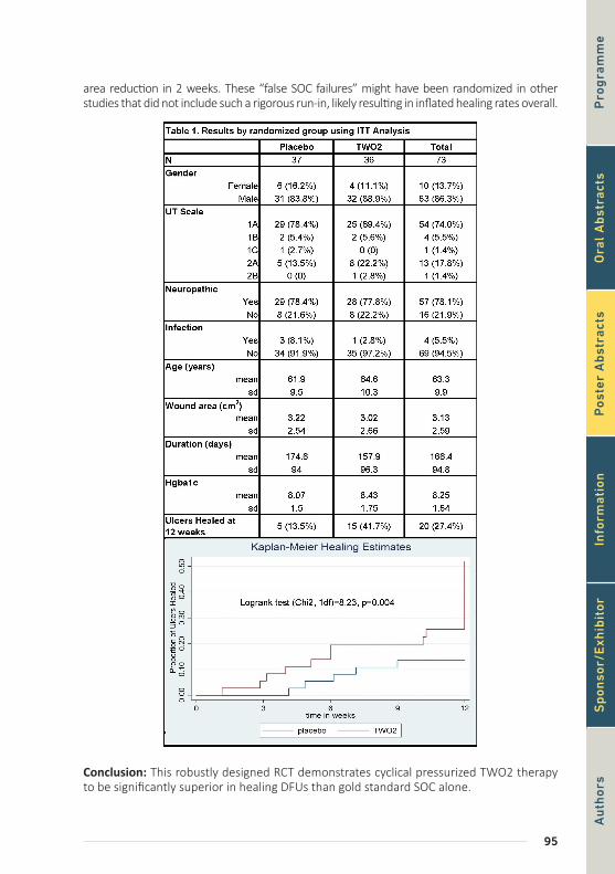
95
area reduction in 2 weeks. These “false SOC failures” might have been randomized in other studies that did not include such a rigorous run-in, likely resulting in inflated healing rates overall.
Conclusion: This robustly designed RCT demonstrates cyclical pressurized TWO2 therapy to be significantly superior in healing DFUs than gold standard SOC alone.
Pro
gra
mm
eA
uth
ors
Ora
l A
bst
ract
sP
oste
r A
bst
ract
sIn
form
atio
nS
pon
sor/
Exh
ibit
or
96
Poster Abstracts
[P017] USE OF LARVAL DEBRIDEMENT THERAPY IN THE MANAGEMENT OF MULTIPLE INFECTED DIABETIC FOOT ULCERS – PREVENTING AMPUTATION
Edward Jude1
1Department of Diabetes, Tameside Hospital NHS Foundation Trust, Ashton Under Lyne, United King-dom
A 48 year old man was referred to the diabetic foot clinic with multiple foot ulcers. He was diagnosed with type 2 diabetes in 2006. He had hypertension, arthritis, diabetic neuropa-thy and retinopathy. He was on Metformin, Gliclazide and a long-acting insulin*. His blood glucose was 26.7 mmol/l in clinic. He was admitted to the medical ward on 28 February 2017. 4 months prior to his presenting to clinic he had a small ulcer on his left lateral and medial malleolus and was under GP care for this. However in the previous few weeks he developed multiple ulcers on both feet; worse on left foot. On admission his HbA1c was 144 mmol/mol (15.3%); CRP 459 mg/l; creatinine 87 umol/l. X-rays ruled out osteomyelitis. He was commenced on intravenous insulin and intravenous antibiotics (clindamycin and flucloxacillin). He had vascular assessment and there was no peripheral arterial disease. He was seen by the surgeons and advised to have an amputation of his left foot in view of the severe foot infection. Patient refused and therefore we proceeded initially for surgical de-bridement which he underwent on 6 March by the surgeon. The wounds resloughed and were infected a week later and it was discussed with the patient to apply larvae therapy to the wound. On 21 March 400 free range larvae were applied using a boot kit. The larvae were removed on 24 March and following further sharp debridement by the podiatrist, 600 free range larvae were applied under a boot kit. This was removed on the 28 March. Patient did complain of slight discomfort following second application and also noticed im-provement in symptoms in his foot. His sleep pattern improved and he felt better clinically and he was very happy with the treatment. 2 months later his HbA1c was 50 mmol/mol (6.7%) and CRP 27 mg/l. Over the next few weeks the wounds continued to improve and no further surgical debridement was required. His left foot was placed in a total contact cast. He was discharged in May. He received antibiotics for 3 months. The wound healed com-pletely 9 months later. This case stresses the need for multidisciplinary involvement in the management of diabetic foot ulcers, improving glycaemic control and the beneficial use of larvae in managing slough and wound debridement and with such treatment amputation can be prevented. Photographs will be presented in the poster.
*Levemir®
97
[P018] COLD ATMOSPHERIC PRESSURE PLASMA AS A NOVEL TREATMENT MO-DALITY IN DIABETIC FOOT ULCERS: A PILOT STUDY
Rimke Lagrand1, Louise Sabelis1, Paulien Smits2, Guus Pemen3, Ana Sobota3, Bas Zeper4, Edgar Peters5
1Vu University Medical Center, Amsterdam, Netherlands2Plasmacure B.V., Eindhoven, Netherlands3Technical University Eindhoven, Eindhoven, Netherlands4Plasmacure , Eindhoven, Netherlands5Vu University Medical Center, Dept. of Internal Medicine, Amsterdam, Netherlands
Aim: Antimicrobial resistance is a growing problem in the treatment of diabetic foot ulcers. Plasma devices generate an ionized gas with a cocktail of highly reactive species, electric fields and UV light. Cold atmospheric plasma (CAP) treatment has advantages over anti-septic or antimicrobial infection prevention and control; it disinfects efficiently, painlessly and instantly, without development of antimicrobial resistance. Concurrently, plasma can stimulate human cell proliferation and migration as well as microcirculation. We studied the safety of a novel CAP device, that is simple to use and can be applied at a patient’s home in the future. Secondary objectives were to examine the effect of CAP treatment on bacterial load and on wound healing.
Method: We included subjects with diabetic foot ulcers without clinical signs of infection or exposed bone or joint in the wound base. Subjects were treated with CAP on a daily basis for ten days in a two-week period, or until their ulcer healed. Primary endpoint of this study was the occurrence of serious adverse events (SAE) as a result of treatment. Safety was defined as: ≤ 10% of subjects experiencing SAE related to treatment other than infection, and ≤ 60% of subjects developing infection within 30 days after treatment. Semi-quantative bacterial load was measured at treatment days 1, 5 and 10, before and after plasma application. Standard protocols for wound treatment were deployed, including weekly debridement and offloading.
Results / Discussion: Twenty subjects were enrolled. Three SAEs (infections) occurred at the site of application within one month of treatment, of which one occurred during treatment. Three SAEs unrelated to treatment occurred: pneumonia, toe amputation on the contralateral foot and a soft tissue infection of the ipsilateral leg. Transient adverse events (AE grade 1) during one or more applications were reported by 55% of subjects. Two wounds (10%) healed during the two-week treatment period. S. aureus bacterial load directly after CAP application decreased significantly compared with before application. Bacterial load of all species combined did not significantly decrease directly after applica-tion compared with before application.
Conclusion: No SAE other than infection occurred as a result of treatment and ≤ 60% of subjects developed an infection. The AEs were low-graded and transient. The results of this study demonstrate that CAP in diabetic foot ulcers is safe and well tolerated. Further research is needed to determine whether CAP significantly improves wound healing and significantly reduces bacterial load of certain species.
Pro
gra
mm
eA
uth
ors
Ora
l A
bst
ract
sP
oste
r A
bst
ract
sIn
form
atio
nS
pon
sor/
Exh
ibit
or
98
Poster Abstracts
[P019] AUTOLOGOUS MONONUCLEAR VERSUS MESENCHYMAL STEM CELLS IN HEALING OF RECALCITRANT NEUROPATHIC DIABETIC FOOT ULCERS
Ahmed Albehairy1, Fady Kyrillos2, Hanan Gawish2, Omnia State2, Hassan Abdelghaffar2, Osama Elbaz2, Ahmed El-Sebaie2, Ahmed Emam2, Manal Tarshoby2
1Faculty of Medicine, Internal Medicine Department, Mansoura, Egypt2Mansoura University, Egypt
Aim: To compare the therapeutic effect of autologous BM-MNCs and BM-MSCs on the healing process in patients with recalcitrant neuropathic diabetic foot ulcers.
Method: Eighteen patients with type 2 diabetes mellitus with neuropathic foot ulcer grade 2 New Texas classification were selected from Diabetic Foot Clinic, Mansoura University, Egypt from May 2016 to August 2017. Only patients who failed to respond to 12 weeks of weekly sharp debridement and proper offloading were included in the study. Patients were randomly assigned to MSCs, MNCs or control group. The study was conducted in Mansoura Regenerative Medicine Centre, Egypt. After aspiration of 20 ml of patients’ own bone marrow under good aseptic technique either mononuclear stem cells (MNCs) were separated or Mesenchymal stem cells (MSCs) were cultured. The bone marrow sample was diluted with phosphate buffer saline then separation using Ficoll hybaqe harvesting the layer of MNCs then washed twice using the complete media. This process revealed 5x10⁶ to 6x10⁶ of MNCs. Mesenchymal stem cells (MSCs) were characterized by adherence, trans-differentiation and CD characterization. Cultured cells were subjected to microbio-logical and karyotyping testing. MSCs number ranged from 1x10⁶ to 2x10⁶. The revealed MNCs or MSCs were dissolved in 2cm saline to be used for injection in the edges of the wound at eight points once. All patients continued on same offloading and dressing and were followed for 12 weeks for the change in ulcer surface area and the presence of any local reactions
Results / Discussion: Percentage reduction of Ulcer surface area was higher in both (MSC) and (MNC) groups 68% and 59% respectively compared to only 6.25 % in control group after 12 weeks of follow up. (P value < 0.05), However, there was no statistical significant difference between the healing rate of (MSC) and (MNC) groups. Complete healing was achieved in one patient in MSC group and in another patient in MNC group
Conclusion: Local injection of both autologous bone marrow derived MSCs and MNCs aug-mented healing of recalcitrant neuropathic diabetic foot ulcers. Using both cells was well tolerated by the patients with no short term complications. The small non-significant bet-ter healing associated with the use of MSCs should be weighed against the fact that MNCs separation is easier and achieved by less manipulation
99
[P020] IMPACT OF COMBINED TREATMENT WITH PATCH APPLICATION AND AEROSOL MIXTURE OF BIO-PROTECTIVE AND REGENERATIVE BIOCHEMICAL AGENTS (KAOLIN,SODIUM HYALURONATE,SILICON AND TITANIUM DIOXIDE NANOCRYSTALS), VERSUS PATCH MONOTHERAPY, IN THE PREVENTION AND TREATMENT OF THE DIABETIC FOOT ULCER
Eleni Matopoulou1, Ilias Kanellos2, Addo Tesfaye Aligaz3, Fofo Pavlidou2, Panagiota Kanoula2, Christina Bami2, Vassiliki Dineni2, Maria Papadopoulou2, Nikolaos Tsokos4, Maria Somali5, Dimitrios Skoutas2, Christos Savopoulos2, Apostolos Hatzitolios6
1Podologiko Kentro Eleni Matopoulou, 1st Floor, General Hospital Papageorgiou Thessaloniki,Diabitic Foot Clinic Dr Christos Manes0, Thessaloniki, Greece21st Propedeutic Dept of Internal Medicine,Ahepa University Hospital, Thessaloniki, Greece3Ahepa University Hospital, A\’prp, Thessaloniki, Greece4Diabetic Foot Clinic, Diabetes Center, General Hospital of Chalkida, Athens , Greece5Private Practice, Endocrinology, Hippokration General Hospital, Department of Endocrinology, Thessaloniki, Greece6Aristotle University of Thessaloniki, Greece, Thessaloniki, Greece
Aim: Comparing healing time, grade of reperfusion of the ulcer bottom, ulcer crater tissue quality and contamination of DU in lower extremities between patients (PA) treated with patch monotherapy and PA treated with combined therapy of patches and aerosol mixture of bio-protective and regenerative biochemical agents (AER).
Method: A group (GA) of 30 diabetes mellitus type I (DM) patients (15 males (M) and 15 fe-males (F), average age of 55 years) with DU on the plantar surface of the foot (PLF) and treated with monotherapy patches, and a second group (GB) of 30 patients (PA) with DM (15 M and 15 F, average age of 55 years) who developed DU on the PLF and treated with combination treatment of patches and AER. Surface area, depth and anatomical location of the DU were similar between the two groups. The following were recorded: time required for a complete healing of DU, quality of healing process, debridement manipulation quality during patches removal and renewal and possible contamination of the DU during the healing time period.
Results / Discussion: Diabetic ulcers (DU), which are not treated early, evolve rapidly, caus-ing a tissue cell necrosis, and eventually lead to amputation. New regimens applied to DU are more appropriate, in order to reduce amputations, in relation to the existing treat-ment. The GA presents an average DU healing time of 64 days, medium grade reperfusion of the ulcer bottom, crater perimeter with crusty quality tissue and contamination of the ulcers in 15% of the patients. The GB presents an average DU healing time of 41 days, high grade reperfusion of the ulcer bottom, crater perimeter with soft - elastic quality tissue and contamination of the ulcers in 5% of the PA.
Conclusion: Adding AER to the standard patch monotherapy results statistically significant difference (23 days, p <0.05) in healing time of diabetic foot ulcers, compared to patches alone. Healthy qualitative tissue characteristics (high grade reperfusion of the ulcer bottom, visible during the debridement manipulation, a crater perimeter with soft-elastic tissue qual-ity, reduction in the percentage of contaminations) were observed, which promote a faster and more effective healing mechanism of the DU. Combined therapy of AER and patches, could replace the present monotherapy reducing the amputations of lower extremities in diabetic patients and simultaneously, improve the quality of life and survival of DM patients, while may reduce the costs of the local health system regarding DM patients concern.
Pro
gra
mm
eA
uth
ors
Ora
l A
bst
ract
sP
oste
r A
bst
ract
sIn
form
atio
nS
pon
sor/
Exh
ibit
or
100
Poster Abstracts
[P021] RISK FACTORS ASSOCIATED WITH UNPLANNED ABOVE KNEE AMPUTA-TION AFTER TRANSTIBIAL AMPUTATION DURING THE PERIOPERATIVE PERIOD
Dane Wukich1, Junho Ahn1, Katherine Raspovic2, George Liu2, Michael Del Core2, Michael Van Pelt2, Trapper Lalli2
1University of Texas Southwestern Medical Center, Department of Orthopaedic Surgery, Dallas, United States2University of Texas Southwestern Medical Center, United States
Aim: Unfortunately, major amputations may be required in up to 20% of patients with severe diabetic foot infections. The purpose of this study was to evaluate diabetes mellitus (DM) and patient variables as possible risk factors for unplanned above-knee amputation (AKA) after primary transtibial amputation (TTA) in the perioperative period.
Method: Patients who underwent TTA between 2011 and 2015 were identified in a data-base*. The perioperative period was defined as the first 30 days after surgery.
Results / Discussion: In this cohort of 7,868 patients, 215 (3%) underwent unplanned AKA. Interestingly, DM (adjusted odds ratio [OR] 0.63, 95% confidence interval [CI] 0.44-0.91) was associated with decreased odds of unplanned AKA. Factors that were independently associated with increased risk of unplanned AKA were age over 65 years (adjusted OR 1.45, 95% CI 1.01-2.08), presence of coagulopathy (adjusted OR 1.58, 95% CI 1.09-2.28), American Society of Anaesthesiologists (ASA) classification grade IV or V (adjusted OR 1.47, 95% CI 1.03-2.10) and surgical site infection at time of surgery (adjusted OR 5.30, 95% CI 2.59-9.92).
Conclusion: Although unplanned AKA after TTA is rare (3%), morbidity is substantial.. Patients with DM were 37% less likely to require revision to AKA compared to patients without DM. Possible explanations for this decreased risk associated with DM include the distal nature of atherosclerosis (i.e. infrapopliteal disease in DM vs. more proximal disease in non-diabetic patients) and the indications for TTA. Diabetic patients typically undergo TTA for severe infections and non-reconstructable deformities (Charcot and osteomyelitis), while patients without DM typically undergo TTA for critical limb ischemia, often due to proximal occlusion (aorto-iliac-femoral stenosis).
* ACS-NSQIP®, American College of Surgeons-National Surgical Quality Improvement Pro-gram®
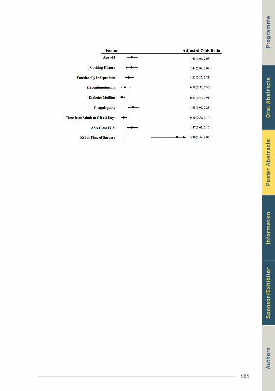
101
Pro
gra
mm
eA
uth
ors
Ora
l A
bst
ract
sP
oste
r A
bst
ract
sIn
form
atio
nS
pon
sor/
Exh
ibit
or
102
Poster Abstracts
[P022] DRASTIC REDUCTION IN LOWER LIMB AMPUTATION RATES IN A EURO-PEAN ISLAND STATE
Ruth Scicluna1, Francesca Theuma1, Samuel Anthony Galea1, Kevin Cassar1
1Vascular Surgery, Surgical Department, Mater Dei Hospital, Birkirkara, Malta
Aim: One of the main goals for arterial reconstructive surgery of the lower limb is to pre-vent major amputation. Until 2008, all vascular procedures in Malta were carried out by general surgeons with a vascular interest. From 2008 a dedicated vascular service was introduced and by 2014 all vascular procedures were performed by the vascular unit. The aim of the study was to compare the rate of open revascularisation and amputation pro-cedures in a defined population before and after the introduction of a vascular surgery service.
Method: Malta is a Southern European island state with a population of around 430,000 served by a centralised national health service. All arterial interventions, minor amputa-tions and major amputations between 1st January 2008 and 31st December 2017 were retrieved from three registers*. Major amputation was defined as amputation above the ankle, minor amputations included trans metatarsal and toe amputations. The indication for amputation in all registered cases was ischaemia with or without diabetes or diabetes without ischaemia. The amputations that were performed for other reasons (trauma or tumour) were excluded from the analysis. The majority of bypass procedures were per-formed for chronic limb threatening ischaemia.
Results / Discussion: After commencement of vascular surgery service the number of re-vascularisation procedures increased rapidly from an average of around 20 per year be-tween 2003 to 2006 to an average of 156 between 2014 and 2017 (>6x increase). This was associated with a dramatic reduction in the number of major amputations from an average of 120 between 2003 to 2006 to 60 between 2014 and 2017 (50% reduction in major amputation rate). The type of amputation shifted from a majority of above knee amputations to below knee amputations. The number of minor amputations increased dramatically after the introduction of vascular surgery service from an average of 100 per year between 2003 and 2006 to an average of 413 between 2014 and 2017. As the number of minor amputations and revascularisation procedures increased the number of major amputations decreased.
Conclusion: Increased revascularisation interventions leads to improved limb salvage rates and reduced amputation rates. The introduction of vascular surgery service in Malta has led to a dramatic reduction in the number of major lower limb amputations.
* The Maltese Vascular Registry, MALTAVASC, the centralised national operation register and the amputee register
103
[P023] PREDICTORS OF FURTHER INTERVENTION AFTER TOE AMPUTATION IN DIABETIC PATIENTS
Jamal Hoballah1, Lamia Jouhar1, Rola Jaafar1, Omar Najjar2, Omar Itani1, Fadi Haddad1
1American University of Beirut, Lebanon2John Hopkins School of Medicine, United States
Aim: Diabetic foot is a major cause of morbidity among diabetic patients. Toe amputation is the most common distal amputation of the foot used mainly to preserve the maximal mobility in patients who need surgical intervention. This study aimed to determine the predictors of further intervention after toe amputation that may help in optimizing and individualizing the treatment plan.
Method: We retrospectively reviewed the results of 85 toe amputations for diabetic foot ulcers treated in a tertiary medical center in Lebanon. The outcome endpoint was requiring further intervention, either debridement or more proximal amputation.
Results / Discussion: 85 patients (65 males, 20 females) with a mean age of 67 ± 12 years and a mean duration of diabetes of 18 ± 7 years were enrolled. 18 patients were lost to follow up. Out of the remaining 67 patients, 48 did not require further intervention and 19 did. Those who required further intervention were older and had more incidence of peripheral arterial disease (p: 0.026 and 0.012 respectively). Toe pressure and Toe Brachial Index were significantly higher in the patients who did not require further intervention (p: 0.027 for both). No difference was found between the two groups regarding glycemic control or glycosylated hemoglobin value. However, patients treated with insulin had a lower further intervention rate compared to those treated with oral antidiabetics only (OR: 0.245, p: 0.026) and delayed primary closure was associated with lower further interven-tion rate compared to primary closure (OR: 0.147, p: 0.027).
Conclusion: Older age and low toe pressure were the main predictors of requiring fur-ther intervention after toe amputation. Diabetes treatment with insulin was associated with lower further intervention rates. Delayed primary closure was the preferred surgical wound closure option with the lowest further intervention rates.
Pro
gra
mm
eA
uth
ors
Ora
l A
bst
ract
sP
oste
r A
bst
ract
sIn
form
atio
nS
pon
sor/
Exh
ibit
or
104
Poster Abstracts
[P024] LONG-TERM PROGNOSIS OF PATIENTS WITH DIABETIC FOOT DISEASE IN TANZANIA: AMPUTATION, SEPSIS AND DEATH OVER THE COURSE OF TWO DECADES
Zulfiqarali G. Abbas1, Stephan Morbach2, Janet Lutale3, Lennox Archibald 4
1Muhas , Amc, Dept. of Internal Medicine, Dar-Es-Salaam, Tanzania2Department of Diabetes and Angiology, Marienkrankenhaus, Soest, , Germany3Muhas , Dar-Es-Salaam, Tanzania4College of Medicine, University of Florida, Gainesville, United States
Aim: We carried out this study to document the interventions for addressing foot dis-ease and outcomes during a follow-up period of 20 years. This information was established through epidemiologic surveillances activities in Tanzania.
Method: This is a prospective analytic cohort study of patients who were seen by one of the authors. A case was defined as any patient who presented to the Muhimbili National Hospital with diabetic foot ulcer (DFU) but had no previous surgical intervention, and who was followed up prospectively till death or lost to follow up. Informed consent was ob-tained. Detailed histories and examination were carried out.
Results / Discussion: Over a two-decade period (June 1998 through Dec 2017), 104 pa-tients met the case-definition. Of these 90 (86%) had type 2 diabetes and 60 (58%) were male. The characteristics of case-patients at presentation were as follows: median age: 51 (range: 10-85) years; median duration of diabetes 5 (range: 0-29) years. Of the 104 study-patients, 84 (80%) had peripheral neuropathy at presentation and all presented with open foot ulcers—63 (60%) in Wagner stage 2 and 13 (13%) in Wagner stage 3. At presen-tation 57 (54%) had pus, malodorous ulcers characteristic of anaerobic infection. Cumu-lative numbers of patients with complete healing at the 1-month, 3-month, 6-month, and 12-month follow-up evaluations were 13 (12%), 38 (36%), 62 (59%), and 75 (72%), respec-tively. First major and minor amputations occurred in 12 (11%), and 17 (16%), respectively. Overall mortality rate of the 104 case-patients was 40% (42/104). Of these, 24 (23%) died from sepsis attributable to limb infection all had significant peripheral arterial disease and peripheral neuropathy.
Conclusion: The findings of this study highlight the importance of long-term follow-up evaluations of patients with DFU disease in Africa. Despite all efforts made recently, ma-jor amputation is still a frequent outcome. Our data suggest that anaerobic infections are playing a significant role in patient outcomes. It is very likely that mortality among patients who are not followed is even higher.
105
[P025] CHOPART AMPUTATION WITH SUBTALAR JOINT ARTHRODESIS AT THE SAME TIME ARE THE EFFECTIVE METHOD OF PREVENTION OF FOOT DEFOR-MITY
Yuta Terabe1
1Tokyo Nishi Tokushukai Hospital, Plastic and Reconstructive Surgery, Akishima, Japan
Aim: Chopart amputation is one of the minor amputations. Chopart amputation cause foot deformation after surgery. Patients after Chopart amputation aren’t able to walk with artificial leg due to some foot deformation. So I performed subtarlar joint or ankle joint arthrodesis after several months. It’s good method of keeping walking ability, but patients were taken reoperation. This time, I operate Chopart amputation and subtarlar joint ar-throdesis at the same time with external fixation
Method: Two patients were performed. Patients were male and 70’s. They have diabetic mellitus, chronic kidney disease(G5d) and critical limb ischemia. External fixation device*1 and surface of Chopart amputation was covered by dermal artificial*2. External fixation fixed for about 4 weeks after checked callus.
Results / Discussion: Two cases were performed and have no infection and non union. They are able to walk by using artificial leg and do not cause deformation for one postop-erative year.
Conclusion: This method is prevention of deformation post Chopart amputation. External fixation is frailer than internal fixation. However, it is enough strong fixation to critical isch-emia patients.
*1 Monotube Triak, Stryker Japan K.K.
*2 Integra, Century Medical, Inc, Japan
Pro
gra
mm
eA
uth
ors
Ora
l A
bst
ract
sP
oste
r A
bst
ract
sIn
form
atio
nS
pon
sor/
Exh
ibit
or
106
Poster Abstracts
[P026] MORTALITY RATES IN DIABETICS UNDERGOING MAJOR LOWER LIMB AMPUTATION, IN A SMALL ISLAND STATE
Francesca Theuma1, Samuel Galea1, Ruth Scicluna2, Kevin Cassar3
1Mater Dei Hospital, Birkirkara, Malta2Vascular Surgery, Surgical Department, Mater Dei Hospital, Birkirkara, Malta3University of Malta, Msida, Malta
Aim: Diabetes remains one of the leading risk factors for major lower limb amputation. The prevalence of diabetes in Malta is one of the highest in Europe and is estimated at 13.8% among the adult population. The aim of the study was to determine the prevalence of diabetes amongst the major amputation population. We also compared the short and long-term mortality in the major amputation population suffering from diabetes compared to non-diabetics, and the general population.
Method: Data on all major lower limb amputations performed between 2011-2017 in Mal-ta was collated prospectively in a national amputee database. This documents all major amputations that occur in government hospitals across the Maltese Islands. Data regarding date of death was obtained from CPAS, the hospital patient administration system.
Results / Discussion: Between 2011 and 2017, 496 major amputations were performed. The mean age of patients was 70.6 years. 80.4% (n=399) of these suffered from diabetes. Among the diabetic amputees, 245 patients underwent transtibial amputation, 153 under-went transfemoral amputation and 1 underwent hip disarticulation. 355 patients (90.0%) had concomitant peripheral arterial disease. Between 2011 and 2017, there was an overall decrease in the number of major amputations carried out: from 85 in 2011 to 57 in 2017. Mean follow up was 1118 days. Overall 30-day mortality following major amputation was 10.3% among our series, 10.5% among those with diabetes and 9.3% among non-diabetics. 1-year mortality was 34.1% for all patients, 35.1% among those with diabetes and 29.9% among non-diabetics. 5-year mortality was 57.3% for all patients, 60.0% among diabetics and 46.4% among non-diabetics. The crude death rate among our series was 593 per 1000, 617 per 1000 among those with diabetes and 485 per 1000 among non-diabetics. This contrasts greatly with the crude death rate of 8.01 per 1000 of the general population, for the same period. Among those discharged from hospital (n=448), 21% were transferred to a nursing home, 37% to a rehabilitation institution, 39% home and 3% to another hospital.
Conclusion: Despite modern advances in healthcare, major amputations are still associat-ed with a high overall mortality, reflecting a highly comorbid patient group.
107
[P028] RECONSTRUCTION OF SEVERE ATHEROSCLEROTIC AND OBSTRUCTIVE DIABETIC FEET USING THORACODORSAL ARTERY PERFORATOR FLAPS WITH LONG VASCULAR PEDICLE
Sang Wha Kim1, Youn Hwan Kim2
1Seoul National University, College of Medicine, Seoul National University Hospital, Department of Plastic and Reconstructive Surgery, Seoul, Korea, Rep. of South2Hanyang University, Seoul, Korea, Rep. of South
Aim: Diabetic foot ulcer often proceed to complicated defect with increased risk of ampu-tation. Free tissue transfer in combination with revascularization has been commonly used for coverage of the major defect in diabetic patients. In this study, we introduce techniques to handle unhealthy vessels with or without revascularization, and evaluate the outcome of foot reconstruction in diabetic patient using latissimus dorsi perforator flap with long vascular pedicle.
Method: Between April 2011 and March 2015, 15 cases, which underwent reconstruction of diabetic foot defects using latissimus dorsi perforator flap with long vascular pedicle (more than 15cm), were included. Fourteen patients were male, and one was female. The mean age of the patients was 54.8 years (range 26-77 years). Nine patient underwent pre-vious angioplasties (percutaneous transluminal angioplasty or bypass surgery).
Results / Discussion: The flap size was ranged from 6x4 cm2 to 18x11 cm2 with the mean pedicle length 16.7 cm (range 15-19 cm). All flap survived. Seven complications were found. Two flaps had marginal wound disruption, which healed without further surger-ies. Four cases had partial loss of the flap; three flaps healed conservatively, but one flap required ray amputation of 1st metartarsal bone. The mean follow-up duration was 35.2 months (ranging 12-60 months).
Conclusion: Diabetic foot patients often have irreversibly damaged vessels due to severe atheroma or calcification and successful reconstruction cannot be guaranteed. The latis-simus dorsi perforator flap can be harvested with long vascular pedicle, more than 15 cm. Using long vascular pedicle, more proximal vessels above ankle level can be reached, and healthier and reliable vessel can be used as recipient vessel. Therefore, latissimus dorsi perforator flap can be a viable choice for the reconstruction of the diabetic foot defects in vascular compromised patients.
Pro
gra
mm
eA
uth
ors
Ora
l A
bst
ract
sP
oste
r A
bst
ract
sIn
form
atio
nS
pon
sor/
Exh
ibit
or
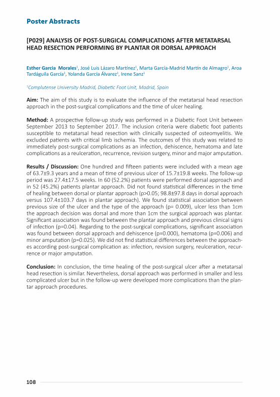
108
Poster Abstracts
[P029] ANALYSIS OF POST-SURGICAL COMPLICATIONS AFTER METATARSAL HEAD RESECTION PERFORMING BY PLANTAR OR DORSAL APPROACH
Esther Garcia Morales1, José Luis Lázaro Martínez1, Marta García-Madrid Martín de Almagro1, Aroa Tardáguila García1, Yolanda García Álvarez1, Irene Sanz1
1Complutense University Madrid, Diabetic Foot Unit, Madrid, Spain
Aim: The aim of this study is to evaluate the influence of the metatarsal head resection approach in the post-surgical complications and the time of ulcer healing.
Method: A prospective follow-up study was performed in a Diabetic Foot Unit between September 2013 to September 2017. The inclusion criteria were diabetic foot patients susceptible to metatarsal head resection with clinically suspected of osteomyelitis. We excluded patients with critical limb ischemia. The outcomes of this study was related to immediately post-surgical complications as an infection, dehiscence, hematoma and late complications as a reulceration, recurrence, revision surgery, minor and major amputation.
Results / Discussion: One hundred and fifteen patients were included with a mean age of 63.7±9.3 years and a mean of time of previous ulcer of 15.7±19.8 weeks. The follow-up period was 27.4±17.5 weeks. In 60 (52.2%) patients were performed dorsal approach and in 52 (45.2%) patients plantar approach. Did not found statistical differences in the time of healing between dorsal or plantar approach (p>0.05; 98.8±97.8 days in dorsal approach versus 107.4±103.7 days in plantar approach). We found statistical association between previous size of the ulcer and the type of the approach (p= 0.009), ulcer less than 1cm the approach decision was dorsal and more than 1cm the surgical approach was plantar. Significant association was found between the plantar approach and previous clinical signs of infection (p=0.04). Regarding to the post-surgical complications, significant association was found between dorsal approach and dehiscence (p=0.000), hematoma (p=0.006) and minor amputation (p=0.025). We did not find statistical differences between the approach-es according post-surgical complication as: infection, revision surgery, reulceration, recur-rence or major amputation.
Conclusion: In conclusion, the time healing of the post-surgical ulcer after a metatarsal head resection is similar. Nevertheless, dorsal approach was performed in smaller and less complicated ulcer but in the follow-up were developed more complications than the plan-tar approach procedures.
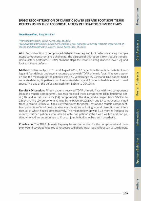
109
[P030] RECONSTRUCTION OF DIABETIC LOWER LEG AND FOOT SOFT TISSUE DEFECTS USING THORACODORSAL ARTERY PERFORATOR CHIMERIC FLAPS
Youn Hwan Kim1, Sang Wha Kim2
1Hanyang University, Seoul, Korea, Rep. of South2Seoul National University, College of Medicine, Seoul National University Hospital, Department of Plastic and Reconstructive Surgery, Seoul, Korea, Rep. of South
Aim: Reconstruction of complicated diabetic lower leg and foot defects involving multiple tissue components remains a challenge. The purpose of this report is to introduce thoraco-dorsal artery perforator (TDAP) chimeric flaps for reconstructing diabetic lower leg and foot soft tissue defects.
Method: Between April 2010 and August 2016, 17 patients with multiple diabetic lower leg and foot defects underwent reconstruction with TDAP chimeric flaps. Nine were wom-en and the mean age of the patients was 57.7 years(range 35-73 years). One patient had 3 separate defects, 14 patients had 2 separate defects, and 2 patients had defects with dead space. The size of the defects ranged from 5x3cm to 20x10cm.
Results / Discussion: Fifteen patients received TDAP chimeric flaps with two components (skin and muscle components), and two received three components (skin, latissimus dor-si (LD), and serratus anterior (SA) components). The skin paddle ranged from 10x3cm to 25x14cm. The LD components ranged from 3x5cm to 20x10cm and SA components ranged from 5x2cm to 8x7cm. All flaps survived except for partial loss of one muscle component. Four patients suffered postoperative complications including wound disruption and infec-tion, all of which healed conservatively. The mean follow-up was 31.3 months (range 8-60 months). Fifteen patients were able to walk, one patient walked with walker, and one pa-tient who had amputation due to Charcot joint infection walked with prosthesis.
Conclusion: The TDAP chimeric flap may be another option for the complicated and com-plex wound coverage required to reconstruct diabetic lower leg and foot soft tissue defects.
Pro
gra
mm
eA
uth
ors
Ora
l A
bst
ract
sP
oste
r A
bst
ract
sIn
form
atio
nS
pon
sor/
Exh
ibit
or
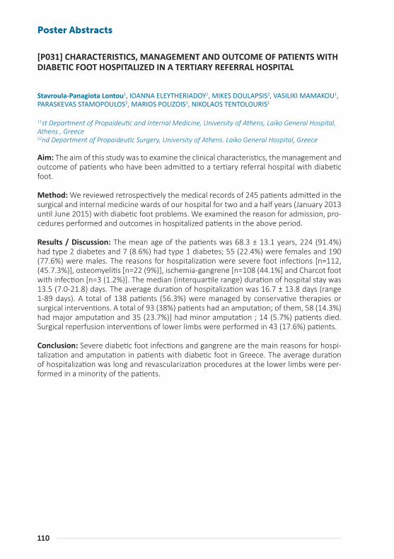
110
Poster Abstracts
[P031] CHARACTERISTICS, MANAGEMENT AND OUTCOME OF PATIENTS WITH DIABETIC FOOT HOSPITALIZED IN A TERTIARY REFERRAL HOSPITAL
Stavroula-Panagiota Lontou1, IOANNA ELEYTHERIADOY1, MIKES DOULAPSIS2, VASILIKI MAMAKOU1, PARASKEVAS STAMOPOULOS2, MARIOS POLIZOIS1, NIKOLAOS TENTOLOURIS1
11st Department of Propaideutic and Internal Medicine, University of Athens, Laiko General Hospital, Athens , Greece22nd Department of Propaideutic Surgery, University of Athens. Laiko General Hospital, Greece
Aim: The aim of this study was to examine the clinical characteristics, the management and outcome of patients who have been admitted to a tertiary referral hospital with diabetic foot.
Method: We reviewed retrospectively the medical records of 245 patients admitted in the surgical and internal medicine wards of our hospital for two and a half years (January 2013 until June 2015) with diabetic foot problems. We examined the reason for admission, pro-cedures performed and outcomes in hospitalized patients in the above period.
Results / Discussion: The mean age of the patients was 68.3 ± 13.1 years, 224 (91.4%) had type 2 diabetes and 7 (8.6%) had type 1 diabetes; 55 (22.4%) were females and 190 (77.6%) were males. The reasons for hospitalization were severe foot infections [n=112, (45.7.3%)], osteomyelitis [n=22 (9%)], ischemia-gangrene [n=108 (44.1%] and Charcot foot with infection [n=3 (1.2%)]. The median (interquartile range) duration of hospital stay was 13.5 (7.0-21.8) days. The average duration of hospitalization was 16.7 ± 13.8 days (range 1-89 days). A total of 138 patients (56.3%) were managed by conservative therapies or surgical interventions. A total of 93 (38%) patients had an amputation; of them, 58 (14.3%) had major amputation and 35 (23.7%)] had minor amputation ; 14 (5.7%) patients died. Surgical reperfusion interventions of lower limbs were performed in 43 (17.6%) patients.
Conclusion: Severe diabetic foot infections and gangrene are the main reasons for hospi-talization and amputation in patients with diabetic foot in Greece. The average duration of hospitalization was long and revascularization procedures at the lower limbs were per-formed in a minority of the patients.
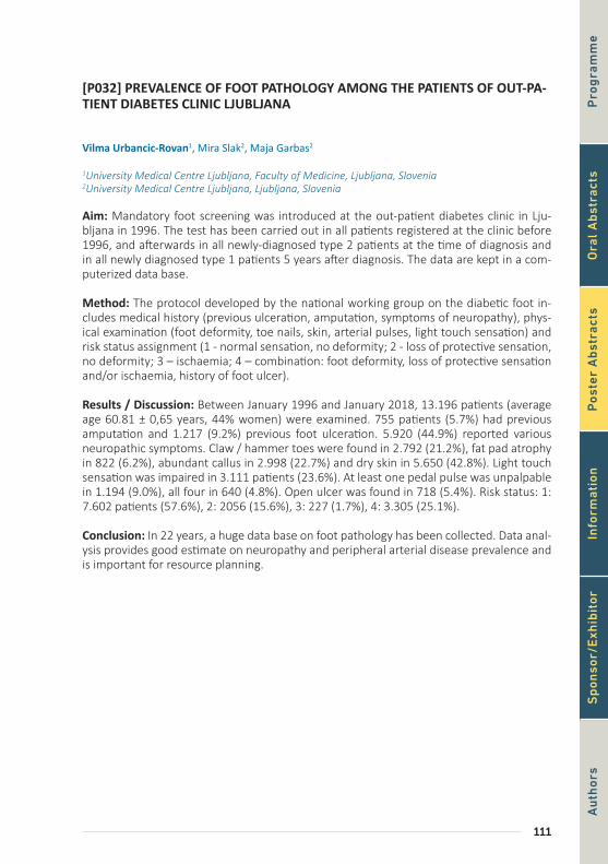
111
[P032] PREVALENCE OF FOOT PATHOLOGY AMONG THE PATIENTS OF OUT-PA-TIENT DIABETES CLINIC LJUBLJANA
Vilma Urbancic-Rovan1, Mira Slak2, Maja Garbas2
1University Medical Centre Ljubljana, Faculty of Medicine, Ljubljana, Slovenia2University Medical Centre Ljubljana, Ljubljana, Slovenia
Aim: Mandatory foot screening was introduced at the out-patient diabetes clinic in Lju-bljana in 1996. The test has been carried out in all patients registered at the clinic before 1996, and afterwards in all newly-diagnosed type 2 patients at the time of diagnosis and in all newly diagnosed type 1 patients 5 years after diagnosis. The data are kept in a com-puterized data base.
Method: The protocol developed by the national working group on the diabetic foot in-cludes medical history (previous ulceration, amputation, symptoms of neuropathy), phys-ical examination (foot deformity, toe nails, skin, arterial pulses, light touch sensation) and risk status assignment (1 - normal sensation, no deformity; 2 - loss of protective sensation, no deformity; 3 – ischaemia; 4 – combination: foot deformity, loss of protective sensation and/or ischaemia, history of foot ulcer).
Results / Discussion: Between January 1996 and January 2018, 13.196 patients (average age 60.81 ± 0,65 years, 44% women) were examined. 755 patients (5.7%) had previous amputation and 1.217 (9.2%) previous foot ulceration. 5.920 (44.9%) reported various neuropathic symptoms. Claw / hammer toes were found in 2.792 (21.2%), fat pad atrophy in 822 (6.2%), abundant callus in 2.998 (22.7%) and dry skin in 5.650 (42.8%). Light touch sensation was impaired in 3.111 patients (23.6%). At least one pedal pulse was unpalpable in 1.194 (9.0%), all four in 640 (4.8%). Open ulcer was found in 718 (5.4%). Risk status: 1: 7.602 patients (57.6%), 2: 2056 (15.6%), 3: 227 (1.7%), 4: 3.305 (25.1%).
Conclusion: In 22 years, a huge data base on foot pathology has been collected. Data anal-ysis provides good estimate on neuropathy and peripheral arterial disease prevalence and is important for resource planning.
Pro
gra
mm
eA
uth
ors
Ora
l A
bst
ract
sP
oste
r A
bst
ract
sIn
form
atio
nS
pon
sor/
Exh
ibit
or
112
Poster Abstracts
[P033] A PROMPT SURGICAL MANAGEMENT OF NECROTIZING FASCIITIS IN DIABETIC FOOT PATIENTS SAVES LIMBS AND LIVES
Chiara Goretti1, Elisabetta Iacopi1, Nicola Riitano1, Letizia Pieruzzi1, Alberto Piaggesi1
1Pisa University Hospital, Medicine Department, Diabetic Foot Section, Pisa, Italy
Aim: Necrotizing fasciitis (NF) is a rapidly progressive, life-threatening infection, involving skin, soft tissue and deep fascia. It requires prompt surgical treatment and appropriate antibiotic therapy. We aimed to evaluate the outcomes of the surgical management of NF in diabetic foot (DF) patients in a tertiary referral centre.
Method: We retrospectively searched the database of our DF Section from 2012 to 2014. All cases were treated according to international guidelines, with prompt extensive surgical debridement and systemic antibiotic therapy. We analysed healing rates, number of surgi-cal procedures and major amputation.
Results / Discussion: In this period 68 patients was referred to our DF clinic for a suspi-cion of NF. The diagnosis was excluded in 14 and confirmed in the remaining 54 (79.4%; male/female 40/14; type 1/ type 2 diabetes 6/48; age 62.8±8.1 yrs; duration of diabetes 13.6±10.1 yrs). Cases was classified, according to bacterial strains, as Type 1 (33-61.1%), Type 2 (7-13.0%) and Type 3 (14-25.9%). No significant differences were observed between the three groups, in terms of demographical and clinical features or glyco-metabolic con-trol. 6 patients (11.1%) underwent to forefoot amputation and 12 (22.2%) to toe or ray amputation. All the remaining cases underwent to decompressive fasciotomy. No major amputation was performed in short-term period and one only during long term follow up. We observed complete healing achieved in 46 patients (85%). Healing time was 94±11 days. 5 patients died (9.2%)
Conclusion: Our experience speaks of a high prevalence of NF in DF; despite these high rates, we observed how, when promptly and aggressively treated, NF has a relatively good prognosis and it is not associated with an excess of limb loss and death.
113
[P034] LOCAL DELIVERY OF ANTIBIOTICS VIA HIGHLY PURIFIED CALCIUM SUL-PHATE BEADS AS AN ADJUNCT TO SURGICAL DEBRIDEMENT IN THE ACUTE MANAGEMENT OF DIABETIC FOOT OSTEOMYELITIS
Michael Pierides1, Alexandros Leonidas Liarakos1
1Kettering General Hospital, Kettering, United Kingdom
Aim: Is there a role for the use of a local delivery device of antibiotics (LDDA) alongside acute surgical debridement to treat cases of septic patients with acute diabetic foot osteo-myelitis (OM)? The use of antibiotics delivered directly into the bone in treating diabetic foot osteomyelitis has been described successfully in case series by a variety of authors eg Jogia et al (2015) and Gauland (2011) but these cases are in chronic/subacute OM and the patients were not admitted as an emergency.
Method: 5 patients underwent acute surgical debridement following an acute admission. The LDDA consists of highly purified synthetic (HPS) calcium sulphate and it was mixed with 1g Vancomycin and 80mg Gentamicin. The operations were carried out by either a vascular surgeon and/or an orthopaedic surgeon with a special interest in diabetic foot disease and who are a formal part of the regional diabetic foot MDT of northamptonshire. The data collection included clinic letters, NHS primary care software database (system1) and individual patient hospital records.
Results / Discussion: All patients had confirmed osteomyelitis with either MRI and/or bone samples. The site of OM and the ulcer varied. 2 patients: hallux and 1st metatarso-phalangeal joint, 2 patients: plantar area on a background of chronic charcot deformity and one patient with calcaneal OM. All patients deteriorated requiring an emergency admis-sion despite being on appropriate oral or intravenous antibiotics (2 patients for 3 weeks, 1 patient for 19 weeks, 1 patient for 38 weeks and 1 patient on/off antibiotics for 44 weeks) as well as offloading and vascular optimisation. 4 out of the 5 patients post operatively remain healed in excess of a year currently. The one who hasn’t yet, is the patient with calcaneal OM who has ongoing improvement.
Conclusion: 4 out of the 5 patients have healed and with only 1 requiring a minor amputa-tion. The numbers are a clear limitation. Our impression which requires scientific backing is that these outcomes are better than for patients with a similar clinical picture locally that had the same management but no local antibiotic therapy. We do not believe that local delivery of antibiotics should replace surgical debridement or treatment of systemic symp-toms with IV antibiotics but more as an adjunct that could well be valuable in preventing relapses down the line as well as reduce the extent of primary or relapse requiring surgery. Further research is needed to validate this theory.
Pro
gra
mm
eA
uth
ors
Ora
l A
bst
ract
sP
oste
r A
bst
ract
sIn
form
atio
nS
pon
sor/
Exh
ibit
or
114
Poster Abstracts
[P035] SEVERE DIABETIC FOOT INFECTION AND OSTEOMYELITIS CAN BE SUC-CESSFULLY TREATED WITH OUTPATIENT PARENTERAL ANTIMICROBIAL THER-APY
Satyan Rajbhandari1, Alison Muir2, Janet Purcell1, Joanne Orton1
1Lancashire Teaching Hospital NHS Trust, Chorley, United Kingdom2Lancashire Teaching Hospitals NHS Trust, Preston, United Kingdom
Aim: Administration of intravenous antimicrobials to a patient with severe diabetic foot infection can be done by outpatient parenteral antimicrobial therapy (OPAT) at home or at day wards without the need for hospitalization. Our microbiology department started OPAT service in 2012 to reduce hospital admissions. Diabetes foot team has used OPAT for severe limb threatening diabetic foot infections. The aim of this study was to assess the clinical outcome of OPAT services and calculate the savings made.
Method: List of subjects accepted by OPAT service over a period last 5 years from diabe-tes foot team was obtained from database. 49 patients (8 males) with mean age of 60.4 (+/- 12.4) years had 57 episodes of severe diabetic foot infections and their records were analysed retrospectively. All patients had appropriate offloading, revascularization and de-bridement as per clinical practice. Choice of parenteral antibiotics was guided by allergy history and culture results. Outcome of foot ulcer was obtained from clinical record and confirmed with photographic record.
Results / Discussion: 32 episodes were stepped down from hospital admission and 25 were referred from foot clinic. The site of infection were toes in 22 cases, MTP area in 17, hind foot in 6, rocker bottom deformity in 5, dorsum of foot in 2 and residual infection of amputation site in 5. Underlying osteomyelitis was present in 47 cases and remaining 10 had cellulites. Cetriaxone 2 gm IV daily was given in 38 cases and Teicoplanin 10mg/kg IV daily in 19 along with metronidazole for anaerobic cover. The mean duration of OPAT was 20.7 (+/- 17.9) days and was followed by oral antibiotics as needed. Most patients had PICC sited for central venous access and were reviewed closely. Complete healing of ulcer was obtained in 42 (84%) episodes. 6 (12%) had amputations and 2 (4%) died. 7 cases of underlying osteomyelitis are still undergoing treatment with parenteral or oral antibiotics. Regarding osteomyelitis, 82.9% healed with OPAT and oral antibiotics. One patient with CKD 4 needed hospitalization due to worsening of renal function. Two had minor adverse event in the form of rash.
Conclusion: Our findings show that severe diabetic foot infections including osteomyelitis can be successfully treated with OPAT. It is safe, well-liked by patients and in the last 5 years OPAT service spared 1138 hospital bed days from severe diabetic foot infection treatment saving £273120 (€ 310, 363).
115
[P036] INFECTIONS AMONG PATIENTS WITH A DIABETIC FOOT ATTACK IN UZ LEUVEN: ANTIBIOTIC STRATEGY AND RESPONSIBLE GERMS
Toon Vissers1, Sabrina Houthoofd1, Sander Wuite1, Giovanni Matricali1
1Uz Leuven, Ku Leuven, Leuven, Belgium
Aim: Insufficiently treating diabetic foot infections (DFI) can result in major tissue destruc-tion of the foot, whereas antibiotic over-treatment can cause excessive side effects and unintended resistance. Starting empiric antibiotics and subsequently tailor them according to wound culture results is essential in treating DFI. This study explored the current clinical practice in the university hospital UZ Leuven (Belgium) concerning empiric and adjusted antibiotic therapy.
Method: In our center we retrospectively assessed antibiotic use and isolated microorgan-isms in all diabetic patients with a clinically proven DFI, defined by a PEDIS infection score ≥ 2 , from January 2016 until October 2017. The local IQED-foot database with additional data search in the electronic medical files was used.
Results / Discussion: In total 96 DFI cases were included, 48 were mild infections and 48 were moderate infections based on the PEDIS infection score. Wound cultures were taken in 88 cases. Altogether 324 cultures were obtained: 205 (63,3%) swab samples and 119 (36,7%) tissue samples. The wound cultures cumulatively contained 456 bac-teria. Most were Gram-positives (71,7%), with predominantly Staphylococcus, Strep-tococcus and Enterococcus species. Gram-negatives (28,3%) mainly comprised E. coli, Proteus, Morganella, Klebsiella and Pseudomonas species. Most bacteria were aerobes (95,6%), with only 4,4% being anaerobic. Multiresistance was seen in 13 (2,9%) isolates. Out of the 96 DFI cases, antibiotics were started in 90 cases: 81 were started empirically whereas in 9 cases culture results were awaited for. Empiric antibiotics for mild infections were predominantly per oral (81,1%) with mainly Clindamycin + Levofloxacin (50%) or Amoxicillin-Clavulanate (36,7%).In moderate infections almost half of empiric antibiotics were intravenously (47,7%) with mainly Piperacillin/Tazobactam (± Vancomycin) (61,9%) or Clindamycin + Levofloxacin (28,6%). Based on culture results, most empiric antibiotics were sufficient, however in 9,9% resistance against empiric therapy was seen.
Conclusion: Empiric antibiotics for DFI in UZ Leuven are generally conform the IDSA and IWGDF guidelines. Gram-positive aerobes were the predominant pathogens and anti-biogram results suggest bacteria remain sensitive to commonly used agents. However definitive antibiotic regimens should always be based on wound culture results, antibiotic susceptibility and patient response to empiric therapy.
Pro
gra
mm
eA
uth
ors
Ora
l A
bst
ract
sP
oste
r A
bst
ract
sIn
form
atio
nS
pon
sor/
Exh
ibit
or
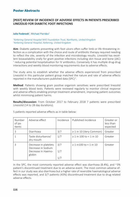
116
Poster Abstracts
[P037] REVIEW OF INCIDENCE OF ADVERSE EFFECTS IN PATIENTS PRESCRIBED LINEZOLID FOR DIABETIC FOOT INFECTIONS
Julie Fosbrook1, Michael Pierides2
1Kettering General Hospital NHS Foundation Trust, Northants, United Kingdom2Kettering General Hospital, Kettering, United Kingdom
Aim: Diabetic patients presenting with foot ulcers often suffer limb or life threatening in-fection as a complication with the choice and route of antibiotic therapy required needing to reflect the site, severity of the infection and microbiology results. Linezolid has excel-lent bioavailability orally for gram positive infections including skin tissue and bone (JAC)
1 reducing potential hospitalisation for IV antibiotics. Conversely it has multiple drug-drug interactions and weekly blood monitoring requirements due to adverse effects.
The study aims to establish whether the adverse effects experienced from prescribed Linezolid in this particular patient group matched the nature and rate of adverse effects reported in the manufacturers published data (SPC) 2.
Method: Patients showing gram positive organism sensitives were prescribed linezolid with weekly blood tests. Patients were reviewed regularly to monitor clinical response and adverse effects enabling prompt treatment amendment, improving patient outcomes whilst minimising patient harms.
Results/discussion: From October 2017 to February 2018 7 patients were prescribed Linezolid (14 to 28 day durations).
5 patients reported adverse effects as in table below:
Number of pa-tients
Adverse effect Incidence Published incidence Greater or less than expected
3 Diarrhoea 3/7 ≥ 1 in 10 (Very Common) Greater
1 Taste disturbance/dry mouth
1/7 ≥ 1 in 100 to < 1 in 10 Greater
3 Decrease in plateletsDecrease in SodiumDecrease in Haemo-globin
3/7
1/7
1/7
≥ 1 in100 to < 1 in 10 Greater
In the SPC, the most commonly reported adverse effect was diarrhoea (8.4%), and ~3% patient’s discontinued treatment due to an adverse event. The most common adverse ef-fect in our study was also diarrhoea but a higher rate of reversible haematological adverse effects was reported, and 3/7 patients (43%) discontinued treatment due to drug related adverse effects.
117
Conclusion: The adverse effects seen were in line with those reported in the SPC however the frequency was higher, perhaps due to the small patient numbers in our study and the course length prescribed. 5 out of 7 patients experienced side effects, this lead to discon-tinuation of treatment in 3 out of 7 patients, a higher rate than that described in the liter-ature. We will continue to build our evidence base and comparisons in this patient group.
Reference:
(1) Matthew S. Dryden, Linezolid pharmacokinetics and pharmacodynamics in clinical treatment, J Antimicrob Chemother 2011; 66 Suppl 4: iv7–iv15 accessed 04/04/18
(2) Summary of product characteristics Linezolid https://www.medicines.org.uk/emc/product/1688/smpc accessed 31/3/18
Pro
gra
mm
eA
uth
ors
Ora
l A
bst
ract
sP
oste
r A
bst
ract
sIn
form
atio
nS
pon
sor/
Exh
ibit
or
118
Poster Abstracts
[P039] A RETROSPECTIEVE STUDY TO REVIEW THE INCIDENCE OF CLOSTRIDIUM DIFFICILE IN DIABETIC FOOT ULCER PATIENTS WHO HAVE BEEN PRESCRIBED CO-AMOXICLAV CLINDAMYCIN CEPHALOSPORINS AND CIPROFLOXACIN
Krishna Gohil1, Michael Pierides2
1Northamptonshire Health Foundation Trust, Kettering , United Kingdom2Kettering General Hospital, United Kingdom
Aim: Most diabetic foot ulcers are polymicrobial (Lipsky 2004), resulting in long courses and combination of antibiotics, which increases the risk of developing Clostridium difficile (Stevens 2011). Antibiotics such as Clindamycin, Co-amoxiclav, Ciprofloxacin, and Cephalo-sporins are associated with a higher risk of c-diff infection. Other risk factors include, aged over 65, use of proton pump inhibitors, immunosuppression, long hospital stays and pre-vious c-diff. The aim of this study was to review the incidence of c-diff in diabetic patients with foot ulcers that attended the diabetic foot ulcer clinic over a 12 month period who had been prescribed Clindamycin, Co-amoxiclav, Ciprofloxacin, and any Cephalosporins.
Method: A retrospective study was carried out from podiatry records over a 12 month pe-riod of December 2016 - December 2017 of diabetic foot ulcer patients who attended the multidisciplinary diabetic foot clinic. Patients who were prescribed Co-amoxiclav Cephalo-sporin Ciprofloxacin and Clindamycin over the 12 month period were included in this study.
Results / Discussion: The total number of days of antimicrobials prescribed was 5322 The total number of days prescribed for Co-amoxiclav 3346 days Cephalosporins 269 days Ci-profloxacin 1546 days and Clindamycin 161 days. The mean number of antimicrobial days per patient was 49.2. The mean age was 65.4. There was one case of c.diff infection gene positive but toxin negative out of 108 patients.
Conclusion: Infection is one of the precipitating factors for lower limb amputation in pa-tients with diabetes. Foot ulcers are classed as mild, moderate or severe and often poly-microbial. Antibiotic exposure in general increases the likelihood of c-difficile infection. In the case of this study there was 1 incidence of c-diff gene positive but toxin negative within the 12 month period which is reassuring. This is not to say that these antibiotics should be used with less caution.
Regular and methodical review and analysis of data retrospectively is essential in optimal antimicrobial prescribing for patients. It ensures that our own practice is aligned with the most current evidence base and clinical governance. The data collected can shape future local and national guidelines so that practice is continually improved and safeguards for patients are in place so that the highest standard of care and excellence are maintained.
119
[P040] FIRST CONSULT AT DIABETIC FOOT UNIT: WHAT (BACTERIA) BRINGS YOU IN TODAY?
Diana Duarte1, Tiago Silva Santos1, André Couto Carvalho2, Cláudia Amaral2, Cláudia Freitas2, Helena Neto2, Joana Martins3, Luís Loureiro3, André Gomes3, Luís Costa3, Pedro Cantista3, Rosa Guimarães3, Joel Pereira3, Sara Pinto2, Rui Carvalho2
1Department of Endocrinology, Diabetes and Metabolism, Centro Hospitalar Do Porto - Hospital Santo António, Porto, Portugal2Department of Endocrinology, Diabetes and Metabolism, Centro Hospitalar Do Porto - Hospital Santo António, Porto(Portugal), Diabetic Foot Unit, Centro Hospitalar Do Porto - Hospital Santo António, Porto(Portugal)3Diabetic Foot Unit, Centro Hospitalar Do Porto - Hospital Santo António, Porto(Portugal)
Aim: Our objective is to present the epidemiology and antimicrobial susceptibility of mod-erate and severe diabetic foot infections (DFI) evaluated in a tertiary referral center.
Method: Retrospective study of diabetic patients presenting for their first consult at Dia-betic Foot Unit between January 2015 and December 2016 (2 years) who collected micro-biological cultures and were admitted to our hospital ward for treatment of moderate to severe DFI during the follow-up of 1 year.
Results / Discussion: A total of 1058 patients were observed during this period. 176 (16.6%) of them were included for the analysis. Positive samples were found in 163 (92.6%) of them. One hundred and forty five of these (82.4%) had neuro-ischaemic ulcer and the remainder 31 patients (17.6%) presented with a pure neuropathic ulcer. The positive sam-ples were polymicrobial in 49.4% (n=87) of the cases. The predominant isolated group was Enterobacteriaceae, present in 46.6% (n=76) of the samples (24 of which were multi-drug resistant species and 4 extended spectrum beta-lactamases producers). Staphylococcus aureus was identified in 31.9% (n=52) (21 of which were methicillin-resistant species). En-terococcus was recognized in 25.8 %( n=42; 4 of which were vancomycin-resistant). Pseu-domonas aeruginosa was isolated in 15.3% of the sample (n=25; 8 of which were fluoro-quinolone resistant species).
Conclusion: Our cohort, despite highly heterogeneous, shows an increasing prevalence of multiresistant species, with Gram-negative bacteria on the lead. This possibly reflects the chronicity of their disease and prolonged exposure to antibiotics and/or hospital en-vironment. This may help update our local empirical antibiotic therapies for this patients, heavily colonized by multi-drug resistant agents
Pro
gra
mm
eA
uth
ors
Ora
l A
bst
ract
sP
oste
r A
bst
ract
sIn
form
atio
nS
pon
sor/
Exh
ibit
or
120
Poster Abstracts
[P041] THE CHANGING BACTERIOLOGY OF DIABETIC MINOR AMPUTATION
Samuel Galea1, Ruth Scicluna2, Francesca Theuma1, Kevin Cassar3
1Mater Dei Hospital , Msida, Malta2Vascular Surgery, Surgical Department, Mater Dei Hospital, Birkirkara, Malta3University of Malta, Msida, Malta
Aim: Malta is the smallest European Union member state and has the highest prevalence of diabetes in the European Union. The number of minor amputations (toe or foot) per capita is also one of the highest in Europe. The aim of this study was to determine the prev-alence of MRSA carriage and CRE rectal colonisation in a population of diabetic patients admitted to hospital for minor amputations as well as determine the bacteriology of tissue and bone specimens submitted for culture and sensitivity.
Method: All diabetic patients admitted to Mater Dei Hospital Vascular Unit between Jan-uary and December 2017 for isolated minor amputation (below ankle) were included. patients undergoing additional procedures such as bypass surgery were excluded. As per hospital policy all patients were screened for MRSA. In addition patients admitted to the vascular ward were also screened for CRE through a rectal swab. Tissue samples from the grossly infected area and bone specimens from the site of the bone division were submit-ted for culture and sensitivity.
Results / Discussion: During the study period 216 minor amputations were performed. 201 (93%) were screened for MRSA during their admission. Of these 21 (10.5%) were found to be MRSA positive. 102 (47.2%) were screened for CRE and of these 21 (20.5%) were found to be positive (8 OXA-48; 13 NDM). 207 (95.8%) had a bone specimen submitted and 206 (95.3%) had tissue submitted for culture and sensitivity. 114 (55.1%) of all bone specimens were positive while 140 (68%) if tissue specimens were positive. The most common or-ganisms grown from both bone and tissue were Staphylococcus aureus (72; 17.4%) and Enterococcus faecalis (71; 17.2%). MRSA was the third commonest organism grown from both bone and tissue (39, 9.4%). These were followed by Pseudomonas aeruginosa, Esche-richea coli and Proteus mirabilis. There were 5 bone specimens that grew CRE.
Conclusion: The prevalence of MRSA carriage in a hospital population of diabetics ad-mitted for minor amputations is significant and this is reflected in the high prevalence of MRSA grown from tissue and bone specimens. The prevalence of CRE in this population is alarmingly high and CRE infection in this category of patients is increasing and presents a worrying new challenge.
121
[P042] RICK CLASSES AND THEIR TREATMENT IN EVERYDAY TEAM WORK
Fabrizia Toscanella1, Vinicio d’Annunzio2, Antonella Lapenna3, Antonio Federici4, Maria Concetta Gugliotta3, Lorenzo Valesini3
1Luigi DI Liegro Hospital, Rome, Italy2Don Gnocchi Rehabilitation Center, Italy3Luigi DI Liegro Hospital, Diabetic Foot Center, Rome, Italy4Private Practice, Italy
Aim: The great challenge of recent years in the diabetic foot (DF) is to prevent ulceration and above all recurrence. In our DF clinic we realized after years of activity that the current classification of classes of risk for the purpose of a prescription of footwear and orthotics was not satisfactory because some patients were not well framed and, moreover, some variables indispensable for a correct therapeutic-diagnostic general overview were not tak-en into account and therefore for the prevention of recurrence. For this reason we have developed a prescriptive protocol enriching the risk classes with details and applied it in clinical practice in the last year.
Method: 352 patients were evaluated according to a classification that included: risk class 0 (no neuropathy or vasculopathy) 0 pts; risk 1 (presence of sensory neuropathy) 42 pts; risk 2 (sensory and / or motor neuropathy, deformity, hyperkeratosis, joint stiffness, vas-culopathy) 168 pts; risk 3 (previous injury / amputation, Charcot’s foot) 142 pts. Patients were also evaluated for the presence of musculoskeletal system disorders that potentially altered the biomechanical structure (consequence of stroke, dysmetry, presence of joint prostheses, severe scoliosis, previous fractures or orthopedic interventions, rheumatologi-cal diseases, etc.) by a physiatrist. Weight (BMI) and age were also taken into consideration, also for the choice of materials. Finally, a prescriptive protocol for orthoses and shoes was applied according to a specific algorithm.
Results / Discussion: In the last year there was a percentage of new ulcerations of 24% (84/350) of which 65% were in class 3, 31% in class 2 and 4% in class 1; most of the patients in class 3 were not assiduous in quarterly controls, due to concomitant hospitalizations. This outcome was better than our previous one and than those from literature.
Conclusion: Knowledge of biomechanical considerations is low, in general, in the team ap-proach to DF because heterogeneous professionals having competence in primary preven-tion and above all in recurrence prevention, are involved, but we have to take into account all the principal features that can influence the gait and the posture of our patients and match them together before prescribing the insole and the shoe, and educate patients to follow the therapy, to be sure not to have new ulcers and recurrence.
Int J Low Extrem Wounds. 2014 The best way to reduce reulcerations: if you understand biomechanics of the diabetic foot, you can do it. Lázaro-Martínez JL1
Pro
gra
mm
eA
uth
ors
Ora
l A
bst
ract
sP
oste
r A
bst
ract
sIn
form
atio
nS
pon
sor/
Exh
ibit
or
122
Poster Abstracts
[P044] HOW TO SUCCESSFULLY IDENTIFY MULTIDISCIPLINARY UNITS FOR PRE-VENTION AND DIABETIC FOOT CARE IN FRANCE?
Jacques Martini1, Julien Vouillarmet2
1Service de Diabétologie, Centre Hospitalier Universitaire de Rangueil, Toulouse, France2Centre Hospitalier Lyon Sud, Pierre Bénite, France
Aim: Reducing the rate of amputation related to the DFU is a major issue of diabetes; for patients in terms of quality of life and prognosis, for carers confronted with serious and complex situations, for society on a medico-economic level. The recommendations pro-vide precise and codified markers on foot management strategies both in terms of wound treatment and the prevention of its occurrence.
The concept of multidisciplinarity and specialized structures on an ambulatory approach has proven its effectiveness in reducing the rate of major amputations. However, the course of patients with foot ulcers is marked by critical points limiting effectiveness.
Apart from the lack of awareness of the recommendations by the caregivers and the lack of patient awareness of the risk, the absence or the difficulties of identifying the specialized structures by the actors of the first resort or their absence within a territory is a problem.
In the “Board Urgo Pied (BUP)”, these findings have confirmed our desire to design a di-rectory of specialized structures at the national level with two objectives: obtain a first national map and strengthen the approach to improve the quality of each structure.
Method: For this, we have defined on the basis of the Internationales guidelines, eight essential criteria defining a specialized foot diabetic center. Our desire is not to make a selection but to create a national dynamic in favor of a better organization of the care of these patients.
Results / Discussion: In a third version, published at the congress of the French speaking Diabetes Society (March 2018), 90 structures were identified. Each center can thus assess its level of development and organization.
Of course, this directory is evolutionary and open to new structures that wish to be listed. It remains an informative tool for caregivers. It can be presented as a map of France to visualize the areas provided and those in need.
Conclusion: The members of the BUP clearly express their lack of legitimacy to affix to this directory a labeling or accreditation, but it can be a tool for reflection for our guardianship on the implementation of a care policy to improve the decision in charge and the prognosis of this pathology.
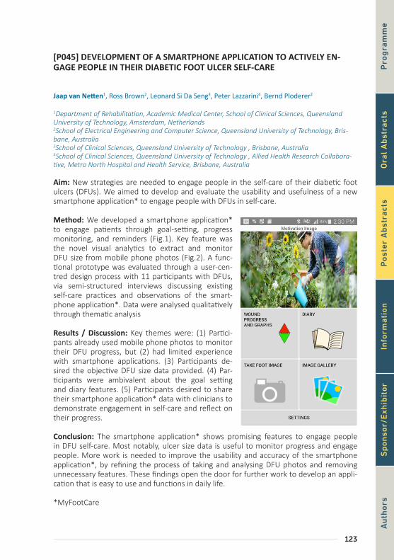
123
[P045] DEVELOPMENT OF A SMARTPHONE APPLICATION TO ACTIVELY EN-GAGE PEOPLE IN THEIR DIABETIC FOOT ULCER SELF-CARE
Jaap van Netten1, Ross Brown2, Leonard Si Da Seng3, Peter Lazzarini4, Bernd Ploderer2
1Department of Rehabilitation, Academic Medical Center, School of Clinical Sciences, Queensland University of Technology, Amsterdam, Netherlands2School of Electrical Engineering and Computer Science, Queensland University of Technology, Bris-bane, Australia3School of Clinical Sciences, Queensland University of Technology , Brisbane, Australia4School of Clinical Sciences, Queensland University of Technology , Allied Health Research Collabora-tive, Metro North Hospital and Health Service, Brisbane, Australia
Aim: New strategies are needed to engage people in the self-care of their diabetic foot ulcers (DFUs). We aimed to develop and evaluate the usability and usefulness of a new smartphone application* to engage people with DFUs in self-care.
Method: We developed a smartphone application* to engage patients through goal-setting, progress monitoring, and reminders (Fig.1). Key feature was the novel visual analytics to extract and monitor DFU size from mobile phone photos (Fig.2). A func-tional prototype was evaluated through a user-cen-tred design process with 11 participants with DFUs, via semi-structured interviews discussing existing self-care practices and observations of the smart-phone application*. Data were analysed qualitatively through thematic analysis
Results / Discussion: Key themes were: (1) Partici-pants already used mobile phone photos to monitor their DFU progress, but (2) had limited experience with smartphone applications. (3) Participants de-sired the objective DFU size data provided. (4) Par-ticipants were ambivalent about the goal setting and diary features. (5) Participants desired to share their smartphone application* data with clinicians to demonstrate engagement in self-care and reflect on their progress.
Conclusion: The smartphone application* shows promising features to engage people in DFU self-care. Most notably, ulcer size data is useful to monitor progress and engage people. More work is needed to improve the usability and accuracy of the smartphone application*, by refining the process of taking and analysing DFU photos and removing unnecessary features. These findings open the door for further work to develop an appli-cation that is easy to use and functions in daily life.
*MyFootCare
Pro
gra
mm
eA
uth
ors
Ora
l A
bst
ract
sP
oste
r A
bst
ract
sIn
form
atio
nS
pon
sor/
Exh
ibit
or
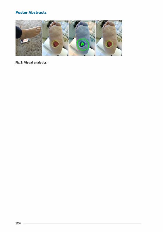
124
Poster Abstracts
Fig.1: Smartphone application* home screen.
Fig.2: Visual analytics.
125
[P046] DOES INTEGRATED FOOT CARE REALLY MATTER?
Matilde Monteiro-Soares1, Jorge Vale-Lima2, João Martiniano3, Sofia Pinheiro-Torres4, Vanessa Dias5, Edward Boyko6
1Medcides - Faculty of Medicine, University of Porto, Cintesis - Faculty of Medicine, University of Porto, Porto, Portugal2Unidade de Saúde Familiar Nova Salus, Agrupamento de Centros de Saúde Grande Porto VII – Vila Nova de Gaia, Portugal3Sociedade Portuguesa de Investigação e Desenvolvimento Da Podologia (Spidp)4Unidade de Saúde Familiar Aldoar, Agrupamento de Centros de Saúde Porto Ocidental - Porto, Portugal5Usf Sto André de Canidelo, Agrupamento de Centros de Saúde Grande Porto VII, Vila Nova de Gaia, Portugal6Seattle Epidemiologic Research and Information Center, Department of Veterans Affairs, Puget Sound Health Care System & University of Washington, Seattle, Wa, USA
Aim: To understand the impact of integrated foot care lower extremity amputation (LEA) risk in people with diabetes (PwD).
Method: Systematic review in MEDLINE for studies published until 02/2016, analysing the association between foot care delivery and LEA occurrence in PwD. A meta-analysis yielded pooled odds ratios (OR) using a random effects model.
Results / Discussion: We retrieved 4415 articles; 35 reported data that allowed us to con-clude if integrated foot care prevented LEA. This included access to healthcare (n= 5), receiv-ing chiropody/ podiatry services (n=5), educating health professionals (n=3), implementing guidelines (n= 2) and structured foot care services (n=18). Access to general practitioner or hospital (in minutes) did not affect LEA risk [OR 0.95, 95% confidence interval (CI) 0.91-1.00 and 1.00, CI 0.99-1.02, respectively]. Taking >1 month to be referred to tertiary care in-creased LEA risk (OR 2.22, CI 1.36-3.64). Provider coordination (programming and feedback) was negatively associated with minor and total LEA risk. Veterans with LEA receiving care at U.S. Veterans Affairs facilities were less likely to have a service-connected disability (p<0.05); there were no differences in visits to a primary care provider or diabetes educators. Receiv-ing regular surveillance before and after a LEA compared to post LEA only had no impact on re-amputation risk. Receiving care from a lower extremity clinician (LEC) specialist or a podiatrist was associated with lower LEA risk regardless of diabetic foot clinical stage [hazard ratio (HR) 0.36 to 0.81, p<0.05]. In univariate analysis, chiropody was not associated with LEA risk; but on multivariate analysis it was protective for LEA or death (HR 0.39, CI 0.05-0.73). Prevalence of podiatrists in a hospital referral region had no impact on LEA incidence among Medicare beneficiaries. In subjects with diabetic foot ulcers, receiving podiatry care in the prior year reduced major LEA risk in the U.S., both in Medicare and non-Medicare eligible populations (p<0.001). To be asked to see a chiropodist ≥1/month (free of charge) was not associated with reduced LEA risk. Educating health professionals significantly decreased LEA in Algeria (Oran) and Tanzania but not in U.S. (New Jersey). After guideline implementation and dissemination in Stockholm and Tuscany, global and major LEA rates significantly de-clined. After creating a multidisciplinary diabetic foot care team or clinical protocols, LEA risk declined in some (n=16), but not all (n=3) settings, with a pooled OR 0.37, CI 0.27-0.52.
Conclusion: The evidence demonstrates that higher quality care prevents LEA in PwD.
Pro
gra
mm
eA
uth
ors
Ora
l A
bst
ract
sP
oste
r A
bst
ract
sIn
form
atio
nS
pon
sor/
Exh
ibit
or
126
Poster Abstracts
[P047] ASSESSMENT OF THE EFFECTIVENESS OF A SPECIALISED DIABETES FOOT CLINIC IN SOUTH INDIA -A 5 YEAR OBSERVATIONAL STUDY
Vijay Viswanathan1
1Prof Viswanathan Diabetes Res Center, Dept. of Diabetology & Podiatry, Chennai, India
Aim: The aim was to see the rate of amputation among people with type 2 diabetes with a healed foot infection, during a five year period.
Method: Type 2 diabetes patients with healed foot infection who attended a specialized diabetes foot clinic in a tertiary diabetes care centre in Chennai, India, for both baseline examination in 2010-11 and subsequent follow up visits till 2015-16, with a minimum of 3 visits in this period were included in the study. A total of 405 subjects with healed foot ulceration and measurement of bio chemical and foot assessment details spanning 5 years were considered for this analysis.
Subjects were divided into two groups, Group A: Patients without any major events during the follow up period (n=243) and Group B: Patients with major events during the follow up (Re-ulceration/Recurrent amputation) (n=162).Data on previous history of ulceration and amputation, smoking habits, presence of any vascular complications were recorded.
Results / Discussion: On an average, subjects in group A visited the consultants two times more than the subjects in group B. The subjects in group B had a longer duration of dia-betes as compared to subjects in group A (18.5Vs12.3 years). There were more smokers in group B (27.2 Vs 19 %) (P<0.06) . It was found that in Group B, 35% of patients had wound debridement, 41.9% had a minor amputation and 22.8% of them had a major amputation. At baseline, the HbA1c was 8.6% and 10.8% respectively in group A and Group B. During the follow up period, patients in Group A showed greater decline in HbA1C (8.6 Vs 7.8%) whereas in Group B, the mean HbA1c was higher when compared with baseline value (10.8 Vs 11.7%) .
Conclusion: Poor glycaemic control, presence of peripheral arterial disease, longer dura-tion of diabetes, smoking can contribute to recurrent diabetic foot complications. Main-tenance of good glycaemic control and regular visits to the foot clinic can help to prevent amputations among people with previous history of diabetic foot infection.
127
[P049] AID CONCEPT FOR MULTIDISCIPLINARY TREATMENT OF DIABETIC FOOT ULCERS
Shinobu Ayabe1
1Yao Tokushukai General Hospital Wound Care Center, Osaka, Japan
Diabetic patients are growing explosively throughout the world, especially in Asia. As a result, patients with a diabetic foot problem are rapidly increasing in these countries.
Arterial insufficiency, infection and deformity are highly associated with healing challenges of diabetic foot ulcers.
In patient with arterial insufficiency ulcers, restoration of blood flow by revascularization is the intervention that will most likely lead to healing. Infected feet require surgical debride-ment to achieve healing. The incision planning for debridement was designed based on the assumption that the defect would be closed by fillet flaps. The wounds were left open, and treated with NPWT. Fillet flaps were gradually advanced by NPWT, and complete wound closure was achieved.
But in ischemic foot, early debridement could result in worsened necrosis by increasing metabolic demand. So we performed marginal debridement without bleeding, not to worsen ischemia. And if necessary, we performed longitudinal incision and drainage not to disturb the skin blood flow.
Foot deformities are a cause of pressure concentrations and create biomechanical stresses that cause ulcerations. The mainstay of treatment for foot deformities is offloading using proper footwear.
Diabetic foot ulcers often require comprehensive, multidisciplinary management.
But the pathogenesis of foot ulceration is complex, clinical presentation variable, and man-agement requires early expert assessment. Since we need to treat diabetic foot in multi-disciplinary, common concept of treatment is required.
Here we propose a new concept of diabetic foot wound management, which we have termed AID (acronym for arterial insufficiency, infection and deformity) concept, and an-alyzed a retrospective series of 73 patients with diabetes mellitus, who were hospitalized because of their diabetic foot.
Pro
gra
mm
eA
uth
ors
Ora
l A
bst
ract
sP
oste
r A
bst
ract
sIn
form
atio
nS
pon
sor/
Exh
ibit
or
128
Poster Abstracts
[P050] THE INTEGRAL ROLE OF THE DIABETIC FOOT CLINICAL NURSE SPECIAL-IST IN THE MULTIDISCIPLINARY FOOT TEAM
Ian Alejandro1, Elicia Bellio1, Enid Joseph1, Maureen Bates1, Chris Manu2, Michael Edmonds1
1King’s College Hospital, Diabetic Foot Clinic, London, United Kingdom2King’s College Hospital, London, United Kingdom
Aim: The Diabetic Foot Clinical Nurse Specialist (DFCNS) is a key member of our Multidisci-plinary Foot Team (MDfT), but it is a role that is less well described. The aim was to evaluate the roles and activities of the DFCNS within our MDfT clinic.
Method: A retrospective analysis of clinical activities over a 12 month period. Electronic patient records and coded clinical activities undertaken during patients clinic visit was ac-quired and analyzed to elaborate the activities undertaken by the DFCNS between January and December 2016.
Results / Discussion: There were 8,056 clinic visits over the 12 months of which 2,762 (34%) of clinic visits were coded as needed an input from the DFCNS. The DFCNS under-took venipuncture for blood test in 2,373 patient visits, 86% of visits that needed their input. Treatment of patients with leg ulceration in 20%, 564 visits. Administration of IM (intramuscular antibiotics) in (14%) 393 patients visits. Application of compression dress-ing 8%, 212 patient visits. IV (Intravenous infusion) of antibiotics in (8%) 206 patient visits. Peripherally Inserted Central Catheter (PICC) line care or removal in (5%) 149 patient visits. Organization for deep cleaning after 107 (4%) patients visits that needed treatment in strict isolation. Organization of patients for admission into hospital from the clinic in (4%) 107 clinic visits. Insertion of intravenous cannulation in (3%) 86 patient visits. Urine analysis and treatment of hypoglycaemia in 7 and 6 clinic visits respectively.
Conclusion: The role and activities of the DFCNS within the MDfT is pivotal to the clinic’s ethos of seeing and assessing diabetic foot patients in a “one-stop” approach. Their role in administering IM, IV antibiotics and PICC line care are also key in managing patients with infected diabetic foot ulcers within the outpatient setting thus avoiding unnecessary hospital admissions.
129
[P051] DOES ATTENDANCE TO A DIABETIC FOOT CLINIC RESULT IN IMPROVED GLYCAEMIC CONTROL WITH OR WITHOUT DIRECT FOCUS ON GLYCAEMIC CONTROL?
Alexandros-Leonidas Liarakos1, Michael Pierides1
1Kettering General Hospital, Kettering, United Kingdom
Aim: To explore the direct and indirect impact of diabetic foot clinics on glycaemic control as defined by their HbA1c levels.
Method: This is a retrospective study focused around diabetic foot patients attending the diabetic foot clinic which is a MDT clinic involving a diabetes consultant, diabetes specialist podiatrist and antimicrobial pharmacist. A diabetes specialist nurse (DSN) is not commis-sioned in the clinic despite NICE guidance (NICE [NG19] 2015). There were 2 periods of di-abetic foot clinics that were explored: the first group where there was no specific emphasis on glycaemic control (due to time constraints), which lasted from April 2013 to October 2014 (Group A), and the second group where there was focused time into glycaemic con-trol from November 2014 to December 2017 (Group B). In the second period there was no increase in time allowance and therefore the clinics ran late with the expected problems. The data collected from the first year of each period, including HbA1c levels before attend-ing clinic and at the end of each year were analysed.
Results / Discussion: 304 diabetic foot patients were evaluated (n: 104 in group A, n: 200 in group B). HbA1c levels decreased by 3.02 mmol/mol in Group A (max drop: 41 mmol/mol, max rise: 76 mmol/mol) and by 5.34 mmol/mol in Group B (max drop: 79 mmol/mol, max rise: 73 mmol/mol).
More specifically, in Group A, from April 2013 to March 2014, HbA1c decreased in 53 patients (n=50.96%), increased in 30 patients (n=28.85%) and unchanged in 7 people (n=6.73%). There were 14 patients (n=13.46%) with no measurement of HbA1c levels during this year.
In Group B, between November 2014 and October 2015, HbA1c decreased in 106 people (n=53%), HbA1c increased in 68 (n=34%), and unchanged in 6 (n= 3%). There were 20 pa-tients (n=10%) with no measurement of HbA1c levels during this year.
Conclusion: Just the attendance to a foot clinic had implications in glycaemic control de-spite no specific dedicated time towards this. Furthermore, there was reassurance that the investment into glycaemic control management resulted in a further improvement. The in-clusion of a DSN to assist in the glycaemic control of patients will possibly free up the clinic to see more patients. Close and holistic monitoring of patients can lead to improvement in glycaemic control, resulting in better clinical outcomes (NICE [NG19] 2015).
Pro
gra
mm
eA
uth
ors
Ora
l A
bst
ract
sP
oste
r A
bst
ract
sIn
form
atio
nS
pon
sor/
Exh
ibit
or
130
Poster Abstracts
[P052] ADOPTION OF IWGDF GUIDANCE ON PREVENTION AND MANAGE-MENT OF FOOT PROBLEMS IN DIABETES FOR IRANIAN VERSION
Neda Mehrdad1, Mahnin Nomali2, Mehrnoosh Yarahmadi1, Maryam Aalaa1, Mahnaz Sanjari3, Mo-hammad Reza Amini3, Mohammad Reza Mohajeri Tehrani4, Hossein Adibi1
1Endocrinology and Metabolism Research Institute, Tehran University of Medical Sciences, Tehran, Iran2Department of Critical Care Nursing, School of Nursing & Midwifery, Tehran University of Medical Sciences, Tehran, Iran3Diabetes Research Center, Endocrinology and Metabolism Clinical Sciences Institute, Tehran Universi-ty of Medical Sciences, Tehran, Iran4Tehran University of Medical Science, Tehran, Iran
Aim: The aim of this study is adoption of the best clinical practice guideline (CPG) on the prevention and management of foot problems in diabetes for Iranian version.
Method: ADAPTE process consisted of three main phases of set-up, adaptation/adoption, and finalization was used for adoption. Set- up phase was consisted of 6 steps and es-tablishing the working group, multidisciplinary team, and primary panel members, giving the research methodology to research team members, preparing the primary adaptation/ adoption plan, holding several meetings, and determining the authorship were conducted. On adaptation/ adoption phase made up of 10 steps, diabetic foot problems CPGs were searched and screened & a large number of retrieved guidelines were reduced. Then, qual-ity, currency, content, and consistency of the selected guideline was assessed. Finally, the results of the CPG assessments were reviewed and the best CPG was selected and pre-pared for adoption. On finalization phase, 5 steps of external reviewing of adopted CPG, consultation with guideline developer and endorsing bodies, acknowledgement of source of documents and producing a final guidance document was conducted.
Results / Discussion: IWGDF 2015 was the only CPG that was eligible for adoption af-ter screening of 20 remained CPGs retrieved by systematic search in different databases and guideline clearinghouses. The CPG obtained the good quality by AGREE II instrument (Score ≥70) and good currency, content, and consistency, too. Each recommendation in different sections of the IWGDF2015 guidance completely accepted (Score ≥7) or modi-fied according to local condition by the working group through Rand consensus and three rounds of Delphi method.
Conclusion: The IWGDF 2015 guidance adopted for local use in different health care set-tings of Iran as a developing country. Thus, further studies are needed to evaluate the effi-cacy of this guidance on prevention and management of foot problems in diabetes in Iran.
131
[P053] THE BENEFITS OF THE WOUND ASSESSEMENT TOOL BY PHOTOGRAPHIC IMAGES IN THE EARLY DIAGNOSIS IN DIABETIC FOOT INFECTION
Irene Sanz1, José Luis Lázaro Martínez2, Raúl Molines Barroso1, Esther Garcia Morales1, Yolanda García Álvarez1, Francisco Javier Alvaro Afonso1
1Diabetic Foot Unit. Complutense University of Madrid, Madrid, Spain2Complutense University Madrid, Diabetic Foot Unit, Madrid, Spain
Aim: We assessed the feasibility of using the photographic foot imaging as a tool to predict diabetic foot infection by the clinical signs of the Triangle Wound Assessment 1 (TWA).
Method: A case-control study of 60 photographs of diabetic foot ulcer (DFU) was per-formed. The inclusion criteria were photographs of DFU in plantar location with more than 1cm and less than 5 cm of surface. One researcher selected 20 cases and 40 matched controls based on size and location, with a 1:2 ratio. The photographs of the cases were recollected from a week prior to the development of the infection and the photographs of the controls were recollected during the healing process without any infection situation. The infection was considered PEDIS grade 2 or 3. A clinician with 5 years of experience in the management of the DFU developed a blind evaluation of all the photographs. The zones evaluated by the TWA in all the photographs were wound bed, wound edge and periwound skin.
Results / Discussion: Statistically significant association was found between the presence of devitalized tissue [p=0.006; OR: 5.4 (1.5-19.4)], necrotic tissue [p=0.042] and macerated edges [p=0.042; OR 3.1 (1-9.4)] in the photographs and the development of infection.
TWA eases by the evaluation of different items the identification of the variables to assess the diagnosis of the diabetic foot infection as a devitalized tissue, necrotic tissue and mac-erated edges.
Health care professionals could profit the new technologies for the management and mon-itoring of DFU. Telemedicine could be a useful tool for aassessment devitalized tissue and macerated edges to early diagnosis the infection in DFU.
Conclusion: The evaluation of the devitalized tissue and the macerated edge by photo-graphic images could be predict the develop of the infection in DFU by the TWA as an evaluation guide.
1. World Union of Wound Healing Societies (WUWHS), Florence Congress, Position Document. Advances in wound care: the Triangle of Wound Assessment Wounds International, 2016.
Pro
gra
mm
eA
uth
ors
Ora
l A
bst
ract
sP
oste
r A
bst
ract
sIn
form
atio
nS
pon
sor/
Exh
ibit
or
132
Poster Abstracts
[P054] BEDSIDE TRANSCUTANEOUS BONE BIOPSY FOR DIAGNOSING DIABETIC FOOT OSTEOMYELITIS: A FEASIBILITY STUDY
Olga-Anna Kosmopoulou1, Isabelle Dumont2
1Chu Brugmann, Bruxelles, Belgium2Foot Centre Ransart, Chu-Brugmann , Bruxelles, Belgium
Aim: To evaluate the feasibility and usefulness of bedside bone biopsy in the setting of diabetic foot osteomyelitis
Method: We identified and studied retrospectively the medical records of neuropathic patients with ulcers and high suspicion of osteomyelitis who underwent a bedside bone biopsy during their consultation between 2012 and 2017 at two outpatient diabetic foot clinics. The bone biopsy (BB) was performed on the examination table during the consul-tation. The suspicion of osteomyelitis was based on the commonly accepted criteria. The treatment included oral antibiotics chosen on the basis of the antibiogram obtained from the bone culture, along with debridement and offloading with the Ransart boot. Cure was defined as the remission of the ulcer (complete epithelialisation) and the absence of any sign of infection until one year after the end of treatment
Results / Discussion: Twenty BBs were performed in 14 neuropathic patients (from which 4 were arteritic): 13 diabetic and 1 non diabetic (neuropathic due to a spina bifida). The pa-tients were aged 61.2 ± 10.8years (mean ±SD); The diabetes duration was of 11±7.6 years. The mean duration of foot ulceration was 7.7 ± 7 months. Probe-to-bone and radiologic testing were positive in all cases. No complication was noted during or after the biopsy. 75% of cultures of BB specimens were positive. Only one microorganism per bone speci-men was cultured. The most common microorganism grown was Staphylococcus aureus. 66% DFU followed-up were cured, two after resection of infected bone. After one year of follow-up, no ulcer had recurred and no patient was amputated
Conclusion: The transcutaneous bedside bone biopsy in the ambulatory setting is a feasi-ble, very useful and safe tool in the management of diabetic foot osteomyelitis
133
[P055] CANDIDA ALBICANS OSTEOMYELITIS IN PATIENT WITH TYPE 2 DIABE-TES
Vasiliki Mamakou1, Stavroula-Panagiota Lontou2, Kiriaki Kakavia3, Xrisovalantis Vergadis4, Paraskev-as Stamopoulos5, Nikolaos Tentolouris2, Georgios L. Daikos7, Michalis Samarkos7
1Dromokaiteio Hospital Greece, 1st Department of Propaideutic and Internal Medicine, University of Athens, Laiko General Hospital, Athens, Greece21st Department of Propaideutic and Internal Medicine, University of Athens, Laiko General Hospital, Athens , Greece31st Department of Surgery , University of Athens, Laiko General Hospital, Greece4Radiology Departmemt, Laiko General Hospital, Greece52nd Department of Propaideutic Surgery, University of Athens, Laiko General Hospital, Greece71st Department of Internal Medicine, University of Athens, Laiko General Hospital, Greece
Aim/ Introduction: Diabetic foot infections are a frequent cause of hospitalization for dia-betic patient, representing a well-recognized risk factor for amputation. Osteomyelitis indi-cates the most severe expression of diabetic foot infection, being present approximately in 10%-15% of moderate and 50% of severe infectious process accordingly. We present a rare case of Candida albicans osteomyelitis in a type 2 diabetes patient.
Case Report/Method/ Results / Discussion: A 60-year-old man with a 20-year history of type 2 diabetes and hypertension was admitted to our department due to diabetic foot in-fection of PEDIS score 4. At this time the patient was presenting with an ulcer at the base of the fifth toe, gangrene at the fourth and fifth toe, as well as cellulitis across the entire left foot exceeding to the lower quarter of the tibia. Additionally, the patient was presenting with fever, tachycardia, and level of consciousness 13/15 according to the Glasgow coma scale. Blood tests revealed leukocytosis, increased serum concentrations of CRP and ESR, anemia, hyponatremia, hyperkalemia and impaired renal function (CrClearance 30 ml/min).
He was empirically treated with piperacillin / tazobactam and vancomycin intravenously and underwent surgical debridement and amputation of the fifth toe. The culture of the removed bone revealed methicillin-resistant Staph.Aureus (MRSA) and despite applying the adequate antimicrobial treatment, the patient did not show clinical or laboratory im-provement. Further, a CT procedure of the affected limb and additionally a 3-phase bone scintigraphy of technetium 99m revealed air bubbles on the lower surface of the foot and correspondingly enhanced concentration of the 99mTc at the distal fourth metatarsal. A few days later, due to the persistent inflammation, an amputation of the fourth toe with exten-sive surgical debridement was performed, followed by a new bone culture, which revealed Candida albicans infection. Administration of fluconazole intravenously resulted in fever remission, additionally to clinical and laboratory improvement.
The patient was discharged after 63 days of hospitalization under oral antifungal treatment after receiving 1500mg dalbavacin for the MRSA infection, according to the protocol of our clinic.
Conclusion: Candida osteomyelitis is a very rare entity. Few cases have been reported global-ly. Bone culture represents the cornerstone and must be applied to all severe diabetic foot in-fection cases. The medical care of such cases involves health staff of many different faculties.
Pro
gra
mm
eA
uth
ors
Ora
l A
bst
ract
sP
oste
r A
bst
ract
sIn
form
atio
nS
pon
sor/
Exh
ibit
or
134
Poster Abstracts
[P056] RADIOLOGICAL AND CLINICAL OUTCOMES IN THE MEDIUM-TERM OF THE USE OF AN ANTIBIOTIC BONE SUBSTITUTE IN THE DIABETIC FOOT
Christine Whisstock1, Mariagrazia Marin2, Sasa Ninkovic2, Marino Bruseghin2, Giovanni Boschetti2, Raffaella Viti2, Valeria De Biasio2, Enrico Brocco2
1Diabetic Foot Unit, Policlinico Abano Terme, Abano Terme, Italy2Policlinico Abano Terme, Abano Terme, Italy
Aim: The aim of this work was to evaluate, via foot and ankle TC scans, the outcomes of the use of a bone substitute* and the growth of native bone in the treatment of osteomyelitis (OM) of the diabetic foot.
Method: In nine patients from July 2014 to December 2016 we used a calcium Sulphate Hemihydrate + Hydroxyapatite + Gentamicin Sulfate (CSH + HA + GS) compound to fill re-sected bone voids following surgical intervention in OM diabetic foot cases.
Of these nine patients, three were female and six were male and their ages were between 49 and 72 years.
Four patients had hindfoot involvement and underwent partial calcanectomy.
Two patients presented a rocker-bottom Charcot foot pattern III according to Sanders and Frykberg’s classification and were treated with esostectomy of the symptomatic bony prominence of the midfoot.
One patient presented OM of the 3°, 4° and 5° metatarsal bones.
One patient underwent partial resection of the midfoot and hindfoot with arthrodesis sta-bilised by an internal-external hybrid fixator.
One patient with a Charcot foot pattern IV-V underwent partial talectomy and calcanecto-my with arthrodesis stabilised by an internal-external hybrid fixator.
In all these patients - after removal of the infected bone - we applied 10 to 20 ml CSH + HA + GS filling the residual spaces with the aim of stabilising the remaining bone fragments. The uniqueness of this product is that it induces native bone growth, while the synthetic bone disappears and antibiotic is released into the surrounding tissues.
In March 2018, the above nine patients underwent foot and ankle TC scans to evaluate bone growth.
135
Results / Discussion: The first four patients showed new bone formation in the calcaneus.
Two patients with previous midfoot destruction showed chaotic but stable bone formation.
The patient with metatarsal OM showed partial bone healing with residual pseudoarthrosis.
Both the two patients who underwent arthrodesis with hybrid fixators showed a planti-grade and stable foot even though a heel wound is still present in one of the patients.
All patients except this one are now wearing suitable shoes as post-operative wounds have healed. The patient still with the heel wound is walking with an aircast brace.
Conclusion: The TC scans have shown new bone formation sufficient to stabilise the foot and allow ambulation. In particular, very good results come from the filling of the calcane-us, probably due to the anatomy of the bone itself.
*CERAMENT|™G
Pro
gra
mm
eA
uth
ors
Ora
l A
bst
ract
sP
oste
r A
bst
ract
sIn
form
atio
nS
pon
sor/
Exh
ibit
or
136
Poster Abstracts
[P057] EVALUATION: LOCAL DELIVERY OF ANTIBIOTICS IN ELECTIVE SURGERY TO CURE CHRONIC DIABETIC FOOT OSTEOMYELITIS AND ITS VALUE TO ANTIBI-OTIC STEWARDSHIP
Paula Grannon1, Michael Pierides2
1Diabetes Centre, Kettering General Hospital, Kettering, United Kingdom2Kettering General Hospital, Kettering, United Kingdom
Aim: Diabetic foot infections (DFIs) are common, burdening and contributing to the growing resistance to antibiotics with demand for excessive use, (Lipsky, 2016). Chronic osteomyelitis is a further complication and significant challenge. Failure to respond to ini-tial conservative measures often requires a radical surgical approach. Locally a cohort of diabetic patients diagnosed with chronic osteomyelitis were identified as highest risk of amputation, all failed to heal despite repeated antibiotics courses, (average 29 weeks) or experienced a relapse.
Method: Clinical care was in partnership with 2 orthopaedic surgeons with a special in-terest in diabetic foot disease who performed the delivery of highly purified synthetic cal-cium sulphate (HPSCS) beads impregnated with antibiotics (Gentamicin and Vancomycin main choice), in conjunction with surgical debridement. Sparse data exists regarding the treatment of DFIs. However, some authors have reported good results with surgical de-bridement with the application of beads impregnated with gentamicin and vancomycin (Markakis, 2017). Selection of these 2 potent antibiotics were based on in vitro sensitivities of isolates obtained from DFI’s, (Jogia et al 2015), the technique of delivery directly into the bone has been shown to be safe and effective without any of the antibiotic dreaded complications (e.g. AKI).
Results/Discussion: Two patient’s deaths were excluded as BKA’s were not options instead of HSPCS beads due to greater surgical risk and both declined BKA. Both died before dis-charge, one from post-operative complications and one from failure of infection control. However, good outcomes were achieved for the majority of the patients, including neu-ropathic, vascular compromised patients, mid and hind foot and previous relapses from surgical debridement without HPSCS. Post operatively in month one 69% changed to a “healing trajectory” with 23% healed. At 12 months 86 % remained healed, one patient underwent a BKA. Many patients, (77%), still required additional antibiotics post opera-tively, however at a significant reduction, (average 7 weeks). The team considered the two deaths and the BKA above to have been too late in selection to have changed these poor outcomes.
Conclusion: The modern world and the complex diabetic foot patients alike are further challenged, with the matter of the relentless rise in antibiotic resistance in all classes, (PHE, 2012/15). To safe guard this precious resource this study has raised that local delivery of antibiotics perhaps plays a role to reduce the burden on antibiotics and contribute to a favorable outcome for chronic osteomyelitis though acknowledging RCT’S are required to demonstrate this.
137
[P058] MULTIDRUG RESISTANT BACTERIA INCREASE RISK OF MINOR AMPUTA-TIONS AND DELAY OF POSTSURGICAL WOUND’S HEALING IN DIABETIC FOOT PATIENTS
Tatiana Zelenina1, Aleksandr Zemlianoi2, Olga Shnejder3
1S. M. Kirov Military Medical Academy , St. Petersburg, Russian Federation2National Medical Surgical Center, Moscow, Russian Federation3Russian Scientific Research Institute of Traumatology and Orthopedics Named after R. R. Vreden, Saint-Petersburg, Russian Federation
Aim: We investigated role of multidrug resistant bacteria in development of diabetic foot deep infection and it’s complications in routine surgical practice.
Method: We included in the study 58 neuropathic diabetic inpatients with deep foot infec-tion who had required surgical operations in the level of foot and had been hospitalized at the diabetic foot surgery department of City Hospital № 14. Cultures were obtained after surgery interventions immediately and on 10-14 days of hospitalization. The patients in critical condition, with serious concomitant somatic pathology and critical limb ischemia were excluded from the study. All patients received Ceftriaxonum for empiric antibiotic therapy. The data were presented as mean ± Standard Deviation (SD). Chi-square tests were used to compare categorical variables between groups. Differences for continuous variables among the study groups were assessed by analysis of variance (ANOVA) test. Statistical procedures were performed with a statistical package*.
Results / Discussion: Mean age of patients was 57.8 ± 1.99 years, duration of diabetes was 10.4 ± 1.74 years. All patients had deep diabetic foot infections, underwent surgery debridement and 34.5% of them were amputated in the level of foot. Mean square of wounds was 16.7±2.57 smІ after operations. Among 58 bacterial foot infections evaluated, 83% had more than one agent isolated and 64% of it were multidrug resistant agents. Staphilococcus aureus was the most frequently isolated agent (68% of cultures), including MRSA in 42%. Gram-negative bacilli were found: Acinetobacter baumanii in 18.9% cases (resistant to Ciprofloxacin in 71.1% and resistant to Carbapenem in 42.9% cases), Coryne-bacterium in 24.5% of isolates (including multiresistant strains in 85.7%), Proteus mirabilis in 15% cases (resistant to Ciptofloxacine in 87.5% isolates). The risk of primary minor am-putations was significantly high in patients with multidrug resistant bacteria (in 1.8 times more in comparing with other patients (XІ=3,09; p=0,07)). We found delay of postsurgical wound healing and/or additional operations in 61.1% patients with multidrug resistant infection after 2 weeks of hospitalization (XІ=3,47; p=0,06). In these patients, the risk of wound healing delay was in 4 times more in comparing with patients with non-multidrug resistant infection.
Conclusion: The multidrug resistant bacteria should expect in most of neuropathic diabetic patients (75%) with deep foot infection, who have required minor amputations. The delay of wound healing will present in more than 60% patients in cases of selecting Cefalosporins of third generation for empiric antibiotic therapy.
*STATISTICA v.10.
Pro
gra
mm
eA
uth
ors
Ora
l A
bst
ract
sP
oste
r A
bst
ract
sIn
form
atio
nS
pon
sor/
Exh
ibit
or
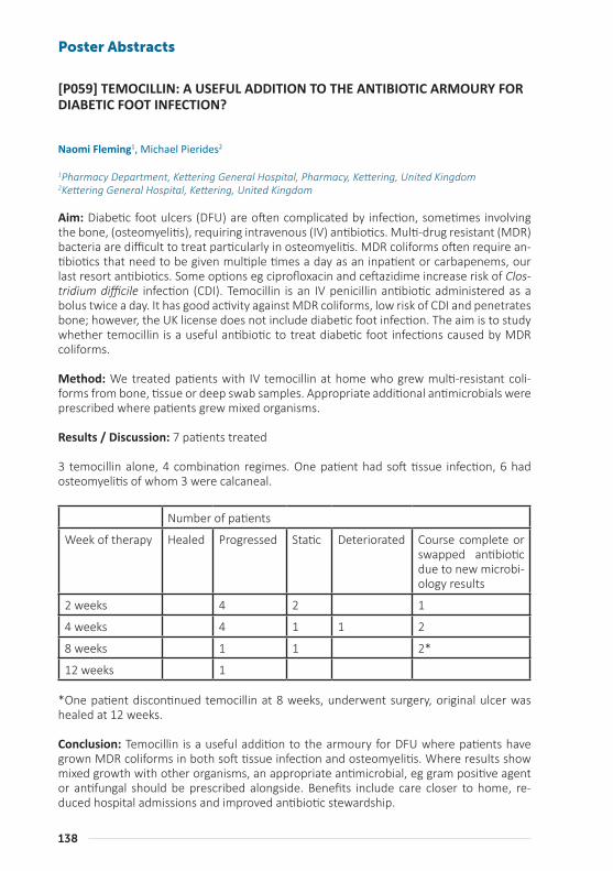
138
Poster Abstracts
[P059] TEMOCILLIN: A USEFUL ADDITION TO THE ANTIBIOTIC ARMOURY FOR DIABETIC FOOT INFECTION?
Naomi Fleming1, Michael Pierides2
1Pharmacy Department, Kettering General Hospital, Pharmacy, Kettering, United Kingdom2Kettering General Hospital, Kettering, United Kingdom
Aim: Diabetic foot ulcers (DFU) are often complicated by infection, sometimes involving the bone, (osteomyelitis), requiring intravenous (IV) antibiotics. Multi-drug resistant (MDR) bacteria are difficult to treat particularly in osteomyelitis. MDR coliforms often require an-tibiotics that need to be given multiple times a day as an inpatient or carbapenems, our last resort antibiotics. Some options eg ciprofloxacin and ceftazidime increase risk of Clos-tridium difficile infection (CDI). Temocillin is an IV penicillin antibiotic administered as a bolus twice a day. It has good activity against MDR coliforms, low risk of CDI and penetrates bone; however, the UK license does not include diabetic foot infection. The aim is to study whether temocillin is a useful antibiotic to treat diabetic foot infections caused by MDR coliforms.
Method: We treated patients with IV temocillin at home who grew multi-resistant coli-forms from bone, tissue or deep swab samples. Appropriate additional antimicrobials were prescribed where patients grew mixed organisms.
Results / Discussion: 7 patients treated
3 temocillin alone, 4 combination regimes. One patient had soft tissue infection, 6 had osteomyelitis of whom 3 were calcaneal.
Number of patients
Week of therapy Healed Progressed Static Deteriorated Course complete or swapped antibiotic due to new microbi-ology results
2 weeks 4 2 1
4 weeks 4 1 1 2
8 weeks 1 1 2*
12 weeks 1
*One patient discontinued temocillin at 8 weeks, underwent surgery, original ulcer was healed at 12 weeks.
Conclusion: Temocillin is a useful addition to the armoury for DFU where patients have grown MDR coliforms in both soft tissue infection and osteomyelitis. Where results show mixed growth with other organisms, an appropriate antimicrobial, eg gram positive agent or antifungal should be prescribed alongside. Benefits include care closer to home, re-duced hospital admissions and improved antibiotic stewardship.
139
[P060] OUTCOME OF DIABETIC FOOT ULCER WITH OSTEOMYELITIS IN EGYPT
Hanan M.S. Gawish1, Fady Kyrillos1, Ahmed Albehairy2, Mohamed Elsayed1, Mohamad Elsherif1, Mamdouh El-Nahas3, Omnia State1, Manal Tarshoby1
1Endocrinology and Diabetes Unit, Mansoura University, Egypt2Faculty of Medicine, Internal Medicine Department , Mansoura, Egypt3 Diabetes Unit, Mansoura University, Port-Said, Egypt
Aim: Osteomyelitis is an important contributing factor to impaired wound healing. Howev-er, the selection of the appropriate therapy and the effect of therapy on ulcer healing is still unclear. The study aims to identify the management outcome of patients with diabetic foot ulcer associated with osteomyelitis at Mansoura Diabetic Foot centre, Egypt and different variables affecting it.
Method: A retrospective single center including all registered cases, who presented with diabetic foot ulcer complicated by osteomyelitis, treated conservatively, were recruited between July 2005 and December 2015 at our tertiary center.
Results / Discussion: Of all 3544 Diabetic Foot Ulcer (DFU) presented to the clinic for this 10 years period; 331 were associated with osteomyelitis. Data of 161 ulcer were excluded; because they were either referred to orthopedic surgery or dropped their follow up.
The mean age of patients was 57±9.91 yrs, with male predominance of 61.8%. PAD was only present in 2.35% and severe infection in 10.6% of patients. Admission was done in 7.1% of the whole group. Sharp debridement was done for 96.5 % patients while ultra-sound debridement was done for 3.5 % patients.
Patients were classified into group 1 who showed complete healing of their ulcers during the study period and group 2 who did not show complete healing. The healing rate was 65.88 % among the group and the time to heal (was 127.05 ± 82.87 days in the first group. None or delayed healing was present in 34.1% of patients. There was no significant dif-ference between the 2 groups as regards the age, gender distribution, or other system comorbidity. Toe ulcers with osteomyelitis showed higher healing rate than ulcers over Metatarsal Heads (P≤0.000). Non-significant difference was present at the other sites of the foot ulceration. The commonest antibiotics used among the healed ulcer group were combination of quinolones and linezolid in 31.4 %.
Recurrence rate of ulceration was 8.9% at the same site of ulcer and new ulceration at other sites occurred in 19.6% of the healed group during the study period.
Conclusion: Around two thirds of patients presenting with osteomyelitis healed conser-vatively without undergoing surgical bone resection. Quinolones/linezolid combination therapy were the commonest antibiotic regimen used among the healers. Osteomyelitis affecting the metatarsal heads was the site significantly associated with delayed ulcer heal-ing or the need for surgical intervention.
Pro
gra
mm
eA
uth
ors
Ora
l A
bst
ract
sP
oste
r A
bst
ract
sIn
form
atio
nS
pon
sor/
Exh
ibit
or
140
Poster Abstracts
[P061] MANAGEMENT OF SEVERE INFECTION OF DIABETIC FOOT IN A LOW RESOURCE ENVIRONMENT
Adalberto Silva1, João Carlos Carranca2, Armando Ribeiro2, Diogo Veiga2
1Chua - Centro Hospitalar Universitário Do Algarve, Portimão, Portugal
Aim: A case report is presented to express the various hardships that a patient undergoes in a low resource environment and to emphasize the importance of a proper diabetic foot program that can prevent those circumstances to happen
Method: A 65-year-old male diabetic patient was admitted with infection of the right foot which extended to the lower third of the leg. Surgical drainage was performed on admis-sion and empirical antibiotherapy was started which was prolonged for ten days.
Microbiology of the pus revealed Streptococcus agalactiae sensitive to amoxycilin and ce-furoxime. He was treated with cefuroxime and metronidazole for 15 days. With this treat-ment the infection was controlled.
Before his admission the patient was taking oral antidiabetics at home.
On the 7th day in hospital he had to have an amputation of the hallux as it was necrotic. He underwent various debridements and on the 50th day presented necrosis of the 2nd toe and underwent amputation of this toe. On the 72nd day he had the 3rd toe amputated because of necrosis.
During his stay in the hospital he also received insulin for better control of the diabetes.
A doppler ultrasound and a CT angiography was performed and revealed severe peripheral arterial occlusive disease in various segments of both lower limbs. He was then referred to a Vascular Surgeon who stated that a vascular procedure was not indicated and that the patient was a candidate for amputation above the knee, which the patient did not agree. We continued the treatment of the wounds with dressings.
Results / Discussion: The wounds improved and he was discharged on the 85th day and he was followed-up as an out-patient. Up to now there has been slow and steady progress of the healing of the wounds.
Conclusion: An appropriate program of prevention of diabetic foot would prevent the di-abetic patient to experience the various hardships, so this case represents the uppermost need to apply such program in our region.
141
[P062] A RETROSPECTIVE STUDY TO DETERMINE HOW COMMON TEICO-PLANIN-INDUCED THROMBOCYCTOPAENIA REALLY IS, IN PATIENTS WITH DIA-BETIC FOOT DISEASE
Jacqueline Mildred1, Michael Pierides1
1Diabetes Centre, Kettering General Hospital, Kettering, Northamptonshire, United Kingdom
Aim: Teicoplanin is a glycopeptide antibiotic, with bactericidal activity against aerobic and anaerobic gram-positive bacteria including multi resistant staphylococci. However, amongst its benefits, Teicoplanin can cause thrombocytopaenia. Electronic Medicines Compendium1 (eMC) recommends haematological monitoring during treatment, [full blood count (FBC) and teicoplanin levels]. We reviewed how common Teicoplanin-induced thrombocytopaenia is, in our cohort of diabetic foot patients, when prescribed Teicoplanin for a diabetic foot infection (DFI).
Method: We identified diabetic patients by searching our electronic database that pre-sented with DFI and received Teicoplanin during 12 months (1st Jan 2017 to 1st Jan 2018). Data was collected on duration of treatment, FBC and Teicoplanin levels. Bloods were tak-en on a routine basis and monitored whilst treated.
Results: Over the 12 months, 25 courses of Teicoplanin were prescribed, to 23 patients. 87% (n=20) were male and 13% (n=3) were female, with a mean age of 69 years. All DFI were from foot ulceration with osteomyelitis. Teicoplanin was dosed at 10mg/kg and levels targeted between 20-60mg/L for bone infection. Levels ranged from 19-57 with a mean of 35.5mg/L. Treatment duration ranged from 1 to 14 weeks, with a mean duration of 4.3 weeks. When levels were above 50mg/L dose reductions were made taking into consideration the patients’ renal function defined as creatinine clearance via Cockcroft Gault method. Platelets defined as normal between 150 to 450x109/L. Thrombocyctopenia was defined as platelets <150x109/L. 64% (n=16) showed a drop in platelets but 100% re-mained within the normal range. Treatment <3.5 weeks showed a mean platelet reduction by 9.2x10⁹/L. However, treatment >3.5 weeks the platelet level dropped by 57.5x10⁹/L. 39% were also on concomitant antibiotics, in addition to Teicoplanin and 55% were on platelet inhibiting drugs.
Conclusion: Teicoplanin-induced thrombocyctopaenia is classed as an infrequent adverse effect1. This study demonstrated there was no development of thrombocyctopenia. How-ever, it did show platelet reduction over duration but not below normality. In addition, Teicoplanin levels increased during treatment time but this failed to translate into a critical drop of platelets although this may have also been prevented by some of the dose reduc-tions. Our data confirms the importance that patients are adequately monitored whilst on Teicoplanin as the risk of a critical drop still remains a possibility but is manageable and preventable if systematic monitoring is in place, as in our cohort of patients.
1www.medicines.org.uk>emc
Pro
gra
mm
eA
uth
ors
Ora
l A
bst
ract
sP
oste
r A
bst
ract
sIn
form
atio
nS
pon
sor/
Exh
ibit
or
142
Poster Abstracts
[P063] PERIPHERAL NEUROPATHY IN PATIENTS WITH SARCOPENIA AND TYPE 2 DIABETES MELLITUS
Julia Onuchina1, Irina Gurieva2
1Federal Bureau of Medical and Social Expertise. Department of Endocrinology, Pirogov Russian National Medical University, The Russian Clinical Research Center for Gerontology, Moscow, Russian Federation2Federal Bureau of Medical and Social Expertise. Department of Endocrinology, Moscow, Russian Federation
Aim: Purpose of this pilot crossectional study is to evaluate clinical and instrumental fea-tures of peripheral neuropathy in patients with sarcopenia and type 2 diabetes mellitus (T2DM).
Method: Ninety-six patients with T2DM over 60 years old were examined. Muscle strength was measured with carpal dynamometry, muscle function was evaluated with short phys-ical performance battery (SPPB) tests. The SPPB includes standing balance, gait speed, timed chair stands test. Skeletal muscle mass index (SMMI) was evaluated with bioimped-ance testing. Patients with decrease of skeletal muscle mass index and/or muscle strength and/or muscle function were diagnosed as sarcopenic - «S+» group and non-sarcopenic - «S-» group. Peripheral neuropathy was studied with calculation of TSS, NDS and NIS-LL scales.
Results / Discussion: Sarcopenia revealed in 27 patients (36%) aged 75 [68;78,5]years and diabetes duration of 11 [6;16]years; HbA1c - 9,3 [6,9;10,8]%; 49 patients aged 69 [66;77]years and diabetes duration of 12 [8;16] years, HbA1c – 8,1 [7;10]% were not sarcopenic.
«S+» patients demonstrated lower composites of SPPB vs «S-» group, such as walking speed (0,79 [0,66;0,8] vs1,25 [1;1,3] m\s, p<0,001), ability to hold balance (31 [14,75;41] vs 11,5 [7;27,75] sec, p=0,002). Both groups demonstrated lower indices of dynamometry.
Diabetic neuropathy was severe in S+» group than in «S-» group (NDSm:5[3;6]; 2[2;3], p=0,000; NIS-LL: 12[7;6], 6 [4,8], p<0,001). «S+» patients had worst vibration score vs «S-» patients (128 Hz tuning fork: 4 [3,5;5] vs 5 [4,6], p=0,006, biothesiometer 17 [15,5;28] vs 12 [8;18], p=0,001). Decrease of pressure perception to monofilament SW5.07 revealed in 52% of «S+» patients in comparison with 8% of «S-» patients (p= 0,001). Pain, tempera-ture, sense of position, Achilles reflexes, muscle weakness of toe extensors and flexors were worse in group «S+»(p<0,05).
«S+» patients noted more often frequency of falls (70%) vs «S-» group- (35%) p=0,004. «S+» demonstrated more often frequency of fractures (67%) than «S-» group (35%) p=0,009.
«S+» group characterized with lower BMI vs patients without sarcopenia (25,7[23,5;29,4] vs 31,6 [29,39; 33,79]kg/m2, p<0,001);smaller waist and neck circumference (94 [89,5;99,5] vs 104 [100;110]cm, p<0,001; 37 [34,5;38] vs 39 [37;41], p<0.001).
«S+» patients received metformin less frequently (48%) than «S-» patients (76%) р=0,009.
143
Conclusion: 1) Severe diabetic neuropathy is more common in patients with sarcopenia 2) Large and small fiber function loss are more common in T2DM patients with sarcopenia 3) Combination of large fiber neuropathies with decreased muscle mass in T2DM patients with sarcopenia increases the risk of falls and fractures.
Pro
gra
mm
eA
uth
ors
Ora
l A
bst
ract
sP
oste
r A
bst
ract
sIn
form
atio
nS
pon
sor/
Exh
ibit
or
144
Poster Abstracts
[P064] EVALUATION OF TWO EXAMINATION PROTOCOLS AGAINST TWO THRESHOLDS OF CLINICAL POLYNEUROPATHY IN TYPE 2 DIABETES MELLITUS
Nikolaos Papanas1, Kalliopi Pafili1, Maria Demetriou1, Soultana Kyroglou1, Dimitrios Papazoglou1, Efstratios Maltezos1
1Diabetes Centre-Diabetic Foot Clinic, , Second Department of Internal Medicine, Democritus Univer-sity of Thrace, Alexandroupolis, Greece
Aim: We examined 2 examination protocols with a portable and useful device* against 2 thresholds of the neuropathy disability score (NDS) for the diagnosis of polyneuropathy (DPN) in patients with type 2 diabetes mellitus (T2DM).
Method: We included 100 T2DM patients with mean age 62.3 years and mean T2DM dura-tion 12.6 years. The device* was used bilaterally to test vibration sensation on the pulp of the hallux (protocol A) or on 3 foot sites (pulp of the hallux, 1st and 3rd metatarsal head: pro-tocol B). With either protocol, the test was considered abnormal if vibration was not felt in ≥1 site. The reference method was NDS, but we used 2 thresholds: NDS≥3 and NDS≥6.
Results: DPN was diagnosed in 46 patients as NDS≥3 and in 38 patients as NDS≥6. Against NDS≥3: a) In protocol A, the device* yielded 91.3% sensitivity, 85.2% specificity, 84% pos-itive predictive value (PPV), 92% negative predictive value (NPV), 6.2 positive likelihood ratio (+LR) and 0.1 negative likelihood ratio (-LR); b) The corresponding values in protocol B were 95.6%, 90.7%, 89.8%, 96.1%, 10.3 and 0.05. Against NDS≥6: a) In protocol A, the de-vice* yielded 100% sensitivity, 95.2% specificity, 92.7% PPV, 100% NPV, 20.8 +LR and 0 -LR; b) The corresponding values in protocol B were 100%, 96.8%, 95.0%, 100%, 31.25 and 0.
Conclusion: With either protocol, the diagnostic performance of the device* is very high in T2DM, rendering it very useful as a screening tool, particularly for the exclusion of DPN. The addition of other than the hallux examination sites (i.e. protocol B) offers a very small improvement in the diagnostic yield. The device performs very well against both NDS thresholds, particularly against NDS≥6.
*VibraTipTM
145
[P065] BASELINE VIBRATION PERCEPTION THRESHOLD DOES NOT AFFECT RE-SPONSE TO FIRST LINE TREATMENT IN PAINFUL DIABETIC NEUROPATHY
Satyan Rajbhandari1, Yassine Noui1
1Lancashire Teaching Hospitals NHS Trust, Chorley, United Kingdom
Aim: Painful neuropathy is due to small fibres or its central connection dysfunction and large fibres are believed to modulate this. Large fibre function can be measured by vibra-tion perception threshold (VPT) and the high threshold (≥ 25 volt) is associated with the development of foot ulcers. There has been no study to demonstrate the effect of large fibres dysfunction in the treatment for neuropathic pain. The aim of this study was to analyse if the response to painful neuropathy treatment varies in patients with or without large fibre dysfunction.
Method: List of subjects with painful neuropathy was obtained from the central database and those who had VPT recorded in baseline and treated with pharmaceutical agents were studied. Subjects were then divided into two groups depending upon baseline VPT of ≥ 25 volts. The response to treatment at follow up after a month was assessed. If patients confirmed improvement in pain and elected to stay on the same dose or discharged after satisfactory response were grouped as responders. If treatment had to be changed or dose titrated upwards due to side effects or poor response were grouped as non-responders.
Results / Discussion: 86 patients had VPT recorded at baseline. 24 patients did not receive pharmacological treatment, 4 patients lost to follow up and 4 subjects who had not yet had second review were excluded. 26 subjects had low VPT and 28 had high VPT. The severity and characteristics of pain was similar in both groups at baseline. The standard treatment as first line was mostly pregabalin (75 mg BD), Amitryptiline (30 mg nocte), Gabapentin (300 mg TDS), Duloxetin (60 mg OD) given in weekly titration dose. Subjects who had high VPT were older [66.7 (+/- 10.5) vs 60.5 (+/- 11.6) years; p = 0.023], but there was no differ-ence in sex, type of diabetes, HbA1c and renal function. 61.5% of low VPT group responded to the first line treatment in comparison to 46.4% of high VPT group but this was not sta-tistically significant (p = 0.29).
Conclusion: Our results show that VPT may not affect the response to the first line treat-ment for painful neuropathy of diabetes. The choice of treatment was guided by co-exist-ing medical condition and concern of side effects. Further clinical trials are needed to see if any specific treatment is more useful in patients with large fibre dysfunction.
Pro
gra
mm
eA
uth
ors
Ora
l A
bst
ract
sP
oste
r A
bst
ract
sIn
form
atio
nS
pon
sor/
Exh
ibit
or
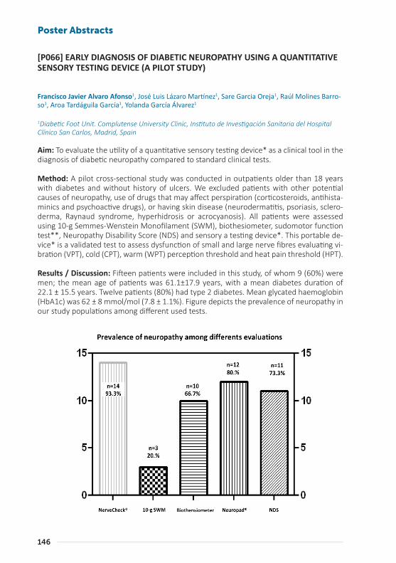
146
Poster Abstracts
[P066] EARLY DIAGNOSIS OF DIABETIC NEUROPATHY USING A QUANTITATIVE SENSORY TESTING DEVICE (A PILOT STUDY)
Francisco Javier Alvaro Afonso1, José Luis Lázaro Martínez1, Sare Garcia Oreja1, Raúl Molines Barro-so1, Aroa Tardáguila García1, Yolanda García Álvarez1
1Diabetic Foot Unit. Complutense University Clinic, Instituto de Investigación Sanitaria del Hospital Clínico San Carlos, Madrid, Spain
Aim: To evaluate the utility of a quantitative sensory testing device* as a clinical tool in the diagnosis of diabetic neuropathy compared to standard clinical tests.
Method: A pilot cross-sectional study was conducted in outpatients older than 18 years with diabetes and without history of ulcers. We excluded patients with other potential causes of neuropathy, use of drugs that may affect perspiration (corticosteroids, antihista-minics and psychoactive drugs), or having skin disease (neurodermatitis, psoriasis, sclero-derma, Raynaud syndrome, hyperhidrosis or acrocyanosis). All patients were assessed using 10-g Semmes-Wenstein Monofilament (SWM), biothesiometer, sudomotor function test**, Neuropathy Disability Score (NDS) and sensory a testing device*. This portable de-vice* is a validated test to assess dysfunction of small and large nerve fibres evaluating vi-bration (VPT), cold (CPT), warm (WPT) perception threshold and heat pain threshold (HPT).
Results / Discussion: Fifteen patients were included in this study, of whom 9 (60%) were men; the mean age of patients was 61.1±17.9 years, with a mean diabetes duration of 22.1 ± 15.5 years. Twelve patients (80%) had type 2 diabetes. Mean glycated haemoglobin (HbA1c) was 62 ± 8 mmol/mol (7.8 ± 1.1%). Figure depicts the prevalence of neuropathy in our study populations among different used tests.
147
The prevalence of diabetic symmetric polyneuropathy (DSPN) in our study population was 93.3% (n=14) using a sensory testing device* versus 20% (n=3) and 66.7% (n=10) using 10-g SWM and Biothensiometer respectively. We observed less difference in the preva-lence of DSPN using test that evaluate dysfunction of small nerve fibres (screening test**, 73.3%, n=11) or using NDS (80%, n=12). In the case of patients who had DSPN using Nerve-Check (n=14), 78,6% (n=11) and 35.7% (n=5) had not DSPN using 10-g SWM and Biothen-siometer respectively. These results show that underdiagnosis of neuropathic patients may occur when using standard neurological tests (mainly used in primary care). Under such circumstances, these patients will not be treated by preventative strategies, which would considerably increase the risk of developing a diabetic foot ulcer.
Conclusion:
A new quantitative sensory testing device could help us in the early detection of diabetic neuropathy in primary care. This device could be added in a care setting where the cate-gorisation of risk is required.
* NerveCheck®
** Neuropad®
Pro
gra
mm
eA
uth
ors
Ora
l A
bst
ract
sP
oste
r A
bst
ract
sIn
form
atio
nS
pon
sor/
Exh
ibit
or
148
Poster Abstracts
[P067] APPLICATION OF THE FREQUENCY RHYTHMIC ELECTRICAL MODULA-TION SYSTEM IN TREATMENT OF NEUROPATHIC PAIN IN PATIENTS WITH DIA-BETIC NEUROPATHY
Ekaterina Zaitseva1, Gagik Galstyan2
1Endocrinology Research Centre, Diabetic Foot Department, Moscow, Russian Federation2Endocrinology Research Centre, Moscow, Russian Federation
Aim: To study the efficiency of the frequency rhythmic electrical modulation system* on the neuropathic pain caused by diabetic neuropathy.
Method: General clinical, assessment of sensitivity by standard methods, severity of neu-ropathy symptoms using the NSS scale, determination of the severity of the pain using a visual analogue scale (VAS).
Results / Discussion: 26 patients (15 women, 11 men) with diabetes mellitus (T1DM - 2, T2DM - 24) were included in the pilot study at the age of 65.8 ± 14.0 years. The average du-ration of DM was 11.9 ± 10 years. Poor glycaemic control was noted – the average HbA1c level was - 8.4 ± 1.4%.
Sensitivity studies in 10 patients (39%) determined a decrease in one or another type of sensitivity, in 16 patients (61%) - complete absence of tactile and temperature sensitivity, vibration sensitivity of 2-3 UE. According to NSS scale, moderate neuropathy was defined in 5 patients (28%), severe neuropathy was found in 17 patients (72%).
15 patients took Pregabalin or Gabapentin in order to reduce pain.
According to VAS 10 patients (40%) characterized leg pain as severe, 6 patients (23%) as very strong, 10 patients (37%) as intolerant.
All patients underwent 9 ± 2 sessions of the frequency rhythmic electrical modulation sys-tem*.
22 patients (86%) at the end of treatment noted a significant improvement in their con-dition, the decrease of the severity of neuropathic symptoms. Three patients managed to cancel the anticonvulsants intake, in 3 patients - to reduce the dose of drugs by 50% of the initial.
After frequency rhythmic electrical modulation system*, 16 patients (64%) rated the pain syndrome as strongly pronounced, 5 patients (18%) - as moderate, 5 patients (18%) - as easily expressed according to the VAS data.
Conclusion: The use of the frequency rhythmic electrical modulation system* has shown short-term effectiveness in the treatment of neuropathic pain in patients with diabetes mellitus. However, this method requires further studies on a larger amount of patients, a comparative study with a control group and a long-term follow-up.
*FREMS-Therapy
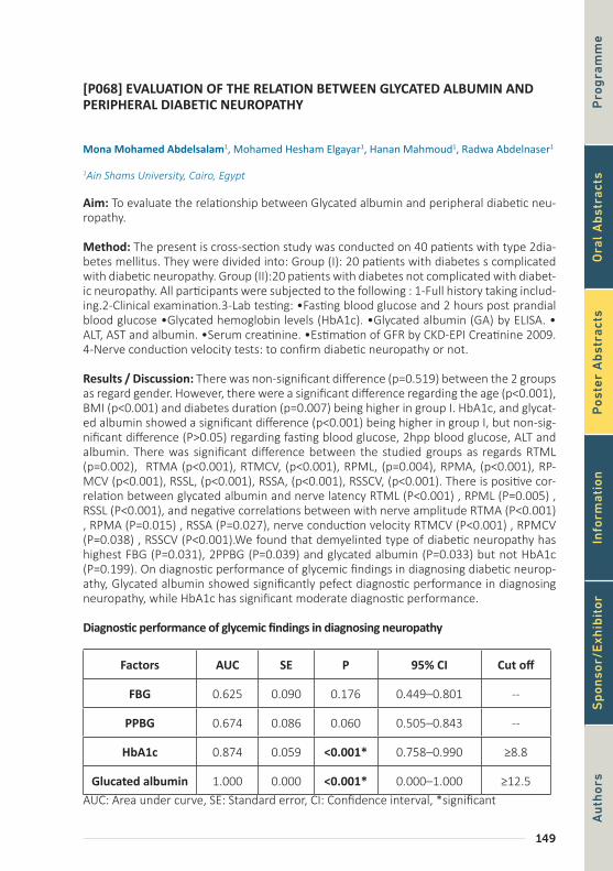
149
[P068] EVALUATION OF THE RELATION BETWEEN GLYCATED ALBUMIN AND PERIPHERAL DIABETIC NEUROPATHY
Mona Mohamed Abdelsalam1, Mohamed Hesham Elgayar1, Hanan Mahmoud1, Radwa Abdelnaser1
1Ain Shams University, Cairo, Egypt
Aim: To evaluate the relationship between Glycated albumin and peripheral diabetic neu-ropathy.
Method: The present is cross-section study was conducted on 40 patients with type 2dia-betes mellitus. They were divided into: Group (I): 20 patients with diabetes s complicated with diabetic neuropathy. Group (II):20 patients with diabetes not complicated with diabet-ic neuropathy. All participants were subjected to the following : 1-Full history taking includ-ing.2-Clinical examination.3-Lab testing: •Fasting blood glucose and 2 hours post prandial blood glucose •Glycated hemoglobin levels (HbA1c). •Glycated albumin (GA) by ELISA. • ALT, AST and albumin. •Serum creatinine. •Estimation of GFR by CKD-EPI Creatinine 2009. 4-Nerve conduction velocity tests: to confirm diabetic neuropathy or not.
Results / Discussion: There was non-significant difference (p=0.519) between the 2 groups as regard gender. However, there were a significant difference regarding the age (p<0.001), BMI (p<0.001) and diabetes duration (p=0.007) being higher in group I. HbA1c, and glycat-ed albumin showed a significant difference (p<0.001) being higher in group I, but non-sig-nificant difference (P>0.05) regarding fasting blood glucose, 2hpp blood glucose, ALT and albumin. There was significant difference between the studied groups as regards RTML (p=0.002), RTMA (p<0.001), RTMCV, (p<0.001), RPML, (p=0.004), RPMA, (p<0.001), RP-MCV (p<0.001), RSSL, (p<0.001), RSSA, (p<0.001), RSSCV, (p<0.001). There is positive cor-relation between glycated albumin and nerve latency RTML (P<0.001) , RPML (P=0.005) , RSSL (P<0.001), and negative correlations between with nerve amplitude RTMA (P<0.001) , RPMA (P=0.015) , RSSA (P=0.027), nerve conduction velocity RTMCV (P<0.001) , RPMCV (P=0.038) , RSSCV (P<0.001).We found that demyelinted type of diabetic neuropathy has highest FBG (P=0.031), 2PPBG (P=0.039) and glycated albumin (P=0.033) but not HbA1c (P=0.199). On diagnostic performance of glycemic findings in diagnosing diabetic neurop-athy, Glycated albumin showed significantly pefect diagnostic performance in diagnosing neuropathy, while HbA1c has significant moderate diagnostic performance.
Diagnostic performance of glycemic findings in diagnosing neuropathy
Factors AUC SE P 95% CI Cut off
FBG 0.625 0.090 0.176 0.449–0.801 --
PPBG 0.674 0.086 0.060 0.505–0.843 --
HbA1c 0.874 0.059 <0.001* 0.758–0.990 ≥8.8
Glucated albumin 1.000 0.000 <0.001* 0.000–1.000 ≥12.5AUC: Area under curve, SE: Standard error, CI: Confidence interval, *significant
Pro
gra
mm
eA
uth
ors
Ora
l A
bst
ract
sP
oste
r A
bst
ract
sIn
form
atio
nS
pon
sor/
Exh
ibit
or
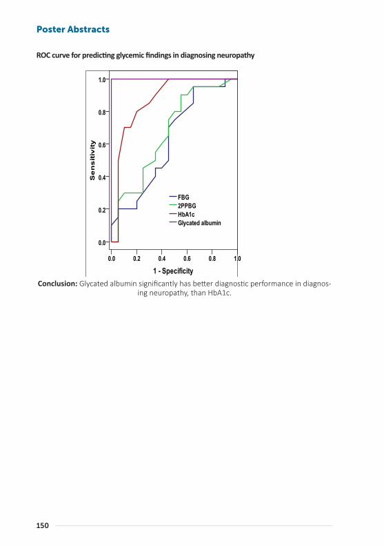
150
Poster Abstracts
ROC curve for predicting glycemic findings in diagnosing neuropathy
Conclusion: Glycated albumin significantly has better diagnostic performance in diagnos-ing neuropathy, than HbA1c.
a
151
ROC curve for predicting glycemic findings in diagnosing neuropathy
Conclusion: Glycated albumin significantly has better diagnostic performance in diagnos-ing neuropathy, than HbA1c.
a
[P070] IMPACT OF THE SERUM LEVEL OF TRACE ELEMENTS ON SYMPTOMS OF PERIPHERAL NEUROPATHY IN TYPE 2 DIABETES
Rania Bahriz1, Sherif Fathy2, Ahmed Albehairy1
1Faculty of Medicine, Internal Medicine Department, Mansoura University, Egypt2Faculty of Medicine, Clinical Pathology Department, Mansoura University, Egypt
Aim: This study was designed in order to estimate the alterations in the levels of these trace elements serum zinc, magnesium and copper in patients with type 2 diabetes mel-litus with diabetic peripheral neuropathy in comparison to those without neuropathy and their relation to duration of diabetes and glycemic control.
Method: Seventy one patients with Type 2 diabetes were recruited from diabetes clinic and diabetic neuropathy clinic in Specialized Medical Hospital, Mansoura University, Egypt from May 2017 to December 2017 divided into 2 groups: Group 1: Patients with type 2 diabetes and diabetic neuropathy including 47 patients and Group 2: Patients with type 2 diabetes without diabetic neuropathy including 24 patients. The presence and absence of neuropathy was assessed using neuropathy disability score and neuropathy symptom score. All the seventy one patients were subjected to:Full history and examination includ-ing (age ,sex, Body mass index ,duration of diabetes and its regimen of treatment and pres-ence or absence of hypertension), neuropathy symptom score and neuropathy disability score. A blood sample was taken for measuring serum zinc ,serum magnesium and serum copper which were estimated by endpoint spectrophotometry. Glycated hemoglobin was also done for both groups. Data were analyzed with SPSS version 21.
Results / Discussion: There was a significant difference between both groups as regard duration of diabetes and serum copper level (P value < 0.05). After performing linear re-gression analysis, the duration of Diabetes was the significant risk factor for the diabetic neuropathy symptoms. On the other hand there was no significant difference between the 2 groups as regard serum zinc, serum magnesium, glycated hemoglobin, age ,sex, and hypertension.
Conclusion: Serum copper is not an independent risk factor for diabetic peripheral neu-ropathy as previously thought inspite of its elevation in both groups of the study that may be attributed to the hyperglycemia itself .On the other hand ,there is no impact of both serum zinc and magnesium on diabetic peripheral neuropathy symptoms.
Pro
gra
mm
eA
uth
ors
Ora
l A
bst
ract
sP
oste
r A
bst
ract
sIn
form
atio
nS
pon
sor/
Exh
ibit
or
152
Poster Abstracts
[P071] TRANSCUTANEOUS OXYGEN PRESSURE - A SUITABLE PARAMETER FOR ASSESSING THE EFFECT OF AUTOLOGOUS CELL THERAPY IN PATIENTS WITH ISCHEMIC DIABETIC FOOT
Andrea Nemcova1, Alexandra Jirkovská2, Michal Dubský2, Robert Bem2, Vladimíra Fejfarová2, Veron-ika Woskova2
1Ikem, Diabetes Centre, Prague, Czech Republic2Diabetes Centre, Institute for Clinical and Experimental Medicine, Prague, Czech Republic
Aim: The main therapeutic benefit of autologous cell therapy (ACT) in patients with diabet-ic foot (DF) and critical limb ischemia (CLI) is increased tissue oxygenation and improved healing of DF. Transcutaneous oxygen pressure (TcPO2) measurement is frequently used in the assessment of the effect of revascularization. The aim of our study was to evaluate TcPO2 measurement in the assessment of the effect of ACT in contrast to standard percu-taneous transluminal angioplasty (PTA) in patients with DF and CLI by comparing changes in TcPO2 between treated and untreated limbs.
Method: Seventy-three patients with DF and CLI treated in our foot clinic during 2008-2016 were enrolled into the study. Thirty-three patients were treated by ACT, 40 patients underwent PTA as a standard method for revascularization and formed the control group. TcPO2 was measured on treated and untreated (control) limbs before and at 1, 3 and 6 months after revascularization.
Results / Discussion: TcPO2 values on the treated limb increased significantly after PTA at 1, 3 and 6 months (from 25.8 to 39.5, 48.3, 47.5 mmHg, respectively, all p ˂ 0.001) in comparison with the untreated limb where no significant changes in TcPO2 were observed at all intervals. TcPO2 values on the treated limb also increased significantly after ACT at 1, 3 and 6 months (from 18.4 to 30.4, 39.9, 42.3 mmHg, respectively, all p ˂ 0.001) while no significant changes in TcPO2 were again seen on the control limb. The reference value for TcPO2 measurement on the clavicle was without a significant change at all intervals both in PTA and ACT groups. The foot/clavicle index increased significantly after ACT on the treated limb at 1, 3 and 6 months from 0.33 to 0.57 (p ˂ 0.01), 0.71 and 0.73 (both p ˂ 0.001) in contrast to the control limb where the indices were without a significant change.
Conclusion: Our study showed a significant increase in TcPO2 levels on the treated limb both after standard revascularization by PTA and after ACT in contrast to the untreated limb. TcPO2 measurement seems to be a reliable tool for assessing the effect of ACT on critical limb ischemia.
Supported by the Ministry of Health of the Czech Republic, grant no. 16-27262A, by project for Development of Research Organization 00023001 (IKEM, Prague, Czech Republic) – In-stitutional support
153
[P072] ROLE OF THERMAL IMAGING FOR DIAGNOSTIC ASSESSMENT OF ACUTE CHARCOT FOOT. A CASE SERIES
Omar Schembri1, Kevin Cassar2, Alison Psaila1, David Coppini3
1Dept of Diabetes, Mater Dei Hospital, Gwardamangia, Malta2Dept of Vasc Surgery, Mater Dei Hospital, Malta3Poole Hospital, Dept of Diabetes, Poole, United Kingdom
Aim: To evaluate the role of thermal imaging in assessment of patients with diabetes pre-senting with possible Charcot foot.
Method: 16 consecutive patients with diabetes (30% type 1, age range 42-75 years, Dia-betes duration 4-35 years) attending the diabetic foot clinic with suspicious Charcot foot received thermal imaging using an infrared camera (resolution 1.2mm/pixel) as part of their diagnostic work up and thermal images of interest with highest temperature differ-ences within same foot and between feet were measured and compared with radiological changes on plain radiography or MR scan.
Results / Discussion: Thermal images showed temperature differences between feet of at least 2°C in 10 patients and areas of highest temperature and maximal colour change compared well with areas of bone oedema or inflammatory change on MR scan in 5 pa-tients. Irrespective of normal thermal imaging in 5 patients, in all subjects a diagnosis of Charcot foot was made based on clinical suspicion supported by thermal imaging, radio-logical change and inflammatory markers (CRP, ESR, WBC). Thermal images in patients with respective radiological change will be presented.
Conclusion: Thermal imaging can be a valuable tool in the diagnostic work up of patients with suspected Charcot foot and is non-invasive, safe and in some cases even correlates with radiological areas suggestive of inflammatory change. Despite its availability, thermal imaging is not routinely used in the diagnostic work up of Charcot foot but our promising data should encourage further studies to help establish its possible role for an often diffi-cult to diagnose diabetes complication.
Pro
gra
mm
eA
uth
ors
Ora
l A
bst
ract
sP
oste
r A
bst
ract
sIn
form
atio
nS
pon
sor/
Exh
ibit
or
154
Poster Abstracts
[PO73] THE USE OF THERMOGRAPHY FOR THE DETECTION OF DIABETIC FOOT COMPLICATIONS
Cynthia Formosa1, Owen Falzon1, Kevin Cassar1, Christian Ellul1, Kenneth Camilleri1, Jean Gauci1, Stephen Mizzi2, Anabelle Mizzi1, Cassandra Sturgeon1, Liberato Camilleri1, Nachiappan Chockaling-ham3, Alfred Gatt1
1University of Malta, Msida, Malta2University of Malta, Faculty of Health Science, Msida, Malta3Staffordshire University, Stoke-on-Trent, United Kingdom
Aim: To investigate the potential of thermography as an assessment tool for the detection of foot complications by evaluating variations in foot temperatures that occur in in type 2 diabetes mellitus (DM).
Method: Participants were categorized into five groups: healthy adults and DM with no complications, DM with peripheral neuropathy, DM with neuroischaemia and DM with pe-ripheral arterial disease (PAD) group based on the medical examination and testing. Ther-mographic imaging of the toes and forefeet were taken.
Results / Discussion: Forty-three neuroischaemic feet, 41 neuropathic feet, 58 PAD feet, 21 DM feet without complications and 126 healthy feet were analyzed. The temperatures of the feet and toes were significantly higher in the complications group when compared to the healthy adult and DM healthy. The higher the temperatures of the foot in DM, the higher the probability that it is affected by neuropathy, neuroischaemia or PAD. This study has confirmed that the mean temperatures of the toes and forefeet of the complications group exhibit significantly higher temperatures than those of the healthy group, whilst each group presents with comparable temperatures within themselves. These results in-dicate that thermography could potentially be used as a screening or clinical investigation tool, however more research in this area is warranted
Conclusion: Significant differences in mean temperatures exist between healthy and DM participants with no known complications when compared to participants with neuroisch-aemia, neuropathy or PAD. As foot temperature rises, so does the probability of the pres-ence of complications of neuropathy, neuroischaemia or peripheral arterial disease.
155
[P074] ASSESSMENT OF PATIENTS’ NEEDS AND PROTOTYPE DEVELOPMENT REGARDING CUSTOM-MADE DIABETIC FOOTWEAR FOR HOME USE
Renske Keukenkamp1, Jaap van Netten2, Tessa Busch-Westbroek1, Sicco Bus1
1Department of Rehabilitation, Academic Medical Center, Amsterdam, Netherlands2Department of Rehabilitation, Academic Medical Center, School of Clinical Sciences, Queensland University of Technology, Amsterdam, Netherlands
Aim: Footwear adherence is a known problem in people with diabetes who are at high ulcer risk. Adherence is lowest indoors, while patients are most active inside their house1. The aim of this study was to assess patients’ needs, expectations and preferences regard-ing custom-made indoor footwear, and develop a prototype with similar plantar pressure reduction compared to patients’ current custom-made footwear while easier to use, to ultimately increase footwear adherence and reduce foot ulceration.
Method: Needs, expectations and preferences regarding custom-made indoor footwear were evaluated with a self-designed questionnaire in 51 diabetic patients who were at high risk for foot ulceration and already wore custom-made shoes. A multidisciplinary team of specialists developed a prototype custom-made shoe for indoor use based on a predefined set of 12 requirements regarding required offloading, ease of use, weight, durability, aes-thetics and costs; the primary requirement was similar peak plantar pressure relief (± 10%) compared to the patient’s current custom-made shoes. The prototype (Fig.1) was made for 9 high-risk diabetic patients and in-shoe plantar pressure measured with Pedar-X (Novel); the other requirements were evaluated by the users and the project team.
Results / Discussion: 82% (n=42) of patients expressed a need for special indoor foot-wear, with 68% (n=35) expecting this to increase their adherence; 83% (n=43) of patients indicated to walk barefoot or on socks/slippers indoors, especially at night when leaving their bed. Peak plantar pressures were similar or lower (± 10%) compared to their current custom-made footwear in 7 of 9 patients, and were below a 200kPa target pressure in 8 of 9 patients. All other requirements were scored as adequate, with the prototype easier to use and lower in costs.
Conclusion: High-risk patients express a clear need for special custom-made footwear for indoor use, and expect such footwear to increase their adherence. We developed a custom-made indoor shoe, showing adequate pressure relief, superior usability, adequate durability and at lower costs than the current custom-made shoe. The effect on footwear adherence will be evaluated in a follow-up study
1. Waaijman et al. Diabetes Care. 2013
Fig.1: Prototype custom-made indoor shoe with corresponding in-shoe peak plantar pres-sures (a) compared to the current custom-made shoe (b) in one of the tested patients
Pro
gra
mm
eA
uth
ors
Ora
l A
bst
ract
sP
oste
r A
bst
ract
sIn
form
atio
nS
pon
sor/
Exh
ibit
or
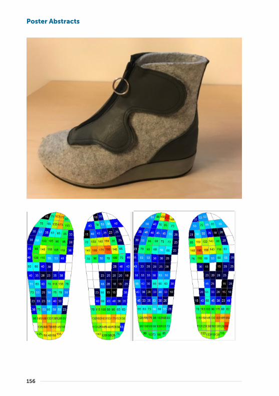
156
Poster Abstracts
b
157
b
[P075] MAXIMIZING COMPLIANCE TO MID-TERM OFFLOADING IN OUTPA-TIENTS WITH RECURRENT DIABETIC FOOT ULCERS: TOLERABILITY AND EFFICA-CY OF ORTHOTIC INSOLES
Giovanni Boschetti1, Marino Bruseghin1, Christine Whisstock1, Mariagrazia Marin1, Michela Pinfi1, Valentina La Rocca1, Deborah Silvestri1, Beatrice Macchi1, Massimo Solidoro1, Sasa Ninkovic1, Enrico Brocco1
1Casa DI Cura Abano Terme; Centro DI Riferimento Per la Diagnosi e la Cura del Piede Diabetico, Abano Terme, Italy
Aim: Both surgical and non-surgical off-loading treatments rely, for success, on strict ad-herence to therapeutic protocols and a span of time of complete off-load of the foot. Low or partial adherence is an avoidable cause of failure and it’s often due to misuse of devices and aids such as post-surgical shoes and offloading insoles. Aim of the ongoing study is to evaluate how adequate training and “tailored” off-loading can improve healing rates through maximizing adherence and proper use of walking aids.
Method: Twenty patients with grade I to III recurrent foot ulcers classified by the University of Texas Diabetic Wound Classification (TUC), had been treated with medications, post-sur-gical shoes and off-loading customizable insoles (FORS) for up to 16 weeks or until healing. All patients either had no indication to complete off-load or couldn’t maintain complete off-load due to personal or professional needs. Lesions where as follows: 4 patients grade 1, 8 patients grade 2, 8 patients grade 3.
Results / Discussion: 69% of patients healed and didn’t show any recurrence at 6 months. 90% of patients showed a significate improvement (at least 1 point in TUC scale) at 16 weeks. 2 patients underwent surgical correction after worsening of the ulcer and therefore achieved complete healing. 2 patients refused both surgical and conservative treatment and dropped out the follow-up.
Conclusion: Off-loading insoles, coupled with post-surgical shoes, can be a well-tolerated and useful addition to the healing process when a surgical approach is not recommended, the patient refuses it or its only partially compliant to total contact casting.
Pro
gra
mm
eA
uth
ors
Ora
l A
bst
ract
sP
oste
r A
bst
ract
sIn
form
atio
nS
pon
sor/
Exh
ibit
or
158
Poster Abstracts
[P076] ATTITUDES AND ATTRIBUTES OF WOMEN AND MEN USING THERAPEU-TIC SHOES
Gustav Jarl1, John Alnemo2, Roy Tranberg3, Lars-Olov Lundqvist4
1University Health Care Research Center and Dept. of Prosthetics and Orthotics, Örebro, Sweden2Sahlgrenska Universitetssjukhus Ortopedteknik, Gothenburg, Sweden3Department of Orthopaedics, Institute of Clinical Sciences, Sahlgrenska Academy, University of Go-thenburg, Mölndal, Sweden4University Health Care Research Center, Faculty of Medicine and Health, Örebro University, Örebro, Sweden
Aim: To compare attitudes and attributes of women and men using therapeutic shoes.
Method: We constructed a questionnaire based on the Health belief model, but also in-cluded questions about adherence to wearing therapeutic shoes, foot complications, gen-eral health, locus of control, self-efficacy, etc. The questionnaire was posted to 1245 peo-ple with diabetes and experience of using therapeutic shoes. Answers of women and men were compared with two-sided chi-square test and Mann–Whitney U test.
Results / Discussion: 469 (38%) questionnaires were analyzed. More men than women (p<0.05) did paid work (22 vs 9%), had someone who reminded them to wear their thera-peutic shoes (27 vs 10%) and had a history of foot ulcers (64 vs 46%) or minor amputation (17 vs 7%). More women than men had disability pension (19 vs 11%). Women reported worse general health, lower internal locus of control regarding prevention of reulcerations and more negative attitudes to some attributes of therapeutic shoes (appearance, price and how it feels using them among other people). Other comparisons were non-signifi-cant (p>0.05): other shoe attributes, adherence, major amputations, satisfaction with shoe services, understanding of neuropathy as a risk factor, belief in shoes’ efficacy, concerns about ulcer healing and reulcerations, self-efficacy, social support, depression and locus of control regarding ulcer healing.
Conclusion: Men had worse foot complications while women had worse general health. Women had more negative attitudes towards therapeutic shoes and lower internal locus of control. Health care needs to pay more attention to the concerns of women using ther-apeutic shoes.
159
[P077] FLEXOR TENOTOMY IN THE TREATMENT OF TOE ULCERS: A FEASIBILITY STUDY
Liesbeth Stokman1, Longueville Lynn1, Eveline Dirinck1, Saskia Van Bouwel2, Christophe Jacobs1, Antoine Roels1, Jeroen Hendriks3, Paul Van Schil3, Christophe De Block1, Somville Johan2, Patrick Lauwers3
1Antwerp University Hospital, Department of Endocrinology, Edegem, Belgium2Antwerp University Hospital, Department of Orthopedic Surgery, Edegem, Belgium3Antwerp University Hospital, Department of Thoracic and Vascular Surgery, Edegem, Belgium
Aims: Toe deformities are common in people with diabetes mellitus, and often lead to ul-ceration with impending risk of osteomyelitis and amputation. Offloading in patients with tip of toe ulcers is possible by using ortheses and adapting shoes and insoles. However, patients can also be surgically offloaded by means of a flexor tenotomy.
The aim of this study is to present preliminary results of percutaneous flexor tenotomy in patients with toe ulcers, secondary to hammer toe formation. A review of current litera-ture is provided as well.
Methods: This is a single-centre prospective cohort study, performed at the diabetic foot clinic of the Antwerp University Hospital (starting Octobre first, 2016; end February 28th, 2018). All patients with a non-rigid hammer toe or claw toe deformity and with an active ulcer on the top of the toe, were offered a percutaneous flexor tenotomy. These proce-dures were done in the outpatient clinic. Local anesthesia was provided on indication. All patients were treated with orthoses as well, to obtain maximal extension of the toe; adapt-ed insoles and shoes were prescribed. A literature review on this topic, using medline, was done.
Results: Twelve patients underwent a percutaneous flexor tenotomy (one patient was treated for two separate diabetic foot ulcers); nine were male, mean age was 74 years (50-84). Nine patients suffered from diabetes (8 DM type 2). All patients had neuropathy, except one. Five patients suffered from peripheral arterial disease, six patients had renal insufficiency (no dialysis). Seven patients had a history of toe ulceration or amputation. Ulcers were localised on the second (n=5), third (n=6) and fourth toe (n=2). Ulcers were present for 10 weeks (1-44). Osteomyelitis was present in 3 cases. Ten ulcers healed after 4 weeks of treatment (1-11); one toe (with active osteomyelitis) was amputated. After a follow up of 26 weeks (1-68), no recurrences occurred.
A literature review revealed only 14 reports on the technique of percutaneous flexor tenot-omy. This procedure is related with over 90% healing rates, short duration to ulcer healing (< 30 days on average), and low ulcer recurrence rates. Complication rates are minimal.
Conclusion: Both in the (scarce) literature as in this preliminary feasibility study, percuta-neous flexor tenotomy is an easy, safe procedure with good results on wound healing. We believe that this technique should be considered in patients with toe ulcers as adjunctive method to other means of offloading.
Pro
gra
mm
eA
uth
ors
Ora
l A
bst
ract
sP
oste
r A
bst
ract
sIn
form
atio
nS
pon
sor/
Exh
ibit
or
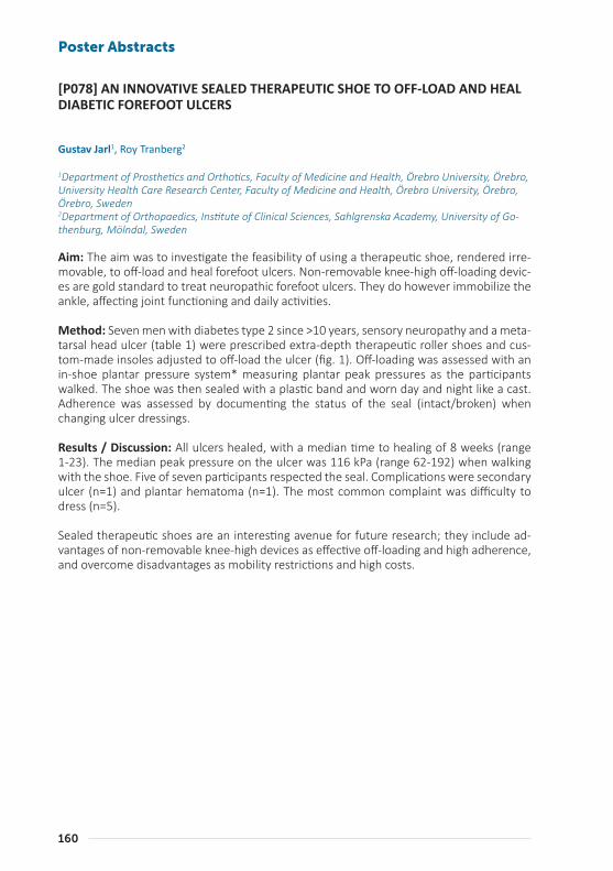
160
Poster Abstracts
[P078] AN INNOVATIVE SEALED THERAPEUTIC SHOE TO OFF-LOAD AND HEAL DIABETIC FOREFOOT ULCERS
Gustav Jarl1, Roy Tranberg2
1Department of Prosthetics and Orthotics, Faculty of Medicine and Health, Örebro University, Örebro, University Health Care Research Center, Faculty of Medicine and Health, Örebro University, Örebro, Örebro, Sweden2Department of Orthopaedics, Institute of Clinical Sciences, Sahlgrenska Academy, University of Go-thenburg, Mölndal, Sweden
Aim: The aim was to investigate the feasibility of using a therapeutic shoe, rendered irre-movable, to off-load and heal forefoot ulcers. Non-removable knee-high off-loading devic-es are gold standard to treat neuropathic forefoot ulcers. They do however immobilize the ankle, affecting joint functioning and daily activities.
Method: Seven men with diabetes type 2 since >10 years, sensory neuropathy and a meta-tarsal head ulcer (table 1) were prescribed extra-depth therapeutic roller shoes and cus-tom-made insoles adjusted to off-load the ulcer (fig. 1). Off-loading was assessed with an in-shoe plantar pressure system* measuring plantar peak pressures as the participants walked. The shoe was then sealed with a plastic band and worn day and night like a cast. Adherence was assessed by documenting the status of the seal (intact/broken) when changing ulcer dressings.
Results / Discussion: All ulcers healed, with a median time to healing of 8 weeks (range 1-23). The median peak pressure on the ulcer was 116 kPa (range 62-192) when walking with the shoe. Five of seven participants respected the seal. Complications were secondary ulcer (n=1) and plantar hematoma (n=1). The most common complaint was difficulty to dress (n=5).
Sealed therapeutic shoes are an interesting avenue for future research; they include ad-vantages of non-removable knee-high devices as effective off-loading and high adherence, and overcome disadvantages as mobility restrictions and high costs.
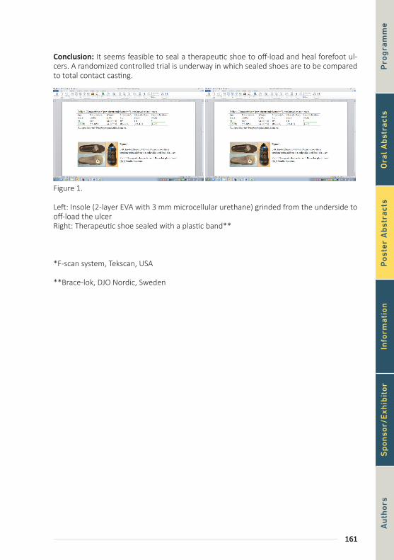
161
Conclusion: It seems feasible to seal a therapeutic shoe to off-load and heal forefoot ul-cers. A randomized controlled trial is underway in which sealed shoes are to be compared to total contact casting.
Figure 1.
Left: Insole (2-layer EVA with 3 mm microcellular urethane) grinded from the underside to off-load the ulcerRight: Therapeutic shoe sealed with a plastic band**
*F-scan system, Tekscan, USA
**Brace-lok, DJO Nordic, Sweden
Pro
gra
mm
eA
uth
ors
Ora
l A
bst
ract
sP
oste
r A
bst
ract
sIn
form
atio
nS
pon
sor/
Exh
ibit
or
162
Poster Abstracts
[P079] REAL LIFE EXPERIENCE OF ORTHOPEDIC BOOTS IN THE MANAGEMENT OF DIABETIC FOOT ULCERS
Satyan Rajbhandari1, Wee Teck Lim2, Helen Robinson3
1Lancashire Teaching Hospital, Chorley, United Kingdom2Manchester Medical School, United Kingdom3Lancashire Teaching Hospitals NHS Trust, Chorley, United Kingdom
Aim: Offloading with Total contact cast (TCC) is the gold standard treatment for diabetic foot ulcers (DFU) and is used as standard off-loading method in our clinic. However, its use can be limited due to underlying conditions such as infection or ischaemia or patients’ re-luctance. We use an orthopedic boot* as a removable walker in such cases. The orthopedic boot* has an outer lightweight shell and the inner lining. The inner lining is the cushion filled with thousands of styrofoam pearls that surrounds the foot. When air is extracted from the cushion with a pump, the body shaped cushion becomes hard as a cast just in a few seconds. This avoids pressure to the DFU and provides stability due to a perfectly shaped orthosis. This aim of this study was to analyse the results of orthopedic boots* in the healing of DFU in real life scenario.
Method: In this retrospective study we analysed records of all subjects supplied with the orthopedic boot* from 2011 to 2017 for the treatment of DFU. The outcome of their ulcers was noted from clinical records. If patients stopped using the orthopedic boot* for any reason their outcome was grouped with those stopped using it. If they changed from other off-loading device to the boot* it was analysed as the outcome of this device.
Results / Discussion: The orthopedic boot* were supplied to 42 patients (35 males) to treat 83 episodes of DFU during this period. The mean age was 56.7 (+/- 11.2) years and duration of diabetes was 18.9 (+/- 13.5) years. Of the 83 episodes of ulcers 41 (49.4%) healed in the median duration of 17 weeks with the use of the orthopedic boot*. 13 (15.6%) are still continuing treatment and 29 (34.9%) stopped using it. The reason for stop-ping its use was the change to other device (n=12), infection (n=3), amputation (n=3), lost to FU (n=7) and other reason (n=4). Out of 83 DFU episodes, 8 episodes were those who changed from TCC to the orthopaedic boot. Similarly, in 10 ulcer episodes, the orthopedic boot* was changed to TCC.
Conclusion: The healing time with the orthopedic boot* was longer than TCC but these included patients with underlying infection and ischaemia, in whom TCC is relatively con-traindicated. Our data shows that the orthopedic boot* is preferred by patients, can be reused when patient has re-ulceration and is as effective as other removable cast walkers.
*VACOped
163
[P080] THE EFFECT OF PERCUTANEOUS FLEXOR TENOTOMY ON HEALING AND PREVENTION OF FOOT ULCERS IN PATIENT WITH CLAW TOE DEFORMITY
Luuk Smeets1, Peter Schmitz1, Ruben Scheffer1, Sophie De Gier2, Daphne Van der Veen-Hobe1, Ruby Krol2
1Rijnstate Hospital, Arnhem, Netherlands2Slingeland Hospital, Doetinchem, Netherlands
Aim: The aim of this study is to investigate if percutaneous flexor tenotomy is an effective surgical method for treatment and prevention of toe ulcers in patients with claw deformity with and without diabetes mellitus.
Method: Percutaneous flexor tenotomy is a simple surgical procedure which can be per-formed under local anesthesia in an out-patient setting. This retrospective study, with a median follow-up of 13.4 (1-45) months, included all consecutive patients who underwent percutaneous flexor tenotomy in two hospitals between May 2014 and April 2017. The procedure was performed in all toes with ulceration and also in toes that were at risk for ul-ceration. Patients who underwent at least one flexor tenotomy on a toe with an ulcer were included in the therapeutic group. Patients who underwent only prophylactic treatments were classified as prophylactic.
Results / Discussion: A total of 256 flexor tenotomies were performed in 75 patients ( mean age 71.4 years, 47 men) on 101 feet; 84 therapeutic and 17 prophylactic. Diabetes mellitus was present in 77% of all patients for a mean period of 17.8 years. In the thera-peutic group 98 (95.1%) of a total of 103 ulcers healed with a median time of 27 days. In this group 11 (13.3%) re-ulcerations, 4 (4.8%) infections were recorded and 1 amputation of the digit was needed eventually. In the prophylactic group 1 local bleeding (6%) and two (12%) ulcers were recorded.
Conclusion: Percutaneous tenotomy of the flexor digitorum longus is a highly effective and safe minimally invasive procedure for the treatment and for the prevention of ulcers and therefore should be integrated and considered in the daily standard care for each diabetic foot patient.
Pro
gra
mm
eA
uth
ors
Ora
l A
bst
ract
sP
oste
r A
bst
ract
sIn
form
atio
nS
pon
sor/
Exh
ibit
or
164
Poster Abstracts
[P081] ADAPTING THE STANDARDLY AVAILABLE SCOTCH CAST BOOT TO ALIGN OUR OWN GOALS WITH OUR PATIENTS’ NEEDS IN RELATION TO OUTCOMES IN DIABETIC FOOT ULCERATION
Mary Gray1, Michael Pierides2
1Diabetic and High Risk Foot Service, Willow Brook Health Centre, Corby, United Kingdom2Kettering General Hospital, Kettering, United Kingdom
Aim: To design and make an alternative offloading device to the current casted boot pro-vided by the Northamptonshire Diabetic Foot Team that will be as effective in offloading Diabetic Foot ulcers (DFU’s) but address non concordance and expedite earlier discharge from hospital. It should also address patients in the community who need immediate off loading or are not suitable to the traditional casted boot used locally.
Method: Our diabetic foot team currently makes a casted boot that requires a plaster saw that creates noise and dust that is not appropriate for a ward setting. Furthermore in addition the boot is sent away to a local laboratory for an outer sole to be added that can further delay provision to the patient by up to two weeks on average. After several design trials and discussions/advice with colleagues from Leicester the team developed a Total contact cast boot which is removable by other healthcare workers to allow a foot inspec-tion. Consequently, the design combines both a hard and soft cast to allow the shoe to be made quickly and without the use of a plaster saw making it ideal for the ward setting with minimal disruption.
Results / Discussion: The results have been positive, as we are able to make the new boot in a ward setting with minimal disruption and significant less time as well as no processing time in the lab. Patients are provided with shoe immediately aiding quicker discharge and offloading.
Conclusion: The new boot is a further offloading device to add to the current arsenal of offloading footwear with its own niche as described above but we would not expect it to replace the standard practice of casted offloading boots. We still need to make our casted boot for better offloading where an aperture is required or biomechanical additions. A more formative scientific review is required to validate its use.
165
[P082] EXAMINATION OF DIABETIC FOOT BY PODIATRISTS IN PRIMARY SECTOR
Alima Ashraf1
1Odense University Hospital, Odense C, Denmark
Background and aim: Since 1982 podiatry for diabetic patients has been entitled to a subsidy of 60 % after prescription by a general practitioner (GP) or a hospital1. But in 2011 did podiatrists in primary sector have a new agreement for diabetic patients and diabetic patients only entitled to a subsidy of 50%. The agreement was decided between Danish Podiatrists Association (DPA) and Danish Health Authority. The aim of this study was how podiatrists in primary sector examined diabetic foot.
Method: 1050 podiatrists have identification code1 in Denmark (DK) and they all have too exa- mination the patients feet once a year. All podiatrists in DK use the same foot examination form.
Examination of diabetic patients’ feet takes about 30 min. Examination process is: Neuropathy – tests with monofilament and biosthesiometer Ischaemia – palpating the pulses, (ADP and ATP) Oedema Foot deformity Check the change in the skin and nails Ulcers Ask about pain in hips, knees, ankles and feet Ask about eye vision and kidney disease
And podiatrist in primary sector (PS) use a lot of time to instructions patient in self-care about the diabetic foot, regularly go to podiatrist for check the feet and remove the callus, makes insoles because foot deformity or pain in lower extremity.
1) After screening the patients assign in a risk group. There are four risk groups. 2) Normal sensibility, no foot deformity, good blood supply.3) Neuropathy, good blood supply, foot deformity, callus, nail diseases, eye vision,4) Neuropathy, reduced blood supply, previous foot ulcer, inactive charcot foot, kidney disease
Diabetic foot ulcer, active charcot foot, amputation, ischemia.Podiatrist will send the result of examination electronic to GP.
Result & Discussion: Podiatrist in primary sector examination 96420 patients in 2016 and 8295 patients were in risk group 41. Most diabetic patients can remain free of ulceration by prophylactic treatment in primary sector.
The conclusion is that all podiatrists in the primary sector make a singularly foot examina-tion and that regularly examination of diabetic feet reduce the risk of foot ulcers and halves the risk of amputations.
1) Danish Podiatrists Association
Pro
gra
mm
eA
uth
ors
Ora
l A
bst
ract
sP
oste
r A
bst
ract
sIn
form
atio
nS
pon
sor/
Exh
ibit
or
166
Poster Abstracts
[P084] TELEMEDICINE AND HOME-MONITORING APPLICATIONS FOR THE DIA-BETIC FOOT: A SYSTEMATIC REVIEW
Wouter aan de Stegge1, Constantijn Hazenberg2, Sicco Bus1, J. G. van Baal3
1Academical Medical Center, Department of Rehabilitation, University of Amsterdam, Amsterdam, Netherlands2Department of Vascular Surgery, University Medical Center Utrecht, the Netherlands3Zgt , Almelo the Netherlands, 1department of Surgery, Ziekenhuis Groep Twente, Almelo, the Nether-lands. , Almelo, Netherlands
Aim: To assess the medical-scientific literature on the feasibility, effectiveness, value, and limitations of using telemedicine and home-monitoring approaches for the prevention and management of diabetic foot disease.
Method: This is a systematic review. A biomedical database* was searched on the 1st of February 2018 for original research articles on telemedicine and home-monitoring ap-proaches used for the assessment, prevention, or treatment of foot disease in patients with diabetes mellitus. All study designs were included and the search was limited to En-glish language papers. Studies with patients with diabetes mellitus and signs of foot dis-ease were included. The included interventions/modalities were dermal thermography, hyperspectral imaging, digital photographic imaging, and video/audio communication tools. Primary and secondary outcome measures were: validity, reproducibility, feasibility, usability, and (cost-) effectiveness.
Results / Discussion: The search yielded 1258 articles of which 57 were found eligi-ble for analysis. Most studies (20) were on the use of dermal infrared thermography as home-monitoring tool; four randomized controlled trials showed this method to effectively diagnose foot temperature changes and contribute to a significantly reduced incidence of foot ulcers compared to usual care. Hyperspectral imaging may contribute to predicting ul-cer healing and ulcer development, but not yet as telemedicine solution. Photographic im-aging can accurately and reliably assess foot ulcers, ulcer area, callus, and absence of foot disease, and is feasible as modality in the patient’s home, however photographic imaging cannot be used as a stand-alone diagnostic instrument. A recent randomized controlled trial showed that an interactive Web-based ulcer record and a mobile phone, enabling counselling and communication between the community nurses and specialist heath care can be an alternative and supplement for usual care in ulcer treatment. Video-assisted treatment of diabetic foot ulcers in the patient’s home showed to be a feasible, and as effective as usual care in ulcer healing. The costs were found to be €2039 less per patient compared to standard monitoring, however, this difference was not statistically significant.
Conclusion: This systematic review shows that telemedicine and home monitoring for the diabetic foot is still in its infancy, but several promising technologies are available that may be of additional value in the assessment, prevention, or treatment of foot disease. The cost-effectiveness and applicability of dermal thermography and the effectiveness, or, in some cases, feasibility, of other modalities should be proven for them to become accepted and used in remote care of the diabetic foot.
*MEDLINE
167
[P085] A SYSTEMATIC REVIEW TO DETERMINE THE EFFECTIVENESS OF MOTI-VATIONAL INTERVIEWING AS AN INTERVENTION TO IMPROVE ADHERENCE BEHAVIOURS FOR THE PREVENTION OF DIABETIC FOOT ULCERATION
Jodi Binning1, Ruth Barn1, Jim Woodburn1, Sicco Bus2
1Glasgow Caledonian University, Glasgow, United Kingdom2Department of Rehabilitation, Academic Medical Center, Amsterdam, Netherlands
Aim: Preventative strategies for diabetic foot ulceration are effective when patients adhere to advice. Therefore interventions aimed at improving adherence are required. A system-atic review was conducted to determine the effectiveness of Motivational Interviewing as an intervention to improve adherence behaviours for the prevention of diabetic foot ulceration.
Method: Electronic searches were run without date or language restrictions across 13 Medical, Health, Psychology and Research databases. Studies were selected if they fulfilled the inclusion criteria: Population: age 18+ with type I or II diabetes at risk of ulceration. Interventions: Motivational Interviewing as the main intervention or as a component. Comparators: all types of control groups were accepted. Outcomes: A new episode of ulceration and/or at least one behavioural outcome measure. RCTs and quasi-experimental prospective studies were accepted. Two review authors independently assessed eligibility using a software tool*. Complete agreement was achieved on 45 of 47 studies. Agreement by discussion was easily reached for the 2 remaining studies. Data on foot risk, duration of diabetes and demographic profile was extracted. Study design, number of participants, intervention description, intervention setting, mode of delivery, outcome measures and time points were recorded. An analysis on intervention content was conducted using the Behavioural Change Taxonomy , (Michie et al. 2013).
Results /Discussion: Five studies met the inclusion criteria and all were assessed as having a high risk of bias. Studies differed in aims, mode and duration of intervention delivery, and measures and outcomes. This prevented the pooling of data to determine overall effec-tiveness of motivational strategies on adherence. Four of five studies used motivational / behavioural techniques as a part of a suite of interventions. These four studies used tech-niques based on goals and planning, social support and identifying consequences of the targeted behaviour. Two of these studies claimed the intervention was effective; however bias and population characteristics indicate that these results are not generalizable. One study used motivational interviewing as the main intervention and found improved short term adherence (from 49% to 84%). This effect returned to baseline after 3 months. This study was exploratory with ten participants. No studies adequately adopted strategies for the least motivated subjects whose barriers to adherence were belief based.
Conclusion: There is insufficient evidence to determine whether motivational interviewing is effective at improving adherence behaviours for the prevention of diabetic foot ulcer-ation. More research is needed to explore relationships between motivation, behaviours, adherence and outcomes for this population.
*Covidence ©
Pro
gra
mm
eA
uth
ors
Ora
l A
bst
ract
sP
oste
r A
bst
ract
sIn
form
atio
nS
pon
sor/
Exh
ibit
or
168
Poster Abstracts
[P086] PEOPLE WITH DIABETES ARE INTERESTED IN EDUCATION ON FOOT HEALTH SELF-MANAGEMENT
Jarmila Jirkovska1, Tamara Hrachovinova2, Alexandra Jirkovská2, Jelena Skibová2, Jozefina Stefan-kova3, Jan Broz4, Alena Adamikova5, Jiri Hradec6, Martin Prazny7, Katerina Cechova2, Vladimira Hav-lova2, Renata Rihankova8
1Medical Department of the First Faculty of Medicine and Military University Hospital, Charles Univer-sity, Prague, Czech Republic2Diabetes Centre, Institute for Clinical and Experimental Medicine, Prague, Czech Republic3Diabetologie - Hk, S.R.O., Hradec Kralove, Czech Republic4Motol University Hospital, Prague, Czech Republic5Hospital of T.Bata, Zlin, Czech Republic6Ide Cr, Chrudim, Czech Republic73rd Department of Internal Medicine, 1st Faculty of Medicine, Charles University, Prague, Czech Republic8University Hospital Plzen, Plzen, Czech Republic
Aim: Foot ulcers are among major complications of diabetes, with high morbidity, mortal-ity, and costs. Prevention is of critical importance to reduce the burden. Various interven-tions are used including patient self-management education. Aim was to implement struc-tured group education programme in diabetes education centers and determine interest of diabetics in foot care education.
Method: Programme was implemented in 9 centers in 2016. Overall 62 Type 2 diabetics were included (63.3±8.2yrs, 53% of men, insulin once-daily at least). Schedule included 4x2hrs of structured education lessons realized within the interval of 14 days for a group of 6-12 individuals. Before initiation patients were inquired about their interest in particular education topics including foot care. Metabolic parameters were measured before as well as within 6 months after the project.
Results / Discussion: Most preferred education topics were foot care (71%) and diet (71%) followed by weight reduction (58%), eye (56%), cardiovascular complications (55%), in-sulinotherapy (48%), nephropathy (48%), exercise (43%), selfmonitoring (31%). HbA1c declined from 70.0±16.2mmol/mol initially to 63.1±12.6 in 6 months (p=0.001). Fasting plasma glucose declined from 8.8±2.8mmol/l initially to 7.7±2.1 in 6 months (p=0.0002). Proportion of individuals with comparatively well diabetes control (HbA1c<60mmol/mol) improved significantly from 26% to 45% in 6 months (p<0.01).
Conclusion: Project demonstrated real implementation into education centers with signif-icant improvement of diabetes control. Self-management foot interventions enable early detection of pre-ulcerative signs. We believe that patients who are interested in preventa-tive behaviour and adherent to the advice given in the education programme are at lower risk of developing a foot ulcer. Project was supported by National Diabetes Society.
169
[P087] DIABETIC FOOT SELF CARE KNOWLEDGE AND PRACTICE IN WOMEN WITH DIABETES
Mohammad Reza Amini1, Mahnaz Sanjari1, Maryam Aalaa2, Neda Mehrdad2, Hossein Adibi2, Mosta-fa Qorbani2, Hamidreza Aghaei Meybodi2
1Diabetes Research Center, Endocrinology and Metabolism Clinical Sciences Institute, Tehran Universi-ty of Medical Sciences, Tehran, Iran2Endocrinology & Metabolism Research Institute, Tehran University of Medical Sciences, Tehran, Iran
Aim: A large number of patients with diabetes mellitus are unaware of foot care principles and are at risk of developing foot ulcer and amputation. The author aimed to examine the knowledge and practice of Diabetic Foot Self-care in women with T2DM.
Method: This is the cross-sectional, multi-centric study, which included 457 women with type 2 diabetes were recruited using random multistage cluster sampling. The data was completed by demographic questionnaire and Diabetic Foot Self Care Questionnaire (DFSQ). This study is investigating demographic and lifestyle factors (age, gender, marital, educational and social economic status), general health status (HbA1c level, FBS, Lipid pro-file, WHR, BMI, BP, life satisfaction, self-rated health) and Diabetic Foot Self Care Question-naire (Personal Self-care, Podiatric Care and Footwear and Socks). The descriptive statistic and Pearson regression were used to assess the Diabetic Foot Self Care status.
Results / Discussion: The mean age of participants was 50 (SD: 7.7), range 28-70 year. The mean total Diabetic Foot Self Care was 60.38 (SD: 9.9). Also, the mean and standard devia-tion of Personal Self-care, Podiatric Care and Footwear and Socks were (M:24.87, SD:7.00), (M:18.02, SD: 2.11), (M: 17.52, SD: 3.95) respectively. The results showed that the 17% of the women with diabetes had poor, 49% median and 34% had acceptable diabetic foot self-care status. The Pearson regression analysis revealed that there was no any correlation between diabetes control, anthropometric and cardiovascular indices, but the BMI level had reverse correlation with Footwear and Socks subcategory of diabetic foot self-care (r: 0.1, P: -0.12). It means that the patients with higher BMI had lower score in Footwear and Socks status.
Conclusion: The results of this study suggest that need to establish and development the diabetic foot care services in outpatient clinics to early detection of foot at risk and provide enhanced patient outcomes and forbid the lower limb amputation.
Pro
gra
mm
eA
uth
ors
Ora
l A
bst
ract
sP
oste
r A
bst
ract
sIn
form
atio
nS
pon
sor/
Exh
ibit
or
170
Poster Abstracts
[P088] PHYSICAL ACTIVITY AND ITS RELATIONSHIP TO THE PSYCHOLOGICAL STATUS IN PATIENTS WITH THE DIABETIC FOOT
Eliška Vrátná1, Andrea Němcová2, Robert Bém2, Michal Dubský2, Anna Pyšná2, Hana Tibenská2, Alexandra Jirkovská2, Veronika Wosková2, Vladimíra Fejfarová2
1Faculty of Physical Education and Sport, Prague, Czech Republic2Institute for Clinical and Experimental Medicine, Prague, Czech Republic
Aim: Previous studies have shown that patients with the diabetic foot (DF) suffered more frequently from depression or poor quality of life (QoL) due to predominantly late diabetes complications and long-lasting immobilisation. This could eventually lead to a decrease in physical activity and subsequently to poor diabetes control and further progression of psychological alterations.
The Aim of our study was to assess physical activity in patients with DF and its correlation with QoL and depression.
Method: Included in our study were 36 patients with DF (mean age 58±12.1 years, dia-betes duration 17.25±12.7 years) treated and followed in our out-patient foot clinic. Pa-tients were divided into 3 groups based on the type of footwear/off-loading – 15 patients using preventive/individual orthopaedic footwear (group 1), 13 patients using off-loading devices (group 2) and 8 patients using wheelchairs (group 3). The study groups did not differ significantly in their basic characteristics including HbA1c and TcPO2 values. All sub-jects met validated tests focusing on physical activities (IPAQ survey), level of depression (GDS - Geriatric Depression Scale), and QoL (WHOQOL-BREF survey detected quality of life through 4 domains – physical, psychological health, social relationship and environment domains). Physical activities, psychological parameters and their possible correlations were compared among the study groups.
Results / Discussion: No significant differences were found in terms of severe and moder-ate physical activities and their duration among the 3 study groups. There was only a trend toward a higher activity volume in group 1. This group also showed higher daily activi-ties including walking; duration of walking was significantly longer in group 1 compared to groups 2 and 3 (82.7±74.4 min/day vs. 64.2±61.8 min/day vs. 18.8±39.5 min/day; p=0.03). Nearly two thirds of patients (66.7%) reported a mild form of depression regardless the group. The study groups differed significantly in two QoL domains – physical and psycho-logical health – the lowest QoL was detected in group 3 using wheelchairs (p=0.025 and p=0.05). There was a positive correlation between physical health and walking (p=0.01 a p=0.001) and a negative correlation between BMI and psychological health (p=0.01).
Conclusion: This study showed insufficient daily physical activities in all patients with DF suffering frequently from a mild form of depression. Therefore, exercise training programs for DF patients that could potentially decrease the depression rate and improve QoL in particular need to be introduced into daily podiatric practice.
Supported by project GAUK 546417, SVV260466.

171
[P089] THE RISK OF DEEP VEIN THROMBOSIS IN PATIENTS SEEN IN OUR MULTI-DISCIPLINARY DIABETIC FOOT CLINIC
Shailesh Gohil1, Rachel Berrington1, Rajesh Jogia1, Marie France Kong1
1Department of Diabetes, University Hospitals of Leicester NHS Trust, Leicester, United Kingdom
Aim: The patients we see in the diabetic foot clinic are in a pro-inflammatory state, with ulcers that may or may not be infected, Charcot neuro-osteoarthropathy and fractures. In addition, we immobilise patients, advising them to offload their feet and often place patients in a cast. This would, in theory, place our patients at risk of developing a deep vein thrombosis (DVT). Our patients often have unilateral leg oedema and this, alongside cast immbolisation, would automatically place our patients at moderate risk of having a DVT according to the Well’s Score. Our aim was to evaluate how many patients were assessed for a DVT with ultrasound compression venography whilst attending the multi-disciplinary diabetic foot clinic and how many new deep vein thromboses were detected.
Method: We searched our electronic database for all patients who were assessed for a DVT whilst attending the multi-disciplinary diabetic foot clinic between 1st January 2016 and 1st January 2018. Patients without diabetes and those who were assessed before referral to the diabetic foot clinic were excluded. Data were collected on the type of foot problem the patient had, whether there was infection present, the method of assessment (either at the nurse led DVT clinic, ambulatory clinic or inpatient) and if a DVT was confirmed.
Results / Discussion: Over a 2 year period, 39 patients were assessed to see if they had a DVT. One patient was assessed twice however one patient did not attend for their ul-trasound scan. 32 scans occurred at the DVT clinic, 3 via another ambulatory clinic, 3 as an inpatient and 1 via the Emergency Department. Of the 39 scans, DVT was detected in 4 patients (10.3%) and thrombophlebitis in 2 (5.1%). The 4 patients who had a DVT had additional risk factors such as previous DVTs, further immobility due to co-morbidities and recent inpatient stay.
Conclusion: Although patients with diabetic foot problems seem to be at high risk of devel-oping DVT, our data suggests this is not the case. Of all the patients seen at our University Hospital multi-disciplinary foot clinic, only 39 had an ultrasound compression venography to assess for DVT and of these, only 4 patients had a DVT, with a further 2 having throm-bophlebitis. Those that did have a DVT had significant additional risk factors which would have been picked up if the Well’s Score was utilised.
Pro
gra
mm
eA
uth
ors
Ora
l A
bst
ract
sP
oste
r A
bst
ract
sIn
form
atio
nS
pon
sor/
Exh
ibit
or
172
Poster Abstracts
[P090] THE OCCURENCE OF OBSTRUCTIVE SLEEP APNEA SYNDROME IN PA-TIENTS WITH DIABETIC FOOT AND IT´S POSSIBLE ASSOCIATION WITH LIMB ISCHEMIA AND WOUND HEALING
Vladimira Fejfarova1, Marta Klementova1, Jan Polak2, Robert Bem1, Michal Dubský1, Alexandra Jirkovska3, Veronika Wosková1, Andrea Němcová1, Anna Pysna1, Eliška Vrátná4, Marta Křížová1, Věra Lánská1
1Diabetes Centre, Institute for Clinical and Experimental Medicine, Prague, Czech Republic2Charles University in Prague, II.Internal Department, Fnkv, Prague, Czech Republic3Institute for Clinical and Experimental Medicine Prague, ., Switzerland4Faculty of Physical Education and Sport, Prague, Czech Republic
Aim: The aim of our study was to assess in patients with DF the occurrence of OSAS and its association with peripheral arterial disease (PAD) and impact on DF healing.
Methods: We included into our study 38 patients with DF whose were treated in our foot clinic, completed screening questionnaires and underwent apnea screening test*(Apnea Hypopnea Index-AHI –patients with OSAS have AHI≥5, severe OSAS-AHI≥30).Patients were divided according to AHI into 2 groups-group A(OSAS positive) and group B(OSAS negative).During the follow-up period (6-9 and 12-15 months) macrovascular and microcirculation status (TcPO2) and DF healing (% of healed patients, ulcer sizes, amputations)were as-sessed.
Results: OSAS was detected in 79% of studied patients(30/38–group A),of whom 30% had the severe form of OSAS(9/30).Trends to higher incidence of PAD(50%vs.25%;p = 0.18)and to lower values of TcPO2 (38.7±12.9 vs. 50.8±10.8 mm Hg; p=0.14)with higher frequency of patients with TcPO2 bellow 40 mm Hg (50%vs.12.5%;p=0.096)were found in group A compared to group B.Patients from group A had significantly larger defects(p=0.03) as at the begging of this study, as after 6-9 (p=0.002)and 12-15 months(p=0.001).The presence of OSAS had no impact on the DF. The sub analysis showed that patients with severe OSAS were significantly older(p=0.03),had more sleep abnormalities(p=0.01),larger DF ulcers at the end of the study (p=0.08)and higher occurrence of new DF ulcers during the follow-up period (p=0.09).
Conclusion: The incidence of OSAS is probably higher in patients with DF compared to general diabetic population. Patients with OSAS had higher occurrence of PAD and lower TcPO2 values. OSAS has probably no clinical impact on the DF prognosis but could nega-tively influence DF characteristics.
VZ 00023001
* ApneaLink
173
[P091] DIABETIC FOOT AND CUTANEOUS T-CELL LYMPHOMA: A CLINICAL CASE
Liliana Fonseca1, Álvaro Machado2, Teresa Pereira1, Ana Lopes3, Lia Ferreira1, Ana Amado1, André Carvalho1, Madalena Sanches2, Ana Martins1, Sofia Teixeira1, Cláudia Amaral1, Helena Rei Neto1, Cláudia Freitas1, Joana Martins1, Luís Loureiro1, Luís Costa1, André Gomes1, Isabel Gonçalves1, Sara Pinto1, Rosa Guimarães1, Joel Pereira1, Rui Carvalho1
1Diabetic Foot Unit - Hospital de Santo António - Oporto, Porto, Portugal2Department of Dermatology, Centro Hospitalar Do Porto3Diabetic Foot Unit - Centro Hospitalar Do Porto, Porto, Portugal
Introduction: The clinical presentation of cutaneous T-cell lymphoma varies from erythem-atous plaques, eczema, hypochromia, erythroderma, chronic ulcers and swelling, and is often confused with inflammatory dermatoses.
Clinical case: A 66-year-old woman, with type 2 diabetes mellitus for 20 years and poor chronic glycemic control (HbA1c 12.4%) and established advanced micro and macrovas-cular complications presented to our Diabetic Foot Clinic with an infected neuroischemic left foot ulcer. After distal limb revascularization and minor amputation of the left foot, her recovery was unremarkable except for the presence of a persistent erythema and des-quamation of the dorsum of the right foot. Firstly assumed as a probable tinea pedis with eczema she was then treated with topical betamethasone and ketoconazole. Due to per-sistence and worsening of the lesions in the following weeks, namely, increased number of erythematous plaques, with more infiltrated and exulcerated areas with well-defined limits (some of them with a diameter of 5 cm), a new evaluation with a skin biopsy of the lesion was performed. The histology result was compatible with the diagnosis of a cutaneous angioimmunoblastic t-cell lymphoma.
Conclusion: Chronic skin lesions are common when evaluating diabetic patients. Clinicians should be aware for less common presentations and any abnormal evolution of skin lesions in these patients. This case study reinforces the need for a multidisciplinary approach in Diabetic Foot problems.
Pro
gra
mm
eA
uth
ors
Ora
l A
bst
ract
sP
oste
r A
bst
ract
sIn
form
atio
nS
pon
sor/
Exh
ibit
or
174
Poster Abstracts
[P092] DIABETIC NEUROPATHY IS A RISK FACTOR OF CHRONIC VENOUS INSUF-FICIENCY
Pavlina Pithova1, Lucie Hoskovcova1, Jana Petrova1, Milan Kvapil1
1Teaching Hospital Motol,, Charles University, Prague, Czech Republic
Aim: Chronic venous insufficiency causes edema and impaired function of microcirculation that could affect the ulcer healing in patients with diabetic foot. The prevalence of skin changes (class C3 and C4 according to the CEAP classification) in patients with chronic ve-nous insufficiency is about 3-11% in general population. Diabetic neuropathy and changed biomechanics of walking could cause impairment of the calf muscle pump and cause func-tional venous insufficiency. We tried to find the prevalence of venous insufficiency in dia-betic foot patients.
Method: We provided the physical examination of calf area in 100 consecutive type 2 diabetic patients with ulcerations (plantar location), confirmed severe diabetic neuropathy (biothesiometer, monofilaments) and excluded severe ischemia (ABI > 0.9 and TBI > 0.5). Patients with skin changes (class C3 and C4 according to the CEAP classification) under-went the duplex ultrasound of venous system at the affected extremity.
Results / Discussion: We found skin changes C3 -C4 in 62 of 100 patients (= 62%) with diabetic foot (the mean age 64.3 ± 12.1 years, mean duration of diabetes was 14.2 ± 4.8 years). In these patients following changes in venous system were found: 3 patients had deep vein thrombosis in personal history and we found post-thrombotic changes in venous system, 1 patient had post-thrombotic changes without deep venous thrombosis in history, 2 patients were after varicectomy in past, 14 patients had reflux in sapheno-femoral or sapheno-popliteal junction, 7 patients suffered from varicosities in superficial venous sys-tem, 8 patients had dilated and insufficient perforating veins; together 56.5% of patients were found with “classical” causes of chronic venous insufficiency but 43.5% of patients had completely normal result of venous duplex ultrasound. The reason for venous insuf-ficiency with skin changes could be impaired function of the calf muscle pump in diabetic neuropathy (and partly also bad involvement of calf muscles during long-term walking with offloading devices).
Conclusion: The prevalence of skin changes C3-C4 of CEAP classification was higher in dia-betic foot patients than in general population (literary data). Nearly ½ of those patients did-n´t exhibit any changes of venous system at the affected extremity – the venous insufficien-cy seems to be functional and caused by diabetic neuropathy and impaired biomechanics.
175
[P093] INDEPENDENT CORRELATIONS BETWEEN THE PRESENCE OF RETINOPA-THY AND KIDNEY DISEASE IN DIABETES AND MEASURES OF BOTH METABOLIC CONTROL AND NEUROPATHY
Dragan Tesic1, Milena Mitrovic1, Jelena Ljikar2, Djordje Popovic 1, Dragica Tesic3, Mirjana Tomic1, Bojan Vukovic1
1Clinical Centre of Vojvodina, Clinic of Endocrinology, Diabetes and Metabolic Disorders , Novi Sad, Serbia2Clinical Centre of Vojvodina, Clinic of Ophthalmology, Novi Sad, Serbia3Institut for Cardiovascular Diseases, Clinic of Cardiology, Sremska Kamenica, Serbia
Aim: The aim of this study was to examine associations between measures of neuropathy, nephropathy and lower-extremity artery disease (LEAD) in people with diabetes compli-cated by nonproliferative retinopathy (NPDR), maculopathy and proliferative retinopathy (PDR).
Method: People were included who attended a specialist service for review between September 2012 and September 2017. Clinical examination of the eyes was performed through dilated pupils using a slit lamp. VPT was measured using a semi quantitative tuning fork C128 and ankle reflexes (AR) were recorded. Sudomotor function was determined as the time until total colour change using a screening test*. LEAD was defined with ABPI >1.4 or <0.8 or Continuous wave Doppler monophasic curve.
Results / Discussion: Of 469 people, 46.1% were male and 68.4% had T2DM. 30 had PDR, 98 had maculopathy, 89 had NPDR and 252 had no evidence of retinopathy. Compared with people without retinopathy, those with retinopathy were older (58±12.5 vs. 52.3±15.1years), with lower VPT (5.1±2.8 vs. 6.6±2), more often with missing ankle reflexes (2.9±1.3 vs. 2.0±1.6), higher prevalence of LEAD (18.4% vs. 7.9%) and arterial hypertension (52.9% vs. 36.5%), lower height (175.4±8.5 vs. 177±5.9cm), higher waist circumference (96.3±12.6 vs. 91±13.6cm) and longer diabetes duration (18.2±8.7 vs. 12.2±8.6years); all p<0.01. After multivariable logistic re-gression analysis (MVLR), the differences in VPT, AR and diabetes duration all persisted (p<0.01). People with PDR compared with controls had worse VPT (3.8 ±3.3 vs. 6.6±2; p<0.001). In a univariate model PDR was related to creatinine (OR 1.014 [95% CI: 1.005-1.023]), triglycerides (1.022 [1.02-1.46]), duration of insulin therapy (1.057 [1.015-1.101]); all p<0.01. After MVLR the differences remained significant (p <0.01) for creatinine and duration on insulin therapy. People with maculopathy had worse sudomotor neuropathy (10±7.3 vs. 7±5.7min; p<0.001). In a univariate model maculopathy was related to NSS (OR 2.19 [1.35-3.04]), screening test* time (1.07 [1.033-1.109]); T2DM (77.6 vs. 62.3%), HbA1c 1.083 [1.058-1.108]); all p<0.01, and to fasting cholesterol (1.035 [1.05-1.71]; p=0.02). After MVLG the significance remained: for NSS, screening test* time, HbA1c (all p <0.05). Both PDR and maculopathy in univariate analyses was related to proteinuria (OR 1.000 [1.0-1.001]) and after MVLR with creatinine clearance (0.976 [0.98-0.99]) and highest life BMI (1.067 [1.029-1.106]); all p=0.000.
Conclusion: Our data showed associations between the presence of different clinical mea-sures and peripheral neuropathy with both retinopathy and kidney disease, complications associated with severe insulin deficient diabetes and severe insulin resistant diabetes. Dia-betic neuropathy reinforce the need to strive to optimise metabolic control.
*Neuropad®
Pro
gra
mm
eA
uth
ors
Ora
l A
bst
ract
sP
oste
r A
bst
ract
sIn
form
atio
nS
pon
sor/
Exh
ibit
or
176
Poster Abstracts
[P094] FACTORS CONTRIBUTING TO INCREASED HOSPITAL LENGTH OF STAY FOR DIABETIC FOOT PATIENTS
Jennifer Tremlett1, Joanne Casey1, Elizabeth Pendry1, Erika Vainieri2, Prash Vas1, Chris Manu2, Mi-chael Edmonds1
1King’s College Hospital, Diabetic Foot Clinic, London, United Kingdom2King’s College Hospital, London, United Kingdom
Aim: The role of the Inpatient Diabetic Foot Practitioner is to coordinate in patient foot care as well as help decrease hospital length of stay (LOS). The aim of this study was to evaluate possible contributors to hospital LOS of diabetic patients who underwent minor amputation over a 12 month period within our centre.
Method: A retrospective analysis of LOS for admissions during which a minor amputation procedures undertaken over a 12 month period, January – December 2016. Data was ex-tracted from electronic patient records, for demographics, details of admission and timing of procedure. Data was analysed to compare 2 groups of admissions as dictated by the Median LOS; Group 1 with long LOS above the Median LOS vs Group 2 with short LOS below the Median LOS.
Results / Discussion: There were 115 admission episodes during which there were 132 minor amputation procedures. Of which 20%(23/115) were done as Day case admissions with 0days as hospital LOS. The remaining 80%(92/115) of admissions had a Median LOS of 23days [Min 4day – Max 223 days]. Group 1, consisting of 47 admissions with hospital LOS ≥23days and Group 2, having 45 admission episodes with hospital LOS <23days. More patients in Group 1 needed more than 1 procedure during their admission 23%(11/47) vs 7%(3/45) in Group 2, [p=0.0257]. On average Group 1 waited longer from admission to first procedure, 17±21days vs 5±4days in Group 2, [p=0.0002]. Patients in Group 1 were older 71±13yrs compared to Group 2, 66±13yrs, [p=0.0317]. There were more Females in Group 1, 38%(15/47) vs 22%(10/45) in Group 2, but not statistically significant, [p=0.0929]. More patients in Group 1 were admitted from outside the hospital catchment area 38%(18/47) vs 29%(13/45) in Group 2, but not statistically significant, [p=0.3421].
Conclusion: Patients age, time to first procedure and number of procedures needed all contributed to an increased hospital LOS. Thus, the need for such patient groups to have more focused input from the Inpatient Diabetic Foot Practitioner to facilitate treatment and discharge.
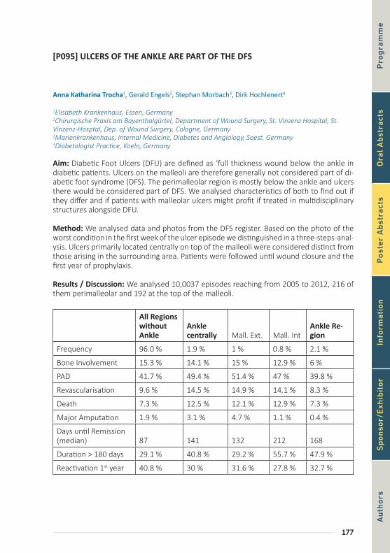
177
[P095] ULCERS OF THE ANKLE ARE PART OF THE DFS
Anna Katharina Trocha1, Gerald Engels2, Stephan Morbach3, Dirk Hochlenert4
1Elisabeth Krankenhaus, Essen, Germany2Chirurgische Praxis am Bayenthalgürtel, Department of Wound Surgery, St. Vinzenz Hospital, St. Vinzenz-Hosptal, Dep. of Wound Surgery, Cologne, Germany3Marienkrankenhaus, Internal Medicine, Diabetes and Angiology, Soest, Germany4Diabetologist Practice, Koeln, Germany
Aim: Diabetic Foot Ulcers (DFU) are defined as ‘full thickness wound below the ankle in diabetic patients. Ulcers on the malleoli are therefore generally not considered part of di-abetic foot syndrome (DFS). The perimalleolar region is mostly below the ankle and ulcers there would be considered part of DFS. We analysed characteristics of both to find out if they differ and if patients with malleolar ulcers might profit if treated in multidisciplinary structures alongside DFU.
Method: We analysed data and photos from the DFS register. Based on the photo of the worst condition in the first week of the ulcer episode we distinguished in a three-steps-anal-ysis. Ulcers primarily located centrally on top of the malleoli were considered distinct from those arising in the surrounding area. Patients were followed until wound closure and the first year of prophylaxis.
Results / Discussion: We analysed 10,0037 episodes reaching from 2005 to 2012, 216 of them perimalleolar and 192 at the top of the malleoli.
All Regions without Ankle
Ankle centrally Mall. Ext. Mall. Int
Ankle Re-gion
Frequency 96.0 % 1.9 % 1 % 0.8 % 2.1 %
Bone Involvement 15.3 % 14.1 % 15 % 12.9 % 6 %
PAD 41.7 % 49.4 % 51.4 % 47 % 39.8 %
Revascularisation 9.6 % 14.5 % 14.9 % 14.1 % 8.3 %
Death 7.3 % 12.5 % 12.1 % 12.9 % 7.3 %
Major Amputation 1.9 % 3.1 % 4.7 % 1.1 % 0.4 %
Days until Remission (median) 87 141 132 212 168
Duration > 180 days 29.1 % 40.8 % 29.2 % 55.7 % 47.9 %
Reactivation 1st year 40.8 % 30 % 31.6 % 27.8 % 32.7 %
Pro
gra
mm
eA
uth
ors
Ora
l A
bst
ract
sP
oste
r A
bst
ract
sIn
form
atio
nS
pon
sor/
Exh
ibit
or
178
Poster Abstracts
Patients with malleolar ulcers, especially laterally, have more arterial problems and worse outcome than those with ulcers in the perimalleolar region. Time to close the ulcer offered an inhomogeneous picture as the ulcers on the inner malleoli needed twice the time of ulcers at the outer malleoli and those in the perimalleolar region were somewhere in be-tween.
Conclusion: Perimalleolar ulcers feature the same differential diagnosis as lower leg ulcers and rarely result in major amputation. PAD, revascularisation, bone involvement and major amputation are strikingly more frequent with ulcers at the top of the malleoli. Patients suf-fering from these ulcers need the treatment offered by multidisciplinary teams and should be considered as part of the DFS in official definitions.
179
[P097] MECHANISMS OF WOUND HEALING IN RATS WITH STREPTOZOTO-CIN-INDUCED DIABETES MELLITUS
Anna Gorbacheva1, Evgeniy Ivanov2, Maria Morozova2, Ekaterina Artemova1, Zera Abdulvapova4, Ekaterina Rzhavina2, Svetlana Gavrilova2, Alexey Erdiakov2, Alla Tokmakova1, Vladiimir Koshelev2, Gagik Galstyan1
1Endocrinology Research Center, Moscow, Russian Federation2Lomonosov Moscow State University, Faculty of Fundamental Medicine, Russian Federation4Endocrinology Research Centre, Diabetic Foot Department, Moscow, Russian Federation
Aim: It was noted that the disturbance of wound healing and the formation of a diabetic foot, a severe disabling complication of diabetes mellitus, is accompanied by the defeat of the nervous system and / or ischemia. Aim of the study was to search for the effect of peripheral innervation disorders on the regulation of tissue repair in the streptozotocin model of diabetes mellitus (DM) in rats.
Method: The study was conducted on male rats (n = 70). After 42 days of persisting DM, (performed by injection of streptozotocin - STZ) or after injection of citrate buffer (CB) animals were/ All rats with diabetes received insulinotherapy. All animals were assessed pain sensitivity. Samples of the skin were taken on the 8th, 16th and 24th days after wound modelling. Sections were stained with hematoxylin and eosin, antibodies specific for Ki67, α1, β1 and β2-adrenoceptors, tyrosine hydroxylase (TH) and cytokeratin 17, n-acetylcho-line receptors were used for immunohistochemical staining. Intact animals were used as a control group.
Results / Discussion: In the DM group, the tail withdrawal time on the 56th day significant-ly increased in comparison with the control group (p = 0.017). In the CB group, a tenden-cy to faster wound healing is observed, but the differences were insignificant (p = 0.64). The intensity of staining on Ki67 was significantly lower in the group of DM (p = 0.045). A progressive decrease in the density of β2-adrenergic receptors was observed in the DM group both at the edge of the wound and in the remote areas of the skin. TH expression was higher in the edge of the wound on the 16th day. Expression of beta1-AR, alpha1-AR, nAchR, cytokeratine 17 did not differ significantly among groups.
Conclusion: The course of STZ-induced DM in rats is accompanied by the development of peripheral sensory neuropathy and a decrease in the speed of wound healing. The regen-eration of the wound in the described model is disturbed and is delayed relative to the control group. The proliferative activity of keratinocytes varies in different experimental groups in different ways, at the same time the peak of the regeneration is accompanied by changes in the activity of the adrenergic and cholinergic systems. The selection and search of markers that will allow to evaluate the changes in sympathetic and parasympathetic innervation of the skin continues, as the observational data clearly indicate the disorders of nervous regulation of wound healing.
Pro
gra
mm
eA
uth
ors
Ora
l A
bst
ract
sP
oste
r A
bst
ract
sIn
form
atio
nS
pon
sor/
Exh
ibit
or
180
Poster Abstracts
[P099] 6-YEARS RESULTS OF THE TREATMENT OF DIABETIC FOOT ULCERS
Vadim Bregovskiy1, Anastasia Demina1, Irina Karpova1
1St-Petersburg City Diabetes Centre, St-Petersburg, Russian Federation
Aim: To evaluate the results of the treatment of foot ulcers in patients with diabetes in the out-patient foot-department.
Method: We analysed the database of the foot department of the City Diabetes Centre from 01/2012 to 11/2017. From 7014 patients admitted during the study period 1714 patients with 2213 diabetic foot ulcers (DFU) were selected. Outcomes (primary healing, healing after toe amputation, healing after minor surgery, forefoot and above the ankle amputation) as well as the duration of the treatment (days, medians with 25th-75th percen-tiles) were calculated.
Results / Discussion: Male/Female: 48,2/51,8%. Mean age 58,9±12,9 years. Duration of diabetes 19,7±12,4 years. Type 1 / 2: 31,4/68,6%. Neuropathic/Neuroischemic foot – 60,8%/39,2%. Relapses were diagnosed in 13,9% of patients, simultaneous multiple ulcers – in 21%, and in 36,6% of patients more than 1 episode of DFU was diagnosed during the study period. Toes were affected in 48,7%, forefoot – 21,6%, midfoot – 6,7%, heel – 5,7%, other – 7,5%, combined – 8,1%. Infection was diagnosed in 47,4% of DFU. 63,5% lesions were superficial, 16,8% - deep and 19,7% - with osteomyelitis. In 492 patients (28,8%) results of the treatment are unknown: 125 patients (7,3%) decided to be treated in their local settings, 234 patients dropped out of follow-up (13,7%), 133 patients (7,8%) declined any treatment at the first visit. Primary healing was achieved in 923 patients (53,9%), heal-ing after toe amputation – in 110 patients (6,4%), healing after minor surgery (resection of metatarsals, interphalangeal joints, surgical drainage of abscesses) – 51 patients (3%), and in 26 patients (1,6%) forefoot and above the ankle amputations were performed. 110 patients remained unhealed (6,4%). Median healing time was 77 days (32 – 154); in neu-ropathic DFU 70 (29 – 146) versus neuroischemic: 85 (43 – 164); p=0,003. Risk factors for refusal of treatment were older age, male gender (OR 1,2; p=0.001), foot ischemia (OR 1,2; p=0.01) and type 2 diabetes mellitus (OR 1,2; p=0.02).
Conclusion: The overall results of 6-year experience of the out-patients treatment of pa-tients with DFU seems to be satisfactory, however, high drop-out rate is the most import-ant negative factor, influenced on the interpretation of results. High prevalence of infection and deep lesions (36,5%) decreased the effectiveness of our treatment and may be a result of the late referral. Male gender, type 2 diabetes, older age and foot ischemia are risk fac-tors of the refuse of treatment.
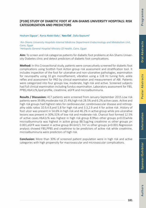
181
[P100] STUDY OF DIABETIC FOOT AT AIN-SHAMS UNIVERSITY HOSPITALS: RISK CATEGORIZATION AND PREDICTORS
Hesham Elgayar1, Rania Abdel-Baky1, Yara Eid1, Dalia Bayoumi1
1Ain-Shams University Hospitals-Internal Medicine Department-Endocrinology and Metabolism Unit, Cairo, Egypt2Heliopolis General Hospital-Ministry Of Health, Cairo, Egypt
Aim: To screen and risk categorize patients for diabetic foot problems at Ain-Shams Univer-sity Diabetes clinic and detect predictors of diabetic foot complications.
Method: In this Crossectional study, patients were consecutively screened for diabetic foot complications using Scottish Foot Action group risk assessment and stratification tool. It includes inspection of the foot for ulcerative and non-ulcerative pathologies, examination for neuropathy using 10 gm monofilament; vibration using a 128 Hz tuning fork; ankle reflex and assessment for PAD by clinical examination and measurement of ABI. Patients were categorized into four groups low, moderate, high risk and active. Screened subjects had full clinical examination including fundus examination. Laboratory assessment for FBS, PPBS,HbA1c%,lipid profile, creatinine, eGFR and microalbuminuria.
Results / Discussion: 417 patients were screened from January-September 2015.Low risk patients were 39.8%;moderate risk 25.4%;high risk 28.5% and 6.2% active cases. Active and high risk groups had highest rates for cardiovascular; cerebrovascular disease and retinop-athy odds ratios 10.9,4.9,and 5.8 for high risk and 3.3,2.9 and 4 for active risk .History of foot ulcer was present in 54.6% in high risk and 46.2% in active group while pre-ulcerative lesions was present in 30%;31% of low risk and moderate risk. Charcot foot formed 12.5% of active cases.HbA1c% was highest in high risk group 8.9%vs other groups p<0.01while microalbuminuria was highest in active group 88.5ug/mg creatinine vs other groups p< 0.001,eGFR was lowest in active group 84.6ml/1.7m2 vs other groups p<0.001.Regression analysis showed FBS,PPBS and creatinine to be predictors of active risk while creatinine, microalbuminuria were predictors of high risk.
Conclusion: More than 30% of screened patient population were in high risk and active categories with high propensity for macrovascular and microvascular complications.
Pro
gra
mm
eA
uth
ors
Ora
l A
bst
ract
sP
oste
r A
bst
ract
sIn
form
atio
nS
pon
sor/
Exh
ibit
or
182
Poster Abstracts
[P101] RECURRENT ULCER VERSUS SINGLE FOOT ULCER: IS THERE ANY DIFFER-ENCE BETWEEN PATIENTS AND OUTCOMES OF THE TREATMENT?
Anastasia Demina1, Vadim Bregovskiy1, Irina Karpova1
1St-Petersburg City Diabetes Centre, St-Petersburg, Russian Federation
Aim: To evaluate the clinical characteristics of patients with recurrent foot ulcers and com-pare the outcomes of their treatment with non-recurrent foot ulcers in patients with dia-betes.
Method: From the database of the foot department of the out-patient foot-clinic 1714 patients with 2213 diabetic foot ulcers were selected (from 01/2012 to 11/2017). 239 patients with rDFU were identified and 1475 patients were relapse-free (rfDFU). Median healing times were calculated. Outcomes (healing, healing after minor amputation, ampu-tation rate and level) were compared between groups.
Results / Discussion: Groups did not significantly differ on age and duration of diabetes. Male/Female: rDFU – 41,8/58,2%; rfDFU – 49,3/50,7% (p=0.03). Type 1/Type 2 diabetes: rDFU – 40,8%/59,2%; nfDFU – 29,9/70,1% (p=0.0001). The history of the previous ampu-tation in rDFU 43% vs 32,7% in rfDFU (p=0.004). Moreover, rDFU were associated with the previous amputation of the great toe (single or with other foot segments): rDFU – 39,2% vs 29,9% in rfDFU, p=0.004; and with Charcot arthropathy (rDFU – 42,2% vs rfDFU – 25,6%; p=0.0001). Inability to feel 10-gr. monofilament was diagnosed in 84,7% in rDFU group and in 67,8% of patients in rfDFU group (p<0.00001). The proportion of the superficial lesions in rDFU group – 72,9% vs 63,3% in rfDFU (p<0.01). The frequency of infection was similar in both groups (42,4 vs 47,3%, ns). Ischemia was more frequent in rfDFU (41,0%) vs 27,6% in rDFU (p=0.0001) and the critical limb ischemia was more typical in rfDFU group (7,5%) vs 2,5% in rDFU (p=0.004). Primary healing: 61,2% in rDFU group and in 52,7% in rfDFU (p=0.025). The median healing time (25th-75th percentiles) in rDFU was longer compared with rfDFU: 109 days (29 – 202) vs 74 days (32 – 142) (p=0.015). Minor amputations rates: 7,2% in rDFU and 6,6% in rfDFU patients. Major amputations were performed in 1 patient of rDFU group and in 20 patients from the rfDFU group. The loss of the follow-up of pa-tients with rDFU was 16% vs 30,7% in rfDFU (p<0.0001).
Conclusion: Risk factors of the recurrent ulcer were: severe foot deformity after previous minor amputation, Charcot foot, type 1 diabetes, ischemia and inability to feel the 10-gr. monofilament. The treatment of recurrent ulcers takes much more time compared with the non-recurrent ulcers, however the healing rate is higher in recurrent ulcers. Results of the present study are significantly influenced by the high drop-out rate in both groups which is more pronounced in patients without recurrent ulcers.
183
[P102] PATIENT AND HEALTH CARE PROFESSIONAL’S PERSPECTIVES ON WHAT IS THE MOST APPROPRIATE CLINICAL OUTCOME FOR PATIENTS AT RISK OF REULCERATION
Katie Gray1, Frances Game2, Kavita Vedhara3, Kristian Pollock3, Lorraine Pinnington 4, Katherine Bradbury5, Katy Sivyer5, Kate Greenwell5, Lucy Yardley6
1Derbyshire Community Health Services NHS Ft, Derby, United Kingdom2Derby Teaching Hospitals NHS Foundation Trust, Derby , United Kingdom3Faculty of Medicine & Health Sciences, University Of Nottingham, United Kingdom4Associate Professor in Rehabilitation, Division of Medical Sciences and Graduate Entry Medicine, University of Nottingham Medical School , Derby , United Kingdom5Psychology, University of Southampton, United Kingdom6Health Psychology, University of Oxford, United Kingdom
Aim: Patients at greatest risk of diabetic foot ulceration are those with a history of previ-ous ulceration, but we know little about the patients’ or healthcare professionals’ (HCP’s) perspective of outcome when managing this high-risk group. The aim of this work was to establish patients and HCP’s priorities following healing of a diabetic foot ulcer (DFU).
Method: Semi-structured qualitative interviews were conducted with 20 patients and 13 HCPs (9 Podiatrists and 4 Consultant Diabetologists). HCP’s were from 5 diabetic foot MDFTs in the UK and patients with a history of DFU who were currently free of ulceration. All partic-ipants were asked to rank 4 outcomes and to discuss the reasons for their rankings. The out-comes has been identified with a focus group of patients from a different health care centre:
(A) ‘future ulcers heal quickly’, (B) ‘I am ulcer free for longer’, (C) ‘I am ulcer free for longer, but when they reoccur they heal quickly’, (D) ‘New ulcers are found at an early stage’
Participants were asked to consider outcomes of most importance in the context of reul-ceration and interviews explored participants understanding of reulceration risk, with fur-ther exploration of healthcare professional’s perspectives of current practice. Transcripts were analysed using thematic analysis.
Results / Discussion: Patient interviews: The most preferred outcome was D (endorsed by 13 patients); followed by B and C (both endorsed by 5 patients each) 5 patients ranked more than one option . Analysis of the reasons patients gave for their rankings made clear, however, that the most important priority for most patients was to be ‘ulcer free’. Patients choosing other outcomes did so because they perceived that their selection was the mech-anism by which they could achieve their goal of being ‘ulcer free (e.g., “if it’s not dealt with early on it goes on and on”)
HCP interviews: HCPs favoured two outcomes: patients being ulcer free for as long as pos-sible and secondly that ulcers were ‘detected quickly’ because, again, ulcers detected early were seen as likely to be less severe and, therefore, to heal more quickly.
Conclusion: The results have been valuable to understand what is important to patients and professionals and will help to develop more relevant future interventions with a greater chance of adoption into practice. Limitations in current research mean many challenges still exist when supporting patients, this work aims to provide a basis for future work on this topic.
Pro
gra
mm
eA
uth
ors
Ora
l A
bst
ract
sP
oste
r A
bst
ract
sIn
form
atio
nS
pon
sor/
Exh
ibit
or

184
Poster Abstracts
[P104] THE NEED FOR LOWER LIMB REVASCULARIZATION OR AMPUTATION IN DIABETIC FOOT INFECTIONS: A COHORT STUDY
Tiago Santos1, Diana Duarte1, André Couto Carvalho2, Claudia Amaral2, Cláudia Freitas2, Helena Neto2, Joana Martins3, Luís Loureiro3, André Gomes3, Luís Costa3, Pedro Cantista3, Rosa Guimarães3, Joel Pereira3, Sara Pinto2, Rui Carvalho2
1Department of Endocrinology, Diabetes and Metabolism, Centro Hospitalar Do Porto - Hospital Santo António, Porto (Portugal), Porto, Portugal2Department of Endocrinology, Diabetes and Metabolism, Centro Hospitalar Do Porto - Hospital Santo António, Porto (Portugal), Diabetic Foot Unit, Centro Hospitalar Do Porto - Hospital Santo António, Porto (Portugal)3Diabetic Foot Unit, Centro Hospitalar Do Porto - Hospital Santo António, Porto (Portugal)
Aim: Our goal is to display the epidemiology and need for lower limb revascularization or amputation of moderate to severe diabetic foot infections (DFI) evaluated in a tertiary referral center.
Method: Retrospective study of diabetic patients presenting for their first consult at our Diabetic Foot Unit between January 2015 and December 2016 (2 years) who collected mi-crobiological cultures and were admitted to our hospital ward for treatment of moderate to severe DFI during the follow-up of 1 year.
Results / Discussion: A total of 1058 patients were observed during this period and 176 of them (16.6%) were included for the analysis. One-hundred and forty five of these (82.3%) had neuro-ischaemic ulcer and the remainder 31 patients (17.6%) presented with a pure neuropathic ulcer. Of neuro-ischaemic ulcer patients, 102 (70.3%) were submitted to a minor lower limb amputation and 29 (20%) to a major lower limb amputation during fol-low-up period. Of the latter, only 9 (31%) were not preceded by a minor lower limb ampu-tation during the follow up. One hundred and nine of these patients (75.1%) experienced a lower limb revascularization procedure: endovascular intervention in 74 (67.9%) and by-pass surgery in the remaining 35 patients (32.1%). With regard to pure neuropathic ulcer patients, 9 (29%) were submitted to minor lower limb amputation. One (3.2%) was later submitted to a major lower limb amputation.
Conclusion: In our neuro-ischaemic ulcer patients, minor lower limb amputation rate was high (~70%), possibly reflecting the surgical need to control acute diabetic foot infections in frail individuals. One fifth of these patients, with severe disease, had to be submitted to a higher level of amputation. As expected, in pure neuropathic ulcer patients, minor lower limb amputation rate was much lower (~30%) with only one major amputation performed during this 2 year period.
185
[P105] RELIABILITY AND USEFULNESS OF CLASSIC PHYSICAL SIGNS OF LOWER EXTREMITY ISCHEMIA IN GENERAL DIABETIC POPULATION
Maria Bahteeva1, Daria Afanaseva1, Oleg Udovichenko2, Darya Ladygina2
1Russian Pirogov Research Medical University, Moscow, Russian Federation2Central Clinical Hospital of the Presidential Affairs Department, Moscow, Russian Federation
Aim: To assess reliability and usefulness of classic physical signs of extremity ischemia in patients without diabetic foot ulcers.
Method: Medical textbooks describe following signs: muscle atrophy, pale skin, atrophic thin skin, hair loss (in men), thickening and lamination of nails, hyperkeratosis, low skin temperature. These signs and palpation of pedal pulses were assessed independently by 2 students and an experienced specialist in 30 consecutive patients (60 legs) with diabetes without foot ulcers or symptoms of CLI who underwent duplex ultrasound scan (DUS) of leg arteries.
Results / Discussion: Mean age of patients was 65 (48-86) yrs, M:F ratio was 9:21. Eleven (37%) had intermittent claudication. Leg ischemia (arterial stenoses of 70% or more at any level) was present in 30% of extremities. Inter-observer agreement (IOA) for physical signs varied in various pairs of investigators from 44-67% (hair loss) to 77-92% (muscle at-rophy). Nevertheless diagnostic accuracy of all these signs was not high enough (43-63%). Relatively high accuracy (due to high specificity but low sensitivity) was demonstrated by muscle atrophy (63%), hyperkeratosis (surprisingly - 58%) and atrophic thin skin (56%).
Palpation of pulses showed high IOA (79-85%) and acceptable accuracy (75%). Sensitivity and specificity of this method was >70% in all investigators with high negative predictive value (86%) but low positive predictive value (58%).
Conclusion: 1). Studied physical signs were described in critical limb ischemia (CLI). They should not be used in general population of patients with diabetes because in patients without severe ischemia their reliability is low. 2). These limitations should be emphasized during education of medical students. 3). Palpation of foot pulses demonstrated better ac-curacy and high IOA. 4). Palpable pulse allow to exclude significant stenoses of the feeding arterial line, but non-palpable pulse has not very high value and need further confirmation of ischemia by instrumental methods.
Pro
gra
mm
eA
uth
ors
Ora
l A
bst
ract
sP
oste
r A
bst
ract
sIn
form
atio
nS
pon
sor/
Exh
ibit
or
186
Poster Abstracts
[P106] CALF MUSCLE ELECTROSTIMULATION EFFECTS VASCULAR PERFUSION AND WALKING CAPACITY IN TYPE 2 DIABETES PATIENTS WITH INTERMITTENT CLAUDICATION
Alfred Gatt1, Christian Ellul1, Cynthia Formosa1
1University of Malta, Msida, Malta
Aim: To determine whether calf muscle electrostimulation effects vascular perfusion and walking capacity in Type 2 diabetes patients with intermittent claudication.
Method: A prospective, pretest-posttest study design was conducted on 40 type 2 diabe-tes participants with Ankle Brachial Index (ABPI) <0.9 who underwent a program of calf muscle electrical stimulation with varying frequency (1-250 Hz) for 1 hour per day for 12 weeks. Spectral waveforms analysis, ABPI, absolute claudication distance (ACD), and ther-mographic temperature patterns across 4 specified regions of interest in the foot at rest and after exercise, were recorded at baseline and following the intervention.
Results / Discussion: A significant improvement in ACD and ABPI was registered follow-ing the intervention (p= 0.000 and p= 0.001, respectively). Resting foot temperatures increased significantly (p=0.000) while the postexercise temperature drops were halved across all regions at follow-up, with hallux (p=0.005) and lateral forefoot (P = .038) reaching statistical significance. Spectral Doppler waveforms were comparable (p=0.304) between both assessments.
Conclusion: Electrical stimulation of varying frequency for 1 hour per day for 12 consec-utive weeks resulted in statistically significant improvement in outcome measures that assess arterial inflow and walking capacity in type 2 diabetes mellitus patients with inter-mittent claudication. These results favour the use of electrostimulation as a therapeutic measure in this high-risk population.

187
Pro
gra
mm
eA
uth
ors
Ora
l A
bst
ract
sP
oste
r A
bst
ract
sIn
form
atio
nS
pon
sor/
Exh
ibit
or
Information

188
General information
CONTACTDFSG SecretariatNordre Fasanvej 113, 2nd floor2000 Frederiksberg CDenmark+45 70 20 03 [email protected]
CONFERENCE VENUEVienna House Andel’s BerlinLandsberger Allee 10610369 BerlinGermany
DISABLED ACCESSAll areas of the venue allow disabled access.
CONFERENCE SECRETARIAT (Registration desk)The conference secretariat is located in the basement in front of the exhibi-tion area and session room. BADGESAll participants and exhibitors should wear the name badge in the conference area at all times. The badge must be visible.
CERTIFICATES OF ATTENDANCECertificates of attendance will be available as self-print after the conference. A link will be provided by e-mail to all participants.
WIFIWifi name: Vienna House Andels BerlinNo password, but conditions must be accepted.
CONFERENCE HOURSn●Friday 28th September12.00 - 19.00 Registration14.00 - 19.10 Scientific sessions14.00 - 20.00 Exhibition19.10 - 20.00 Welcome reception (open to all delegates)
n●Saturday 29th September07.00 Podartis Diabetes
Run/walk, 4 km (open to all delegates, no pre-registration)
08.30 - 18.00 Registration09.00 - 18:00 Scientific sessions10.00 - 16.15 Exhibition18.05 - 18.45 DFSG Business Meeting (for DFSG members only)19.30 - 24.00 Conference dinner
at Brauhaus Lemke am Alex, Berlin. Not included in the registration fee, a separate ticket must be purchased.
n●Sunday 30th September07.30 - 15.00 Registration08.00 - 15.00 Scientific sessions10.15 - 14.00 Exhibition15.00 - 15.45 Closing Words and
Farewell in the Hotel lobby
LANGUAGE The language for the DFSG 2018 conference is English.

189
ENTITLEMENTSn●Presenting authors: Accommodation in a single room (in 28 - out 30 September including breakfast), participation in all scientific sessions, programme and book of abstracts, coffee Friday, lunch and coffee Saturday and Sunday, partici- pation in Welcome Reception Friday.
n●Non-pres. members:Accommodation in a single room (in 28 - out 30 September including breakfast), participation in all scientific sessions, programme and book of ab-stracts, coffee Friday, lunch and coffee Saturday and Sunday, participation in Welcome Reception Friday
n●Non-members: Participation in all scientific sessions, programme and book of abstracts, coffee Friday, lunch and coffee Saturday and Sunday, participation in Welcome Reception Friday.
n●Exhibitors: All exhibitors must register through the online registration system. Coffee Friday, lunch and coffee Saturday and Sunday is included in price. Exhibitors may participate in social events for an extra fee, please contact the secre-tariat for details. Exhibitors do not have access to the scientific sessions.
n●Non-exhibiting, non-sponsoring industry participants: Participation in all scientific sessions, programme and book of abstracts, coffee Friday, lunch and coffee Saturday and Sunday, participation in Welcome Reception Friday
LUNCH AND COFFEE Lunch and coffee is available in the exhibition area. See programme for exact time of breaks.
PARKINGYou can park your car on the under-ground garage of the venue. Price for parking: 17€ / 24 hours / car. Please note the maximum height of 2 m.
SPEAKER INFORMATION Please bring your presentation to the Session Room before your session starts. We recommend you upload your presentation at least 2 hours before your session. A technician will be present to assist in the upload if necessary.Please bring your presentation on a USB. Use of personal laptops is not allowed. Unless otherwise agreed all presentations will be deleted after the conference in order to secure that no copyright issues will arise at the end of the conference.
MOBILE PHONES All mobile phones must be on silent mode during the sessions.
LOST AND FOUND Found items should be returned to the registration desk. If you lose something, please report to this desk for assistance.
NO SMOKING POLICY Smoking is prohibited at the venue. There are dedicated outdoor smoking areas available.
Pro
gra
mm
eA
uth
ors
Ora
l A
bst
ract
sP
oste
r A
bst
ract
sIn
form
atio
nS
pon
sor/
Exh
ibit
or

190
General information
POSTERS Posters can be mounted from Friday 28th September 2018, 12.00 and must be removed by the end of the conference on Sunday 30th September 2018, 15.00The posters will be affixed to the poster boards with adhesive which will be provided to you by the conference staff.
CME CREDITSEuropean CME creditsThe DFSG 2018 has been accredited 11 European CME credits (ECMEC®s) by the European Accreditation Council for Continuing Medical Education (EACCME®).To receive the CME credits, please sign the attendance sheet at the registration desk each day after 14.00. The CME certificates will be sent by e-mail after the conference.
German CME creditsThe DFSG 2018 has been accredited 12 CME credits by Ärztekammer Berlin.To receive the CME credits, please sign and add your Barcode or EFN number to the German CME attendance sheet at the registration desk each day.
PHOTOGRAPHINGPhotographing is prohibited during sessions: Please respect the intellec- tual property right of the presenter.
PRIZES AND AWARDS n●Oral abstract prizeThe 3 best oral abstracts will be presented on Saturday 29th September 2018. Participants will have the oppor-tunity to vote for the best presentation after the 3 presentations.1st prize for the best oral presentation will be announced at the Conference Dinner Saturday evening.
n●Poster prize Poster Prize winners will be chosen from the posters in the session “Top 10 poster abstracts”1st and 2nd prize for the best posters will be announced at the Conference Dinner Saturday evening.
n●Life Time Achievement AwardThe DFSG Life Time Achievement award will be announced at the Conference dinner.
n●The Paul Wilson Brand Repetitive Stress AwardThe prize is awarded to a presenter who has submitted an abstract to a regular DFSG meeting, presenting new clinical or experimental data on the effects of repetitive mechanical stress on soft tissues, joints or bones.The winner of the prize will present his/her results in a session on Saturday 29th September.
n●First time presenter grantWinners of the First time presenter grant will be announced at the Confer-ence dinner.
191
Social Events
WELCOME RECEPTION n●●Friday 28th September 19.10 - 20.00 The welcome reception takes place in the exhibition
area. Join your colleagues for snacks and wine/soft drinks. Included in the registration fee. Please note that the reception is not a dinner.
PODARTIS DIABETES RUN n●29th September at 7.00. Start the day by running or walking a route of 4 km.
We meet in the entrance of Vienna House Andel’s Berlin. Map and water will be provided provided. The run is sponsored by Podartis.
CONFERENCE DINNER n●29th September 2018 The conference dinner of DFSG 2018 will take place
at Brauhaus Lemke am Alex, located on Alexander-platz and close to the Berlin TV tower. The confer-ence dinner will offer a unique experience in the brewery, where the expertly art of brewing meets honest and regional food.
Brauhaus Lemke am Alex Karl-Liebknecht-Str. 13, 10178 Berlin For more information, visit the website www.lemke.berlin/am_alex
The conference dinner is not included in the registration fee. Tickets must be purchased before the conference.
5 busses will leave with a 5 min interval from Vienna House Andel’s Berlin from 19.00-19.20.
Busses will return to Vienna House Andel’s at: 22.00 / 22.30 / 23.00 / 23.30 / 24.00
If you choose to come or leave by other means of transportation, please note that this is not covered by the conference.
CLOSING WORDS & FAREWELL n●Sunday 30th September, 15.00 We close the conference with a snack and drink in
the lobby of Vienna House Andel’s Berlin. The event is open to all participants.
Pro
gra
mm
eA
uth
ors
Ora
l A
bst
ract
sP
oste
r A
bst
ract
sIn
form
atio
nS
pon
sor/
Exh
ibit
or
Podartis Diabetes RunJoin your colleagues in a 4 km morning run or walk on Saturday 29th September, 7.00.
The run will take you through the streets and parks of Berlin and we encourage all participants to join to get a fresh start of the day and to see the city waking up.
Water and map will be handed out at the Lobby of Vienna House Andel’s at 7.00.
No pre-registration for the run is necessary, just show up!
The run is sponsored by Podartis who will supply all running participants with a pair of running socks. Socks can be picked up at the Podartis stand on Friday 28th September.
192
193
Industry Sponsored Satellite Symposia
Protex HealthcareFriday 28 September 201817.00 - 18.00
Modern fast & ad hoc diagnostics and follow up of diabetic foot wound ulcers with hyper spectral analysis. More attention is needed for intra wound oedema. How to manage?
Kristien van Acker, Belgium
Molliter Saturday 29 September 201811.15 - 12.15
DFA: The Diabetic Foot Offloading AlgorithmChair: Alberto Piaggesi, Italy
IntroductionAlberto Piaggesi, Italy
Stabilization and Post SurgeryArmin Koller, Germany
How to use the algorithm in a specialized df unit Giacomo Clerici, Italy
Open discussion
Urgo Saturday 29 September 201814.45 - 15.45
How do we close more wounds around us? Let’s start todayChair: José Luis Lázaro Martinez, Spain
What are the key challenges when treating DFU patients?José Luis Lázaro Martinez, Spain
Why and how to develop standardized care?Kristien Van Acker, Belgium
The UK experience in patient protocol implementationGraham Bowen, United Kingdom
Pro
gra
mm
eA
uth
ors
Ora
l A
bst
ract
sP
oste
r A
bst
ract
sIn
form
atio
nS
pon
sor/
Exh
ibit
or

194
Sponsor and exhibitor information
Advanced Oxygen Therapy IncTel: +1 760 672 1920 [email protected]
AOTI offers a wide range of Single-use and Reusable Topical Wound Oxygen (TWO2) therapy systems that allow for our patented wound care approach to be applied effortlessly at all care sites including; hospitals, wound care clinics, nursing homes and in the patient’s home. TWO2 therapy has been proven in numerous clinical trials to completely heal non-healing chronic wounds (Diabetic, Venous and Pressure) at an unprecedented rate. TWO2 therapy is also effective in treating post-surgical and burn wounds. Over 1,500,000 TWO2 treatments have be delivered to patients to date.
CollPlant Ltd.Tel.: +972 73 232 5613 [email protected]
CollPlant is a regenerative medicine company focused on 3D bioprinting of tissues and organs, and on developing and commercializing tissue repair products for orthobiologics, and advanced wound care markets. Our products are based on our rhCollagen (recombinant human collagen) that is produced with CollPlant’s proprietary plant based genetic engineering technology. Our products address indications for diverse fields of organ and tissue repair, and are ushering in a new era in regenerative medicine. Our unique Vergenix line of rhCollagen products includes a wound repair matrix to promote a rapid optimal healing of acute and chronic wounds.
ConvaTecTel.: +49 800 78 66 [email protected]/wound-skin
ConvaTec is a global medical company focused on therapies for the management of chronic conditions. The Group’s Advanced Wound Care franchise provides advanced wound dressings and skin care products used for the management of among others, diabetic foot ulcers. Key products include our AQUACEL® line of advanced dressings, which features ConvaTec’s proprietary Hydrofiber®Technology. The benefits of this technology can now also be found in our Avelle™ Negative Pressure Wound Therapy System. ConvaTec’s recently introduced disposable NPWT system is designed to give patients more mobility, helping them resume their daily lives as their wounds heal.
DARCO (Europe) GmbHTel: + 49 8807 922 8-0 [email protected]
DARCO is dedicated to being one of the leading providers of post op, trauma and wound care solutions to the global foot and ankle community.

195
Dm SystemsTel : + 1 847 328 [email protected]
DM Systems is a global provider of clinically proven positioning and offloading products for the treatment and prevention of pressure injuries. Our innovative products and clinical research are at the leading edge of pressure injury prevention and treatment
Essity is a leading global hygiene and health company dedicated to improving well-being through our products and solutions, essentials for everyday life. Essity’s Medical Solutions product segment originates from the acquisition of BSN medical, a leading medical solutions company acquired on April 3, 2017. BSN medical develops, manufactures and sells products within wound care, compression therapy and orthopedics.
Medicap homecare GmbHTel.: +49 6645 970 0 [email protected]
Medicap was founded in 1981 in Ulrichstein/Germany. Medicap’s product portfolio today consists of certified medical devices like oxygen concentrators, oxygen conserving devices, IPPB devices and optical transcuta-neous oximetry devices/tcpO2 for the field of vascular medicine, Diabetology, angiology and wound therapy.
Mölnlycke Health Care ABTel.: +46 31 722 30 [email protected]
Mölnlycke is a world leading medical solutions company. We develop and bring to market innovative wound care and surgical solutions along the entire continuum of care – from prevention to post-acute settings. Our solutions provide value for money, supported by clinical and health economic evidence. Mölnlycke was founded in 1849. Nowadays, our solutions are available in around 100 countries; we are the number one global provider of advanced wound care and single-use surgical products; and we are Europe’s largest provider of customized trays. Our headquarters are in Gothenburg, Sweden and we have about 7,700 employees around the world. Learn more at molnlycke.com.
Optima Molliter SRLTel: + 39 0733 80 [email protected]
OPTIMA MOLLITER is a Company that produces prevention shoes (MAC1-2-3), Daily Activity Shoes (YDA) and Offloading Walkers for the management of the Diabetic foot ulceration or post-surgery/trauma (SBi).
Pro
gra
mm
eA
uth
ors
Ora
l A
bst
ract
sP
oste
r A
bst
ract
sIn
form
atio
nS
pon
sor/
Exh
ibit
or
196
Perimed+46 70 772 07 74eva.weberandersson@ perimed-instruments.comwww.perimed-instruments.com
PERIMED is a global provider of diagnostic solutions for patients with peripheral vascular diseases and complex diabetic foot ulcers. Our PeriFlux 6000 offers a unique combination of tests: ABI, toe pressure and tcpO2.PeriFlux 6000 Combined System – ABI/TBI, toe pressure, tcpO2. PeriFlux 6000 Pressure System – ABI/TBI, toe pressure. PeriFlux 6000 tcpO2 Stand-alone – tcpO2. For more information, please visit.
Podartis Srl Tel.: +39 [email protected]@[email protected]
Podartis is specialized in the production of shoes, plantars and walkers for the prevention and the treatment of the major gait diseases. Source of pride for the Company are the cutting edge solutions designed for the diabetic foot, that are all strictly clinically tested to ensure maximum benefit and comfort. The continuous product innovation through a strong and ongoing commitment in research, the medical/scientific collaborations with the most authoritative experts, and the several patents that characterize Podartis footwear, are the direct evidence of the mission of the company: to help people walk well, in order to better live.
Protex Healthcare Tel.: +44 1926 21 78 [email protected]
Protex Healthcare is a provider of wound management solutions, supplying patients and clinicians across the world with our unique Vacutex™ products. Our experienced clinical support team is dedicated to working with clinicians and patients, providing objective advice concerning advanced wound care by means of TIVITA hyperspectral analysis support. Wherever in the world you are, you can rely on Protex Healthcare to deliver innovative wound management solutions that help to heal wounds – helping to save feet, legs and lives.Vacutex™ is a Rapid Capillary Action Wound Dressing for use on difficult to heal wounds with moderate or heavy exudate or slough.
ReapplixTel.: +45 8816 [email protected] www.reapplix.com
Reapplix is a Danish company that has developed the CE-marked and FDA cleared 3C Patch® Device (Formerly LeucoPatch). The 3C Patch® is produced from the patient’s own blood without any reagents and harnesses the patients healing potential by generating a concentrate of cells and growth factors, which is then reapplied to the patient’s wound. The clinical effect of 3C Patch® on the healing of diabetic foot ulcers has recently been proven in a large randomized controlled trial (N=269) showing significantly more wounds healed in less time. Reapplix is currently introducing the 3C Patch® in selected EU markets and the US.
Sponsor and exhibitor information
197
Saluber SRLTEL +39 0424 [email protected]
Saluber Srl, based in Italy, is the manufacturer of exquisetely made non-custom orthotics and of the FORS™-15 offloading insole for the treatment of diabetic foot ulcers which has recently been evaluated at multiple academic institutions with excellent results.
Smith & Nephew Advanced Wound ManagementTel +44 1482 [email protected]/wound
Smith & Nephew supports healthcare professionals in http://www.smith-nephew.com/about-us/where-we-operate/ in their daily efforts to improve the lives of their patients. We do this by taking a http://www.smith-nephew.com/commercial-resources/research/ to the design of our http://www.smith-nephew.com/key-products/, by securing wider access to our diverse technologies for more customers globally, and by enabling better outcomes for patients and healthcare systems.
Urgo MedicalTel.: + 33 380 545 000 www.urgomedical.com
Urgo Medical is the Healing Company committed each day to improve wound care treatments for both patients and health care professionals by offering highly innovative solutions.
Woundcare-Circletel: + 49 8807 922 [email protected]
The 3 Woundcare-Circle founders, OPTIMA, Heelift and DARCO are international market leaders providing innovative product solutions. The group permanently supports research & science as well as the advancement.
Pro
gra
mm
eA
uth
ors
Ora
l A
bst
ract
sP
oste
r A
bst
ract
sIn
form
atio
nS
pon
sor/
Exh
ibit
or
198
Aalaa, Maryam OP30, P052, P087aan de Stegge, Wouter P084Abbas, Zulfiqarali G. P024Abbott, Caroline Paul Brand Award OralAbdel-Baky, Rania P100Abdelghaffar, Hassan P019Abdelnaser, Radwa P068Abdulvapova, Zera P097, OP28Adamikova, Alena P086Adibi, Hossein OP30, P052, P087Afanaseva, Daria P105Aghaei Meybodi, Hamidreza OP30, P087Ahluwalia, Raju OP36Ahn, Junho OP15, OP21, P003, P021Ainarkar, S OP06, OP08Albehairy, Ahmed OP03, P060, P019, P070Alejandro, Ian P007, P044Allen, J OP06, OP08Alnemo, John P076Alvaro Afonso, Francisco Javier OP12, OP18, P053, P066Amado, Ana P091Amaral, Cláudia P040, P091, P104Amini, Mohammad Reza OP30, P052, P087Andersen, Henning OP26Andersen, Henrik Ullits OP33, P014Anichini, Roberto OP22Anson, Matthew P007Archibald , Lennox P024Arsos, Georgios OP37Artemova, Ekaterina OP28, P097Ashraf, Alima P082Assaloni, Roberta P006Ayabe, Shinobu P049Azard, Marie OP01Azmy Kyrillos, Fady OP03Bahriz, Rania P070Bahteeva, Maria P105Baikstiene, Ieva P002Bami, Christina P020Banchellini, Elisa P015Barn, Ruth Prize Oral 3, P085Barro, Enrica P006Bates, Maureen P007, P044Bayoumi, Dalia P100Bellio, Elicia P044Bem, Robert P001, P004, P071, P088, P090, OP38Berrington, Rachel OP19, P089Bevans, J OP06, OP08
Authors

199
Binning, Jodi P085Bloom, Jonathan OP31Boddington, Lucy OP25Bond, Hannah OP16Boschetti, Giovanni P056, P075Boulton, Andrew Paul Brand Award OralBowling, Frank Paul Brand Award OralBoyko, Edward P046Bradbury, Katherine P102Brantley , Jonathan P016Bregovskiy, Vadim P099, P101Brocco, Enrico P056, P075Brown, Ross P045Broz, Jan P086Brunato, Barbara P006Bruseghin, Marino P056, P075Bruyninckx, Herman OP11Buncová, Marie OP38Bus, Sicco Prize Oral 3, P074, P084, P085Busch-Westbroek, Tessa P074Camilleri, Kenneth PO73Camilleri, Liberato PO73Cantista, Pedro P040, P104Carlucci, Silvana P006Carranca, João Carlos P061Carvalho, André P091Carvalho, Rui P040, P091, P104Casey, Joanne OP29, P009, P094Cassar, Kevin P022, P026, P041, P072, P073Cassin, Matteo OP24Cechova, Katerina P086Cerniauskiene, Kristina P002Cervesato, Eugenio OP24Chantelau, Ernst OP34Chatwin, Katie Paul Brand Award OralChen, Min Qi OP05Chockalingham, Nachiappan PO73Cohen, Siew P005Coppini, David P072Costa, Luís P040, P091, P104Couto Carvalho , André P040, P104Da Porto, Andrea OP24Da Ros, Roberto P006, OP24Daikos, Georgios L. P055D'Annunzio, Vinicio P042Danyte, Evalda P002De Bellis, Alessandra OP22De Biasio, Valeria P056
Pro
gra
mm
eO
ral
Ab
stra
cts
Pos
ter
Ab
stra
cts
Info
rmat
ion
Sp
onso
r/E
xhib
itor
Au
thor
s
200
De Block, Christophe P010, P077De Gier, Sophie P080Del Core, Michael P021Demetriou, Maria P064Demina, Anastasia P099, P101deSancha, Maximilian Prize Oral 1Deschamps, Kevin OP11Desmet, Dirk OP11Devriendt, Eveline P010Diane, Whitham Prize Oral 2Dias, Vanessa P046Didangelos, Triantafillos OP37Dineni, Vassiliki P020Dirinck, Eveline P010, P077Donaldson, AN OP06, OP08Donaldson, NK OP08Doulapsis, Mikes P031Dove, Cyaandi P016Duarte, Diana P040, P104Dubský, Michal OP38, P001, P004, P071, P088, P090Dumont, Isabelle P054Edmonds, Michael Prize Oral 1, OP02, OP06, OP08, OP27, OP29,
P007, P008, P009, P012, P016, P044, P094Eid, Yara P100Ejskjaer, Niels OP26Elbaz, Osama P019Eleanor, Harrison Prize Oral 2Eleytheriadoy, Ioanna P031Elgayar, Hesham P100Elias, David Prize Oral 1Elkashef, Wagdi OP03Ellul, Christian PO73, P106El-Nahas, Mamdouh P060Elsayed, Mohamed P060El-Sebaie, Ahmed P019Elsherif, Mohamad P060Emam, Ahmed P019Engberg, Susanne OP04, OP33, P014Engels, Gerald OP14, OP23, P095Erdiakov, Alexey P097Falzon, Owen PO73Fathy, Sherif P070Federici, Antonio P042Fejfarová, Vladimíra OP38, P001, P004, P071, P088, P090Ferreira, Lia P091Fialova, Libuse OP13Fleming, Naomi P059Foden, Philip Paul Brand Award OralFonseca, Liliana P091Formosa, Cynthia PO73, P106
201
Fosbrook, Julie P037Foussard, Ninon OP01Fowler, Duncan OP25Francesconi, Paolo OP22Francia, Piergiorgio OP22Franks, Peter P016Franssen, Catya P010Freitas, Cláudia P040, P091, P104Frøkjær, Johnny P013Frykberg, Robert P016Galea, Samuel Anthony P022, P026, P041Galstyan, Gagik OP28, P067, P097Game, Frances Prize Oral 2, P102Garbas, Maja P032García Álvarez, Yolanda OP10, P029, P053, P066Garcia Klepzig, Jose Luis OP10, OP18Garcia Oreja , Sara P066García-Madrid Martín de Almagro, Marta
P029
Garoufalis, Matthew P016Gatt, Alfred PO73, P106Gauci, Jean PO73Gavrilova, Svetlana OP28, P097Gawish, Hanan M.S. P019, P060Geluardi, Matteo P011Georga, Stamata OP37Gibson, Lesley Prize Oral 3Giurato, Laura OP09Gohil, Krishna P039Gohil, Shailesh P089Gomes, André P040, P091, P104Gonçalves, Isabel P091Gorbacheva, Anna OP28, P097Goretti, Chiara P011, P015, P033Gouni, Ravikanth OP16Grandis, Umberto OP24Grannon, Paula P057Gray, Katie P102Greenwell, kate P102Grotemeyer, Dirk P016Gugliotta, Maria Concetta P042Guimarães, Rosa P040, P091, P104Gurieva, Irina P063Haddad, Fadi P023Hadjadj, Samy OP01Hamborg-Petersen, Ellen P013Hasan, Ahmad Paul Brand Award OralHatzitolios, Apostolos P020Have, Laerke Walther Junggreen OP26Havlova, Vladimira P086
Pro
gra
mm
eO
ral
Ab
stra
cts
Pos
ter
Ab
stra
cts
Info
rmat
ion
Sp
onso
r/E
xhib
itor
Au
thor
s

202
Hazenberg, Constantijn P084Hedegaard Andersen, Jonas OP04Hendriks, Jeroen P010, P077Herdova, Katerina OP13Hesham Elgayar, Mohamed P068Hoballah, Jamal P023Hochlenert, Dirk OP14, OP23, P095Holstein, Per OP35Hoskovcova, Lucie P092Houthoofd, Sabrina P036Hrachovinova, Tamara P086Hradec, Jiri P086Hurst, Joanne Prize Oral 3Iacopi, Elisabetta P011, P015, P033Isabelle, Morelec OP17Itani, Omar P023Ivanov, Evgeniy P097, OP28Izzo, Valentina OP09Jaafar, Rola P023Jacobs, Christophe P077Jacues, Martini OP27Jansen, Rasmus OP35Jarl, Gustav P076, P078Jeffcoate, William Prize Oral 2Jemmott, Timothy OP02, P012Jeremy, Tordo OP17Jirkovska, Jarmila OP13, P086Jirkovská, Alexandra P001, P004, P071, P086, P088, P090Jogia, Rajesh OP19, P089Johan, Somville P077Joseph, Enid P044Jouhar, Lamia P023Jude, Edward P017Judith, Jacobsen Prize Oral 2Julien, Dubreuil OP17Jørgensen, Bo OP35Kakavia, Kiriaki P055Kanellos, Ilias P020Kanoula, Panagiota P020Karalliedde, Janaka P005, P007Karpova, Irina P101, P099Kavarthapu, Venu OP36Kennon, Brian Prize Oral 3Keukenkamp, Renske P074Kim, Sang Wha P028, P030Kim, Youn Hwan P028, P030Kirketerp-Møller, Klaus OP04, OP33, OP35, P014Klementova, Marta P090Kluwe, B OP06, OP08Kong, Marie France OP19, P089
203
Koshelev, Vladiimir P097Kosmopoulou, Olga-Anna P054Kreivaitiene, Egle P002Křížová, Marta P090Krol, Ruby P080Kvapil, Milan P092Kymantas, Vytautas P002Kyrillos, Fady P019, P060Kyroglou, Soultana P064La Rocca, Valentina P075Lachgar, Karim P016Ladygina, Darya P105Lagrand, Rimke P018Lalli, Trapper P021Lander Svendsen, Ole OP35Lánská, Věra P090Lapenna, Antonella P042Lauwers, Patrick P010, P077Lavery, Lawrence OP31Lazzarini, Peter OP32, P045Lee, Aliza P016Leow, Wenxiam OP32Li, Jeffrey OP15Liarakos, Alexandros-Leonidas P034, P051Lim, Wee Teck P079Lise, Tarnow Prize Oral 2Liu, George OP15, OP21, P003, P021Liu, Jennifer OP15, P003Ljikar , Jelena P093Lobmann, Ralf OP27Lomas, C OP08Löndahl, Magnus Prize Oral 2Lontou, Stavroula-Panagiota P031, P055Lopes, Ana P091López Moral, Mateo OP10, OP12Loureiro, Luís P040, P091, P104Lucas, Jody P012Lundqvist, Lars-Olov P076Lutale, Janet P024Lynn, Longueville P077Macchi, Beatrice P075MacDonald, A OP06, OP08Macdougall, Ian P007Machado, Álvaro P091Machin, G OP06, OP08MacMillan, J OP08Mahmoud, Hanan P068Maltezos, Efstratios P064Mamakou, Vasiliki P055Mamakou, Vasiliki P031
Pro
gra
mm
eO
ral
Ab
stra
cts
Pos
ter
Ab
stra
cts
Info
rmat
ion
Sp
onso
r/E
xhib
itor
Au
thor
s

204
Manes, Christos OP37Manu, Chris OP02, OP29, P007, P008, P009, P012, P044, P094Marin, Mariagrazia P056, P075Martínez, José Luis Lázaro OP10, OP12, OP18, OP27 P029, P053, P066Martini, Jacques 222Martiniano, João P046Martins, Ana P091Martins, Joana P040, P091, P104Matopoulou, Eleni P020Matricali, Giovanni OP11, P036Mehrdad, Neda OP30, P052, P087Meloni, Marco OP09Metcalf, Lisa OP16Milbourn, Victoria OP25Mildred, Jacqueline P062Miranda, Cesare OP24, P006Mitrovic , Milena P093Mizzi, Anabelle PO73Mizzi, Stephen PO73Modha, Deborah OP19Mohajeri Tehrani, Mohammad Reza P052Mohamed Abdelsalam, Mona P068Mohammedi, Kamel OP01Molines Barroso, Raúl OP10, OP12, P053, P066Monteiro-Soares, Matilde P046Morales, Esther Garcia OP12, OP18, P029, P053Morbach, Stephan OP14, OP23, P024, P095Morozova, Maria P097Mottolini, Nicole P008Muir, Alison P035Musa, Nadim Paul Brand Award OralMyriam, Moret OP17Najjar, Omar P023Nasole, Emanuele OP20Navrátil, Kamil P001Němcová, Andrea OP38, P001, P004, P071, P088, P090, Neri, Riccardo OP24Neto, Helena P040, P104Nicoletti, Cristian OP20Ninkovic, Sasa P056, P075Nobels, Frank OP11Nomali, Mahnin P052Noui, Yassine P065Nyvltova, Monika OP13Onuchina, Julia P063Orton, Joanne P035Pafili, Kalliopi P064Papadopoulou, Maria P020Papanas, Nikolaos P064Papazoglou, Dimitrios P064

205
Patel, Tejal P005Patkauskiene, Egle P002Paul, Michon OP17Pavlidou, Fofo P020Pedersen, Anne Sofie Bruno OP26Peiffer, Frida P010Pemen, Guus P018Pendry, Elizabeth OP29, P009, P094Pereira, Joel P040, P091, P104Pereira, Teresa P091Peters, Edgar P018Petersen, Brian OP31Petrova, Jana P092Petrova, Nina Prize Oral 1, OP02, OP06, OP08, OP29, P008 Piaggesi, Alberto OP27, P011, P015, P033Pierides, Michael P034, P037, P039, P051, P057, P059, P062Pieruzzi, Letizia P011, P015, P033Pijadin, Andrei P002Pinfi, Michela P075Pinheiro-Torres, Sofia P046Pinnington , Lorraine P102Pinto, Katharine OP25Pinto, Sara P040, P091, P104Pithova, Pavlina P092Plassmann, P OP06, OP08Ploderer, Bernd P045Polak, Jan P090Policardo, Laura OP22Polizois, Marios P031Pollock, Kristian P102Popovic , Djordje P093Prazny, Martin P086Psaila, Alison P072Purcell, Janet P035Pyšná, Anna P004, P088, P090Qorbani, Mostafa OP30, P087Rajbhandari, Satyan Paul Brand Award Oral, P035, P065, P079Rashid, Hisham OP02Rasmussen, Anne OP04, OP33, P014Raspovic, Katherine OP15, OP21, P003, P021 Reach, Gerard P016Reeves, Neil Paul Brand Award OralRei Neto, Helena P091Reichert, Ines OP36Ribeiro, Armando P061Rigalleau, Vincent OP01Rihankova, Renata P086Riitano, Nicola P011, P015, P033Ring, F OP06, OP08Robinson, Helen P079
Pro
gra
mm
eO
ral
Ab
stra
cts
Pos
ter
Ab
stra
cts
Info
rmat
ion
Sp
onso
r/E
xhib
itor
Au
thor
s

206
Roels, Antoine P077Rogers, L OP06, OP08Roosen, Philip OP11Rösgen, Lena OP23Roshdi, Mohamed OP03Rothenberg, Gary OP31Rzhavina, Ekaterina P097Sabelis, Louise P018Salutini, Elisabetta OP22Samarkos, Michalis P055Sanches, Madalena P091Sange, Chandbi Paul Brand Award OralSanjari, Mahnaz OP30, P052, P087Santos, Tiago P104Sanz, Irene OP12, OP18, P029, P053Savopoulos, Christos P020Scheffer, Ruben P080Schembri, Omar P072Schmitz, Peter P080Scicluna, Ruth P022, P026, P041Seghieri, Giuseppe OP22Sharma, Angelica P005Sharma, Sanjeev OP25Sharon, Ellender Prize Oral 2Shnejder, Olga P058Si Da Seng, Leonard P045Sidhu, Harvinder Raj Singh OP05Silva, Adalberto P061Silva Santos, Tiago P040Silvestri, Deborah P075Simmgen, Marcus OP36Simpson, R OP06, OP08Sivyer, Katy P102Skibová, Jelena P086Skoutas, Dimitrios OP37, P020Slak, Mira P032Sliauzys, Viktoras P002Smeets, Luuk P080Smits, Paulien P018Snogdal, Lena Soender P013Sobota, Ana P018Solagna, Elena OP20Solar, Svatopluk OP13Solidoro, Massimo P075Somali, Maria P020Spazzapan, Luca OP20Staes, Filip OP11Stamopoulos, Paraskevas P031, P055State, Omnia P019, P060Stefankova, Jozefina P086

207
Stocking, Katie Paul Brand Award OralStokman, Liesbeth P077Stravinskas, Mindaugas P002Sturgeon, Cassandra PO73Sutoris, Karol P001, P004Swan, Christer Zoylner OP26Tang, Wegin OP06, OP08Tardáguila García, Aroa OP10, OP18, P029, P066Tarshoby, Manal OP03, P019, P060Tedeschi, Anna OP22Teixeira, Sofia P091Tentolouris, Nikolaos P031, P055Teot, Luc P016Terabe, Yuta P025Tesfaye Aligaz, Addo P020Tesic, Dragan P093Tesic, Dragica P093Theuma, Francesca P022, P026, P041Thomas, Stephen P005Thompson, Janette P016Tibenská, Hana P088Tits, Jozef OP11Tiwari, Alok OP05Tokmakova, Alla OP28, P097Tomic, Mirjana P093Toscanella, Fabrizia P042Tranberg, Roy P078, P076Tremlett, Jennifer OP29, P009, P094Trocha, Anna Katharina P095Tsokos, Nikolaos P020Uccioli, Luigi OP09Udovichenko, Oleg P105Urbancic-Rovan, Vilma P032Vainieri, Erika P012, P094Vale-Lima, Jorge P046Valesini, Lorenzo P042van Baal, J. G. OP07, P084Van Bouwel, Saskia P010, P077Van Craenenbroeck, Amaryllis P010van der Heijden, F. OP07Van der Veen-Hobe, Daphne P080van Doremalen, R.F.M. OP07van Netten, Jaap OP07, OP32, P045, P074Van Pelt, Michael P021Van Schil, Paul P010, P077Vas, Prash OP29, P005, P007, P008, , P009, P012, P094Vaznaisiene, Danguole P002Vedhara, Kavita P102Veiga, Diogo P061Velicka, Linas P002
Pro
gra
mm
eO
ral
Ab
stra
cts
Pos
ter
Ab
stra
cts
Info
rmat
ion
Sp
onso
r/E
xhib
itor
Au
thor
s

Venerová, Johana OP13Vergadis, Xrisovalantis P055Verkauskiene, Rasa P002Vijayan, Vikram OP05Vileikyte, Loretta Paul Brand Award OralVissers, Toon P036Viswanathan, Vijay P047Viti, Raffaella P056Viti, Secondina OP22Vollenbroek, M.M.R. OP07Vouillarmet, Julien OP17, P044Vrátná, Eliška P088, P090Vukovic, Bojan P093Walton, Daina OP02, P008Whisstock, Christine P056, P075Whittam , A OP06Wild, Thomas P016Winkley, Kirsty OP02Woodburn, James Prize Oral 3Woodburn, Jim P085Wosková, Veronika OP38, P001, P004, P071, P088, P090Wuite, Sander OP11, P036Wukich, Dane OP15, OP21, P003, P021 Wylie, David Prize Oral 3Yarahmadi, Mehrnoosh P052Yardley, Lucy P102Yderstræde, Knud P013Zaitseva, Ekaterina P067Zalinkevicius, Rimantas P002Zanette, Giorgio OP24Zavoral, Miroslav OP13Zelenina, Tatiana P058Zemlianoi, Aleksandr P058Zeper, Bas P018Zweck, Brigitte OP34
208

209
Notes
Pro
gra
mm
eO
ral
Ab
stra
cts
Pos
ter
Ab
stra
cts
Info
rmat
ion
Sp
onso
r/E
xhib
itor
Au
thor
s

Notes
210

The 15th meeting of the DFSG is organised with the highly appreciated support of:
GOLD SPONSORS
BRONZE SPONSORS
EXHIBITORS
www.dfsg.org