Embed Size (px)
DESCRIPTION
diabetes emergencias
Citation preview
February 2004Volume 6, Number 2
Author
Charles Stewart, MD, FACEPColorado Springs, CO.
Peer Reviewers
Catherine A. Marco, MD, FACEPAssociate Professor, Department of Surgery,Medical College of Ohio; AttendingPhysician, St. Vincent Mercy Medical Center,Toledo, OH.
Amal Mattu, MDDirector, Emergency Medicine Residency,University of Maryland School of Medicine,Baltimore, MD.
CME Objectives
Upon completing this article, you should beable to:1. discuss the pathophysiology and risk factors for
diabetes and related hyperglycemic complications;2. describe the signs and symptoms of hyperglycemic
emergencies such as diabetic ketoacidosis andhyperglycemic hyperosmolar syndrome;
3. explain ways to target the history, physicalexamination, and laboratory studies in order toidentify potentially lethal complications of diabetessuch as diabetic ketoacidosis and hyperglycemichyperosmolar syndrome; and
4. discuss complications of diabetic ketoacidosis andhyperglycemic hyperosmolar syndrome, such as co-existing infection and/or other illnesses, includingcerebral edema.
Date of original release: February 1, 2004.Date of most recent review: January 9, 2004.
See “Physician CME Information” on back page.
Associate Editor
Andy Jagoda, MD, FACEP, Vice-Chair of Academic Affairs,Department of EmergencyMedicine; Residency ProgramDirector; Director, InternationalStudies Program, Mount SinaiSchool of Medicine, New York, NY.
Editorial Board
Judith C. Brillman, MD, AssociateProfessor, Department ofEmergency Medicine, TheUniversity of New Mexico HealthSciences Center School ofMedicine, Albuquerque, NM.
W. Richard Bukata, MD, ClinicalProfessor, Emergency Medicine,Los Angeles County/USC MedicalCenter, Los Angeles, CA; MedicalDirector, Emergency Department,
San Gabriel Valley MedicalCenter, San Gabriel, CA.
Francis M. Fesmire, MD, FACEP,Director, Heart-Stroke Center,Erlanger Medical Center;Assistant Professor of Medicine,UT College of Medicine,Chattanooga, TN.
Valerio Gai, MD, Professor andChair, Department of EmergencyMedicine, University of Turin,Italy.
Michael J. Gerardi, MD, FAAP, FACEP,Clinical Assistant Professor,Medicine, University of Medicineand Dentistry of New Jersey;Director, Pediatric EmergencyMedicine, Children’s MedicalCenter, Atlantic Health System;Department of EmergencyMedicine, MorristownMemorial Hospital.
Michael A. Gibbs, MD, FACEP, Chief,Department of EmergencyMedicine, Maine Medical Center,Portland, ME.
Gregory L. Henry, MD, FACEP,CEO, Medical Practice RiskAssessment, Inc., Ann Arbor,MI; Clinical Professor, Departmentof Emergency Medicine,University of Michigan MedicalSchool, Ann Arbor, MI; PastPresident, ACEP.
Jerome R. Hoffman, MA, MD, FACEP,Professor of Medicine/Emergency Medicine, UCLASchool of Medicine; AttendingPhysician, UCLA EmergencyMedicine Center; Co-Director, TheDoctoring Program, UCLA Schoolof Medicine, Los Angeles, CA.
Francis P. Kohrs, MD, MSPH, LifelongMedical Care, Berkeley, CA.
Michael S. Radeos, MD, MPH,Attending Physician, Departmentof Emergency Medicine, LincolnMedical and Mental HealthCenter, Bronx, NY; AssistantProfessor in Emergency Medicine,Weill College of Medicine, CornellUniversity, New York, NY.
Steven G. Rothrock, MD, FACEP,FAAP, Associate Professorof Emergency Medicine,University of Florida; OrlandoRegional Medical Center; MedicalDirector of Orange CountyEmergency Medical Service,Orlando, FL.
Alfred Sacchetti, MD, FACEP,Research Director, Our Lady ofLourdes Medical Center, Camden,NJ; Assistant Clinical Professorof Emergency Medicine,Thomas Jefferson University,
Philadelphia, PA.
Corey M. Slovis, MD, FACP, FACEP,Professor of Emergency Medicineand Chairman, Department ofEmergency Medicine, VanderbiltUniversity Medical Center;Medical Director, Metro NashvilleEMS, Nashville, TN.
Mark Smith, MD, Chairman,Department of EmergencyMedicine, Washington HospitalCenter and GeorgetownUniversity School of Medicine,Washington, DC.
Charles Stewart, MD, FACEP,Colorado Springs, CO.
Thomas E. Terndrup, MD, Professorand Chair, Department ofEmergency Medicine, Universityof Alabama at Birmingham,Birmingham, AL.
EMERGENCY MEDICINE PRACTICEA N E V I D E N C E - B A S E D A P P R O A C H T O E M E R G E N C Y M E D I C I N E
EMPRACTICE.NET
Diabetic Emergencies:Diagnosis And ManagementOf Hyperglycemic Disorders
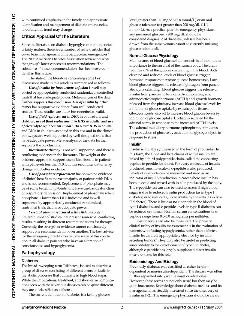
2:20 a.m.: Prehospital personnel call with a case of vomiting and altered mentalstatus in a 9-year-old girl. The child’s mother found her unconscious in bed in a pool ofvomit. The child’s mother notes that the child had been vomiting earlier but hadconsumed clear liquids for supper. She also notes that her child had recently started adiet and was doing exceptionally well at her planned weight loss; she also commentsthat for the past two days, her daughter kept a pitcher of water at the bedside and wouldwake frequently and drink. The child felt that the extra water was helping her weightloss program. The paramedics note that the child’s respiratory rate is 50, she has a heartrate of 128, and her bedside glucose test measures “HHH” on their glucometer. They’vebeen unable to obtain intravenous access in this child.
DIABETES is a chronic disease that requires long-term medical attention.As many as 5%-6% of the United States population have either diagnosed
or undiagnosed diabetes.1
Diabetic emergencies are common in patients with diabetes, and the effectscan be devastating. Hypoglycemia and hyperglycemia represent two extremesin the emergency presentations of the diabetic patient. This edition of EmergencyMedicine Practice reviews the acute management of two of the most serioushyperglycemic disorders—diabetic ketoacidosis (DKA) and hyperglycemichyperosmolar syndrome (HHS)—and the controversy that surrounds themanagement of these diseases.
Over 100,000 cases of DKA, 5000 cases of diabetic coma, and 10,000 cases ofhyperosmolar coma were recorded in the United States between 1989 and 1991.2
DKA is the most common cause of diabetes-related death in childhood and is asignificant cause of mortality in adults.101 There is considerable overlap—aboutone-fifth to one-third of patients with otherwise classic DKA will also havehyperosmolarity. About one-half to three-fourths of patients with uncontrolleddiabetes will have an osmolarity of 320 mOsm or more.3
Despite the plethora of guidelines and protocols, all with meticulousdetails of fluid and electrolyte replacement and insulin therapy, the mortality ofdiabetic emergencies has remained unchanged for the past 10 years.111 However,
Emergency Medicine Practice 2 www.empractice.net • February 2004
CO
PY
RIG
HTE
D M
ATER
IAL—
DO
NO
T P
HO
TOC
OP
Y O
R D
ISTR
IBU
TE E
LEC
TRO
NIC
ALL
Y W
ITH
OU
T W
RIT
TEN
CO
NSE
NT
OF
EB P
RA
CTI
CE,
LLC with continued emphasis on the timely and appropriate
identification and management of diabetic emergencies,hopefully this trend may change.
Critical Appraisal Of The Literature
Since the literature on diabetic hyperglycemic emergenciesis fairly mature, there are a number of review articles thatcover basic management of hyperglycemic emergencies.4
The 2003 American Diabetes Association review presentsthat group’s latest consensus recommendations.4 Thesubstance of these recommendations has been covered indetail in this article.
The state of the literature concerning some keydiscussions made in this article is summarized as follows.
Use of insulin by intravenous infusion is well-sup-ported by appropriately conducted randomized, controlledtrials that have adequate power. Meta-analysis of the datafurther supports this conclusion. Use of insulin by otherroutes has supportive evidence from well-conductedstudies. These studies are older, but nonetheless valid.
Use of fluid replacement in DKA in both adults andchildren, use of fluid replacement in HHS in adults, and useof electrolyte replacement in both DKA and HHS in adultsand DKA in children, as noted in this text and in the clinicalpathways, are well-supported by well-designed trials thathave adequate power. Meta-analysis of the data furthersupports the conclusions.
Bicarbonate therapy is not well-supported, and there isconflicting evidence in the literature. The weight of theevidence appears to support use of bicarbonate in patientswith pH levels less than 7.0, but this recommendation maychange with better evidence.
Use of phosphate replacement has shown no evidenceof clinical benefit to the vast majority of patients with DKAand is not recommended. Replacement of phosphate maybe of some benefit in patients who have cardiac dysfunctionor respiratory depression. Replacement of phosphate whenphosphate is lower than 1.0 is indicated and is well-supported by appropriately conducted randomized,controlled trials that have adequate power.
Cerebral edema associated with DKA has only alimited number of studies that present somewhat conflictingresults, resulting in different recommendations for therapy.Currently, the strength of evidence cannot conclusivelysupport one recommendation over another. The best advicefor the emergency practitioner is to be wary of this condi-tion in all diabetic patients who have an alteration ofconsciousness and hyperglycemia.
Pathophysiology
DiabetesThe broad, sweeping term “diabetes” is used to describe agroup of diseases consisting of different errors or faults inmetabolic processes that culminate in high blood sugar.While the implications, treatment, and short-term complica-tions seen with these various diseases can be quite different,they are all classified as diabetes.
The current definition of diabetes is a fasting glucose
level greater than 140 mg/dL (7.8 mmol/L) or an oralglucose tolerance test greater than 200 mg/dL (11.1mmol/L). As a practical point to emergency physicians,any measured glucose > 200 mg/dL should beconsidered diagnostic of diabetes (unless it has beendrawn from the same venous runoff as currently infusingglucose solutions!).
Normal Glucose PhysiologyMaintenance of blood glucose homeostasis is of paramountimportance to the survival of the human body. The brainrequires 75% of the glucose circulating in the blood. Bothelevated and reduced levels of blood glucose triggerhormonal responses to restore glucose homeostasis. Lowblood glucose triggers the release of glucagon from pancre-atic alpha cells. High blood glucose triggers the release ofinsulin from pancreatic beta cells. Additional signals,adrenocorticotropic hormone (ACTH) and growth hormonereleased from the pituitary, increase blood glucose levels byinhibition of glucose uptake by extrahepatic tissues.Glucocorticoids also act to increase blood glucose levels byinhibition of glucose uptake. Cortisol is secreted by theadrenal cortex in response to the increased ACTH levels.The adrenal medullary hormone, epinephrine, stimulatesthe production of glucose by activation of glycogenolysis inresponse to stress.
InsulinInsulin is initially synthesized in the form of proinsulin. Inthis form, the alpha and beta chains of active insulin arelinked by a third polypeptide chain, called the connectingpeptide (c-peptide for short). For every molecule of insulinproduced, one molecule of c-peptide is also produced.Levels of c-peptide can be measured and used as anindicator of insulin production in cases where insulin hasbeen injected and mixed with insulin produced by the body.The c-peptide test can also be used to assess if high bloodsugar is due to reduced insulin production (as in type Idiabetes) or to reduced glucose intake by the cells (as in typeII diabetes). There is little or no c-peptide in the blood oftype I diabetics, and c-peptide levels in type II diabetics canbe reduced or normal. Normal serum concentrations of c-peptide range from 0.5-3.0 nanograms per milliliter.
Insulin levels can also be measured. The primaryclinical utility of insulin measurement is in the evaluation ofpatients with fasting hypoglycemia, rather than diabetes.Insulin levels are inappropriately elevated by insulin-secreting tumors.9 They may also be useful in predictingsusceptibility to the development of type II diabetes,although c-peptide has largely supplanted direct insulinmeasurements for this role.
Epidemiology And EtiologyPreviously, diabetes was classified as either insulin-dependent or non-insulin-dependent. The disease was oftenfurther separated into juvenile onset or adult onset.However, these terms are not only passé, but they may bequite inaccurate. Knowledge about diabetes mellitus and itsmanagement has steadily increased since the discovery ofinsulin in 1921. The emergency physician should be aware
3 Emergency Medicine PracticeFebruary 2004 • www.empractice.net
CO
PY
RIG
HTED
MATER
IAL—
DO
NO
T PH
OTO
CO
PY
OR
DISTR
IBU
TE ELECTR
ON
ICA
LLY W
ITHO
UT W
RIT
TEN C
ON
SENT O
F EB P
RA
CTIC
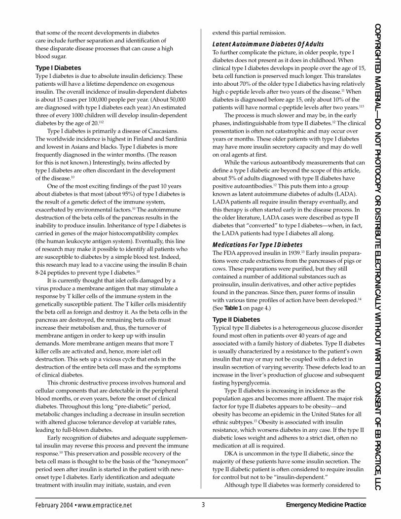
E, LLCthat some of the recent developments in diabetescare include further separation and identification ofthese disparate disease processes that can cause a highblood sugar.
Type I DiabetesType I diabetes is due to absolute insulin deficiency. Thesepatients will have a lifetime dependence on exogenousinsulin. The overall incidence of insulin-dependent diabetesis about 15 cases per 100,000 people per year. (About 50,000are diagnosed with type I diabetes each year.) An estimatedthree of every 1000 children will develop insulin-dependentdiabetes by the age of 20.112
Type I diabetes is primarily a disease of Caucasians.The worldwide incidence is highest in Finland and Sardiniaand lowest in Asians and blacks. Type I diabetes is morefrequently diagnosed in the winter months. (The reasonfor this is not known.) Interestingly, twins affected bytype I diabetes are often discordant in the developmentof the disease.10
One of the most exciting findings of the past 10 yearsabout diabetes is that most (about 95%) of type I diabetes isthe result of a genetic defect of the immune system,exacerbated by environmental factors.10 The autoimmunedestruction of the beta cells of the pancreas results in theinability to produce insulin. Inheritance of type I diabetes iscarried in genes of the major histocompatibility complex(the human leukocyte antigen system). Eventually, this lineof research may make it possible to identify all patients whoare susceptible to diabetes by a simple blood test. Indeed,this research may lead to a vaccine using the insulin B chain8-24 peptides to prevent type I diabetes.10
It is currently thought that islet cells damaged by avirus produce a membrane antigen that may stimulate aresponse by T killer cells of the immune system in thegenetically susceptible patient. The T killer cells misidentifythe beta cell as foreign and destroy it. As the beta cells in thepancreas are destroyed, the remaining beta cells mustincrease their metabolism and, thus, the turnover ofmembrane antigen in order to keep up with insulindemands. More membrane antigen means that more Tkiller cells are activated and, hence, more islet celldestruction. This sets up a vicious cycle that ends in thedestruction of the entire beta cell mass and the symptomsof clinical diabetes.
This chronic destructive process involves humoral andcellular components that are detectable in the peripheralblood months, or even years, before the onset of clinicaldiabetes. Throughout this long “pre-diabetic” period,metabolic changes including a decrease in insulin secretionwith altered glucose tolerance develop at variable rates,leading to full-blown diabetes.
Early recognition of diabetes and adequate supplemen-tal insulin may reverse this process and prevent the immuneresponse.10 This preservation and possible recovery of thebeta cell mass is thought to be the basis of the “honeymoon”period seen after insulin is started in the patient with new-onset type I diabetes. Early identification and adequatetreatment with insulin may initiate, sustain, and even
extend this partial remission.
Latent Autoimmune Diabetes Of AdultsTo further complicate the picture, in older people, type Idiabetes does not present as it does in childhood. Whenclinical type I diabetes develops in people over the age of 15,beta cell function is preserved much longer. This translatesinto about 70% of the older type I diabetics having relativelyhigh c-peptide levels after two years of the disease.11 Whendiabetes is diagnosed before age 15, only about 10% of thepatients will have normal c-peptide levels after two years.113
The process is much slower and may be, in the earlyphases, indistinguishable from type II diabetes.12 The clinicalpresentation is often not catastrophic and may occur overyears or months. These older patients with type I diabetesmay have more insulin secretory capacity and may do wellon oral agents at first.
While the various autoantibody measurements that candefine a type I diabetic are beyond the scope of this article,about 5% of adults diagnosed with type II diabetes havepositive autoantibodies.12 This puts them into a groupknown as latent autoimmune diabetes of adults (LADA).LADA patients all require insulin therapy eventually, andthis therapy is often started early in the disease process. Inthe older literature, LADA cases were described as type IIdiabetes that “converted” to type I diabetes—when, in fact,the LADA patients had type I diabetes all along.
Medications For Type I DiabetesThe FDA approved insulin in 1939.13 Early insulin prepara-tions were crude extractions from the pancreases of pigs orcows. These preparations were purified, but they stillcontained a number of additional substances such asproinsulin, insulin derivatives, and other active peptidesfound in the pancreas. Since then, purer forms of insulinwith various time profiles of action have been developed.14
(See Table 1 on page 4.)
Type II DiabetesTypical type II diabetes is a heterogeneous glucose disorderfound most often in patients over 40 years of age andassociated with a family history of diabetes. Type II diabetesis usually characterized by a resistance to the patient’s owninsulin that may or may not be coupled with a defect ininsulin secretion of varying severity. These defects lead to anincrease in the liver’s production of glucose and subsequentfasting hyperglycemia.
Type II diabetes is increasing in incidence as thepopulation ages and becomes more affluent. The major riskfactor for type II diabetes appears to be obesity—andobesity has become an epidemic in the United States for allethnic subtypes.15 Obesity is associated with insulinresistance, which worsens diabetes in any case. If the type IIdiabetic loses weight and adheres to a strict diet, often nomedication at all is required.
DKA is uncommon in the type II diabetic, since themajority of these patients have some insulin secretion. Thetype II diabetic patient is often considered to require insulinfor control but not to be “insulin-dependent.”
Although type II diabetes was formerly considered to
Emergency Medicine Practice 4 www.empractice.net • February 2004
CO
PY
RIG
HTE
D M
ATER
IAL—
DO
NO
T P
HO
TOC
OP
Y O
R D
ISTR
IBU
TE E
LEC
TRO
NIC
ALL
Y W
ITH
OU
T W
RIT
TEN
CO
NSE
NT
OF
EB P
RA
CTI
CE,
LLC be a disease of people older than 40, emergency physicians
are seeing increasing numbers of patients in their 20s or 30swith type II diabetes. It is seen more frequently in youngerpeople in association with childhood obesity.16,17 Type IIdiabetes is even found in adolescents. (These patients havethe slow clinical course of type II diabetics without thedevelopment of DKA.)
High blood sugars in type II diabetics can be associatedwith obesity, high blood pressure, renal failure, andaccelerated coronary artery disease. Prolonged hyperglyce-mia will increase the rate of development of all of thesediseases.18 Prolonged high blood sugars will also reduce theeffect of insulin (insulin resistance) and decrease thesecretion of insulin (glucose toxicity).19 When the pancreasquits making insulin, then the type II diabetic needs insulinas much as the type I diabetic.20 (As noted, these patientsoften will secrete enough insulin to stave off ketoacidosis,but not enough to prevent profound hyperglycemia.)
Maturity Onset Diabetes Of The YoungMaturity onset diabetes of the young (MODY) is afamilial form of type II diabetes that was described asa separate entity from type II diabetes by many research-ers.21-24 MODY represents a very small subset of type IIdiabetes. It is estimated that only 1%-5% of all type IIdiabetics may have MODY.21-24
A typical MODY patient has a diagnosis of diabetesthat is not type I diabetes, is less than 25 years old, and has apositive family history with a dominant mode of inherit-ance. These patients are often lean and do not appear to beinsulin-resistant. There are several types that have beenidentified and classified by the type of genetic defectinvolved. A monogenic defect in insulin secretion isresponsible for all forms of MODY, and genetic tests canconfirm the clinical suspicion.25-31
Medications For Type II DiabetesOral medications for the treatment of type II diabetes havebeen around since the 1950s. Recently, there has been amarked increase in the number and kinds of drugs that are
seen by the emergency physician. (See Table 2 on page 5.)Oral medications work by causing the pancreas to
make more insulin, by making the tissues more sensitive tothe effects of insulin, by decreasing hepatic output ofglucose, or by decreasing absorption of glucose from thegut. No single medication uses all four mechanisms, butmost will have one or more effects.
A new, cutting-edge drug, exenatide, actually causesthe pancreas to produce more islet cells and hence producemore natural insulin.32
Secondary DiabetesSevere pancreatic disorders, including pancreatic cancer,chronic pancreatitis, hemachromatosis, and cystic fibrosis,can lead to insulin deficiency and subsequent diabetes.Since the pancreatic tissue in these individuals is destroyed,these patients clinically resemble type I diabetics andrequire insulin.
Other patients may have diseases or hormonalsyndromes that interfere with insulin secretion (such aspheochromocytoma) or insulin use (such as acromegaly,Cushing’s syndrome, and pheochromocytoma). These casesmore closely resemble type II diabetes.
Several common medications can impair the body’s useof insulin and produce diabetes. The most commonly seen isprobably associated with glucocorticosteroids such asmethylprednisolone or prednisone. Treatment for highblood pressure (furosemide, clonidine, and thiazidediuretics), other drugs with hormonal activity (oral contra-ceptives, thyroid hormone, and progestins), and the anti-inflammatory drug indomethacin can also cause or exacer-bate diabetes. Numerous other drugs can impair glucoseabsorption, such as haloperidol, lithium, phenothiazines,tricyclic antidepressants, isoniazid, nicotinic acid, heparin,and cimetidine.
Drug-induced diabetes may or may not requireinsulin and may simply resolve after the drug is stopped.Drug-induced diabetes may resemble either type I ortype II diabetes.
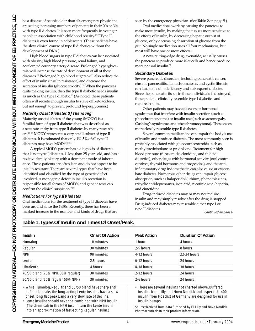
Table 1. Types Of Insulin And Times Of Onset/Peak.
Insulin Onset Of Action Peak Action Duration Of Action
Humalog 10 minutes 1 hour 4 hours
Regular 30 minutes 2-5 hours 8 hours
NPH 90 minutes 4-12 hours 22-24 hours
Lente 2.5 hours 6-12 hours 24 hours
Ultralente 4 hours 8-18 hours 30 hours
70/30 blend (70% NPH, 30% regular) 30 minutes 2-12 hours 24 hours
50/50 blend (50% regular, 50% NPH) 30 minutes 2-6 hours 24 hours
• While Humalog, Regular, and 50/50 blend have sharp anddefinable peaks, the long-acting Lente insulins have a slowonset, long flat peaks, and a very slow rate of decline.
• Lente insulins should never be combined with NPH insulin.(The chemicals in the NPH insulin turn the Lente insulininto an approximation of fast-acting Regular insulin.)
• There are several insulins not charted above. Bufferedinsulins from Lilly and Novo Nordisk and a special U-400insulin from Hoechst of Germany are designed for use ininsulin pumps.
Source: Derived from data furnished by Eli Lilly and Novo NordiskPharmaceuticals in their product information.
Continued on page 6
5 Emergency Medicine PracticeFebruary 2004 • www.empractice.net
CO
PY
RIG
HTED
MATER
IAL—
DO
NO
T PH
OTO
CO
PY
OR
DISTR
IBU
TE ELECTR
ON
ICA
LLY W
ITHO
UT W
RIT
TEN C
ON
SENT O
F EB P
RA
CTIC
E, LLC
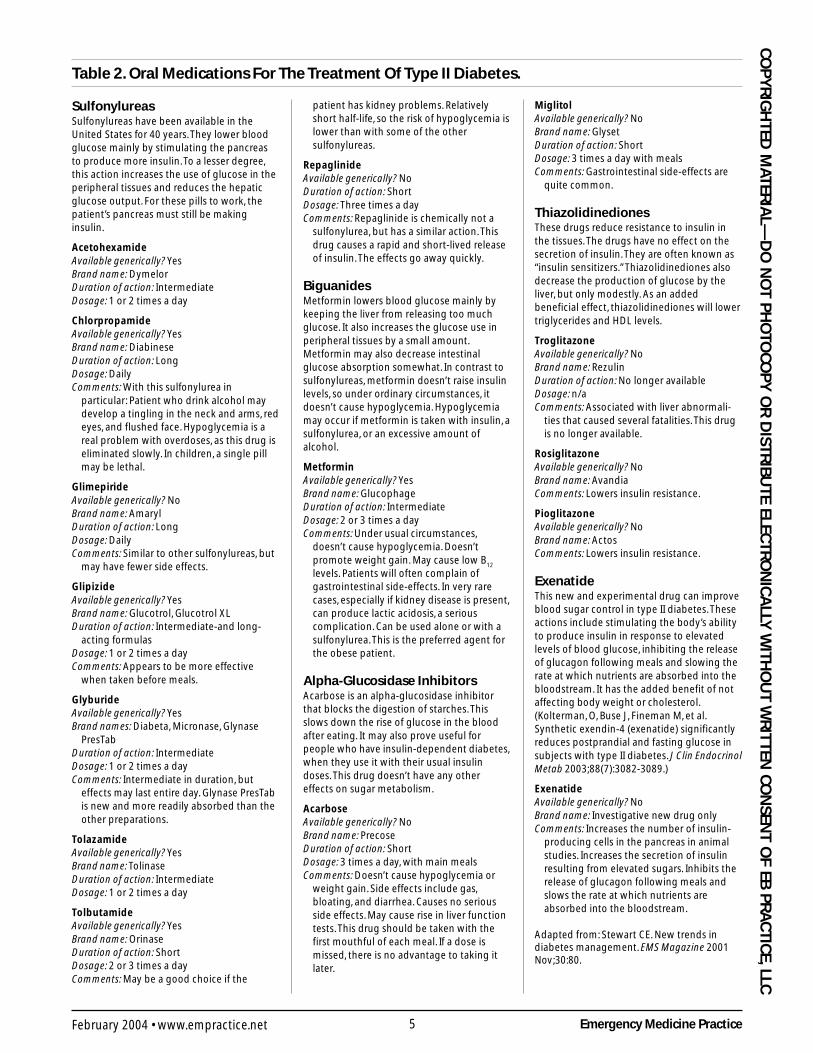
Table 2. Oral Medications For The Treatment Of Type II Diabetes.
SulfonylureasSulfonylureas have been available in theUnited States for 40 years. They lower bloodglucose mainly by stimulating the pancreasto produce more insulin. To a lesser degree,this action increases the use of glucose in theperipheral tissues and reduces the hepaticglucose output. For these pills to work, thepatient’s pancreas must still be makinginsulin.
AcetohexamideAvailable generically? YesBrand name: DymelorDuration of action: IntermediateDosage: 1 or 2 times a day
ChlorpropamideAvailable generically? YesBrand name: DiabineseDuration of action: LongDosage: DailyComments: With this sulfonylurea in
particular: Patient who drink alcohol maydevelop a tingling in the neck and arms, redeyes, and flushed face. Hypoglycemia is areal problem with overdoses, as this drug iseliminated slowly. In children, a single pillmay be lethal.
GlimepirideAvailable generically? NoBrand name: AmarylDuration of action: LongDosage: DailyComments: Similar to other sulfonylureas, but
may have fewer side effects.
GlipizideAvailable generically? YesBrand name: Glucotrol, Glucotrol XLDuration of action: Intermediate-and long-
acting formulasDosage: 1 or 2 times a dayComments: Appears to be more effective
when taken before meals.
GlyburideAvailable generically? YesBrand names: Diabeta, Micronase, Glynase
PresTabDuration of action: IntermediateDosage: 1 or 2 times a dayComments: Intermediate in duration, but
effects may last entire day. Glynase PresTabis new and more readily absorbed than theother preparations.
TolazamideAvailable generically? YesBrand name: TolinaseDuration of action: IntermediateDosage: 1 or 2 times a day
TolbutamideAvailable generically? YesBrand name: OrinaseDuration of action: ShortDosage: 2 or 3 times a dayComments: May be a good choice if the
patient has kidney problems. Relativelyshort half-life, so the risk of hypoglycemia islower than with some of the othersulfonylureas.
RepaglinideAvailable generically? NoDuration of action: ShortDosage: Three times a dayComments: Repaglinide is chemically not a
sulfonylurea, but has a similar action. Thisdrug causes a rapid and short-lived releaseof insulin. The effects go away quickly.
BiguanidesMetformin lowers blood glucose mainly bykeeping the liver from releasing too muchglucose. It also increases the glucose use inperipheral tissues by a small amount.Metformin may also decrease intestinalglucose absorption somewhat. In contrast tosulfonylureas, metformin doesn’t raise insulinlevels, so under ordinary circumstances, itdoesn’t cause hypoglycemia. Hypoglycemiamay occur if metformin is taken with insulin, asulfonylurea, or an excessive amount ofalcohol.
MetforminAvailable generically? YesBrand name: GlucophageDuration of action: IntermediateDosage: 2 or 3 times a dayComments: Under usual circumstances,
doesn’t cause hypoglycemia. Doesn’tpromote weight gain. May cause low B
12
levels. Patients will often complain ofgastrointestinal side-effects. In very rarecases, especially if kidney disease is present,can produce lactic acidosis, a seriouscomplication. Can be used alone or with asulfonylurea. This is the preferred agent forthe obese patient.
Alpha-Glucosidase InhibitorsAcarbose is an alpha-glucosidase inhibitorthat blocks the digestion of starches. Thisslows down the rise of glucose in the bloodafter eating. It may also prove useful forpeople who have insulin-dependent diabetes,when they use it with their usual insulindoses. This drug doesn’t have any othereffects on sugar metabolism.
AcarboseAvailable generically? NoBrand name: PrecoseDuration of action: ShortDosage: 3 times a day, with main mealsComments: Doesn’t cause hypoglycemia or
weight gain. Side effects include gas,bloating, and diarrhea. Causes no seriousside effects. May cause rise in liver functiontests. This drug should be taken with thefirst mouthful of each meal. If a dose ismissed, there is no advantage to taking itlater.
MiglitolAvailable generically? NoBrand name: GlysetDuration of action: ShortDosage: 3 times a day with mealsComments: Gastrointestinal side-effects are
quite common.
ThiazolidinedionesThese drugs reduce resistance to insulin inthe tissues. The drugs have no effect on thesecretion of insulin. They are often known as“insulin sensitizers.” Thiazolidinediones alsodecrease the production of glucose by theliver, but only modestly. As an addedbeneficial effect, thiazolidinediones will lowertriglycerides and HDL levels.
TroglitazoneAvailable generically? NoBrand name: RezulinDuration of action: No longer availableDosage: n/aComments: Associated with liver abnormali-
ties that caused several fatalities. This drugis no longer available.
RosiglitazoneAvailable generically? NoBrand name: AvandiaComments: Lowers insulin resistance.
PioglitazoneAvailable generically? NoBrand name: ActosComments: Lowers insulin resistance.
ExenatideThis new and experimental drug can improveblood sugar control in type II diabetes. Theseactions include stimulating the body’s abilityto produce insulin in response to elevatedlevels of blood glucose, inhibiting the releaseof glucagon following meals and slowing therate at which nutrients are absorbed into thebloodstream. It has the added benefit of notaffecting body weight or cholesterol.(Kolterman, O, Buse J, Fineman M, et al.Synthetic exendin-4 (exenatide) significantlyreduces postprandial and fasting glucose insubjects with type II diabetes. J Clin EndocrinolMetab 2003;88(7):3082-3089.)
ExenatideAvailable generically? NoBrand name: Investigative new drug onlyComments: Increases the number of insulin-
producing cells in the pancreas in animalstudies. Increases the secretion of insulinresulting from elevated sugars. Inhibits therelease of glucagon following meals andslows the rate at which nutrients areabsorbed into the bloodstream.
Adapted from: Stewart CE. New trends indiabetes management. EMS Magazine 2001Nov;30:80.
Emergency Medicine Practice 6 www.empractice.net • February 2004
CO
PY
RIG
HTE
D M
ATER
IAL—
DO
NO
T P
HO
TOC
OP
Y O
R D
ISTR
IBU
TE E
LEC
TRO
NIC
ALL
Y W
ITH
OU
T W
RIT
TEN
CO
NSE
NT
OF
EB P
RA
CTI
CE,
LLC
Gestational DiabetesGestational diabetes is glucose intolerance during preg-nancy. About 4% of all pregnancies are complicated bygestational diabetes.114 Mothers with gestational diabeteshave higher rates of cesarean delivery and chronic hyperten-sion.114 Their children may have macrosomia, hypoglycemia,hypocalcemia, and hyperbilirubinemia. (A completediscussion of gestational diabetes is beyond the scope ofthis review.)
Impaired Glucose ToleranceAn impaired glucose tolerance is defined as a fastingglucose of greater than 110 mg/dL (6.1 mmol/L) but lessthan 140 mg/dL.115,116 It is also defined as a patient who hasa glucose tolerance test with ranges between 140 mg/dL (7.8mmol/L) and 199 mg/dL.115,116 Impaired glucose tolerancewas formerly known as borderline, chemical, or latentdiabetes.115,116 (Impaired glucose tolerance and its implica-tions are not covered in this review.)
Differential Diagnosis: HyperglycemicComplications Of Diabetes
DKA and HHS are the most lethal hyperglycemic complica-tions of diabetes. Patients may present with some combina-tion of hyperglycemia, altered mental status, and dehydra-tion. Significantly, both can be associated with coexistingmedical conditions that may obfuscate the clinical encoun-ter, and both can be associated with a patient’s initialpresentation of diabetes. The history, physical examination,and laboratory studies must therefore be tailored to detectDKA, HHS, and any possible coexisting illnesses. (See alsoTable 3 and Table 4 on page 7.)
The basic underlying mechanism for both DKA andHHS is a reduction in the net effective action of circulatinginsulin coupled with a concomitant elevation ofcounterregulatory hormones, such as glucagon, catechola-mines, cortisol, and growth hormone.
The differential diagnosis for the patient who has nohistory of diabetes and is found in a coma starts with the
very long differential for altered mental status. Since thesepatients may have complex medical histories andcomorbidities, this evaluation should not stop at the firstdisease encountered.
Not all patients with ketoacidosis have DKA.The differential diagnosis for the patient who has aknown history of diabetes and presents with the findingsof DKA includes:
• HHS• Alcoholic ketoacidosis• Starvation• Sepsis• Lactic acidosis• Uremia
Starvation ketosis and alcoholic ketoacidosis areexcluded by the clinical history and by a plasma glucosethat ranges from mildly elevated (rarely ≥ 250 mg/dL) tofrank hypoglycemia. In addition, although alcoholicketoacidosis can result in profound acidosis, the serumbicarbonate concentration in starvation ketosis is usuallyless pronounced than in DKA.
Diabetic KetoacidosisThe pathology of DKA is due to the inability of the cells totake up and use glucose when insulin is not present. Insulinis the most significant hormone of blood glucose regulation.It increases the ability of the cell to take in glucose andstimulates the manufacture of glycogen.
DKA can be caused by either an absolute or relativedeficiency of insulin and is exacerbated by the concomitantincrease in the insulin counter-regulatory hormones:glucagon, epinephrine, cortisol, and growth hormone. InDKA, the classic triad of hyperglycemia, ketosis, andacidosis is present. The counter-regulatory hormonesfurther shift metabolism toward hyperglycemia, acidosis,and ketosis.
DKA is characterized by hyperglycemia with glucoseover 250 mg/dL, although it has been recently recognizedthat some patients may present with only mild hyperglyce-mia, but with marked ketoacidosis (notably pregnant
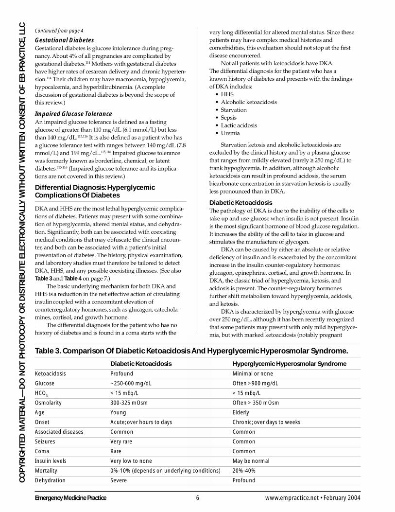
Table 3. Comparison Of Diabetic Ketoacidosis And Hyperglycemic Hyperosmolar Syndrome.
Diabetic Ketoacidosis Hyperglycemic Hyperosmolar Syndrome
Ketoacidosis Profound Minimal or none
Glucose ~250-600 mg/dL Often >900 mg/dL
HCO3
< 15 mEq/L > 15 mEq/L
Osmolarity 300-325 mOsm Often > 350 mOsm
Age Young Elderly
Onset Acute; over hours to days Chronic; over days to weeks
Associated diseases Common Common
Seizures Very rare Common
Coma Rare Common
Insulin levels Very low to none May be normal
Mortality 0%-10% (depends on underlying conditions) 20%-40%
Dehydration Severe Profound
Continued from page 4
7 Emergency Medicine PracticeFebruary 2004 • www.empractice.net
CO
PY
RIG
HTED
MATER
IAL—
DO
NO
T PH
OTO
CO
PY
OR
DISTR
IBU
TE ELECTR
ON
ICA
LLY W
ITHO
UT W
RIT
TEN C
ON
SENT O
F EB P
RA
CTIC
E, LLCwomen).115 Typically, patients will be overtly acidotic withpH levels less than 7.35, low serum bicarbonate (commonlyunder 15 mmol/L), and positive serum ketones. (If a patientpresents with a marked ketosis, but a glucose under 250mg/dL, the physician should consider the possibility thatthe ketosis is related to another cause such as alcoholicketosis, rather than DKA.)
Insulin must be administered to correct the underlyingmetabolic abnormalities.
HyperglycemiaThe main counter-regulatory hormones (glucagon, cortisol,catecholamines, and growth hormones) are increasedbecause the cells can’t use the available glucose. Increasedhepatic glucose production and decreased peripheraluptake of glucose are major causes of the hyperglycemiaseen in diabetes.
In the absence of insulin, glucagon becomes theprimary driving hormone for hepatic carbohydratemetabolism. Glucagon stimulates the release of glucosefrom the liver by gluconeogenesis and glycogenolysis.Liver glycogen stores are broken down into sugar andreleased into the bloodstream. Deficiency of insulin andconcomitant increases in glucagon will enhance the liverproduction of glucose by breakdown of fat and protein. Thefatty acid oxidation leads to ketone body formation andinhibits the conversion of acetyl CoA, by acetyl CoAcarboxylase, to malonyl CoA, which is the first intermediatein the lipogenesis pathway. This inhibition means that fattyacids are unable to enter the citric acid cycle and moveinstead into the mitochondria, where they are oxidized andfurther ketone bodies (acetoacetate and beta-hydroxybutyrate) are formed.33
At the same time, peripheral uptake of glucose isimpaired by both lack of insulin and excess of glucagon,so the excess glucose accumulates in the bloodstream.Insulin deficiency alone, or in combination with theinsulin counter-regulatory hormone increases willincrease protein breakdown, providing amino acidsfor increased gluconeogenesis.
Poorly controlled diabetes may result in
hypertriglyceridemia (found in as many as 50% ofpatients with DKA).34,35 This extreme hypertriglyceridemiacan cause the blood sugar, sodium, and bicarbonate to befactitiously lowered.36
Blood glucose levels will rise above the renal thresholdfor glucose reabsorption, so an osmotic diuresis occurs, asdiscussed below.
AcidosisInsulin inhibits the lipolytic action of cortisol and growthhormone. A deficiency of insulin will increase the circulatinglevels of fatty acids. These fatty acids are metabolized byalternative metabolic pathways. The breakdown products ofthe alternative metabolic pathways cause the characteristicacetone byproducts and a resultant metabolic acidosis. Theacidosis of DKA is mostly due to the ketoacids, althoughexcess fatty acids and lactic acid (produced by poor tissueperfusion) also contribute to the lowered pH. Theseincreased ketoacids include acetone, beta-hydroxybutyricacid, and acetoacetic acid, although the major derangementin DKA is an increased level of beta-hydroxybutyric acidrather than acetone or acetoacetate (on the order of 10:1).115
Volume DepletionThe kidney plays an important role in the development ofDKA. The normal renal threshold for glucose reabsorptionis greater than 240 mg/dL.37 When the patient is well-hydrated and normal kidney function is maintained, theserum glucose level is maintained at about 240 mg/dL byspillage into the urine.
The osmotic diuresis results in significant volumedepletion unless the patient drinks copious amounts offluids. When hypovolemia occurs, the glomerular filtrationrate falls and the hyperglycemia is exacerbated. The diuresisalso leads to significant urinary losses of potassium,sodium, phosphate, chloride, and magnesium ions.
Hyperglycemic Hyperosmolar SyndromeEpidemiologyHHS has a mean age of onset in the seventh decade of life.115
Males are affected slightly less often than females.115
Residents of nursing facilities who are both elderly and
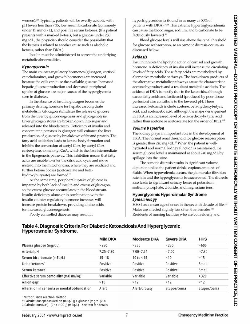
Table 4. Diagnostic Criteria For Diabetic Ketoacidosis And HyperglycemicHyperosmolar Syndrome.
Mild DKA Moderate DKA Severe DKA HHS
Plasma glucose (mg/dL) >250 >250 >250 >600
Arterial pH 7.25–7.30 7.00–7.24 <7.00 >7.30
Serum bicarbonate (mEq/L) 15–18 10 to <15 <10 >15
Urine ketones* Positive Positive Positive Small
Serum ketones* Positive Positive Positive Small
Effective serum osmolality (mOsm/kg)† Variable Variable Variable >320
Anion gap‡ >10 >12 >12 <12
Alteration in sensoria or mental obtundation Alert Alert/drowsy Stupor/coma Stupor/coma
* Nitroprusside reaction method† Calculation: 2[measured Na (mEq/L)] + glucose (mg/dL)/18‡ Calculation: (Na+) - (Cl- + HCO
3-) (mEq/L)—see text for details
Emergency Medicine Practice 8 www.empractice.net • February 2004
CO
PY
RIG
HTE
D M
ATER
IAL—
DO
NO
T P
HO
TOC
OP
Y O
R D
ISTR
IBU
TE E
LEC
TRO
NIC
ALL
Y W
ITH
OU
T W
RIT
TEN
CO
NSE
NT
OF
EB P
RA
CTI
CE,
LLC
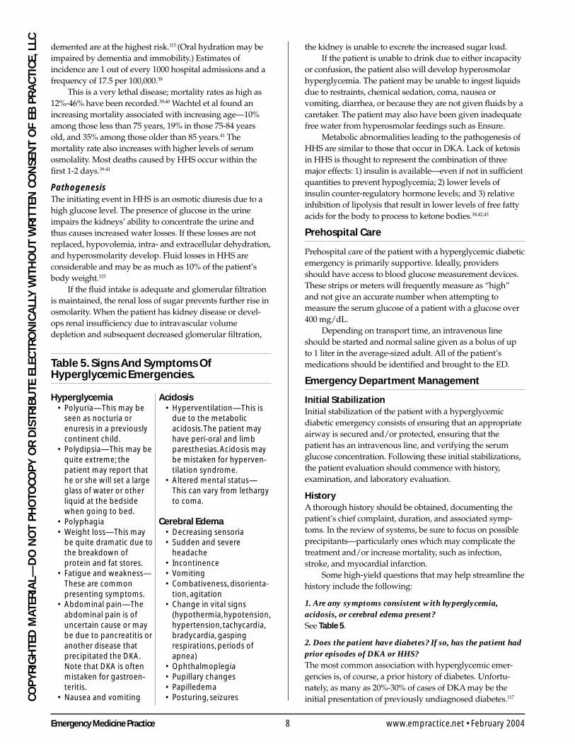
Table 5. Signs And Symptoms OfHyperglycemic Emergencies.
Hyperglycemia• Polyuria—This may be
seen as nocturia orenuresis in a previouslycontinent child.
• Polydipsia—This may bequite extreme; thepatient may report thathe or she will set a largeglass of water or otherliquid at the bedsidewhen going to bed.
• Polyphagia• Weight loss—This may
be quite dramatic due tothe breakdown ofprotein and fat stores.
• Fatigue and weakness—These are commonpresenting symptoms.
• Abdominal pain—Theabdominal pain is ofuncertain cause or maybe due to pancreatitis oranother disease thatprecipitated the DKA.Note that DKA is oftenmistaken for gastroen-teritis.
• Nausea and vomiting
Acidosis• Hyperventilation—This is
due to the metabolicacidosis. The patient mayhave peri-oral and limbparesthesias. Acidosis maybe mistaken for hyperven-tilation syndrome.
• Altered mental status—This can vary from lethargyto coma.
Cerebral Edema• Decreasing sensoria• Sudden and severe
headache• Incontinence• Vomiting• Combativeness, disorienta-
tion, agitation• Change in vital signs
(hypothermia, hypotension,hypertension, tachycardia,bradycardia, gaspingrespirations, periods ofapnea)
• Ophthalmoplegia• Pupillary changes• Papilledema• Posturing, seizures
demented are at the highest risk.115 (Oral hydration may beimpaired by dementia and immobility.) Estimates ofincidence are 1 out of every 1000 hospital admissions and afrequency of 17.5 per 100,000.38
This is a very lethal disease; mortality rates as high as12%-46% have been recorded.39,40 Wachtel et al found anincreasing mortality associated with increasing age—10%among those less than 75 years, 19% in those 75-84 yearsold, and 35% among those older than 85 years.41 Themortality rate also increases with higher levels of serumosmolality. Most deaths caused by HHS occur within thefirst 1-2 days.39-41
PathogenesisThe initiating event in HHS is an osmotic diuresis due to ahigh glucose level. The presence of glucose in the urineimpairs the kidneys’ ability to concentrate the urine andthus causes increased water losses. If these losses are notreplaced, hypovolemia, intra- and extracellular dehydration,and hyperosmolarity develop. Fluid losses in HHS areconsiderable and may be as much as 10% of the patient’sbody weight.115
If the fluid intake is adequate and glomerular filtrationis maintained, the renal loss of sugar prevents further rise inosmolarity. When the patient has kidney disease or devel-ops renal insufficiency due to intravascular volumedepletion and subsequent decreased glomerular filtration,
the kidney is unable to excrete the increased sugar load.If the patient is unable to drink due to either incapacity
or confusion, the patient also will develop hyperosmolarhyperglycemia. The patient may be unable to ingest liquidsdue to restraints, chemical sedation, coma, nausea orvomiting, diarrhea, or because they are not given fluids by acaretaker. The patient may also have been given inadequatefree water from hyperosmolar feedings such as Ensure.
Metabolic abnormalities leading to the pathogenesis ofHHS are similar to those that occur in DKA. Lack of ketosisin HHS is thought to represent the combination of threemajor effects: 1) insulin is available—even if not in sufficientquantities to prevent hypoglycemia; 2) lower levels ofinsulin counter-regulatory hormone levels; and 3) relativeinhibition of lipolysis that result in lower levels of free fattyacids for the body to process to ketone bodies.38,42,43
Prehospital Care
Prehospital care of the patient with a hyperglycemic diabeticemergency is primarily supportive. Ideally, providersshould have access to blood glucose measurement devices.These strips or meters will frequently measure as “high”and not give an accurate number when attempting tomeasure the serum glucose of a patient with a glucose over400 mg/dL.
Depending on transport time, an intravenous lineshould be started and normal saline given as a bolus of upto 1 liter in the average-sized adult. All of the patient’smedications should be identified and brought to the ED.
Emergency Department Management
Initial StabilizationInitial stabilization of the patient with a hyperglycemicdiabetic emergency consists of ensuring that an appropriateairway is secured and/or protected, ensuring that thepatient has an intravenous line, and verifying the serumglucose concentration. Following these initial stabilizations,the patient evaluation should commence with history,examination, and laboratory evaluation.
HistoryA thorough history should be obtained, documenting thepatient’s chief complaint, duration, and associated symp-toms. In the review of systems, be sure to focus on possibleprecipitants—particularly ones which may complicate thetreatment and/or increase mortality, such as infection,stroke, and myocardial infarction.
Some high-yield questions that may help streamline thehistory include the following:
1. Are any symptoms consistent with hyperglycemia,acidosis, or cerebral edema present?See Table 5.
2. Does the patient have diabetes? If so, has the patient hadprior episodes of DKA or HHS?The most common association with hyperglycemic emer-gencies is, of course, a prior history of diabetes. Unfortu-nately, as many as 20%-30% of cases of DKA may be theinitial presentation of previously undiagnosed diabetes.117
9 Emergency Medicine PracticeFebruary 2004 • www.empractice.net
CO
PY
RIG
HTED
MATER
IAL—
DO
NO
T PH
OTO
CO
PY
OR
DISTR
IBU
TE ELECTR
ON
ICA
LLY W
ITHO
UT W
RIT
TEN C
ON
SENT O
F EB P
RA
CTIC
E, LLCWhen DKA occurs as the initial presentation in the new
diabetic, the symptoms are often gradual in onset withprogressive dehydration and slowly developing ketosis. Thestress and symptoms of another illness may both mask theonset of the DKA and precipitate the entire process.
In the established diabetic, DKA can develop quiterapidly. This may occur during an illness or when insulintherapy has been forgotten, deliberately omitted, ordisrupted. When this happens, the ketoacidosis maypredominate, and DKA may present with only a modestelevation of the blood glucose (250 mg/dL).
DKA most often occurs in patients with type I diabetes,although it may occur in patients with type II diabetes.118 Inone series, 13% of cases of DKA occurred in patients over 60years of age and 31% of mixed DKA and HHS cases were inpatients over the age of 60.44
3. Is there an associated infection?An infection—including pneumonia, urinary tract infection,or sepsis—is the most common precipitating factor of bothDKA and HHS.44-47 The physician must search diligently inthese patients for a focus of infection such as sinusitis,middle ear infections, prostatitis, perirectal abscess, orinfected decubiti. Rectal and pelvic examinations areimportant parts of the evaluation if the patient has noobvious focus of infection identified.
4. Is there another associated illness?Associated medical illnesses are commonly present in HHS.These may include:
• Cerebrovascular accidents (both stroke and intracranialhemorrhage)
• Myocardial infarction• Acute pancreatitis• Pulmonary embolus• Mesenteric thrombosis• Renal failure• Heat stroke and heat stress• Gastrointestinal hemorrhage• Hypothermia• Alcohol consumption or cocaine use
Furthermore, trauma, febrile illnesses, or even psycho-logical turmoil may elevate the counter-regulatory hor-mones (glucagon, epinephrine, growth hormone, andcortisol) and precipitate DKA. Other precipitating diseasesinclude alcohol abuse and pancreatitis. In older patients, thestress of myocardial infarction or stroke can precipitateDKA. The stress of pregnancy may also precipitate DKA indiabetic patients.
Steroid excess due to exogenous steroid administrationor endogenous production of steroids (Cushing’s syndrome)is a common precipitating factor of DKA. Excess of counter-regulatory hormones, such as is found in pheochromocy-toma, may cause DKA. Thyrotoxicosis and hyperthyroidismcan precipitate DKA.
5. Has the patient been receiving adequate insulin?Thorough questioning may be necessary in order toevaluate this issue adequately. Failure to take insulin is themost common cause of recurrent DKA, particularly in
adolescents.5 The patient may also run out of insulin, have acalibration error in the injection device, use the wrongconcentration or type of insulin, or inadvertently inject aninadequate dose of insulin. Underinsurance may mean thatthe patient can’t afford insulin or that funds were divertedto other uses (e.g., syringes).5 A change of diet or exercisemay also mean that the insulin administered is inadequate.
In young patients with type I diabetes, psychologicalproblems complicated by eating disorders may be acontributing factor in up to 20% of cases of recurrentketoacidosis.5,48 Factors that decrease compliance in theyoung include fear of weight gain, fear of hypoglycemia,rebellion, and the stress of chronic disease.48
Noncompliance with insulin or other drugs is one ofthe most commonly cited precipitating factors for HHS.38
The noncompliant adult diabetic may have repeatedepisodes of HHS. Noncompliance may be due to psychiatricdisorders, neglect/abuse, onset of dementia, or inability topurchase medications or obtain refills.38 Each of these causesshould be explored in the patient who presents withrecurrent HHS and considered carefully in a patient withnew HHS.
It is also important to note that patients may skipinsulin when ill. Insulin must always be administeredduring illness, even when eating is markedly diminished.Infection induces insulin resistance and may requireincreased or supplemental doses of insulin. Failing toincrease insulin during periods of illness may contribute toan increased incidence of DKA during this period of stress.49
The blood sugar level and the presence of ketones help todetermine the optimal supplemental insulin dosage.
6. What other medications has the patient been taking?Drugs such as sympathomimetics, pentamidine, thiazides,phenytoin, and calcium-channel blocker agents canprecipitate diabetes and DKA.48 As noted earlier, corticoster-oids can not only precipitate diabetes, they can alter theglucose tolerance of normal patients.
The use of drugs and therapies that are known to causehyperglycemia may precipitate HHS. These includeazathioprine, beta-blocking agents, cimetidine, diazoxide,diuretics, glycerol, phenothiazines, calcium-channelblocking agents, phenytoin, and steroids.50-56 Initiation oftotal parenteral nutrition (parenteral hyperalimentation) iscommonly implicated as a precipitating factor, but this is notusually seen in the ED.57
Physical ExaminationThe physical examination for cases of suspected DKA orHHS is essentially the same. In such cases, be sure tocarefully assess vital signs. Also, check and document thepatient’s weight, extent of dehydration, level of conscious-ness, whether Kussmaul respirations are present, and besure to evaluate for the possibility of infection.
Certain physical findings can help determine whetherthe patient has DKA, HHS, or both.
Physical Findings In Both Diabetic Ketoacidosis AndHyperglycemic Hyperosmolar SyndromeUniformly, the patient will be dehydrated. The typical
Emergency Medicine Practice 10 www.empractice.net • February 2004
CO
PY
RIG
HTE
D M
ATER
IAL—
DO
NO
T P
HO
TOC
OP
Y O
R D
ISTR
IBU
TE E
LEC
TRO
NIC
ALL
Y W
ITH
OU
T W
RIT
TEN
CO
NSE
NT
OF
EB P
RA
CTI
CE,
LLC deficit in body water is 20%-25%, which is about 12% of the
patient’s total body weight.58 The physical examination mayreflect this profound dehydration. The mucous membranesmay be quite dry. Skin turgor may be decreased, but thismay be difficult to evaluate in the elderly patient. Thepatient’s eyes may be sunken.
Tachycardia and hypotension may be present. Thepulse may be weak and thready. Use of beta-blockers mustbe taken into consideration when evaluating the vital signs.If infection is present, the patient may be febrile; however,those patients who are immunocompromised or septic maybe normothermic or even hypothermic.48
Abdominal pain or tenderness, nausea and vomiting,lack of bowel sounds, and ileus may be found in manypatients with uncontrolled diabetes. These same findingsmay also be due to an intra-abdominal process that causedthe patient to decompensate. This is not uncommon in theelderly. The development of findings due to decompensa-tion of the diabetes follows the onset of symptoms ratherthan precedes it. The symptoms should improve markedlyafter the patient’s elevated blood sugar and dehydration areproperly treated.
Physical Findings In Diabetic KetoacidosisThe physical signs of DKA can be quite variable. Typicalsigns include fatigue, malaise, thirst, polyuria, reducedskin elasticity (poor skin turgor), dry mucous membranes,hypotension, and tachycardia from the volume deficits.Protracted vomiting may markedly increase the waterloss. Some water loss may also occur due to the compensa-tory hyperventilation from the metabolic acidosis (Kuss-maul respiration).
The patient may note weight loss if there is a longonset. As the patient becomes more ill, he or she will beginto vomit and may complain of abdominal pain.
The exact cause of the abdominal pain associatedwith DKA is not known. Prostaglandins I2 and E2, which
are generated in adipose tissue, are increased duringDKA.59 These prostaglandins decrease peripheral vascularresistance and may cause tachycardia, hypotension,nausea, vomiting, and abdominal pain. The samesymptoms occur when PGI2 is infused over severalhours into normal humans.
The abdominal pain associated with diabetes isdisconcerting, since it may be from either DKA or from apathologic process that caused the crisis, such as pyelone-phritis, appendicitis, or pancreatitis.48 Indeed, in somepatients with DKA, surgeons have been asked to evaluatethe patient for intra-abdominal pathology before thediagnosis of DKA is made.
The respiratory rate may be normal or somewhatrapid. If the patient is carefully examined, the rapid, deepbreathing typical of Kussmaul respirations is often found.If Kussmaul respirations are present, serum CO2 is likelyto be less than 10 mEq/L. A fruity odor to the breath, dueto the acetone and ketone bodies associated with DKA, isoften reported.60
Lethargy is common, and some patients will presentin a coma. Mental status changes may occur in DKA andmay be the result of DKA or may be due to an underlyingprocess that caused the patient to develop DKA. If a mentalstatus change is present, it is imperative to find the cause.Coma may result from the hyperosmolality associatedwith DKA. A calculated osmolality greater than 320mOsm/L is often associated with coma.61,62 Calculatedor measured values below this level would not explain acoma, and another cause such as meningitis or strokeshould be pursued. The clinician should also be wary ofcerebral edema associated with DKA, as described later inthis article.
Up to 25% of patients with DKA have emesis,which may be coffee-ground in appearance and guaiac-positive.47 Endoscopy has shown this to be the result ofhemorrhagic gastritis.48
Cost- And Time-Effective Strategies For Hyperglycemic Disorders
1. Check the glucose in children, even if the chief complaintdoes not appear to relate to diabetes.
A major impact that the emergency physician can make isto ensure that no child with new-onset diabetes is missed.Since the original presenting symptom often masqueradesas gastroenteritis, abdominal pain, or is concomitant withanother illness, the emergency physician’s best strategy isto ensure that a glucose measurement is always obtained.
2. Dipstick the urine in all patients with vomiting.Since glucose is spilled into the urine when the bloodsugar exceeds about 200 mg/dL, the easiest cost- andtime-saving measure is to simply dipstick the urine andensure that there is no significant sugar in the urine inpatients with vomiting.
3. Bedside glucose measurements are fast, inexpensive, andrelay clinically relevant information.
Bedside glucose measurements done from fingerstick or
other samples can both save time and costs. Use of abedside glucose measurement on all patients who requirean IV for hydration can both rapidly and inexpensivelyensure that the vast majority of DKA patients will not bemissed. Use of bedside glucose measurements on allpatients with alteration of consciousness is bothappropriate and will rapidly identify those who have hypo-or hyperglycemia as either contributing or primary causesof the alteration of consciousness.
4. In many cases, phosphate replacement is unnecessaryand can be potentially harmful.
Use of phosphate replacement in diabetes has shown nobenefit on clinical outcome in the majority of patients withDKA. Overzealous use of phosphate can cause severehypocalcemia. Unless the patient has a phosphate of lessthan 1.0 mg/dL, the clinician can decrease costs byomitting this mineral in intravenous fluids used in DKA. ▲
11 Emergency Medicine PracticeFebruary 2004 • www.empractice.net
CO
PY
RIG
HTED
MATER
IAL—
DO
NO
T PH
OTO
CO
PY
OR
DISTR
IBU
TE ELECTR
ON
ICA
LLY W
ITHO
UT W
RIT
TEN C
ON
SENT O
F EB P
RA
CTIC
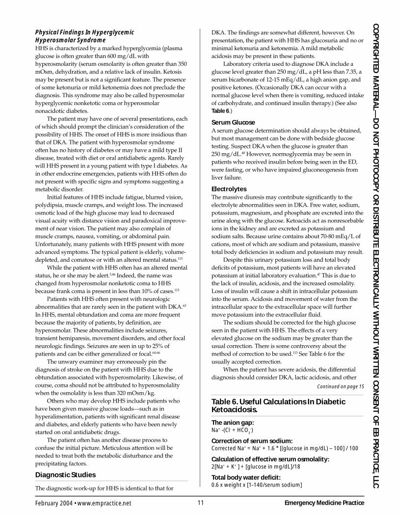
E, LLCPhysical Findings In HyperglycemicHyperosmolar SyndromeHHS is characterized by a marked hyperglycemia (plasmaglucose is often greater than 600 mg/dL withhyperosmolarity (serum osmolarity is often greater than 350mOsm, dehydration, and a relative lack of insulin. Ketosismay be present but is not a significant feature. The presenceof some ketonuria or mild ketonemia does not preclude thediagnosis. This syndrome may also be called hyperosmolarhyperglycemic nonketotic coma or hyperosmolarnonacidotic diabetes.
The patient may have one of several presentations, eachof which should prompt the clinician’s consideration of thepossibility of HHS. The onset of HHS is more insidious thanthat of DKA. The patient with hyperosmolar syndromeoften has no history of diabetes or may have a mild type IIdisease, treated with diet or oral antidiabetic agents. Rarelywill HHS present in a young patient with type I diabetes. Asin other endocrine emergencies, patients with HHS often donot present with specific signs and symptoms suggesting ametabolic disorder.
Initial features of HHS include fatigue, blurred vision,polydipsia, muscle cramps, and weight loss. The increasedosmotic load of the high glucose may lead to decreasedvisual acuity with distance vision and paradoxical improve-ment of near vision. The patient may also complain ofmuscle cramps, nausea, vomiting, or abdominal pain.Unfortunately, many patients with HHS present with moreadvanced symptoms. The typical patient is elderly, volume-depleted, and comatose or with an altered mental status.115
While the patient with HHS often has an altered mentalstatus, he or she may be alert.3,46 Indeed, the name waschanged from hyperosmolar nonketotic coma to HHSbecause frank coma is present in less than 10% of cases.113
Patients with HHS often present with neurologicabnormalities that are rarely seen in the patient with DKA.63
In HHS, mental obtundation and coma are more frequentbecause the majority of patients, by definition, arehyperosmolar. These abnormalities include seizures,transient hemiparesis, movement disorders, and other focalneurologic findings. Seizures are seen in up to 25% ofpatients and can be either generalized or focal.64-66
The unwary examiner may erroneously pin thediagnosis of stroke on the patient with HHS due to theobtundation associated with hyperosmolarity. Likewise, ofcourse, coma should not be attributed to hyperosmolalitywhen the osmolality is less than 320 mOsm/kg.
Others who may develop HHS include patients whohave been given massive glucose loads—such as inhyperalimentation, patients with significant renal diseaseand diabetes, and elderly patients who have been newlystarted on oral antidiabetic drugs.
The patient often has another disease process toconfuse the initial picture. Meticulous attention will beneeded to treat both the metabolic disturbance and theprecipitating factors.
Diagnostic Studies
The diagnostic work-up for HHS is identical to that for
DKA. The findings are somewhat different, however. Onpresentation, the patient with HHS has glucosuria and no orminimal ketonuria and ketonemia. A mild metabolicacidosis may be present in these patients.
Laboratory criteria used to diagnose DKA include aglucose level greater than 250 mg/dL, a pH less than 7.35, aserum bicarbonate of 12-15 mEq/dL, a high anion gap, andpositive ketones. (Occasionally DKA can occur with anormal glucose level when there is vomiting, reduced intakeof carbohydrate, and continued insulin therapy.) (See alsoTable 6.)
Serum GlucoseA serum glucose determination should always be obtained,but most management can be done with bedside glucosetesting. Suspect DKA when the glucose is greater than250 mg/dL.48 However, normoglycemia may be seen inpatients who received insulin before being seen in the ED,were fasting, or who have impaired gluconeogenesis fromliver failure.
ElectrolytesThe massive diuresis may contribute significantly to theelectrolyte abnormalities seen in DKA. Free water, sodium,potassium, magnesium, and phosphate are excreted into theurine along with the glucose. Ketoacids act as nonresorbableions in the kidney and are excreted as potassium andsodium salts. Because urine contains about 70-80 mEq/L ofcations, most of which are sodium and potassium, massivetotal body deficiencies in sodium and potassium may result.
Despite this urinary potassium loss and total bodydeficits of potassium, most patients will have an elevatedpotassium at initial laboratory evaluation.47 This is due tothe lack of insulin, acidosis, and the increased osmolality.Loss of insulin will cause a shift in intracellular potassiuminto the serum. Acidosis and movement of water from theintracellular space to the extracellular space will furthermove potassium into the extracellular fluid.
The sodium should be corrected for the high glucoseseen in the patient with HHS. The effects of a veryelevated glucose on the sodium may be greater than theusual correction. There is some controversy about themethod of correction to be used.113 See Table 6 for theusually accepted correction.
When the patient has severe acidosis, the differentialdiagnosis should consider DKA, lactic acidosis, and other
Continued on page 15
Table 6. Useful Calculations In DiabeticKetoacidosis.
The anion gap:Na+ -(Cl- + HCO
3-)
Correction of serum sodium:Corrected Na+ = Na+ + 1.6 * [(glucose in mg/dL) – 100] / 100
Calculation of effective serum osmolality:2[Na+ + K+ ] + [glucose in mg/dL]/18
Total body water deficit:0.6 x weight x [1-140/serum sodium]
Emergency Medicine Practice 12 www.empractice.net • February 2004
CO
PY
RIG
HTE
D M
ATER
IAL—
DO
NO
T P
HO
TOC
OP
Y O
R D
ISTR
IBU
TE E
LEC
TRO
NIC
ALL
Y W
ITH
OU
T W
RIT
TEN
CO
NSE
NT
OF
EB P
RA
CTI
CE,
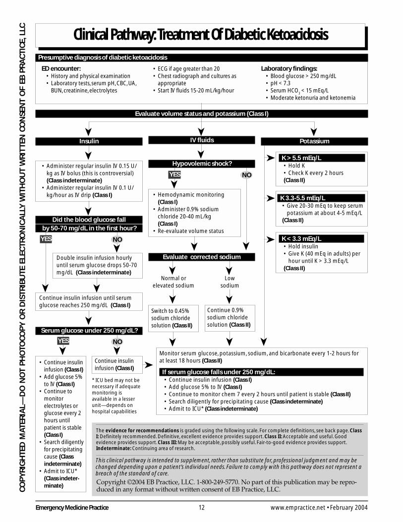
LLC Clinical Pathway: Treatment Of Diabetic Ketoacidosis
The evidence for recommendations is graded using the following scale. For complete definitions, see back page. ClassI: Definitely recommended. Definitive, excellent evidence provides support. Class II: Acceptable and useful. Goodevidence provides support. Class III: May be acceptable, possibly useful. Fair-to-good evidence provides support.Indeterminate: Continuing area of research.
This clinical pathway is intended to supplement, rather than substitute for, professional judgment and may bechanged depending upon a patient’s individual needs. Failure to comply with this pathway does not represent abreach of the standard of care.
Copyright ©2004 EB Practice, LLC. 1-800-249-5770. No part of this publication may be repro-duced in any format without written consent of EB Practice, LLC.
Presumptive diagnosis of diabetic ketoacidosis
Laboratory findings:• Blood glucose > 250 mg/dL• pH < 7.3• Serum HCO
3 < 15 mEq/L
• Moderate ketonuria and ketonemia
ED encounter:• History and physical examination• Laboratory tests, serum pH, CBC, UA,
BUN, creatinine, electrolytes
➤ ➤➤
Evaluate volume status and potassium (Class I)
Potassium
K > 5.5 mEq/L• Hold K• Check K every 2 hours(Class II)
K 3.3-5.5 mEq/L• Give 20-30 mEq to keep serum
potassium at about 4-5 mEq/L(Class II)
K < 3.3 mEq/L• Hold insulin• Give K (40 mEq in adults) per
hour until K > 3.3 mEq/L(Class II)
➤
➤
➤
➤
• ECG if age greater than 20• Chest radiograph and cultures as
appropriate• Start IV fluids 15-20 mL/kg/hour
IV fluids
➤
Hypovolemic shock?➤
NO➤YES
• Hemodynamic monitoring(Class I)
• Administer 0.9% sodiumchloride 20-40 mL/kg(Class I)
• Re-evaluate volume status
Evaluate corrected sodium
➤
➤ ➤
Normal orelevated sodium
Lowsodium
➤ ➤
Switch to 0.45%sodium chloridesolution (Class II)
Continue 0.9%sodium chloridesolution (Class II)
➤ ➤
Insulin
• Administer regular insulin IV 0.15 U/kg as IV bolus (this is controversial)(Class indeterminate)
• Administer regular insulin IV 0.1 U/kg/hour as IV drip (Class I)
➤
➤NO
➤
YES
Double insulin infusion hourlyuntil serum glucose drops 50-70mg/dL (Class indeterminate)
Serum glucose under 250 mg/dL?
➤
Did the blood glucose fallby 50-70 mg/dL in the first hour?
➤➤
Continue insulin infusion until serumglucose reaches 250 mg/dL (Class I)
➤YES ➤NO
Continue insulininfusion (Class I)
• Continue insulininfusion (Class I)
• Add glucose 5%to IV (Class I)
• Continue tomonitorelectrolytes orglucose every 2hours untilpatient is stable(Class I)
• Search diligentlyfor precipitatingcause (Classindeterminate)
• Admit to ICU*(Class indeter-minate)
Monitor serum glucose, potassium, sodium, and bicarbonate every 1-2 hours forat least 18 hours (Class II)
If serum glucose falls under 250 mg/dL:• Continue insulin infusion (Class I)• Add glucose 5% to IV (Class I)• Continue to monitor chem 7 every 2 hours until patient is stable (Class II)• Search diligently for precipitating cause (Class indeterminate)• Admit to ICU* (Class indeterminate)
➤
* ICU bed may not benecessary if adequatemonitoring isavailable in a lesserunit—depends onhospital capabilities
13 Emergency Medicine PracticeFebruary 2004 • www.empractice.net
CO
PY
RIG
HTED
MATER
IAL—
DO
NO
T PH
OTO
CO
PY
OR
DISTR
IBU
TE ELECTR
ON
ICA
LLY W
ITHO
UT W
RIT
TEN C
ON
SENT O
F EB P
RA
CTIC
E, LLC
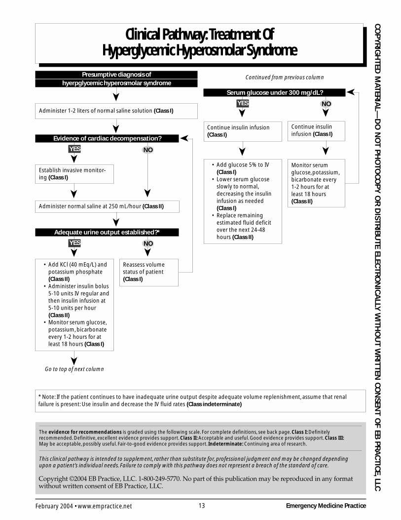
Clinical Pathway: Treatment OfHyperglycemic Hyperosmolar Syndrome
The evidence for recommendations is graded using the following scale. For complete definitions, see back page. Class I: Definitelyrecommended. Definitive, excellent evidence provides support. Class II: Acceptable and useful. Good evidence provides support. Class III:May be acceptable, possibly useful. Fair-to-good evidence provides support. Indeterminate: Continuing area of research.
This clinical pathway is intended to supplement, rather than substitute for, professional judgment and may be changed dependingupon a patient’s individual needs. Failure to comply with this pathway does not represent a breach of the standard of care.
Copyright ©2004 EB Practice, LLC. 1-800-249-5770. No part of this publication may be reproduced in any formatwithout written consent of EB Practice, LLC.
Presumptive diagnosis ofhyerpglycemic hyperosmolar syndrome
Administer 1-2 liters of normal saline solution (Class I)
➤➤
Evidence of cardiac decompensation?
➤YES
➤
NO
Establish invasive monitor-ing (Class I)
➤
Administer normal saline at 250 mL/hour (Class II)
Adequate urine output established?*
➤YES
➤
➤NO
• Add KCl (40 mEq/L) andpotassium phosphate(Class II)
• Administer insulin bolus5-10 units IV regular andthen insulin infusion at5-10 units per hour(Class II)
• Monitor serum glucose,potassium, bicarbonateevery 1-2 hours for atleast 18 hours (Class I)
Reassess volumestatus of patient(Class I)
➤
Serum glucose under 300 mg/dL?
➤YES ➤NO
➤
Go to top of next column
Continue insulin infusion(Class I)
➤Monitor serumglucose, potassium,bicarbonate every1-2 hours for atleast 18 hours(Class II)
• Add glucose 5% to IV(Class I)
• Lower serum glucoseslowly to normal,decreasing the insulininfusion as needed(Class I)
• Replace remainingestimated fluid deficitover the next 24-48hours (Class II)
Continue insulininfusion (Class I)
➤
* Note: If the patient continues to have inadequate urine output despite adequate volume replenishment, assume that renalfailure is present: Use insulin and decrease the IV fluid rates (Class indeterminate)
➤
Continued from previous column
Emergency Medicine Practice 14 www.empractice.net • February 2004
CO
PY
RIG
HTE
D M
ATER
IAL—
DO
NO
T P
HO
TOC
OP
Y O
R D
ISTR
IBU
TE E
LEC
TRO
NIC
ALL
Y W
ITH
OU
T W
RIT
TEN
CO
NSE
NT
OF
EB P
RA
CTI
CE,
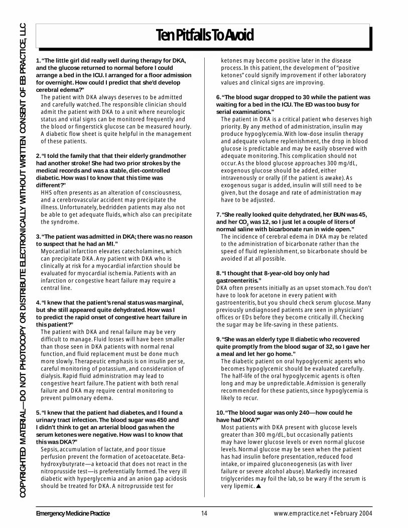
LLC Ten Pitfalls To Avoid
1. “The little girl did really well during therapy for DKA,and the glucose returned to normal before I couldarrange a bed in the ICU. I arranged for a floor admissionfor overnight. How could I predict that she’d developcerebral edema?”
The patient with DKA always deserves to be admittedand carefully watched. The responsible clinician shouldadmit the patient with DKA to a unit where neurologicstatus and vital signs can be monitored frequently andthe blood or fingerstick glucose can be measured hourly.A diabetic flow sheet is quite helpful in the managementof these patients.
2. “I told the family that that their elderly grandmotherhad another stroke! She had two prior strokes by themedical records and was a stable, diet-controlleddiabetic. How was I to know that this time wasdifferent?”
HHS often presents as an alteration of consciousness,and a cerebrovascular accident may precipitate theillness. Unfortunately, bedridden patients may also notbe able to get adequate fluids, which also can precipitatethe syndrome.
3. “The patient was admitted in DKA; there was no reasonto suspect that he had an MI.”
Myocardial infarction elevates catecholamines, whichcan precipitate DKA. Any patient with DKA who isclinically at risk for a myocardial infarction should beevaluated for myocardial ischemia. Patients with aninfarction or congestive heart failure may require acentral line.
4. “I knew that the patient’s renal status was marginal,but she still appeared quite dehydrated. How was Ito predict the rapid onset of congestive heart failure inthis patient?”
The patient with DKA and renal failure may be verydifficult to manage. Fluid losses will have been smallerthan those seen in DKA patients with normal renalfunction, and fluid replacement must be done muchmore slowly. Therapeutic emphasis is on insulin per se,careful monitoring of potassium, and consideration ofdialysis. Rapid fluid administration may lead tocongestive heart failure. The patient with both renalfailure and DKA may require central monitoring toprevent pulmonary edema.
5. “I knew that the patient had diabetes, and I found aurinary tract infection. The blood sugar was 450 andI didn’t think to get an arterial blood gas when theserum ketones were negative. How was I to know thatthis was DKA?”
Sepsis, accumulation of lactate, and poor tissueperfusion prevent the formation of acetoacetate. Beta-hydroxybutyrate—a ketoacid that does not react in thenitroprusside test—is preferentially formed. The very illdiabetic with hyperglycemia and an anion gap acidosisshould be treated for DKA. A nitroprusside test for
ketones may become positive later in the diseaseprocess. In this patient, the development of “positiveketones” could signify improvement if other laboratoryvalues and clinical signs are improving.
6. “The blood sugar dropped to 30 while the patient waswaiting for a bed in the ICU. The ED was too busy forserial examinations.”
The patient in DKA is a critical patient who deserves highpriority. By any method of administration, insulin mayproduce hypoglycemia. With low-dose insulin therapyand adequate volume replenishment, the drop in bloodglucose is predictable and may be easily observed withadequate monitoring. This complication should notoccur. As the blood glucose approaches 300 mg/dL,exogenous glucose should be added, eitherintravenously or orally (if the patient is awake). Asexogenous sugar is added, insulin will still need to begiven, but the dosage and rate of administration mayhave to be adjusted.
7. “She really looked quite dehydrated, her BUN was 45,and her CO
2 was 12, so I just let a couple of liters of
normal saline with bicarbonate run in wide open.”The incidence of cerebral edema in DKA may be relatedto the administration of bicarbonate rather than thespeed of fluid replenishment, so bicarbonate should beavoided if at all possible.
8. “I thought that 8-year-old boy only hadgastroenteritis.”DKA often presents initially as an upset stomach. You don’thave to look for acetone in every patient withgastroenteritis, but you should check serum glucose. Manypreviously undiagnosed patients are seen in physicians’offices or EDs before they become critically ill. Checkingthe sugar may be life-saving in these patients.
9. “She was an elderly type II diabetic who recoveredquite promptly from the blood sugar of 32, so I gave hera meal and let her go home.”
The diabetic patient on oral hypoglycemic agents whobecomes hypoglycemic should be evaluated carefully.The half-life of the oral hypoglycemic agents is oftenlong and may be unpredictable. Admission is generallyrecommended for these patients, since hypoglycemia islikely to recur.
10. “The blood sugar was only 240—how could hehave had DKA?”
Most patients with DKA present with glucose levelsgreater than 300 mg/dL, but occasionally patientsmay have lower glucose levels or even normal glucoselevels. Normal glucose may be seen when the patienthas had insulin before presentation, reduced foodintake, or impaired gluconeogenesis (as with liverfailure or severe alcohol abuse). Markedly increasedtriglycerides may foil the lab, so be wary if the serum isvery lipemic. ▲
15 Emergency Medicine PracticeFebruary 2004 • www.empractice.net
CO
PY
RIG
HTED
MATER
IAL—
DO
NO
T PH
OTO
CO
PY
OR
DISTR
IBU
TE ELECTR
ON
ICA
LLY W
ITHO
UT W
RIT
TEN C
ON
SENT O
F EB P
RA
CTIC
E, LLC
non-HHS disease processes. Vomiting or the concomitantuse of a thiazide diuretic can cause a metabolic alkalosis thatcan mask the severity of the acidosis. This situation mightbe found if the combined anion gap and the measuredHCO3 are greater than expected.
Complete Blood CountThe complete blood count often shows a leukocytosis. Thismay be in part due to the hemoconcentration from dehydra-tion. White blood cell counts of 20,000 cells/mm3 are notuncommon. If the patient has an elevated band count(bandemia) on peripheral smear, then an infectious processis likely.67
Serum pHAn arterial blood gas or venous gas should be sent early inthe evaluation of the patient considered to have DKA.68,69
(The correlation between arterial and venous pH is quiteclose, and the two can be used interchangably for theevaluation of DKA patients.) This will help determine thedegree of acidosis and bicarbonate loss. When the pH is lessthan 7.35 and a low HCO3
- is found, then the diagnosis ofDKA should be seriously considered.
Blood Urea Nitrogen And CreatinineThe patient will often have some elevation of the blood ureanitrogen due to dehydration. The clinician should carefullyconsider the possibility of chronic renal failure. The patientwith both diabetes and renal failure may be quite difficult tomanage. Fluid losses may be smaller than when the patienthas normal renal function, and fluid replacement must bemuch more conservative. Therapeutic emphasis will switchto insulin, careful monitoring of potassium, and consider-ation of dialysis.
Urinalysis And Urine CultureCheck the urine on every patient who presents with apossible diagnosis of DKA in order to identify possibleurosepsis causing the DKA. Likewise, in females of child-bearing age, a urine pregnancy test is equally important.
Serum KetonesSerum ketone testing may not correlate with the degree ofketoacidosis. Beta-hydroxybutyric acid is the major ketoacidproduced in DKA. It does not react with nitroprusside, sourine or blood testing for ketones may be negative or onlyslightly positive.
During the treatment of DKA, beta-hydroxybutyricacid will be converted to acetoacetic acid. The resultantincrease in acetoacetic acid may make the serum or urinetesting for ketones more positive, despite clinical improve-ment and an increasing pH and decreasing anion gap.While some literature has suggested that urine ketones canbe used to rule out DKA, serum ketones are more accurateand should be used instead.
Serum OsmolalityBecause biological membranes are readily permeable towater molecules, there is a continuing movement orexchange of water between various fluid compartments.
The forces governing the movement of water are bothhydrostatic and osmotic pressures. The osmotic pressuresgreatly exceed hydrostatic pressures in the body.
Osmotic pressure is a property of solutions thatdepends on the number of osmotically active molecules insolution. It does not depend on the ionic charge, the size ofthe molecule, or the chemical properties of the molecule.One gram-molecular weight (1 mole) of a compound is alsotermed 1 osmole (osmol or Osm). Most biological concentra-tions are expressed in milliosmolar (mOsm) ormicroosmolar (µOsm) concentrations. Ionized moleculessuch as sodium chloride will dissociate in solution, and eachion exerts its own osmotic force. (This means that 1 mOsmof NaCl exerts 2 mOsm of osmotic pressure.) Osmolality isthe expression of this quantity of osmotically active particlesper 1000 grams of water or solvent. The more common term,osmolarity, expresses the number of osmotically activeparticles per liter of solvent. The difference betweenosmolarity and osmolality is due to protein and fat, whichcomprise about 6%-8% of the solutes in plasma.
Hyperosmolality in diabetes is largely due tohyperglycemia.
Lumbar PunctureLumbar puncture is indicated in patients with an acutealteration of consciousness and clinical features suggestiveof possible central nervous system infection.
Hemoglobin A1c DeterminationGlycosylated hemoglobin measurement is not needed forthe emergent management of the patient’s diabetes. Thehemoglobin A1c will give the clinician some idea of theduration or degree of control of the patient’s diabetes priorto the emergency.
Treatment
Successful treatment of both DKA and HHS includescorrection of the dehydration and hyperglycemia, resolutionand anticipation of the electrolyte abnormalities, identifica-tion of precipitating or comorbid illnesses. (See also theClinical Pathways on page 12 and page 13.)
Infection is quite common as a precipitating factor.Pneumonia, urinary infections, and sepsis are the mostcommon etiologies. This high incidence of infection in HHSpatients has led to a recommendation for early empiricantibiotic therapy, even if no source is identified.41,70 This iscontroversial; others recommend waiting for definitiveevidence since both fever and leukocytosis can also beprecipitated by other factors.71
The current mortality rate of DKA is about 2%-5%.72
Without insulin therapy, the mortality rate of DKA is 100%.Remember that the underlying pathology may be the
reason for mortality in the patient with HHS. Re-evaluatethe patient for infection, myocardial infarction, and underly-ing diseases. Early mortality in HHS is more likely to becaused by sepsis, shock, or an underlying illness.
The most common complications of DKA and HHSinclude hypoglycemia due to overtreatment with insulin,hypokalemia due to insulin administration in the potas-
Continued from page 11
Emergency Medicine Practice 16 www.empractice.net • February 2004
CO
PY
RIG
HTE
D M
ATER
IAL—
DO
NO
T P
HO
TOC
OP
Y O
R D
ISTR
IBU
TE E
LEC
TRO
NIC
ALL
Y W
ITH
OU
T W
RIT
TEN
CO
NSE
NT
OF
EB P
RA
CTI
CE,
LLC sium-depleted patient and treatment of acidosis with
bicarbonate, and hyperglycemia due to interruption ofintravenous insulin before coverage with other forms ofinsulin has been established.
Diabetic KetoacidosisThe specific treatment goals of the patient with DKA are:
1. Improve tissue perfusion and correct hypovolemia2. Decrease the serum glucose3. Reverse acidosis and ketonemia4. Correct electrolyte imbalances5. Treat the disease process that precipitated the patient’s
DKA (if present)
This is not a rigid treatment protocol but a guideline.Each aspect of each case must be assessed individually andreassessed frequently.
FluidsRehydration is the most important intervention in thetherapy of DKA; however, the optimal fluid managementfor DKA is uncertain. The practitioner is faced with a patientwho is often dehydrated, yet rapid fluid administration maypredispose the patient to lethal complications.
If the patient is hypotensive, administer a bolus of 1-2liters of normal saline (0.9%) over the first hour (pediatricdose, 20-40 mL/kg). If hypotension persists, then giveanother bolus. If the patient is normotensive, then use 0.9%saline at 1000 cc per hour. Subsequent choice of fluidsdepends on the state of hydration, serum electrolytes, andthe urinary output. When giving this much fluid to olderpatients, pulmonary edema is a real possibility. The patient’slung sounds should be carefully monitored. If the patienthas renal failure or has a history of congestive heart failure,then invasive monitoring with Swan catheters may be anappropriate step. This is a clinical judgment.
In general, normal saline (0.9%) infused at 4-14 mL/kg/hour is appropriate if the corrected serum sodium isnormal or elevated.113 Once renal function is ensured, theinfusion should include 20-30 mEq/L of potassium until thepatient is stable and can tolerate oral intake.
Provide maintenance fluids and electrolytes evenlyover the first 24 hours. Deficit fluids are usually replaced ina bi-level fashion: One half of the deficit is replaced over thefirst eight hours, and the other half of the deficit is replacedover the next 16 hours.113
Successful fluid replacement is judged by hemody-namic stability, measurement of fluid input/output, andclinical examination of the patient. Overhydration is aconcern when treating children with DKA, adults withcompromised renal or cardiac function, and elderly patientswith incipient congestive heart failure.
In patients with renal or cardiac insufficiency, monitor-ing of cardiac, renal, and mental status must be diligentlyperformed during the fluid resuscitation to avoid aniatrogenic fluid overload.
In pediatric patients, the need for volume expansionmust be balanced with the risk of cerebral edema, asdiscussed later in this article. Therapy must include
monitoring of mental status, which may be the firstindication of cerebral edema in the diabetic patient.
InsulinReplacing insulin reverses the mobilization of free fattyacid and the hepatic production of glucose and ketoacids.Insulin treats the ketosis and the acidosis as well as thehyperglycemia. Insulin can be given intravenously orintramuscularly. Some physicians advocate preceding theinfusion with a bolus of insulin; however, this has nophysiologic advantage.
Continuous intravenous insulin infusion has severaladvantages over intramuscular insulin.113 Smaller insulindoses are needed for clinical efficacy when given intrave-nously. An insulin drip gives therapeutic plasma insulinlevels with a smooth correction and flexibility to increase ordecrease the dose easily. A common intravenous insulinadministration regimen is continuous infusion of 0.1 unitsper kilogram per hour (about 6-8 units per hour in theaverage adult). If the insulin infusion is connected through aseparate line or through a “piggy-back” connection, theinfusion can be regulated separately from the rate of fluidadministration.
An alternative method of dosing would be to give anequivalent dose of rapid-acting insulin intramuscularlyevery hour. Since vasoconstriction decreases medicationabsorption in hypotensive patients, this method is less thanoptimal in the critically ill patient.
Subcutaneous insulin is not recommended duringthe treatment of any critically ill patient. In thesepotentially hypotensive and/or dehydrated patients,the absorption from subcutaneous tissues may be inad-equate or unpredictable.
Research indicates that bolus administration of insulinmay not be the best possible therapy for the patient.113 Bolusinsulin causes plasma insulin levels to rise within minutesto over 3000 microunits per milliliter. This level far exceedsphysiological levels of insulin. Potassium levels are signifi-cantly reduced when these boluses of insulin are used. Thiscould be a clinical problem if the patient is hypokalemicbefore insulin is given.
The endpoint for continuous insulin infusion should beresolution of hyperketonemia (3-hydroxybutyrate < 0.5mmol/L) rather than the normalization of blood sugar.73 Toprevent the recurrence of DKA, the patient should resumehis or her usual subcutaneous dose of insulin before theinsulin infusion is stopped. The insulin drip should not beconsidered as part of fluid replacement calculations.
The glucose level can be anticipated to fall by 50-100mg/dL (2.5-5.0 mmol/L) per hour.74 The initial rate ofdecrease may be more rapid as the renal function improveswith improved hydration. When the glucose level reaches250 mg/dL (12.5 mmol/L), the possibility of hypoglycemiashould be anticipated. When the patient’s glucose levelreaches 250 mg/dL, the rate of infusion of insulin should becut in half to decrease the possibility of hypoglycemia. Insome patients, D5 normal saline or even D10 normal sailinemay be used to prevent hypoglycemia and allow intrave-
17 Emergency Medicine PracticeFebruary 2004 • www.empractice.net
CO
PY
RIG
HTED
MATER
IAL—
DO
NO
T PH
OTO
CO
PY
OR
DISTR
IBU
TE ELECTR
ON
ICA
LLY W
ITHO
UT W
RIT
TEN C
ON
SENT O
F EB P
RA
CTIC
E, LLCnous infusion of insulin until the subcutaneous administra-tion of insulin is resumed.113
If the patient’s glucose is falling steadily, the anion gapis decreasing, and the pH is improving, then the insulininfusion is appropriate. If the glucose level does not fall inthe presence of adequate hydration and urine output, thenthe patient may have insulin resistance. In this case, theinsulin dose can be doubled each hour where there is nodecrease. This insulin resistance should remind the clinicianthat the patient may have sepsis, occult infection (e.g.,endocarditis, perirectal abscess, prostatitis, or sinusitis, toname a few), myocardial infarction, or an intra-abdominalillness that precipitated the crisis. A few patients may have aprimary insulin resistance. The serum glucose level maydecline more rapidly in the older patient, so carefullymonitor glucose levels in these patients.
This insulin administration regimen is only a guideline.Consider the patient’s previously injected long-actinginsulin, concurrent stress (such as a myocardial infarction),or the concurrent administration of steroids.
BicarbonateThe use of bicarbonate is not recommended for DKA.Bicarbonate is used to treat metabolic acidosis. The rationalefor the use of bicarbonate in DKA is based on the assump-tion that the acidosis may contribute to morbidity in thesepatients.75,76 This ignores the issue in the patient with DKAthat the appropriate treatment for ketoacidosis is notbicarbonate, but insulin. Use of bicarbonate merely masksthe ketoacidosis. Studies of cerebral edema (outlined later inthis article) show that there is an increased risk of cerebraledema when bicarbonate is used. Given all of these factors,bicarbonate can’t be recommended for patients in DKA.
The American Diabetes Association guidelines suggestthe use of bicarbonate when the pH is below 6.9.6 There areno prospective, randomized studies concerning the use ofbicarbonate in DKA in either children or adults. The supportfor this part of these American Diabetes Associationguidelines, therefore, is incredibly weak.
PotassiumThe major electrolyte lost during DKA is potassium. Thetotal body potassium deficit ranges from 300-1000 mEq totalbody deficit for an adult.77 Hypokalemia is a potentiallylethal complication of DKA. The patient may have a mildlyelevated potassium level upon initial evaluation and stillhave a tremendous total potassium deficit. Much potassiumis lost as the potassium salts of ketoacidosis.
Continued potassium losses are the rule during thetreatment of the patient with normal renal function andDKA. Resolution of acidosis and concomitant administra-tion of insulin will force the remaining extracellularpotassium into the cells. The clinician should anticipate themassive shift of potassium carried into the cells with insulinand the endogenous glucose loading. Insulin therapyshould not be started until the potassium level is known, inorder to prevent lethal hypokalemia.113
The electrocardiogram should be evaluated for thesigns of hyperkalemia, such as tall peaked T waves,
disappearance of the P wave, and widening of the QRScomplex. (Remember that the T waves usually peak if theserum potassium is over 6 mEq/L.) Hypertonicity, deficit ofinsulin, and acidosis can combine to cause extracellularhyperkalemia. In reality, the patient has lost total bodypotassium, but the lack of insulin and acidosis canmobilize remaining intracellular potassium into theextracellular fluid. An ECG may show these signs ofhyper- or hypokalemia before the electrolyte determinationhas returned.
Multiple algorithms are described in the literature forthe replacement of potassium during therapy of DKA. Themost important part of any such algorithm is adequatemonitoring of the electrolytes during the process. If theserum potassium is less than 5.0 mEq/L and there is a recenthistory of urination, and/or the ECG has no peaked Twaves, then potassium can be started safely. Before anypotassium is given, kidney function and adequate urineoutput must be established.
Phosphate, Magnesium, And CalciumIn DKA, calcium, magnesium, and phosphate are alsodeficient. The prevalence and clinical need for replacementof these electrolytes are not universally accepted.
PhosphateWhole body phosphate deficits in DKA are substantial.Despite this, several small studies have not shown any clearbenefit to phosphate replacement in DKA.78-80 Excessivephosphate replacement may cause hypocalcemia and soft-tissue metastatic calcifications.
The clinical manifestations of severe hypophos-phatemia are diverse. Alterations in the function of skeletaland smooth muscles can lead to rhabdomyolysis, ileus,respiratory failure, and cardiac dysfunction. Decrease inintracellular adenosine triphosphate can result in hemolysis,thrombocytopenia, and impaired phagocytosis. Patients alsomay demonstrate metabolic encephalopathy, progressingfrom irritability to coma. Indications for the clinical reple-tion of phosphate include left ventricular dysfunction orfailure to clear mental confusion despite improvement incirculating volume, hyperosmolarity, and acidosis.
If the patient’s phosphate level is less than 1.0mg/dL or if one of the above indications for phosphatetherapy is present, then give 30-60 mM of phosphate overa 24-hour period. Obtain phosphate levels before the startof treatment, after an infusion, and at 24 and 48 hoursinto treatment.
MagnesiumThe kidney is the primary organ responsible for magnesiumhomeostasis. Under normal conditions, 15% of filteredmagnesium is reabsorbed in the proximal tubule through anactive transcellular process, 60%-70% is reabsorbed pas-sively in the thick ascending limb of the loop of Henledriven by transepithelial voltage, and 10% is reabsorbed inthe distal tubule.119 Because magnesium reabsorption isproportional to urine volume and flow, volume expansionor osmotic diuresis can lead to magnesium wasting.
Emergency Medicine Practice 18 www.empractice.net • February 2004
CO
PY
RIG
HTE
D M
ATER
IAL—
DO
NO
T P
HO
TOC
OP
Y O
R D
ISTR
IBU
TE E
LEC
TRO
NIC
ALL
Y W
ITH
OU
T W
RIT
TEN
CO
NSE
NT
OF
EB P
RA
CTI
CE,
LLC Significant magnesium depletion usually is associated
with hypocalcemia and hypokalemia, since they have thesimilar mechanisms.
Clinical manifestations of hypomagnesemia arenonspecific and include weakness, dizziness, tremors,cramps, paresthesias, and seizures.81 Severe hypo-magnesemia can lead to both atrial and ventriculararrhythmias. If the patient has a magnesium level of lessthan 1.8 mEq/L or has tetany, then magnesium sulfate maybe started as 5 grams in 500 mL of normal saline adminis-tered over five hours.
CalciumIf the patient has symptomatic hypocalcemia, then 10-20 mL(1-2 grams) of 10% calcium gluconate may be givenintravenously over 10 minutes.
Hyperosmolar Hyperglycemic SyndromeAdequate management of HHS requires fluid replacement;this is the first priority of treatment. HHS patients are oftenabout 25% dehydrated and may require 9-12 liters of fluidreplenishment. Unfortunately, many patients with thisdisease cannot tolerate either the disease or the treatmentrequired. Careful monitoring during this fluid therapy iscrucial, because people with hyperosmolar coma may havecompromised cardiovascular function.
Most patients can be treated with normal saline at 500-1000 cc per hour until the blood pressure is normal and thepatient has good urine output. The clinician should plan onreplacing about 50% of the fluid deficit in the first 12 hoursand the remaining 50% in the subsequent 24-36 hours.113
Controversy also abounds about the proper choice ofrehydration fluid in HHS. Normal saline replaces theintracellular volume more quickly but also increases thepossibility of fluid overload. It may provoke adult respira-tory distress syndrome, particularly in patients withpotential cardiac and respiratory compromise. Hypotonicfluids such as half-normal saline (0.45%) may be moreeffective in replacing the lost free water, but risk too rapidcorrection of hyponatremia with subsequent pontinemyelinonecrolysis. (Severe hypertonicity is associated withlarge sodium deficits and hypovolemic shock.40,82)
An appropriate compromise is an initial fluid replace-ment of one liter of normal saline and then switching to halfnormal saline when urine output is adequate.
Insulin, while not present in quantities sufficient toprevent hyperglycemia, is usually present in patients withHHS and may serve to prevent the formation of ketonebodies. Relative insulin deficiency is exacerbated by thepatient’s dehydration. Insulin therapy is not as important asit is in DKA. Indeed, the use of too much insulin, given toorapidly, may cause vascular collapse as glucose and waterare moved into the cells and out of the volume-depletedintravascular space. As in the patient with DKA, once theglucose drops to 250 mg/dL, the patient must receivedextrose in the intravenous fluids.
Although patients are generally not hypokalemic byinitial measurement of serum potassium, the total bodydeficit may be quite high as in DKA. Lethal hypokalemia
may accompany the administration of insulin as potassiumis forced into the cells with insulin and sugar. (This is nowquite rare since patients are now treated with lower-dosecontinuous insulin infusion rather than large boluses ofinsulin.113) The clinician must follow serum potassium levelsfrequently and administer a minimum of 10 mEq per hourof potassium chloride. Severe hypokalemia is often corre-lated with frequent emesis, nasogastric suction, or the use ofthiazide diuretic agents. As with DKA, do not initiatepotassium replacement until it has been confirmed by thelaboratory and adequate urine output has been established.
Hypokalemia is often accompanied by hypo-magnesemia. Again, the initial serum levels of magnesiumand phosphate are typically normal or elevated whenthe disease process is diagnosed and may fall withinsulin administration.
Careful MonitoringThe sicker the patient, the more frequently he or she shouldbe monitored. Do not wait for results to come back beforesending the next set of tests. The following parametersshould be monitored and recorded on a flow sheet:
• Glucose—every 1-2 hours by fingerstick (confirmed bylaboratory correlation when indicated)
• Basic metabolic profile for anion gap and serumpotassium—at the onset of treatment, at one and twohours after the onset of treatment, and at two- to four-hour intervals until the patient is substantially better
• Arterial or venous pH—follow as indicated to monitorclinical status
• Insulin dose and route of administration—every hour• IV fluids—every hour• Urine output—every hour
Special Circumstances
Pulmonary Edema And Adult RespiratoryDistress SyndromeCardiogenic and non-cardiogenic pulmonary edema canoccur in association with the treatment of DKA. Thesecomplications more often are noted during the ICU stay, butthey may be found by the emergency physician. Excessivefluid replacement may cause pulmonary edema even whenthe cardiac function is normal. These patients should havecontinuous pulse oximeter monitoring. Avoiding overzeal-ous fluid administration should prevent cases related todecreased colloid osmotic pressure.
Adult respiratory distress syndrome is a rare butpotentially fatal complication of the treatment of DKA.83,84 Apatient who has an increased alveolar-to-arterial oxygengradient on arterial blood gas or who presents with ralesmay have an increased risk of adult respiratory distresssyndrome. This rare complication appears to result from anincreased capillary membrane permeability and is conse-quently more difficult to prevent. Treatment requires adelicate balance between adequate rehydration and fluidrestriction needed for the treatment of the pulmonaryedema. These patients require invasive hemodynamicmonitoring and intensive pulmonary therapy.
19 Emergency Medicine PracticeFebruary 2004 • www.empractice.net
CO
PY
RIG
HTED
MATER
IAL—
DO
NO
T PH
OTO
CO
PY
OR
DISTR
IBU
TE ELECTR
ON
ICA
LLY W
ITHO
UT W
RIT
TEN C
ON
SENT O
F EB P
RA
CTIC
E, LLCVascular ComplicationsDKA is a hypercoagulable state. The factors that precipitatethrombosis include the hypercoagulability in the DKApatient, stasis, and endothelial damage.85,86 The severedehydration and immobility that occur in DKA alsodecrease perfusion of vital organs and promote stasis andhypercoagulability.
Vascular occlusions (such as myocardial infarction,cerebrovascular accidents, and mesenteric artery occlusions)are important complications of both DKA and HHS.113
Predisposing factors include dehydration with resultantincreased viscosity, and preexisting coagulation abnormali-ties. With aggressive rehydration of the patient, theexpected incidence of these complications can be reducedto about 2%.113
RhabdomyolysisSubclinical rhabdomyolysis, possibly owing to shrinkage ofmuscle cells and impaired glucose use, is a common findingin patients with HHS (and to a lesser degree with DKA), butsecondary acute renal failure is extremely rare.113 Manypatients do not develop myoglobinuria; thus, monitoringserum creatinine phosphokinase levels is the most sensitiveway to screen for this potentially serious complication.
Acute Gastric DilatationAcute gastric dilatation is a relatively uncommon butpotentially lethal problem. If the patient is distended, thenother causes of intra-abdominal pathology must be soughtfirst. A nasogastric tube will make the diagnosis and serveas therapy if the patient continues to vomit.
Electrolyte DisordersTreatment for DKA can cause life-threatening, predictable,and avoidable acute complications such as hypokalemia,hyponatremia, and fluid overload. These are common yetpreventable complications of the treatment of diabeticemergencies.
Hyperchloremic Metabolic AcidosisHyperchloremic metabolic acidosis with a normal anion gapoften persists after the resolution of ketonemia. This acidosishas no adverse clinical effects and is gradually correctedover the subsequent 24-48 hours by enhanced renal acidexcretion.87 Hyperchloremia can be aggravated by excessivechloride administration during the rehydration phase.
SeizuresSeizures associated with HHS are often resistant to typicalantiseizure medications.120 Phenytoin may further exacer-bate the HHS.120 The emergency physician should beaware of this problem with seizure control and proceedto alternative agents such as barbiturates if benzodiazepinesare not effective.
Cerebral Edema—A Pediatric ProblemCerebral edema is the most feared complication of DKA andmay have driven many of the changes in the managementof DKA over the past few decades.
Cerebral edema occurs in about 1% of DKA cases and isthe most common cause of diabetes-related death in young
diabetic patients.88,89 It is almost always a disease of children;over 95% of cases in the largest reported series occurredunder the age of 20, and one-third of cases occurredunder the age of 5 years.88 One-fourth of the childrenaffected die from this complication. When cerebral edemadoes occur in adults, it is quite similar in presentation tothe pediatric disease.
After initial improvement of clinical, biochemical, andcentral nervous system signs in a patient with DKA, thepatient develops increasing irritability, central nervoussystem depression, seizures, and coma. Cerebral edemacomplicating DKA generally occurs in children who seem tobe metabolically returning to normal about 3-12 hours afterthe onset of therapy.6,7
The risk factors for cerebral edema are incompletelydefined. Recent work shows that children who have a lowCO2 and a high blood urea nitrogen and then are treatedwith bicarbonate are at increased risk for cerebral edema.8,90
Children who had a smaller increase in serum sodiumduring therapy were also at more risk of cerebral edema.The highest risk was found with the use of bicarbonate anda low CO2.
The initial serum glucose and the rate of change in theserum glucose were not associated with the development ofcerebral edema in this study. The traditional dogma hasbeen that these values are related to cerebral edema;however, this appears to be completely unsubstantiated bythis study.
Previous investigations of cerebral edema in childrenwith DKA implicated a younger age, a new onset of thedisease,91 and a rapid rate of fluid administration as factorsassociated with cerebral edema.88,92 In these studies, childrenwith cerebral edema were not compared with controls.
Why does it occur in children and not adults? There isno clear answer. Some speculate that it may relate torelatively greater brain volume, more rapid changes inplasma osmolarity, lack of sex steroids, less developedmechanisms for brain cell volume regulation, a difference intaurine or other osmolyte metabolism, a difference in blood-brain barrier efficiency, or unidentified causes.89
Why does cerebral edema with the treatment ofDKA occur at all? The pathogenesis remains an enigma.There are a number of hypotheses attempting to explainthe development of cerebral edema, none of which areentirely satisfactory and have remained unproven.8,93-106
The mechanisms that lead to the development of cerebraledema in DKA probably involve a complex interaction ofmany of these hypothetical mechanisms. How they relateto fluid and electrolyte balance during the treatment ofDKA is also not known. Much more study is needed toanswer these questions.
Management Of Cerebral EdemaChildren with the biochemical features of cerebral edemashould be monitored carefully during therapy. If any sign ofneurologic deterioration is noted, hyperosmolar therapyshould be readily available and neurologic consultationsought. Two of the risk factors identified in the study by
Emergency Medicine Practice 20 www.empractice.net • February 2004
CO
PY
RIG
HTE
D M
ATER
IAL—
DO
NO
T P
HO
TOC
OP
Y O
R D
ISTR
IBU
TE E
LEC
TRO
NIC
ALL
Y W
ITH
OU
T W
RIT
TEN
CO
NSE
NT
OF
EB P
RA
CTI
CE,
LLC Glaser et al were blood urea nitrogen and CO2 at presenta-
tion, which can’t be altered by ED therapy. However, thethird risk factor—bicarbonate administration—is directlywithin the realm of ED care. Bicarbonate is associated withseveral risks, including cerebral edema, and should beavoided if possible.
Initial therapy of cerebral edema should be the use ofmannitol as an osmotic diuretic to lower the intracranialpressure. Recent studies show that for maximum effect,mannitol should be given within 5-10 minutes of the initialdeterioration in neurologic function.107,108
A more favorable outcome has been reported whenintracranial pressure monitoring is used in addition tomannitol.109 The role of dexamethasone and diuretics hasnot been established.
HHS And Cerebral EdemaCerebral edema is generally not seen in adults with HHS.Because undertreatment of HHS carries an increased risk formortality, rapid correction of hyperosmolarity to an effectiveserum osmolarity under 320 mOsm/L and correction of theglucose to about 250-300 mg/dL is the goal of earlytherapy.110 This approach differs from the one used to correcthyperosmolarity in DKA.
Cutting Edge
There are many exciting developments in the managementof the diabetic patient that have not been covered in thisarticle. Inhaled delivery of insulin is in the final stages offield testing.121 Gene therapy for some of the variantsof diabetes is in planning stages.122 Insulin pumps thatmeasure the glucose and deliver the exact dose—anartificial pancreas—are under development.123 This devicewould make DKA much less common, as the patient wouldnot be able to forget or misread the delivered dose ofinsulin. Sick days and exercise doses of insulin would beautomatically compensated.
Disposition
The patient with DKA or HHS requires hospital admissionto a unit where the patient’s neurologic status and vitalsigns can be monitored frequently and blood glucose (orfingerstick glucose) can be measured hourly.
Severely decompensated diabetes should be managedin the ICU. Admit the patient to the ICU if:
• the patient has hemodynamic instability;• the patient is unable to defend the airway (this patient
should be promptly intubated);• the patient is obtunded (this patient should also be
promptly intubated);• abdominal distention, acute abdominal signs, or
symptoms suggestive of acute gastric dilatation arepresent (surgical consultation should be considered forthese patients);
• insulin infusions cannot be administered on the ward;• monitoring cannot be provided on the ward; or• the patient has a co-morbid disease such as sepsis,
myocardial infarction, or trauma.
Summary
DKA and HHS are among the most lethal complications ofdiabetes. Furthermore, they can occur with other seriouscomorbid illnesses or infection—or as the patient’s initialpresentation of diabetes. With the growing incidencediabetes, it is all the more necessary for the prompt identifi-cation and effective management of these complications tobe a part of every emergency physician’s armamentarium.
The major complications related to the treatment ofDKA and HHS include hypoglycemia, hypokalemia, and,rarely, cerebral edema. The use of low-dose insulin regi-mens, potassium replacement early in management, theaddition of glucose to intravenous solutions, and attentivemonitoring of therapeutic response should significantlyreduce these complications. ▲
References
Evidence-based medicine requires a critical appraisal of theliterature based upon study methodology and number ofsubjects. Not all references are equally robust. The findingsof a large, prospective, randomized, and blinded trialshould carry more weight than a case report.
To help the reader judge the strength of each reference,pertinent information about the study, such as the type ofstudy and the number of patients in the study, will beincluded in bold type following the reference, whereavailable. In addition, the most informative references citedin the paper, as determined by the authors, will be noted byan asterisk (*) next to the number of the reference.
1. No authors listed. Diabetes: 1991 vital statistics. Alexandria, VA:American Diabetes Association; 1991. (Statistical compilation)
2. Fishbein H, Palumbo PJ. Acute metabolic complications in diabetes. In:Diabetes in America. Washington, DC, NIH Publication No. 95-1468,283-291. (Statistical compilation)
3.* Carroll P, Matz R. Uncontrolled diabetes mellitus in adults: experiencein treating diabetic ketoacidosis and hyperosmolar nonketotic comawith low-dose insulin and a uniform treatment regimen. Diabetes Care1983 Nov-Dec;6(6):579-585. (Retrospective; 275 cases)
4. Kitabchi AE, Umpierrez GE, Murphy MB; American DiabetesAssociation. Hyperglycemic crises in patients with diabetes mellitus.Diabetes Care 2003 Jan;26 Suppl 1:S109-S117. (Review)
5.* Rewers A, Chase HP, Mackenzie T, et al. Predictors of acute complica-tions in children with type I diabetes. JAMA 2002 May 15;287(19):2511-2518. (Prospective cohort over four years; 1243 patients)
6. Bratton SL, Krane EJ. Diabetic ketoacidosis: pathophysiology,management and complications. J Intensive Care Med 1992;7:199-211.(Review)
7. Krane EJ, Rockoff MA, Wallman JK, et al. Subclinical brain swelling inchildren during treatment of diabetic ketoacidosis. N Engl J Med 1985May 2;312(18):1147-1151. (Convenience sampling; 6 children—showsthat even asymptomatic children may have signs of cerebral edemaon CT scan)
8. Glaser N, Barnett P, McCaslin I, et al; Pediatric Emergency MedicineCollaborative Research Committee of the American Academy ofPediatrics. Risk factors for cerebral edema in children with diabeticketoacidosis. The Pediatric Emergency Medicine CollaborativeResearch Committee of the American Academy of Pediatrics. N EnglJ Med 2001 Jan 25;344(4):264-269. (Retrospective, case-control; 61children)
9. Boden G. Glucagonomas and insulinomas. Gastroenterol Clin North Am1989 Dec;18(4):831-845. (Review)
10. Kukreja A, Maclaren NK. Autoimmunity and diabetes. J ClinEndocrinol Metab 1999 Dec;84(12):4371-4378. (Review)
11. Decochez K, Keymeulen B, Somers G, et al; Belgian Diabetes Registry.Use of an islet cell antibody assay to identify type I diabetic patientswith rapid decrease in C-peptide levels after clinical onset. Belgian
21 Emergency Medicine PracticeFebruary 2004 • www.empractice.net
CO
PY
RIG
HTED
MATER
IAL—
DO
NO
T PH
OTO
CO
PY
OR
DISTR
IBU
TE ELECTR
ON
ICA
LLY W
ITHO
UT W
RIT
TEN C
ON
SENT O
F EB P
RA
CTIC
E, LLCDiabetes Registry. Diabetes Care 2000 Aug;23(8):1072-1078. (Caseseries; 172 patients)
12. Leslie RD, Pozzilli P. Type I diabetes masquerading as type II diabetes.Possible implications for prevention and treatment. Diabetes Care 1994Oct;17(10):1214-1219. (Review)
13. Bliss M. The Discovery of Insulin. Chicago: University of Chicago Press;1982:112-113. (Historical reference)
14. White JR Jr, Campbell RK, Hirsch I. Insulin analogues: new agents forimproving glycemic control. Postgrad Med 1997 Feb;101(2):58-60, 63-65,70. (Review)
15. Crawford PB, Story M, Wang MC, et al. Ethnic issues in the epidemiol-ogy of childhood obesity. Pediatr Clin North Am 2001 Aug;48(4):855-878. (Review)
16.* No authors listed. Type II diabetes in children and adolescents.American Diabetes Association. Diabetes Care 2000 Mar;23(3):381-389.(Practice guideline, consensus statement)
17. Fagot-Campagna A, Pettitt DJ, Engelgau MM, et al. Type II diabetesamong North American children and adolescents: an epidemiologicreview and a public health perspective. J Pediatr 2000 May;136(5):664-672. (Multicase review)
18. Edelman SV. Importance of glucose control. Med Clin North Am 1998Jul;82(4):665-687. (Review)
19.* Le Roith D, Zick Y. Recent advances in our understanding of insulinaction and insulin resistance. Diabetes Care 2001 Mar;24(3):588-597.(Review)
20. Buse JB. The use of insulin alone and in combination with oral agentsin type II diabetes. Prim Care 1999 Dec;26(4):931-950. (Review)
21. Fajans SS, Floyd JC Jr, Pek S, et al. The course of asymptomaticdiabetes in young people, as determined by levels of blood glucoseand plasma insulin. Trans Assoc Am Physicians 1969;82:211-224.(Historical review)
22. Fajans SS, Floyd JC Jr, Conn JW, et al. The course of asymptomaticdiabetes of children, adolescents, and young adults. Adv Metab Disord1970;1:Suppl 1:377. (Historical review)
23. Tattersall RB, Fajans SS. A difference between the inheritance ofclassical juvenile-onset and maturity-onset type diabetes of youngpeople. Diabetes 1975 Jan;24(1):44-53. (Historical review)
24. Tattersall RB. Mild familial diabetes with dominant inheritance. Q JMed 1974 Apr;43(170):339-357. (Historical review)
25. Fajans SS. Scope and heterogeneous nature of MODY. Diabetes Care1990 Jan;13(1):49-64. (Review)
26. Hattersley AT. Maturity-onset diabetes of the young: clinicalheterogeneity explained by genetic heterogeneity. Diabet Med 1998Jan;15(1):15-24. (Review)
27.* Froguel P. Glucokinase and MODY: from the gene to the disease.Diabet Med 1996 Sep;13(9 Suppl 6):S96-S97. (Review)
28. Fajans SS, Bell GI, Polonsky KS. Molecular mechanisms and clinicalpathophysiology of maturity-onset diabetes of the young. N Engl J Med2001 Sep 27;345(13):971-980. (Review)
29. Byrne MM, Sturis J, Fajans SS, et al. Altered insulin secretory responsesto glucose in subjects with a mutation in the MODY1 gene onchromosome 20. Diabetes 1995 Jun;44(6):699-704. (Case-control; 10patients)
30. Byrne MM, Sturis J, Clement K, et al. Insulin secretory abnormalities insubjects with hyperglycemia due to glucokinase mutations. J ClinInvest 1994 Mar;93(3):1120-1130. (Case control; 6 patients)
31. Byrne MM, Sturis J, Menzel S, et al. Altered insulin secretory responsesto glucose in diabetic and nondiabetic subjects with mutations in thediabetes susceptibility gene MODY3 on chromosome 12. Diabetes 1996Nov;45(11):1503-1510. (Case control; 18 patients)
32. Kolterman OG, Buse JB, Fineman MS, et al. Synthetic exendin-4(exenatide) significantly reduces postprandial and fasting plasmaglucose in subjects with type II diabetes. J Clin Endocrinol Metab 2003Jul;88(7):3082-3089. (Controlled clinical trial [Phase III]; 37 patients)
33. Fleckman AM. Diabetic ketoacidosis. Endocrinol Metab Clin North Am1993 Jun;22(2):181-207. (Review)
34.* Kitabchi AE, Murphy MB. Diabetic ketoacidosis and hyperosmolarhyperglycemic nonketotic coma. Med Clin North Am 1988Nov;72(6):1545-1563. (Review)
35. Bagdade JD, Porte D Jr, Bierman EL. Diabetic lipemia. A form ofacquired fat-induced lipemia. N Engl J Med 1967 Feb 23;276(8):427-433.(Review, case report)
36. Rumbak MJ, Hughes TA, Kitabchi AE. Pseudonormoglycemia indiabetic ketoacidosis with elevated triglycerides. Am J Emerg Med 1991Jan;9(1):61-63. (Case report)
37. Rose BD. Proximal tubule. In: Rose BD. Clinical Physiology of Acid-Baseand Electrolyte Disorders. New York: McGraw-Hill; 1989:102. (Review)
38. Lorber D. Nonketotic hypertonicity in diabetes mellitus. Med Clin
North Am 1995 Jan;79(1):39-52. (Review)39. Khardori R, Soler NG. Hyperosmolar hyperglycemic nonketotic
syndrome. Report of 22 cases and brief review. Am J Med 1984Nov;77(5):899-904. (Review, case report)
40. Arieff AI, Carroll HJ. Nonketotic hyperosmolar coma with hyperglyce-mia: clinical features, pathophysiology, renal function, acid-basebalance, plasma-cerebrospinal fluid equilibria and the effects oftherapy in 37 cases. Medicine (Baltimore) 1972 Mar;51(2):73-94.(Retrospective; 37 cases)
41. Wachtel TJ, Silliman RA, Lamberton P. Prognostic factors in thediabetic hyperosmolar state. J Am Geriatr Soc 1987 Aug;35(8):737-741.(Retrospective; 135 patients)
42. Lorber D. Nonketotic hypertonicity in diabetes mellitus. Med ClinNorth Am 1995 Jan;79(1):39-52. (Review)
43. Umpierrez GE, Khajavi M, Kitabchi AE. Review: diabetic ketoacidosisand hyperglycemic hyperosmolar nonketotic syndrome. Am J Med Sci1996 May;311(5):225-233. (Review)
44. Wachtel TJ, Tetu-Mouradjian LM, Goldman DL, et al. Hyperosmolarityand acidosis in diabetes mellitus: a three-year experience in RhodeIsland. J Gen Intern Med 1991 Nov-Dec;6(6):495-502. (Retrospective; 613patients)
45. Ellemann K, Soerensen JN, Pedersen L, et al. Epidemiology andtreatment of diabetic ketoacidosis in a community population. DiabetesCare 1984 Nov-Dec;7(6):528-532. (Epidemiological statistical review)
46. Umpierrez GE, Kelly JP, Navarrete JE, et al. Hyperglycemic crises inurban blacks. Arch Intern Med 1997 Mar 24;157(6):669-675. (Prospec-tive; 144 patients)
47. Kitabchi AE, Umpierrez GE, Murphy MB, et al. Management ofhyperglycemic crises in patients with diabetes. Diabetes Care 2001Jan;24(1):131-153. (Review)
48.* No authors listed. American Diabetes Association. Position statement:hyperglycemic crises in patients with diabetes mellitus. Diabetes Care2002;25:S100-S108. (Consensus statement, review)
49. Laffel L. Sick-day management in type I diabetes. Endocrinol Metab ClinNorth Am 2000 Dec;29(4):707-723. (Review)
50. Bress P, DeFronzo RA. Drugs and diabetes. Diabetes Rev 1994;2:53-84.(Review)
51. Harrison BD, Rutter TW, Taylor RT. Severe non-ketotic hyperglycaemicpre-coma in a hypertensive patient receiving diazoxide. Lancet 1972Sep 16;2(7777):599-600. (Review, case report)
52. Ahmad S. Diltiazem and hyperglycemia-coma. J Am Coll Cardiol 1985Aug;6(2):494. (Letter, case report)
53. Fonseca V, Phear DN. Hyperosmolar non-ketotic diabetic syndromeprecipitated by treatment with diuretics. Br Med J (Clin Res Ed) 1982 Jan2;284(6308):36-37. (Case report)
54. Hollis WC. Aggravation of diabetes mellitus during treatment withchlorothiazide. JAMA 1961 Jun 17;176:947-949. (Case report)
55. Podolsky S, Pattavina CG. Hyperosmolar nonketotic diabetic coma: acomplication of propranolol therapy. Metabolism 1973 May;22(5):685-693. (Case report)
56. Hiler B. Hyperglycemia and glycosuria following chlorpromazinetherapy. JAMA 1956;162:1651-1652. (Case report)
57. Sypniewski E Jr, Mirtallo JM, Schneider PJ. Hyperosmolar, hyperglyce-mic, nonketotic coma in a patient receiving home total parenteralnutrient therapy. Clin Pharm 1987 Jan;6(1):69-73. (Case report)
58. Ennis ED, Stachl EJ, Kreisberg RA. The hyperosmolar hyperglycemicsyndrome. Diabetes Rev 1994;2:115-126. (Review)
59. Axelrod L. Insulin, prostaglandins, and the pathogenesis ofhypertension. Diabetes 1991 Oct;40(10):1223-1227. (Review)
60. Axelrod L. Diabetic ketoacidosis. Endocrinologist 1992;2:375-383.(Review)
61. Fulop M, Tannenbaum H, Dreyer N. Ketotic hyperosmolar coma.Lancet 1973 Sep 22;2(7830):635-639. (Retrospective; 70 cases)
62. Arieff AI, Carroll HJ. Cerebral edema and depression of sensorium innonketotic hyperosmolar coma. Diabetes 1974 Jun;23(6):525-531.(Retrospective; 53 cases)
63. Morres CA, Dire DJ. Movement disorders as a manifestation ofnonketotic hyperglycemia. J Emerg Med 1989 Jul-Aug;7(4):359-364.(Review, case report; 2 patients)
64. Lorber D. Nonketotic hypertonicity in diabetes mellitus. Med ClinNorth Am 1995 Jan;79(1):39-52. (Review)
65. Harden CL, Rosenbaum DH, Daras M. Hyperglycemia presentingwith occipital seizures. Epilepsia 1991 Mar-Apr;32(2):215-220. (Casereport)
66. Duncan MB, Jabbari B, Rosenberg ML. Gaze-evoked visual seizures innonketotic hyperglycemia. Epilepsia 1991 Mar-Apr;32(2):221-224. (Casereport)
67. Slovis CM, Mork VG, Slovis RJ, et al. Diabetic ketoacidosis and
Emergency Medicine Practice 22 www.empractice.net • February 2004
CO
PY
RIG
HTE
D M
ATER
IAL—
DO
NO
T P
HO
TOC
OP
Y O
R D
ISTR
IBU
TE E
LEC
TRO
NIC
ALL
Y W
ITH
OU
T W
RIT
TEN
CO
NSE
NT
OF
EB P
RA
CTI
CE,
LLC infection: leukocyte count and differential as early predictors of serious
infection. Am J Emerg Med 1987 Jan;5(1):1-5. (Retrospective; 153patients)
68. Brandenburg MA, Dire DJ. Comparison of arterial and venous bloodgas values in the initial emergency department evaluation of patientswith diabetic ketoacidosis. Ann Emerg Med 1998 Apr;31(4):459-465.(Retrospective; 38 patients with 44 episodes of DKA)
69. Gokel Y, Paydas S, Koseoglu Z, et al. Comparison of blood gas andacid-base measurements in arterial and venous blood samples inpatients with uremic acidosis and diabetic ketoacidosis in theemergency room. Am J Nephrol 2000 Jul-Aug;20(4):319-323. (Retrospec-tive; 100 uremic patients, 21 diabetic patients, and 31 normal controlswith a total of 152 venous and arterial samples)
70. Small M, Alzaid A, MacCuish AC. Diabetic hyperosmolar non-ketoticdecompensation. Q J Med 1988 Mar;66(251):251-257. (Retrospective; 22patients)
71. Levine SN, Sanson TH. Treatment of hyperglycaemic hyperosmolarnon-ketotic syndrome. Drugs 1989 Sep;38(3):462-472. (Review)
72. Scibilia J, Finegold D, Dorman J, et al. Why do children with diabetesdie? Acta Endocrinol Suppl (Copenh) 1986;279:326-333. (Retrospective;55 cases)
73.* Wiggam MI, O’Kane MJ, Harper R, et al. Treatment of diabeticketoacidosis using normalization of blood 3-hydroxybutyrateconcentration as the endpoint of emergency management. Arandomized controlled study. Diabetes Care 1997 Sep;20(9):1347-1352.(Randomized, controlled trial; 22 adult patients [11 in each studyarm])
74. Kitabchi AE, Young R, Sacks H, et al. Diabetic ketoacidosis: reappraisalof therapeutic approach. Annu Rev Med 1979;30:339-357. (Review)
75. Morris LR, Murphy MB, Kitabchi AE. Bicarbonate therapy in severediabetic ketoacidosis. Ann Intern Med 1986 Dec;105(6):836-840.(Randomized, controlled trial; 21 adult patients)
76. Viallon A, Zeni F, Lafond P, et al. Does bicarbonate therapy improvethe management of severe diabetic ketoacidosis? Crit Care Med 1999Dec;27(12):2690-2693. (Retrospective; 39 patients)
77. Van Gaal L, De Leeuwen I, Beaker J. Serum potassium levels inuntreated diabetic ketoacidosis. Diabetoliga 1981;21:338A. (Retrospec-tive; 22 cases)
78. Fisher JN, Kitabchi AE. A randomized study of phosphate therapy inthe treatment of diabetic ketoacidosis. J Clin Endocrinol Metab 1983Jul;57(1):177-180. (Randomized, controlled trial; 30 patients)
79. Wilson HK, Keuer SP, Lea AS, et al. Phosphate therapy in diabeticketoacidosis. Arch Intern Med 1982 Mar;142(3):517-520. (Randomized,controlled trial; 44 patients)
80. Zipf WB, Bacon GE, Spencer ML, et al. Hypocalcemia, hypo-magnesemia, and transient hypoparathyroidism during therapy withpotassium phosphate in diabetic ketoacidosis. Diabetes Care 1979 May-Jun;2(3):265-268. (Case report)
81. Bell DR, Woods RL, Levi JA. cis-Diamminedichloroplatinum-inducedhypomagnesemia and renal magnesium wasting. Eur J Cancer ClinOncol 1985 Mar;21(3):287-290. (Prospective; 50 patients; describessymptoms of hypomagnesemia well)
82. Pinies JA, Cairo G, Gaztambide S, et al. Course and prognosis of 132patients with diabetic non ketotic hyperosmolar state. Diabetes Metab1994 Jan-Feb;20(1):43-48. (Retrospective; 132 cases)
83. Carroll P, Matz R. Adult respiratory distress syndrome complicatingseverely uncontrolled diabetes mellitus: report of nine cases and areview of the literature. Diabetes Care 1982 Nov-Dec;5(6):574-580. (Casereport; 9 patients)
84. Oh MS, Carroll HJ, Uribarri J. Mechanism of normochloremic andhyperchloremic acidosis in diabetic ketoacidosis. Nephron 1990;54(1):1-6. (Review)
85. Kitches CS. Concepts of hypercoagulability: A review of its develop-ment, clinical application, and recent progress. Semin Thromb Hemostas1985;11:293-315. (Review)
86. Lavis VR, D’Souza D, Brown SD. Decompensated diabetes. Newfeatures of an old problem. Circulation 1994 Dec;90(6):3108-3112.(Review, clinical conference)
87. DeFronzo RA, Matsuda M, Barret EJ. Diabetic ketoacidosis Acombined metabolic-nephrologic approach to therapy. Diabetes Rev1994;2:209-238. (Review)
88. Rosenbloom AL. Intracerebral crises during treatment of diabeticketoacidosis. Diabetes Care 1990 Jan;13(1):22-33. (Retrospective; 69cases)
89. Edge JA. Cerebral oedema during treatment of diabetic ketoacidosis:are we any nearer finding a cause? Diabetes Metab Res Rev 2000 Sep-Oct;16(5):316-324. (Review)
90. Mahoney CP, Vlcek BW, DelAguila M. Risk factors for developing
brain herniation during diabetic ketoacidosis. Pediatr Neurol 1999Oct;21(4):721-727. (Retrospective; 153 children)
91.* Edge JA, Hawkins MM, Winter DL, et al. The risk and outcome ofcerebral oedema developing during diabetic ketoacidosis. Arch DisChild 2001 Jul;85(1):16-22. (Prospective, population-based statisticalstudy; 20 children with DKA and cerebral edema)
92. Duck SC, Wyatt DT. Factors associated with brain herniation in thetreatment of diabetic ketoacidosis. J Pediatr 1988 Jul;113(1 Pt 1):10-14.(Retrospective [33 cases]; case report [9 cases])
93. Arieff AI, Kleeman CR. Studies on mechanisms of cerebral edema indiabetic comas. Effects of hyperglycemia and rapid lowering of plasmaglucose in normal rabbits. J Clin Invest 1973 Mar;52(3):571-583.(Animal study)
94. Arieff AI, Kleeman CR. Cerebral edema in diabetic comas. II. Effects ofhyperosmolality, hyperglycemia and insulin in diabetic rabbits. J ClinEndocrinol Metab 1974 Jun;38(6):1057-1067. (Animal study)
95. Rother KI, Schwenk WF 2nd. Effect of rehydration fluid with 75mmol/L of sodium on serum sodium concentration and serumosmolality in young patients with diabetic ketoacidosis. Mayo Clin Proc1994 Dec;69(12):1149-1153. (Retrospective; 18 episodes)
96. Felner EI, White PC. Improving management of diabetic ketoacidosisin children. Pediatrics 2001 Sep;108(3):735-740. (Retrospective; 520patients)
97. Rosenbloom AL. Cerebral edema in diabetic ketoacidosis. J ClinEndocrinol Metab 2000;85:508-509. (Editorial, case report; 2 cases)
98.* Rutledge J, Couch R. Initial fluid management of diabetic ketoacidosisin children. Am J Emerg Med 2000 Oct;18(6):658-660. (Retrospective; 49cases of DKA in 37 patients)
99. Glasgow AM. Devastating cerebral edema in diabetic ketoacidosisbefore therapy. Diabetes Care 1991 Jan;14(1):77-78. (Letter, case report)
100. Couch RM, Acott PD, Wong GW. Early onset fatal cerebral edema indiabetic ketoacidosis. Diabetes Care 1991 Jan;14(1):78-79. (Letter, casereport)
101. Edge JA, Ford-Adams ME, Dunger DB. Causes of death in childrenwith insulin dependent diabetes 1990-96. Arch Dis Child 1999Oct;81(4):318-323. (Retrospective; 83 deaths caused by diabetes)
102. Clements RS Jr, Blumenthal SA, Morrison AD, et al. Increasedcerebrospinal fluid pressure during therapy for diabetic acidosis. TransAssoc Am Physicians 1971;84:102-112. (Case report)
103. Fein IA, Rachow EC, Sprung CL, et al. Relation of colloid osmoticpressure to arterial hypoxemia and cerebral edema during crystalloidvolume loading of patients with diabetic ketoacidosis. Ann Intern Med1982 May;96(5):570-575. (Prospective; 18 patients)
104. Hoffman WH, Pluta RM, Fisher AQ, et al. Transcranial Dopplerultrasound assessment of intracranial hemodynamics in children withdiabetic ketoacidosis. J Clin Ultrasound 1995 Nov-Dec;23(9):517-523.(Convenience sample)
105. Hoffman WH, Steinhart CM, el Gammal T, et al. Cranial CT in childrenand adolescents with diabetic ketoacidosis. AJNR Am J Neuroradiol1988 Jul-Aug;9(4):733-739. (Convenience sample; 9 patients)
106. Hoffman WH, Casanova MF, Bauza JA, et al. Computer analysis ofmagnetic resonance imaging of the brain in children and adolescentsafter treatment of diabetic ketoacidosis. J Diabetes Complications 1999Jul-Aug;13(4):176-181. (Convenience sample; 6 patients)
107. Duck SC, Kohler E. Cerebral edema in diabetic ketoacidosis. J Pediatr1981 Apr;98(4):674-676. (Letter)
108. Franklin B, Liu J, Ginsberg-Fellner F. Cerebral edema and ophthal-moplegia reversed by mannitol in a new case of insulin-dependentdiabetes mellitus. Pediatrics 1982 Jan;69(1):87-90. (Case report)
109. Wood EG, Go-Wingkun J, Luisiri A, et al. Symptomatic cerebralswelling complicating diabetic ketoacidosis documented byintraventricular pressure monitoring: survival without neurologicsequela. Pediatr Emerg Care 1990 Dec;6(4):285-288. (Case report)
110. Matz R. Management of the hyperosmolar hyperglycemic syndrome.Am Fam Physician 1999 Oct 1;60(5):1468-1476. (Review)
111. Inward CD, Chambers TL. Fluid management in diabetic ketoacidosis.Arch Dis Child 2002 Jun;86(6):443-444. (Review)
112. LaPorte RE, Matsushima M, Chang Y-F. Prevalence and incidence ofinsulin dependent diabetes. http://diabetes.niddk.nih.gov/dm/pubs/America/pdf/chapter3.pdf. (Statistical analysis)
113.* Magee MF, Bhatt BA. Management of decompensated diabetes.Diabetic ketoacidosis and hyperglycemic hyperosmolar syndrome. CritCare Clin 2001 Jan;17(1):75-106. (Review)
114. U.S. preventative services task force. Screening for gestational diabetes:Recommendations and rationale. http://www.ahcpr.gov/clinic/3rduspstf/gdmrr.pdf. (Consensus practice guidelines)
115. Delaney MF, Zisman A, Kettyle WM. Diabetic ketoacidosis andhyperglycemic hyperosmolar nonketotic syndrome. Endocrinol Metab
23 Emergency Medicine PracticeFebruary 2004 • www.empractice.net
CO
PY
RIG
HTED
MATER
IAL—
DO
NO
T PH
OTO
CO
PY
OR
DISTR
IBU
TE ELECTR
ON
ICA
LLY W
ITHO
UT W
RIT
TEN C
ON
SENT O
F EB P
RA
CTIC
E, LLCClin North Am 2000 Dec;29(4):683-705, V. (Review)
116.* No authors listed. Standards of medical care in diabetes. Diabetes Care2004 Jan;27(Suppl 1):S15-S35. (Position statement, clinical guideline)
117. White NH. Diabetic ketoacidosis in children. Endocrinol Metab ClinNorth Am 2000 Dec;29(4):657-682. (Review)
118.* Adler E, Paauw D. Medical myths involving diabetes. Prim Care 2003Sep;30(3):607-618. (Review)
119. Kapoor M, Chan GZ. Fluid and electrolyte abnormalities. Crit Care Clin2001 Jul;17(3):503-529. (Review)
120. Trence DL, Hirsch IB. Hyperglycemic crises in diabetes mellitus type 2.Endocrinol Metab Clin North Am 2001 Dec;30(4):817-831. (Review)
121. No authors listed. Inhaled insulin could become a reality soon. DiabetesMonitor http://www.diabetesmonitor.com/inhaledi.htm. (Newsrelease of phase III trials)
122. No authors listed. VA researchers develop potential gene therapy fordiabetes. VA Research and Development http://www1.va.gov/resdev/highlights/diabetes.htm. (News release of animal studies)
123. Gano S. Artificial pancreas. Energy Science News http://www.pnl.gov/energyscience/06-01/ws.htm. (News release)
Physician CME Questions
17. Type I diabetes results from a decreased secretionof insulin, and type II diabetes results frominsulin resistance.a. Trueb. False
18. Latent autoimmune diabetes of adults:a. presents very similarly to the way diabetes
presents in children.b. has a rapid onset.c. can often be managed on oral agents at first, but all
patients will eventually require insulin therapy.d. is a form of type II diabetes.
19. Prolonged hyperglycemia in the type II diabetic cancause all of the following except:a. obesity.b. type I diabetes.c. high blood pressure.d. renal failure.
20. Oral medications for type II diabetes work by:a. causing the pancreas to make more insulin.b. making the tissues more sensitive to the effects
of insulin.c. decreasing hepatic output of glucose.d. decreasing absorption of glucose from the gut.e. any of the above.
21. Which of the following is not known to causediabetes?a. Pancreatic disorders, including pancreatic
cancer, chronic pancreatitis, hemachromatosis,and cystic fibrosis
b. Pregnancyc. Use of glucocorticosteroids such as methylpred-
nisolone or prednisoned. Vegetarianism
22. DKA most frequently occurs in the elderly, whileHHS most frequently occurs in children.a. Trueb. False
23. Ketoacidosis:a. is profound in DKA but is minimal in HHS.b. is profound in HHS but is minimal in DKA.c. is profound in both DKA and HHS.d. rarely occurs except in severe cases of DKA.
24. Dehydration:a. is severe in DKA but is minimal in HHS.b. is severe in HHS but is minimal in DKA.c. is severe in both DKA and HHS.d. rarely occurs in hyperglycemic disorders.
25. Neurologic symptoms such as seizures and coma:a. are common in DKA but rare in HHS.b. are common in HHS but rare in DKA.c. are common in both DKA and HHS.d. rarely occur in hyperglycemic disorders.
26. Which of the following is not a common sign/symptom of hyperglycemia?a. Nervousness/perspirationb. Polyuriac. Polydipsiad. Fatigue and weakness
27. Hyperglycemic emergencies:a. rarely occur in patients with previously undiag-
nosed diabetes.b. are associated with very low morbidity.c. often occur in conjunction with other illnesses
or infection.d. rarely require hospital admission.
28. Which of the following medications is not known tocause or complicate diabetes?a. Glucocorticosteroids (methylprednisolone,
prednisone)b. NSAIDsc. High blood pressure medications (furosemide,
clonidine, and thiazide diuretics)d. Drugs with hormonal activity (oral contraceptives,
thyroid hormone, and progestins)
29. Which of the following factors may prevent a patientfrom receiving adequate insulin?a. Lack of funds to buy insulin, syringes, etc.b. Psychological problems, including eating disorders
or dementiac. Recent changes in diet or exercised. Recent illness, especially if the patient is unable to
eat normallye. Any of the above
30. Laboratory findings of a glucose level greaterthan 250 mg/dL, a pH less than 7.35, a serumbicarbonate of 12-15 mEq/L, a high anion gap, andpositive ketones suggest:a. DKA.b. HHS.c. both DKA and HHS.d. neither DKA nor HHS.
Emergency Medicine Practice 24 www.empractice.net • February 2004
CO
PY
RIG
HTE
D M
ATER
IAL—
DO
NO
T P
HO
TOC
OP
Y O
R D
ISTR
IBU
TE E
LEC
TRO
NIC
ALL
Y W
ITH
OU
T W
RIT
TEN
CO
NSE
NT
OF
EB P
RA
CTI
CE,
LLC
Emergency Medicine Practice is not affiliated with any pharmaceutical firm or medical device manufacturer.
Direct all editorial or subscription-related questions to EB Practice, LLC: 1-800-249-5770 • Fax: 1-770-500-1316 • Non-U.S. subscribers, call: 1-678-366-7933
EB Practice, LLC • 305 Windlake Court • Alpharetta, GA 30022E-mail: [email protected] • Web Site: http://www.empractice.net
Emergency Medicine Practice (ISSN 1524-1971) is published monthly (12 times per year) by EB Practice, LLC, 305 Windlake Court, Alpharetta, GA 30022. Opinions expressed are not necessarilythose of this publication. Mention of products or services does not constitute endorsement. This publication is intended as a general guide and is intended to supplement, rather thansubstitute, professional judgment. It covers a highly technical and complex subject and should not be used for making specific medical decisions. The materials contained herein are notintended to establish policy, procedure, or standard of care. Emergency Medicine Practice is a trademark of EB Practice, LLC. Copyright 2004 EB Practice, LLC. All rights reserved. No part of thispublication may be reproduced in any format without written consent of EB Practice, LLC. Subscription price: $299, U.S. funds. (Call for international shipping prices.)
President and CEO: Robert Williford. Publisher: Heidi Frost. Research Editors: Ben Abella, MD, University of Chicago; Richard Kwun, MD, Mount Sinai School of Medicine.
Physician CME InformationThis CME enduring material is sponsored by Mount Sinai School of Medicine andhas been planned and implemented in accordance with the Essentials andStandards of the Accreditation Council for Continuing Medical Education. Creditmay be obtained by reading each issue and completing the printed post-testsadministered in December and June or online single-issue post-testsadministered at www.empractice.net.
Target Audience: This enduring material is designed for emergency medicinephysicians.
Needs Assessment: The need for this educational activity was determined by asurvey of medical staff, including the editorial board of this publication; reviewof morbidity and mortality data from the CDC, AHA, NCHS, and ACEP; andevaluation of prior activities for emergency physicians.
Date of Original Release: This issue of Emergency Medicine Practice was publishedFebruary 1, 2004. This activity is eligible for CME credit through February 1,2007. The latest review of this material was January 9, 2004.
Discussion of Investigational Information: As part of the newsletter, faculty maybe presenting investigational information about pharmaceutical products thatis outside Food and Drug Administration approved labeling. Informationpresented as part of this activity is intended solely as continuing medicaleducation and is not intended to promote off-label use of any pharmaceuticalproduct. Disclosure of Off-Label Usage: This issue of Emergency Medicine Practicediscusses no off-label use of any pharmaceutical product.
Faculty Disclosure: In compliance with all ACCME Essentials, Standards, andGuidelines, all faculty for this CME activity were asked to complete a fulldisclosure statement. The information received is as follows: Dr. Stewart, Dr.Marco, and Dr. Mattu report no significant financial interest or other relationshipwith the manufacturer(s) of any commercial product(s) discussed in thiseducational presentation.
Accreditation: Mount Sinai School of Medicine is accredited by the AccreditationCouncil for Continuing Medical Education to sponsor continuing medicaleducation for physicians.
Credit Designation: Mount Sinai School of Medicine designates this educationalactivity for up to 4 hours of Category 1 credit toward the AMA Physician’sRecognition Award. Each physician should claim only those hours of creditactually spent in the educational activity. Emergency Medicine Practice is approvedby the American College of Emergency Physicians for 48 hours of ACEP Category1 credit (per annual subscription). Emergency Medicine Practice has been reviewedand is acceptable for up to 48 Prescribed credit hours by the American Academyof Family Physicians. Emergency Medicine Practice has been approved for 48Category 2-B credit hours by the American Osteopathic Association.
Earning Credit: Two Convenient Methods
• Print Subscription Semester Program: Paid subscribers with current andvalid licenses in the United States who read all CME articles during eachEmergency Medicine Practice six-month testing period, complete the post-test and the CME Evaluation Form distributed with the December and Juneissues, and return it according to the published instructions are eligible forup to 4 hours of Category 1 credit toward the AMA Physician’s RecognitionAward (PRA) for each issue. You must complete both the post-test and CMEEvaluation Form to receive credit. Results will be kept confidential. CMEcertificates will be delivered to each participant scoring higher than 70%.
• Online Single-Issue Program: Paid subscribers with current and validlicenses in the United States who read this Emergency Medicine Practice CMEarticle and complete the online post-test and CME Evaluation Form atwww.empractice.net are eligible for up to 4 hours of Category 1 credittoward the AMA Physician’s Recognition Award (PRA). You must completeboth the post-test and CME Evaluation Form to receive credit. Results willbe kept confidential. CME certificates may be printed directly from the Website to each participant scoring higher than 70%.
Class I• Always acceptable, safe• Definitely useful• Proven in both efficacy and
effectiveness
Level of Evidence:• One or more large prospective
studies are present (withrare exceptions)
• High-quality meta-analyses• Study results consistently
positive and compelling
Class II• Safe, acceptable• Probably useful
Level of Evidence:• Generally higher levels
of evidence• Non-randomized or retrospec-
tive studies: historic, cohort, orcase-control studies
• Less robust RCTs• Results consistently positive
Class III• May be acceptable• Possibly useful• Considered optional or
alternative treatments
Level of Evidence:• Generally lower or intermediate
levels of evidence
• Case series, animal studies,consensus panels
• Occasionally positive results
Indeterminate• Continuing area of research• No recommendations until
further research
Level of Evidence:• Evidence not available• Higher studies in progress• Results inconsistent,
contradictory• Results not compelling
Significantly modified from: TheEmergency Cardiovascular CareCommittees of the American HeartAssociation and representativesfrom the resuscitation councils ofILCOR: How to Develop Evidence-Based Guidelines for EmergencyCardiac Care: Quality of Evidenceand Classes of Recommendations;also: Anonymous. Guidelines forcardiopulmonary resuscitation andemergency cardiac care. EmergencyCardiac Care Committee andSubcommittees, American HeartAssociation. Part IX. Ensuringeffectiveness of community-wideemergency cardiac care. JAMA1992;268(16):2289-2295.
Class Of Evidence Definitions
Each action in the clinical pathways section of Emergency Medicine Practicereceives an alpha-numerical score based on the following definitions.
31. DKA treatment goals are to improve tissue perfusionand correct hypovolemia; decrease the serum glucose;reverse acidosis and ketonemia; correct electrolyteimbalances; and treat coexisting disease.a. Trueb. False
32. Cerebral edema:a. is almost always a disease of adults and rarely
occurs in children.b. is more common in patients who have a low CO2, a
high blood urea nitrogen, and who are then treatedwith bicarbonate.
c. rarely requires a neurologic consult.d. is common among adults with HHS.
Coming in Future Issues:Acute Coronary Syndromes • Gastrointestinal Hemorrhage •
Deep Venous Thrombosis