Embed Size (px)
Citation preview
TERATOLOGY M73-83 (1996)
Developmental Toxicity Study of 60 Hz (Power Frequency) Magnetic Fields in Rats BERNADETTE M. RYAN, EDDIE MALLETT, JR., TIM R. JOHNSON, JAMES R. GAUGER, AND DAVID L. McCORMICK Life Sciences Department (B.M.R., E.M., D.L.M.) and Electromagnetic and Electronic Systems Department (T.R.J., J.R.G.), IIT Research Institute, Chicago, Illinois 60616
ABSTRACT Considerable public concern has developed regarding possible adverse repro- ductive outcomes resulting from exposure to power frequency magnetic fields (MF). To identify possible effects of MF exposure on fetal development, timed-pregnant female Sprague-Dawley rats ( 5 9 group) received continuous exposure to linearly polarized, transient-free 60 Hz MF at field strengths of 0 Gauss (G; sham control), 0.02 G, 2 G, or 10 G, or intermittent (1 hr on/l hr off) exposure to 10 G fields. Dams received MF or sham exposures for 18.5 hr/day on gestation days 6 through 19. A pos- itive control group of 15 dams received daily oral doses of 85 mg ethylenethiourea (ETU)/kg body weight on gestation days 1 1, 12, and 13; positive control dams received no MF exposure. Ambient and experimentally generated MF were monitored continuously throughout the study. Experimentally generated MF were within 2% of the target field strengths at all times, and ambient MF to which sham controls were exposed did not exceed 0.7 mG at any point in the study. No evidence of ma- ternal toxicity was identified in any MF-exposed dam; mean maternal body weight and organ weights in groups exposed to MF did not differ from those in sham controls. Comparisons of fetal viability and body weight demonstrated no biolog- ically significant differences between MF-exposed groups and sham controls. Similarly, a battety of gross external, visceral, skeletal, and cephalic ex- aminations demonstrated no significant differences in the incidence of fetal malformations or anoma- lies in MF-exposed groups vs. sham controls. By contrast, 100% of the fetuses in the positive control group treated with ETU demonstrated malforma- tions and reduced body weight. Exposure of preg- nant Sprague-Dawley rats to 60 Hz at field strengths up to 10 G during gestation days &19 did not produce biologically significant effects in either dams or fetuses. These results do not support the hypothesis that exposure to pure, linearly po- larized 60 Hz MF is a significant risk factor for the developing fetus. o 1996 WiIey-Liss, Inc.
The safety of overhead power transmission lines, transformers, household applicances, and other electri- cal equipment that generate magnetic fields (MF) is an issue of continuing debate in both the scientific com- munity and the general public. Paramount among these issues is the potential for exposure to power fre- quency (50 and 60 Hz) MF to increase the risk of cancer or to induce adverse effects on reproduction (reduced fertility and/or developmental toxicity). Although a number of experimental and epidemiological studies have been conducted to evaluate the potential terato- genicity of MF exposure, no consensus has developed concerning the magnitude of the risk (if any) that MFs pose to the developing fetus.
Early reproductive and developmental toxicity stud- ies in bioelectromagnetics focused on the effects of elec- tric, rather than magnetic, fields. In a study in which miniature swine were exposed to 30 kV/m electric fields, both positive and negative results were reported (Sikov et al., '87). In this study, the development of the F1 was unaffected by parental exposure to electric fields, but an increased incidence of malformations was observed in F2 offspring produced by mating F1 ani- mals. Surprisingly, however, when F1 offspring were rebred under comparable exposure conditions, no in- crease in anomalies was observed among the offspring. Variable results have also been reported from develop- mental toxicity studies in which rats were exposed to electric fields of up to 100 kV/m (Rommereim et al., '87). In this study, a trend toward an increased inci- dence of terata was seen in one cohort of exposed rats, but was not replicated in a second cohort. Subsequent studies by the same group (Rommereim et al., '90) us- ing higher electric field strengths (130 kV/m) did not confirm their earlier positive results. On this basis, they concluded that exposure to electric fields is not
Received February 7, 1996; accepted July 25,1996. Address reprint requeets to Bernadette M. Ryan, Life Sciences De- partment, IIT Research Institute, 10 West 35th Street, Chicago, IL 60616. Presented in part at the Annual Meeting of the Bioelectromagnetics Society, Copenhagen, Denmark, June 1994.
0 1996 WILEY-LISS, INC.
74 B.M. RYAN ET AL.
associated with adverse effects on the developing rat fetus.
Because the use of electricity generally results in greater population exposure to MF than to electric fields generated by the same sources, recent research in bioelectromagnetics has shifted to studies of the biological activity and potential toxicity of MF. The potential teratogenicity of MF has been evaluated in several experimental model systems (reviewed by Chernoff et al., '92; Brent et al., '93). However, as was seen with electric field studies, both positive and neg- ative outcomes have been reported. More critically, weaknesses in study design have limited the value of many of these studies for possible application to human risk assessment, and the existing experimental data base cannot be considered adequate to support such assessments.
Previous experimental studies designed to evaluate the developmental toxicity of MF have been character- ized by a number of weaknesses. Several studies have been conducted using non-standard model systems whose relevance to human developmental biology has not been demonstrated. As such, the relevance of even a strong positive finding to human health is unclear. Other studies have been designed with very small group sizes; the use of small experimental groups may be at least partially responsible for difficulties in rep- lication of findings. The use of small group sizes poses particular difficulties in the event of a negative find- ing, as these studies often lack the statistical power to estimate a minimal level of exposure at which effects are not observed.
Conversely, positive evidence of developmental tox- icity has been demonstrated in several studies in which extremely high MF strengths (in the hundreds of Gauss) were used. Although these data may be of some relevance to risk assessment for developmental toxic- ity, high field strengths may induce artifactual ther- mal effects that are not seen at field strengths that are even two to three orders of magnitude greater than those to which humans might reasonably be ex- posed. As a result, effects observed at very high field strengths may result from tissue heating, a physical mechanism that is irrelevant to human MF exposures. A similar limitation is applicable to certain epidemio- logical studies. For example, Wertheimer and Leeper ('86) reported a possible correlation between exposure to MF generated by electric blankets and heated water beds and effects on the developing fetus. Although these effects appear to be associated with the use of electrically heated beds, one cannot distinguish be- tween effects due to local heating and those that may result from MF exposure.
Perhaps the most frequent deficiencies in the design of experimental studies to evaluate the biological ef- fects of MF, however, are related to inadequate char- acterization of the background (or ambient) MF envi- ronment to which controls are exposed, and inadequate
characterization and monitoring of experimentally generated MF to which experimental groups are ex- posed. Lacking an essentially complete definition of the MF exposure environment for both control and exper- imental groups, adequate conclusions concerning the biological effects of MF cannot be drawn.
The lack of consensus concerning the possible devel- opmental toxicity of MF suggests a need for critical evaluation of the existing data base, and emphasizes the requirement that the biological effects of MF be evaluated systematically, using well-studied and fully validated experimental model systems, study designs of adequate statistical power, and a MF exposure en- vironment that is extensively characterized and con- tinuously monitored. It is to these goals that the study described herein was designed and conducted.
MATERIALS AND METHODS MF exposure instrumentation
MF generation and monitoring equipment were de- signed and constructed in collaboration with Electric Research and Management, Inc. (Pittsburgh, PA). The MF exposure system consists of five identical field-gen- erating coil sets, one located in each of five identical animal exposure rooms. Each coil set consists of a se- ries of seven pairs of rectangular, vertically oriented coils with uniform spacing. Coil pairs are stacked one directly above another, thereby forming two solenoids with opposing MF. The opposing solenoid fields tend to cancel one another outside of the exposure modules.
Each coil is approximately 4 ft tall and 8 ft wide, and weighs approximately 80 lb. The five center coils of both solenoids consist of 50 turns of 2 in. wide by 0.064 in. thick aluminum conductor. The end coils of both solenoids and two steering coils located at the end of each coil set consist of 56 turns of the same conductor. Additional turns are needed on the end coils to main- tain field uniformity near the ends of the animal expo- sure volume. The coils are supported by a fiberglass supporting structure, and are enclosed with plexiglass to keep moisture away from the coils. Durometer neo- prene pads are in place between the coils and the sup- port structure to isolate coil vibrations. During expo- sure periods, animal cage racks are placed in identical locations in exposure bays located between adjacent coil sets.
Exposure module coils are connected in series with compensating capacitors between each coil. The com- pensating capacitors minimize the voltage required to circulate the necessary current through the coils by canceling the inductive reactance of the coils a t 60 Hz. Under this condition, the circuits are series resonant at 60 Hz, and the voltage required to circulate the current is dependent only on the resistance of the circuit. The capacitors also control the voltage differential between adjacent coils, thereby minimizing the electric field.
The rectangular solenoid arrangement provides for
DEVELOPMENTAL TOXICITY OF 60 HZ MF 75
an animal exposure volume with linearly polarized 60 Hz MF magnitudes within 25% of the target exposure level. Field frequency content is nearly pure 60 Hz, with less than 3% total harmonic distortion. When the exposure system is turned on or off, fields are “ramped” on and off over seven to nine cycles to prevent the in- duction of high frequency transients. Prior to the ini- tiation of experimental studies, the operation of the MF exposure and monitoring systems was independently validated by the National Institute of Standards and Technology.
MF monitoring instrumentation MFs were monitored continuously throughout the
studies using a Multiwaven data collection and anal- ysis system (Electric Research and Management, Inc.). The system consists of an industrial grade microcom- puter, an external tie-in box, an enclosure containing data multiplexors, and a series of alternating current (ac) MF probes, ac voltage probes, and environmental sensors. Two three-axis ac MF probes are located in each exposure room, and in the quarantine/breeding room. During the field “on” periods (18.5 hrlday), the probes measure experimentally generated MF. In the sham control exposure room, in the quarantine/breed- ing room, and during field “off” periods, probes provide an accurate measure of the background field magni- tudes. In addition to MF exposure parameters, rack vibration, acoustic noise, light intensity, temperature, and humidity were monitored continuously throughout the study.
MF exposures Animals received sham exposure or exposure to 60
Hz MF for 18.5 hr/day, 7 daydweek. The target MF strengths were 0 Gauss (G; sham control), 0.02 G, 2 G, 10 G (intermittent; 1 hr on/l hr off), and 10 G (contin- uous). Field strengths used in the study were selected by a panel of experts convened by the National Toxi- cology Program; the maximum field strength of 10 G is approximately 5,000-20,000 times higher than that to which humans are routinely exposed in normal resi- dential environments (Kaune et al., ’87) and was se- lected as the highest field strength reasonably attain- able within an experimental exposure system without possible confounding by vibration and thermal effects. Exposure modules were energized between the hours of 11:OO AM and 3:OO PM, C.S.T., and 4:30 PM and 7:OO AM, C.S.T.; exposure system downtime allowed for hus- bandry operations and observations of the study ani- mals. MF exposure parameters did not vary more than 2% from the target during the course of the study, and total harmonic distortion was uniformly less than 3%. Maximum ambient MF strength to which the sham exposure group was exposed was 0.7 mG, a value com- parable to that encountered in residential environ- ments (Kaune et al., ’87).
Experimental design The study was conducted using five groups, each con-
sisting of 55 timed-pregnant primiparous dams. Four groups were exposed to the MF’ strengths described above; the fifth group was not exposed to MF and served as a sham control. The control dams were han- dled in an identical manner to the MF-exposed dams. In addition, a positive control group consisting of 15 timed-pregnant dams was included; the positive con- trol group was included to demonstrate the ability of the model system to develop terata and to demonstrate that the evaluators were able to identify known terato- genic effects. Dams in the positive control group re- ceived daily oral doses of 85 mg ethylenethiourea (ETU; Aldrich Chemical Co., Milwaukee, WI)/kg body weight on gestation days (GD) 11,12, and 13. ETU was administered as a suspension in distilled water; a con- stant dosing volume of 7.4 ml/kg body weight was used.
Animals and animal husbandry Female and male Sprague-Dawley rats, 6-7 weeks
old upon receipt, were purchased from Taconic Farms (Germantown, NY) and were held in quarantine for 10 days in the IIT Research Institute MF facility quaran- tine/breeding room. All animals had free access to cer- tified NIH-07 open formula diet (Zeigler Bros., Gard- ner, PA) and fresh drinking water a t all times during the study. During quarantine, all rats were singly housed in stainless steel mesh bottom cages. After mat- ing, female rats were housed singly in polycarbonate shoebox cages on certified hardwood bedding (Beta Chips@, Northeastern Products, Warrensburg, NY) for the remainder of the study.
Environmental conditions (temperature, humidity, and air flows) were monitored continuously in the quarantine and breeding room and the animal expo- sure rooms. Temperature was maintained in the range of 72 2 3°F and relative humidity was maintained in the range of 50 2 15%. Fluorescent lighting was pro- vided for 12 hr daily (5:OO AM to 5:OO PM, C.S.T.), fol- lowed by 12 hr of darkness. Fluorescent light ballasts were located remotely in order that the MF generated by the ballasts would not interfere with the MF envi- ronment in animal exposure rooms or in the quaran- tine and breeding room.
Mating and MF exposures For 3-4 consecutive days prior to mating, vaginal
smears were collected from all female rats in order to verify cyclicity. During the mating period, one female rat and one male rat were cohabitated in a polycarbon- ate cage until evidence of mating was obtained. Mated females were identified by a sperm-positive vaginal smear; the day a sperm-positive smear was collected was considered to be GD 0. Mating was done in two replicates, with each replicate consisting of approxi- mately 145 dams. Replicates were staggered by 4
76 B.M. RYAN ET AL.
weeks, and mating was staggered over 5 days for each replicate. Rats mated on each day were randomized in approximately equal numbers into experimental groups.
All dams designated to receive MF or sham exposure were transferred from the quarantinelbreeding room to the appropriate exposure room on GD 6 and were housed in the MF exposure room until GD 19. On GD 19, females were returned to the quarantinelbreeding room. Positive control dams remained in the quaran- tine room throughout gestation. Male rats were used for breeding purposes only and were not exposed to either MF or the positive control article.
Clinical observations and data collection MF- and sham-exposed rats were observed for mor-
tality or evidence of moribundity on a daily basis dur- ing quarantine, mating, and GD 0-5, and twice daily during the MF exposure period (GD 6-19). Physical examinations were performed on GD 5 (prior to initia- tion of MF exposure) and immediately prior to eutha- nasia on GD 20. Positive control dams were observed daily on GD 11-20 for evidence of toxicity related to ETU administration.
Each dam was weighed on GD 0 (plug date), 3, 5 (randomization), 6 (first day of MF exposure), 9,12,15, 18, and 20 (study termination). Food consumption was monitored over the following intervals: GD 0-3, 3-6, 6-9,9-12,12-15, and 15-19.
Postmortem examination procedures On GD 20, all dams were sacrificed by COz asphyx-
iation, and were subjected to a cesarean section and gross necropsy to determine their pregnancy status and identify lesions and/or abnormal conditions in the dams and fetuses. The uterine horns, fetuses, and ova- ries were removed intact, trimmed free of adherent tis- sue, and weighed. After weighing, ovaries were re- moved, and corpora lutea were counted. Fetuses were excised from the uterine horns, assigned identification numbers that were unique within the litter, and were weighed individually after the total number and dispo- sition of each implant were recorded. Each fetus re- ceived a gross external morphologic examination and a wet visceral examination by a modified method of Sta- ples (Stuckhardt and Poppe, '84). Approximately one- half of each litter was randomly assigned to be decap- itated. The heads were examined using a modified Wilson's razor blade sectioning technique (Wilson, '65). All fetal carcasses, including the torsos of the decapi- tated pups, were processed for skeletal examinations, using a modification of the double staining technique of Marr et al. ('88). All skeletal anomalies were classified according to established in-house criteria as variations or as malformations. The variations were graded and assigned a severity score ranging from 1 to 4, with a score of 1 equivalent to a variation within normal lim- its, a score of 2 indicative of a slight variation, a score
of 3 indicative of a moderate variation, and a score of 4 indicative of a severe variation. Skeletal variations were classified as being within normal limits if that variation is present at an incidence of 3% or greater in our in-house historical control data base.
Statistical analysis Means and standard errors of the means were calcu-
lated for all parameters. Dam body weights, dam body weight gains, and food consumption were analyzed by a multivariate analysis of variance (ANOVA) for re- peated measures (Bock, '73, in order to determine the significance of the exposure response relationship via a test for linear trend and an ANOVA. Uterus weights, dam kidney and liver weights, and litter weights were analyzed using ANOVA. In the presence of significant main effects, post hoc comparisons between MF-ex- posed or ETU-treated groups and the sham control group were performed using Dunnett's test (Snedecor and Cochran, '80). For viability data, a one-factor (i.e., treatment group) ANOVA was used for live implants, total implants, total corpora lutea, the percent live im- plants, percent resorption, percent dead, percent non- live Le., dead + resorptions), percent preimplantation loss, and percent maledlitter. An arcsine-square root transformation was performed on all litter-derived per- centage data prior to analysis by ANOVA (Winer, '62). Data for skeletal, visceral, and cephalic anomalies in MF-exposed and sham control groups were tested for statistical significance using chi-square analysis. A minimum significance level of P 5 0.05 was used in the comparisons. The following comparisons were made: sham control, 0.02, 2, 10 G intermittent, 10 G contin- uous, and positive control; sham control, 0.02, 2, 10 G intermittent, and 10 G continuous; sham control and positive control; sham control, 0.02, 2, and 10 G con- tinuous; and sham control, 10 G intermittent and 10 G continuous.
RESULTS MF strengths and exposure conditions
Experimentally generated MF strengths were within 2% of the target field strength at all times during the study. Mean field strengths measured in the 0.02 G, 2 G, 10 G continuous, and 10 G intermittent exposure rooms while exposure modules were energized were 0.020,2.00,9.93, and 9.89 G, respectively. The average field strength in the sham control exposure room was 0.0004 G (0.4 mG). The field strengths in the quaran- tine/breeding room and in the exposure rooms when the fields were de-energized ranged from 0.3 to 0.9 mG.
Temperature within the animal exposure area was not changed by energization of the MF exposure sys- tem, and temperature, relative humidity, and air flow were maintained by a single, facility-dedicated system. The noise level in the exposure rooms averaged 69-70 dB in all rooms, and did not change when the exposure
DEVELOPMENTAL TOXICITY OF 60 HZ MF 77
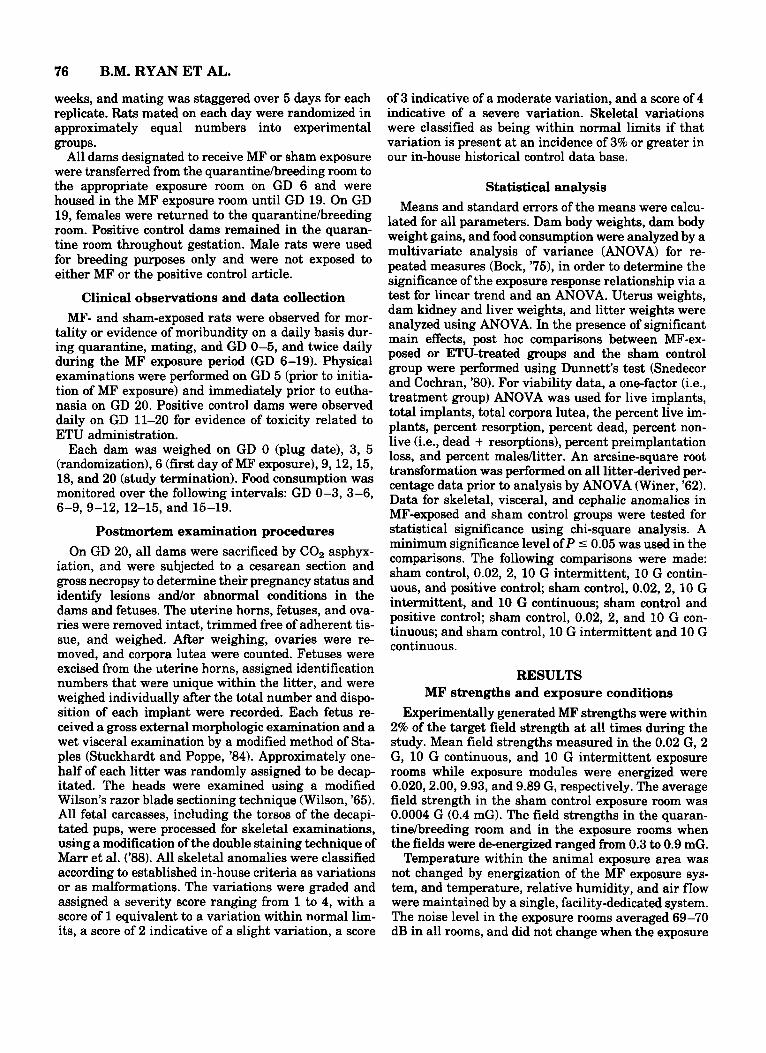
Fig. 1. Effect of 60 Hz MF on mean dam body weight during gestation. No statistically significant (P 5 0.05) differences were noted between the mean body weight of dams exposed to MF and that of the sham control dams. Inter. = intermittent; Cont. = continuous.
system was energized. Vibration in animal exposure racks did not differ when the fields were energized or de-energized.
Maternal toxicity No deaths occurred in any group during the study,
and clinical observations were unremarkable in all groups. No clinical evidence of toxicity related to expo- sure to either MF or the positive control article was noted in any animal.
When compared to sham controls, no statistically significant differences in body weight (Fig. l), liver and kidney weights, uterine weight, or corrected body weights were found in any group exposed to MF or the positive control article. Small but statistically signifi- cant reductions in body weight gain between GD 12 and 15 were observed in the 10 G intermittent expo- sure group and in the positive control group over GD 9-12. Cumulative body weight gain was also signifi- cantly reduced on day 15 in the 10 G intermittent group and on days 12 and 15 in the positive control group (Table 1). These differences were transient, however, as body weight gains in the dams from these exposure groups did not differ from sham control at GD 18.
Food consumption data are presented in Table 2. Minimal but statistically significant changes in food consumption were noted in the 2 G and 10 G continu- ous group dams during GD 15-19 and in the positive control dams during GD 0-3 and 12-15. None of these differences is considered to be biologically significant.
Maternal reproduction and litter viability No gross lesions were noted in any dam in any study
group. Viable uterine implants were noted in 54 of 55 dams (98%) in the sham control group, 48 of 55 dams (87%) in the 0.02 G MF group, 44 of 55 dams (80%) in the 2 G MF group, 50 of 55 dams (91%) in the 10 G intermittent MF group, 50 of 55 dams (91%) in the 10 G continuous MF group, and 15 of 15 dams (100%) in the positive control group (Table 3). Of those dams in which viable implants were not observed, nonviable implants were noted in 1/1 sham control dams, 017 dams in the 0.02 G MF group, 2/11 dams in the 2 G MF group, 1/5 dams in the 10 G intermittent MF group, and 1/5 dams in the 10 G continuous MF group. In dams without visible implants, the uterus was stained to determine early pregnancy loss.
Embryofetotoxicity observations Similar numbers of resorptions were observed in all
groups (Table 3). The total number of corpora lutea and percent preimplantation loss were highest in the sham control group. This resulted in a statistically signifi- cant decrease in the percent preimplantation loss for dams in the 2 and 10 G continuous MF groups. The ma1e:female ratio was approximately 1:l in all groups; however, the percent of maledlitter was significantly higher in the group exposed to 2 G MF.
Gross external examination of fetuses revealed no effects of MF exposure (Table 4). By contrast, 100% of the fetuses from positive control dams treated with
78 B.M. RYAN ET AL.
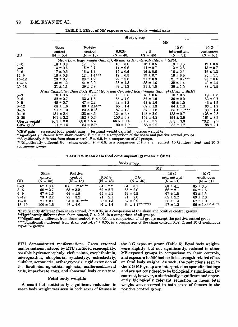
TABLE I. Effect of MF exposure on dam body weight gain
Study group MF
Sham Positive 10 G 10 G control control 0.02G 2 G intermittent continuous
GD (N = 55) (N = 15) (N = 48) (N = 46) (N = 51) (N = 51) Mean Dam Body Weight Gain (g), 48 and 72 Hr Intervals (Mean f SEM)
3-0 18 f 0.6 17 f 3.2 18 f 0.6 18 f 0.6 18 f 0.6 19 f 0.8 13 4 0.7 6-3 14 f 0.6 15 f 2.7 14 f 0.6 14 f 0.7
9-6 17 f 0.5 16 f 1.4 15 f 0.6 16 f 0.6 16 f 0.5 15 f 1.3 12-9 18 f 0.6 12 f 1.4*~** 17 f 0.5 19 f 0.7 18 f 0.6 20 4 1.1 15-12 23 2 0.7 23 f 1.2 22 2 0.8 21 f 0.9 21 f 0.7*** 23 f 0.6 18-15 40 2 1.3 41 f 3.0 38 f 1.3 38 f 1.6 38 f 1.4 40 ? 1.4 20-18 31 2 1.1 29 2 2.9 32 f 1.3 31 f 1.5 30 f 1.5 33 4 1.5
Mean Cumulative Dam Body Weight Gain and Corrected Body Weight Gain (g) (Mean f SEM) 0-3 18 f 0.6 17 f 3.2 18 f 0.6 18 f 0.6 18 t 0.6 19 2 0.8 0-6 32 f 0.7 32 f 1.6 33 f 1.0 32 f 1.0 30 f 0.9 32 * 0.7 0-9 49 & 0.7 47 f 2.2 48 f 1.2 48 f 1.0 46 f 1.0 46 f 1.5 0-12 68 2 1.0 60 f 2.4*3** 65 f 1.4 67 f 1.3 64 f 1.3 66 4 1.2 0-15 91 f 1.3 83 t 2.3* 87 f 1.8 87 +- 1.8 85 f 1.7*** 88 2 1.4
128 2 2.3 0-18 131 2 2.4 123 f 4.5 124 f 2.8 126 f 3.0 123 f 2.7 0-20 161 & 3.2 152 f 5.3 156 f 3.8 157 f 4.2 154 f 3.9 161 * 3.3 Uterus weight 70.9 2 3.0 68.0 f 5.4 66.5 f 3.4 70.6 f 3.2 68.3 f 3.3 73.2 5 2.9 CBW gain' 91 f 1.5 84 f 2.7* 90 f 1.9 86 f 2.0 85 f 1.7 88 ? 2.1
lCBW gain = corrected body weight gain = terminal weight gain (g) - uterus weight (g). *Significantly different from sham control, P 5 0.5, in a comparison of the sham and positive control groups. **Significantly different from sham control, P 5 0.5, in a comparison of all groups. ***Significantly different from sham control, P I 0.5, in a comparison of the sham control, 10 G intermittent, and 10 G continuous groups.
13 f 0.6
TABLE 2. Mean dam food consumption (g) (mean 2 SEMI
Study group MF
Sham Positive 10 G 10 G control control 0.02 G 2 G intermittent continuous
GD (N = 55) (N = 15) (N = 48) (N = 46) (N = 51) (N = 51) 65 f 3.0 0-3 67 f 3.4 106 f 12.6*,** 64 f 2.2 64 f 3.1 68 f 4.1
3-6 68 f 2.7 65 f 3.2 69 f 2.7 66 f 2.0 66 f 2.1 64 f 1.6 6-9 67 f 1.5 64 2 1.8 65 f 1.1 64 f 0.9 67 f 1.8 63 t 1.5 9-12 73 f 2.2 72 f 3.2 71 f 2.1 71 f 2.6 69 f 2.2 69 f 2.6 12-15 71 f 2.1 94 f 10.7*'** 69 f 1.3 67 f 0.9 68 f 1.4 67 f 1.0
*Significantly different from sham control, P 5 0.05, in a comparison of the sham and positive control groups. **Significantly different from sham control, P 5 0.05, in a comparison of all groups. ***Significantly different from sham control, P 5 0.05, in a comparison of all groups except the positive control group. ****Significantly different from sham control, P 5 0.05, in a comparison of the sham control, 0.02, 2, and 10 G continuous exposure groups.
15-19 100 f 1.5 96 f 4.0 97 f 1.4 94 f 1.4***,**** 97 f 1.3 96 f 1.4***-****
ETU demonstrated malformations. Gross external malformations induced by ETU included exencephaly, possible hydranencephaly, cleft palate, exophthalmia, micrognathia, ablepharia, syndactyly, ectrodactyly, clubfoot, acromicria, arthrogryposis, rigid extension of the forelimbs, agnathia, aglossia, malformed/absent tails, imperforate anus, and abnormal body curvature.
Fetal body weights A small but statistically significant reduction in
mean body weight was seen in both sexes of fetuses in
the 2 G exposure group (Table 5). Fetal body weights were slightly, but not significantly, reduced in other MF-exposed groups in comparison to sham controls, and exposure to MF had no field strength-related effect on fetal body weight. As such, the reductions seen in the 2 G MF group are interpreted as sporadic findings and are not considered to be biologically significant. By contrast, however, a statistically significant and appar- ently biologically relevant reduction in mean fetal weight was observed in both sexes of fetuses in the positive control group.
DEVELOPMENTAL TOXICITY OF 60 HZ MF 79
TABLE 3. Summary of maternal reproduction and litter viability
Parameter Initial group size (sperm-positive) Actual group size (gravid) Viable litters' (at least 1 live implant) Non-viable litters (no live implants) Male: female ratioflitter % Maleflitter Corpora lutea
Live implants
Total implants
% Preimplantation loss3 (mean f SEM) Non-live implants (resorptions)
Mean 2 SEM Non-live implants (deaths)
Mean 2 SEM Malformed fetuses4 Mean % non-live and malformed/litter Litters with resorptions % litters with resorptions Litters with deaths % Litters with deaths Litters with malformations % Litters with malformations
Mean 2 SEM'
Mean 2 SEM
Mean 2 SEM
Study group
Sham control
55 55 54 1
1.2:l 49
798 14.5 f 0.4
623 11.3 2 0.5
651 11.8 & 0.5 19.9 f 2.8
28 0.51 & 0.12
0 0 0 6.3
19 34.5 0 0 0 0
Positive control
MF 10 G 10 G
0.02 G 2 G intermittent continuous 15 15 15 0
151 52
217 14.5 f 0.6
177 11.8 f 0.9
186 12.4 f 0.9 15.1 f 5.9
7 0.47 f 0.13
2 0.13 f 0.13
177
7 46.7 1 6.7
15
100.05
100'
55 48 48 0
1.O:l 46
635 13.5 t 0.5
512 10.7 f 0.6
542 11.3 f 0.6 17.5 f 3.3
30 0.63 f 0.17
0 0 0 7.0
21 43.8 0 0 0 0
55 46 44
2 1.5:l 56* 626
13.9 f 0.5 533
11.6 f 0.5 568
12.3 2 0.5 11.9 f 2.3*
35 0.76 f 0.19
0 0 0 9.2
18 39.1 0 0 0 0
55 51 50 1
1.2:l 50
690 13.8 f 0.4
570 11.2 f 0.6
606 11.9 f 0.5 15.3 f 3.1
35 0.69 f 0.22
1 0.02 t 0.02
1 6.7
16 31.4 1 2.0 1 2.0
55 51 50 1
1.2:l 50
696 13.9 f 0.3
604 11.8 f 0.5
623 12.2 f 0.5 11.6 f 2.1*
19 0.37 f 0.15
0 0 0 5.4
12 23.5 0 0 0 0
Litters with resorptions/deaths 19 7 21 18 17 12 % Litters with resorptionsldeaths 34.5 46.7 43.8 39.1 33.3 23.5
'Viable litters refer to the number of dams with at least 1 live implant on GD 20. q h e mean reported utilizes the litter as the unit of observation f SEM; the N used is the number gravid unless the parameter requires an intact fetus for evaluation (e.g., sex, malformed, etc.), in which case the N utilized is viable litters. 3% Preimplantation loss = (corpora lutea - total implants)/corpora lutea x 100. When the number of implants is greater than the number of corpora lutea a zero was used, indicating 0% preimplantation loss. 4Fetuses with any combination of malformation(s) as determined by fetal examination. 'Statistical analysis not performed since all fetuses (100%) were malformed. *Significantly different from the sham control, P I 0.05, in a comparison of continuous exposure groups.
Wet visceral evaluations
No pattern of exposure-related visceral anomalies was seen in fetuses from dams exposed to MF (Table 6). The most prominent anomalies noted in fetuses ex- posed to MF were hydroureter andlor hydronephrosis. However, these conditions were observed in all groups, including the sham controls; most were of slight to mild severity, and are considered to be normal variations of development (Woo and Hoar, '72). Other anomalies ob- served in a few instances (per exposure group) included reversed umbilical artery (i.e., single umbilical artery courses the left side of the bladder rather than the right side of the bladder), kidney-pelvis red (i.e., pelvis con- gestedlred), and short/absent innominate artery. In contrast to the lack of significant increases in the in- cidence of visceral anomalies in groups exposed to MF, the incidence of anomalies observed in fetuses exposed in utero to ETU was significantly increased from sham control. Anomalies observed in ETU-exposed fetuses
included moderate to severe hydroureterhydronephro- sis, deflated stomach, and undescended testes.
Skeletal evaluations A similar pattern of skeletal anomalies was observed
in MF-exposed and sham control groups; the results of skeletal examinations are summarized in Table 7. Commonly observed anomalies in all groups included incomplete ossification or reduction of bones in the sternebrae, centra, pelvic girdle, and skull, and unos- sified bones of the sternebrae and sacral and lumbar vertebral arches. Enlarged sutura and misshapen (i.e., dumbbell and bipartite) centra were also observed. Oc- casional observations in all groups included rudimen- tary 14th rib, cervical ribs, variations of the 13th rib, and misshapen ribs. The incidence of these anomalies was similar in the sham control and all MF-exposed groups.
The incidence of variations observed in the thoracic
80 B.M. RYAN ET AL. TABLE 4. Summary of gross external anomalies'
Study group MF
Sham control Positive control 0.02 G 2 G 10 G intermittent 10 G continuous Observation (F/L = 623154) (F/L = 177115) (FIL = 512148) (Fa = 533144) (F/L = 570150) (F/L = 604150) Red mark/bruises2
Head3 Tail Limbs Body
Mascerated Exencephaly Possible
Ablepharia Exophthalmia Cleft lip Agnathia Micrognathia Syndactyly Ectrodactyly Acromicria Clubfoot
hydranencephaly
Arthrogryposis Tail
Short and/or kinked Stumped Absent Rudimentary
Imperforate anus Abnormal
body curvature Forelimbs rigid
and extended Aglossia (no tongue) Edema
717 717
16/14 716 -/- - I -
-/- -/- -/- -/- -/- -/- -/- -/- -I- -/- -/-
-/- -/- -/- -/- -/-
-/-
-/- -/- -/-
212 -/- 412 913 -/-
176115
43/12 915
16/10 1612 111
131114 158115 149115
311 171/15 2714
1214 2017 94/13 50111
107114
177115
158114 111 111
313 412
1119 414 -/- - I -
-/- -/- -/- -/- -/- -/- -/- -/- -/- -/- -/-
-/- -/- -/- -/- -/-
-/-
-/- -/- -/-
1018 816
1519 917 -/- -/-
-/- -/- -/- -/- -/- -/- -/- -/- -/- -/- -/-
-/- -/- -/- -/- -/-
-/-
-/- -/- -/-
816 111
-/-
-/- -/- -/- - I - -/- -/- -/- -/- -/- -/- -/-
-/- -/- -/- -/- -/-
-I-
-/- -/- -/-
23/15 313
16/13 815 -/- -/-
-/- -/- -/- -/- -/- -/- -/- -I-- - I - -/- -/-
-/- -/- -/- -I- -/-
--I-
-/- -I- -/-
'Fa = number of fetuses (F)/number of litters (L) examined. -I- = zero incidence. 2Statistical analysis not performed on red marWbruises since they were considered an artifact of handling during removal from the uterus. 3Red mark head includes jaw, face, nose, etc. These were collapsed into a general category (i.e., head).
TABLE 5. Fetal body weight (g) (mean fetal body weightflitter f SEMI
Study group MF
10 G 10 G Parameter Sham control Positive control 0.02 G 2 G intermittent continuous Litters/group' 54 15 48 44 50 50 Male fetal weight 4.32 2 0.05' 2.96 2 0.08*%** 4.20 2 O.0ti3 4.15 2 0.04****** 4.20 & 0.054 4.23 f 0.04 Female fetal weight 4.06 2 0.05 2.79 k 0.075**v** 4.00 f 0.053 3.90 ~f: 0.04** 3.97 f 0.034 4.02 2 0.044 Combined fetal weight 4.19 2 0.05 2.90 * 0.08*,** 4.09 2 0.05 4.04 f 0.03 4.10 f 0.05 4.13 2 0.04
'The mean reported utilizes the average fetal weightilitter; therefore, the litter is the unit of observation (N size). 2N = 53. 3N = 47. 4N = 48. 5N = 14. *Significantly different from sham control, P 5 0.05, in a comparison of the sham and positive control groups. **Significantly different from sham control, P 5 0.05, in a comparison of all groups. ***Significantly different from sham control, P 5 0.05, in a comparison of sham control, 0.02,2, and 10 G continuous groups.
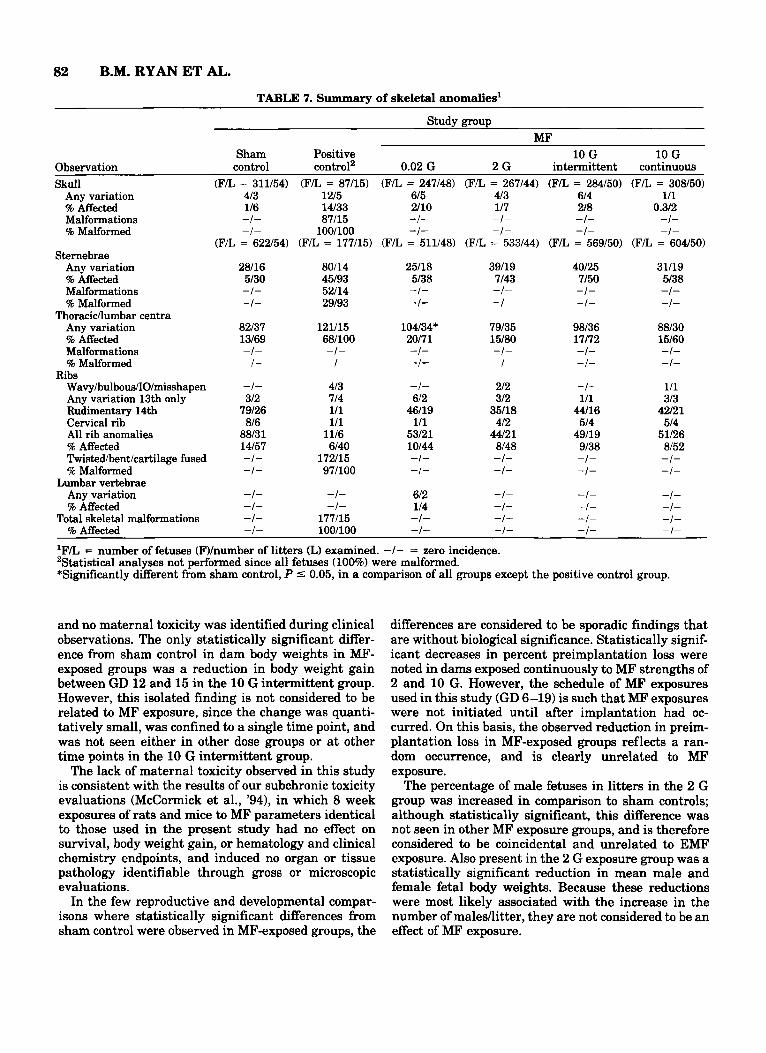
and lumbar centra was significantly increased in the groups exposed to MF. Several common variations group exposed to 0.02 G MF. No other statistically sig- (e.g., incomplete ossification of sternebrae) were seen nificant differences from sham control incidences of in groups exposed to MF', but were considered to be skeletal anomalies or variations were present in within normal limits. Other identified anomalies were
DEVELOPMENTAL TOXICITY OF 60 HZ MF 81
TABLE 6. Summary of visceral anomalies'
Study group
Sham Positive 10 G 10 G control control2 0.02 G 2 G intermittent continuous
MF
Observation (FL = 623154) (FL = 177115) (FL = 512148) (FIL = 533144) (FL = 570150) (FL = 604150) Hydroureter
Slight-mild 1019 37/11 18/10 15/10 18/12 1019 Moderate-severe 111 136115 -/- -/- 111 -/-
% Affected 2/19 981100 412 1 3/23 3126 2/18 Hydronephrosis
Slight-mild 817 54/12 1016 414 1218 918 Moderate-severe -/- 106113 -/- -/- 111 -/-
% Affected 1/13 9OI100 2/13 119 2/18 1/16 Extrahepatic Lobe -/- -1- -/- -I- -/- 111 Heart
Shortlabsent 111 312 111 212 -1- -/- Reversed umbilical artery -1- -I- 111 111 -I- -I- Left carotid originates at innominate's origin -/- -1- -/- -/- -/- 111 Left andlor right carotid absent -I- 212 -I- -I- -I- -I-
Kidney Pelvis red -/- -/- 111 111 111 111 Dark -/- 212 -/- -/- -/- -1-
Green material in ureter -/- 2/2 -/- -I- -/- -I- Stomach-deflatedl
convoluted -/- 176115 -/- -1- -I- -/- Testesundescended -/- 22/10 -/- -/- -1- -/- Liver-enlarged -/- 212 -/- -1- -/- -1- Bladder-deflated -/- 311 -I- -/- -I- -I-
'FIL = number of fetuses (F)/number of litters (L) examined. -/- = zero incidence. 'Statistical analyses not performed since all fetuses (100%) were malformed.
Innominate
considered to be variations with a range of severity; the majority of these variations were slight, within normal limits, and occurred in similar incidence in all groups.
Skeletal malformations were present in 100% of pos- itive control fetuses. Malformations were consistent with the gross external appearance of the positive control fetuses and included cleft palate; acrania; fusedlcleaved sternebrae; severly twisted ribs with as- sociated abnormal body curvature; irregular shaped pubis/tibia/fibia and scapula/radius/ulna/umerus (con- sistent with the gross external appearance of clubfoot/ rigid extension of forelimbs); fusedlabsent bones in the metacarpals, metatarsals, and phalanges (consistent with the gross external appearance of syndactyly and ectrodactyly); and fusdabsent sacral vertebrae (con- sistent with the gross external tail anomalies).
Cephalic evaluations With the exception of anophthalmia in one fetus in
the 10 G intermittent group, the results of cephalic examinations were unremarkable for sham controls and all MF-exposed groups. Cephalic anomalies ob- served in fetuses from the positive control group in- cluded exencephaly, anencephaly, hydrocephaly, cleft
palate, absentlsmall olfactory bulbs, absent cerebel- lum, and general malformed brain (i.e., lobedventri- cles not discernible).
DISCUSSION The results of the present study demonstrated no ev-
idence of developmental toxicity in rats exposed in utero to 60 Hz MF strengths ranging from 0.02 G to 10 G. Occasional statistically significant differences from sham control were seen in several parameters in fe- tuses exposed to MF; however, most changes observed in MF-exposed fetuses were minor variations that were also present in sham controls, and no clear pattern sug- gestive of adverse effects on fetal development was identifiable.
The 10 G field strength used in this study is consid- ered to be the maximum level to which animals can be exposed without the generation of possibly confounding vibration and thermal effects within the animal expo- sure volume. On this basis, the maximum field strength studied was selected on the basis of engineer- ing considerations and possible confounding factors, rather than on its induction of minimal maternal tox- icity. No maternal mortality was seen in any group,
82 B.M. RYAN ET AL. TABLE 7. Summary of skeletal anomalies'
Study group
Sham Positive 10 G 10 G MF
Observation control control' 0.02 G 2 G intermittent continuous Skull
Any variation % Affected Malformations % Malformed
Sternebrae Any variation % Affected Malformations % Malformed
Any variation % Affected Malformations % Malformed
Thoracicflumbar centra
(F/L = 311154) 413 116 -I- -/-
(F/L = 622154)
28/16 5/30 -I- -/-
82/37 13/69 -1- -/-
(FIL = 87/15) (FIL = 247148) (FIL = 267144) 1215 615 413 14/33 2/10 117 87/15 -/- -1- 100/100 -1- -1-
(FIL = 177115) (F/L = 511148) (F/L = 533144)
80114 25/18 39/19 45/93 5/38 7/43 52/14 -/- -I- 29/93 -/- -1-
121115 104/34* 79/35 68/100 20171 15180 -1- -/- -1- -/- -I- -/-
( F a = 284150) (F/L = 308150) 614 111 218 0.312 -/- -I- -/- -I-
(FIL = 569150) (FIL = 604150)
40125 31/19 7/50 5/38 -1- -1- -/- -I-
98/36 88/30 11/72 15/60 -/- -1- -/- -I-
Ribs Wavyhulbous/IO/misshapen -1- 413 -I- 212 -1- 111 Any variation 13th only 312 714 612 312 111 313 Rudimentary 14th 79/26 111 46/19 35/18 44/16 42/21 Cervical rib 816 111 111 412 514 514 All rib anomalies 8813 1 1116 53/21 44/21 49/19 51/26 % Affected 14/57 6/40 10144 8/48 9/38 8/52 Twiskdhent/cartilage fused -1- 172115 -1- -1- -1- -1- % Malformed -1- 971100 -1- -1- -1- -1-
Any variation -1- -1- 612 -I- -/- -I- % Affected -1- -1- 114 -I- -/- - I -
Total skeletal malformations -1- 177115 -/- -1- -1- -1- % Affected -1- 100/100 -/- -1- -/- -1-
'F/L = number of fetuses (F)/number of litters (L) examined. -/- = zero incidence. 'Statistical analyses not performed since all fetuses (100%) were malformed. *Significantly different from sham control, P 5 0.05, in a comparison of all groups except the positive control group.
Lumbar vertebrae
and no maternal toxicity was identified during clinical observations. The only statistically significant differ- ence from sham control in dam body weights in MF- exposed groups was a reduction in body weight gain between GD 12 and 15 in the 10 G intermittent group. However, this isolated finding is not considered to be related to MF exposure, since the change was quanti- tatively small, was confined to a single time point, and was not seen either in other dose groups or at other time points in the 10 G intermittent group.
The lack of maternal toxicity observed in this study is consistent with the results of our subchronic toxicity evaluations (McCormick et al., '94), in which 8 week exposures of rats and mice to MF parameters identical to those used in the present study had no effect on survival, body weight gain, or hematology and clinical chemistry endpoints, and induced no organ or tissue pathology identifiable through gross or microscopic evaluations.
In the few reproductive and developmental compar- isons where statistically significant differences from sham control were observed in MF-exposed groups, the
differences are considered to be sporadic findings that are without biological significance. Statistically signif- icant decreases in percent preimplantation loss were noted in dams exposed continuously to MF strengths of 2 and 10 G. However, the schedule of MF exposures used in this study (GD 6-19) is such that MF exposures were not initiated until after implantation had oc- curred. On this basis, the observed reduction in preim- plantation loss in MF-exposed groups reflects a ran- dom occurrence, and is clearly unrelated to MF exposure.
The percentage of male fetuses in litters in the 2 G group was increased in comparison to sham controls; although statistically signXicant, this difference was not seen in other MF exposure groups, and is therefore considered to be coincidental and unrelated to EMF exposure. Also present in the 2 G exposure group was a statistically significant reduction in mean male and female fetal body weights. Because these reductions were most likely associated with the increase in the number of males/litter, they are not considered to be an effect of MF exposure.
DEVELOPMENTAL TOXICITY OF 60 HZ MF 83
A statistically significant increase in the incidence of skeletal variations was observed in the thoracic and lumbar centra of fetuses exposed to 0.02 G MF; this increase was not seen in groups exposed to higher field strengths. Although their incidence was increased, most findings in the 0.02 G group consisted of dumbbell shaped centra and other common variations that are considered to represent normal stages of development; all variations were observed in both the sham control group and the 0.02 G group. While other variations were also found at low incidence during visceral, skel- etal, and cephalic examinations, these too were consid- ered to reflect normal stages of maturation, and there- fore are incidental in nature and without biological consequence.
In contrast to the overall lack of effect of MF expo- sure on fetal development in this study, significant ma- ternal and developmental toxicity were observed in the positive control group exposed to ETU. Although it pro- duced minimal maternal toxicity, ETU did induce a pattern of anomalies that is consistent with previous assessments of its teratogenicity (Khera, ’73; Ruddick and Khera, ’75). All fetuses from ETU-treated dams demonstrated malformations; skeletal and cephalic malformations were consistent with the gross external appearance of the malformed fetuses. Mean body weights were also significantly reduced in litters re- ceiving in utero exposure to ETU.
The present study was conducted to address an im- portant gap in the data base relevant to the biological effects of MF. The experiment was conducted using a well-defined animal model that has been used widely to assess the developmental toxicity of chemical and physical agents. Group sizes in the study were in- creased well beyond the standard for developmental toxicity evaluations in order to increase statistical power, and thereby increase the probability of de- tecting weak effects. It should be noted, however, that with its focus on pure, transient-free, linearly polar- ized MF, the design of the present study did not address the potential adverse effects of MF exposure para- digms other than pure 60 Hz fields. As such, our results do not preclude the possibility of adverse developmen- tal outcomes resulting from exposure to higher order harmonics or MF transients that are commonly en- countered in residential and occupational environ- ments.
The results of the present study do not support the hypothesis that exposure to 60 Hz MF causes signifi- cant risk of toxicity to the developing fetus. On this basis, and in consideration of the broad predictability of the experimental model used in these studies, it is concluded from these data that human exposure to pure 60 Hz MF is unlikely to pose a significant risk of toxicity to the fetus.
ACKNOWLEDGMENTS Research was supported by contract N01-ES-25351
from the National Toxicology Program, National Insti- tute of Environmental Sciences, Department of Health and Human Services. The authors thank Tanya Bryan, Mary Ann Cahill, Lisa Pomeranz, and Richard Syman- ski for their expert technical assistance and Dr. Gary Boorman of the National Institute of Environmental Health Sciences for his support and review of the manuscript.
LITERATURE CITED Bock, R.D. (1975) Multivariate Statistical Analysis in Behavioral Re-
search. New York McGraw-Hill, chap. 7. Brent, R.L., W.E. Gordon, W.R. Bennett, and D.A. Beckman (1993)
Reproductive and teratologic effecta of electromagnetic fields. Re- prod. Toxicol., 7:535-580.
Chernoff, N., J.M. Rogers, and R. Kavet (1992) A review of the liter- ature on potential reproductive and developmental toxicity of elec- tric and magnetic fields. Toxicology, 74:91-126.
Kaune, W.T., R.G. Stevens, N.J. Callahan, R.K. Severson, and D.B. Thomas (1987) Residential magnetic and electric fields. Bioelectro- magnetics, 8:315-335.
Khera, K.S. (1973) Ethylenethiourea: Teratogenicity in rats and rab- bits. Teratology, 7~242-252.
Marr, M.C., C.B. Myers, J.D. George, and C.J. Price (1988) Compar- ison of single and double staining for evaluation of skeletal devel- opment The effects of ethylene glycol (EG) in CD rats. Teratology, 37:476.
McCormick, D.L., B.M. Ryan, J.C. Findlay, J.B. Harder, J.R. Gauger, T.R. Johnson, M.J. Tomlinson, L.H. Brennecke, and G.A. Boorman (1994) Eight week toxicity study of 60 Hz magnetic fields in F344 rats and B6C3F1 mice. Proc. Bioelectromagn. Soc., 16:83.
Rommereim, D.N., W.T. Kaune, R.L. Buschbom, R.D. Phillips, and M.R. Sikov (1987) Reproduction and development in rats chroni- cally exposed to 60-Hz electric fields. Bioelectromagnetics, 8:243 - 258.
Rommereim, D.N., R.L. Rommereim, M.R. Sikov, R.L. Buschbom, and L.E. Anderson (1990) Reproduction, growth, and development of rats during chronic exposure to multiple field strengths of 60-Hz electric fields. Fundam. Appl. Toxicol., 14:608-621.
Ruddick, J.A., and K.S. Khera (1975) Pattern of anomalies following single oral doses of ethylenethiourea to pregnant rats. Teratology, 12:277-282.
Sikov, M.R., D.N. Rommereim, J.L. Beamer, R.L. Buschbom, W.T. Kaune, and R.D. Phillips (1987) Developmental studies of Hanford miniature swine exposed to 60-Hz electric fields. Bioelectromagnet- ics, 8:229-242.
Snedecor, G.W., and W.G. Cochran (1980) Statistical Methods. 7th Ed. Ames: Iowa State University Press.
Stuckhardt, J.L., and S.M. Poppe (1984) Fresh visceral examination of rat and rabbit fetuses used in teratogenicity testing. Teratogen. Carcinogen. Mutagen., 4:181-188.
Wertheimer, N., and E. Leeper (1986) Possible effects of electric blan- kets and heated waterbeds on fetal development. Bioelectromagnet- ics, 7:13-22.
Wilson, J.G. (1965) Embryological considerations in teratology. In: Teratology: Principles and Techniques. J.G. Wilson and J. War- kany, eds. Chicago: University of Chicago Press, pp. 251-277.
Winer, B.J. (1962) Statistical Principles in Experimental Design. New York McGraw-Hill.
Woo, D.C., and R.M. Hoar (1972) “Apparent hydroenphrosis” as a normal aspect of renal development in late gestation of rats: The effects of methyl salicylate. Teratology, 6:191-196.


![Repeated-dose toxicity of common ragweed on rats · cumanin BW5147 [52] trypanocidal [53] antileishmanial [53] anti-inflammatory [54] ... Repeated-dose toxicity of common ragweed](https://img.pdfslide.us/doc/110x75/5b8306107f8b9a940b8c2e41/repeated-dose-toxicity-of-common-ragweed-on-rats-cumanin-bw5147-52-trypanocidal.jpg)