Embed Size (px)
Citation preview
Developmental Dysplasiaof Hip
• CDH is old term
• Congenital means: condition existed at birth
• DDH is more representative of the wide range of abnormalities seen in this condition
• embryonic, fetal, and infantile periods
• It includes congenital and those that are developmental, incorporating subluxation, dislocation, and dysplasia of the hip
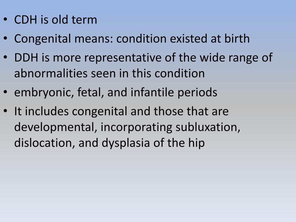
• In the newborn, the term dysplasia refers to any hip with a positive Ortolani sign
• Dysplasia has anatomical and radiological definition.
Anteroposterior view at 15 years of age. Note the bilateral anatomic dysplasia. The right hip is radiographically dysplastic, and the left hip is radiographically subluxated
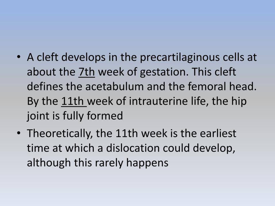
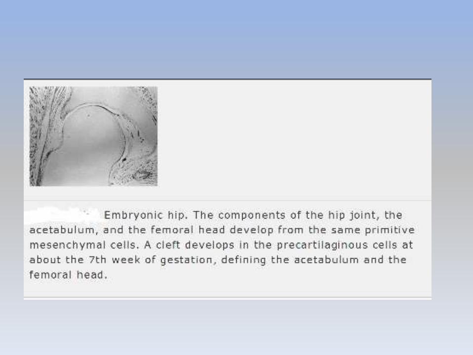
• A cleft develops in the precartilaginous cells at about the 7th week of gestation. This cleft defines the acetabulum and the femoral head. By the 11th week of intrauterine life, the hip joint is fully formed
• Theoretically, the 11th week is the earliest time at which a dislocation could develop, although this rarely happens
• In normal hip at birth, the femoral head is deeply seated in the acetabulum and held within the confines of the acetabulum by the surface tension of the synovial fluid
• It is extremely difficult to dislocate a normal infant's hip, even after incising the hip joint capsule
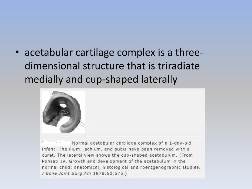
• acetabular cartilage complex is a three-dimensional structure that is triradiatemedially and cup-shaped laterally
w
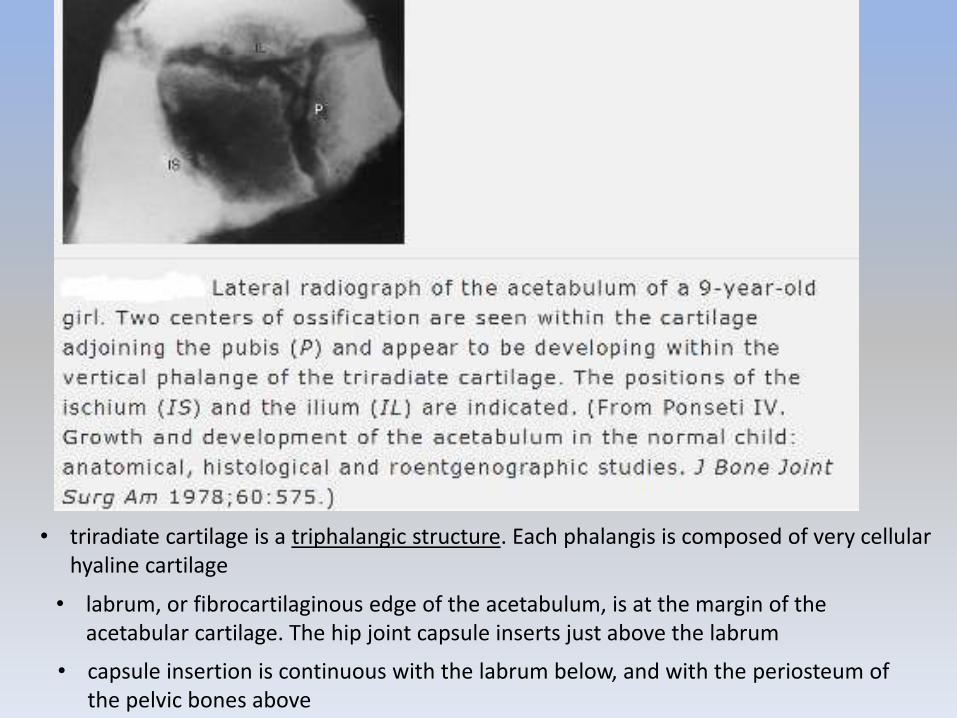
• triradiate cartilage is a triphalangic structure. Each phalangis is composed of very cellular hyaline cartilage
• labrum, or fibrocartilaginous edge of the acetabulum, is at the margin of the acetabular cartilage. The hip joint capsule inserts just above the labrum
• capsule insertion is continuous with the labrum below, and with the periosteum of the pelvic bones above
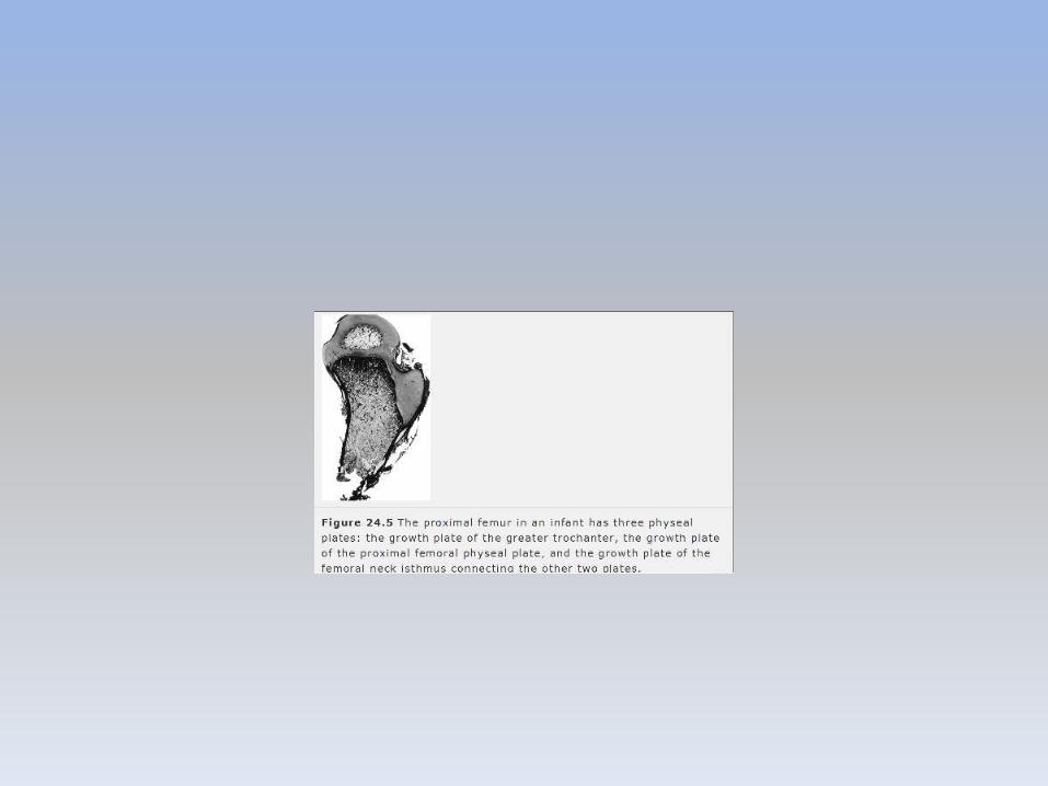
• In the infant the entire proximal end of the femur is composed of cartilage
• Between the 4th and 7th months of life, the proximal femoral ossification center appears
• three main growth areas in the proximal femur are the physeal plate, the growth plate of the greater trochanter and the femoral neck isthmus
• growth of the proximal femur is affected by muscle pull, the forces transmitted across the hip joint by weightbearing, normal joint nutrition, circulation, and muscle tone
• Experimental studies and clinical findings in humans with unreduced dislocations suggest that the main stimulus for the concave shape of the acetabulum is the presence of a spherical femoral head
• Growth of the acetabular cartilage occurs by means of interstitial growth within the cartilage and appositional growth under the perichondrium
Pathoanatomy
• tight fit between the femoral head and the acetabulum is lost. The femoral head can be made to glide in and out of the acetabulum
• DDH in the newborn refers to a spectrum of anatomic abnormalities, from mild dysplastic changes to the severe
• most common pathologic change in the newborn with DDH is a hypertrophied ridge of acetabular cartilage in the superior, posterior, and inferior aspects of the acetabulum.
• This ridge was referred to by Ortolani as the neolimbus
• neolimbus is composed of hypertrophied acetabular cartilage
• There is empiric evidence that the pathologic changes are reversible in the typical newborn with DDH, because there is a 95% success rate of treatment using simple devices such as the Pavlik harness
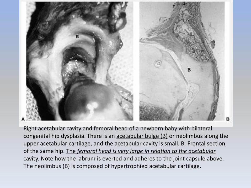
Right acetabular cavity and femoral head of a newborn baby with bilateral congenital hip dysplasia. There is an acetabular bulge (B) or neolimbus along the upper acetabular cartilage, and the acetabular cavity is small. B: Frontal section of the same hip. The femoral head is very large in relation to the acetabularcavity. Note how the labrum is everted and adheres to the joint capsule above. The neolimbus (B) is composed of hypertrophied acetabular cartilage.
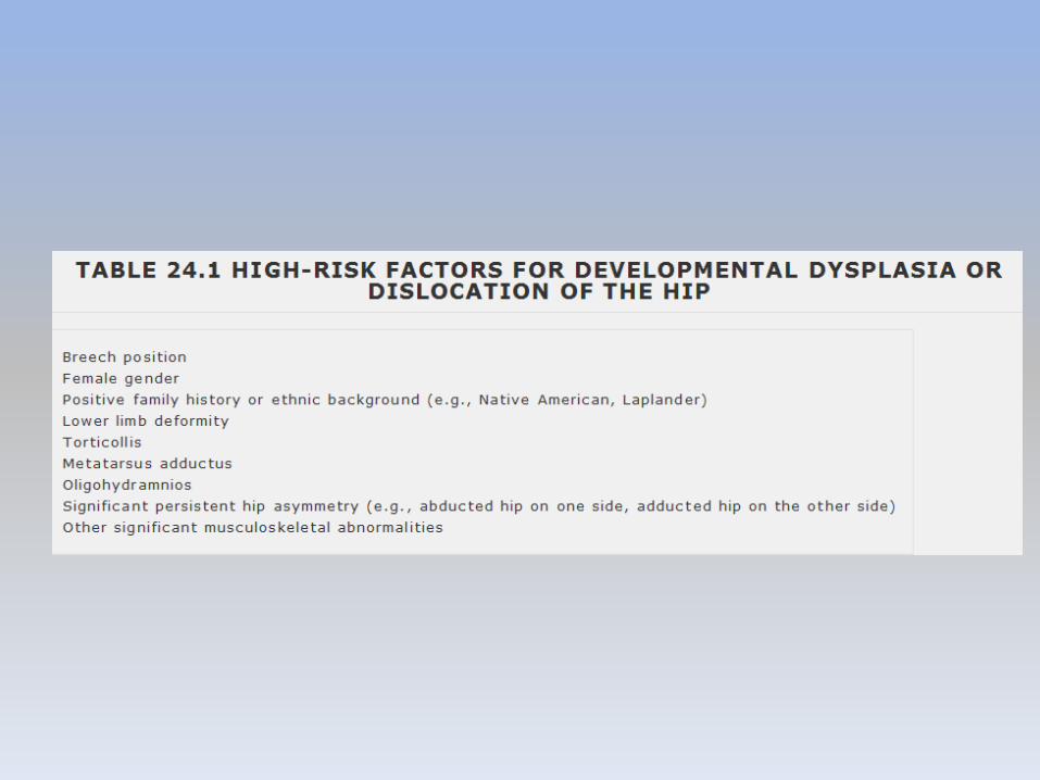
Causes
• incidence of DDH as high as 25 to 50 in 1000 live births among Lapps and Native Americans
• very low rate among the southern Chinese population and persons of African descent
• positive family history for DDH may be found in 12% to 33%
– tenfold increase among the parents and a sevenfold increase among siblings
• Intrauterine mechanical factors, such as breech position or oligohydramnios, and neuromuscular mechanisms such as myelomeningocele
• first-born child (primigravida)
• high rate of association of DDH with other intrauterine molding abnormalities, such as torticollis and metatarsus adductus (crowding theory)
• left hip
• more common among girls
• breech presentation
• Twice as many girls as boys are born breech
• Fifty-nine percent of breech presentations are first-born children
• postnatal environment (swaddling)
Diagnosis
• Neonates screening (ortolani)
• High-pitched soft tissue clicks are often elicited in the hip examination of newborns. These clicks are usually transmitted from the trochanteric region or the knee and have no diagnostic significance
• This led to overdiagnosis
• If only “clicks” wait for one month prior to doing an u/s as normal infant laxity with subluxation mostly resolves.
• Some physicians make treatment decisions on the basis of whether they feel that the hip is Ortolani-positive rather than Barlow-positive, the general opinion being that the Barlow-positive hip is more stable and hence may stabilize spontaneously
• Newborn clinical screening programs estimate that 1 of every 100 newborns examined has evidence of some hip instability (i.e., positive Ortolani or Barlow sign), although the incidence of true dislocation is reported to be between 1 and 1.5 cases per 1000 live births
• Complete irreducible dislocations are extremely rare in newborns and are usually associated with other generalized conditions, such as arthrogryposis, myelodysplasia, and other syndromes. These perinatal teratologicdislocations are at the extreme end of the DDH pathologic spectrum and account for only 2%
Late Diagnosis
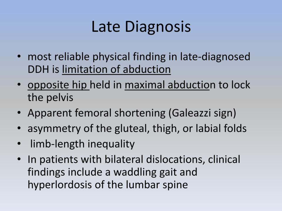
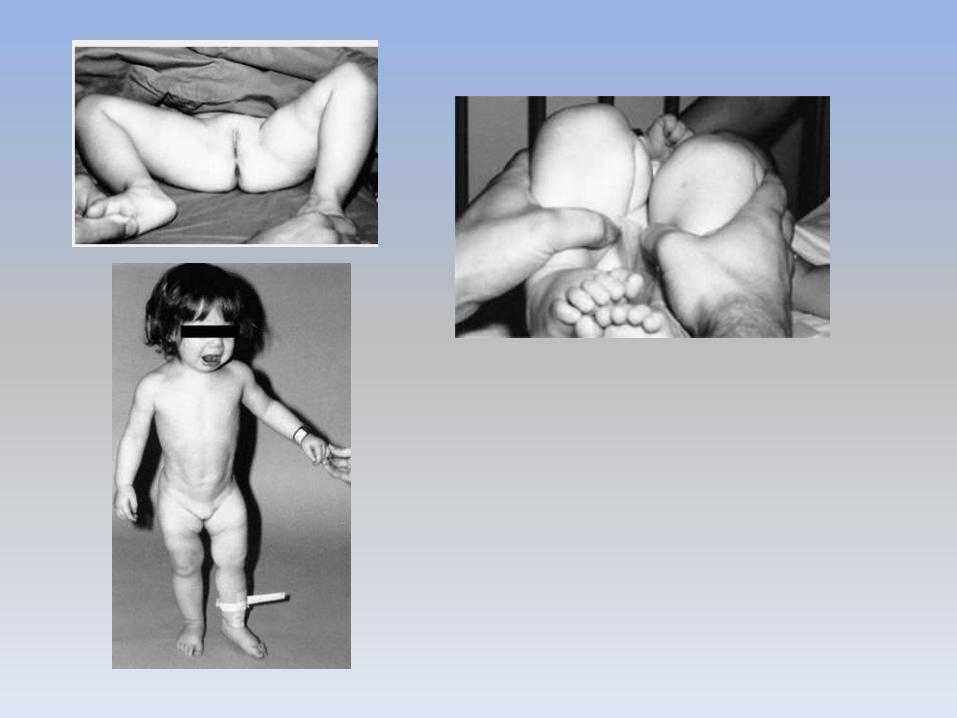
• most reliable physical finding in late-diagnosed DDH is limitation of abduction
• opposite hip held in maximal abduction to lock the pelvis
• Apparent femoral shortening (Galeazzi sign)
• asymmetry of the gluteal, thigh, or labial folds
• limb-length inequality
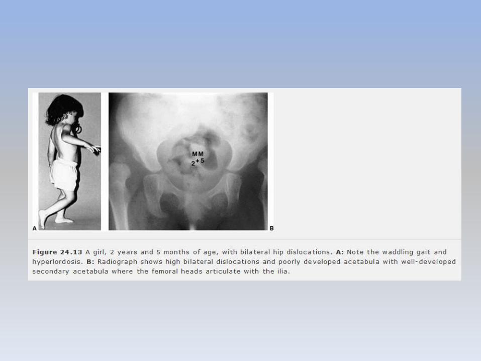
• In patients with bilateral dislocations, clinical findings include a waddling gait and hyperlordosis of the lumbar spine
• If DDH goes undetected, normal hip joint growth and development are impaired. With increasing age at detection and reduction, and particularly in children older than 6 months, the obstacles (intraarticular and extraarticular) to concentric reduction become increasingly difficult to overcome by simple treatment methods such as use of the Pavlik harness, and closed or open reduction usually must be performed under general anesthesia
intraarticular obstacles to reduction in late-diagnosed DDH
• ligamentum teres
• transverse acetabular ligament
• constricted anteromedial joint capsule (most segnificant)
• rarely an inverted and hypertrophied labrum
Diagnostic Imaging and Radiography
• clinical examination remains the gold standard
• Ultrasonography– Morphogenic
– Dynamic
• Its routine use in newborn nurseries has resulted in overdiagnosis
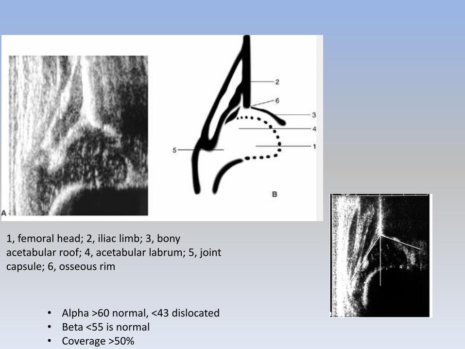
1, femoral head; 2, iliac limb; 3, bony acetabular roof; 4, acetabular labrum; 5, joint capsule; 6, osseous rim
• Alpha >60 normal, <43 dislocated• Beta <55 is normal• Coverage >50%
• Some centers advocate the use of ultrasonography in all Ortolani-positive infants to assess stability at the completion of treatment
• An ideal use for ultrasonography is for monitoring the progress of reduction of a subluxated or dislocated hip being treated in a Pavlik harness
• US 7- to 10-day intervals to check the progress of reduction of the hip and its stability during Pavlik
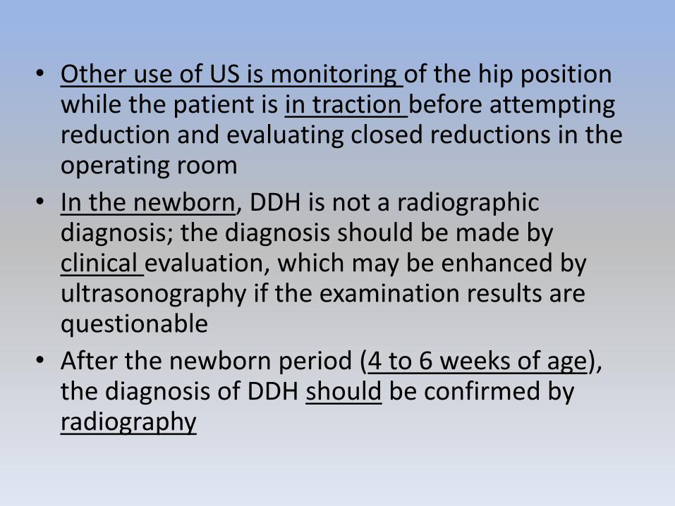
• Other use of US is monitoring of the hip position while the patient is in traction before attempting reduction and evaluating closed reductions in the operating room
• In the newborn, DDH is not a radiographic diagnosis; the diagnosis should be made by clinical evaluation, which may be enhanced by ultrasonography if the examination results are questionable
• After the newborn period (4 to 6 weeks of age), the diagnosis of DDH should be confirmed by radiography
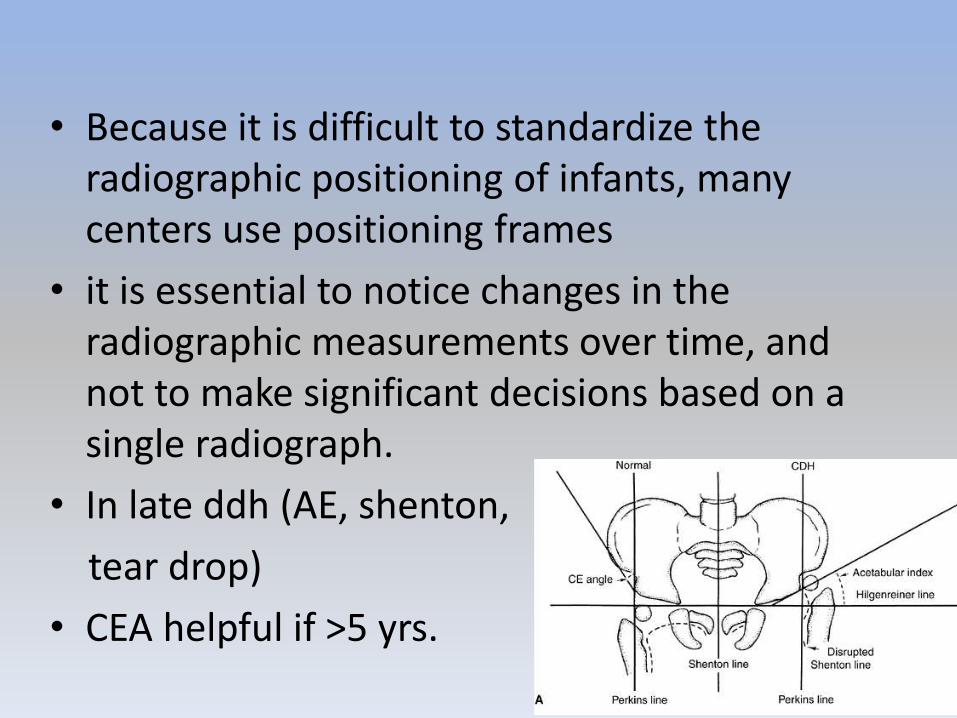
• Because it is difficult to standardize the radiographic positioning of infants, many centers use positioning frames
• it is essential to notice changes in the radiographic measurements over time, and not to make significant decisions based on a single radiograph.
• In late ddh (AE, shenton,
tear drop)
• CEA helpful if >5 yrs.
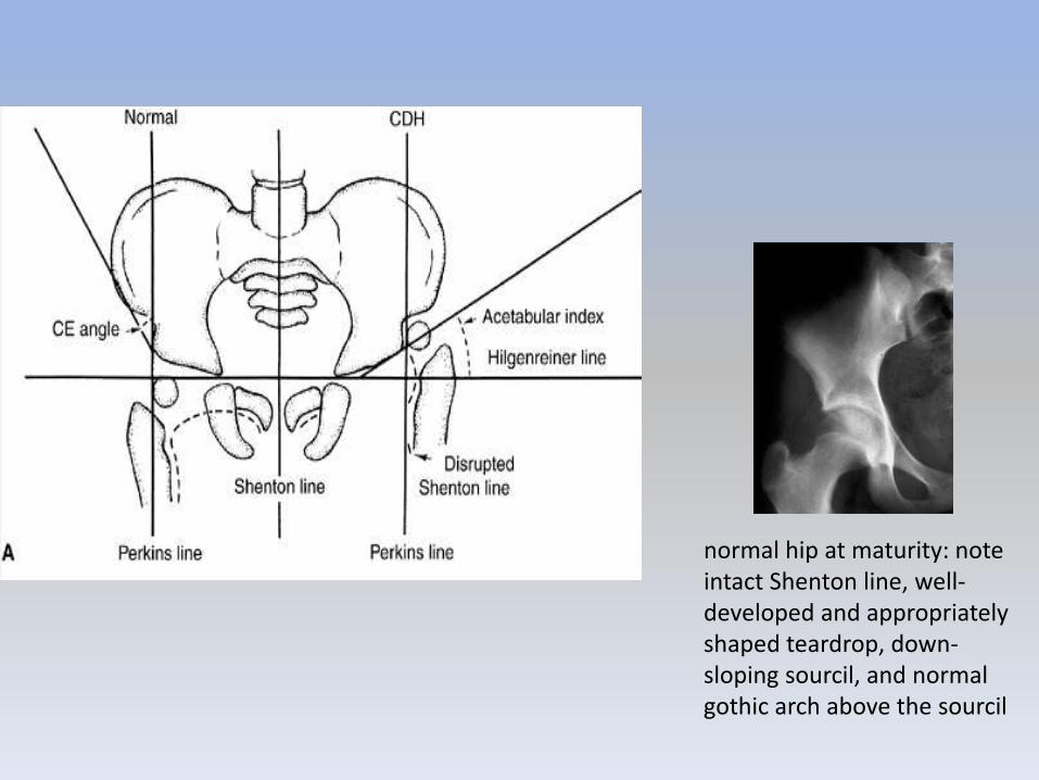
normal hip at maturity: note intact Shenton line, well-developed and appropriately shaped teardrop, down-sloping sourcil, and normal gothic arch above the sourcil
• Because it is not possible to predict the outcome of unstable hips in newborns, all newborns with clinical hip instability, as manifested by a positive Ortolani or Barlow sign, should be treated
Course in adults
• depend of laterality and pewsence of well-developed false acetabulum
• Worse in false acetabulum
• In unilateral complete dislocations, secondary problems of limb-length inequality, ipsilateralknee deformity and pain, scoliosis, and gait disturbance are common
• Pt with subluxation presented earlier than completely dislocated
Treatment
• treatment goals in DDH are the same, regardless of the age
• first goal is to obtain reduction and maintain that reduction to provide an optimal environment for the development of the femoral head and acetabulum
• acetabulum has the potential for development for many years after reduction as long as the reduction is maintained
• femoral head and femoral anteversion can remodel if the reduction is maintained
• The later the diagnosis of DDH is made, the less potential
• Triple diapers or abduction diapers have no place in the treatment of DDH
• most commonly used device for the treatment of DDH in the newborn is the Pavlik harness
• von Rosen splint, Frejka pillow
• before child able to stand in presence of reducible hip
• Pavlik prevents hip axt. And adduction but allows further abduction and flexion
• 95% resolve of hip instability if used 6/52 with full-time basis
• After 6/12: 50% failure (difficult to maintain active crawling Pt)
• failures of treatment result from problems related to the physician, the device, or the patient.
• Physician factors:• Inappropriate application
• CIC:– muscle imbalance, such as those with myelodysplasia or
cerebral palsy
– significant stiffness of the joints, such as children with arthrogryposis
– excessive ligamentous laxity, as seen in Ehlers-Danlossyndrome
• Not all pavlik are exactly the same
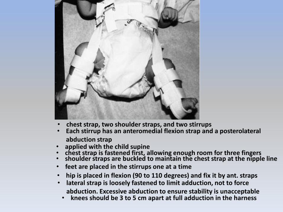
• chest strap, two shoulder straps, and two stirrups• Each stirrup has an anteromedial flexion strap and a posterolateral
abduction strap• applied with the child supine • chest strap is fastened first, allowing enough room for three fingers • shoulder straps are buckled to maintain the chest strap at the nipple line• feet are placed in the stirrups one at a time
• hip is placed in flexion (90 to 110 degrees) and fix it by ant. straps• lateral strap is loosely fastened to limit adduction, not to force
abduction. Excessive abduction to ensure stability is unacceptable• knees should be 3 to 5 cm apart at full adduction in the harness
• US is a useful for documenting relocation of the Ortolani-positive hip
• Used full time for 6-12/52
• Some recommend 6/52 more after stability is reached
• Most hips stabelize in days or weeks
• Check every 7-10/7
• adjust straps for infant growth
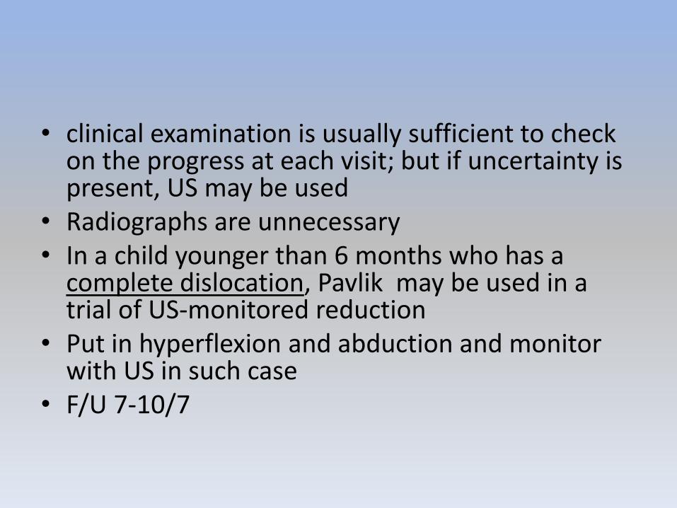
• clinical examination is usually sufficient to check on the progress at each visit; but if uncertainty is present, US may be used
• Radiographs are unnecessary• In a child younger than 6 months who has a
complete dislocation, Pavlik may be used in a trial of US-monitored reduction
• Put in hyperflexion and abduction and monitor with US in such case
• F/U 7-10/7
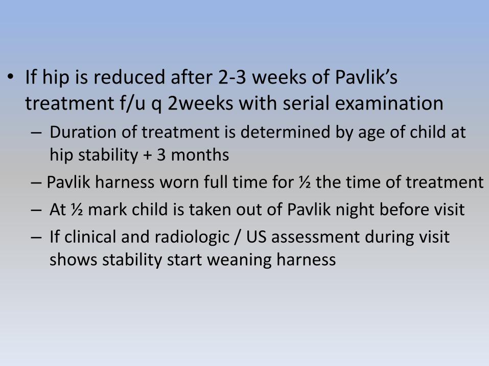
• If hip is reduced after 2-3 weeks of Pavlik’streatment f/u q 2weeks with serial examination
– Duration of treatment is determined by age of child at hip stability + 3 months
– Pavlik harness worn full time for ½ the time of treatment
– At ½ mark child is taken out of Pavlik night before visit
– If clinical and radiologic / US assessment during visit shows stability start weaning harness
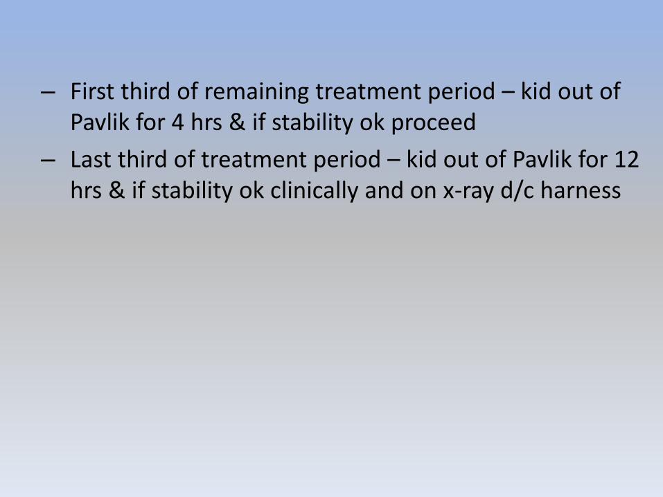
– First third of remaining treatment period – kid out of Pavlik for 4 hrs & if stability ok proceed
– Last third of treatment period – kid out of Pavlik for 12 hrs & if stability ok clinically and on x-ray d/c harness
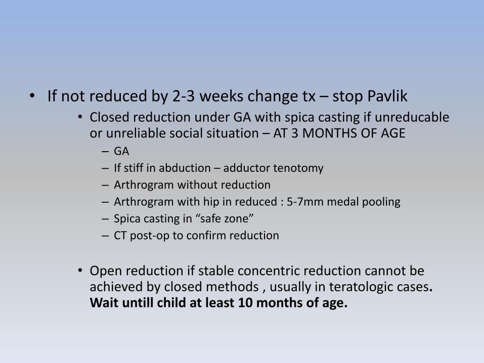
• If not reduced by 2-3 weeks change tx – stop Pavlik• Closed reduction under GA with spica casting if unreducable
or unreliable social situation – AT 3 MONTHS OF AGE– GA
– If stiff in abduction – adductor tenotomy
– Arthrogram without reduction
– Arthrogram with hip in reduced : 5-7mm medal pooling
– Spica casting in “safe zone”
– CT post-op to confirm reduction
• Open reduction if stable concentric reduction cannot be achieved by closed methods , usually in teratologic cases. Wait untill child at least 10 months of age.
• Coplications
– Inferior dislocation
– Femoral n. compression (generaly improves after removal of pavlik)
– Imp. To check quadrecips during visits.
– Brachial pluxes inj.
– Knee subluxation.
– Skin breakdown (mainly in groin)
• most disastrous consequence of Pavlikharness treatment is damage to the cartilaginous femoral head and the proximal femoral physeal plate

Thank you …