Embed Size (px)
Citation preview

Developmental Cell
Article
The Vps27/Hse1 Complex Is a GAT Domain-BasedScaffold for Ubiquitin-Dependent SortingGali Prag,1 Hadiya Watson,2 Young C. Kim,3 Bridgette M. Beach,1 Rodolfo Ghirlando,1 Gerhard Hummer,3
Juan S. Bonifacino,2 and James H. Hurley1,*1 Laboratory of Molecular Biology, National Institute of Diabetes and Digestive and Kidney Diseases2 Cell Biology and Metabolism Branch, National Institute of Child Health and Human Development3 Laboratory of Chemical Physics, National Institute of Diabetes and Digestive and Kidney Diseases
National Institutes of Health, U.S. Department of Health and Human Services, Bethesda, MD 20892, USA
*Correspondence: [email protected]
DOI 10.1016/j.devcel.2007.04.013
SUMMARY
The yeast Vps27/Hse1 complex and the homol-ogous mammalian Hrs/STAM complex deliverubiquitinated transmembrane proteins to theESCRT endosomal-sorting pathway. The Vps27/Hse1 complex directly binds to ubiquitinatedtransmembrane proteins and recruits bothubiquitin ligases and deubiquitinating enzymes.We have solved the crystal structure of the coreresponsible for the assembly of the Vps27/Hse1complex at 3.0 A resolution. The structure con-sists of two intertwined GAT domains, eachconsisting of two helices from one subunitand one from the other. The two GAT domainsare connected by an antiparallel coiled coil,forming a 90 A-long barbell-like structure. Thisstructure places the domains of Vps27 andHse1 that recruit ubiquitinated cargo anddeubiquitinating enzymes close to each other.Coarse-grained Monte Carlo simulations of theVps27/Hse1 complex on a membrane showhow the complex binds cooperatively to lipidsand ubiquitinated membrane proteins andacts as a scaffold for ubiquitination reactions.
INTRODUCTION
Protein ubiquitination is a widespread, multifunctional reg-
ulatory mechanism. Ubiquitin is conjugated to proteins via
an isopeptide bond between the C terminus of ubiquitin
and Lys residues in the ubiquitinated protein. This reaction
is carried out by a ubiquitin-activating enzyme (E1), a ubiq-
uitin-conjugating enzyme (E2), and a ubiquitin protein li-
gase (E3) (Hershko et al., 2000; Hochstrasser, 2000; Pick-
art, 2001; Weissman, 2001). Ubiquitination is a major
regulator of endocytosis and vesicular trafficking (Hicke,
2001; Raiborg et al., 2003). Ubiquitinated proteins are tar-
geted to and regulate the vesicular trafficking machinery
via interactions between the ubiquitin moiety and proteins
Develo
that contain ubiquitin-binding domains (Harper and Schul-
man, 2006; Hicke et al., 2005; Hurley et al., 2006).
The ESCRT protein network targets ubiquitinated trans-
membrane proteins for degradation in the lysosome or
yeast vacuole (Babst, 2005; Bowers and Stevens, 2005;
Hurley and Emr, 2006; Slagsvold et al., 2006). These pro-
teins were discovered in yeast, in which defects in their
genes lead to an enlarged cargo-rich compartment adja-
cent to the vacuole (Bowers and Stevens, 2005). This phe-
notype is referred to as a class E vacuolar protein-sorting
(VPS) defect. Yeast class E VPS genes encode the sub-
units of four hetero-oligomeric protein complexes: the
Vps27/Hse1 complex (Bilodeau et al., 2003; Bowers and
Stevens, 2005; Piper et al., 1995) and ESCRT-I, -II, and -III
(Babst, 2005; Bowers and Stevens, 2005; Hurley and
Emr, 2006; Slagsvold et al., 2006). The ESCRT network
is conserved from yeast to human and sorts ubiquitinated
transmembrane proteins into small vesicles that bud into
the lumen of endosomes, thus forming multivesicular bod-
ies (MVBs) (Gruenberg and Stenmark, 2004; Piper and
Luzio, 2001). In mammalian cells, the ESCRT network
directs the lysosomal degradation of signaling molecules
such as the EGF receptor (Clague and Urbe, 2001;
Haglund et al., 2003; Katzmann et al., 2002; Slagsvold
et al., 2006). Further, this network is hijacked by viruses
such as HIV, which use a process topologically equivalent
to MVB formation to bud from cells (Demirov and Freed,
2004; Morita and Sundquist, 2004).
Vps27/Hse1 is a multifunctional complex required for
MVB sorting of ubiquitinated cargo molecules as well as
the efficient recycling of late Golgi proteins including the
carboxypeptidase Y (CPY) sorting receptor, Vps10 (Bilo-
deau et al., 2002, 2003; Piper et al., 1995). Human
Vps27 is known as Hrs (hepatocyte growth factor receptor
substrate), and Hse1 has two human orthologs, STAM1
and STAM2 (signal-transducing adaptor molecule) (Ko-
mada and Kitamura, 2005) (Figure 1A). The Vps27/Hse1
and Hrs/STAM complexes sort cargo proteins from early
endosomes to the ESCRT-I complex (Bilodeau et al.,
2003; Katzmann et al., 2003) via clathrin-coated domains
(Lloyd et al., 2002; Raiborg et al., 2002). The Vps27/Hse1
complex is targeted to early endosomes via the FYVE do-
mains of Vps27 or Hrs (Raiborg et al., 2001), which bind to
phosphatidylinositol 3-phosphate (PI(3)P). The Vps27/Hse1
pmental Cell 12, 973–986, June 2007 ª2007 Elsevier Inc. 973
Developmental Cell
Structural Organization of the Vps27/Hse1 Complex
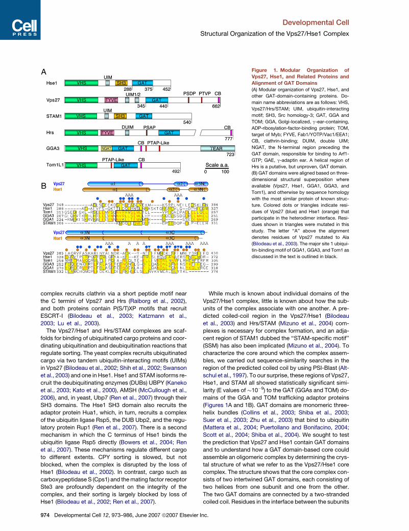
Figure 1. Modular Organization of
Vps27, Hse1, and Related Proteins and
Alignment of GAT Domains
(A) Modular organization of Vps27, Hse1, and
other GAT-domain-containing proteins. Do-
main name abbreviations are as follows: VHS,
Vps27/Hrs/STAM; UIM, ubiquitin-interacting
motif; SH3, Src homology-3; GAT, GGA and
TOM; GGA, Golgi-localized, g-ear-containing,
ADP-ribosylation-factor-binding protein; TOM,
target of Myb; FYVE, Fab1/YOTP/Vac1/EEA1;
CB, clathrin-binding; DUIM, double UIM;
NGAT, the N-terminal region preceding the
GAT domain, responsible for binding to Arf1-
GTP; GAE, g-adaptin ear. A helical region of
Hrs is a putative, but unproven, GAT domain.
(B) GAT domains were aligned based on three-
dimensional structural superposition where
available (Vps27, Hse1, GGA1, GGA3, and
Tom1), and otherwise by sequence homology
with the most similar protein of known struc-
ture. Colored dots or triangles indicate resi-
dues of Vps27 (blue) and Hse1 (orange) that
participate in the heterodimer interface. Resi-
dues shown in triangles were mutated in this
study. The letter ‘‘A’’ above the alignment
denotes residues of Vps27 mutated to Ala
(Bilodeau et al., 2003). The major site 1 ubiqui-
tin-binding motif of GGA1, GGA3, and Tom1 as
discussed in the text is outlined in black.
complex recruits clathrin via a short peptide motif near
the C termini of Vps27 and Hrs (Raiborg et al., 2002),
and both proteins contain P(S/T)XP motifs that recruit
ESCRT-I (Bilodeau et al., 2003; Katzmann et al.,
2003; Lu et al., 2003).
The Vps27/Hse1 and Hrs/STAM complexes are scaf-
folds for binding of ubiquitinated cargo proteins and coor-
dinating ubiquitination and deubiquitination reactions that
regulate sorting. The yeast complex recruits ubiquitinated
cargo via two tandem ubiquitin-interacting motifs (UIMs)
in Vps27 (Bilodeau et al., 2002; Shih et al., 2002; Swanson
et al., 2003) and one in Hse1. Hse1 and STAM isoforms re-
cruit the deubiquitinating enzymes (DUBs) UBPY (Kaneko
et al., 2003; Kato et al., 2000), AMSH (McCullough et al.,
2006), and, in yeast, Ubp7 (Ren et al., 2007) through their
SH3 domains. The Hse1 SH3 domain also recruits the
adaptor protein Hua1, which, in turn, recruits a complex
of the ubiquitin ligase Rsp5, the DUB Ubp2, and the regu-
latory protein Rup1 (Ren et al., 2007). There is a second
mechanism in which the C terminus of Hse1 binds the
ubiquitin ligase Rsp5 directly (Bowers et al., 2004; Ren
et al., 2007). These mechanisms regulate different cargo
to different extents. CPY sorting is slowed, but not
blocked, when the complex is disrupted by the loss of
Hse1 (Bilodeau et al., 2002). In contrast, cargo such as
carboxypeptidase S (Cps1) and the mating factor receptor
Ste3 are profoundly dependent on the integrity of the
complex, and their sorting is largely blocked by loss of
Hse1 (Bilodeau et al., 2002; Ren et al., 2007).
974 Developmental Cell 12, 973–986, June 2007 ª2007 Elsevier
While much is known about individual domains of the
Vps27/Hse1 complex, little is known about how the sub-
units of the complex associate with one another. A pre-
dicted coiled-coil region in the Vps27/Hse1 (Bilodeau
et al., 2003) and Hrs/STAM (Mizuno et al., 2004) com-
plexes is necessary for complex formation, and an adja-
cent region of STAM1 dubbed the ‘‘STAM-specific motif’’
(SSM) has also been implicated (Mizuno et al., 2004). To
characterize the core around which the complex assem-
bles, we carried out sequence-similarity searches in the
region of the predicted coiled coil by using PSI-Blast (Alt-
schul et al., 1997). To our surprise, these regions of Vps27,
Hse1, and STAM all showed statistically significant simi-
larity (E values of �10�9) to the GAT (GGAs and TOM) do-
mains of the GGA and TOM trafficking adaptor proteins
(Figures 1A and 1B). GAT domains are monomeric three-
helix bundles (Collins et al., 2003; Shiba et al., 2003;
Suer et al., 2003; Zhu et al., 2003) that bind to ubiquitin
(Mattera et al., 2004; Puertollano and Bonifacino, 2004;
Scott et al., 2004; Shiba et al., 2004). We sought to test
the prediction that Vps27 and Hse1 contain GAT domains
and to understand how a GAT domain-based core could
assemble an oligomeric complex by determining the crys-
tal structure of what we refer to as the Vps27/Hse1 core
complex. The structure shows that the core complex con-
sists of two intertwined GAT domains, each consisting of
two helices from one subunit and one from the other.
The two GAT domains are connected by a two-stranded
coiled coil. Residues in the interface between the subunits
Inc.

Developmental Cell
Structural Organization of the Vps27/Hse1 Complex
are shown to be essential for the normal sorting functions
of these proteins. Finally, the role of the core in organizing
the cooperative interactions of other domains of the
Vps27/Hse1 complex was explored by using coarse-
grained Monte Carlo simulations.
RESULTS
The Vps27/Hse1 Core Complex
Based on secondary structure predictions and the puta-
tive homology to known GAT domains, a protein construct
comprising residues 345–440 of Vps27 fused to an N-ter-
minal hexahistidine tag was coexpressed in Escherichia
coli with a second untagged construct comprising resi-
dues 275–375 of Hse1. The Vps27 and Hse1 fragments
coeluted from the chelating column and comigrated on
size-exclusion chromatography (Figure 2A). All of the ma-
terial consisted of the binary complex, and there was no
observable population of free monomeric subunits. Since
these portions of Vps27 and Hse1 are competent to form
a highly stable binary complex, and there is no indication
that other domains of these proteins are involved in com-
plex formation, we refer to this complex as the ‘‘Vps27/
Hse1 core complex’’ throughout the manuscript.
To determine the oligomeric state of the Vps27/Hse1
core complex in solution, the Vps27/Hse1 complex was
analyzed by analytical ultracentrifugation. The global
data analysis was consistent with a single ideal solute
with a molecular mass of 23.2 ± 0.4 kDa. This compares
well to the calculated molecular mass of 23,703 Da for a
1:1 complex of the Vps27 and Hse1 core fragments. The
experimental stoichiometry of 1:1 heterodimers is n =
0.98 ± 0.02 (Figures 2B and 2C; Figure S1, see the Supple-
mental Data available with this article online). The core
region of Hse1 expressed alone was too unstable to char-
acterize by analytical ultracentrifugation. The isolated core
region of Vps27 was found to be relatively stable, how-
ever. The isolated core region of Vps27 was characterized
by sedimentation equilibrium experiments (Figure 2D).
The global data analysis in terms of a single ideal solute re-
sulted in poor data fits, but analysis in terms of two non-
interacting solutes returned excellent fits consistent with
the presence of monomeric Vps27 and a higher-molecular
mass species (Figure 2D; Figure S2). The best fit showed
that the isolated Vps27 core sample contained 94%
monomer and 6% aggregate on a molar basis.
Structure of the Vps27/Hse1 Core Complex
The crystal structure of the Vps27/Hse1 core complex was
determined at 3.0 A resolution by a two-wavelength multi-
wavelength anomalous dispersion experiment at the Se
edge (Figure 3A). The overall structure is barbell like,
with dimensions of roughly 90 3 20 3 20 A (Figures 3B
and 3C). The structures of the two subunits are very similar
to each other, with an rmsd of 1.8 A for the overlay of 67 Ca
atoms. The two subunits are intimately intertwined such
that 2104 A2 of solvent-accessible surface area is buried
per subunit (Figures 3D and 3E). The N termini of the sub-
units are close together (17 A apart) at the middle of the
Develo
barbell. In contrast, the C termini are at opposite ends of
the barbell, 87 A away from each other.
Each subunit consists of three a helices. At 89 and 72 A,
respectively, the a3 helices are the longest in the struc-
ture. The a3 helices have three distinct structural roles.
The N- and C-terminal portions of a3 contribute to the for-
mation of three-helix bundles at each end of the barbell.
One bundle (HHV) consists of a1 (residues 288–312) and
a3-N (322–341) of Hse1, together with a3-C of Vps27
(410–438). The second bundle (VVH) consists of a1 (resi-
dues 351–371) and a3-N (381–399) of Vps27, together
Figure 2. Hydrodynamic Properties of the Vps27/Hse1 Core
Complex
(A) Gel filtration analysis of the recombinant Vps27/Hse1 core showing
comigration at an apparent mass of 29 kDa. This is slightly higher than
the calculated mass of 23.2 kDa for a 1:1 complex, and is therefore
consistent with an elongated 1:1 complex, but is not consistent with
any oligomer with a greater number of subunits.
(B) Sedimentation equilibrium profiles at 4.0�C plotted as a distribution
of the absorbance at 280 nm versus r at equilibrium. Data were col-
lected at 13 (orange), 16 (yellow), 19 (green), 22 (cyan), 25 (blue), and
28 (brown) krpm at a loading A280 of 0.75. The solid lines show the
best-fit global analysis (carried out for the three loading concentra-
tions) in terms of a single ideal solute; the corresponding residuals
are shown in the panels above the plot.
(C) Sedimentation equilibrium profile at 4.0�C and 22 krpm plotted in
terms of lnA280 versus r2. The data shown correspond to a loading
A280 of 0.25. The solid line indicates the plot expected for a monodis-
perse 1:1 Vps27:Hse1 complex.
(D) Sedimentation equilibrium profile of the Vps27 core region in isola-
tion at 4.0�C and 22 krpm plotted in terms of lnA280 versus r2. The data
shown correspond to a loading A280 of 0.60. The solid line indicates the
plot expected for a monomeric Vps27, indicating the presence of
higher oligomers.
pmental Cell 12, 973–986, June 2007 ª2007 Elsevier Inc. 975

Developmental Cell
Structural Organization of the Vps27/Hse1 Complex
Figure 3. Crystal Structure of the Vps27/Hse1 Complex
(A) Density-modified MAD Fourier synthesis (green) contoured at 1.0s and Se anomalous difference Fourier (red) contoured at 4.0s superimposed on
the refined structure; SeMet residues are highlighted.
(B) Overall structure of the core heterodimer; Vps27 is blue, and Hse1 is orange.
(C) Superposition of the core portions of the Vps27 and Hse1 monomers.
(D) Surface of Hse1 showing interactions with labeled residues of Vps27.
(E) Surface of Vps27 showing interactions with labeled residues of Hse1.
with a3-C of Hse1 (354–372). The narrow center of the bar-
bell consists of a two-stranded coiled coil formed by the
central portion of each a3 helix. The coiled-coil region
spans residues 396–414 of Vps27 and 338–356 of Hse1.
Several residues of the coiled coil are thus also part of
the helical bundles.
976 Developmental Cell 12, 973–986, June 2007 ª2007 Elsevie
Domain-Swapped GAT Domains in Vps27 and Hse1
The two three-helix bundles closely resemble the struc-
tures of GAT domains (Figures 4A and 4B). A search of
the structural database with Dali (Holm and Sander,
1995), using the HHV bundle as the probe structure, iden-
tified the GAT domain of Tom1 (Akutsu et al., 2005) as the
r Inc.

Developmental Cell
Structural Organization of the Vps27/Hse1 Complex
Figure 4. GAT Domains in Vps27 and Hse1
(A) The HHV helical bundle.
(B) The VVH helical bundle.
(C) The GAT domain of Tom1 shown in the same orientation as in (A) and (B).
(D) Superposition of the HHV and VVH bundles and the Tom1 GAT domain.
(E) Model for the closed monomeric conformation of the Vps27 GAT domain, generated by superimposing the Vps27 structure on the Tom1 GAT
domain monomer.
top-scoring match, with a Z score of 7.7 (Figure 4C). The
Tom1-GAT domain overlays the HHV bundle with an
rmsd of 2.6 A over 73 Ca positions. The VVH bundle over-
lays with an rmsd of 2.2 A over 47 Ca positions. These rmsd
values compare to values of 1.6–1.8 A over 86–90 Ca po-
sitions for superpositions of the structures of GAT domains
of GGA1, GGA3, and Tom1 with each other. Given the sig-
nificant sequence and structural similarity, it seems appro-
priate to designate the Vps27/Hse1 three-helix bundles as
Develo
members of the GAT domain family (Figures 1A and 1B).
The a3-N segment corresponds to a2 of the GAT domain,
and a3-C of the opposing subunit corresponds to a3 of the
GAT domain. The major difference between the GAT do-
mains of Vps27/Hse1 compared to GGAs and Tom1 is
that the former are heterodimeric, whereas the latter are
monomeric. Further, the Vps27/Hse1 GAT domains con-
tain a very short helix, a2 (Figures 4A and 4B), which has
no counterpart in the GGA GAT structures.
pmental Cell 12, 973–986, June 2007 ª2007 Elsevier Inc. 977

Developmental Cell
Structural Organization of the Vps27/Hse1 Complex
Figure 5. The Vps27/Hse1 Interface Is Required for Sorting
(A) Expression levels and coimmunoprecipitation of HA-tagged Vps27 and myc-tagged Hse1 proteins. Left panels. Whole-cell lysates from vps27D
hse1D cells cotransformed with Hse1-myc and Vps27-HA constructs were lysed and subjected to SDS-PAGE and immunoblot analysis with anti-HA
and anti-myc antibodies. Right panels. Rabbit anti-HA immunoprecipitation from the above-described lysates, followed by SDS-PAGE and immu-
noblotting with mouse anti-HA and mouse anti-myc antibodies.
(B–I) Fluorescence microscopy images of GFP chimeras of Ste3 and Cps1 in vps27D cells expressing Vps27-WT or the indicated core-complex
mutants.
(J) CPY maturation in vps27D hse1D cells expressing various constructs. Cells were metabolically labeled with 35S-metionine for 10 min (pulse) and
chased for 15 min in complete medium, and endogenous CPY was immunoprecipitated with anti-CPY antibody. The 15 min point samples were
analyzed by SDS-PAGE followed by fluorography.
(K) CPY colony blot assay on vps27D hse1D cells coexpressing Hse1-WT and various Vps27 constructs. Colonies from each strain were spotted onto
selective medium and overlayed with nitrocellulose. Secreted CPY was detected by immunoblotting the nitrocellulose with an anti-CPY antibody.
Mutational Analysis of the Heterodimer Interface
To confirm the physiological importance of the subunit
contacts observed in the Vps27/Hse1 core-complex crys-
tal structure, point mutations were introduced into the het-
erodimer interface. Vps27 residues Leu410, Ile417, and
Ile420 are deeply buried in the dimer interface (Figure 3E)
and were selected for mutagenesis to the charged residue
Asp. The mutations were introduced into a full-length HA-
tagged Vps27 construct to generate the mutants Vps27-
I417D, Vps27-I420D, and Vps27-L410D. For comparison,
a UIM mutation (Vps27-A266Q) that was predicted not to
abolish core-complex assembly was also generated.
These point mutants, as well as wild-type (WT) HA-tagged
978 Developmental Cell 12, 973–986, June 2007 ª2007 Elsevie
Vps27 (Vps27-WT), were coexpressed with wild-type
Hse1-myc in vps27D hse1D yeast cells. To determine
the expression levels of these proteins, whole-cell lysates
from these strains were subjected to SDS-PAGE and im-
munoblot analysis with antibodies to the HA and myc epi-
topes. No significant difference in expression level was
detected among Vps27-WT, core-complex mutants, or
the Vps27-A266Q mutant (Figure 5A, lanes 1–5). Hse1-
myc levels in each strain were also equivalent. The lysates
were then subjected to immunoprecipitation with anti-HA
antibody and were analyzed by immunoblotting with anti-
HA and anti-myc antibodies. As expected, Hse1-myc pro-
tein coimmunoprecipitated with both Vps27-WT and the
r Inc.

Developmental Cell
Structural Organization of the Vps27/Hse1 Complex
UIM mutant, Vps27-A266Q (Figure 5A, lanes 6 and 7);
however, this interaction was abolished by all three
core-complex mutations (Figure 5A, lanes 8–10).
We next sought to determine the functional conse-
quence of core-complex mutations. MVB sorting was
tested by using GFP chimeras of the biosynthetic cargo
protein Cps1 (GFP-Cps1) and the plasma membrane re-
ceptor Ste3 (Ste3-GFP) expressed in vps27D cells. Both
GFP-Cps1 and Ste3-GFP accumulated in the class-E
compartment in vps27D cells expressing only empty vec-
tor (Figures 5B and 5C). In addition, GFP-Cps1 labeled the
limiting membrane of the vacuole (Figure 5B). Transforma-
tion of the vps27D strain with Vps27-WT restored trans-
port of both cargo proteins into the lumen of the vacuole
(Figures 5D and 5E). In contrast, neither of the core-
complex mutants, Vps27-L410D or Vps27-I420D, was
capable of restoring this transport (Figures 5F–5I).
To assess the sorting of the soluble vacuolar hydrolase
CPY, the proteolytic maturation of the Golgi precursor (p2)
form to the vacuolar mature (m) form was examined by
pulse-chase analysis of vps27D hse1D cells transformed
with various Vps27 and Hse1 constructs (Figure 5J). After
a 15 min chase period, CPY species were isolated by im-
munoprecipitation and were analyzed by SDS-PAGE
(Figure 5J). vps27D hse1D cells coexpressing Vps27-WT
and Hse1-WT exhibited normal CPY maturation, and the
majority of the hydrolase migrated as mature CPY (Fig-
ure 5J, lane 1). Significant differences were observed in
CPY maturation in vps27D hse1D cells expressing either
Vps27-WT alone or Hse1-WT alone (Figure 5J, lanes 2
and 3). Specifically, cells expressing Vps27-WT alone
(Figure 5J, lane 2) exhibited a partial CPY-maturation de-
fect, as compared to cells expressing Hse1-WT alone
(Figure 5J, lane 3), in which virtually all of the CPY migrated
as the p2 form. These findings are consistent with past re-
sults that Vps27 is essential for CPY maturation, while loss
of Hse1 results in only a modest reduction in CPY pro-
cessing (Bilodeau et al., 2002). Interestingly, none of the
core-complex mutants (Vps27-I420D and Vps27-L410D
shown) fully complemented the loss of Vps27 in vps27D
hse1D cells expressing Hse1-WT alone (Figure 5J, lanes
5 and 6), while Vps27-A266Q behaved identically to
Vps27-WT (Figure 5J, lane 4). Similar results were ob-
tained when the effects of the Vps27-A266Q and core-
complex mutations were analyzed in a CPY secretion col-
ony blot assay (Figure 5K). In this assay, secreted CPY
was undetectable in vps27D hse1D cells expressing
Vps27-WT, and it was barely visible in vps27D hse1D cells
expressing Vps27-A266Q (Figure 5K, lanes 1 and 2); how-
ever, significant amounts of CPY secretion were detected
in vps27D hse1D cells expressing either Vps27-I420D or
Vps27-L410D (Figure 5K, lanes 3 and 4). These data are
also consistent with previous studies showing no signifi-
cant role for the UIM of Vps27 in CPY maturation (Bilodeau
et al., 2002). These results thus show that point mutants in
Vps27 that abrogate binding to Hse1 affect CPY sorting to
roughly the same extent as the deletion of the Hse1 gene.
Taken together with the results showing that these mu-
tants block sorting of Cps1 and Ste3, these data demon-
Develo
strate that the Vps27-interface residues observed in the
crystal structure are important for the cellular functions
of the Vps27/Hse1 complex.
Monte Carlo Simulation Analysis of the Properties
of the Vps27/Hse1 Complex
The Vps27/Hse1 core is the last folded domain of the
Vps27/Hse1 complex to have its three-dimensional struc-
ture solved. The complex consists of several folded do-
mains linked by unstructured segments (Figure 6). This
structural information allowed us to conduct a theoretical
analysis of the organization, dynamics, and interactions of
this complex with the model ubiquitinated transmembrane
cargo, Cps1 (Ub-Cps1), by using a coarse-grained Monte
Carlo (MC) approach. The UIMs of Vps27 and Hse1 were
modeled as interacting with ubiquitin moieties on Lys8 of
the Cps1 cytosolic tail (Katzmann et al., 2001). The dis-
tance distributions of all three Ub-Cps1 molecules to a
noninteracting domain used as a reference point are found
to be equivalent over the course of the simulation, indicat-
ing that the system is well sampled and thoroughly equil-
ibrated (Figure 7A).
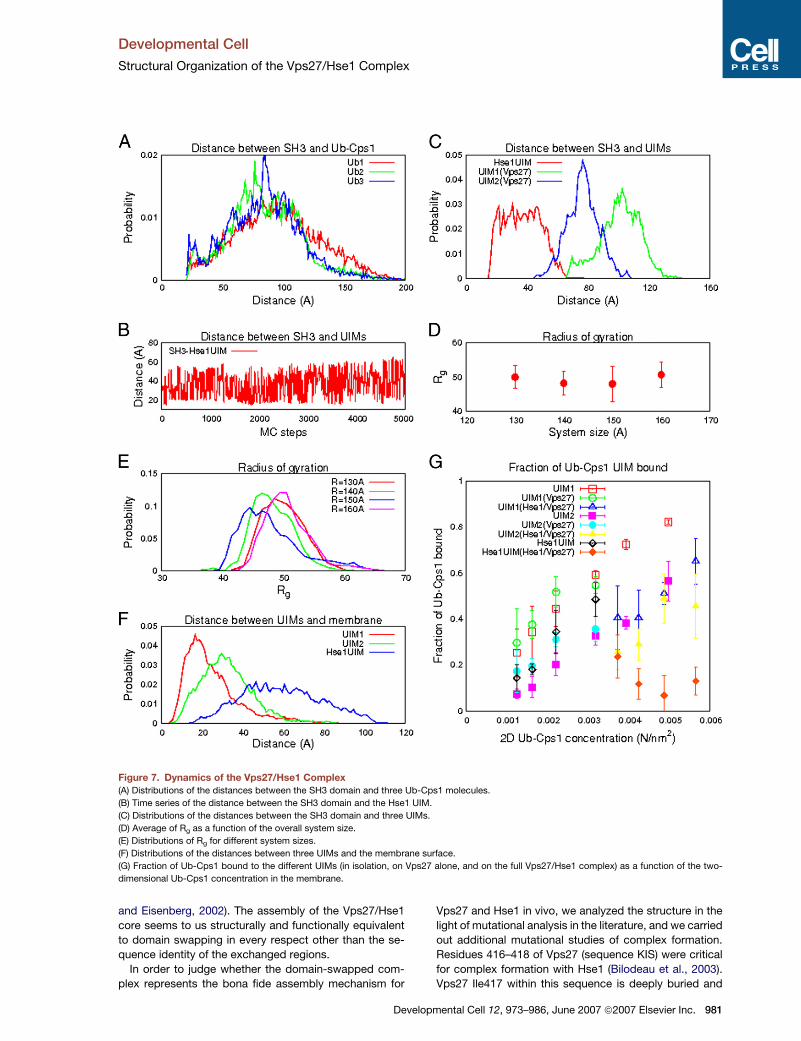
The MC simulations showed this complex to be flexible
and dynamic, capable of binding multiple ubiquitin moie-
ties situated at different distances from the membrane.
Even with the overall topology maintained, the Vps27/
Hse1 complex can undergo large conformational changes,
as indicated by the time series and distributions of dis-
tances between nonspecifically interacting domains (Fig-
ures 7B and 7C). Along the MC simulation trajectories,
the Vps27/Hse1 core retains an extended and open
configuration with a radius of gyration, Rg, between �40
and �60 A (Figures 7D and 7E). A globular protein of the
same molecular weight as the simulated portion of
Vps27/Hse1 would be expected to be much more com-
pact, with an Rg of �28 A.
Interactions of the Vps27 FYVE domain with PI(3)P and
the two Vps27 UIM domains with Ub-Cps1 keep this con-
tiguous portion of the Vps27 complex near the membrane
(Figure 7F). Unlike the two Vps27 UIM domains, the single
Hse1 UIM does not directly adjoin a membrane-binding
domain in the primary sequence. Therefore, on average,
the Hse1 UIM is found farther from the membrane (Fig-
ure 7F). To explore the relative contributions of the differ-
ent UIMs to overall binding on the membrane, the fraction
of Ub-Cps1 molecules bound to the three different UIMs in
isolation, in the full Vps27/Hse1 complex, and in Vps27
alone were calculated as a function of concentration (Fig-
ure 7G). The affinities of the Vps27 UIMs in the context of
the full complex are comparable to those of the isolated
UIMs. In contrast, the affinity of the Hse1 UIM for Ub-Cps1
is substantially reduced in the complex. Several factors
contribute to the difference. Within the complex,
Ub-Cps1/UIM binding is limited by steric restrictions and
internally competitive interactions that are not present
for the isolated UIM. This negative steric contribution to
binding is balanced by cooperative interactions with the
membrane and membrane-bound Ub-Cps1 molecules.
This positive cooperative contribution is stronger for
pmental Cell 12, 973–986, June 2007 ª2007 Elsevier Inc. 979

Developmental Cell
Structural Organization of the Vps27/Hse1 Complex
Figure 6. A Unified Model for the Interaction of Vps27/Hse1 with Membranes and Ubiquitinated Cargo
(A and B) The figure shows a single snapshot from the MC simulation. (A) View looking directly down toward the membrane. (B) View normal to the
plane of the membrane (the membrane surface is indicated by the green line). Vps27 is shown in blue, Hse1 is shown in orange, and ubiquitinated
cargo is shown in red.
Vps27 than for Hse1 because the FYVE domain and two
UIMs of Vps27 are close together. Therefore, the Vps27
UIM domains appear to have much stronger interactions
with Ub-Cps1 than the Hse1 UIM.
DISCUSSION
The structure of the Vps27/Hse1 core fills the last major
gap in our understanding of the organization of this com-
plex and led us to several unexpected observations. First,
the region involved in forming the core is more extensive
than anticipated and extends beyond the predicted
980 Developmental Cell 12, 973–986, June 2007 ª2007 Elsevie
coiled-coil regions in both the N- and C-terminal direc-
tions. Second, the core contains two GAT domains. Third,
the core assembles by the interchange of the homologous
C-terminal halves of the a3 helices from each GAT do-
main. In addition, the resolution of this structure has
allowed us to model its function as a scaffold for ubiqui-
tin-binding and ubiquitination-deubiquitination reactions
at the endosomal membrane.
The interchange of the homologous a3 C-terminal
halves from Vps27 and Hse1 is reminiscent of the mecha-
nism of ‘‘domain swapping.’’ As originally defined, domain
swapping refers to the oligomerization of identical proto-
mers by interchange of identical regions of subunits (Liu
r Inc.
Developmental Cell
Structural Organization of the Vps27/Hse1 Complex
Figure 7. Dynamics of the Vps27/Hse1 Complex
(A) Distributions of the distances between the SH3 domain and three Ub-Cps1 molecules.
(B) Time series of the distance between the SH3 domain and the Hse1 UIM.
(C) Distributions of the distances between the SH3 domain and three UIMs.
(D) Average of Rg as a function of the overall system size.
(E) Distributions of Rg for different system sizes.
(F) Distributions of the distances between three UIMs and the membrane surface.
(G) Fraction of Ub-Cps1 bound to the different UIMs (in isolation, on Vps27 alone, and on the full Vps27/Hse1 complex) as a function of the two-
dimensional Ub-Cps1 concentration in the membrane.
and Eisenberg, 2002). The assembly of the Vps27/Hse1
core seems to us structurally and functionally equivalent
to domain swapping in every respect other than the se-
quence identity of the exchanged regions.
In order to judge whether the domain-swapped com-
plex represents the bona fide assembly mechanism for
Develo
Vps27 and Hse1 in vivo, we analyzed the structure in the
light of mutational analysis in the literature, and we carried
out additional mutational studies of complex formation.
Residues 416–418 of Vps27 (sequence KIS) were critical
for complex formation with Hse1 (Bilodeau et al., 2003).
Vps27 Ile417 within this sequence is deeply buried and
pmental Cell 12, 973–986, June 2007 ª2007 Elsevier Inc. 981
Developmental Cell
Structural Organization of the Vps27/Hse1 Complex
almost completely surrounded by hydrophobic residues
from Hse1 (Figure 3), consistent with a critical role in func-
tion. We mutated Vps27 residues Leu410, Ile417, and
Ile420 individually to Asp, and we found that these muta-
tions prevented formation of the complex with Hse1. For
each cargo tested, Cps1, Ste3, and CPY, these mutations
resulted in a loss of function mirroring that seen in the de-
letion of Hse1. This establishes that the protein:protein in-
terface observed in the structure is responsible for the as-
sembly of the Vps27/Hse1 complex in yeast.
Despite the fact that Vps27 and Hse1 are subunits of
a tightly assembled heterodimer, deletion of the gene en-
coding each subunit results in a quantitatively different de-
fect in cargo sorting. Deletion of VPS27 causes a much
stronger CPY-missorting phenotype than deletion of
HSE1 (Bilodeau et al., 2002) (also see Figure 5). The mis-
sorting of Cps1 and Ste3 is also more severe in VPS27-
than HSE1-deletion mutants (Bilodeau et al., 2002).
HSE1-disruption phenotypes are more manifest in certain
genetic backgrounds, like that of the SF8389D yeast strain
used in these studies (Bilodeau et al., 2002). Our observa-
tions shed light on the probable cause for these pheno-
typic differences. Although the Vps27 core domain prefers
to assemble as a heterodimer with the Hse1 core domain,
it is nonetheless stable as a monomer when expressed in
the absence of the Hse1 core domain. This is likely due to
its ability to form an intramolecular GAT fold. In contrast,
the Hse1 core domain expressed in isolation tends to ag-
gregate and be degraded. Deletion of the VPS27 gene
may thus lead to loss of both the Vps27 and Hse1 proteins,
whereas deletion of the HSE1 gene would still leave
enough Vps27 protein to sustain a modicum of function.
In addition, Vps27 contains the main determinant of at-
tachment of the complex to membranes, the FYVE do-
main, such that monomeric Hse1 is likely incapable of ef-
ficient recruitment to endosomes. Conversely, monomeric
Vps27 could bind to membranes independently of Hse1,
thus bringing its ubiquitin-, ESCRT-I-, and clathrin-binding
activities to bear on MVB sorting. Finally, the MC simula-
tions show that the two UIM domains of Vps27 are closer
to the membrane and exhibit more cooperativity than the
single UIM domain of Hse1.
The human Hrs/STAM complex has been intensively
studied, but the structural basis for its assembly remains
unknown. The significant sequence homology between
STAM, Hse1, and Vps27 allows us to predict that the
core region of STAM will adopt the same structural fold
as Vps27 and Hse1. The so-called SSM, which is needed
for Hrs/STAM complex formation (Mizuno et al., 2004),
corresponds to the C-terminal half of helix a1 and a few
residues immediately following a1. Several of the con-
served residues in the SSM correspond to key hydropho-
bic anchor residues in the subunit interface. The sequence
of the core region of Hrs diverges from those of Vps27,
Hse1, and STAM. However, the region of Hrs correspond-
ing to the Vps27 GAT domain is predicted to be a-helical.
Further, the examples of Vps27 and Hse1 suggest that the
STAM GAT domain requires a complementary GAT do-
main in Hrs with which to associate.
982 Developmental Cell 12, 973–986, June 2007 ª2007 Elsevie
The unexpected observation of GAT domains in Vps27
and Hse1 highlights the parallel roles of these proteins
with other GAT-domain-containing trafficking adaptors,
the GGAs and Tom1 and Tom1-like proteins. The GGAs
are modular proteins that contain a receptor-binding
VHS domain; an Arf-binding helical hairpin domain; a ubiq-
uitin-, Rabex-5, and ESCRT-I-binding GAT domain; an un-
structured region containing autoinhibitory and clathrin-
binding domains; and a GAE domain that binds to various
accessory proteins (Bonifacino, 2004). Tom1 and its rela-
tives Tom1L1 and Tom1L2 have similar modular struc-
tures to the GGAs (Figure 1A), and they bind to ubiquitin
via their GAT domains (Katoh et al., 2004; Yamakami
et al., 2003) and to ESCRT-I (Puertollano, 2005). Collec-
tively, the GGAs, Tom1, and the Tom1-like proteins and
the Vps27/Hse1 and Hrs/STAM complexes comprise
a class of endosomal clathrin-binding proteins that sort
ubiquitinated cargo proteins into the ESCRT pathway
(Raiborg et al., 2006). These similarities highlight the
GAT domain proteins collectively (Figure 1A) as a family
of proteins that sort ubiquitinated cargo into the ESCRT
system.
The GAT domains of GGA1, GGA3, and Tom1 bind
ubiquitin (Katoh et al., 2004; Puertollano and Bonifacino,
2004; Scott et al., 2004; Shiba et al., 2004) with affinities
ranging from 180 to 410 mM (Akutsu et al., 2005; Kawasaki
et al., 2005; Prag et al., 2005). However, no ubiquitin bind-
ing to the Vps27/Hse1 core was detected by isothermal
titration calorimetry or surface plasmon resonance at con-
centrations of up to 8.0 mM and 2.0 mM, respectively
(data not shown). Known ubiquitin-binding GAT domains
contain two ubiquitin-binding sites. Ubiquitin binds to
the GGA and Tom1 GAT domains at site 1 on helices a1
and a2 and site 2 on helices a2 and a3. These sites are
incompletely conserved in Vps27 and Hse1 (Figure 1B).
Unlike Vps27 and Hse1, the GGAs and Tom1 do not con-
tain ubiquitin-binding UIMs. If the family of GAT-domain-
containing adaptors evolved from a common ancestor,
the GAT domains have served multiple purposes in ubiq-
uitin binding, dimerization, and other functions. By the
same token, different ubiquitin-binding, GAT-domain-
containing proteins acquired different mechanisms for
binding ubiquitinated cargo, some binding through the
GAT domain, and others via their UIMs.
The structures and the ubiquitin and membrane affini-
ties of individual domains from the Vps27/Hse1 and Hrs/
STAM complexes are known (Diraviyam et al., 2003;
Fisher et al., 2003; Hirano et al., 2006; Kaneko et al.,
2003; Mao et al., 2000; Misra and Hurley, 1999; Stahelin
et al., 2002; Swanson et al., 2003). Despite a wealth of in-
formation on individual domains, it has not been possible
to integrate this knowledge into a unified model of the
Vps27/Hse1 complex. The structure determination of the
Vps27/Hse1 core provides the missing link that allows
for the integration of the domain. The simulations show
that cooperativity between the Vps27 and FYVE and
UIM domains in membrane and ubiquitinated membrane
protein binding offsets steric constraints imposed in the
complex. The simulations portray the complex as open
r Inc.

Developmental Cell
Structural Organization of the Vps27/Hse1 Complex
and dynamic. Vps27/Hse1 traffics a variety of ubiquiti-
nated cargo. The molecular weight, the size of the cyto-
solic domain, and the location of the ubiquitination sites
on these cargoes vary widely. An open, dynamic complex
such as Vps27/Hse1 can adapt to these differences in
cargo in ways that a rigid complex could not. Finally, the
simulations show that the Hse1 SH3 domain, which tar-
gets DUBs that potentially deubiquitinate cargo, samples
conformational space that frequently approaches within
20 A of ubiquitinated cargo. This suggests that the action
of the Hse1 UIM and SH3 domains coordinates ubiquiti-
nated cargo-binding and deubiquitination reactions.
A model has been proposed for Hrs, based on a 16 A
resolution cryo-EM structure of an Hrs hexamer deter-
mined in the absence of STAM (Pullan et al., 2006). In
the Hrs model, three sets of membrane and ubiquitin-
binding domains are located 175 A apart from each other
at two sets of end caps. It is difficult to compare these
models given the substantial differences in the Hrs and
Vps27 core sequences and the presence of a 1:1 hetero-
dimer in one structure versus a homohexamer in the other.
The core of the Vps27/Hse1 complex contains an ele-
gant variation on the GAT domain, which does not bind
ubiquitin directly, but instead coordinates ubiquitinated
cargo-binding and ubiquitination and deubiquitination re-
actions. The structure of the Vps27/Hse1 complex shows
how the complex can spatially confine ubiquitinated cargo
and coordinate the action of DUBs (e.g., Ubp7) against
a tightly localized subpopulation of substrate. Such
a mechanism could help account for the biological speci-
ficity of this DUB, given the large number of potential sub-
strates in the cell. Most importantly, the structure of the
Vps27/Hse1 core complex has provided us with a unifying
framework for understanding the integrated action of the
many modular domains of these two proteins.
EXPERIMENTAL PROCEDURES
Protein Expression, Purification, and Crystallization
DNA sequences encoding Vps27 and Hse1 genes were amplified by
PCR from Saccharomyces cerevisiae genomic DNA. DNA coding for
residues 345–440 of Vps27 was subcloned in frame with a hexahisti-
dine tag followed by a TEV protease cleavage sequence into the
pST39 (Tan, 2001) expression vector. DNA coding for residues 275–
375 of Hse1 was subsequently cloned into the same vector without
a tag to yield a polycistronic expression vector for the Vps27/Hse1
core complex (pHisVps27/Hse1). The Hse1 GAT domain with similar
boundaries was cloned into pHIS-Parallel.2 in frame to an N-terminal
His tag and a TEV recognition site. Site-directed mutagenesis of
pHisVps27/Hse1 was performed by using the QuikChange kit (Strata-
gene) with appropriate DNA primers, and the entire sequences were
verified by DNA sequencing.
The individual GAT domains of Vps27 or Hse1 were overexpressed
in Escherichia coli BL21 (lDE3) Rosetta cells (Novagen) overnight at
20�C with 1 mM IPTG. Pellets were resuspended in 0.5 M NaCl,
10% glycerol, and 50 mM Tris-HCl (pH 8.0) supplemented with prote-
ase inhibitors. Cells were disrupted by lysozyme treatment, followed
by sonication and centrifugation. The proteins were then purified on
a Ni-NTA agarose column. The His tags were removed by cleavage
with TEV protease, and the samples were passed through a second
Ni-NTA column, followed by gel filtration. The Vps27/Hse1 core com-
plex was overexpressed and purified in the same manner. The seleno-
Develo
methionyl form of the complex was expressed in the methionine auxo-
troph E. coli strain B834 (lDE3) grown in defined media that contained
selenomethionine and was purified as described above.
The native and selenomethionyl forms of the Vps27/Hse1 core com-
plex were concentrated to 18 mg/ml and were crystallized in 1.1–1.4 M
ammonium sulfate, 0.1 M Tris-HCl (pH 8.85) in hanging drops at a 1:1
ratio of protein:precipitant. Crystals were improved by microseeding
into similar conditions with 1.25 M ammonium sulfate. Individual crys-
tals were cryoprotected with Paratone-N (Hampton Research) and
were frozen in liquid nitrogen.
Structure Determination
A two-wavelength MAD data set was collected from a single seleno-
methionyl crystal at the Advanced Photon Source (APS) synchrotron,
Argonne National Laboratory at 95 K. Energies for the MAD experiment
were chosen at the inflection point and remote at 12,658 and 12,750
eV, respectively. Data were analyzed with DENZO and Scalepack
(HKL Research). Eight selenium atoms were located by a direct
methods search with SnB (Weeks and Miller, 1999), and phases
were calculated by using Phasit (Furey and Swaminathan, 1997). Den-
sity modification of the initial maps was performed by using RESOLVE
(Terwilliger, 2000), DM, and Solomon of the CCP4 suite of programs
(CCP4, 1994). The resulting maps were used to build atomic models
in O (Jones et al., 1991) and Coot (Emsley and Cowtan, 2004). The
model was then refined with CNS (Brunger et al., 1998) (Table 1) at
3.0 A against the inflection point data set.
Gel Filtration
In vitro analysis of the oligomeric state of purified recombinant GAT
domains from the individual proteins and from the complex was per-
formed on a Superdex 200 HR 30/10 column (Amersham Pharmacia
Biotech) in 150 mM NaCl, 50 mM Tris-HCl (pH 8.0).
Sedimentation Equilibrium
Sedimentation equilibrium experiments were conducted at 4.0�C
on a Beckman Optima XL-A analytical ultracentrifuge. Samples in
50 mM Tris (pH 8.0) were loaded at concentrations corresponding to
measured A280 values of 0.25, 0.50, and 0.75. Data were acquired at
13, 16, 19, 22, 25, and 28 krpm as an average of 4 absorbance mea-
surements at 280 nm and a radial spacing of 0.001 cm. Equilibrium
was achieved within 48 hr. Data were analyzed globally in SEDPHAT
4.3 (Schuck, 2003) (http://www.analyticalultracentrifugation.com/
sedphat/sedphat.htm) by using solution densities (r) and protein
partial specific volumes (v) calculated in SEDNTERP (J. Philo; http://
www.jphilo.mailway.com/) as described in the text. Excellent fits
were observed in all cases. For the sample consisting of the Vps27
core region alone, the mass of the smallest species was fixed to that
expected for the monomer to return a value of 48 kDa for the oligomeric
species (n = 4.3). This higher-mass species contributes �20% of the
total absorbance; therefore, a solution with an absorbance at
280 nm of 1.0 will contain 130 mM of the monomer and 9 mM of the ag-
gregate, demonstrating that the Vps27 monomer is the major species.
Coimmunoprecipitation
Twenty OD600 of cells grown to midlog phase were lysed in TBS-T
(10 mM Tris, 140 mM NaCl, 0.1% Tween 20 [pH 7.5]) supplemented
with protease inhibitor cocktail (Roche, Indianapolis, IN) by using glass
bead lysis. Lysates were centrifuged at 14,000 rpm at 4�C, and super-
natants were precleared by incubation for 60 min at 4�C with 30 ml
Protein A-Sepharose beads (Amersham Pharmacia Biotech, Piscat-
away, NJ) and centrifugation at 8,000 3 g for 5 min. The precleared
lysates were incubated for 2 hr at 4�C with 30 ml Protein A-Sepharose
beads bound to rabbit polyclonal anti-HA antibody (Covance, Prince-
ton, NJ). The beads were then washed four times with TBS-T and sub-
jected to SDS-PAGE and immunoblotting analysis with either mouse
monoclonal anti-HA or mouse monoclonal anti-myc (9E10) (Covance,
Princeton, NJ).
pmental Cell 12, 973–986, June 2007 ª2007 Elsevier Inc. 983

Developmental Cell
Structural Organization of the Vps27/Hse1 Complex
Fluorescence Microscopy
Cells were grown to midlog phase in selective media and viewed on an
Olympus IX-70 fluorescence microscope (excitation, 560 nm; dichroic
mirror at 595 nm; emission, 630 nm). Images were captured with an
IMAGO charge-coupled device camera controlled by TILLvisION soft-
ware (TILL Photonics, Eugene, OR) and were processed by using
Adobe Photoshop 5.0 (Adobe Systems, Mountain View, CA).
CPY Maturation Assay
Yeast vps27D hse1D transformants were metabolically labeled with
the 35S Express reagent (Perkin Elmer Life Sciences) for 10 min (pulse)
and chased for 30 min (Bonifacino and Dell’Angelica, 1998). Immuno-
precipitations with mouse anti-CPY (Molecular Probes, Eugene, OR)
were performed overnight at 4�C, and the immunoprecipitates were
analyzed by SDS-PAGE and fluorography.
CPY Secretion Assay
The CPY secretion colony blot assay was performed as described
(Mullins and Bonifacino, 2001). Strains were grown to 1–1.5 U of
OD600/ml at 30�C and were concentrated to 0.2 U of OD600/ml. A total
of 5 ml of each strain was spotted on selective medium. Plates were
incubated overnight at 30�C to allow for cell growth, overlayed with
nitrocellulose, and incubated 12–14 hr. Membranes were rinsed with
distilled H2O and were analyzed by immunoblotting with anti-CPY
antibodies (Molecular Probes, Eugene, OR).
Table 1. Crystallographic Data Processing, Phasing,and Refinement Statistics
Collection Energies Inflection Remote
Data Processing
Wavelength (A) 0.97948 0.97242
Space group P3221 P3221
Cell dimensions
a, b, c (A) 62.4, 62.4,94.5
62.4, 62.4,94.6
a, b, g (�) 90, 90, 120 90, 90, 120
Resolution (A) 3.0 3.2
I/sI 18.1 (2.5) 12.7 (0.4)
Redundancy 4.1 (2.3) 3.7 (1.2)
Completeness (%) 97.5 (88.1) 82.5 (17.3)
Number of reflections 4415 3798
Rsym 0.052 (0.336) 0.059 (0.744)
Refinement
Resolution (A) 3.0
Rwork/Rfree 0.22/0.28
Number of protein atoms 1429
Number of water molecules 29
B factors
Average of main chains 65.3
Average of side chains 76.3
Water molecules 65.4
Geometric rmsd
Bond length (A) 0.007
Bond angles (�) 1.2
984 Developmental Cell 12, 973–986, June 2007 ª2007 Elsevie
Simulations
To obtain a starting model, the Vps27 FYVE domain (Misra and Hurley,
1999) was docked to the membrane (Diraviyam et al., 2003). The Vps27
UIM1-ubiquitin complex and the Vps27 tandem UIM1-UIM2 con-
structs (Swanson et al., 2003) were used to model the ubiquitin com-
plex with both Vps27 UIMs and the Hse1 UIM-ubiquitin complex.
The Hse1 SH3 domain was modeled from the STAM2 SH3 domain
(Kaneko et al., 2003). The VHS domains of Vps27 and Hse1 were
modeled from the Hrs VHS domain (Mao et al., 2000). The core was
positioned such that linkers to the nearest domains would be able to
reach them. The predicted unstructured (Rost and Liu, 2003) C-termi-
nal regions of Vps27/Hse1 were omitted.
Long-range electrostatic interactions were included at the Debye-
Huckel level. The relative interaction strengths have previously been
calibrated to reproduce measured protein second virial coefficients.
Flexible linkers were represented as polymers at the amino acid level,
and the folded protein domains were treated as rigid bodies. The
resulting model has been validated by predictions of a series of
protein-complex structures and binding affinities (Y.C.K. and G.H.,
unpublished data). Homology modeling was carried out manually in
O (Jones et al., 1991) or semimanually in Coot (Emsley and Cowtan,
2004).
Protein-membrane interactions were represented by a combination
of residue-dependent short-range interactions (Miyazawa and Jerni-
gan, 1996) and a Gouy-Chapmann-type electrostatic potential
between the flat membrane and the amino acids (Y.C.K. and G.H.,
unpublished data). To control molecular concentrations, the proteins
were confined into semispheres of different sizes above the membrane
plane. The FYVE domain was anchored to the membrane by a
harmonic potential between the known PI(3)P-binding site (Misra and
Hurley, 1999) and the membrane surface. The Ub-Cps1 molecules
were modeled as a covalent link between Gly76 of ubiquitin and
Lys8 of Cps1. Cps1 residues were modeled explicitly through residue
18, which was anchored to the membrane surface by a strict distance
constraint.
An equilibrium ensemble of complex structures was obtained by
performing replica-exchange Monte Carlo (MC) simulations, with 20
replicas covering the temperature range from 0.8T–1.7T, where T is
room temperature. For each system, a total of 20,000–30,000 complex
structures were obtained, which amounted to �107–108 MC steps. To
test for convergence, results from three independent runs were
compared.
To gauge whether the simulations provided a realistic estimate of af-
finity, the affinities of the isolated UIMs for ubiquitin were calculated.
We obtained a calculated value of Kd = �800 mM for Vps27 UIM1, as
compared to the experimental value of Kd = �300 mM (Fisher et al.,
2003). The calculated Kd of Vps27 UIM1 was lower than that of
Vps27 UIM2, also in agreement with the experimental value (Fisher
et al., 2003; Swanson et al., 2003). The simulations predicted the struc-
ture of the Vps27 UIM1-ubiquitin complex to within 3 A rmsd (Ca) of the
solution structure (Swanson et al., 2003). We found that the overall to-
pology of the Vps27/Hse1 multiprotein complex inferred from known
domain interactions was roughly maintained (Figure 7), despite
substantial conformational dynamics. Ubiquitin-binding events were
thoroughly equilibrated. These observations suggest that the simula-
tions provide a well-sampled and quantitatively reasonable reflection
of the system.
Supplemental Data
Supplemental Data include descriptions of methods and sedimentation
equilibrium data and are available at http://www.developmentalcell.
com/cgi/content/full/12/6/973/DC1/.
ACKNOWLEDGMENTS
We thank X. (Snow) Ren and W. Smith for assistance with biochemical
experiments; R. Piper for strains and plasmids; J. Chrzas, J. Gonczy,
M. Graham, and the entire staff of SER-CAT beamline ID-22 for user
r Inc.

Developmental Cell
Structural Organization of the Vps27/Hse1 Complex
support; and Y. Ye and A. Hickman for comments on the manuscript.
Use of the Advanced Photon Source was supported by the U.S.
Department of Energy, Basic Energy Sciences, Office of Science, un-
der Contract No.W-31-109-Eng-38. This research was supported by
National Institutes of Health intramural support, National Institute of
Diabetes and Digestive and Kidney Diseases (J.H.H. and G.H.),
National Institute of Child Health and Human Development (J.S.B.),
and Intramural AIDS Targeted Antiviral Program (J.H.H. and J.S.B.).
Received: December 12, 2006
Revised: February 20, 2007
Accepted: April 18, 2007
Published: June 4, 2007
REFERENCES
Akutsu, M., Kawasaki, M., Katoh, Y., Shiba, T., Yamaguchi, Y., Kato,
R., Kato, K., Nakayama, K., and Wakatsuki, S. (2005). Structural basis
for recognition of ubiquitinated cargo by Tom1-GAT domain. FEBS
Lett. 579, 5385–5391.
Altschul, S.F., Madden, T.L., Schaffer, A.A., Zhang, J.H., Zhang, Z.,
Miller, W., and Lipman, D.J. (1997). Gapped BLAST and PSI-BLAST:
a new generation of protein database search programs. Nucleic Acids
Res. 25, 3389–3402.
Babst, M. (2005). A protein’s final ESCRT. Traffic 6, 2–9.
Bilodeau, P.S., Urbanowski, J.L., Winistorfer, S.C., and Piper, R.C.
(2002). The Vps27p-Hse1p complex binds ubiquitin and mediates en-
dosomal protein sorting. Nat. Cell Biol. 4, 534–539.
Bilodeau, P.S., Winistorfer, S.C., Kearney, W.R., Robertson, A.D., and
Piper, R.C. (2003). Vps27-Hse1 and ESCRT-I complexes cooperate to
increase efficiency of sorting ubiquitinated proteins at the endosome.
J. Cell Biol. 163, 237–243.
Bonifacino, J.S. (2004). The GGA proteins: adaptors on the move. Nat.
Rev. Mol. Cell Biol. 5, 23–32.
Bonifacino, J.S., and Dell’Angelica, E.C. (1998). Protein labeling
and immunoprecipitation. In Current Protocols in Cell Biology,
J.S. Bonifacino, M. Dasso, J.B. Harford, J. Lippincott-Schwartz, and
K.M. Yamada, eds. (New York: John Wiley and Sons, Inc.),
pp. 7.2.1–7.2.14.
Bowers, K., and Stevens, T.H. (2005). Protein transport from the late
Golgi to the vacuole in the yeast Saccharomyces cerevisiae. Biochim.
Biophys. Acta 1744, 438–454.
Bowers, K., Lottridge, J., Helliwell, S.B., Goldthwaite, L.M., Luzio, J.P.,
and Stevens, T.H. (2004). Protein-protein interactions of ESCRT com-
plexes in the yeast Saccharomyces cerevisiae. Traffic 5, 194–210.
Brunger, A.T., Adams, P.D., Clore, G.M., DeLano, W.L., Gros, P.,
Grosse-Kunstleve, R.W., Jiang, J.S., Kuszewski, J., Nilges, M., Pannu,
N.S., et al. (1998). Crystallography & NMR system: a new software
suite for macromolecular structure determination. Acta Crystallogr. D
Biol. Crystallogr. 54, 905–921.
CCP4 (Collaborative Computational Project, Number 4) (1994). The
CCP4 suite: programs for protein crystallography. Acta Crystallogr. A
50, 760–763.
Clague, M.J., and Urbe, S. (2001). The interface of receptor trafficking
and signalling. J. Cell Sci. 114, 3075–3081.
Collins, B.M., Watson, P.J., and Owen, D.J. (2003). The structure of the
GGA1-GAT domain reveals the molecular basis for ARF binding and
membrane association of GGAs. Dev. Cell 4, 321–332.
Demirov, D.G., and Freed, E.O. (2004). Retrovirus budding. Virus Res.
106, 87–102.
Diraviyam, K., Stahelin, R.V., Cho, W., and Murray, D. (2003). Com-
puter modeling of the membrane interaction of FYVE domains.
J. Mol. Biol. 328, 721–736.
Emsley, P., and Cowtan, K. (2004). Coot: model-building tools for mo-
lecular graphics. Acta Crystallogr. D Biol. Crystallogr. 60, 2126–2132.
Develo
Fisher, R.D., Wang, B., Alam, S.L., Higginson, D.S., Robinson, H.,
Sundquist, W.I., and Hill, C.P. (2003). Structure and ubiquitin binding
of the ubiquitin-interacting motif. J. Biol. Chem. 278, 28976–28984.
Furey, W., and Swaminathan, S. (1997). PHASES-95: a program pack-
age for processing and analyzing diffraction data from macromole-
cules. Methods Enzymol. 277, 590–608.
Gruenberg, J., and Stenmark, H. (2004). The biogenesis of multi-
vesicular endosomes. Nat. Rev. Mol. Cell Biol. 5, 317–323.
Haglund, K., Di Fiore, P.P., and Dikic, I. (2003). Distinct monoubiquitin
signals in receptor endocytosis. Trends Biochem. Sci. 28, 598–603.
Harper, J.W., and Schulman, B.A. (2006). Structural complexity in
ubiquitin recognition. Cell 124, 1133–1136.
Hershko, A., Ciechanover, A., and Varshavsky, A. (2000). The ubiquitin
system. Nat. Med. 6, 1073–1081.
Hicke, L. (2001). Protein regulation by monoubiquitin. Nat. Rev. Mol.
Cell Biol. 2, 195–201.
Hicke, L., Schubert, H.L., and Hill, C.P. (2005). Ubiquitin-binding do-
mains. Nat. Rev. Mol. Cell Biol. 6, 610–621.
Hirano, S., Kawasaki, M., Ura, H., Kato, R., Raiborg, C., Stenmark, H.,
and Wakatsuki, S. (2006). Double-sided ubiquitin binding of Hrs-UIM in
endosomal protein sorting. Nat. Struct. Mol. Biol. 13, 272–277.
Hochstrasser, M. (2000). Evolution and function of ubiquitin-like pro-
tein-conjugation systems. Nat. Cell Biol. 2, E153–E157.
Holm, L., and Sander, C. (1995). Dali: a network tool for protein-struc-
ture comparison. Trends Biochem. Sci. 20, 478–480.
Hurley, J.H., and Emr, S.D. (2006). The ESCRT complexes: structure
and mechanism of a membrane-trafficking network. Annu. Rev. Bio-
phys. Biomol. Struct. 35, 277–298.
Hurley, J.H., Lee, S., and Prag, G. (2006). Ubiquitin binding domains.
Biochem. J. 399, 361–372.
Jones, T.A., Zou, J.Y., Cowan, S.W., and Kjeldgaard, M. (1991). Im-
proved methods for building protein models in electron-density
maps and the location of errors in these models. Acta Crystallogr. A
47, 110–119.
Kaneko, T., Kumasaka, T., Ganbe, T., Sato, T., Miyazawa, K., Kita-
mura, N., and Tanaka, N. (2003). Structural insight into modest binding
of a non-PXXP ligand to the signal transducing adaptor molecule-2 Src
homology 3 domain. J. Biol. Chem. 278, 48162–48168.
Kato, M., Miyazawa, K., and Kitamura, N. (2000). A deubiquitinating
enzyme UBPY interacts with the Src homology 3 domain of Hrs-bind-
ing protein via a novel binding motif PX(V/I)(D/N)RXXKP. J. Biol. Chem.
275, 37481–37487.
Katoh, Y., Shiba, Y., Mitsuhashi, H., Yanagida, Y., Takatsu, H., and
Nakayama, K. (2004). Tollip and Tom1 form a complex and recruit
ubiquitin-conjugated proteins onto early endosomes. J. Biol. Chem.
279, 24435–24443.
Katzmann, D.J., Babst, M., and Emr, S.D. (2001). Ubiquitin-dependent
sorting into the multivesicular body pathway requires the function of
a conserved endosomal protein sorting complex, ESCRT-I. Cell 106,
145–155.
Katzmann, D.J., Odorizzi, G., and Emr, S.D. (2002). Receptor down-
regulation and multivesicular-body sorting. Nat. Rev. Mol. Cell Biol.
3, 893–905.
Katzmann, D.J., Stefan, C.J., Babst, M., and Emr, S.D. (2003). Vps27
recruits ESCRT machinery to endosomes during MVB sorting. J. Cell
Biol. 162, 413–423.
Kawasaki, M., Shiba, T., Shiba, Y., Yamaguchi, Y., Matsugaki, N., Igar-
ashi, N., Suzuki, M., Kato, R., Kato, K., Nakayama, K., and Wakatsuki,
S. (2005). Molecular mechanism of ubiquitin recognition by GGA3 GAT
domain. Genes Cells 10, 639–654.
Komada, M., and Kitamura, N. (2005). The Hrs/STAM complex in the
downregulation of receptor tyrosine kinases. J. Biochem. (Tokyo)
137, 1–8.
pmental Cell 12, 973–986, June 2007 ª2007 Elsevier Inc. 985

Developmental Cell
Structural Organization of the Vps27/Hse1 Complex
Liu, Y., and Eisenberg, D. (2002). 3D domain swapping: as domains
continue to swap. Protein Sci. 11, 1285–1299.
Lloyd, T.E., Atkinson, R., Wu, M.N., Zhou, Y., Pennetta, G., and Bellen,
H.J. (2002). Hrs regulates endosome membrane invagination and tyro-
sine kinase receptor signaling in Drosophila. Cell 108, 261–269.
Lu, Q., Hope, L.W.Q., Brasch, M., Reinhard, C., and Cohen, S.N.
(2003). TSG101 interaction with HRS mediates endosomal trafficking
and receptor down-regulation. Proc. Natl. Acad. Sci. USA 100,
7626–7631.
Mao, Y.X., Nickitenko, A., Duan, X.Q., Lloyd, T.E., Wu, M.N., Bellen, H.,
and Quiocho, F.A. (2000). Crystal structure of the VHS and FYVE tan-
dem domains of Hrs, a protein involved in membrane trafficking and
signal transduction. Cell 100, 447–456.
Mattera, R., Puertollano, R., Smith, W.J., and Bonifacino, J.S. (2004).
The trihelical bundle subdomain of the GGA proteins interacts with
multiple partners through overlapping but distinct sites. J. Biol.
Chem. 279, 31409–31418.
McCullough, J., Row, P.E., Lorenzo, O., Doherty, M., Beynon, R.,
Clague, M.J., and Urbe, S. (2006). Activation of the endosome-associ-
ated ubiquitin isopeptidase AMSH by STAM, a component of the multi-
vesicular body-sorting machinery. Curr. Biol. 16, 160–165.
Misra, S., and Hurley, J.H. (1999). Crystal structure of a phosphatidyli-
nositol 3-phosphate-specific membrane-targeting motif, the FYVE do-
main of Vps27p. Cell 97, 657–666.
Miyazawa, K., and Jernigan, R.L. (1996). Residue-residue potentials
with a favorable pair term and an unfavorable high packing density
term, for simulation and threading. J. Mol. Biol. 256, 623–644.
Mizuno, E., Kawahata, K., Okamoto, A., Kitamura, N., and Komada, M.
(2004). Association with Hrs is required for the early endosomal local-
ization, stability, and function of STAM. J. Biochem. (Tokyo) 135, 385–
396.
Morita, E., and Sundquist, W.I. (2004). Retrovirus budding. Annu. Rev.
Cell Dev. Biol. 20, 395–425.
Mullins, C., and Bonifacino, J.S. (2001). Structural requirements for
function of Yeast GGAs in vacuolar protein sorting, a-factor matura-
tion, and interactions with clathrin. Mol. Cell. Biol. 21, 7981–7994.
Pickart, C.M. (2001). Mechanisms underlying ubiquitination. Annu.
Rev. Biochem. 70, 503–533.
Piper, R.C., and Luzio, J.P. (2001). Late endosomes: sorting and par-
titioning in multivesicular bodies. Traffic 2, 612–621.
Piper, R.C., Cooper, A.A., Yang, H., and Stevens, T.H. (1995). Vps27
controls vacuolar and endocytic traffic through a prevacuolar com-
partment in Saccharomyces cerevisiae. J. Cell Biol. 131, 603–617.
Prag, G., Lee, S.H., Mattera, R., Arighi, C.N., Beach, B.M., Bonifacino,
J.S., and Hurley, J.H. (2005). Structural mechanism for ubiquitinated-
cargo recognition by the Golgi-localized, g-ear-containing, ADP-
ribosylation-factor-binding proteins. Proc. Natl. Acad. Sci. USA 102,
2334–2339.
Puertollano, R. (2005). Interactions of Tom1L1 with the multivesicular
body sorting machinery. J. Biol. Chem. 280, 9258–9264.
Puertollano, R., and Bonifacino, J.S. (2004). Interactions of GGA3 with
the ubiquitin sorting machinery. Nat. Cell Biol. 6, 244–251.
Pullan, L., Mullapudi, S., Huang, Z., Baldwin, P.R., Chin, C., Sun, W.,
Tsujimoto, S., Kolodziej, S.J., Stoops, J.K., Lee, J.C., et al. (2006).
The endosome-associated protein Hrs is hexameric and controls
cargo sorting as a ‘‘master molecule’’. Structure 14, 661–671.
Raiborg, C., Bremnes, B., Mehlum, A., Gillooly, D.J., D’Arrigo, A.,
Stang, E., and Stenmark, H. (2001). FYVE and coiled-coil domains de-
termine the specific localisation of Hrs to early endosomes. J. Cell Sci.
114, 2255–2263.
Raiborg, C., Bache, K.G., Gillooly, D.J., Madshus, I.H., Stang, E., and
Stenmark, H. (2002). Hrs sorts ubiquitinated proteins into clathrin-
coated microdomains of early endosomes. Nat. Cell Biol. 4, 394–398.
986 Developmental Cell 12, 973–986, June 2007 ª2007 Elsevie
Raiborg, C., Rusten, T.E., and Stenmark, H. (2003). Protein sorting into
multivesicular endosomes. Curr. Opin. Cell Biol. 15, 446–455.
Raiborg, C., Wesche, J., Malerod, L., and Stenmark, H. (2006). Flat cla-
thrin coats on endosomes mediate degradative protein sorting by
scaffolding Hrs in dynamic microdomains. J. Cell Sci. 119, 2414–2424.
Ren, J., Kee, Y., Huibergtse, J.M., and Piper, R.C. (2007). Hse1, a com-
ponent of the yeast Hrs-STAM ubiquitin sorting complex, associates
with ubiquitin peptidases and a ligase to control sorting efficiency
into multivesicular bodies. Mol. Biol. Cell 18, 324–335.
Rost, B., and Liu, J.F. (2003). The PredictProtein server. Nucleic Acids
Res. 31, 3300–3304.
Schuck, P. (2003). On the analysis of protein self-association by sedi-
mentation velocity analytical ultracentrifugation. Anal. Biochem. 320,
104–124.
Scott, P.M., Bilodeau, P.S., Zhdankina, O., Winistorfer, S.C., Hau-
glund, M.J., Allaman, M.M., Kearney, W.R., Robertson, A.D., Boman,
A.L., and Piper, R.C. (2004). GGA proteins bind ubiquitin to facilitate
sorting at the trans-Golgi network. Nat. Cell Biol. 6, 252–259.
Shiba, T., Kawasaki, M., Takatsu, H., Nogi, T., Matsugaki, N., Igarashi,
N., Suzuki, M., Kato, R., Nakayama, K., and Wakatsuki, S. (2003).
Molecular mechanism of membrane recruitment of GGA by ARF in
lysosomal protein transport. Nat. Struct. Biol. 10, 386–393.
Shiba, Y., Katoh, Y., Shiba, T., Yoshino, K., Takatsu, H., Kobayashi, H.,
Shin, H.W., Wakatsuki, S., and Nakayama, K. (2004). GAT (GGA and
Tom1) domain responsible for ubiquitin binding and ubiquitination.
J. Biol. Chem. 279, 7105–7111.
Shih, S.C., Katzmann, D.J., Schnell, J.D., Sutanto, M., Emr, S.D., and
Hicke, L. (2002). Epsins and Vps27p/Hrs contain ubiquitin-binding do-
mains that function in receptor endocytosis. Nat. Cell Biol. 4, 389–393.
Slagsvold, T., Pattni, K., Malerod, L., and Stenmark, H. (2006). Endo-
somal and non-endosomal functions of ESCRT proteins. Trends Cell
Biol. 16, 317–326.
Stahelin, R.V., Long, F., Diraviyam, K., Bruzik, K.S., Murray, D., and
Cho, W. (2002). Phosphatidylinositol 3-phosphate induces the mem-
brane penetration of the FYVE domains of Vps27p and Hrs. J. Biol.
Chem. 277, 26379–26388.
Suer, S., Misra, S., Saidi, L.F., and Hurley, J.H. (2003). Structure of the
GAT domain of human GGA1: a syntaxin amino-terminal domain fold in
an endosomal trafficking adaptor. Proc. Natl. Acad. Sci. USA 100,
4451–4456.
Swanson, K.A., Kang, R.S., Stamenova, S.D., Hicke, L., and Radhak-
rishnan, I. (2003). Solution structure of Vps27 UIM-ubiquitin complex
important for endosomal sorting and receptor downregulation.
EMBO J. 22, 4597–4606.
Tan, S. (2001). A modular polycistronic expression system for overex-
pressing protein complexes in Escherichia coli. Protein Expr. Purif. 21,
224–234.
Terwilliger, T.C. (2000). Maximum-likelihood density modification.
Acta Crystallogr. D Biol. Crystallogr. 56, 965–972.
Weeks, C.M., and Miller, R. (1999). The design and implementation of
SnB v2.0. J. Appl. Crystallogr. 32, 120–124.
Weissman, A.M. (2001). Themes and variations on ubiquitylation. Nat.
Rev. Mol. Cell Biol. 2, 169–178.
Yamakami, M., Yoshimori, T., and Yokosawa, H. (2003). Tom1, a VHS
domain-containing protein, interacts with tollip, ubiquitin, and clathrin.
J. Biol. Chem. 278, 52865–52872.
Zhu, G.Y., Zhai, P., He, X.Y., Terzyan, S., Zhang, R.G., Joachimiak, A.,
Tang, J., and Zhang, X.J.C. (2003). Crystal structure of the human
GGA1 GAT domain. Biochemistry 42, 6392–6399.
Accession Numbers
Crystallographic coordinates have been deposited in the Protein Data
Bank with accession code 2PJW.
r Inc.

1
Developmental Cell 12
Supplemental Data
The Vps27/Hse1 Complex Is a GAT Domain-Based
Scaffold for Ubiquitin-Dependent Sorting Gali Prag, Hadiya Watson, Young C. Kim, Bridgette M. Beach, Rodolfo Ghirlando, Gerhard Hummer, Juan S. Bonifacino, and James H. Hurley
Supplemental Experimental Procedures Yeast Strains and Plasmid Construction
Yeast Strains. Saccharomyces cerevisiae strains were grown in standard yeast extract–peptone–dextrose (YPD) or synthetic medium with dextrose supplemented with the appropriate amino acids as required for plasmid maintenance. The following yeast strains were generous gifts from Robert Piper: hse1∆::Kanr (PLY2464), vps27∆::LEU2 (PLY2228), and hse1∆::Kanr vps27∆::LEU2 (PLY2784). These strains are derived from SF8389D (MATα ura3-52 leu2-3-112 his4-519 gal pep4-3) and carry the pep4 mutation to inactivate vacuolar proteases. For the pulse-chase CPY maturation assay, where functional proteases were needed, an hse1∆::Kanr vps27∆::LEU2 strain was generated by PCR-mediated gene replacement of the VPS27 locus in an hse1∆::Kanr Research Genetics strain (MATα his3∆1 leu2∆0 lys2∆0 ura3∆0). Yeast Plasmids. Hse1-myc (p1772) was a gift from Robert Piper. This plasmid is URA3-based (Bilodeau et al., 2002). VPS27 was PCR-amplified from genomic DNA, using oligonucleotides that generated an EcoRI site at the 5’ end, and an HA tag, followed by a stop codon and BamHI at the 3’ end. The resulting PCR fragments were cloned into the EcoRI/BamHI sites of a pRS316 vector containing an ADH1 promoter (KpnI/EcoRI) and CYC1 terminator (XhoI/SacI) to generate Vps27-WT. QuikChange Mutagenesis (Stratagene) was used to generate the A266Q, L410D, I417D, and I420D mutations within Vps27-WT. For co-expression studies, KpnI/Sac II fragments from the Vps27-WT and mutant constructs were subcloned from pRS316 (ADH1/CYC1) into the KpnI/SacII sites within pRS413 vector. STE3 and CPS1 were PCR amplified from genomic DNA. STE3 was cloned into the BamHI/XbaI sites of a pRS316 (ADH1/CYC1) vector containing a GFP fragment (XbaI/XhoI), generating Ste3p-GFP. CPS1 was cloned into the BamHI/XhoI sites of a pRS316 (ADH1/CYC1) vector containing a GFP fragment (EcoRI/BamHI), generating GFP-Cps1p.

2
Figure S1. Vps27 and Hse1 Form a 1:1 Complex
(A) Sedimentation equilibrium profiles obtained for the Vps27 and Hse1 complex shown in terms of A280 versus the radius r for data collected at a loading A280 of 0.25 (left), 0.50 (center) and 0.75 (right). Data were collected at 4.0°C and rotor speeds of 13 (orange), 16 (yellow), 19 (green), 22 (cyan), 25 (blue) and 28 (brown) krpm and analyzed globally in terms of a single ideal solute. Best fits, corresponding to a molecular mass of 23.2 ± 0.4 kDa, are shown as black lines through the experimental points. For clarity, alternate data points have been omitted. The corresponding distributions of the residuals are shown in the accompanying plot (B).

3
Figure S2. Isolated Vps27 Is Predominantly Monomeric
(A) Sedimentation equilibrium profiles obtained for the Vps27 shown in terms of A280 versus the radius r for data collected at a loading A280 of 0.20 (left) and 0.60 (right). Data were collected at 4.0°C and rotor speeds of 13 (orange), 16 (yellow), 19 (green), 22 (cyan), 25 (blue) and 28 (brown) krpm and analyzed globally in terms of two non-interacting ideal solutes using mass conservation. Best fits, corresponding to a Vps27 monomer and aggregate (n = 4.3, representing 20% of the total absorbance), are shown as black lines through the experimental points. For clarity, alternate data points have been omitted. The corresponding distributions of the residuals are shown in the accompanying plot (B).

4
Supplemental Reference
Bilodeau, P. S., Urbanowski, J. L., Winistorfer, S. C., and Piper, R. C. (2002). The Vps27p-Hse1p complex binds ubiquitin and mediates endosomal protein sorting. Nat. Cell Biol. 4, 534-539.