Embed Size (px)
Citation preview

Development of the Mouse Inner Ear and Origin ofIts Sensory Organs
Hakim Morsli,1 Daniel Choo,1 Allen Ryan,2 Randy Johnson,3 and Doris K. Wu1
1National Institute on Deafness and Other Communication Disorders, Rockville, Maryland 20850, 2Departments ofSurgery/Otolaryngology and Neurosciences, School of Medicine, and Veterans Administration Medical Center, Universityof California at San Diego, La Jolla, California 92093-0666, and 3M. D. Anderson Cancer Center, University of Texas,Department of Biochemistry and Molecular Biology, Houston, Texas 77030-4095
The molecular mechanisms dictating the morphogenesis anddifferentiation of the mammalian inner ear are largely unknown.To better elucidate the normal development of this organ, twoapproaches were taken. First, the membranous labyrinths ofmouse inner ears ranging from 10.25 to 17 d postcoitum (dpc)were filled with paint to reveal their gross development. Partic-ular attention was focused on the developing utricle, saccule,and cochlea. Second, we used bone morphogenetic protein 4(BMP4) and lunatic fringe (Fng) as molecular markers to identifythe origin of the sensory structures. Our data showed thatBMP4 was an early marker for the superior, lateral, and poste-rior cristae, whereas Fng served as an early marker for the
macula utriculi, macula sacculi, and the sensory portion of thecochlea. The posterior crista was the first organ to appear at11.5 dpc and was followed by the superior crista, the lateralcrista, and the macula utriculi at 12 dpc. The macula sacculiand the cochlea were present at 12 dpc but became distin-guishable from each other by 13 dpc. Based on the geneexpression patterns, the anterior and lateral cristae may sharea common origin. Similarly, three sensory organs, the maculautriculi, macula sacculi, and cochlea, seem to arise from asingle region of the otocyst.
Key words: inner ear development; sensory organs; lunaticfringe; BMP4; NT-3; Brn3.1
The mammalian inner ear is an unusually complex organ. Thevestibular sensory organs including the macula utriculi, maculasacculi, and cristae are responsible for detecting gravity andlinear and angular acceleration. These functions are necessary formaintaining normal balance. The coiled cochlea contains theauditory machinery necessary for hearing. One of the most re-markable aspects of the inner ear is that its elaborate three-dimensional structure, as well as the ganglion that innervates itssensory organs, arise from a simple hollow sphere of epithelium,the otic vesicle. To better visualize the normal morphogenesis ofthe mouse inner ear, and in particular, the cochlea, a solution ofwhite latex paint was injected into the lumen of mouse inner earsat different stages of development (Martin and Swanson, 1993).The gross anatomical changes of the inner ear were correlatedwith the appearance of each sensory organ that was identified bygenes specifically expressed in sensory regions before histologicaldifferentiation. One such candidate gene was bone morphogeneticprotein 4 (BMP4), a member of the transforming growth factor-bgene family. Previous studies showed that BMP4 is an earlymarker for all the presumptive sensory organs in the chickeninner ear (Wu and Oh, 1996). The early BMP4 gene expressionpattern in the chicken otocyst (an anterior and posterior focus)
appears to be similar to those observed in Xenopus (Hemmati-Brivanlou and Thomsen, 1995) and mouse (Jones et al., 1991)otocysts. However, although BMP4 is expressed in hair cells ofthe chicken basilar papilla (cochlea) before hatching (Oh et al.,1996), it has been reported to be expressed exclusively by Clau-dius’ cells of the mouse cochlea (Takemura et al., 1996).
Lunatic fringe (Fng), the murine homolog of Drosophila fringe,has been implicated in the formation of boundaries during em-bryogenesis (Cohen et al., 1997; Johnston et al., 1997). In the oticvesicle, Fng is expressed in restricted domains in both the mouseand the chicken (Johnston et al., 1997; Laufer et al., 1997). Moredetailed studies of the chicken inner ear indicate that Fng isexpressed in some presumptive sensory organs at early stages (F.Nunes and D. K. Wu, unpublished observations).
Neurotrophin-3 (NT-3) and Brain-3.1 (Brn-3.1) were also con-sidered good candidates for sensory organ markers. NT-3 hasbeen reported to be expressed in hair cells of the embryonic ratcochlea (Ylikoski et al., 1993; Wheeler et al., 1994). A morerecent study suggests that NT-3 expression in the mouse may bebroader and concentrated in supporting cells of the inner ear(Fritzsch et al., 1997a,b). Brn-3.1, a member of the POU domaintranscription factor family, is expressed in sensory hair cells of theinner ear (Erkman et al., 1996; Ryan, 1997; Xiang et al., 1997). Inthe present study, using BMP4, Fng, NT-3, and Brn-3.1 as markers,the origin and time at which each sensory organ was molecularlydefined in the mouse inner ear were determined.
MATERIALS AND METHODSEmbryos. Pregnant CD-1 mice (Charles River Laboratories, Wilming-ton, MA) were killed, and the litters were collected according to the NIHGuide for the Care and Use of Laboratory Animals protocol. Embryos wereindividually staged according to the method of Theiler (1989).
Probes. The in situ hybridization probe for Fng was obtained by
Received Dec. 15, 1997; revised Feb. 18, 1998; accepted Feb. 23, 1998.This work was supported in part by National Institutes of Health/National
Institute on Deafness and Other Communication Disorders Grant DC00139 to A.R.We are indebted to the staff of Biocomputation Center of the Ames ResearchCenter at NASA for their help with ROSS software. We also thank Drs. JamesBattey and Susan Sullivan for critically reviewing this manuscript, Drs. Brigid Hoganand Linda Erkman for providing plasmids for riboprobe generation, and MireneBoerner for editing.
Correspondence should be addressed to Dr. Doris K. Wu, National Institute onDeafness and Other Communication Disorders, 5 Research Court, Room 2B34,Rockville, MD 20850.Copyright © 1998 Society for Neuroscience 0270-6474/98/183327-09$05.00/0
The Journal of Neuroscience, May 1, 1998, 18(9):3327–3335

screening an 8.5 d postcoitum (dpc) embryonic mouse lgt10 library (agift from Brigid Hogan) with a mixture of human (Expressed sequencetags, Research Genetics) and chicken Fng (Laufer et al., 1997) cDNAs.Several strongly hybridizing clones were obtained, and one of these,pMFR1, was selected for further analysis. The 2.2 kb insert of pMFR1contains the entire coding region of murine Fng (Cohen et al., 1997;Johnston et al., 1997). The antisense RNA probe was generated by usingT3 RNA polymerase after HindIII restriction digest of pMFR1, and thesense RNA probe was generated by T7 RNA polymerase after XbaIrestriction digest. The NT-3 RNA probe was generated from an EST(881879, Genome Systems) containing a fragment of mouse NT-3 cDNAfrom the 59 end of the open reading frame to nucleotide 402 in pT7T3D-Pac vector (Pharmacia, Piscataway, NJ). The NT-3 antisense RNA probewas generated by using T3 RNA polymerase after restriction digest of theplasmid with EcoRI, and the sense RNA probe was generated by using T7RNA polymerase after restriction digest with NotI. The in situ hybrid-ization probe for BMP4 was generated from a 1550 bp full-length mouseBMP4 cDNA (kindly provided by Brigid Hogan, Vanderbilt University,Nashville, TN) (Jones et al., 1991). For generation of Brn-3.1 antisenseand sense RNA probes, a 209 bp XhoI mouse genomic fragment up-stream of the POU domain was used (a gift from Linda Erkman,University of California at San Diego, LaJolla, CA). None of the senseRNA probes used in this study yielded any specific hybridizationpatterns.
Paint injection. Mouse embryos ranging from 10.25 to 17 dpc wereharvested and fixed overnight in Bodian’s fixative. Specimens were thendehydrated in ethanol and cleared in methyl salicylate. The inner earswere visualized by injecting 0.1% white latex paint in methyl salicylateinto the membranous labyrinth as previously described (Martin andSwanson, 1993; Bissonnette and Fekete, 1996). The micropipette wasinserted in the lateral surface of otocysts. For more mature ears, thesuperior ampulla, the utricle, or the common crus were targeted depend-ing on the ease of visualization of the lumen. At a minimum, five innerears were injected for each stage presented.
Whole-mount in situ hybridization. Whole-mount in situ hybridizationwas performed (Riddle et al., 1993) with the following modifications. Allembryos were permeabilized with proteinase K (Boehringer Mannheim,Indianapolis, IN) using concentrations from 1 to 15 mg/ml. Hybridiza-tion, washings, and detection procedures were performed as described byRiddle et al. (1993).
In situ hybridization of f rozen sections. Frozen sections of mouse em-bryos were processed for in situ hybridization (Wu and Oh, 1996).Embryos were fixed overnight in 4% paraformaldehyde in PBS, dehy-drated in 30% sucrose, and embedded in OCT (Tissue-Tek). Embryoswere then sectioned at 12 mm thickness onto superfrost slides (VWRScientific) and stored at 280°C. Before in situ hybridization, slides werewarmed to room temperature, rehydrated, post-fixed, and permeabilizedusing 10 mg/ml proteinase K for 2–5 min. Hybridization was performedin Seal-A-Meal bags (Kapak). Each bag contained four slides and 5 ml ofhybridization solution with a probe concentration of ;0.2 mg/ml.
Three-dimensional reconstruction. Images of serial sections of themouse inner ear after in situ hybridization were captured from anAxiophot microscope (Zeiss) onto a Macintosh computer using a CCDcamera and NIH Image software. Images were transferred to a SiliconGraphics workstation. Contours of the inner ear of each section weretraced, aligned, and reconstructed into three-dimensional images usingROSS software (Biocomputation Center, Ames Research Center,NASA).
RESULTSGross anatomy of the developing inner earEight to 12 dpcThe inner ear arose from a thickening of the ectoderm known asthe otic placode that invaginated to form an otocyst (data notshown). At 10.75 dpc, a tube-like structure known as the en-dolymphatic duct projected dorsally from the medial part of theotocyst (Fig. 1A, ed). In addition, the cochlear anlage emerged asa ventral bulge (Fig. 1A, co). At 11.5 dpc, the endolymphatic ductwas more distinct (Fig. 1B, ed), and the cochlear anlage continuedto expand ventrally (Fig. 1B, co). The vertical canal plate, whichrepresented the primordium for the posterior and superior semi-
circular canals, began to form in the dorsolateral part of theotocyst (Fig. 1B, vpl).
Significant changes occurred at 12 dpc. Two regions in theanterior and posterior parts of the vertical canal plate (Fig. 1C,asterisks) started to reabsorb, thereby delineating the superiorand posterior semicircular canals (Martin and Swanson, 1993).The horizontal canal plate, which is the primordium for thelateral semicircular canal, appeared as a small bulge in the lateralpart of the otocyst (Fig. 1C, hp). The utricle, which houses themacula utriculi, appeared as a protrusion in the anterior part ofthe inner ear ventral to the vertical plate (Fig. 1C, u). At this time,the cochlea acquired a more elaborate shape consisting of aproximal and a distal part (Fig. 1C, arrows and arrowheads,respectively). The proximal part extended ventromedially,whereas the distal part started to extend anteriorly, adapting ahook-like shape (Fig. 1C, inset).
Thirteen to 17 dpcAt 13 dpc, the membranous labyrinth adopted a much thinnerand more mature appearance (Fig. 1D). The endolymphatic ductbecame thin, whereas the dorsal portion of the duct formed theprimordium for the endolymphatic sac (Fig. 1D, es). All threecanals were well formed (Fig. 1D, ssc, psc, lsc), with the superiorand posterior semicircular canals joined at the common cruslocated posterior to the endolymphatic duct (Fig. 1D, cc). Incomparison to the utricle at 12 dpc (Fig. 1C, u), the floor of theutricle at 13 dpc had adopted a more horizontal orientation (Fig.1D, u). The saccular anlage appeared ventral to the utricle as ananterior expansion of the proximal part of the cochlea (Fig. 1D,s). The proximal part of the cochlea further expanded ventrome-dially (Fig. 1D, arrows), whereas the distal part began coiling (Fig.1D, arrowheads). The cochlea consisted of half a turn at this point(Fig. 1D, inset). By 15 dpc, all primordial structures underwentfurther refinements, approximating their mature shape. Thedome-shaped ampullae, which house the cristae, were now ap-parent (Fig. 1E, sa, pa, la). The saccular connections to the utricleand the cochlea, the utriculosaccular and cochleosaccular ducts,respectively, were also apparent at this stage (Fig. 1E, usd, csd).The proximal part of the cochlea expanded further ventromedi-ally (Fig. 1E, arrows) with its most dorsal tip now being distinctand located anteroventrolateral to the posterior ampulla. Thedistal part of the cochlea continued to coil (Fig. 1E, arrowheads)and completed one and one-half turns by 15 dpc (Fig. 1E, inset).By 17 dpc, the membranous labyrinth had attained its matureshape (Fig. 1F), and the coiling process of the cochlea hadreached one and three-quarters turns (Fig. 1F, inset). To demon-strate the relative increase in the size of inner ears from 10.75 to17 dpc, the structures in Figure 1A–F are shown in Figure 1G atthe same magnification. The arrows in Figure 1G illustrate thegrowth of the proximal part of the cochlea from 13 to 17 dpc.
Presumptive sensory organs in the mouse inner earIn an effort to identify genes that could serve as markers for thepresumptive sensory organs in the mouse inner ear, two criteriawere imposed: (1) the gene should be activated at an early otocyststage; and (2) its expression should continue until the sensoryorgans could be identified histologically. Among multiple genestested for this purpose, BMP4 and Fng fulfilled both criteria.Results from .30 in situ hybridization experiments and three-dimensional reconstructions of critical developmental stagesshowed that BMP4 is an early marker for the three cristae in themouse inner ear, whereas Fng is an early marker for the macula
3328 J. Neurosci., May 1, 1998, 18(9):3327–3335 Morsli et al. • Development of the Mouse Inner Ear
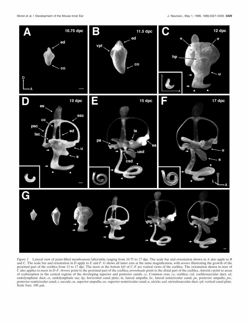
Figure 1. Lateral view of paint-filled membranous labyrinths ranging from 10.75 to 17 dpc. The scale bar and orientation shown in A also apply to Band C. The scale bar and orientation in D apply to E and F. G shows all inner ears at the same magnification, with arrows illustrating the growth of theproximal part of the cochlea from 13 to 17 dpc. The insets at the bottom lef t of C–F are ventral views of the cochlea. The orientation shown in inset ofC also applies to insets in D–F. Arrows point to the proximal part of the cochlea; arrowheads point to the distal part of the cochlea. Asterisks point to areasof reabsorption in the central regions of the developing superior and posterior canals. cc, Common crus; co, cochlea; csd, cochleosaccular duct; ed,endolymphatic duct; es, endolymphatic sac; hp, horizontal canal plate; la, lateral ampulla; lsc, lateral semicircular canal; pa, posterior ampulla; psc,posterior semicircular canal; s, saccule; sa, superior ampulla; ssc, superior semicircular canal; u, utricle; usd, utriculosaccular duct; vpl, vertical canal plate.Scale bars, 100 mm.
Morsli et al. • Development of the Mouse Inner Ear J. Neurosci., May 1, 1998, 18(9):3327–3335 3329

Figure 2. Top. Gene expression patterns of BMP4 and Fng in developing mouse inner ear from 9 to 10.25 dpc by whole-mount in situ hybridization. At9 dpc, BMP4 expression was detected in the posterior margin of the otic cup (A, arrow), whereas Fng transcripts were in the most ventral part of the oticcup (D, arrow). At 9.5 dpc, BMP4 was diffusely expressed in the posterior part of the otocyst (B, arrow). Fng transcripts were localized to the mostanteroventral part of the otocyst (E, arrow). At 10.25 dpc, BMP4 was expressed in two distinct areas, a posterior focus and an anterior streak (C, arrow,arrowhead, respectively). Fng transcripts were restricted to the most anteroventral quadrant of the otocyst (F, arrow). Orientation: A, anterior; D, dorsal.Scale bar, 100 mm.
3330 J. Neurosci., May 1, 1998, 18(9):3327–3335 Morsli et al. • Development of the Mouse Inner Ear

utriculi, the macula sacculi, and the cochlea. In both cases,patches of expression initially observed in the otocyst persistedand could be traced until the various sensory structures were welldefined both histologically and morphologically. A more detailedaccount of BMP4 and Fng gene expression during inner eardevelopment is described below.
BMP4 and Fng expression from 9 to 11 dpcAnalyses of BMP4 and Fng gene expression patterns during earlyinner ear development were performed using whole-mount in situhybridization. At 9 dpc when the placode started to invaginate,BMP4 mRNA was detected in the posterior margin of the otic cup(Fig. 2A, arrow). At 9.5 dpc, the invagination deepened to form anotocyst. BMP4 transcripts remained in the posterior portion ofthe otocyst as a rather diffuse signal (Fig. 2B, arrow). At 10.25dpc, the posterior hybridization signal became restricted to aposterior focus (Fig. 2C, arrow). In addition, a streak of hybrid-ization signal appeared in the anterolateral part of the otocyst(Fig. 2C, arrowhead). This anterior streak seemed to ariseabruptly and independently of the posterior hybridization signal.At 11 dpc, the anterior streak of BMP4 signal remained the same,whereas the posterior focus expanded ventrally (data not shown).
From 9 to 11 dpc, Fng expression was broader than that ofBMP4. At 9 dpc, Fng mRNA was detected in the most ventralportion of the otic cup (Fig. 2D, arrow). At 9.5 dpc, Fng wasexpressed as a “comma” shape at the most anteroventral part ofthe otocyst (Fig. 2E, arrow). By 10.25 dpc, Fng expression wasrestricted to the most anteroventral quadrant of the otocyst (Fig.2F, arrow). A similar Fng expression pattern was observed at 11dpc. Although the anterior streak of BMP4- and Fng-positiveareas appeared to be in close proximity to each other at 10.75 and11 dpc, probing alternate sections for BMP4 and Fng mRNAsindicated that the two positive areas did not overlap with eachother (data not shown). Further analyses of BMP4 and Fngexpression patterns at later stages were performed using serialcryosections of the inner ear.
BMP4 expression from 11.5 to 13 dpcAt 11.5 dpc, the anterior streak of BMP4 on the lateral side of theotocyst persisted (Fig. 3A,B). However, at 12 dpc, this BMP4hybridization signal split into an anterior and a lateral focus.These two foci corresponded to the presumptive superior andlateral cristae, respectively (Fig. 4A, sc, lc). The relative positionsof the two cristae are illustrated in the three-dimensional recon-struction of a 12 dpc inner ear in Figure 5, A and B. At 13 dpc,BMP4 transcripts persisted in the anterior and lateral cristae, asillustrated by the three-dimensional reconstruction of a 13 dpcinner ear (Fig. 5C,D). By this age, the morphology of the inner earwas more distinct (compare Figs. 1D, 5C), and the relative posi-tions of the two cristae approximate those of a mature inner ear.
At 11.5 dpc, the posterior focus of BMP4 signal split into adorsal spot and a ventral streak, which corresponded to theposterior crista (Fig. 3B, pc) and the lateral cochlear hybridiza-
tion signal (lco), respectively (Fig. 3D,E, lco). The lco was locatedin the posterior pole of the otocyst. It originated ventrolateral tothe posterior crista (Fig. 3D) and expanded ventrally to the distaltip of the cochlea anlage (Fig. 3E). At 12 dpc, BMP4 transcriptspersisted in the posterior crista (Fig. 4B, pc), whereas the hybrid-ization signal in the lateral cochlea became more complex. Thedorsal tip of the lco was restricted and located in the lateral partof the inner ear (Fig. 4D, lco). As this hybridization signalexpanded ventrally, it wrapped around the posterior pole of theinner ear (Fig. 4E, lco) and continued along the greater curvatureof the coiling cochlea (Fig. 4F, lco). The pattern of the lco can bebetter appreciated in the three-dimensional reconstruction of theinner ear in Figure 5, A and B. From this reconstruction, it isapparent that the lco of BMP4 followed the shape of the futurecochlea. This hybridization signal was likened to a ribbon origi-nating anteroventrolateral to the presumptive posterior crista andextending into the greater curvature of the cochlea (compareFigs. 1E, 5A,B). At 13 dpc, BMP4 transcripts remained in theposterior crista and the greater curvature of the cochlea, asillustrated in the three-dimensional reconstruction in Figure 5, Cand D.
Fng expression from 11.5 to 13 dpcAt 11.5 dpc, the most dorsal boundary of the Fng-positive areawas ventral to the anterior streak of BMP4 signal (Fig. 3C). ThisFng signal originated on the anterolateral part of the otocyst andextended both ventrally and medially (Fig. 3C,D,E). In the ven-tral portion of the otocyst, Fng was expressed on the medial side,encompassing the lco of BMP4 (Fig. 3, compare D,D9, E,E9).However, Fng transcripts were highly abundant in an anterome-dial region (Fig. 3E, brackets), whereas BMP4 transcripts concen-trated posteromedial to this Fng-positive domain (Fig. 3E, brack-ets). At 12 dpc, the broad Fng expression domain divided into twofoci, one dorsal and one ventral. The dorsal focus marked thepresumptive macula utriculi, localized in the lateral part of theotocyst, ventral to the presumptive superior and lateral cristae(Figs. 4C, 5A,B, mu). The ventral focus was the medial cochlearhybridization signal (mco), localized in the medial part of theotocyst (Fig. 4E, mco), originating at the level of the cochlearanlage and expanding ventrally into the lesser curvature of thecoiling cochlea (Fig. 4F, mco). Fng and BMP4 transcripts werecoexpressed in a small area at the tip of the coiling cochlea (Figs.4F,F9, brackets, 5A, white stripes). To determine whether theBMP4 or Fng expression domain gave rise to sensory cells, wecompared their expression to other potential presumptive sensorycell markers such as NT-3 and Brn-3.1. NT-3 proved to be a usefulmarker in this case. Our results show that the gene expressionpattern of NT-3 was similar to that of Fng from 10.75 to 12 dpc(data not shown). The hybridization signal of NT-3 at 12 dpcoverlapped with the Fng-positive domain (Fig. 4, compare F,F0),suggesting that this is the area that will develop into sensory cells.
At 13 dpc, the Fng signal in the macula utriculi was more
4
Figure 3. Bottom. Gene expression patterns of BMP4 and Fng in developing inner ear at 11.5 dpc. All panels are horizontal sections such that theanterior part of the embryo is toward the top. The level of each section is represented in the ear diagram at the top lef t. D, D9, E, and E9 are 12 mmadjacent sections. BMP4 was expressed in three distinct areas. In the dorsolateral part of the otocyst, BMP4 was expressed as an anterior streak ( A, B,as). The posterior focus of BMP4 signal in previous stages had now split into two signals: the posterior crista (B, pc) and the lateral cochlear hybridizationsignal (D, E, lco). Fng was expressed as one signal originating anterolaterally in the middle of the otocyst (C) and expanding ventrally and medially (D9,E9). At the tip of the cochlea, Fng transcripts concentrated in an anteromedial region (E9, brackets), whereas BMP4 transcripts concentratedposteromedial to this Fng positive domain (E, brackets). as, Anterior streak; lco, lateral cochlear hybridization signal; pc, posterior crista. Orientation:A, anterior; L, lateral. Scale bar, 100 mm.
Morsli et al. • Development of the Mouse Inner Ear J. Neurosci., May 1, 1998, 18(9):3327–3335 3331

horizontally oriented than at 12 dpc (Fig. 5, compare A,C). Thedorsal part of the mco expanded anteriorly, giving rise to themacula sacculi, as illustrated by the three-dimensional structure(Fig. 5C,D, ms). The ventral part of the mco restricted to thelesser curvature of the cochlea. Fng and BMP4 remained coex-pressed in a small area at the tip of the cochlea (Fig. 5D, asterisk).In addition, at 13 dpc Fng transcripts appeared in all three cristaeand thus were coexpressed with BMP4 (Fig. 5C,D, sc, pc, lc).
BMP4 and Fng expression from 14 dpc topostnatal day 1At 14 and 15 dpc, BMP4 expression persisted in all three cristae.By 16 dpc, BMP4 transcripts were concentrated in the supportingcells of the cristae (data not shown). In addition, at 16 dpc BMP4was expressed in the supporting cells of the macula utriculi andsacculi (data not shown). From 14 to 18 dpc, the lco of BMP4remained localized to the greater curvature of the coiling cochlea.
At 14 dpc, Fng expression persisted in all six sensory organs. Inaddition, the macula sacculi and the cochlea were now distinctbased on Fng expression pattern. By 16 dpc, Fng transcriptsconcentrated in supporting cells. In the cochlea, the BMP4- andFng-positive areas were now juxtaposed. At postnatal day 1 (P1),the histology of the cochlea was more distinct. Figure 6 illustratesFng transcripts being restricted to the supporting cells beneaththe differentiating inner and outer hair cells, as indicated by theBrn-3.1 gene expression pattern (Fig. 6B,C, arrows, open arrow,respectively). BMP4 transcripts were localized to cells lateral tothe outer hair cells, which most likely gave rise to Hensen’sand/or Claudius’ cells (Fig. 6A, arrows). In addition, at P1 BMP4was expressed in the mesenchyme surrounding the cochlea, aspreviously reported by Takemura et al. (1996) (Fig. 6A,arrowheads).
DISCUSSIONMorphogenesis of the inner earThe gross anatomy of the inner ear in several mammalian specieshas been well described (Retzius, 1884; Larsell et al., 1935; Bastand Anson, 1949). In the mouse, the histology of the inner earduring development has also been described in detail (Kikuchiand Hilding, 1965; Sher, 1971; Lim and Anniko, 1985). However,given the phenomenal morphogenesis that this organ undergoesto reach maturation, it is difficult to correlate the histologicaldifferentiation with gross anatomical changes. By using a paint-filling technique previously described (Martin and Swanson,1993) and three-dimensional reconstructions of gene expressionpatterns, the development of the sensory organs in relation to thegross anatomy of the inner ear can be appreciated. The ages ofthe developing inner ears described here were usually 1 d earlierthan those previously reported, which is most likely attributableto differences in staging (Theiler, 1989). Nonetheless, the innerear morphogenesis is in general agreement with previous reports(Sher, 1971; Lim and Anniko, 1985).
Based solely on the paint injection data, one might interpretthat the outpouch for the utricle at 12 dpc (Fig. 1C, u) is actuallythe primordial structure of the saccule (Fig. 1D, s). However,three-dimensional reconstructions demonstrating the positions ofthe sensory organs indicate that this interpretation is incorrect(Fig. 5). The utricle and its macula were located in a verticalposition at 12 dpc and became more horizontal by 13 dpc (Figs.1C,D, 5A,C). In contrast, the saccular anlage was not yet apparentat 12 dpc. When the saccule and its macula were distinguishableat 13 dpc, they were located ventral to the utricle and close to the
beginning of the first turn of the cochlea (Fig. 5A,B). As the innerear matured, the distance between the saccule and the first turn ofthe cochlea increased considerably (Fig. 1G, see distance betweenarrows). Furthermore, study of the paint-injected inner earsshowed that the increase in length of the proximal portion of thecochlea occurred concurrently with the coiling of the distalportion.
Origin of sensory organsBMP4 and Fng are expressed early in the otic cup stage and wellbefore the histological differentiation of sensory organs. At thesestages, BMP4 served as a marker for the three cristae, and Fngserved as a marker for the two maculae and cochlea. Although theexpression of these genes overlapped in most sensory organs atolder ages, we have used them as markers to determine the timeof appearance as well as the approximate location of each pre-sumptive sensory organ in the mouse inner ear. A presumptivesensory organ was considered molecularly defined when either itsBMP4 or Fng expression domain was distinct. Based on ourresults, the posterior crista appeared at 11.5 dpc. The maculautriculi and the superior and lateral cristae appeared at 12 dpc.The macula sacculi and the cochlea were distinguishable at 13 dpcbut remained connected until 14 dpc. The location of each pre-sumptive sensory organ described here is consistent with the invitro fate-mapping study of the mouse otocyst performed at 11and 12 dpc (Li et al., 1978).
An earlier study in chickens using BMP4 as a sensory organmarker suggested that all sensory organs in the chicken inner eararise independently from each other (Wu and Oh, 1996). Incontrast, results presented here suggest that in the mouse, thesuperior and lateral cristae may share a common origin as evidentby the single BMP4-positive area (anterior streak) in the anteriorportion of the otocyst that was later seen as two distinct domains.Likewise, the macula utriculi, macula sacculi, and cochlea mayshare a common origin as well, based on the gene expressionpatterns of Fng. Interestingly, a previous histological study hasshown that in some amphibian species, two of the sensory organs(amphibian papilla and papilla neglecta) are initially joined butseparated later in development (Fritzsch and Wake, 1988). How-ever, it is important to note that it remains speculative to extrap-olate lineage relationships among sensory organs from staticimages of hybridization signals or histology. Furthermore, be-cause the markers used for sensory organ identification were notidentical for the chicken and mouse studies, it is not clear whetherthese results reflect a fundamental difference in the origin ofsensory organ generation between the two species (for model onsensory organ generation in chicken, see Fekete, 1996; Kiernan etal., 1997). Further evidence will have to come from more com-parative studies and fate mapping using cell tracers.
Nevertheless, despite the issue of common sensory origin, theshared hybridization areas among the two cristae (superior andlateral) and the two maculae and cochlea suggest that sensoryorgans may be organized in clusters such that those within a clusterare related to each other developmentally. It has long been sus-pected that the macula sacculi and the cochlea are developmentallylinked. This belief stems from the identification of a group ofmutations in mice (Steel and Brown, 1994) and humans (Jackler,1993) in which only the saccule and the cochlea are affected. Inmice, these defects result from a malformation of the stria vascu-laris, leading to the loss of endocochlear potential within thecochlea (Steel et al., 1987) and subsequent degeneration of sensoryhair cells in the sensory organs. Therefore, these defects most likely
3332 J. Neurosci., May 1, 1998, 18(9):3327–3335 Morsli et al. • Development of the Mouse Inner Ear

reflect the mutual dependence of these two sensory organs for theintegrity of the endocochlear potential rather than the fact thatthey may actually share a common origin. In humans, however, thedefects described as cochleosaccular dysplasia are more suggestiveof a common developmental problem in the saccule and the co-chlea (Ormerod, 1960; Jackler, 1993). The Fng expression results
described here provide the first molecular evidence that these twosensory organs are developmentally related.
Functions of BMP4 and Fng in inner earIn most of the sensory organs, both Fng and BMP4 were expressedinitially in cells associated with sensory organ formation and later
Figure 4. Gene expression patterns ofBMP4 and Fng in the developing inner earat 12 dpc. All panels are horizontal sectionssuch that the anterior part of the embryo istoward the top. The level of each section isrepresented in the ear diagram at the bot-tom right. F and F9 are 12 mm adjacentsections. BMP4 was expressed in four dis-tinct areas. The anterior streak of BMP4signal at 11.5 dpc had now split into thepresumptive superior and lateral cristae(A, sc, lc), and BMP4 was still expressed inthe posterior crista (B, pc). The lco ofBMP4 became more elaborate, originatingin the lateral part of the inner ear andexpanding into the greater curvature of thecochlea (D, E, F, lco). Fng was expressed intwo distinct areas. The most dorsal areawas the presumptive macula utriculi (C,mu). The most ventral area was in the co-chlea, where the signal originated in themedial part of the basal turn expandinginto the lesser curvature of the cochlea (E9,F9, mco). BMP4 and Fng were coexpressedin a small area at the tip of the cochlea (F,F9, brackets). NT-3 gene expression over-lapped with that of Fng in the cochlea (F0).lc, Lateral crista; lco, lateral cochlear hy-bridization signal; mco, medial cochlear hy-bridization signal; mu, macula utriculi; pc,posterior crista; sc, superior crista. Orien-tation: A, anterior; L, lateral. Scale Bar,100 mm.
Figure 5. Three-dimensional reconstruc-tions of BMP4 and Fng domains of expres-sion in a 12 dpc (A, B) and 13 dpc (C, D)mouse inner ear. BMP4-positive areas aredisplayed in blue, and Fng-positive areasare in red. Areas positive for both BMP4and Fng are in blue with red stripes. A and Cwere tilted to give a ventrolateral view ofthe right inner ear. B and D are dorsome-dial views. The inner ears of 12 and 13 dpcwere reconstructed from 56 and 83 hori-zontal 12 mm serial sections, respectively.Alternate sections were probed for BMP4and Fng. The outline of the inner ears wasobtained by tracing the inner border of theotic epithelium of each section. The inset inA is a 12 dpc paint-filled inner ear shown ina view similar to the three-dimensional re-construction. White stripes in A representcoexpression of BMP4 and Fng in the distaltip of the cochlea. The asterisk in D pointsto the distal tip of the cochlea (not revealedin the reconstruction) where BMP4 and Fngexpression overlapped. cc, Common crus;ed, endolymphatic duct; lc, lateral crista;lco, lateral cochlear hybridization signal;lsc, lateral semicircular canal; mco, medialcochlear hybridization signal; ms, maculasacculi; mu, macula utriculi; pc, posteriorcrista; psc, posterior semicircular canal; sc,superior crista; ssc, superior semicircularcanal. Orientation: A, anterior; D, dorsal;L, lateral; M, medial. Scale Bars, 100 mm.
Morsli et al. • Development of the Mouse Inner Ear J. Neurosci., May 1, 1998, 18(9):3327–3335 3333

in supporting cells. The Drosophila fringe protein and its verte-brate homologs, such as lunatic (described in this study), radical,and manic fringe, are thought to specify cell fate through aNotch-signaling pathway (Johnston et al., 1997). Therefore, Fngmay play an important role in sensory hair cell and supportingcell determination. In addition, fringe proteins are thought to beimportant in boundary formation during embryogenesis. For ex-ample, radical fringe is important for positioning the apical ecto-dermal ridge at the dorsoventral boundary of the vertebrate limb(Laufer et al., 1997; Rodriguez-Esteban et al., 1997). Lunaticfringe may be important in establishing the boundary of individ-
ual somites (Cohen et al., 1997; Johnston et al., 1997). It isinteresting that in the mouse cochlea, the Fng and BMP4 domainsof expression form a boundary that runs along the third row ofouter hair cells, raising the possibility that these genes may play arole in specifying the position of the sensory and nonsensory (i.e.,Hensen’s and Claudius’) cells within the cochlea (Fig. 6).
The early BMP4 expression pattern in the mouse otocyst issimilar to that of the chicken (Wu and Oh, 1996), in which thehybridization signals correspond to the locations of the presump-tive cristae. Interestingly, the early BMP4 expression patternreported in the Xenopus otocyst resembled that of the chicken andmouse, suggesting that BMP4 is also a marker for the cristae infrogs (Hemmati-Brivanlou and Thomsen, 1995). The conservedpattern of expression across species suggests that BMP4 may playan important role in the induction and/or differentiation of cris-tae. In contrast, in the cochlea, expression of BMP4 is not con-served among chickens and mice. BMP4 is expressed in thesensory hair cells of the basilar papilla in the chicken (Oh et al.,1996), whereas it is only expressed in Hensen’s and/or Claudius’cells of the mouse cochlea. Although the expression domain ofBMP4 was not distributed across the entire lateral wall of thecochlea (Fig. 5C), it outlined the shape of the mouse cochlea earlyin development and was intimately associated with the Fng-positive sensory region. Therefore, it would not be surprising ifBMP4 also participates in patterning the shape of the mousecochlea. In addition, the histology of the mouse cochlea is muchmore complicated and contains many more cell types than thebasilar papilla of the chicken (for review, see Rubel, 1978; Smith,1985; Cohen and Cotanche, 1992; Fritzsch et al., 1998). Theabsence of BMP4 expression in sensory hair cells of the mousecochlea may contribute to the fine structural differences betweenthe mouse and chicken cochlea. In conclusion, our study serves asa basis for understanding the molecular mechanisms underlyingthe development of the mammalian inner ear and for decipheringmalformations resulting from mutations.
REFERENCESBast TH, Anson BJ (1949) The otic labyrinth. In: The temporal bone
and the ear, pp 30–105. Springfield, IL: Thomas.Bissonnette JP, Fekete DM (1996) Standard atlas of the gross anatomy
of the developing inner ear of the chicken. J Comp Neurol 174:1–11.Cohen B, Bashirullah A, Dagnino L, Campbell C, Fisher WW, Leow CC,
Whiting E, Ryan D, Zinyk D, Boulianne G, Hui CC, Gallie B, PhillipsRA, Lipshitz HD, Egan SE (1997) Fringe boundaries coincide withNotch-dependent patterning centres in mammals and alter Notch-dependent development in Drosophila. Nat Genet 16:283–288.
Cohen GM, Cotanche DA (1992) Development of the sensory receptorsand their innervation in the chick cochlea. In: Development of auditoryand vestibular systems, Vol 2 (Romand AL, ed), pp 101–138. New York:Elsevier.
Erkman L, McEvilly RJ, Luo L, Ryan AK, Hooshmand F, O’Connell SM,Keithley EM, Rapaport DH, Ryan AF, Rosenfeld MG (1996) Role oftranscription factors Brn-3.1 and Brn-3.2 in auditory and visual systemdevelopment. Nature 381:603–606.
Fekete DM (1996) Cell fate specification in the inner ear. Curr OpinNeurobiol 6:533–541.
Fritzsch B, Wake MH (1988) The inner ear of gymnophione amphibiansand its nerve supply: a comparative study of regressive events in acomplex sensory system (Amphibia, Gymnophiona). Zoomorphology108:201–217.
Fritzsch B, Farinas I, Reichardt LF (1997a) Lack of neurotrophin-3causes losses of both classes of spiral ganglion neurons in the cochlea ina region-specific fashion. J Neurosci 17:6213–6225.
Fritzsch B, Farinas I, Reichardt L (1997b) The development of NT-3expression as revealed with a Lac-Z reporter and of innervation deficitsin NT-3 mutant mice. Soc Neurosci Abstr 23:351.13.
Fritzsch B, Barald K, Lomax M (1998) Early embryology of the verte-
Figure 6. Gene expression of BMP4, Fng, and Brn-3.1 in developingcochlea at P1. A–C are 12 mm adjacent sections. BMP4 transcripts werelocalized to specialized cells lateral to the outer hair cells: Hensen’sand/or Claudius’ cells (A, arrow). BMP4 was also expressed in the mes-enchyme surrounding the cochlea (A, arrowheads). Fng transcripts wererestricted to the supporting cells underneath the inner and outer hair cells(B, arrow, open arrow, respectively). Brn-3.1 was expressed in the innerand outer hair cells (C, arrow, open arrow, respectively). sv, Spiral vessel.Orientation: A, anterior; L, lateral. Scale Bar, 100 mm.
3334 J. Neurosci., May 1, 1998, 18(9):3327–3335 Morsli et al. • Development of the Mouse Inner Ear

brate ear. In: Development of the auditory system, Springer handbookof auditory research (Rubel E, Popper A, Fay RS, eds), pp 81–145. NewYork: Springer.
Hemmati-Brivanlou A, Thomsen GH (1995) Ventral mesodermal pat-terning in Xenopus embryos: expression patterns and activities ofBMP-2 and BMP-4. Dev Genet 17:78–89.
Jackler RK (1993) Congenital malformations of the inner ear. In:Otolaryngology–head and neck surgery, Vol 4 (Patterson AS, Ryan JD,eds), pp 2756–2771. St. Louis: Mosby.
Johnston SH, Rauskolb C, Wilson R, Prabhakaran B, Irvine KD, Vogt TF(1997) A family of mammalian fringe genes implicated in boundarydetermination and the Notch pathway. Development 124:2245–2254.
Jones MC, Lyons KM, Hogan BLM (1991) Involvement of bone mor-phogenetic protein-4 (BMP-4) and Vgr-1 in morphogenesis and neu-rogenesis in the mouse. Development 111:531–542.
Kiernan AE, Nunes F, Wu DK, Fekete DM (1997) The expressiondomain of two related homeobox genes defines a compartment in thechicken inner ear that may be involved in semicircular canal formation.Dev Biol 191:215–229.
Kikuchi K, Hilding D (1965) The development of the organ of Corti inthe mouse. Acta Otolaryngol 60:207–222.
Larsell O, McCrady E, Zimmermann A (1935) Morphological and func-tional development of the membranous labyrinth in the opossum.J Comp Neurol 63:95–118.
Laufer E, Dahn R, Orozco OE, Yeo CY, Pisenti J, Henrique D, AbbottUK, Fallon JF, Tabin C (1997) Expression of radical fringe in limb-bud ectoderm regulates apical ectodermal ridge formation. Nature386:366–373.
Li CW, Van Der Water TR, Ruben RJ (1978) The fate mapping of theeleventh and twelfth day mouse otocyst: an in vitro study of the sites oforigin of the embryonic inner ear sensory structures. J Morphol157:249–268.
Lim DJ, Anniko M (1985) Developmental morphology of the mouseinner ear. Acta Otolaryngol Suppl 422:1–69.
Martin P, Swanson GJ (1993) Descriptive and experimental analysis ofthe epithelial remodellings that control semicircular canal formation inthe developing mouse inner ear. Dev Biol 159:549–558.
Oh SH, Johnson R, Wu DK (1996) Differential expression of bonemorphogenetic proteins in the developing vestibular and auditory sen-sory organs. J Neurosci 16:6463–6475.
Ormerod FC (1960) The pathology of congenital deafness. J LaryngolOtol 74:919–950.
Retzius G (1884) Das Gehororgan der Wirbeltiere: II Das Gehororgander Amnioten, pp 201–358. Stockholm: Samson und Wallin.
Riddle RD, Johnson RL, Laufer E, Tabin C (1993) Sonic hedgehogmediates the polarizing activity of the ZPA. Cell 75:1401–1416.
Rodriguez-Esteban C, Schwabe JWR, De La Pena J, Foys B, Eshelman B,Belmonte JCI (1997) Radical fringe positions the apical ectodermalridge at the dorsoventral boundary of the vertebrate limb. Nature386:360–366.
Rubel EW (1978) Ontogeny of structure and function in the vertebrateauditory system. In: Handbook of sensory physiology: development ofsensory systems, Vol 9 (Jacobson M, ed), pp 135–237. New York:Springer.
Ryan AF (1997) Transcription factors and the control of inner ear de-velopment. Semin Cell Dev Biol 8:249–256.
Sher A (1971) The embryonic and postnatal development of the innerear of the mouse. Acta Otolaryngol Suppl 285:1–77.
Smith CA (1985) Inner ear. In: Form and function in birds, Vol 3, (KingAS, McLelland J, eds) pp 273–310. London: Academic.
Steel KP, Barkway C, Bock G (1987) Strial dysfunction in mice withcochleo-saccular abnormalities. Hear Res 27:11–26.
Steel KP, Brown SDM (1994) Genes and deafness. Trends Genet10:428–435.
Takemura T, Sakagami M, Takebayashi K, Masanori U, Nakase T,Takaoka K, Kubo T, Kitamura Y, Nomura S (1996) Localization ofbone morphogenetic protein-4 messenger RNA in developing mousecochlea. Hear Res 95:26–32.
Theiler K (1989) The house mouse: atlas of embryonic development.New York: Springer.
Wheeler EF, Bothwell M, Schecterson LC, von Bartheld CS (1994)Expression of BDNF and NT-3 mRNA in hair cells of the organ ofCorti: quantitative analysis in developing rats. Hear Res 73:46–56.
Wu DK, Oh SH (1996) Sensory organ generation in the chick inner ear.J Neurosci 16:6454–6462.
Xiang M, Gan L, Li D, Chen ZY, Zhou L, O’Malley BWJ, Klein W,Nathans J (1997) Essential role of POU-domain factor Brn-3c in au-ditory and vestibular hair cell development. Proc Natl Acad Sci USA94:9445–9450.
Ylikoski J, Pirvola U, Moshnyakov M, Palgi J, Arumae U, Saarma M(1993) Expression patterns of neurotrophin and their receptormRNAs in the rat inner inner ear. Hear Res 65:69–78.
Morsli et al. • Development of the Mouse Inner Ear J. Neurosci., May 1, 1998, 18(9):3327–3335 3335


![Inner Ear Anatomy[1]](https://img.pdfslide.us/doc/110x75/5528566b4979591c048b47a6/inner-ear-anatomy1.jpg)