Embed Size (px)
DESCRIPTION
DEVELOPMENT OF the Kidneys and Ureters. Dr Rania Gabr. Objectives. 1- Describe the stages of development of the kidneys and ureters 2-Discuss the congenital anomalies of the kidneys and ureters. Stages of Development of the kidney:. - PowerPoint PPT Presentation
Citation preview

DEVELOPMENT OFDEVELOPMENT OFthe Kidneys and the Kidneys and
UretersUreters
Dr Rania GabrDr Rania Gabr

ObjectivesObjectives
• 1- Describe the stages of 1- Describe the stages of development of the kidneys and development of the kidneys and uretersureters
• 2-Discuss the congenital anomalies 2-Discuss the congenital anomalies of the kidneys and uretersof the kidneys and ureters
Stages of Development of the Stages of Development of the kidney:kidney:
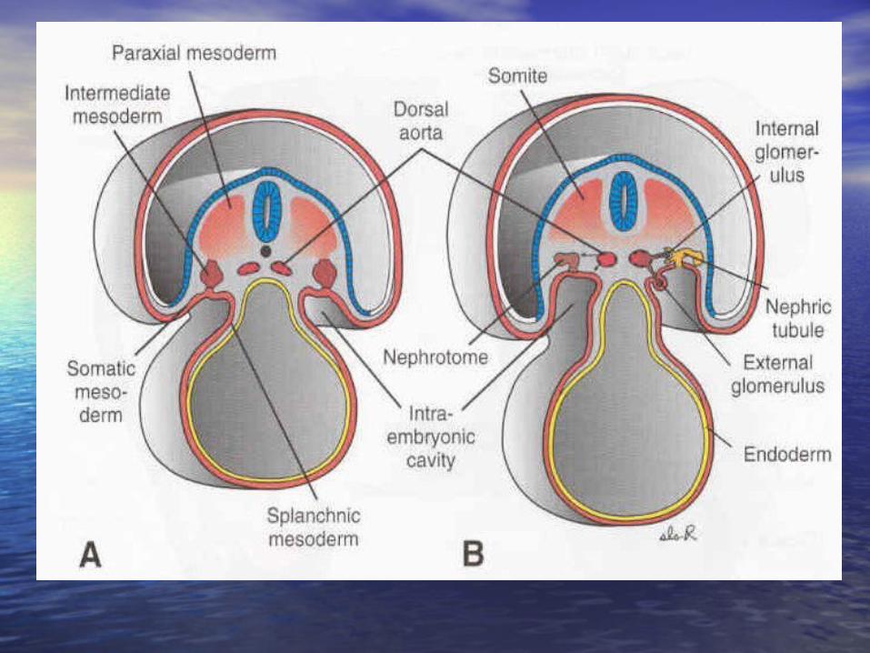
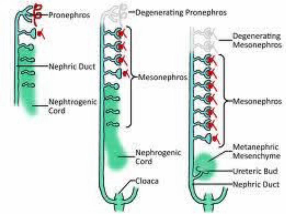
• Human kidney is developed from the Human kidney is developed from the intermediate mesoderm and passes intermediate mesoderm and passes through 3 stages :through 3 stages :
1- 1- PronephrosPronephros
2- 2- MesonephrosMesonephros
3- 3- MetanephrosMetanephros


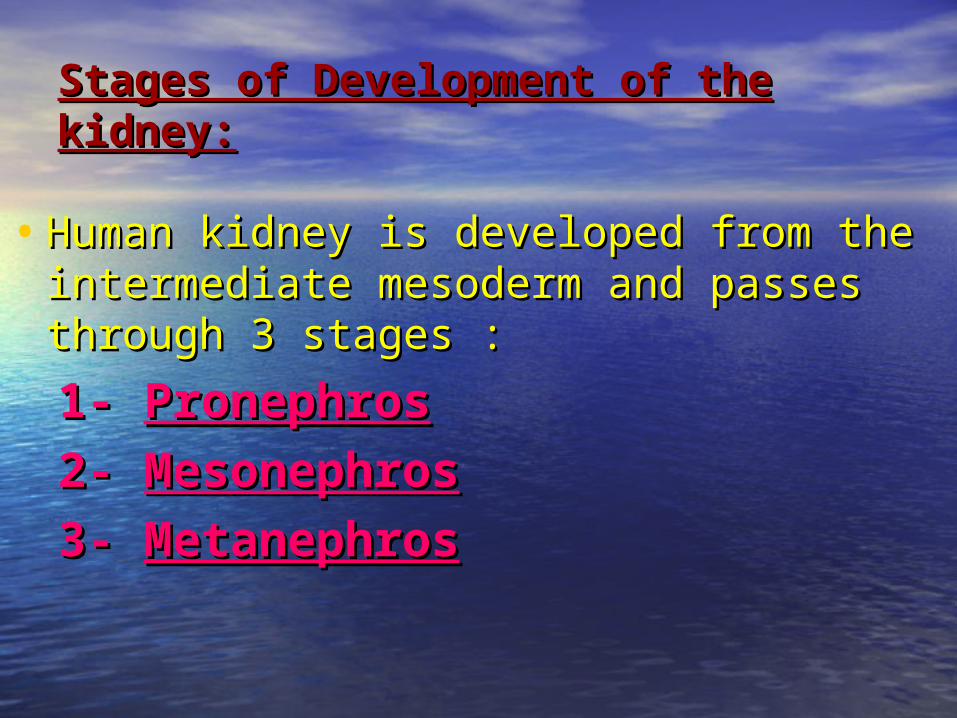
PRONEPHROSPRONEPHROS• Appears at 4Appears at 4thth week. In the week. In the cervicalcervical
region, the cranial part of the region, the cranial part of the intermediate mesoderm is segmented intermediate mesoderm is segmented into 7 cell clusters called “into 7 cell clusters called “nephrotomes”.nephrotomes”.
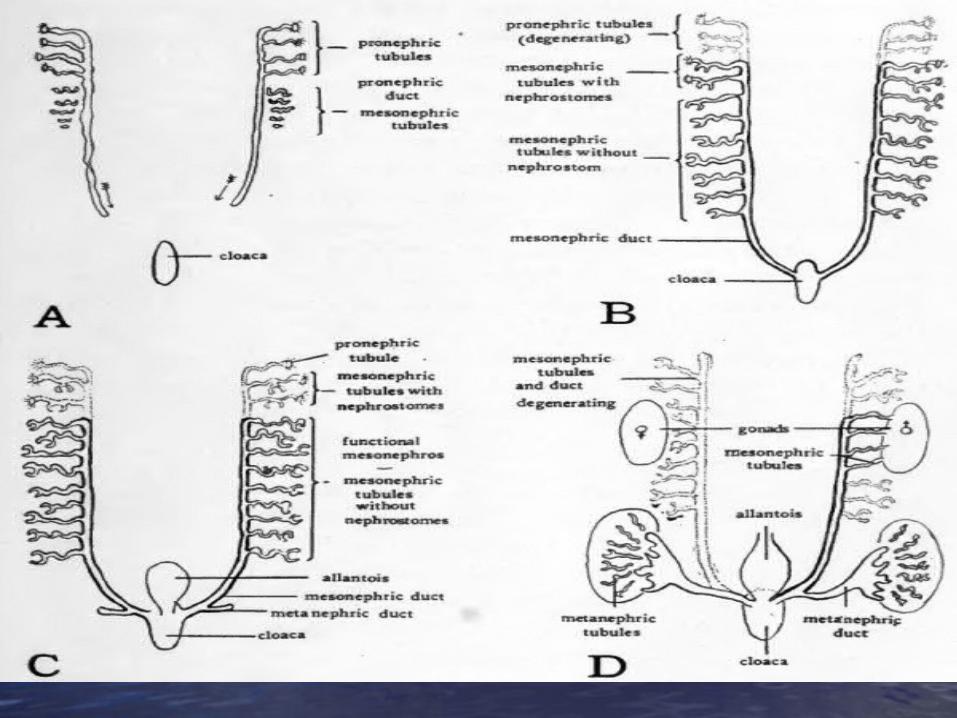
• Those become cavitated to form 7 Those become cavitated to form 7 ““pronephric tubules”.pronephric tubules”.
• Each tubule has 2 ends: Each tubule has 2 ends:
1- 1- The lateral end:The lateral end: joins to form pronephric joins to form pronephric duct. duct.
2-2- The medial end:The medial end: joins the coelomic joins the coelomic cavity.cavity.
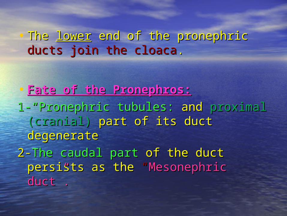
• The The lowerlower end of the pronephric end of the pronephric ducts join the cloacaducts join the cloaca..
• Fate of the Pronephros:Fate of the Pronephros:
1-“Pronephric tubules: 1-“Pronephric tubules: and and proximal proximal (cranial)(cranial) part of its duct degenerate part of its duct degenerate
2-2-The caudal part The caudal part of the duct persists of the duct persists as theas the “ “Mesonephric duct”. Mesonephric duct”.


MESONEPHROSMESONEPHROS• It appears at the It appears at the 66thth week week as 2 bulges on as 2 bulges on
posterior abdominal wall forming ovoid posterior abdominal wall forming ovoid mesonephric ridges.mesonephric ridges.
• It develops in the middle part of the It develops in the middle part of the (Intermediate mesoderm) that lies in the(Intermediate mesoderm) that lies in the thoracicthoracic and and upper lumbar upper lumbar region.region.
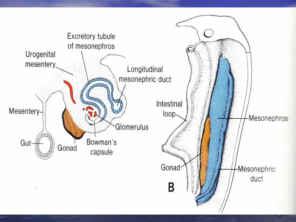
• It is divided into segments which become It is divided into segments which become canalised to form the “canalised to form the “mesonephric mesonephric tubules”tubules”..
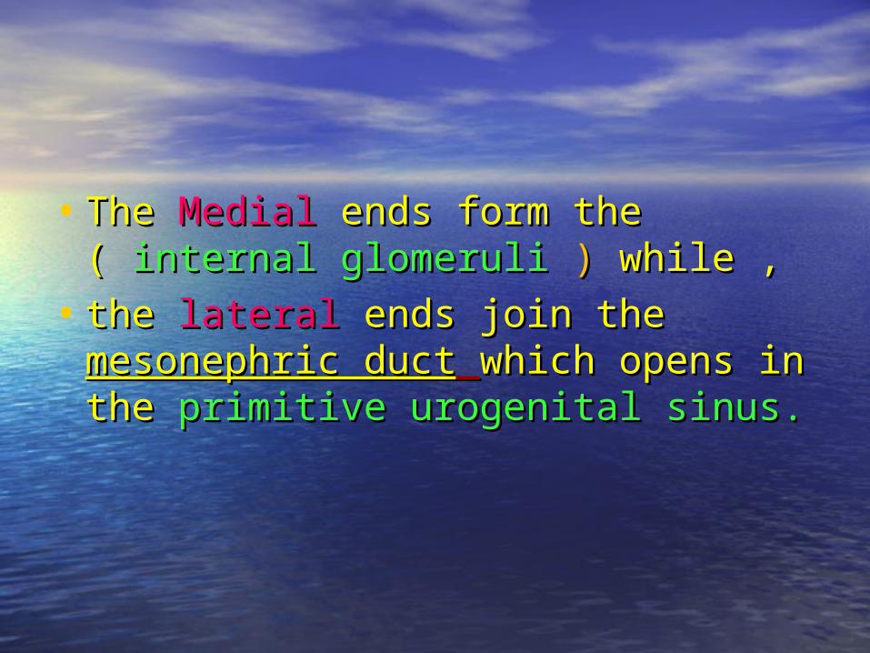
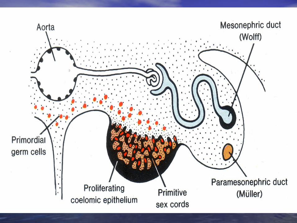
• The The MedialMedial ends form the ( ends form the ( internal internal glomeruli glomeruli ) ) while ,while ,
• the the laterallateral ends join the ends join the mesonephric mesonephric ductduct which opens in the which opens in the primitive primitive urogenital sinus.urogenital sinus.
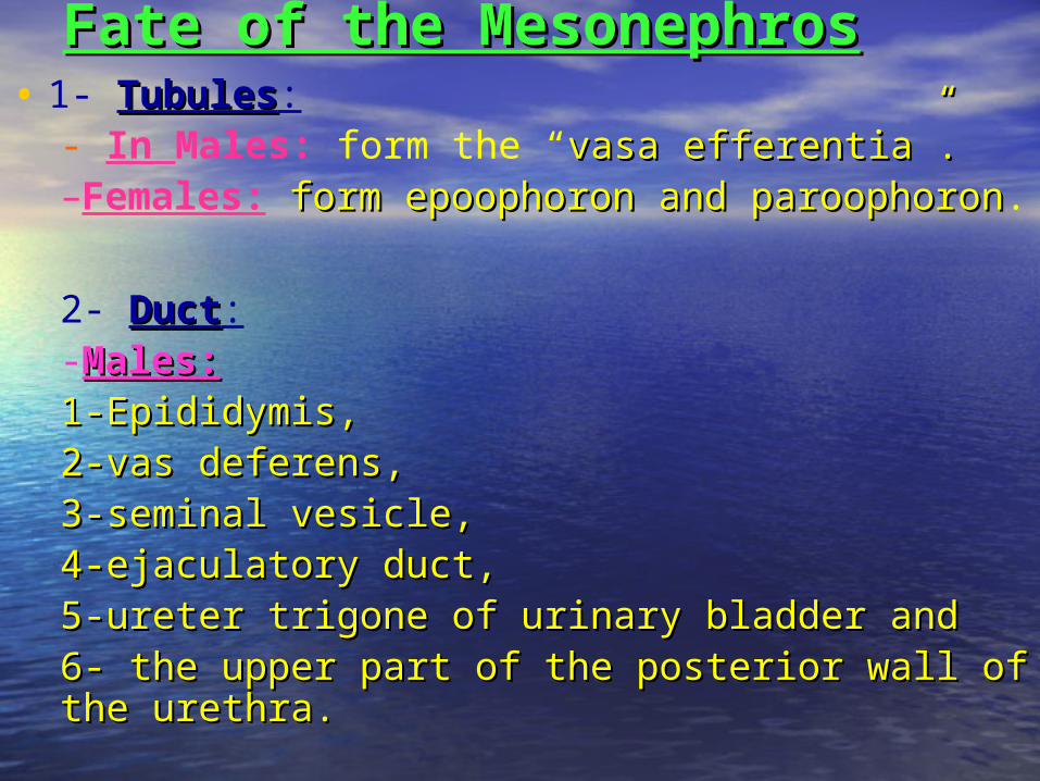
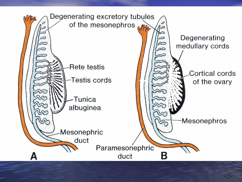
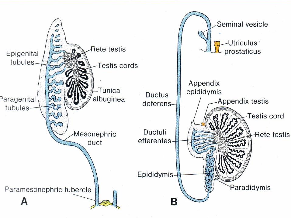
Fate of the MesonephrosFate of the Mesonephros• 1- TubulesTubules:
- In Males: form the “vasa efferentia”.vasa efferentia”.–Females: form epoophoron and paroophoronform epoophoron and paroophoron.
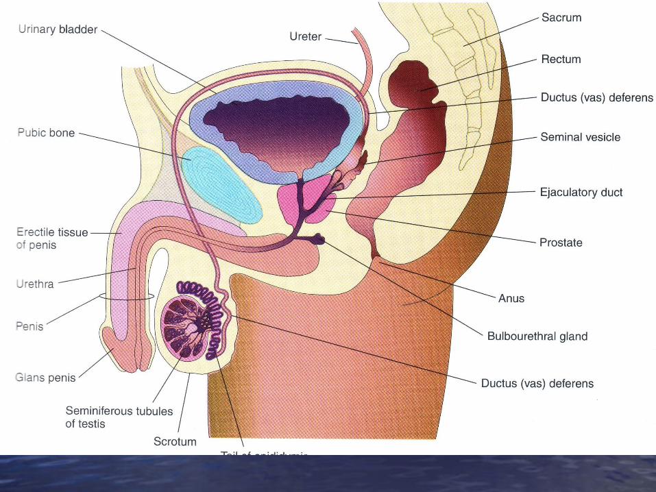
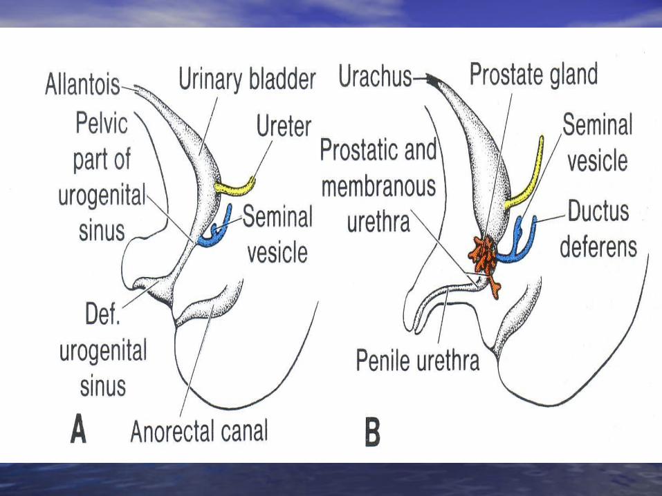
2- DuctDuct:-Males:Males: 1-Epididymis, 1-Epididymis, 2-vas deferens, 2-vas deferens, 3-seminal vesicle, 3-seminal vesicle, 4-ejaculatory duct,4-ejaculatory duct,5-ureter trigone of urinary bladder and5-ureter trigone of urinary bladder and6- the upper part of the posterior wall of the 6- the upper part of the posterior wall of the urethra.urethra.
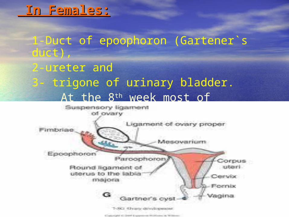
In Females:In Females:
1-Duct of epoophoron (Gartener`s duct), 2-ureter and3- trigone of urinary bladder.
At the 8th week most of mesonephric tubules degenerate.

KIDNEY AND URETERKIDNEY AND URETER• The human kidney develops from 2 The human kidney develops from 2
main parts:main parts:
1- The ureteric bud, and1- The ureteric bud, and
2- The Metanephros2- The Metanephros

The ureteric budThe ureteric bud
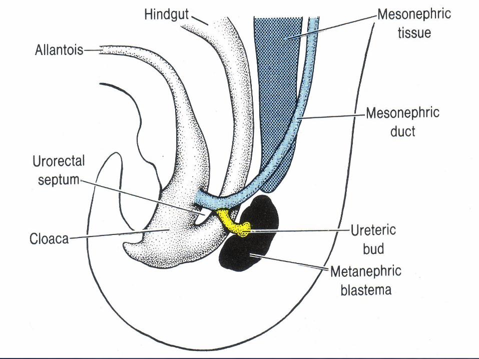
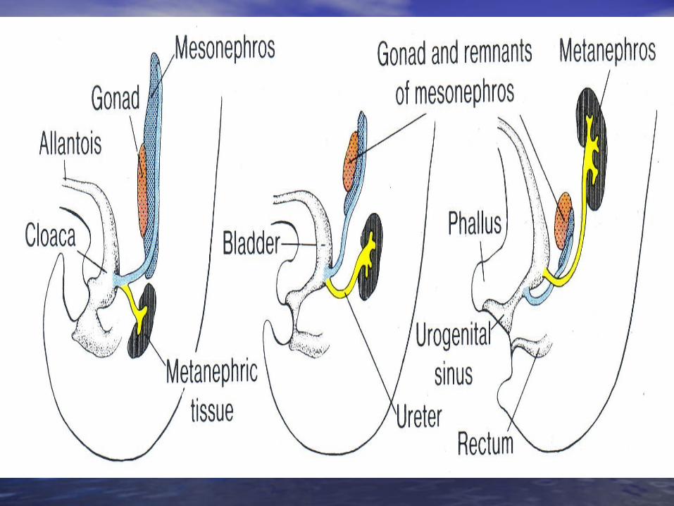
• The ureteric bud arises as an The ureteric bud arises as an outgrowth of the outgrowth of the mesonephric duct mesonephric duct close to the entrance of the duct at close to the entrance of the duct at the cloaca the cloaca
• The ureteric bud is responsible for The ureteric bud is responsible for the development of the the development of the collecting collecting systemsystem

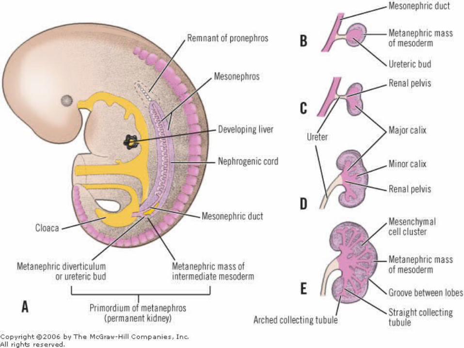
Ureter:Ureter:• The ureteric bud The ureteric bud elongates dorso-craniallyelongates dorso-cranially to be to be
in contact with the in contact with the metanephrosmetanephros which will form which will form the the metanephric capmetanephric cap..
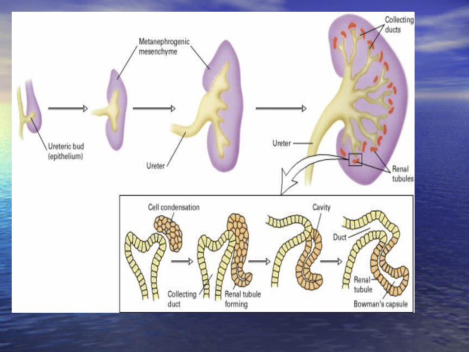
• The upper end of the ureter divides to form The upper end of the ureter divides to form 2 – 2 – 3 major calyces3 major calyces, which further divide into many , which further divide into many minor calycesminor calyces which divide into which divide into collecting collecting tubulestubules..

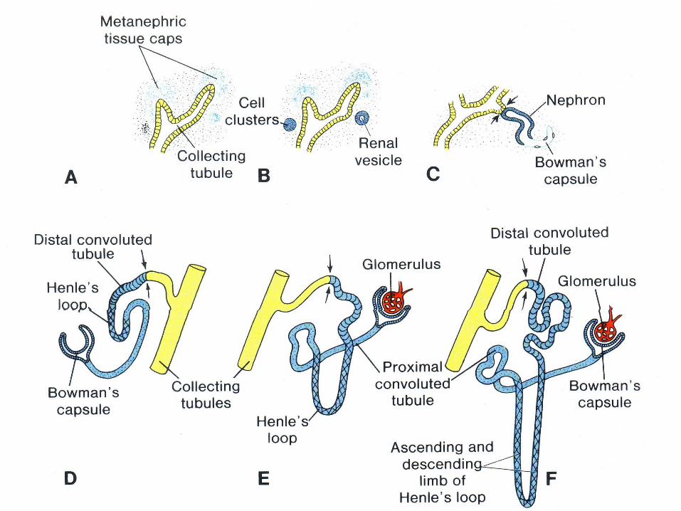
• Each collecting tubule will be covered Each collecting tubule will be covered with a with a piece ofpiece of the the metanephric capmetanephric cap which form which form renal vesicles renal vesicles which form which form the rest of the nephron the rest of the nephron exceptexcept the the collecting tubules which is developed collecting tubules which is developed from dividing ureteric bud.from dividing ureteric bud.
• Collecting tubules Collecting tubules communicatecommunicate with with the rest of the nephron.(canalization)the rest of the nephron.(canalization)
KIDNEY KIDNEY
Metanephros:Metanephros: • It is the It is the caudal part of the intermediate caudal part of the intermediate
mesoderm mesoderm in the pelvic cavity.in the pelvic cavity.
• It forms the It forms the metanephric capmetanephric cap (blastema) (blastema) which divides into small masses following which divides into small masses following divisions of the divisions of the ureteric budureteric bud. .
• Each mass is called Each mass is called renal vesiclesrenal vesicles..
• Each Each vesicle vesicle will form:will form:
1- Bowman’s capsule,1- Bowman’s capsule,
2- proximal convoluted tubule,2- proximal convoluted tubule,
3- loop of Henle and 3- loop of Henle and
4- distal convoluted tubule.4- distal convoluted tubule.


Changes of the external features of the Changes of the external features of the developing kidney:developing kidney:
• 1- Ascent of the kidney:1- Ascent of the kidney: It ascends from pelvic cavity to its adult It ascends from pelvic cavity to its adult
site in the lumbar region on the site in the lumbar region on the posterior abdominal wall.posterior abdominal wall.
This is done by the dorso-cranial This is done by the dorso-cranial elongation of the ureter pushing the elongation of the ureter pushing the kidney.kidney.

• 2- Change of the blood supply:2- Change of the blood supply:
During its ascent, the arterial supply of During its ascent, the arterial supply of the kidney shifts to the nearest main the kidney shifts to the nearest main artery .artery .
1-1- First it is supplied by the Median First it is supplied by the Median sacral artery, then sacral artery, then 2-2- Internal iliac, Internal iliac, 3-3- Common Iliac, and finally Common Iliac, and finally 4-4- Abdominal aortaAbdominal aorta

• 3- Loss of fetal lobulation:3- Loss of fetal lobulation:
•The surface of the kidney becomes surface of the kidney becomes smoothsmooth
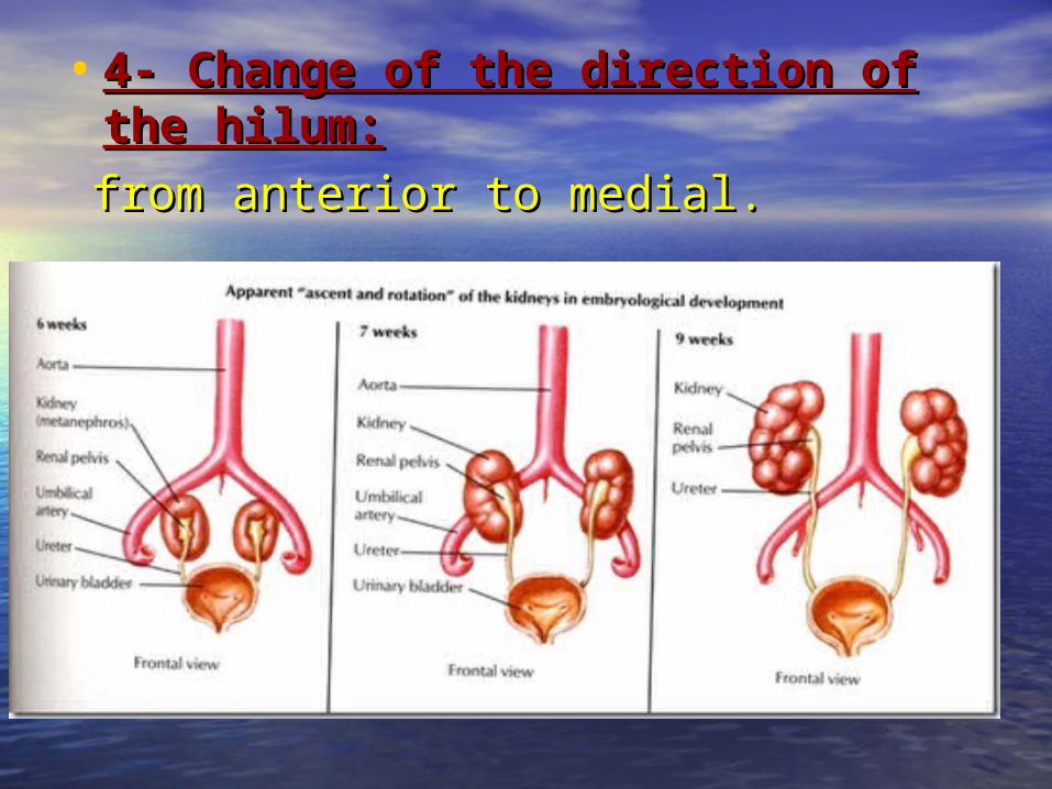
• 4- Change of the direction of the 4- Change of the direction of the hilum:hilum:
from anterior to medial.from anterior to medial.

CONGENITAL ANOMALIESCONGENITAL ANOMALIESA- A- KIDNEYKIDNEY
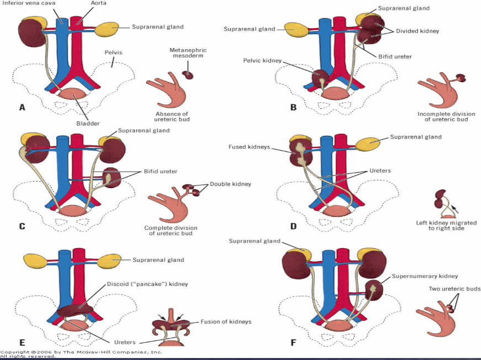
• 1-1- Renal agenesis: Renal agenesis: Unilateral or bilateral.Unilateral or bilateral.

2- 2- Congenital polycystic kidney:Congenital polycystic kidney:
due to failure of communication between due to failure of communication between the collecting tubules and rest of the the collecting tubules and rest of the nephron.nephron.
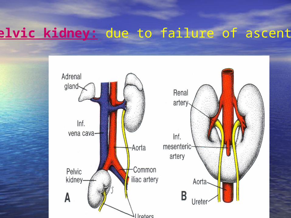
3- Pelvic kidney: due to failure of ascent.
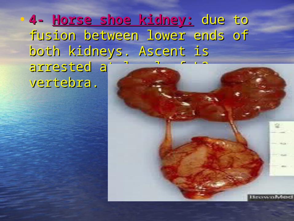
• 4- 4- Horse shoe kidney:Horse shoe kidney: due to due to fusion between lower ends of both fusion between lower ends of both kidneys. Ascent is arrested at level of kidneys. Ascent is arrested at level of L3 vertebra.L3 vertebra.

• 5- Rosette kidney:5- Rosette kidney:Due to fusion of the upper and lower
poles
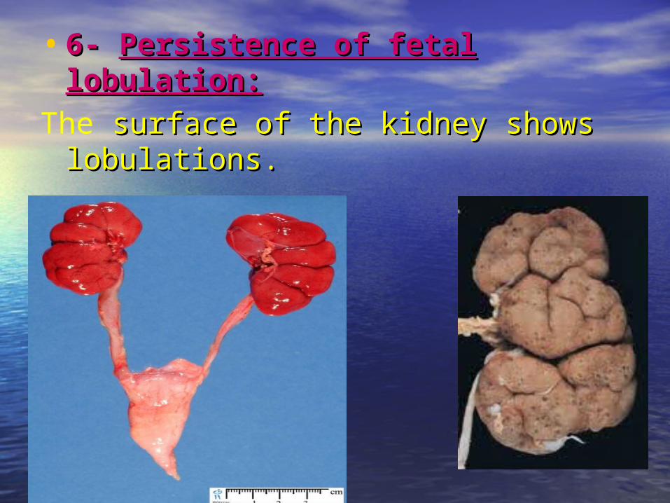
• 6- 6- Persistence of fetal lobulation:Persistence of fetal lobulation:The surface of the kidney shows surface of the kidney shows
lobulations.lobulations.
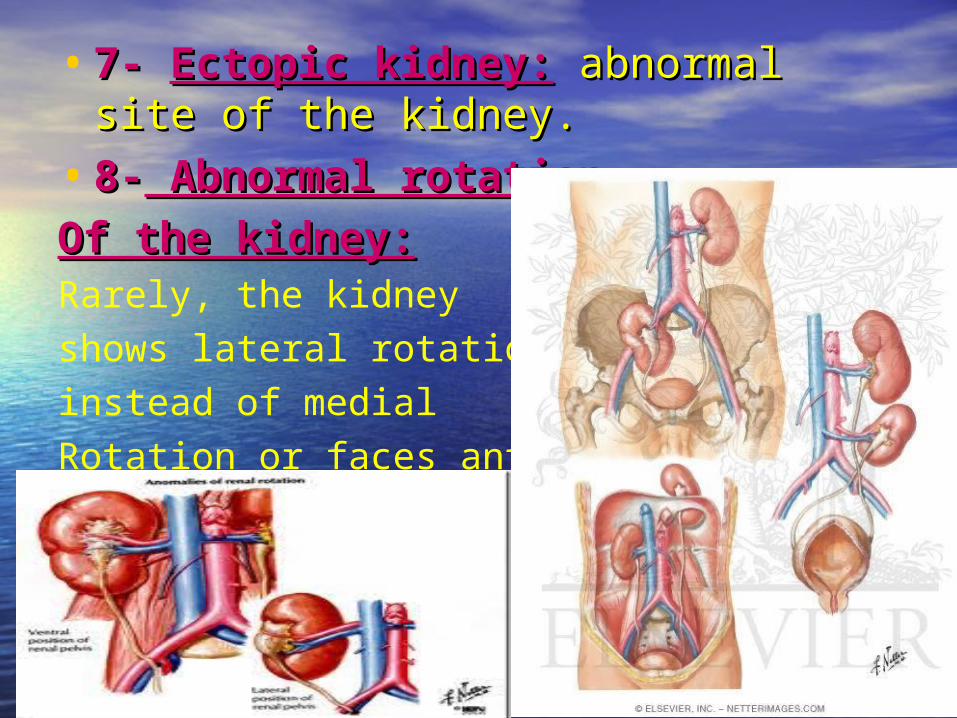
• 7- 7- Ectopic kidney:Ectopic kidney: abnormal site of abnormal site of the kidney.the kidney.
• 8-8- Abnormal rotation Abnormal rotation
Of the kidney:Of the kidney:Rarely, the kidneyshows lateral rotationinstead of medial Rotation or faces anteriorly
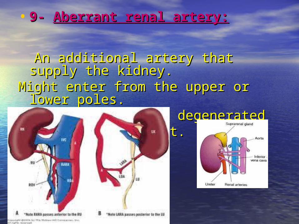
• 9- 9- Aberrant renal artery:Aberrant renal artery:
An additional artery that supply the An additional artery that supply the kidney.kidney.
Might enter from the upper or lower Might enter from the upper or lower poles.poles.
It is usually a non degeneratedIt is usually a non degeneratedArtery during ascent.Artery during ascent.
CONGENITAL ANOMALIESCONGENITAL ANOMALIESB- B- UreterUreter
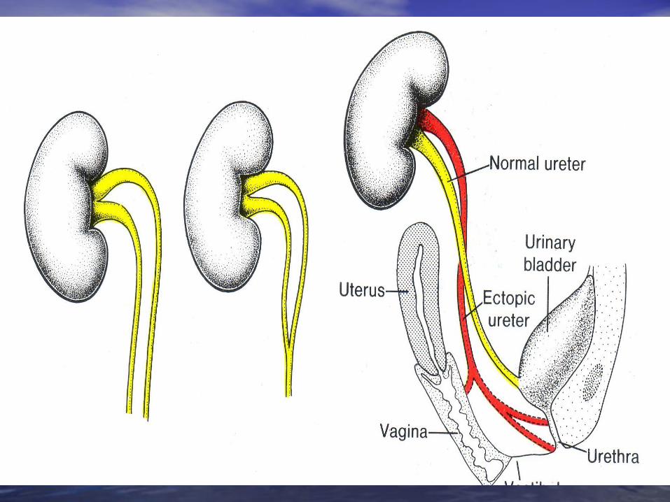
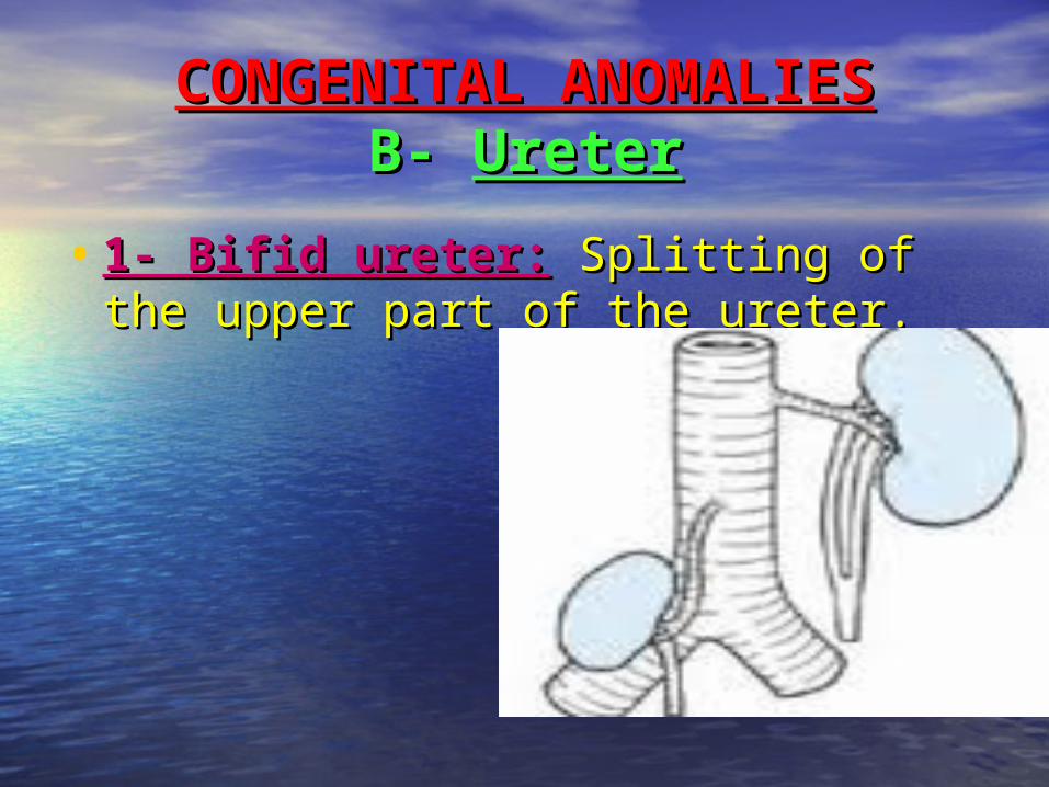
• 1- Bifid ureter:1- Bifid ureter: Splitting of the Splitting of the upper part of the ureter.upper part of the ureter.
• 2- Double ureter and Ectopic 2- Double ureter and Ectopic ureter:ureter: Two separate ureters due to Two separate ureters due to formation of 2 ureteric buds.formation of 2 ureteric buds.
•One will open normally in the bladder,
The other (Ectopic ureter): Male: Opens in the urethraFemale: Opens either in :a- the vestibule of the vagina, orb- in the urethra