Embed Size (px)
Citation preview

L E C T U R E 5
DEVELOPMENT OF THE CIRCULATORY SYSTEM

REVIEW OF CARDIAC ANATOMY
• Heart
• 4 chambers
• Base and apex
• Valves
• Pericardial sac
• 3 layers: epi, myo, endo cardium
• Major blood vessels
• Aorta and its branches
• Formation of the vena cavas
• Coronary arteries

FORMATION OF BLOOD VESSELS
• Derived from mesoderm
• Precursor cells called angioblasts develop into
blood vessels
• 3 primary mechanisms
• 1. Vasculogenesis = generally produce veins (and dorsal
aorta)
• 2. Angiogenesis = produce arteries
• 3. combination of both = produce capillaries and small
arterioles and venules
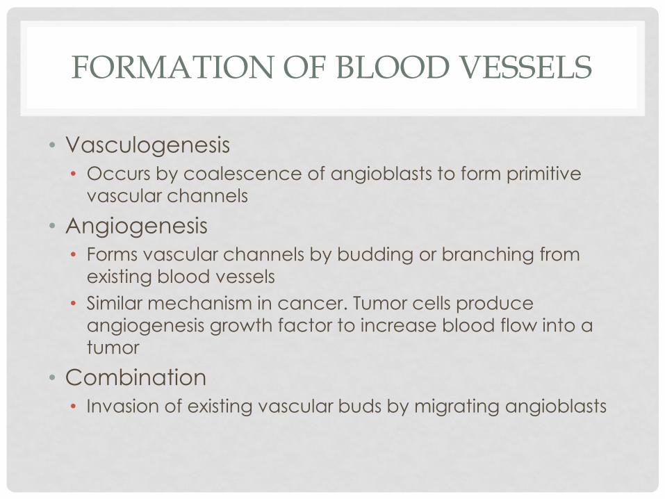
FORMATION OF BLOOD VESSELS
• Vasculogenesis
• Occurs by coalescence of angioblasts to form primitive
vascular channels
• Angiogenesis
• Forms vascular channels by budding or branching from existing blood vessels
• Similar mechanism in cancer. Tumor cells produce
angiogenesis growth factor to increase blood flow into a
tumor
• Combination
• Invasion of existing vascular buds by migrating angioblasts

FORMATION OF THE HEART
• Arise from splanchnic mesoderm at the level of the
pharynx
• Mesoderm from both sides fuse together as the
foregut is produced, creating a single tube of
splanchnic mesoderm = future heart
• Cardiac primordia are composed initially of 2 layers
• Endocardium: inner layer endothelial layer
• Myocardium: outer layer (eventually becomes the middle layer) muscular layer
• As the cardiac mesoderm fuse, a third layer is formed from the cardiac primordia = the epicardium becomes the
pericardium or cardiac sac


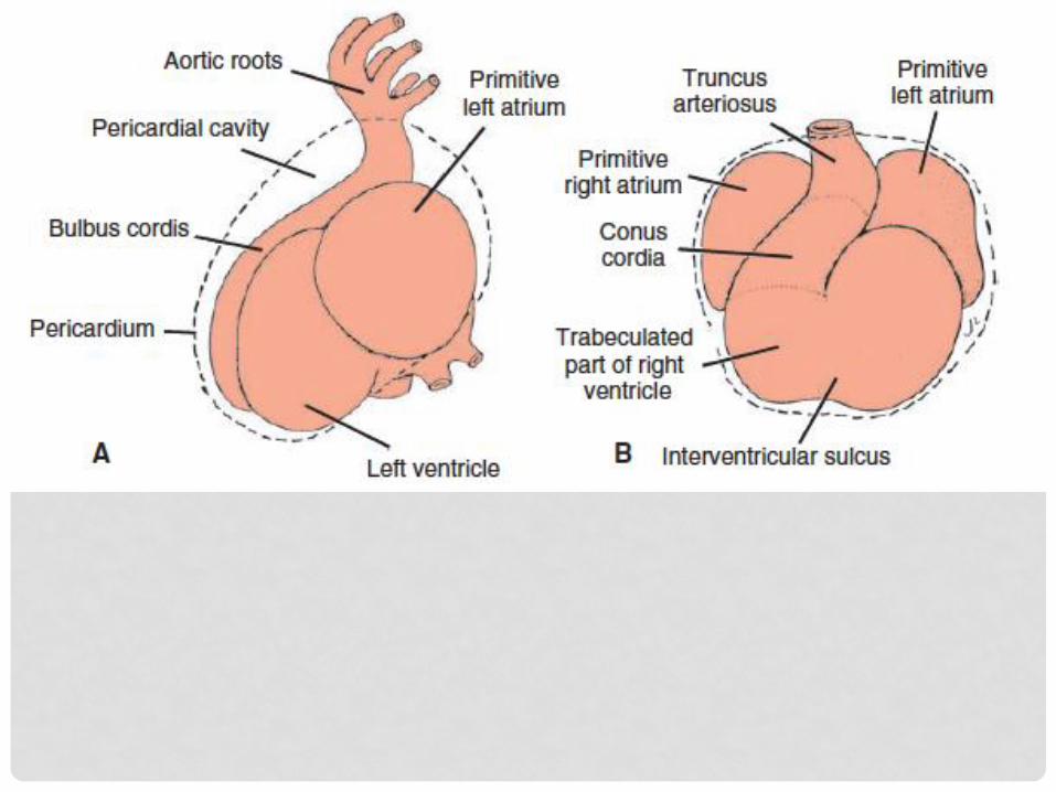
FORMATION OF THE HEART
• From a straight tubular form, the heart changes in
shape into an S-shaped configuration
• The bottom (caudal) part will form the atrium
• The top (cephalic) part will form the ventricles
• The veins converging to enter the heart become
confluent into a chamber called the sinus venosus,
which opens into the atrium
• From the ventricle, blood will flow out of the heart
into the bulbus cordis and truncus arteriosus.
• The bulbus cordis will merge with the ventricle to form part of the right ventricle.

FORMATION OF THE HEART
• The S-shaped single tube heart further bends upon
itself, producing the normal configuration of the
atrium and ventricles (atrium is cephalic, and
ventricles are caudal)
• The ventricle will move forward and the atrium will move to the back of the ventricle.
• Separating the atrium and ventricle is a narrowed
portion called the atrioventricular junction,
containing endocardial cushions, which eventually
form the atrio-ventricular valves


PARTITIONING OF THE HEART
• Eventually, the atrium and ventricle further dilate
allowing a septum to divide each chamber into
right and left halves
• Within these chambers are constricted areas due to
thickenings in the endocardium, called endocardialcushion tissue
• Within the endocardial cushion tissue are masses of
connective tissue matrix called cardiac jelly

PARTITIONING OF THE HEART
• These endocardial cushions grow from from 3 areas,
one from each side of the heart tube, and finally at
the atrioventricular junction, to form the heart
septa, dividing the heart into right and left halves


PARTITIONING OF THE HEART
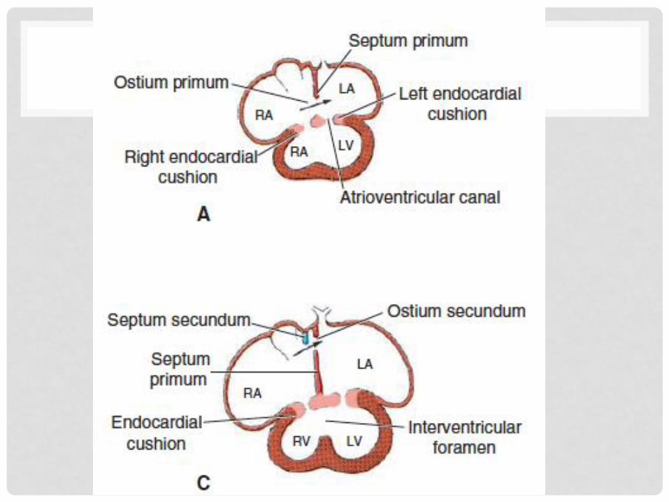
• Interatrial septum
• One partition grows from the middle endocardial cushion,
called the septum primum, which has an opening in its
middle or the ostium primum (interatrial foramen primum),
allowing blood flow from the right side to the left side of the
heart
• Interventricular septum
• A septum grows from the endocardial cushion at the apex
and from the endocardial cushion at the AV junction.


PARTITIONING OF THE HEART
• As the ostium primum closes, another hole opens
above the septum primum called the ostium
secundum
• Starts as small perforations, due to apoptosis of some cells in
the area
• These rapidly expand and unite to form a single opening
called the ostium secundum (interatrial foramen secundum)
• This allows blood flow from the RA into the LA of the heart,
to prevent the left heart from collapsing on itself
• Remember: there is as yet no pulmonary circulation to bring
blood into the left side of the heart

PARTITIONING OF THE HEART
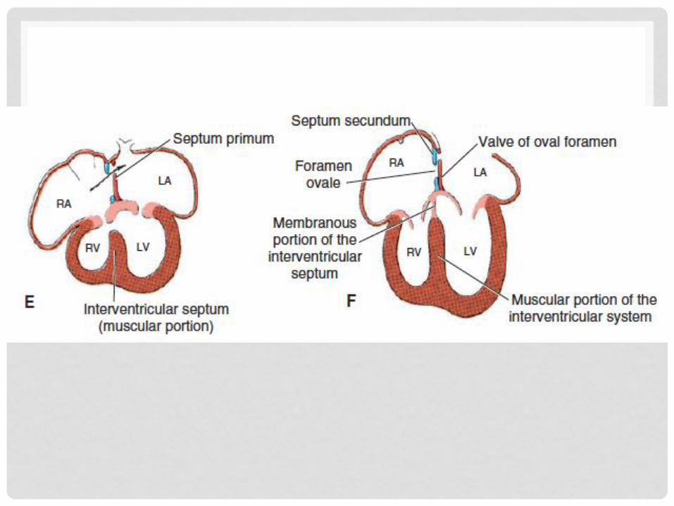
• A second interatrial septum is formed from the top
margin of the atrium, just to the right of the first
septum, called septum secundum
• This forms a partial blockage of the foramen secundum,
producing a valve
• The septum secundum will have a small opening into the
ostium secundum called the foramen ovale
• The foramen ovale and ostium secundum acts as a one
way valve from the right atrium to the left atrium
• This has important structural and physiological aspects in
fetal circulation.


PARTITIONING OF THE HEART
• The sinus venosus shifts from the midline to the right,
and opens exclusively into the right atrium
• This eventually combines with the right atrium, receiving
blood from two major vessels, the superior vena cava and
inferior vena cava
• Formation of the interventricular septum
• Similar with formation of the interatrial septum
• Ridges grow from the apex and from the endocardial
cushion at the AV junction and these meet at the middle.

PARTITIONING OF THE HEART
• There is complete division of the right and left sides
of the heart, except at the foramen ovale and
foramen secundum or ostium secundum, which is
essential for development
• Formation of the AV valves
• Endocardial cushion at the AV junction grow into valves
and papillary muscles, forming the tricuspid and bicuspid
(mitral) valves


PARTITIONING OF THE HEART
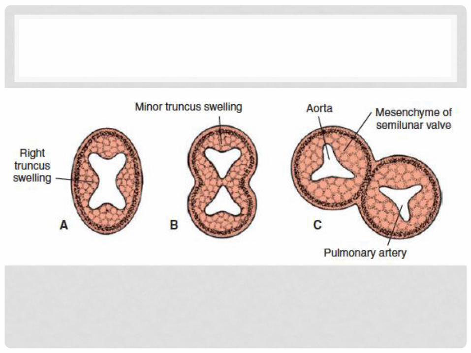
• The truncus arteriosus also divides into the aorta and pulmonary arteries• Mechanism is similar to the endocardial cushions and heart
partitioning, but called truncoconal ridges.
• Initially the truncus arteriosus exits primarily from the right ventricle. The truncoconal ridges then divide it at the middle, which eventually joins the interventricular septum. This allows formation of the aorta and pulmonary arteries.
• These ridges also form the semilunar valves of the PA and Ao
• The truncus arteriosus twists or spirals, such that the PA is in the left side and the Ao is in the right side and behind the PA.

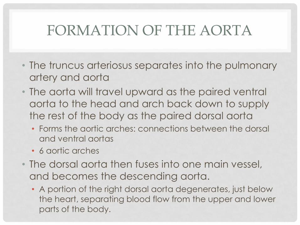
FORMATION OF THE AORTA
• The truncus arteriosus separates into the pulmonary
artery and aorta
• The aorta will travel upward as the paired ventral
aorta to the head and arch back down to supply
the rest of the body as the paired dorsal aorta
• Forms the aortic arches: connections between the dorsal
and ventral aortas
• 6 aortic arches
• The dorsal aorta then fuses into one main vessel,
and becomes the descending aorta.
• A portion of the right dorsal aorta degenerates, just below
the heart, separating blood flow from the upper and lower
parts of the body.
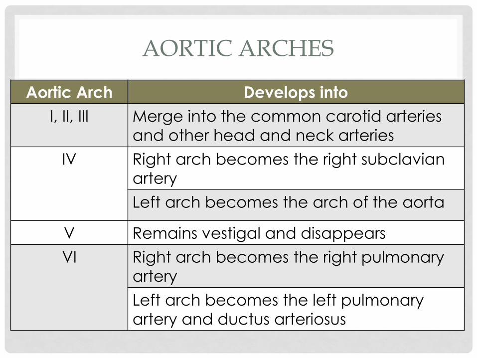
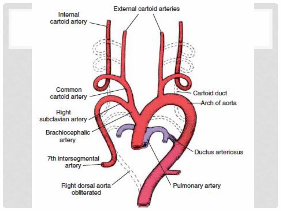
AORTIC ARCHES
Aortic Arch Develops into
I, II, III Merge into the common carotid arteries
and other head and neck arteries
IV Right arch becomes the right subclavian
artery
Left arch becomes the arch of the aorta
V Remains vestigal and disappears
VI Right arch becomes the right pulmonary
artery
Left arch becomes the left pulmonary
artery and ductus arteriosus

FORMATION OF THE MAJOR VEINS
• The systemic venous system consists of the anterior
and posterior cardinal veins which empty into the
right and left common cardinal veins, and
ultimately into the sinus venosus.
• The right common cardinal vein
• The right anterior cardinal vein becomes the superior vena
cava
• The right posterior cardinal vein becomes the inferior vena
cava
• The left common cardinal vein
• becomes the coronary sinus (draining blood from the
coronary vessels which supply blood to the heart muscles)

FORMATION OF THE MAJOR VEINS
• The vitelline veins (omhalomesenteric veins) drains
blood from the yolk sac
• The umbilical veins brings blood from the placenta
to the embryo
• All these drain into the sinus venosus. The sinus
venosus eventually merges with the right atrium.
• The pulmonary veins arise from the lung buds and
drain directly into the left atrium (although a few
short pulmonary veins grow from the left atrium to
the lungs)

FORMATION OF THE MAJOR VEINS
• The inferior vena cava is formed as follows
• The vitelline veins and umbilical veins join together to meet
with the posterior cardinal vein to become the inferior vena
cava
• The vitelline veins develop into the hepatic portal system which drains blood from the developing intestines and liver.
All blood from the intestines first drains into the liver for
processing before going to the heart for circulation.
• The umbilical vein, is initially paired. The right umbilical vein
degenerates, leaving the left as the sole vessel from the
placenta to the embryo. As it enters the fetus and is at the level of the liver, this becomes the ductus venosus


BACK VIEW

CHANGES IN CIRCULATION FROM FETUS TO BIRTH
• 2 major changes• Abrupt cutting off of the placental blood flow
• Immediate assumption by the lungs for respiration
• Fetal blood flow• Placenta umbilical veins ductus venosus inferior
vena cava (joined by the hepatic veins) right atrium foramen ovale left atrium left ventricle aorta head
• Superior vena cava right atrium right ventricle pulmonary artery lungs pulmonary veins left atrium left ventricle aorta dorsal aorta body
• Pulmonary artery ductus arteriosus aorta
• Aorta umbilical arteries placenta


CHANGES IN CIRCULATION FROM FETUS TO BIRTH
• At birth• Cessation of placental flow shrinks the umbilical vessels.
• Umbilical vein close and the ductus venosus becomes the round ligament of the liver (part of the falciform ligament)
• Umbilical arteries close and becomes the medial umbilical ligaments
• Ductus arteriosus closes by muscular contractions through the following mechanisms
• As the lungs expand, it releases a substance called bradykininwhich stimulates contraction of the muscles in the DA
• Becomes the ligamentum arteriosum
• Umbilical blood flow ceases decreasing pressure in the right atrium, while ductus arteriosus closes increasing blood flow to the lungs and to the left atrium. This increased pressure functionally closes the foramen ovale.


CONGENITAL HEART DEFECTS
• Atrial Septal Defect (ASD)
• Ventricular Septal Defect (VSD)
• Patent Ductus Arteriosus (PDA)
• Transposition of the Great Arteries (TGA)
• Persistent truncus arteriosus
• Double outlet right ventricle (DORV)
• Tetralogy of Fallot (TOF)