Embed Size (px)
Citation preview
Nanomedicine (Epub ahead of print) ISSN 1743-5889
part of
10.2217/NNM.13.225 © 2014 Future Medicine Ltd
Research Article
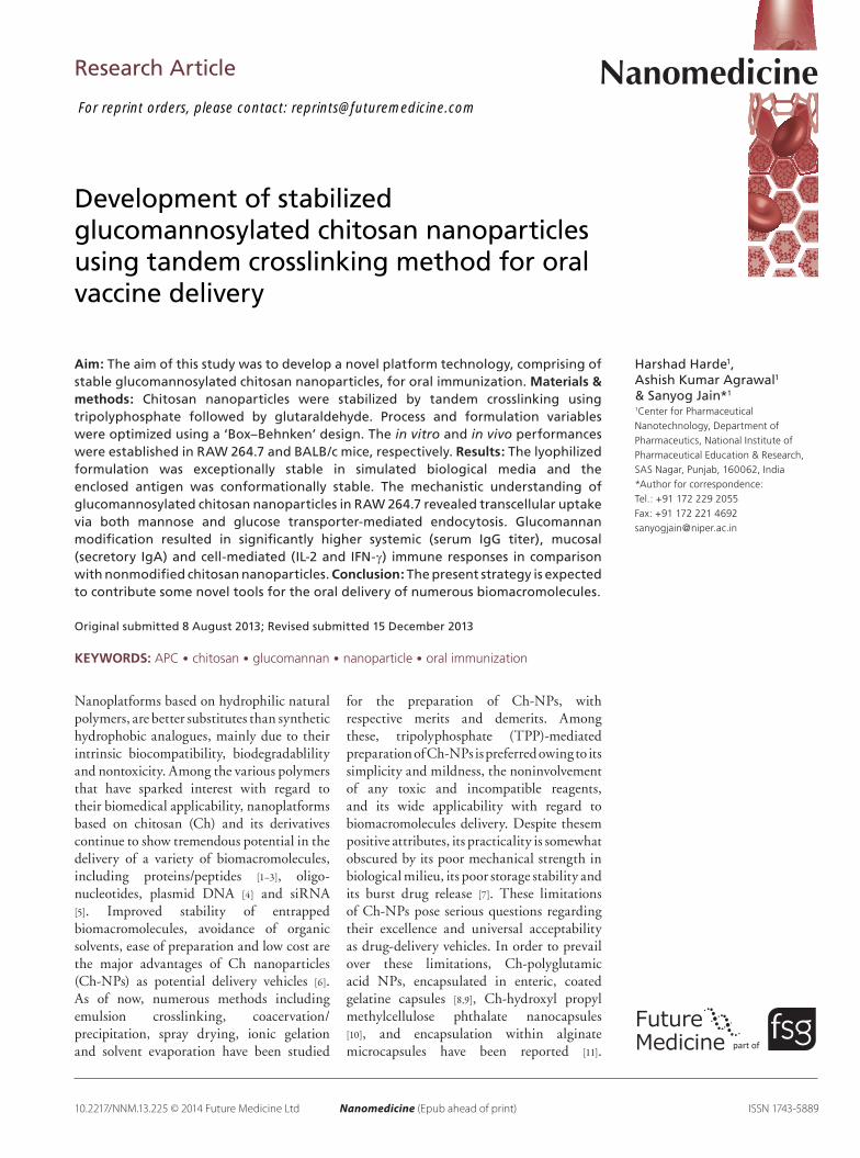
Development of stabilized glucomannosylated chitosan nanoparticles using tandem crosslinking method for oral vaccine delivery
Aim: The aim of this study was to develop a novel platform technology, comprising of stable glucomannosylated chitosan nanoparticles, for oral immunization. Materials & methods: Chitosan nanoparticles were stabilized by tandem crosslinking using tripolyphosphate followed by glutaraldehyde. Process and formulation variables were optimized using a ‘Box–Behnken’ design. The in vitro and in vivo performances were established in RAW 264.7 and BALB/c mice, respectively. Results: The lyophilized formulation was exceptionally stable in simulated biological media and the enclosed antigen was conformationally stable. The mechanistic understanding of glucomannosylated chitosan nanoparticles in RAW 264.7 revealed transcellular uptake via both mannose and glucose transporter-mediated endocytosis. Glucomannan modification resulted in significantly higher systemic (serum IgG titer), mucosal (secretory IgA) and cell-mediated (IL-2 and IFN-g) immune responses in comparison with nonmodified chitosan nanoparticles. Conclusion: The present strategy is expected to contribute some novel tools for the oral delivery of numerous biomacromolecules.
Original submitted 8 August 2013; Revised submitted 15 December 2013
KEYWORDS: APC • chitosan • glucomannan • nanoparticle • oral immunization
Nanoplatforms based on hydrophilic natural polymers, are better substitutes than synthetic hydrophobic analogues, mainly due to their intrinsic biocompatibility, biodegradablility and nontoxicity. Among the various polymers that have sparked interest with regard to their biomedical applicability, nanoplatforms based on chitosan (Ch) and its derivatives continue to show tremendous potential in the delivery of a variety of biomacromolecules, including proteins/peptides [1–3], oligonucleotides, plasmid DNA [4] and siRNA [5]. Improved stability of entrapped biomacromolecules, avoidance of organic solvents, ease of preparation and low cost are the major advantages of Ch nanoparticles (ChNPs) as potential delivery vehicles [6]. As of now, numerous methods including emulsion crosslinking, coacervation/precipitation, spray drying, ionic gelation and solvent evaporation have been studied
for the preparation of ChNPs, with respective merits and demerits. Among these, tripolyphosphate (TPP)mediated preparation of ChNPs is preferred owing to its simplicity and mildness, the noninvolvement of any toxic and incompatible reagents, and its wide applicability with regard to biomacromolecules delivery. Despite thesem positive attributes, its practicality is somewhat obscured by its poor mechanical strength in biological milieu, its poor storage stability and its burst drug release [7]. These limitations of ChNPs pose serious questions regarding their excellence and universal acceptability as drugdelivery vehicles. In order to prevail over these limitations, Chpolyglutamic acid NPs, encapsulated in enteric, coated gelatine capsules [8,9], Chhydroxyl propyl methylcellulose phthalate nanocapsules [10], and encapsulation within alginate microcapsules have been reported [11].
Harshad Harde1, Ashish Kumar Agrawal1 & Sanyog Jain*1
1Center for Pharmaceutical Nanotechnology, Department of Pharmaceutics, National Institute of Pharmaceutical Education & Research, SAS Nagar, Punjab, 160062, India *Author for correspondence: Tel.: +91 172 229 2055 Fax: +91 172 221 4692 [email protected]
For reprint orders, please contact: [email protected]

10.2217/NNM.13.225 Nanomedicine (Epub ahead of print) future science group
Research Article Harde, Agrawal & Jain
Unfortunately, instability in acidic milieu, higher particle size, up to micrometer range, and complex methods of preparation further limit their use. In this article, we propose a tandem crosslinking strategy to produce ChNPs of the desired particle size and with an improved stability profile. This simple troubleshooting strategy consist of the preparation of ChNPs using TPP as a primary crosslinker, followed by a second surface crosslinking using glutaraldehyde. The amount of glutaraldehyde used is too little to cause any detectable toxicity after oral administration and complies with regulatory norms. This dual approach, involving the sequential use of two crosslinking agents for preparation and stabilization of the ChNPs, is unique and has not been reported on before.
To further demonstrate the potential usefulness of these newly synthesized ChNPs in terms of their biomedical applicability, we explored these NPs for use in oral immunization. We were particularly interested in this application because oral immunization using ChNPs has not been very successful till date. Oral mass immunization is not only the easiest approach for widespread use of a vaccine, but also superior for induction of the mucosal immune response (secretory IgA [sIgA]), which is the predominant entry site for most infectious pathogens [12,13]. However, degradation of the antigen by harsh proteolytic milieu, inability to breach the defensive mucosal barrier and poor passive diffusion are the major hurdles for antigen delivery via the oral route [13]. Therefore, surface functionalization by ligand grafting is an additionally promising approach along with the use of stabilized ChNPs, which have been tried for targeted delivery and potentiation of host immunity through rapid internalization into compartments that contain elements of the antigenprocessing machinery [14,15]. Among the various ligand molecules glucomannosylation has been recognized as a potential approach, since receptors for mannose are not only overexpressed on cells of the immune system, namely macrophages and dendritic cells, but it is also upregulated in activated macrophages (i.e., macrophages associated with tissue repair and humoral immunity) [14,16,17]. Therefore, a water soluble, high molecular weight, linear copolymer of dmannose and dglucose, namely konjac glucomannan (GM), was explored as a ligand for mannose receptors. The rational for selection of GM is that it is not hydrolyzed by digestive enzymes and has been reported to improve in stability in acidic pH or high salt solutions by forming robust particulate systems [18,19]. Along with improved stability, GM has the ability to interact favorably with some biological surfaces that are particularly rich in mannose receptors, such as the Mcells overlying the Peyer’s patches and macrophages. In the present study,
we also speculated that glucomannosylation using GM can fortify the efficacy by providing additional stability to ChNPs, while increasing the specialized uptake by providing a higher density of mannose molecules on their surface.
Cumulatively, the objective of the present study was to develop Ch nanoformulations with the dual specification of improved stability and site specificity for improved oral immunization using bovine serum albumin (BSA) as the model antigen and GM as the sitedirecting ligand.
Materials & methodsMaterials & reagents Medium molecular weight Ch (190–300 kDa; deacetylation degree: 87%), pentasodium TPP, bicinchoninic acid, dextrose, sucrose, lactose, trehalose, mannitol, sodium dodecyl sulfate (SDS), acrylamide, bisacrylamide, ammonium persulpahte, N,N,N ,́N ́tetramethylethylenediamine, and pilocarpine were all purchased from SigmaAldrich (MO, USA). Copper sulfate (pentavalent), panacreatin, pepsin, and glutaraldehyde (25% w/v) were procured from Loba Chemie Pvt. Ltd (Mumbai, India). Sodium dihydrogen phosphate and dipotassium hydrogen phosphate were acquired from Central Drug House (New Delhi, India). Acetic acid was obtained from Fisher Scientific (Mumbai, India). Bromophenol blue, coomassie brilliant blue G, glycine, bmercaptoethanol, and tris buffer were obtained from HiMedia (Mumbai, India). GM was procured from Megazyme International Ireland Ltd (Wicklow, Ireland). All other chemicals and reagents used were of analytical grade. Ultrapure water (LaboStar™ TWF UV 7, Siemens, MA, USA) was prepared in house and used throughout the experimentation.
Antigen, antibodies & ELISA kitsBSA (67 kD), BSA–fluorescein isothiocyanate conjugate (BSA–FITC), antimouse IgA (achain specific)–peroxidase conjugate, anti-mouse IgG (g-chain specific)–peroxidase conjugate, 3,3 ,́5,5́ tetramethylbenzidine and Nunc Immuno™ Maxisorb F 96well solid plates were procured from SigmaAldrich. Mouse IL2 and IFNg Legend ELISA MAX™ Deluxe kits were obtained from Biolegend Inc. (CA, USA).
Preparation & optimization of Ch nanoformulations Preparation of Ch-NPsChNPs were prepared by an ionotropic gelation method using TPP as a primary polyanionic crosslinking agent [1,20,21]. Briefly, Ch was dissolved in acetic acid solution
10.2217/NNM.13.225future science group
Glucomannosylated chitosan nanoparticles for oral vaccine delivery Research Article
(0.1% w/v; pH 3.2) and the pH of the resultant solution was adjusted to pH 5.5–6.5 with the help of NaOH (1N). Crosslinking solution was prepared separately, by dissolving TPP along with BSA in distilled water, and then added to the Ch solution in 1:4 volume ratio under continuous stirring at room temperature, which led to the formation of ChNPs. Different process and formulation variables were optimized using fourfactor, threelevel triplicates of ‘Box–Behnken’ response surface designs, using the pH of the Ch solution (X1), TPP concentration (X2), speed of the stirrer (X3) and antigen loading (X4), as explanatory variables, against particle size (Y1), entrapment efficiency (EE%; Y2) and polydispersity index (PDI; Y3), as response variables. The SAS Institute JMP® 10.0.0 statistical software (trial version, NC, USA) was used to study the effect of the explanatory variables on the responses using ‘actualbypredicted plots’, ‘sorted parameter estimates’, a prediction profiler, a contour profiler and a surface profiler. The three different points within the design space of the contour profiler were validated using the optimum explanatory variables and experimental values of the response variables. The prediction error was calculated based on a comparison between predicted values versus experimental values.
Glucomannosylation of Ch-NPsA total of 1 ml of GM (10–30% w/v) was incorporated along with 4 ml of Ch (0.1% w/v; 6.0 pH), followed by the addition of 1 ml of TPP (0.1% w/v) at 1500 rpm for 10 min to create glucomannosylated of ChNPs (ChGMNPs). The optimization was performed by considering GM as an independent variable while considering particle size, PDI and EE% as dependent variables. The amount of free GM was calculated using a concentrated sulfuric acid–phenol (SAP) assay [22], while surface GM functionalization was confirmed by a concanavalin A agglutination assay (see Supplementary Material online at www.futuremedicine.com/doi/suppl/10.2217/NNM.13.225) [16,17].
Stabilization of Ch-NPS & Ch-GM-NPsFor the stabilization of ChNPS and ChGMNPs (sChNPs and sChGMNPs), 1 ml of different concentrations of glutaraldehyde (0.0125–0.2% w/v) solutions were added to 6 ml of NP dispersion. The resulting sChGMNPs were characterized for particle size, PDI and EE%. The residual glutaraldehyde content in the Ch nanoformulations was determined by gas chromatography [23] and SAP colorimetric assay [22].
Characterization of Ch nanoformulations The particle size and PDI of Ch nanoformulations (refering to all ChNPs, ChGMNPs, sChNPs
and sChGMNPs) were determined by dynamic light scattering using a the zetasizer Nano ZS (Malvern Instruments, Malvern, UK). The EE% was evaluated by a direct incubation method, in which the amount of unentrapped BSA in the supernatant was measured using a validated microbicinchoninic acid colorimetric method (Powerwave XS2, BioTek Instruments Inc., VT, USA). The shape and surface morphology of Ch nanoformulations was analysed by scanning electron microscopy (S3400N, Hitachi Global, Hitachi, Japan).
LyophilizationThe Ch nanoformulations were lyophilized (Wizard 2.0, VirTis, NY, USA) using different cryoprotectants (5% w/v), namely dextrose, sucrose, lactose, trehalose and mannitol, in the preliminary screening. Later, sucrose was finalized and further optimized in the range of 2.5–10% w/v. A lyophilization cycle comprising the steps of freezing at 45°C for 8 h at 400 bar, primary drying at 45 to 20°C for 36 h at 200 mbar and secondary drying at 25°C for 6 h at 100 mbar was used for this study, which is well developed in our laboratory [24,25]. The lyophilized formulations were examined for appearance of the cake, redispersibility index and redispersibility score. The residual glutaraldehyde content in lyophilized Ch nanoformulations was determined by gas chromatography [23].
Stability studiesIn-process stability of entrapped BSA in Ch nanoformulationsChemical stability The chemical stability of BSA against degradation and/or aggregation was determined by SDS polyacrylamide gel electrophoresis [26–28]. Briefly, BSA, released from lyophilized Ch nanoformulations upon incubation with phosphatebuffer saline (PBS; pH 7.4) for 24 h, was separated by centrifugation and subjected to vertical SDS polyacrylamide gel electrophoresis. An equivalent amount of standard BSA (control) and BSA obtained from lyophilized Ch nanoformulations were loaded on to individual wells of a 7.5% polyacrylamide gel. Samples were electrophoresed using a miniVE electrophoresis unit (BioRad Laboratories, CA, USA) at a constant voltage (200 V). The resolved protein bands were stained using coomassie blue dye and visualized using GelDoc (BioRad Laboratories). In addition, protein markers were also used to estimate molecular weights.
Conformational stability The conformational integrity of BSA was determined using far UV circular dichroism (CD) spectroscopy
10.2217/NNM.13.225 Nanomedicine (Epub ahead of print) future science group
Research Article Harde, Agrawal & Jain
(J815; Jasco, Tokyo, Japan) [29,30]. Briefly, a standard BSA solution and BSA released from lyophilized Ch nanoformulations were subjected to CD analysis in the far UV region of 250–190 nm at a data pitch of 0.5 nm and a scanning speed of 50 nm/min. The average of three accumulations for each sample was taken and a baseline correction with distilled water was carried out to obtain the CD spectrum.
Stability in simulated biological mediaThe stability of Ch nanoformulations were determined at different pHs and enzymatic conditions simulated in different biological media, namely water, normal saline (0.9% NaCl), PBS (pH 7.4), HCl (pH 1.2), simulated gastric fluid (pH 1.2) and simulated intestinal fluid (pH 6.8) [31–34]. For the stability study, 1 ml of NP dispersion was added in 4 ml of stability media and incubated at 37°C for 2 h in simulated gastric fluid, and 4 h in simulated intestinal fluid, PBS, saline and water. The stability of Ch nanoformulations was evaluated by measuring the change in particle size, PDI, zetapotential and EE%.
Mechanistic understanding of uptake by antigen-presenting cells The murine macrophage cell line (RAW 264.7) was incubated with BSA–FITCloaded Ch nanoformulations against free BSA–FITC (negative control) at different concentrations (10–100 µg/ml) for 1, 2 and 3 h. In addition, the uptake of ChGMNPs was studied after GM or mannose pretreatment for mechanistic understanding of receptorbased endocytosis. Following incubation, cells were thoroughly washed with PBS (pH 7.4) and analysed by 2D confocal laser scanning microscopy (CLSM; FluoView® 1000, Olympus, Tokyo, Japan). For quantitative determination of cellular uptake, cells were lysed using 1% w/v of Triton™ X100 (Sigma-Aldrich) and fluorescence was measured at 490 and 520 nm excitation and emission wavelength, respectively [24,35].
In vivo intestinal permeation For evaluation of in vivo intestinal permeation, standard deviation rats received 200 µl of BSA–FITCloaded Ch nanoformulations (4 µg/ml) and free BSA–FITC (in equivalent concentrations) via the oral route. The animals were sacrificed after 4 h, followed by extraction of the duodenal portion of their intestine. The intestine was washed thoroughly with PBS (pH 7.4) and sectioned transversely in optimal cuttingtemperature compound medium using a Cryomicrotome (MICROM, Walldorf, Germany). For visualization of the intestinal permeation, transverse
sections as well as apical views of intestine were observed using CLSM [16].
Immunization studiesAnimalsMale BALB/c mice of 6–8 weeks of age (20–25 g weight) and Sprague–Dawley rats of 6–7 weeks old (200–220 g weight) were procured from Central Animal Facility, the National Institute of Pharmaceutical Education and Research (SAS Nagar, Punjab, India). All animal protocols were duly approved by the Institutional Animal Ethics Committee of the National Institute of Pharmaceutical Education and Research. All experimentations were performed in accordance with the guidelines of the Committee for the Purpose of Control and Supervision of Experiments on Animals (Delhi, India).
Immunization Animals were divided into six groups with six animals in each group. Group I received an aluminium hydroxideadsorbed BSA (BSAAL) suspension by intramuscular (im.) route (control; 50 µg/mice); group II received BSAAL by peroral route (equivalent to 2 mg/kg); group III–VI received BSAloaded ChNPs, sChNPs, ChGMNPs and sChGMNPs, respectively (equivalent to 2 mg/kg of BSA) by peroral route. Each group also received a booster dose at day 21 [16,36,37].
Sample collectionThe blood samples were withdrawn from the retroorbital plexus of mice on day 1, before immunization (preimmune), 7, 14, 21 (postimmunization), 28 and 35 (postbooster) for IgG determination. Blood samples were allowed to coagulate and centrifuged at 10000 rpm for 8 min. Subsequently, serum was separated and stored at 20°C until it was analysed for its IgG titer using ELISA [36,38].
The sIgA level was estimated in biological fluids, namely saliva and intestinal fluids, at the end of the study. Saliva was collected using a Gilson pipette (Gilson Inc., WI, USA) following intraperitonial injection of pilocarpine (0.2 ml; 80 mg/kg) [39]. Subsequently, animals were sacrificed, dissected and intestinal sections of equal lengths (small intestine) were incubated in PBS (pH 7.4) at 2–8°C for 24 h. The extracted antibody was separated from the tissue content by centrifugation at 20000 rpm for 8 min. The supernatant was collected and analyzed for sIgA titer using ELISA [40].
Endogenous levels of cytokines (IL2) and IFNg were determined in the spleen homogenate. Briefly, the spleen was homogenized in cold PBS using a tissue homogenizer (Polytron® PT 4000, Kinematica,

10.2217/NNM.13.225future science group
Glucomannosylated chitosan nanoparticles for oral vaccine delivery Research Article
NY, USA) at the end of study (35th day), it was then centrifuged and the supernatant was collected. Cytokine levels were analysed using ELISA [16,41].
Measurement of mean antibody titer and cytokine levels ELISA was used to determine the mean antibody titer in the serum. Briefly, 100 µl of BSA (10 µg/ml) in PBS (pH 7.4) was added to a 96well plates and incubated overnight at 4°C, followed by thorough washing (three times) with PBS containing 0.05% w/v Tween® 20 (SigmaAldrich). Subsequently, serum samples were serially diluted with PBS, and 100 µl of diluted serum samples were added in each well and incubated at 37°C for 2 h, followed by thorough washing with Tween 20. Subsequently, 100 µl of rabbit antimouse IgG–peroxidase conjugate (1:15000) in PBS was added in each well and incubated for 1 h at 37°C, followed by thorough washing with Tween 20. Finally, 3,3 ,́5,5´tetramethylbenzidine substrate (100 µl) was added in each well and incubated for 30 min at room temperature, followed by the addition of 100 µl of stop solution (H
2SO
4; 2N). The
developed colour was measured at 450 nm on a UV ELISA plate reader (Biotek Powerwave XS2, BioTek Instruments Inc.) [36,38].
The mean sIgA titer in the biological fluids was estimated following the same procedure used for the determination of IgG, except with the addition of 100 µl of rabbit antimouse sIgA–peroxidase conjugate (1:10000) in PBS in place of antimouse IgG–peroxidase conjugate [16,17].
IL2 and IFNg levels were determined using Legend ELISA MAX™ Deluxe kits (BioLegend Inc.), strictly following the manufacturer’s protocol [16].
Statistical methodsOptimization of multiple explanatory variables was accomplished using JMP 10.0.0 statistical software. The statistical analysis of in vitro and in vivo results were performed using SigmaStat (version 3.5, Systat Software Inc., IL, USA) utilizing oneway analysis of variance followed by Tukey’s pairwise multiple comparison procedures. Significance was evaluated at a pvalue of 0.05.
ResultsOptimization & data analysis of Ch-NPs using the Box–Behnken designBox–Behnken surface design was used to optimize the ChNPs on the basis of the effect of the explanatory variables on the response variables. The ‘actualbypredicted plot’ and ‘sorted parameter estimate’ suggested that response variables were
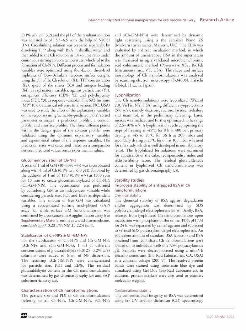
significantly affected by every explanatory variable (Supplementary Figure S2). The ‘prediction profiler’ was plotted to obtained the maximum desirabilitydemonstrating pH (6), TPP concentration (1 mg/ml), speed (1800 rpm) and drug loading (5% w/v) as optimized explanatory variables, which produced a desirable formulation of 144 nm size, 0.22 PDI, 89 EE% at 95% CI (Supplementary Figure 3S).
The response surface and contour plots were used to study the interaction between allied explanatory variables and responses when other factors were kept constant (Figure 1) [42]. ChNPs with desired quality attributes were obtained when the pH of the Ch solution varied in the range of 5.8–6.0, while the TPP ratio was close to one. Likewise, a stirring speed greater than 1200 rpm and antigen loading below 12% produced smaller NPs with >80 EE%. The design space is the desired tangible region where highly appropriate formulation with optimized parameters were obtained when a higher limit of <200 nm for size, <0.3 for PDI and lower limit for >80 EE% were fixed (Figure 1). Three different points on the design space were selected for the evaluation of the model (Table 1). It was observed that the experimental values were in the range of the predicted values within the 10% of prediction error. Therefore, the Box–Behnken design for optimization of ChNPs was validated.
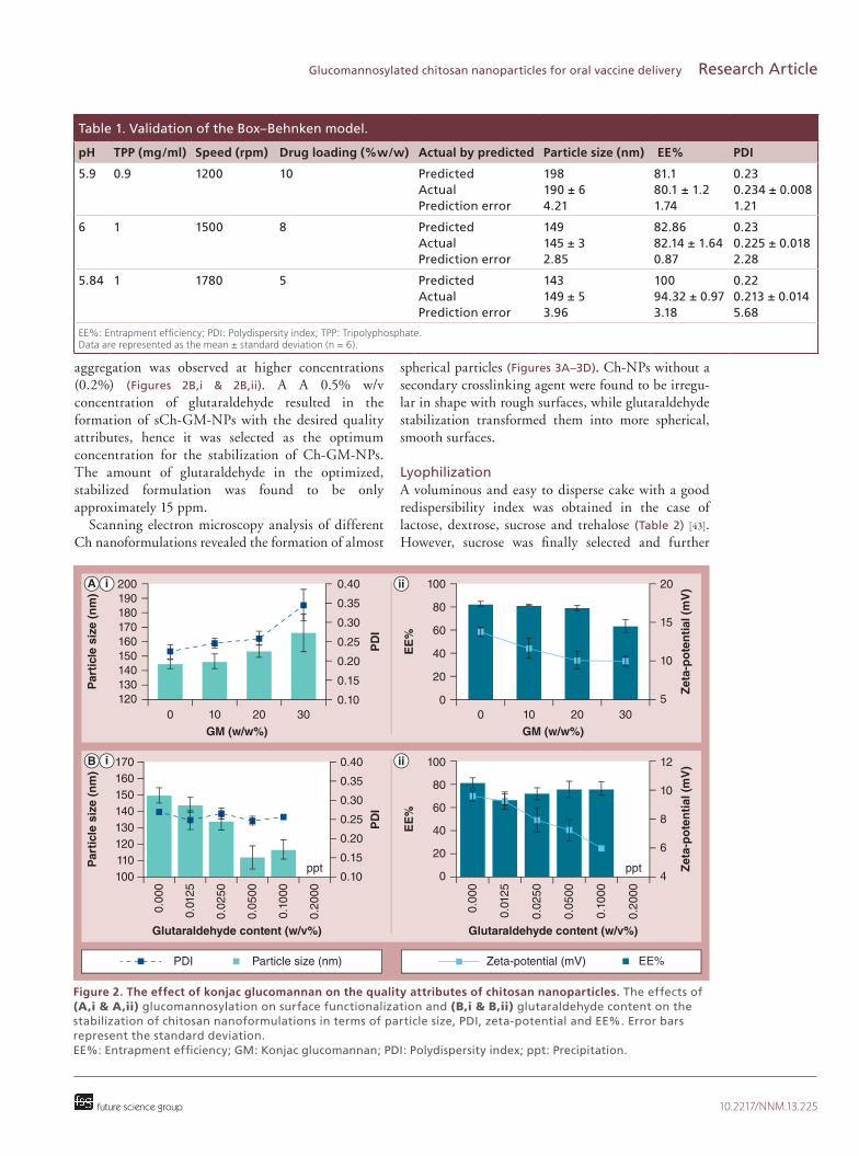
Preparation of Ch-GM-NPsThe effect of GM on the quality attributes of ChNPs is shown in Figures 2A,i & 2A,ii. No significant change in quality attributes was observed at lower concentrations of GM (<10% w/v), while particle structure was markedly disturbed (high particle size and poor PDI) at high GM concentrations (>30% w/v). Similarly, no significant effect on EE% was observed upon varying the concentration of GM. Therefore, GM (20%) was considered the optimum for the preparation of ChGMNPs.
Almost negligible yellow–orange colour developed in the SAP colorimetric assay, demonstrating that >98% of GM was presented within nanoformulations. The concanavalin A agglutination assay confirmed the surface functionalization of nanoformulations by GM, based on higher absorbances at 550 nm in the case of glucomannosylated nanoformulations than nonglucomannosylated nanoformulations.
Preparation of sCh-GM-NPsA significant (p < 0.001) decrease in particle size and zetapotential was observed upon increasing the concentration of glutaraldehyde from 0.0125 to 0.05% w/v, which assured the crosslinking, while
10.2217/NNM.13.225 Nanomedicine (Epub ahead of print) future science group
Research Article Harde, Agrawal & Jain
D) )
B) E) H)
C) F) I)
14001800
A B C
D E F
G H I
J K L
Par
ticl
e si
ze (
nm
)E
E%
PD
I
pH (5.5, 6.5)
12010080604020
0-20-40-60
EE
%100
80604020
0-20-40
EE
%
10080604020
0-20-40-60
6.06.45.6
pH (5.5, 6.5)
6.06.4 5.6
pH (5.5, 6.5)6.06.4
5.6
6001000
14001800
pH (5.5, 6.5)
6001000
Speed (rpm)
Speed (rpm)
Speed (rpm)
Sp
eed
(R
PM
)
pH (5.5, 6.5)
600
1000
1400
1800
1614
1210
864
pH (5.5, 6.5)5.4 5.8 6.25.4 5.8 6.2
5.4 5.8 6.2
1614
1210
86
4
Drug loading (%w/w)
Drug loading (%w/w)
Drug loading (%w/w)
Dru
g lo
adin
g (
%w
/w)
pH (5.5, 6.5)
1614
1210
86
4
0.550.500.450.400.350.300.250.20
PD
I0.40
0.35
0.30
0.250.20
PD
I
0.45
0.40
0.35
0.30
0.25
0.200.5
0.60.7
0.80.9
1
pH (5.5, 6.5)
0.50.6
0.70.8
0.91
TPP (mg/ml)
TPP (mg/ml)
TPP (mg/ml)
TP
P (
mg
/ml)
pH (5.5, 6.5)6.0
6.4
5.66.0
6.4
5.66.0
6.4
5.6
0.50.6
0.70.8
0.91
600500400300200100
Par
ticl
e si
ze (
nm
)
550500
400450
300350
250200
Par
ticl
e si
ze (
nm
) 550500
400450
350300250200
1.0
0.9
0.8
0.7
0.6
0.5
5.5 5.75 6 6.25 6.5
pH
1750
1500
1250
1000
750
5.5 5.75 6 6.25 6.5
pH
15
12.5
10
7.5
5
5.5 5.75 6 6.25 6.5
pH
EE% Particle size PDI EE% Particle size PDI EE% Particle size PDI
Figure 1. Strategic optimization of chitosan nanoparticles using ‘Box–Behnken’ design. (A–I) Response surface profiler representing the effect of two different explanatory variables on each response. The columns represent allies of explanatory variables, namely (A, D & G) pH × TPP, (B, E & H) pH × speed and (C, F & I) pH × drug loading, while the rows represent the effects of explanatory variables on (A–C) particle size, (D–F) EE% and (G–I) PDI. (J–L) Contour profiler representing a desirable ‘design space (white region)’ based on the effect of explanatory variables on the responses, namely particle size, EE% and PDI. EE%: Entrapment efficiency; PDI: Polydispersity index; TPP: Tripolyphosphate.
10.2217/NNM.13.225future science group
Glucomannosylated chitosan nanoparticles for oral vaccine delivery Research Article
aggregation was observed at higher concentrations (0.2%) (Figures 2B,i & 2B,ii). A A 0.5% w/v concentration of glutaraldehyde resulted in the formation of sChGMNPs with the desired quality attributes, hence it was selected as the optimum concentration for the stabilization of ChGMNPs. The amount of glutaraldehyde in the optimized, stabilized formulation was found to be only approximately 15 ppm.
Scanning electron microscopy analysis of different Ch nanoformulations revealed the formation of almost
spherical particles (Figures 3A–3D). ChNPs without a secondary crosslinking agent were found to be irregular in shape with rough surfaces, while glutaraldehyde stabilization transformed them into more spherical, smooth surfaces.
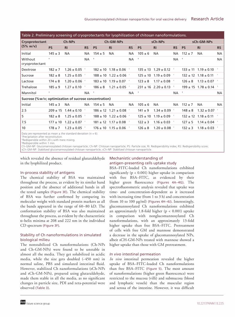
LyophilizationA voluminous and easy to disperse cake with a good redispersibility index was obtained in the case of lactose, dextrose, sucrose and trehalose (Table 2) [43]. However, sucrose was finally selected and further
A
B
i
i
ii
ii
Par
ticl
e si
ze (
nm
)P
arti
cle
size
(n
m)
GM (w/w%)
PD
IP
DI
EE
%E
E%
Zet
a-p
ote
nti
al (
mV
)Z
eta-
po
ten
tial
(m
V)
Glutaraldehyde content (w/v%) Glutaraldehyde content (w/v%)
GM (w/w%)
200190180170160150140130120
170160150140130120110100
ppt ppt
0
0.00
0
10 20 30
0.40
0.35
0.30
0.25
0.20
0.15
0.10
0.40
0.35
0.30
0.25
0.20
0.15
0.10
0.01
25
0.02
50
0.05
00
0.10
00
0.20
00
0.00
0
0.01
25
0.02
50
0.05
00
0.10
00
0.20
00
20
15
10
50 10 20 30
12
10
8
6
4
100
80
60
40
20
0
100
80
60
40
20
0
Particle size (nm) EE%PDI Zeta-potential (mV)
Figure 2. The effect of konjac glucomannan on the quality attributes of chitosan nanoparticles. The effects of (A,i & A,ii) glucomannosylation on surface functionalization and (B,i & B,ii) glutaraldehyde content on the stabilization of chitosan nanoformulations in terms of particle size, PDI, zeta-potential and EE%. Error bars represent the standard deviation. EE%: Entrapment efficiency; GM: Konjac glucomannan; PDI: Polydispersity index; ppt: Precipitation.
Table 1. Validation of the Box–Behnken model.
pH TPP (mg/ml) Speed (rpm) Drug loading (%w/w) Actual by predicted Particle size (nm) EE% PDI
5.9 0.9 1200 10 PredictedActualPrediction error
198190 ± 64.21
81.180.1 ± 1.21.74
0.230.234 ± 0.0081.21
6 1 1500 8 PredictedActualPrediction error
149145 ± 32.85
82.8682.14 ± 1.640.87
0.230.225 ± 0.0182.28
5.84 1 1780 5 PredictedActualPrediction error
143149 ± 53.96
10094.32 ± 0.973.18
0.220.213 ± 0.0145.68
EE%: Entrapment efficiency; PDI: Polydispersity index; TPP: Tripolyphosphate. Data are represented as the mean ± standard deviation (n = 6).

10.2217/NNM.13.225 Nanomedicine (Epub ahead of print) future science group
Research Article Harde, Agrawal & Jain
optimized for the different concentrations in the range of 2.5–10%. Sucrose at 5% w/v exhibited a good redispersibility index (close to one) along with a good reconstitution score, while no significant change in lyophilization parameters was observed with further increases of sucrose (>5% w/v) concentration. However,
a lower concentration (≤2.5%) of sucrose exhibited a significant increase (p < 0.05) in the redispersibility index. Therefore, 5% w/v sucrose was selected as the optimum concentration for lyophilization. Residual glutaraldehyde content was also determined in the lyophilized formulation by using gas chromatography,
Ch-NPsCh-GM-NPssCh-NPssCh-GM-NPsSTD BSA
80 kD
60 kD
A B
C D
E F 0
200 210 220 230 240 250
-1
-2
-3
-4
-5
-6
-7
-8
1 µm
1 µm 1 µm
1 µm
CD
(m
deg
)
Wavelength (nm)
i ii iii iv v
Figure 3. Surface morphology and in-process antigen stability of chitosan nanoformulations. Scanning electron microscopy of (A) Ch-NPs; (B) sCh-NPs; (C) Ch-GM-NPs; and (D) sCh-GM-NPs. The in-process stability of BSA released from Ch nanoformulations using (E) sodium dodecyl sulfate polyacrylamide gel electrophoresis ana lysis portraying (E,i) standard BSA, and BSA released from (E,ii) Ch-NPs, (E,iii) sCh-NPs, (E,iv) Ch-GM-NPs and (E,v) sCh-GM-NPs, respectively; and (F) CD ana lysis. BSA: Bovine serum albumin; CD: Circular dichroism; Ch-GM-NP: Glucomannosylated chitosan nanoparticle; Ch-NP: Chitosan nanoparticle; sCh-GM-NP: Stabilized glucomannosylated chitosan nanoparticle; sCh-NP: Stabilized chitosan nanoparticle; STD: Standard.
10.2217/NNM.13.225future science group
Glucomannosylated chitosan nanoparticles for oral vaccine delivery Research Article
which revealed the absence of residual glutaraldehyde in the lyophilized product.
In-process stability of antigens The chemical stability of BSA was maintained throughout the process, as evident by its similar band position and the absence of additional bands in all the tested samples (Figure 3E). The chemical stability of BSA was further confirmed by comparing its molecular weight with standard protein markers as all the bands appeared in the range of 60–80 kD. The conformation stability of BSA was also maintained throughout the process, as evident by the characteristic ahelix minima at 208 and 222 nm in the individual CD spectrum (Figure 3F).
Stability of Ch nanoformulations in simulated biological milieu The nonstabilized Ch nanoformulations (ChNPs and ChGMNPs) were found to be unstable in almost all the media. They get solubilized in acidic media, while the size gets doubled (~450 nm) in normal saline, PBS and simulated intestinal fluid. However, stabilized Ch nanoformulations (sChNPs and sChGMNPs), prepared using glutaraldehyde, made them stable in all the media, as no significant changes in particle size, PDI and zetapotential were observed (Table 3).
Mechanistic understanding of antigen-presenting cells uptake studyBSA–FITCloaded Ch nanoformulations exhibited significantly (p < 0.001) higher uptake in comparison with free BSA–FITC, as evidenced by their higher green fluorescence (Figures 4A–4G). The spectrofluorometric analysis revealed that uptake was time and concentrationdependent as it increased with increasing time (from 1 to 3 h) and concentration (from 10 to 100 µg/ml) (Figures 4H–4J). Interestingly, glucomannosylated Ch nanoformulations exhibited an approximately 1.8fold higher (p < 0.001) uptake in comparison with nonglucomannosylated Ch nanoformulations, with an approximately 13fold higher uptake than free BSA–FITC. Pretreatment of cells with free GM and mannose demonstrated a decrease in the uptake of glucomannosylated NPs, albeit sCHGMNPs treated with mannose showed a higher uptake than those with GM pretreatment.
In vivo intestinal permeation In vivo intestinal permeation revealed the higher uptake of BSA–FITCloaded Ch nanoformulations than free BSA–FITC (Figure 5). The most amount of nanoformulations (higher green fluorescence) were restricted to the mucosa (villi) and submucosa (blood and lymphatic vessels) than the muscular region and serosa of the intestine. However, it was difficult
Table 2. Preliminary screening of cryoprotectants for lyophilization of chitosan nanoformulations.
Cryoprotectant (5% w/v)
Ch-NPs Ch-GM-NPs sCh-NPs sCh-GM-NPs
PS Ri RS PS Ri RS PS Ri RS PS Ri RS
Initial 145 ± 3 NA NA 154 ± 5 NA NA 105 ± 6 NA NA 112 ± 7 NA NA
Without cryoprotectant
† NA † NA † NA † NA
Dextrose 182 ± 7 1.26 ± 0.05 ‡ 182 ± 10 1.18 ± 0.06 ‡ 135 ± 13 1.29 ± 0.12 ‡ 133 ± 11 1.19 ± 0.10 ‡
Sucrose 182 ± 8 1.25 ± 0.05 ‡ 188 ± 10 1.22 ± 0.06 ‡ 125 ± 10 1.19 ± 0.09 ‡ 132 ± 12 1.18 ± 0.11 ‡
Lactose 174 ± 8 1.20 ± 0.06 ‡ 183 ± 10 1.19 ± 0.07 ‡ 123 ± 8 1.17 ± 0.08 ‡ 126 ± 8 1.13 ± 0.07 ‡
Trehalose 185 ± 9 1.27 ± 0.10 ‡ 186 ± 8 1.21 ± 0.05 ‡ 231 ± 16 2.20 ± 0.13 § 199 ± 15 1.78 ± 0.14 §
Mannitol † NA † NA † NA † NA
Sucrose (%w/v; optimization of sucrose concentration)
Initial 145 ± 3 NA NA 154 ± 5 NA NA 105 ± 6 NA NA 112 ± 7 NA NA
2.5 209 ± 15 1.44 ± 0.10 ‡ 186 ± 12 1.21 ± 0.08 ‡ 141 ± 9 1.34 ± 0.09 ‡ 148 ± 8 1.32 ± 0.07 ‡
5 182 ± 8 1.25 ± 0.05 ‡ 188 ± 10 1.22 ± 0.06 ‡ 125 ± 10 1.19 ± 0.09 ‡ 132 ± 12 1.18 ± 0.11 ‡
7.5 177 ± 10 1.22 ± 0.07 ‡ 181 ± 12 1.17 ± 0.08 ‡ 122 ± 3 1.16 ± 0.03 ‡ 127 ± 5 1.14 ± 0.04 ‡
10 178 ± 7 1.23 ± 0.05 ‡ 176 ± 10 1.15 ± 0.06 ‡ 126 ± 8 1.20 ± 0.08 ‡ 132 ± 3 1.18 ± 0.03 ‡
Data are represented as mean ± the standard deviation (n = 6). †Precipitation after reconstitution. ‡Redispersible within 20 s with mere mixing. §Redispersible within 1 min. Ch-GM-NP: Glucomannosylated chitosan nanoparticle; Ch-NP: Chitosan nanoparticle; PS: Particle size; Ri: Redispersibility index; RS: Redispersibility score; sCh-GM-NP: Stabilized glucomannosylated chitosan nanoparticle; sCh-NP: Stabilized chitosan nanoparticle.

10.2217/NNM.13.225 Nanomedicine (Epub ahead of print) future science group
Research Article Harde, Agrawal & Jain
to visualise the Peyer’s patches, as well as normal enterocytes, in numerous sections using CLSM. The apical 3D view of the intestine (150 µm) demonstrated that dyeloaded Ch nanoformulations were distributed throughout the intestine.
Immunization studies The orally administered conventional BSAAL elicited very poor immune responses (3500 unit mean IgG titer) compared with other formulations, while Ch nanoformulations elicited significantly higher immune responses (p < 0.001) than orally administered BSAAL suspension (Figure 6A). The mean IgG titer measured in the case of ChNPs, sChNPs, ChGMNPs and sChGMNPs was 1.4, 3.4, 2.2 and 5.4fold higher in comparison with an orally administered BSAAL suspension, respectively. Stabilization of ChNPs by glutaraldehyde (sChNPs) resulted in a 2.4fold higher IgG titer in comparison with ChNPs, while stabilization along
with glucomannosylation resulted in 3.9 and 1.6fold higher mean IgG titer than ChNPs and sChNPs, respectively. However, the immune response elicited by the BSAAL suspension (im.) was comparable with stabilized Ch nanoformulations.
The mean sIgA titer estimated in the saliva and intestinal fluids was found to be higher in Ch nanoformulations than BSAAL suspensions (Figure 6). Interestingly, the trend was quite similar with our observation of serum IgG level as sChGMNPs > sChNPs > ChGMNPs > ChNPs. ChNPs elicited a 1.4 and 1.6fold higher sIgA level in salivary fluid and intestinal secretions, respectively, compared with orally administered BSAAL, while this was 3.2 and 4.0fold higher in the case of sChNPs. Glucomannosylation of stabilized NPs (sChGMNPs) elicited a 4.0 and 3.8fold higher sIgA titer in salivary fluid and intestinal content, respectively, compared with sChNPs.
The endogenous cytokine levels of IL2 and IFNg estimated in the spleen homogenate at day 35 is
Table 3. Stability of chitosan nanoformulations in simulated biological media.
Formulations Biological media
Particle size (nm) PDI Zeta-potential (mV) EE%
Ch-NPs InitialWaterNormal salinePBS (pH 7.4)SIF (pH 6.8)HCl (pH 1.2)SGF (pH 1.2)
145 ± 3147 ± 5421 ± 47425 ± 51475 ± 23Solubilization of NPsSolubilization of NPs
0.225 ± 0.0180.225 ± 0.0280.256 ± 0.0220.212 ± 0.0150.318 ± 0.034Solubilization of NPsSolubilization of NPs
13.78 ± 0.4513.57 ± 1.1216.40 ± 0.9211.13 ± 0.9613.05 ± 0.07Solubilization of NPsSolubilization of NPs
82.14 ± 1.6444.39 ± 2.4529.96 ± 1.2229.82 ± 0.2025.78 ± 1.02Solubilization of NPsSolubilization of NPs
Ch-GM-NPs InitialWaterNormal salinePBS (pH 7.4)SIF (pH 6.8)HCl (pH 1.2)SG (pH 1.2)
154 ± 5151 ± 5369 ± 33335 ± 8345 ± 13Solubilization of NPsSolubilization of NPs
0.259 ± 0.0180.213 ± 0.0230.227 ± 0.0140.222 ± 0.0140.240 ± 0.059Solubilization of NPsSolubilization of NPs
10.09 ± 1.109.76 ± 0.388.78 ± 0.278.51 ± 0.067.31 ± 0.46Solubilization of NPsSolubilization of NPs
79.20 ± 2.1042.80 ± 3.4728.66 ± 2.2429.82 ± 2.6526.64 ± 0.20Solubilization of NPsSolubilization of NPs
sCh-NPs InitialWaterNormal salinePBS (pH 7.4)SIF (pH 6.8)HCl (pH 1.2)SGF (pH 1.2)
105 ± 6117 ± 3120 ± 2120 ± 6140 ± 6152 ± 7156 ± 7
0.200 ± 0.0220.201 ± 0.0180.218 ± 0.0180.221 ± 0.0070.233 ± 0.0110.204 ± 0.0210.223 ± 0.014
9.08 ± 0.406.75 ± 0.356.14 ± 0.156.32 ± 0.344.81 ± 0.4615.53 ± 0.8621.00 ± 2.26
80.89 ± 1.3157.08 ± 1.6344.82 ± 2.2443.95 ± 3.0639.05 ± 1.4339.77 ± 2.8637.32 ± 1.84
sCh-GM-NPs InitialWaterNormal salinePBS (pH 7.4)SIF (pH 6.8)HCl (pH 1.2)SGF (pH 1.2)
112 ± 7121 ± 3121 ± 3121 ± 1138 ± 6150 ± 6149 ± 6
0.270 ± 0.0200.190 ± 0.0160.190 ± 0.0450.205 ± 0.0070.236 ± 0.0120.182 ± 0.0080.166 ± 0.030
7.24 ± 0.736.23 ± 0.555.80 ± 0.308.58 ± 0.773.26 ± 0.6914.77 ± 1.1222.30 ± 1.27
75.60 ± 2.3054.48 ± 2.8641.36 ± 1.4343.52 ± 1.2237.61 ± 2.6539.34 ± 4.6936.60 ± 2.86
Data are represented as the mean ± the standard deviation. Ch-GM-NP: Glucomannosylated chitosan nanoparticle; Ch-NP: Chitosan nanoparticle; EE%: Entrapment efficiency; PDI: Polydispersity index; PBS: Phosphate-buffer saline; sCh-GM-NP: Stabilized glucomannosylated chitosan nanoparticle; sCh-NP: Stabilized chitosan nanoparticle; SGF: Sphasimulated gastric fluid; SIF: Simulated intestinal fluid.

10.2217/NNM.13.225future science group
Glucomannosylated chitosan nanoparticles for oral vaccine delivery Research Article
sCh-GM-NPs
APCs
Free
BS
A-F
ITC
Ch
-NP
s
sCh
-NP
s
Ch
-GM
-NP
s
sCh
-GM
-NP
s
sCh
-GM
-NP
s+
GM
sCh
-GM
-NP
s+
M
50
40
30
20
10
0
Up
take
(%
)
Free
BS
A-F
ITC
Ch
-NP
s
sCh
-NP
s
Ch
-GM
-NP
s
sCh
-GM
-NP
s
sCh
-GM
-NP
s+
GM
sCh
-GM
-NP
s+
M
40
30
20
10
0
Up
take
(%
)
Free
BS
A-F
ITC
Ch
-NP
s
sCh
-NP
s
Ch
-GM
-NP
s
sCh
-GM
-NP
s
sCh
-GM
-NP
s+
GM
sCh
-GM
-NP
s+
M
40
30
20
10
0
Up
take
(%
)
40953071
2047
10230
40953071
2047
10230
100 µm 100 µm
100 µm 100 µm
100 µm 100 µm
100 µm 100 µm
100 µm 100 µm
100 µm 100 µm
100 µm 100 µm
A i ii
B i ii
C i ii
D i ii
E i ii
F i ii
G i
i
ii
ii
‡,***†,*** †,***
†,***
§,***
‡ †
§
H
I
J
10 µg/ml 30 µg/ml100 µg/ml
10 µg/ml 30 µg/ml100 µg/ml
10 µg/ml 30 µg/ml100 µg/ml
MR
GLUT
GM Manosea b
cX
X
X
Clathrinpits
Late endosome
K K
Figure 4. Antigen-presenting cells uptake study. Confocal laser scanning microscopy ana lysis representing the APC’s uptake of (A) free BSA–FITC, (B) Ch-NPs, (C) Ch-GM-NPs, (D) sCh-NPs, (E) sCh-GM-NPs, (F) sCh-GM-NPs + GM, and (G) sCh-GM-NPs + M. (A,i–G,i) Represent fluorescence images, while (A,ii–G,ii) represent fluorescence overlay with differential interference contrast image. Quantitative uptake of BSA–FITC formulations after (H) 1 h, (I) 2h and (J) 3 h of incubation. (K) Schematic presentation of uptake mechanism of APCs. (K,i) Uptake mechanism of NPs by: (a) phagocytosis, (b) macropinocytosis and (c) receptor mediated endocytosis. (K,ii) Pretreatment and blockage of receptor. Ch-GM-NPs are taken up via MR and GLUT-encoded clathrin coated pits. Pretreatment with GM or M saturate these receptors present on APCs, leaving behind only phagocytosis and macropinocytosis mechanisms for uptake. The error bars represent the standard deviation. †, ‡, §Represent the statistical differences between the groups; ***Represents the significant difference at p < 0.001 within the groups. APC: Antigen-presenting cell; BSA: Bovine serum albumin; FITC: Fluorescein isothiocyanate conjugate; Ch-GM-NP: Glucomannosylated chitosan nanoparticle; Ch-NP: Chitosan nanoparticle; GLUT: Glucose transporter; M: Mannose; MR: Mannose receptor; sCh-GM-NP: Stabilized glucomannosylated chitosan nanoparticle; sCh-NP: Stabilized chitosan nanoparticle.

10.2217/NNM.13.225 Nanomedicine (Epub ahead of print) future science group
Research Article Harde, Agrawal & Jain
A
B
C
D
E
F
i ii iii iv
i ii iii iv
i ii iii iv
i ii iii iv
i ii iii iv
i ii iii iv
4095
3071
2047
1023
4095
3071
2047
1023
500 µm
500 µm
500 µm
500 µm
500 µm
500 µm
500 µm
500 µm
500 µm
500 µm
500 µm
500 µm
Figure 5. Intestinal uptake of dye-loaded chitosan formulations. (A) Normal (without any treatment), (B) free bovine serum albumin–fluorescein isothiocyanate conjugate, (C) chitosan nanoparticles (Ch-NPs), (D) glucomannosylated Ch-NPs, (E) stabilized Ch-NPs, and (F) stabilized glucomannosylated Ch-NPs using confocal laser scanning microscopy. (A,i–F,i) Fluorescence image of transverse section; (A,ii–F,ii) overlay image of transverse section; (A,iii–F,iii) apical 3D view of overlay image; and (A,iv–F,iv) apical 3D view of fluorescence image of intestine using confocal laser scanning microscopy. The dotted arrow indicates the mucosal region, the solid arrow represents the submucosa and the dashed arrow shows the muscular region of intestine in the transverse section.

10.2217/NNM.13.225future science group
Glucomannosylated chitosan nanoparticles for oral vaccine delivery Research Article
A
B C
D E
Immunization schedule (day)
Mea
n Ig
G e
nd
po
int
tite
rM
ean
IgA
en
d p
oin
t ti
ter
7 14 21 28 350
0
5000
10,000
15,000
20,000
25,000
30,000
BSA-AL, IM
BSA-AL, oral
Ch-NPs
sCh-NPs
Ch-GM-NPs
sCH-GM-NPsB
SA
-AL
, IM
BS
A-A
L, o
ral
Ch
-NP
s
sCh
-NP
s
Ch
-GM
-NP
s
sCH
-GM
-NP
s
1000
2000
3000
4000
5000
6000
******
******
******
*
**
*****
**
**
IL-2
leve
l (p
g/m
g)
0
BS
A-A
L, I
M
Co
ntr
ol
BS
A-A
L, o
ral
Ch
-NP
s
sCh
-NP
s
Ch
-GM
-NP
s
sCH
-GM
-NP
s
5
10
15
20
25
30
******
******
***
***
**
IFN
-γ le
vel (
pg
/mg
)
15
BS
A-A
L, I
M
Co
ntr
ol
BS
A-A
L, o
ral
Ch
-NP
s
sCh
-NP
s
Ch
-GM
-NP
s
sCH
-GM
-NP
s20
25
30
******
******
***
***
***
Mea
n Ig
A e
nd
po
int
tite
r
0B
SA
-AL
, IM
BS
A-A
L, o
ral
Ch
-NP
s
sCh
-NP
s
Ch
-GM
-NP
s
sCH
-GM
-NP
s
1000
2000
3000
4000
5000
6000
7000
******
******
**
*
Figure 6. Immunostimulatory response of chitosan nanoformulations. Estimation of the (A) serum mean IgG titer, and the secretory sIgA titer in (B) saliva and (C) intestinal fluid, as well as the cytokine levels of (D) IL-2 and (E) IFN-g in spleen homogenate following administration of the BSA-AL suspension (oral and intermuscular) and BSA-loaded Ch nanoformulations. The arrows represent the days of dosing. Data are presented as mean ± standard error of the mean (n = 6). *p <0.05 **p < 0.01 ***p < 0.001 BSA-AL: Aluminium hydroxide-adsorbed bovine serum albumin; Ch-GM-NP: Glucomannosylated chitosan nanoparticle; Ch-NP: Chitosan nanoparticle; sCh-GM-NP: Stabilized glucomannosylated chitosan nanoparticle; sCh-NP: Stabilized chitosan nanoparticle.
10.2217/NNM.13.225 Nanomedicine (Epub ahead of print) future science group
Research Article Harde, Agrawal & Jain
shown in Figures 6D & 6E, respectively. Significantly higher levels of both IL2 and IFNg were elicited by sChGMNPs in comparison with the control, BSAAL suspensions administered by both im. and oral routes, ChNPs, ChGMNPs (p < 0.001), and sChNPs (p < 0.01).
DiscussionIn this study, ChNPs were stabilized by a tandem crosslinking technique and proposed as a platform technology for a variety of biomacromolecules by taking BSA as a model antigen. It was well reported that particle size is an important parameter that plays a crucial role in effective and nonerratic absorption of nanocarriers through the oral route [44,45]. Therefore, Box–Behnken surface design was used for extensive optimization of the process and formulation variables in order to get NPs with the desired quality attributes. Box–Behnken avoids all the corner and star points in design, making it simple and cost effective by having fewer runs. The ‘actualbypredicted plot’ and ‘sorted estimate parameter’ demonstrated all explanatory variables and allies had a significant (p < 0.0001) effect on the responses and, thus, were critically important in predicting the performance of ChNPs. pH is a very important explanatory variable, which not only affects the stability of the protein but also affects the EE% and formation of NPs. A very high positive charge on Ch at a pH ≤5.5 resulted in sudden aggregation, while neutralization (decreased degree of protonation of Ch) at a pH ≥6.5 led to less interaction between Ch and TPP [10,46]. In addition, changes in the intra/intermolecular electric repulsions, due to change in pH, cause swelling and deswelling, which may produce unstable NPs. Similarly, higher particle size and PDI at lower/higher Ch to TPP ratios could be attributed to interactionmediated aggregation [10]. Stirring speed has been reported as one of the size reduction methods of NPs. Insufficient energy input to break the complexes of Ch and TPP at lower stirring speeds (≤1200) could be ascribed for the formation of larger size particles. Drug loading had no major effect on particle size and PDI; however, the EE% was slightly decreased with an increase in drug loading. Ionic interaction between negatively charged BSA and positively charged Ch could possible lead to high EE% (70–90%) at different drug loadings. Finally, model validation suggested reproducibility within 5% of prediction errors.
For glucomannosylation, GM was used strategically as its polymeric nature can provide higher a density of mannose molecules over the surface, which can result in improved performance due to specific recognition and binding to mannose receptors overexpressed on
antigenpresenting cells (APCs). A 20% w/v of GM was selected as the optimized concentration, as higher concentrations (>20%) resulted in larger particle sizes, poor PDI and poor mechanical strength, which might be the consequence of interference with the crosslinking. However, glucomannosylation using GM did not improve the stability and was found to be unstable in the presence of acidic media. Our results are in agreement with previous reports, in which ChNPs have been reported to exhibit poor mechanical strength in biological milieu [7]. SAP colorimetric assays and concanavalin A agglutination assays further suggested that part of GM may act as a matrixforming polymer and part may act as a ligand. The part at the surface is supposed to assist in the ligand–receptor interaction with mannose receptors.
In another strategy these NPs were stabilized using glutaraldehyde as a secondary crosslinking agent. Aggregation at higher concentrations (>0.1% w/v) could be attributed to the induction of interparticle crosslinking. The shrinking of particle size after stabilization may be the result of reduced swelling and hydration in the presence of water, while excellent stability after glutaraldehyde incubation could be attributed to surface crosslinking leading to less surface ionization and solubilization of ChNPs. During crosslinking, free NH
2 groups of the Ch
(and not the GM) are easily available for crosslinking with the CHO of glutaraldehyde, leaving behind free radiating GM ligandlike structure on the surface of nanoformulations.
Lyophilization is an important technique to improve the longterm physicochemical stability of the product. From preliminary study, sucrose was selected as a cryoprotectant of choice on the basis of its high glasstransition temperature, low cost and stable crystallization during lyophilization. Lactose and dextrose, being a reducing sugar, may responsible for the Maillard reaction with Ch [47] and, thus, they were excluded, even though they formed good redispersible cakes without any sign of discoloration. Formation of a dry elegant cake with excellent redispersibility may be ascribed to the formation of a cryoprotective bulking network around NPs. Although sucrose at lower concentrations (≤2.5% w/v) provides poor cryoprotection due to the insufficient amount needed to form a proper network to protect the formulations against the undue stress of lyophilization [48]. Importantly, a nondetectable residual glutaraldehyde content in the lyophilized product was found that revealed that the maximum amount glutaraldehyde was sublimed in the lyophilization cycle, which made the product suitable for human administration by eliminating toxicityrelated issues.

10.2217/NNM.13.225future science group
Glucomannosylated chitosan nanoparticles for oral vaccine delivery Research Article
The chemical and conformational stability of proteins are very important in maintaining their bioactivity [49]. the similar band positions observed in SDS polyacrylamide gel electrophoresis indicated the suitability of the process and formulation for delivery of antigens. Furthermore, superimposed spectra and characteristic bands of ahelicity at 208 and 222 nm in CD analysis further supported the suitability of the method and formulation for the delivery of antigens. The Ch nanoformulations were tested for their stability in different biological media to check their suitability for oral administration. Glutaraldehyde stabilized ChNPs exhibited excellent stability that could be attributed to the crosslinking of amino groups, which, ultimately, led to a reduction in ionization and solubilization in acidic media.
Macrophages are the APCs present in the vicinity of Peyer’s patches, which are responsible for uptake and mucosal immunity, and, thus, can be helpful for evaluation of vaccine adjuvants [16,50]. The chances of presentation, internalization and subsequent processing of antigens inside the APCs notably increases only if the extent of the uptake of NPs inside APCs takes place. A similar principle was used for the im. delivery of any antigen adsorbed on an aluminium base [51]. CLSM and spectrofluorometric analysis confirmed the uptake of BSA–FITC-loaded Ch nanoformulations within cells was higher than of the free antigen, as evidenced by green fluorescence. In addition, glucomannosylation resulted in a higher uptake, which could be attributed to the selective recognition due to ligand–receptor interaction, uptake and internalization of nanoformulations. To study this hypothesis, pretreatment of sCHGMNPs with GM and mannose were accomplished which revealed that mannose and glucose receptors (MR and GLUT, respectively) present on the surface of the macrophages may assist in the uptake of NPs (Figure 4L). The MRdependent receptormediated uptake was decreased after incubation with mannose, albeit GLUT remained untouched, demonstrating higher uptake than with GM pretreatment. While pretreatment with GM resulted in blockage of both MR and GLUT, contributing even lower uptake than with mannose pretreatment. The role of MR in receptormediated endocytosis has been extensively studied and reported by scientist, but the role of GLUT was still unscathed. However, few reports evidenced that the association of clathrincoated pits with GLUT may assist receptormediated endocytosis via ligand–receptor interactions, which may be beneficial in the present delivery system. Therefore, it clearly indicated that GM has dual functionality assisting in the receptormediated endocytosis of ChNPs via both MR and GLUT.
The intestinal uptake of BSA–FITC loaded Ch nanoformulations revealed widespread uptake throughout the intestinal region, as evidenced by the green fluorescence observed in the CLSM images compared with free BSA–FITC (Figure 5). Ch nanoformulations demonstrated higher uptake in the mucosa and submucosa (blood and lymphatic vessels), which could be attributed to the ongoing absorption process from enterocytes, as well as absorbed nanoformulations in lymphatic vessels. The difficultly in distinguishing the uptake of vesicular formulations by Peyer’s patches as well as nonPeyer’s patches regions suggested the uptake of vesicular formulations via both transcellular and paracellular routes, as evidenced by the distribution of green fluorescence in the whole intestinal region [52]. It could be attributed to the mucoadhesive interactions of positively charged Ch nanoformulations with the tight junction proteins (Occludin, ZO1 and redistribution of Factin), which causes slight destabilization of the plasma membrane and decrease the transepithelial electrical resistance of Caco2 cell monolayers [53–55].
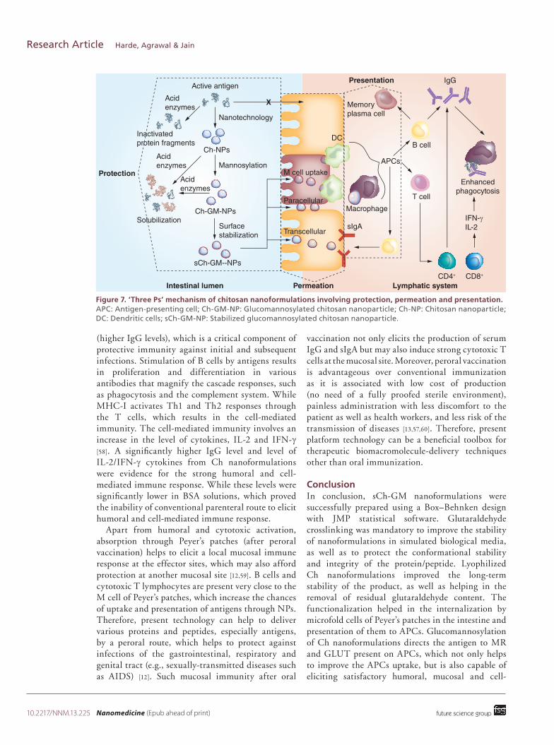
A significant improvement in humoral, mucosal and cellmediated responses was observed in Ch nanoformulations campared with the free antigen. This improved performance could be explained by a hypothesis of the ‘Three Ps’, namely protection, permeation and presentation (Figure 7). First, the entrapped antigen within the NPs was protected from the harsh biological milieu. Second, the absorption of NPs was improved due to specialized permeation mechanisms through microfold cells of Peyer’s patches, as well as partially via the trancellular or paracellular pathways of intestine [53,56]. M cellassisted absorption of numerous nanotechnologybased formulations and further improvement of the bioavailability of different bioactives is also a wellreported fact in literature [16,17,56]. After absorption, presentation and processing of the antigen by the APCs immunostimulate the subsequent biological processes [57]. APCs, such as DCs or macrophages, are key regulators of B and Tcell immunity [58]. Strategic modification of Ch nanoformulations using glucomannosylation could improve recognition and receptormediated internalization. Fusion of endocytosized nanoformulations and endosomes resulted in the formation of immunoproteasomes, which caused degradation of the protein into epitopic peptides. Incorporation of the antigenic peptide pool into MHCI and MHCII, as well as presentation to B and T cells, further activated the cascade response [51]. Presentation of antigens to APCs activates the B cells, which help to induce the humoral immunity
10.2217/NNM.13.225 Nanomedicine (Epub ahead of print) future science group
Research Article Harde, Agrawal & Jain
(higher IgG levels), which is a critical component of protective immunity against initial and subsequent infections. Stimulation of B cells by antigens results in proliferation and differentiation in various antibodies that magnify the cascade responses, such as phagocytosis and the complement system. While MHCI activates Th1 and Th2 responses through the T cells, which results in the cellmediated immunity. The cellmediated immunity involves an increase in the level of cytokines, IL2 and IFNg [58]. A significantly higher IgG level and level of IL2/IFNg cytokines from Ch nanoformulations were evidence for the strong humoral and cellmediated immune response. While these levels were significantly lower in BSA solutions, which proved the inability of conventional parenteral route to elicit humoral and cellmediated immune response.
Apart from humoral and cytotoxic activation, absorption through Peyer’s patches (after peroral vaccination) helps to elicit a local mucosal immune response at the effector sites, which may also afford protection at another mucosal site [12,59]. B cells and cytotoxic T lymphocytes are present very close to the M cell of Peyer’s patches, which increase the chances of uptake and presentation of antigens through NPs. Therefore, present technology can help to deliver various proteins and peptides, especially antigens, by a peroral route, which helps to protect against infections of the gastrointestinal, respiratory and genital tract (e.g., sexuallytransmitted diseases such as AIDS) [12]. Such mucosal immunity after oral
vaccination not only elicits the production of serum IgG and sIgA but may also induce strong cytotoxic T cells at the mucosal site. Moreover, peroral vaccination is advantageous over conventional immunization as it is associated with low cost of production (no need of a fully proofed sterile environment), painless administration with less discomfort to the patient as well as health workers, and less risk of the transmission of diseases [13,57,60]. Therefore, present platform technology can be a beneficial toolbox for therapeutic biomacromoleculedelivery techniques other than oral immunization.
ConclusionIn conclusion, sChGM nanoformulations were successfully prepared using a Box–Behnken design with JMP statistical software. Glutaraldehyde crosslinking was mandatory to improve the stability of nanoformulations in simulated biological media, as well as to protect the conformational stability and integrity of the protein/peptide. Lyophilized Ch nanoformulations improved the longterm stability of the product, as well as helping in the removal of residual glutaraldehyde content. The functionalization helped in the internalization by microfold cells of Peyer’s patches in the intestine and presentation of them to APCs. Glucomannosylation of Ch nanoformulations directs the antigen to MR and GLUT present on APCs, which not only helps to improve the APCs uptake, but is also capable of eliciting satisfactory humoral, mucosal and cell
Protection
Active antigen
Acidenzymes
Acidenzymes
Acidenzymes
SolubilizationSurfacestabilization
Inactivatedprotein fragments
Nanotechnology
Ch-NPs
Mannosylation
Ch-GM-NPs
sCh-GM--NPs
M cell uptake
Paracellular
Transcellular
Intestinal lumen Permeation Lymphatic system
Presentation
Memoryplasma cell
IgG
B cellDC
Macrophage
APCs
T cell
sIgA
Enhancedphagocytosis
CD4+ CD8+
IL-2IFN-γ
X
Figure 7. ‘Three Ps’ mechanism of chitosan nanoformulations involving protection, permeation and presentation. APC: Antigen-presenting cell; Ch-GM-NP: Glucomannosylated chitosan nanoparticle; Ch-NP: Chitosan nanoparticle; DC: Dendritic cells; sCh-GM-NP: Stabilized glucomannosylated chitosan nanoparticle.
10.2217/NNM.13.225future science group
Glucomannosylated chitosan nanoparticles for oral vaccine delivery Research Article
mediated immune responses. The application of present strategies can be extended to therapeutic antigens such as toxoids and conjugate polysaccharide antigens, studies of which are undergoing in our laboratory and will be reported in due course.
Future perspectiveMass immunization of therapeutic antigens is beyond reach due to severe challenges posed by conventional vaccines. The present platform nanotechnology opens the door for mass immunization as it is painless, comfortable and devoid of needlebased infections. It also demonstrates improved immune efficiency due to protection, permeation and presentation of antigens to the cells of the immune system compared with conventional vaccines. However, cytotoxic T lymphocyte assays and fluorescence-activated cell sorting analysis, which are underway in our laboratory, further strengthen the basis of current platform technology. It can also be easily transferred to an industrial level owing to its simplicity, low cost, ease of processing and scaleup. Moreover, the present platform technology harnesses a novel systemic avenue and can also be exploited for oral bioavailability enhancement of candidate proteins and peptides, such as insulin, leptin, octreotide and many more.
AcknowledgementsAuthors are thankful to the Director of the National
Institute of Pharmaceutical Education and Research (Punjab,
India) for providing necessary infrastructure facilities.
Technical assistance provided by Rahul Mahajan is also duly
acknowledged.
Financial & competing interests disclosureThis project work was financially supported by the De-
partment of Biotechnology of the Government of India
(New Delhi, India). The authors have no other relevant af-
filiations or financial involvement with any organization or
entity with a financial interest in or financial conflict with
the subject matter or materials discussed in the manuscript
apart from those disclosed.
No writing assistance was utilized in the production of this
manuscript.
Ethical conduct of research The authors state that they have obtained appropriate
insti tutional review board approval or have followed the
princi ples outlined in the Declaration of Helsinki for all hu-
man or animal experimental investigations. In addition, for
investi gations involving human subjects, informed consent
has been obtained from the participants involved.
Executive summary
Development of platform technology: stable glucomannosylated chitosan nanoparticles• Chitosan nanoparticles were stabilized by tandem crosslinking using tripolyphosphate as a primary crosslinker
followed by surface crosslinking using glutaraldehyde. • Process and formulation variables were optimized using a four-factor, three-level, two-replicate ‘Box–Behnken’
design, and design spaces were selected based on desirability using surface and contour profiler.• The lyophilized formulations helped in the removal of residual glutaraldehyde content, exhibited excellent
stability in simulated biological media and enclosed conformationally stable antigens.Uptake by antigen-presenting cells • Glucomannosylation resulted in a higher uptake than nonglucomannosylated nanoformulations due to the
selective recognition and internalization via ligand–receptor interaction in RAW 264.7 mice.• Glucomannan has dual functionality assisting in receptor-mediated endocytosis via both mannose and glucose
transporter (clathrin-assisted endocytosis) receptor-mediated endocytosis.In vivo evidence of oral immunization• Glucomannan modification resulted in significantly higher systemic (serum IgG), mucosal (secretory IgA), and
cell mediated (IL-2 and IFN- g) immune responses in comparison with unmodified chitosan nanoparticles and conventional vaccines in BALB/c mice.
• The improved performance could be explained by the ‘3 Ps’ mechanism including protection, permeation and presentation of nanoparticles.
Feasibility for mass immunization • Peroral vaccination is advantageous over conventional immunization as it is associated with low cost of
production (no need of a fully proofed sterile environment), painless administration with less discomfort to the patient as well as health workers and less risk of the transfection of diseases.
• The proposed technology systems have industrial significance due to their simplicity, economy, ease of processing and scale-up.
• This approach can also be explored for delivery of therapeutic antigens, such as toxoids, as well as bioavailability enhancement of the proteins and peptides, such as insulin, leptin, octreotide and many more.
10.2217/NNM.13.225 Nanomedicine (Epub ahead of print) future science group
Research Article Harde, Agrawal & Jain
ReferencesPapers of special note have been highlighted as: l of interest l l of considerable interest
1 Pan Y, Li Y, Zhao H et al. Bioadhesive polysaccharide in protein delivery system: chitosan nanoparticles improve the intestinal absorption of insulin in vivo. Int. J. Pharm. 249(1–2), 139–147 (2002).
2 Xu Y, Du Y. Effect of molecular structure of chitosan on protein delivery properties of chitosan nanoparticles. Int. J. Pharm. 250(1), 215–226 (2003).
3 Calvo P, Remuñán López C, Vila Jato JL, Alonso MJ. Novel hydrophilic chitosan polyethylene oxide nanoparticles as protein carriers. J. Appl. Polym. Sci. 63(1), 125–132 (1997).
4 Csaba N, KopingHoggard M, Alonso MJ. Ionically crosslinked chitosan/tripolyphosphate nanoparticles for oligonucleotide and plasmid DNA delivery. Int. J. Pharm. 382(1–2), 205–214 (2009).
5 Katas H, Alpar HO. Development and characterisation of chitosan nanoparticles for siRNA delivery. J. Control. Release 115(2), 216–225 (2006).
6 Bowman K, Leong KW. Chitosan nanoparticles for oral drug and gene delivery. Int. J. Nanomed. 1(2), 117–128 (2006).
7 Lopez Leon T, Carvalho ELS, Seijo B, Ortega Vinuesa JL, Bastos Gonzalez D. Physicochemical characterization of chitosan nanoparticles: electrokinetic and stability behavior. J. Colloid. Interf. Sci. 283(2), 344–351 (2005).
l Article explaining the instability of chitosan nanoparticles.
8 Sung HW, Sonaje K, Liao ZX, Hsu LW, Chuang EY. pHresponsive nanoparticles shelled with chitosan for oral delivery of insulin: from mechanism to therapeutic applications. Acc. Chem. Res. 45(4), 619–629 (2012).
9 Sonaje K, Chen YJ, Chen HL et al. Entericcoated capsules filled with freeze-dried chitosan/poly (gglutamic acid) nanoparticles for oral insulin delivery. Biomaterials 31(12), 3384–3394 (2010).
10 Makhlof A, Tozuka Y, Takeuchi H. Design and evaluation of novel pHsensitive chitosan nanoparticles for oral insulin delivery. Eur. J. Pharm. Sci. 42(5), 445–451 (2011).
11 Sarmento B, Ribeiro A, Veiga F, Sampaio P, Neufeld R, Ferreira D. Alginate/chitosan nanoparticles are effective for oral insulin delivery. Pharm. Res. 24(12), 2198–2206 (2007).
12 Tiwari S, Agrawal GP, Vyas SP. Molecular basis of the mucosal immune system: from fundamental concepts to advances in liposomebased vaccines. Nanomedicine 5(10), 1617–1640 (2010).
13 Fahmy TM, Demento SL, Caplan MJ, Mellman I, Saltzman WM. Design opportunities for actively targeted nanoparticle vaccines. Nanomedicine 3(3), 343–355 (2008).
14 Keler T, Ramakrishna V, Fanger MW. Mannose receptortargeted vaccines. Exp. Opin. Biol. Ther. 4(12), 1953–1962 (2004).
15 Sheng KC, Kalkanidis M, Pouniotis DS et al. Delivery of antigen using a novel mannosylated dendrimer potentiates immunogenicity in vitro and in vivo. Eur. J. Immunol. 38(2), 424–436 (2008).
16 Jain S, Harde H, Indulkar A, Agrawal AK. Improved stability and immunological potential of tetanus toxoid containing surface engineered bilosomes following oral administration. Nanomedicine 10(2), 431–440, (2014).
l l Article describing the extensive oral immunization and ELISA protocols.
17 Jain S, Indulkar A, Harde H, Agrawal AK. Oral mucosal immunization using glucomannosylated bilosomes. J. Biomed. Nanotechnol. 10(6), 932–947 (2014).
18 Du J, Sun R, Zhang S et al. Novel polyelectrolyte carboxymethyl konjac glucomannanchitosan nanoparticles for drug delivery. Macromol. Rapid Comm. 25(9), 954–958 (2004).
l First article reporting on the use of glucomannan as a delivery vehicle.
19 Du J, Sun R, Zhang S, Zhang LF, Xiong CD, Peng YX. Novel polyelectrolyte carboxymethyl konjac glucomannanchitosan nanoparticles for drug delivery. I. Physicochemical characterization of the carboxymethyl konjac glucomannanchitosan nanoparticles. Biopolymers 78(1), 1–8 (2005).
20 FernandezUrrusuno R, Calvo P, RemunanLopez C, VilaJato JL, Alonso MJ. Enhancement of nasal absorption of insulin using chitosan nanoparticles. Pharm. Res. 16(10), 1576–1581 (1999).
21 Ma Z, Yeoh HH, Lim LY. Formulation pH modulates the interaction of insulin with chitosan nanoparticles. J. Pharm. Sci. 91(6), 1396–1404 (2002).
22 Boratynski J, Zal T. Colorimetric micromethods for glutaraldehyde determination by means of phenol and sulfuric acid or phenol and perchloric acid. Anal. Biochem. 184(2), 259–262 (1990).
23 Lyman GW, Johnson RN, Kho B. Gas chromatographic determination of glutaraldehyde. J. Chromatogr. A. 156(2), 285–291 (1978).
24 Jain AK, Swarnakar NK, Godugu C, Singh RP, Jain S. The effect of the oral administration of polymeric nanoparticles on the efficacy and toxicity of tamoxifen. Biomaterials 32(2), 503–515 (2011).
25 Jain S, Rathi VV, Jain AK, Das M, Godugu C. Folatedecorated PLGA nanoparticles as a rationally designed vehicle for the oral delivery of insulin. Nanomedicine 7(9), 1311–1337 (2012).
26 Xie SY, Wang SL, Zhao BK, Han C, Wang M, Zhou WZ. Effect of PLGA as a polymeric emulsifier on preparation of hydrophilic proteinloaded solid lipid nanoparticles. Colloids Surf. B. 67(2), 199–204 (2008).
27 Amidi M, Romeijn SG, Borchard G, Junginger HE, Hennink WE, Jiskoot W. Preparation and characterization of proteinloaded Ntrimethyl chitosan nanoparticles as nasal delivery system. J. Control. Release 111(1–2), 107–116 (2006).
28 Sayin B, Somavarapu S, Li XW et al. MonoNcarboxymethyl chitosan (MCC) and Ntrimethyl chitosan (TMC) nanoparticles for noninvasive vaccine delivery. Int. J. Pharm. 363(1–2), 139–148 (2008).
29 Jelvehgari M, ZakeriMilani P, SiahiShadbad MR et al. Development of pHsensitive insulin nanoparticles using

10.2217/NNM.13.225future science group
Glucomannosylated chitosan nanoparticles for oral vaccine delivery Research Article
eudragit L100–55 and chitosan with different molecular weights. AAPS PharmSciTech. 11(3), 1237–1242 (2010).
30 Zhang N, Li J, Jiang W, Ren C, Xin J, Li K. Effective protection and controlled release of insulin by cationic betacyclodextrin polymers from alginate/chitosan nanoparticles. Int. J. Pharm. 393(1–2), 213–219 (2010).
31 Shukla A, Katare OP, Singh B, Vyas SP. Mcell targeted delivery of recombinant hepatitis B surface antigen using cholera toxin B subunit conjugated bilosomes. Int. J. Pharm. 385(1–2), 47–52 (2010).
32 Roger E, Lagarce F, Benoit JP. The gastrointestinal stability of lipid nanocapsules. Int. J. Pharm. 379(2), 260–265 (2009).
33 Kalaria DR, Sharma G, Beniwal V, Ravi Kumar MNV. Design of biodegradable nanoparticles for oral delivery of doxorubicin: in vivo pharmacokinetics and toxicity studies in rats. Pharm. Res. 26(3), 492–501 (2009).
34 Wiecinski PN, Metz KM, Mangham AN, Jacobson KH, Harriers RJ, Pedersen JA. Gastrointestinal biodurability of engineered nanoparticles: development of an in vitro assay. Nanotoxicology 3(3), 202–214 (2009).
35 Shan X, Liu C, Yuan Y et al. In vitro macrophage uptake and in vivo biodistribution of longcirculation nanoparticles with poly(ethylene-glycol)-modified PLA (BAB type) triblock copolymer. Colloids Surf. B. 72(2), 303–311 (2009).
36 Sarti F, Perera G, Hintzen F et al. In vivo evidence of oral vaccination with PLGA nanoparticles containing the immunostimulant monophosphoryl lipid A. Biomaterials 32(16), 4052–4057 (2011).
37 Garinot M, Fievez V, Pourcelle V et al. PEGylated PLGAbased nanoparticles targeting M cells for oral vaccination. J. Control. Release 120(3), 195–204 (2007).
38 Jain S, Singh P, Mishra V, Vyas S. Mannosylated niosomes as adjuvant–carrier system for oral genetic immunization against hepatitis B. Immunol. Lett. 101(1), 41–49 (2005).
39 Jain S, Sharma RK, Vyas SP. Chitosan nanoparticles encapsulated vesicular systems for oral immunization: preparation, in vitro and in vivo characterization. J. Pharm. Pharmacol. 58(3), 303–310 (2006).
40 Vila A, Sanchez A, Janes K et al. Low molecular weight chitosan nanoparticles as new carriers for nasal vaccine delivery in mice. Eur. J. Pharm. Biopharm. 57(1), 123–131 (2004).
41 Zhu Y, Li X, Chen C et al. Effects of aluminum trichloride on the trace elements and cytokines in the spleen of rats. Food Chem. Toxicol. 50(8), 2911–2915 (2012).
42 Rahman Z, Zidan AS, Khan MA. Nondestructive methods of characterization of risperidone solid lipid nanoparticles. Eur. J. Pharm. Biopharm. 76(1), 127–137 (2010).
43 Jain S, Kumar D, Swarnakar NK, Thanki K. Polyelectrolyte stabilized multilayered liposomes for oral delivery of paclitaxel. Biomaterials 33(28), 6758–6768 (2012).
44 Kreuter J. Peroral administration of nanoparticles. Adv. Drug Del. Rev. 7(1), 71–86 (1991).
45 Harde H, Das M, Jain S. Solid lipid nanoparticles: an oral bioavailability enhancer vehicle. Exp. Opin. Drug Del. 8(11), 1–18 (2011).
l Important review on the factors affecting the absorption of nanoparticles.
46 Gan Q, Wang T, Cochrane C, Mccarron P. Modulation of surface charge, particle size and morphological properties of chitosanTPP nanoparticles intended for gene delivery. Colloids Surf. B. 44(2–3), 65–73 (2005).
47 Ying GQ, Xiong WY, Wang H, Sun Y, Liu HZ. Preparation, water solubility and antioxidant activity of branchedchain chitosan derivatives. Carbohyd. Polym. 83(4), 1787–1796 (2011).
48 Abdelwahed W, Degobert G, Stainmesse S, Fessi H. Freezedrying of nanoparticles: Formulation, process and storage considerations. Adv. Drug Del. Rev. 58(15), 1688–1713 (2006).
l l Excellent summary on the freeze drying of nanoparticles.
49 Van De Weert M, Hennink WE, Jiskoot W. Protein instability in poly (lacticcoglycolic acid) microparticles. Pharm. Res. 17(10), 1159–1167 (2000).
50 Hopkins S, Niedergang F, CorthesyTheulaz I, Kraehenbuhl J. A recombinant Salmonella typhimurium vaccine strain is taken up and survives within murine Peyer’s patch dendritic cells. Cell Microbiol. 2(1), 59–68 (2000).
51 Tacken PJ, De Vries IJM, Torensma R, Figdor CG. Dendriticcell immunotherapy: from ex vivo loading to in vivo targeting. Nat. Rev. Immunol. 7(10), 790–802 (2007).
52 Mcclean S, Prosser E, Meehan E et al. Binding and uptake of biodegradable polydllactide microand nanoparticles in intestinal epithelia. Eur. J. Pharm. Sci. 6(2), 153–163 (1998).
53 Mathiowitz E, Jacob JS, Jong YS et al. Biologically erodable microspheres as potential oral drug delivery systems. Nature 386(6623), 410–414 (1997).
l l Article explaining the specialized permeation mechanism through M cells of the Peyer’s patches in the intestine.
54 Artursson P, Lindmark T, Davis SS, Illum L. Effect of chitosan on the permeability of monolayers of intestinal epithelial cells (Caco2). Pharm. Res. 11(9), 1358–1361 (1994).
55 Dodane V, Amin Khan M, Merwin JR. Effect of chitosan on epithelial permeability and structure. Int. J. Pharm. 182(1), 21–32 (1999).
56 Rieux AD, Pourcelle V, Cani PD, MarchandBrynaert J, Préat V. Targeted nanoparticles with novel nonpeptidic ligands for oral delivery. Adv. Drug Del. Rev. 65(6), 833–844 (2013).
57 Des Rieux A, Fievez V, Garinot M, Schneider YJ, Préat V. Nanoparticles as potential oral delivery systems of proteins and vaccines: a mechanistic approach. J. Control. Release 116(1), 1–27 (2006).
58 Luo Z, Li P, Deng J et al. Cationic polypeptide micellebased antigen delivery system: a simple and robust adjuvant to improve vaccine efficacy. J. Control. Release 170(2), 259–267 (2013).
59 Mackay IR, Rosen FS, Ada G. Vaccines and vaccination. N. Engl. J. Med. 345(14), 1042–1053 (2001).
l l Excellent review about the immunological pathways of vaccinations.
60 Leleux J, Roy K. Micro and nanoparticlebased delivery systems for vaccine immunotherapy: an immunological and materials perspective. Adv. Healthc. Mater. 2(1), 72–94 (2013).