Embed Size (px)
Citation preview

Development of Prostatic Exosomal Protein Lateral Flow Chromatographic Strips for
Detection of Chronic Prostatitis
Jiao Zhang MD, PhD.The Brody School of Medicine East Carolina UniversityGreenville, NC 27834Email: [email protected]
Prostate Cancer June 22-24, 2015 Florida, USA
Background• Chronic prostatitis/chronic pelvic pain syndrome
(CP/CPPS) is a common condition in urological practice,
may be linked to an increased risk of prostate cancer,
yet proper diagnosis and treatments are still lacking.
• Traditional diagnoses are based primarily on the clinical
symptoms and signs, routine urine test, bacterial culture
as well as the expressed prostatic secretion (EPS) indexes.
• There are no gold-standard or objective diagnostic
surrogates for non-bacterial CP/CPPS.
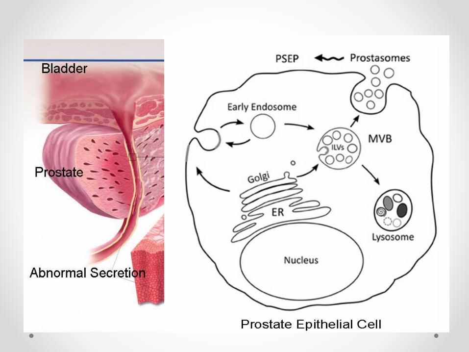
• Recently, PSEP (Prostatic exosomal protein) ELISA
methodology was developed to be used as a
potential indicator of CP/CPPS.
• Multi-center clinical trials performed in China
demonstrated that CP/CPPS patients present
elevated PSEP in the void urine when compared to
that of the healthy men.
• Our current study is designed to further develop a
PSEP point-of-care (POC) platform for large-scale
screening of CP/CPPS patients.
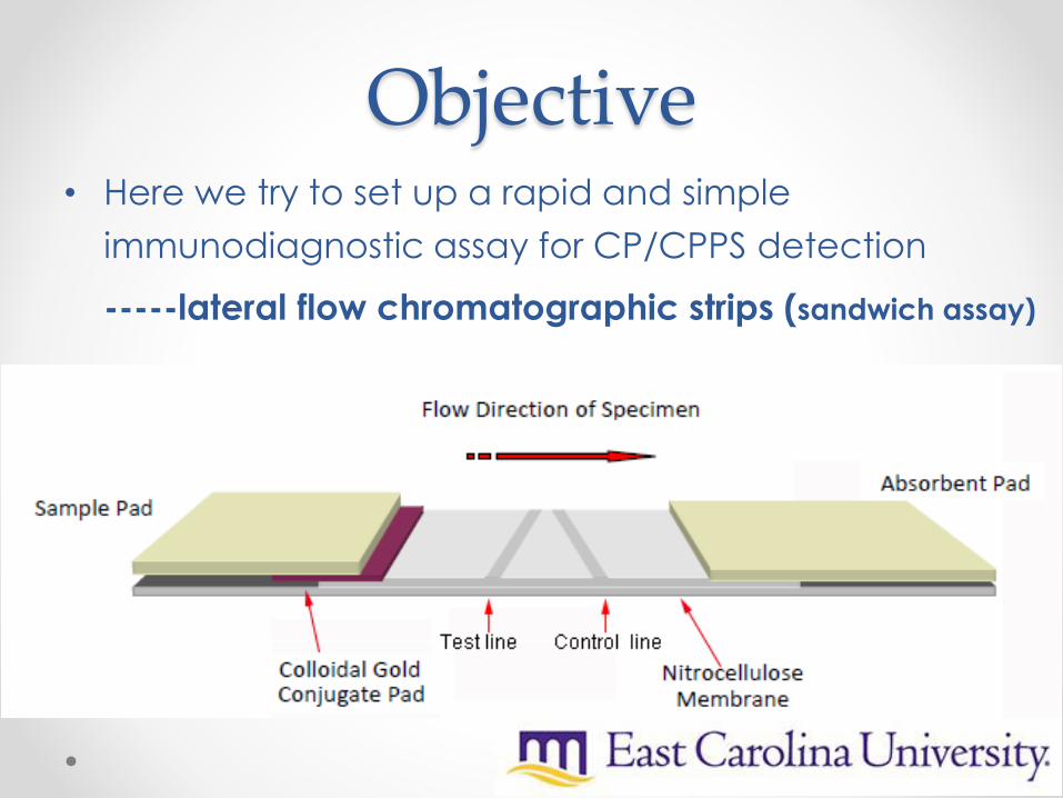
Objective• Here we try to set up a rapid and simple
immunodiagnostic assay for CP/CPPS detection
-----lateral flow chromatographic strips (sandwich assay)
Research Design• PSEP Antibody production
• Development of Lateral-Flow Test Device
o Preparation of Colloidal Gold Particles
o Preparation of Colloidal Gold Labeled mAb
o Preparation of Nitrocellulose Capture Membranes
o Test Procedure and Principle
• Determination of Performance
o Sensitivity of the Test Strip
o Detection of Urine Sample
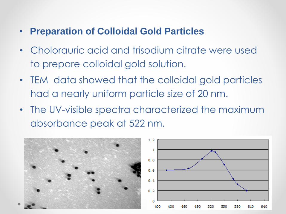
• Cholorauric acid and trisodium citrate were used
to prepare colloidal gold solution.
• TEM data showed that the colloidal gold particles
had a nearly uniform particle size of 20 nm.
• The UV-visible spectra characterized the maximum
absorbance peak at 522 nm.
• Preparation of Colloidal Gold Particles
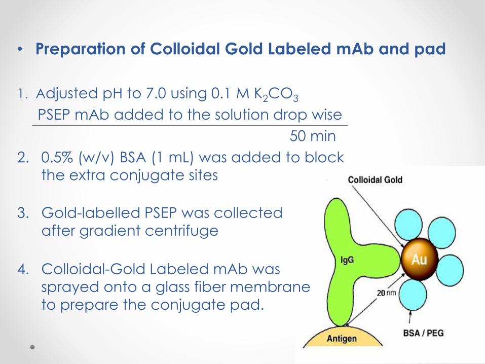
1. Adjusted pH to 7.0 using 0.1 M K2CO3
PSEP mAb added to the solution drop wise
50 min
2. 0.5% (w/v) BSA (1 mL) was added to block
the extra conjugate sites
3. Gold-labelled PSEP was collected
after gradient centrifuge
4. Colloidal-Gold Labeled mAb was
sprayed onto a glass fiber membrane
to prepare the conjugate pad.
• Preparation of Colloidal Gold Labeled mAb and pad
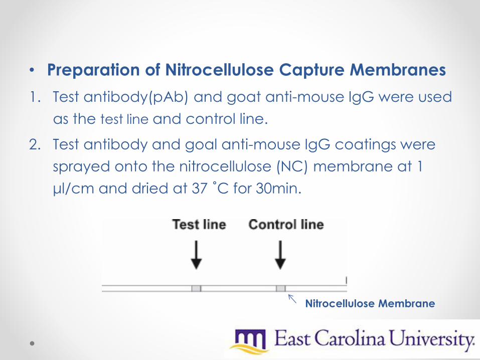
• Preparation of Nitrocellulose Capture Membranes
1. Test antibody(pAb) and goat anti-mouse IgG were used
as the test line and control line.
2. Test antibody and goal anti-mouse IgG coatings were
sprayed onto the nitrocellulose (NC) membrane at 1
μl/cm and dried at 37 ˚C for 30min.
Nitrocellulose Membrane
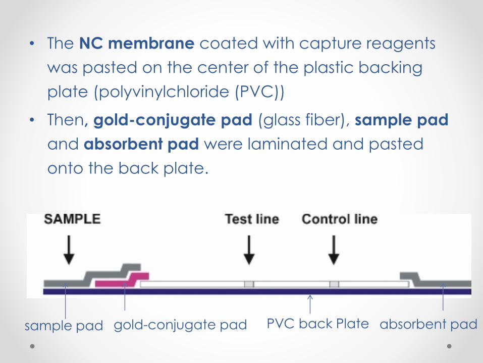
• The NC membrane coated with capture reagents
was pasted on the center of the plastic backing
plate (polyvinylchloride (PVC))
• Then, gold-conjugate pad (glass fiber), sample pad
and absorbent pad were laminated and pasted
onto the back plate.
PVC back Plategold-conjugate pad sample pad absorbent pad
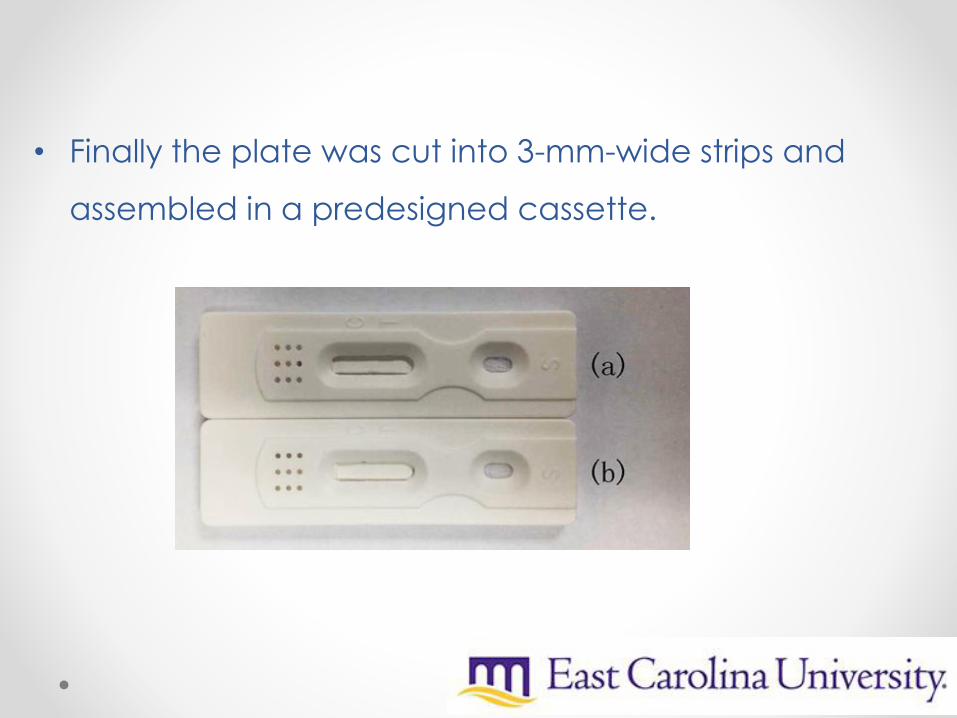
• Finally the plate was cut into 3-mm-wide strips and
assembled in a predesigned cassette.
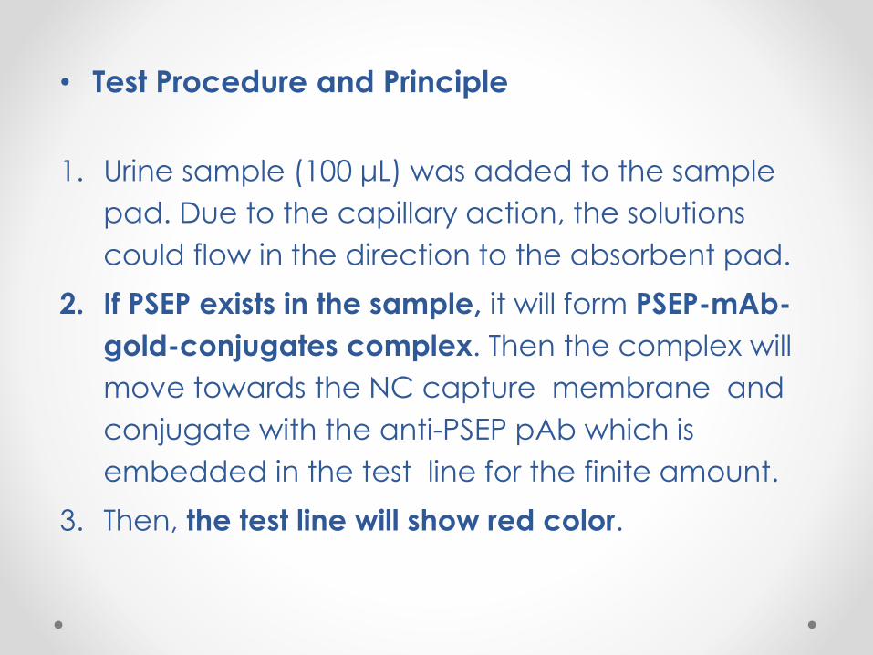
• Test Procedure and Principle
1. Urine sample (100 μL) was added to the sample
pad. Due to the capillary action, the solutions
could flow in the direction to the absorbent pad.
2. If PSEP exists in the sample, it will form PSEP-mAb-
gold-conjugates complex. Then the complex will
move towards the NC capture membrane and
conjugate with the anti-PSEP pAb which is
embedded in the test line for the finite amount.
3. Then, the test line will show red color.
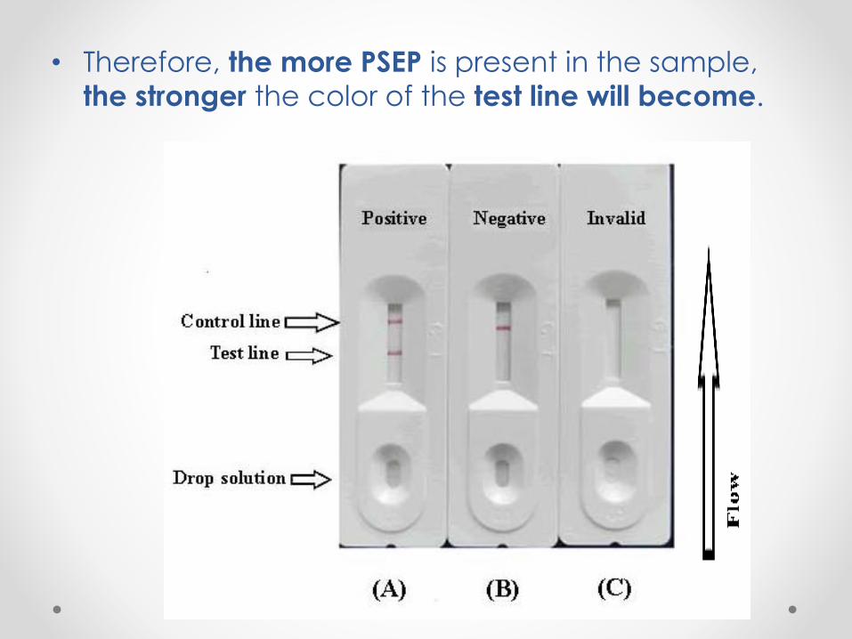
• Therefore, the more PSEP is present in the sample,
the stronger the color of the test line will become.
• Determination of Performance
• Sensitivity of the Test Strips
1. PSEP was diluted at concentrations of 0, 0.2, 0.5 1,
2, 5, and 10 ng/mL in 0.01 M PBS (pH 7.4).
2. The sensitivity of the test strip was determined by
testing PSEP reference spiked samples.
3. Three minutes later, the lower detection limit (LDL)
with naked eyes was defined at the certain
amount of PSEP concentration which produce a
clearly visible test line on the test device compare
with the blank sample(0 ng/mL ).
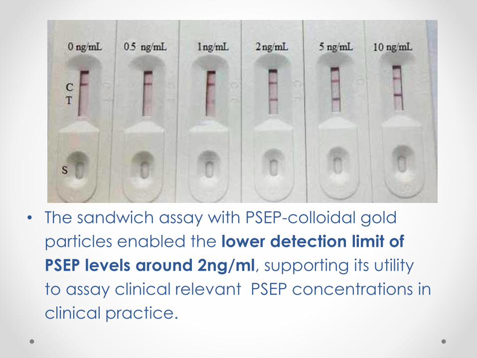
• The sandwich assay with PSEP-colloidal gold
particles enabled the lower detection limit of
PSEP levels around 2ng/ml, supporting its utility
to assay clinical relevant PSEP concentrations in
clinical practice.
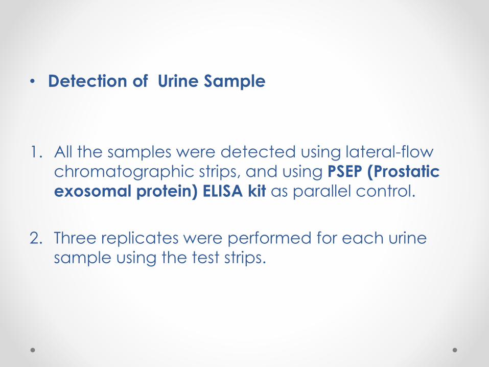
• Detection of Urine Sample
1. All the samples were detected using lateral-flow
chromatographic strips, and using PSEP (Prostatic
exosomal protein) ELISA kit as parallel control.
2. Three replicates were performed for each urine
sample using the test strips.
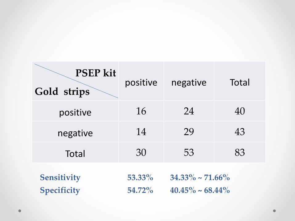
PSEP kit
Gold strips positive negative Total
positive 16 24 40
negative 14 29 43
Total 30 53 83
Sensitivity 53.33% 34.33% ~ 71.66%
Specificity 54.72% 40.45% ~ 68.44%
1. The test device enabled the detection
level of PSEP as low as 2ng/ml.
2. These lateral flow chromatographic
strips showed potential value in assisting
CP/CPPS POC diagnosis.
Above result indicated that :
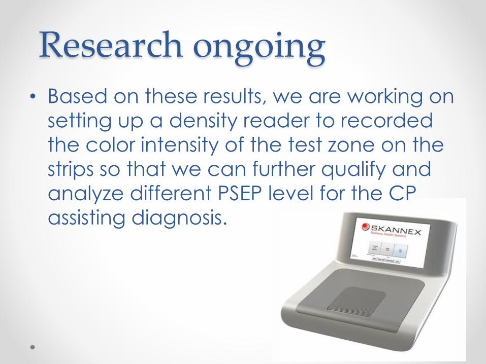
Research ongoing
• Based on these results, we are working on
setting up a density reader to recorded
the color intensity of the test zone on the
strips so that we can further qualify and
analyze different PSEP level for the CP
assisting diagnosis.