Embed Size (px)
Citation preview
Development of novel antiviral
drugs to combat human pathogenic
arenaviruses
Thesis submitted for the degree Candidata pharmaciae to the
Department of Microbiology, School of Pharmacy,
Faculty of Mathematics and Natural Sciences, University of Oslo
2006
Anette Tølløfsrud Gundersen
The Scripps Research Institute, Department of Immunoloy
10550 North Torrey Pines Road
La Jolla, California 92037
TABLE OF CONTENTS ACKNOWLEDGEMENTS…………………………………………………………………………. iii ABSTRACT………………………………………………………………………………………….. ix A. INTRODUCTION…………………………………………………............................................... 1
A.1 Arenaviruses………………………………………………………………………………….. 1 A.1.1 Classification of arenaviruses………………………………………………………….. 1 A.1.2 Structure, genome and proteins of arenaviruses………………………………………... 2 A.1.3 The GP of arenaviruses mediate host cell attachment and entry……………………….. 4 A.1.4 Alpha-dystroglycan is the cellular receptor for arenaviruses…………………………... 5
A.2 Lassa fever represents a severe threat for human health and a major humanitarian problem... 6 A.3 Current therapy against arenaviruses…………………………………………………………. 7 A.4 Targeting arenavirus receptor binding and entry is a promising new anti-viral strategy…….. 9 A.5 Small molecule inhibitors of protein interactions derived from combinatorial chemical
libraries……………………………………………………………………………………….. 10 B. MATERIALS AND METHODS………………………………………………………………… 13
B.1 Reagents and antibodies……………………………………………………………………… 13 B.2 Cell lines……………………………………………………………………………………… 13 B.3 Virus strains, purification and quantification………………………………………………… 13 B.4 Detection of LCMVGP in ELISA……………………………………………………………. 14 B.5 Immunoblotting………………………………………………………………………………. 14 B.6 Molecular biology techniques………………………………………………………………… 15
B.6.1 Polymerase chain reaction (PCR)………………………………………………………. 15 B.6.2 Phenol chloroform extraction…………………………………………………………... 15 B.6.3 Agarose gel electrophoresis…………………………………………………………….. 16 B.6.4 Gel extraction…………………………………………………………………………… 16 B.6.5 Treatment of DNA fragments with calf intestinal phosphatase (CIP)………………….. 17 B.6.6 Ligation of DNA fragments with T4 DNA ligase……………………………………… 17 B.6.7 Transformation of E.coli………………………………………………………………………... 17 B.6.8 Plasmid DNA purification using the QIAprep spin miniprep kit………………………. 18
B.7 Generation of pseudotyped retroviral vectors………………………………………………… 18 B.8 Infection of human cells with retroviral pseudotypes………………………………………… 19 B.9 Steady-Glo® luciferase assay………………………………………………………………… 19 B.10 Screening of chemical libraries: inhibition of the infection with retroviral pseudotypes by
compounds from combinatorial chemical libraries…………………………………………... 20 B.11 Target-specificity of compound mixtures…………………………………………………… 21 B.12 Determination of the dose-response characteristics of selected candidate compounds……... 21 B.13 Blocking of pseudotype infection with sulfated polysaccarides…………………………….. 22 B.14 Intracellular FACS staining for LCMV-NP using mAb 113………………………………... 23 B.15 Detection of LCMV-NP by immunofluorescence staining…………………………………. 23
C. RESULTS…………………………………………………………………………………………. 25 C.1 Discovery of novel small molecule inhibitors of Lassa fever virus (LFV) infection from
combinatorial chemical libraries……………………………………………………………... 25 C.1.1 Production and characterization of retroviral pseudotypes……………………………... 25 C.1.2 Screening of combinatorial chemical libraries…………………………………………. 28 C.1.3 Determination of the target specificities of candidate compounds……………………... 29 C.1.4 Validation of candidate compounds in different human and primate cells…………….. 30 C.1.5 Dose-response curves of candidate compounds………………………………………... 31
i
C.1.6 Determination of the activity of candidate compounds against other human pathogenic arenaviruses……………………………………………………………………………… 31
C.2 Discovery of novel small molecule inhibitors of Lassa fever virus (LFV) infection from natural product libraries………………………………………………………………………. 35
C.2.1 Screening of natural product libraries…………………………………………………... 35 C.2.2 Deconvolution of compound mixtures…………………………………………………. 36
C.3 Evaluation of polyaninonic compounds as anti-arenaviral drugs…………………………….. 38 C.3.1 Selection of candidate polymers: heparin, HS, DS and fucoidan………………………. 38 C.3.2 Test of heparin, HS, DS, fucoidan and polymer X for anti-viral activity against
arenaviruses……………………………………………………………………………… 39 C.3.3 What is the role of HS and glycosaminoglycans in arenavirus infection?....................... 41 C.3.4 Evaluation of polymer X as a drug against LCMV…………………………………….. 43
C.4 Production of recombinant Lassa fever virus glycoprotein…………………………………... 47 C.4.1 Design of the LFVGP-foldon fusion protein…………………………………………… 49 C.4.2 Expression and characterization of the GP1-foldon fusion proteins…………………… 50
D. DISCUSSION……………………………………………………………………………………... 51 D.1 Discovery of novel small molecule inhibitors of Lassa fever virus (LFV) infection from
combinatorial chemical libraries and natural products libraries……………………………... 51 D.2 Evaluation of polyanionic compounds as anti-arenaviral drugs……………………………… 53 D.3 Production of recombinant Lassa fever virus glycoprotein…………………………………... 54 D.4 Outlook……………………………………………………………………………………….. 55
D.4.1 Further validation of candidate compounds……………………………………………. 55 D.4.2 Determination of the mechanism of action of candidate drug………………………….. 56 D.4.3 Extension of small molecule screening to other drug targets and other emerging
pathogens………………………………………………………………………………… 56 E. REFERENCES……………………………………………………………………………………. 58
ii
ACKNOWLEDGEMENTS
I would like to thank the following people:
Stefan Kunz, my tutor at TSRI, for his enthusiasm regarding my project, his guidance and expertise,
and for all his help when making the figures and writing my thesis.
Professor Tor Gjøen, the supervisor of my master thesis at the University of Oslo, for good support and
for providing me the opportunity to come to The Scripps Research Institute.
Dr. Michael B. A. Oldstone, the head of the Viral-Immunobiology laboratory at TSRI for his generous
support, Ms. Jillian M. Rojek (TSRI) for technical and scientific advice, Dr. Dale Boger and Dr. Jin
Wei (TSRI) for the chemical libraries, Dr. Kevin P. Campbell (Howard Hughes Medical Institute,
University of Iowa), Dr. Juan-Carlos de la Torre (TSRI), and Dr. Michael Buchmeier (TSRI) for
materials. The retroviral construct pLZRs-Luc-gfp was kindly provided by Dr. Gary Nabel.
For this work, I received a stipend from “S.G. Sønnelands Foundation” and a loan from “Statens
Lånekasse”. My laboratory expenses at TSRI were covered by US Public Health grant AI55540
(M.B.A.O., S.K.), and grant 1U54 AI065359 of the Pacific Southwest Regional center of Excellence
for Emerging Infectious Disease (S.K.).
iii
ABSTRACT
Arenaviruses merit significant attention both as powerful models to study viral pathogenesis and as
important human pathogens. Lymphocytic choriomeningitis virus (LCMV) infection of its natural host,
the mouse, represents a powerful experimental system that provided novel concepts in immunology
and virology that have been extended to other viruses, bacteria, and parasites. With over 200, 000
infections per year and several thousand deaths, Lassa virus (LFV) is by far the most important among
the human pathogenic arenaviruses and represents a severe threat for human health. There are no
licensed arenavirus vaccines and the current therapies are not optimal. Therefore, it is important to
develop better antiviral drugs to combat the threats of arenavirus infections.
A fundamental reason for the high mortality of infections with human pathogenic arenaviruses is a
failure of the host’s immune system to control viral replication, leading to an unchecked viremia
associated with hemorrhagic disease. Since rapid dissemination of the virus critically depends on
attachment and entry into host cells, drugs targeting these steps will give the host’s immune system a
wider window of opportunity for the generation of an efficient anti-viral immune response. The goal of
my research was therefore the development of novel anti-viral drugs that are able to block these initial
steps of infection. In a first approach to identify such “gatekeeper” drugs I performed high-throughput
small molecule screening using combinatorial chemical libraries, which represent a powerful
technology for discovery of specific inhibitors of receptor-ligand interactions. In a collaborative effort
with the laboratory of Dr. Dale Boger (Department of Chemistry, Scripps) I used a high-throughput
screening assay for inhibitors of LFV infection using retroviral pseudotypes containing LFVGP in their
envelope and a luciferase reporter gene. My screening identified several small molecule compounds
that show specific blocking of LFV infection in several human and primate cell types with IC50 values
in the range of 1-10 µM. Some of the candidates were also active against retroviral pseudotypes the
South American hemorrhagic fever viruses Junin, Machupo, and Guanarito.
In a second approach, I evaluated the activity of anionic polymers like heparin, dextrane sulfate, and
fucoidan, previously shown to have anti-viral effects, against human pathogenic arenaviruses. In
contrast to published data, I found no significant activity of heparin and heparan sulfate against
arenavirus infection and could demonstrate that heparan sulfate and other glycosaminoglycans are
generally not involved in arenavirus infection. Dextrane sulfate and fucoidan indeed showed some
anti-viral activity, however, they were far less efficient than claimed by other researchers. This
motivated me to start to evaluate novel polymer drugs for activity against arenaviruses.
ix
Introduction
A. INTRODUCTION
Viral infectious diseases have threatened humankind throughout our existence. Although advances in
medical science have changed the demographics of mortality with an increase in life expectancy to
more than seventy-five years in the developed world, viral infections are still rampant in
underdeveloped countries with life expectancies of less than 40 years in the poorest nations. Beyond
contributing to mortality, viral infections also leave affected individuals often physically and mentally
disabled, exacerbating human suffering and the socio-economic impact caused by these diseases.
A.1 Arenaviruses
A.1.1 Classification of arenaviruses
Two major groups of arenaviruses are currently recognized: the Old World arenaviruses with
lymphocytic choriomeningitis virus (LCMV) and Lassa fever virus (LFV) as representatives and the
larger group of the New World arenaviruses, which are divided into three major Clades, A, B, and C
(Buchmeier et al., 2001). LCMV infection of its natural host, the mouse, represents a powerful
experimental system that provided novel concepts in immunology and virology that have been
extended to other viruses, bacteria, and parasites (Buchmeier et al., 2001; Oldstone, 2002). LCMV is
also a prevalent human pathogen and congenital infections of the central nervous system by LCMV are
considered an important problem in human pediatric medicine.
Arenaviruses are also the causative agents of several severe hemorrhagic fevers (HF) with high
mortality in humans (Buchmeier et al., 2001; Geisbert and Jahrling, 2004). Lassa fever is the second
most important human viral HF after Dengue and affects an estimated 180 million individuals living in
its endemic regions in Western Africa. With over 200, 000 infections per year and several thousand
deaths, LFV represents a severe threat for human health and a major humanitarian problem
(McCormick and Fisher-Hoch, 2002). There is currently neither an efficient cure nor an efficacious
vaccine for this disease and survivors are often left with substantial neurological impairment. Among
the New World arenaviruses, Junin virus causes Argentine HF, and is a serious public health problem
in Argentina. Machupo virus causes Bolivian HF, and Guanarito and Sabia virus have emerged as
etiological agents of severe HF in Venezuela and Brazil, respectively. Infections with these
arenaviruses in humans result in high mortality of 15 to 30%. Apart from the severe humanitarian
1
Introduction
burden caused in endemic regions, air travel regularly imports arenavirus HF cases into metropolitan
areas around the globe, placing local populations at risk.
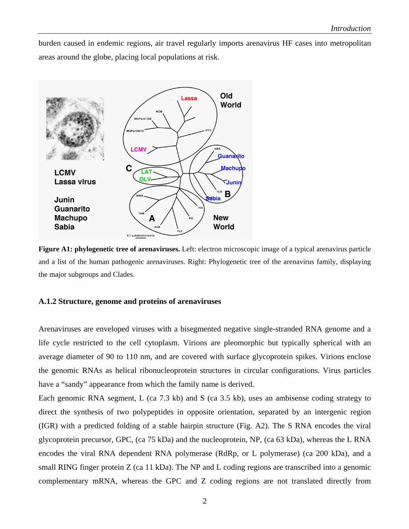
Figure A1: phylogenetic tree of arenaviruses. Left: electron microscopic image of a typical arenavirus particle
and a list of the human pathogenic arenaviruses. Right: Phylogenetic tree of the arenavirus family, displaying
the major subgroups and Clades.
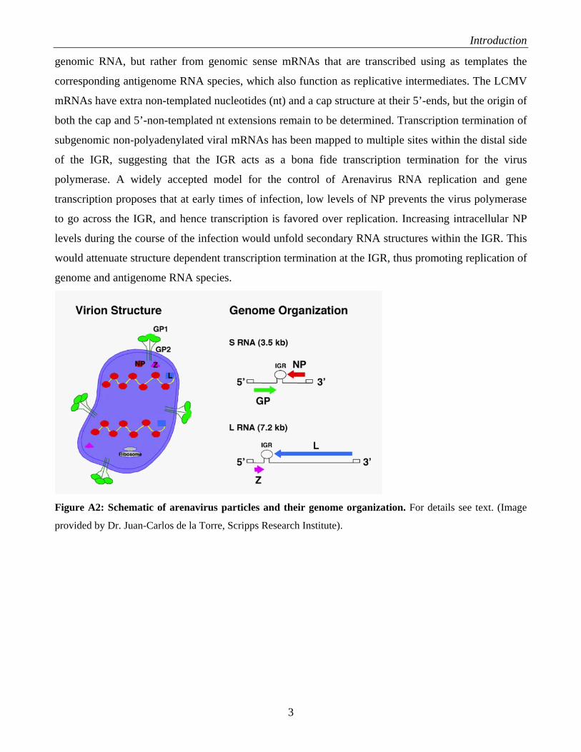
A.1.2 Structure, genome and proteins of arenaviruses
Arenaviruses are enveloped viruses with a bisegmented negative single-stranded RNA genome and a
life cycle restricted to the cell cytoplasm. Virions are pleomorphic but typically spherical with an
average diameter of 90 to 110 nm, and are covered with surface glycoprotein spikes. Virions enclose
the genomic RNAs as helical ribonucleoprotein structures in circular configurations. Virus particles
have a “sandy” appearance from which the family name is derived.
Each genomic RNA segment, L (ca 7.3 kb) and S (ca 3.5 kb), uses an ambisense coding strategy to
direct the synthesis of two polypeptides in opposite orientation, separated by an intergenic region
(IGR) with a predicted folding of a stable hairpin structure (Fig. A2). The S RNA encodes the viral
glycoprotein precursor, GPC, (ca 75 kDa) and the nucleoprotein, NP, (ca 63 kDa), whereas the L RNA
encodes the viral RNA dependent RNA polymerase (RdRp, or L polymerase) (ca 200 kDa), and a
small RING finger protein Z (ca 11 kDa). The NP and L coding regions are transcribed into a genomic
complementary mRNA, whereas the GPC and Z coding regions are not translated directly from
2
Introduction
genomic RNA, but rather from genomic sense mRNAs that are transcribed using as templates the
corresponding antigenome RNA species, which also function as replicative intermediates. The LCMV
mRNAs have extra non-templated nucleotides (nt) and a cap structure at their 5’-ends, but the origin of
both the cap and 5’-non-templated nt extensions remain to be determined. Transcription termination of
subgenomic non-polyadenylated viral mRNAs has been mapped to multiple sites within the distal side
of the IGR, suggesting that the IGR acts as a bona fide transcription termination for the virus
polymerase. A widely accepted model for the control of Arenavirus RNA replication and gene
transcription proposes that at early times of infection, low levels of NP prevents the virus polymerase
to go across the IGR, and hence transcription is favored over replication. Increasing intracellular NP
levels during the course of the infection would unfold secondary RNA structures within the IGR. This
would attenuate structure dependent transcription termination at the IGR, thus promoting replication of
genome and antigenome RNA species.
Figure A2: Schematic of arenavirus particles and their genome organization. For details see text. (Image
provided by Dr. Juan-Carlos de la Torre, Scripps Research Institute).
3
Introduction
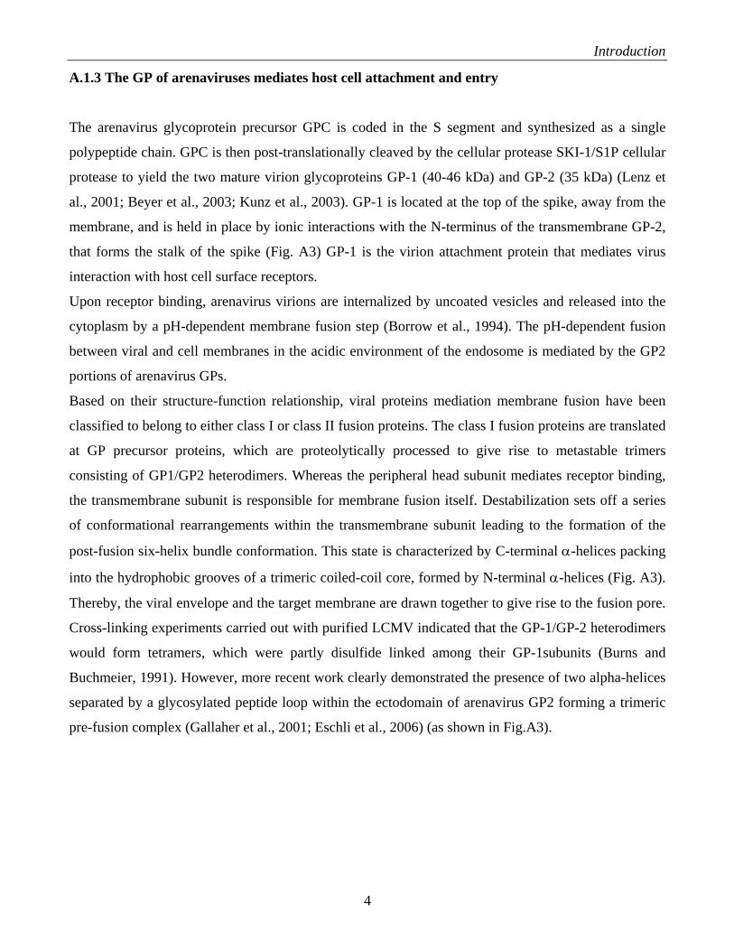
A.1.3 The GP of arenaviruses mediates host cell attachment and entry
The arenavirus glycoprotein precursor GPC is coded in the S segment and synthesized as a single
polypeptide chain. GPC is then post-translationally cleaved by the cellular protease SKI-1/S1P cellular
protease to yield the two mature virion glycoproteins GP-1 (40-46 kDa) and GP-2 (35 kDa) (Lenz et
al., 2001; Beyer et al., 2003; Kunz et al., 2003). GP-1 is located at the top of the spike, away from the
membrane, and is held in place by ionic interactions with the N-terminus of the transmembrane GP-2,
that forms the stalk of the spike (Fig. A3) GP-1 is the virion attachment protein that mediates virus
interaction with host cell surface receptors.
Upon receptor binding, arenavirus virions are internalized by uncoated vesicles and released into the
cytoplasm by a pH-dependent membrane fusion step (Borrow et al., 1994). The pH-dependent fusion
between viral and cell membranes in the acidic environment of the endosome is mediated by the GP2
portions of arenavirus GPs.
Based on their structure-function relationship, viral proteins mediation membrane fusion have been
classified to belong to either class I or class II fusion proteins. The class I fusion proteins are translated
at GP precursor proteins, which are proteolytically processed to give rise to metastable trimers
consisting of GP1/GP2 heterodimers. Whereas the peripheral head subunit mediates receptor binding,
the transmembrane subunit is responsible for membrane fusion itself. Destabilization sets off a series
of conformational rearrangements within the transmembrane subunit leading to the formation of the
post-fusion six-helix bundle conformation. This state is characterized by C-terminal α-helices packing
into the hydrophobic grooves of a trimeric coiled-coil core, formed by N-terminal α-helices (Fig. A3).
Thereby, the viral envelope and the target membrane are drawn together to give rise to the fusion pore.
Cross-linking experiments carried out with purified LCMV indicated that the GP-1/GP-2 heterodimers
would form tetramers, which were partly disulfide linked among their GP-1subunits (Burns and
Buchmeier, 1991). However, more recent work clearly demonstrated the presence of two alpha-helices
separated by a glycosylated peptide loop within the ectodomain of arenavirus GP2 forming a trimeric
pre-fusion complex (Gallaher et al., 2001; Eschli et al., 2006) (as shown in Fig.A3).
4
Introduction
Figure A3: Proposed class I model of the arenavirus GP. (A) Arenavirus GP in its hypothetical pre-fusion
state. GP-1 forms the globular head subunit, whereas GP-2 forms the membrane fusion-mediating subunit. GP-1
is covered by a dense carbohydrate shield, with the exception of the receptor binding site, depicted in red. (B)
GP spike in its hypothetical low-energy post-fusion state, after formation of the viral fusion pore. The membrane
of the infected host cell is colored in yellow and the viral membrane in light gray. The GP-2 subunit is shown in
its predicted six-helix bundle conformation; thus, the α-helix 2 packs in an anti-parallel fashion into the grooves
of the N-terminal coiled-coil core. Taken from Eschli et al., (2006).
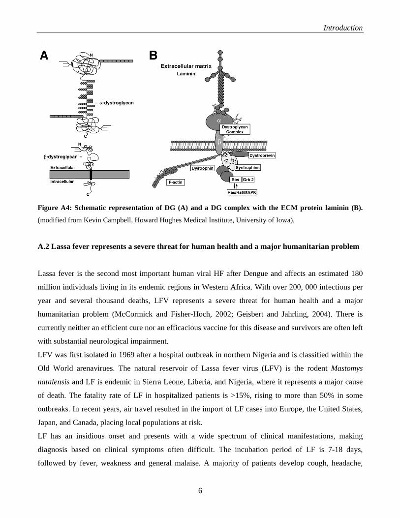
A.1.4 Alpha-dystroglycan is the cellular receptor for arenaviruses
The first cellular receptor for LFV, LCMV and Clade C New World arenaviruses is α-dystroglycan (α-
DG) (Cao et al., 1998; Spiropoulou et al., 2002), the extracellular part of dystroglycan (Fig. A4). DG
was initially identified as the central component of the dystrophin-glycoprotein complex (DGC) in the
membrane of skeletal muscle (Barresi and Campbell, 2006). In vertebrates, DG is encoded as a single
protein, which is cleaved into α-DG, a peripheral protein, and β-DG, a membrane protein (Fig. A4A).
DG is expressed in most developing and adult tissues and provides a molecular link between the
extracellular matrix (ECM) and the actin-based cytoskeleton (Fig. A4B).
5
Introduction
Figure A4: Schematic representation of DG (A) and a DG complex with the ECM protein laminin (B).
(modified from Kevin Campbell, Howard Hughes Medical Institute, University of Iowa).
A.2 Lassa fever represents a severe threat for human health and a major humanitarian problem
Lassa fever is the second most important human viral HF after Dengue and affects an estimated 180
million individuals living in its endemic regions in Western Africa. With over 200, 000 infections per
year and several thousand deaths, LFV represents a severe threat for human health and a major
humanitarian problem (McCormick and Fisher-Hoch, 2002; Geisbert and Jahrling, 2004). There is
currently neither an efficient cure nor an efficacious vaccine for this disease and survivors are often left
with substantial neurological impairment.
LFV was first isolated in 1969 after a hospital outbreak in northern Nigeria and is classified within the
Old World arenavirues. The natural reservoir of Lassa fever virus (LFV) is the rodent Mastomys
natalensis and LF is endemic in Sierra Leone, Liberia, and Nigeria, where it represents a major cause
of death. The fatality rate of LF in hospitalized patients is >15%, rising to more than 50% in some
outbreaks. In recent years, air travel resulted in the import of LF cases into Europe, the United States,
Japan, and Canada, placing local populations at risk.
LF has an insidious onset and presents with a wide spectrum of clinical manifestations, making
diagnosis based on clinical symptoms often difficult. The incubation period of LF is 7-18 days,
followed by fever, weakness and general malaise. A majority of patients develop cough, headache,
6
Introduction
sore throat, and gastrointestinal manifestations. Signs of increased vascular permeability such as facial
edema and pleural effusions indicate a poor prognosis. In lethal cases, deterioration is rapid with
progressive signs and symptoms of pulmonary edema, respiratory distress, and shock, accompanied by
bleeding from mucosal surfaces. A highly predictive factor for disease outcome is the extent of
viremia. Patients developing a fatal LFV infection have higher viral loads and are unable to limit
viremia while survivors have lower viral load and clear the virus.
The host’s control of LFV replication is primarily mediated by cellular immunity. Antibodies play a
modest role in acute LFV infection as patients can recover in the absence of a neutralizing antibody
response. Like in other human viral HF, hallmark of fatal LF in humans is a marked
immunosuppression (Geisbert and Jahrling, 2004).
Despite recent progress in the development of a LF vaccine (Fisher-Hoch, 2004; Geisbert et al., 2005),
to my knowledge, to date no human vaccine trials have taken place. The only drug currently available
for treatment of acute LF is the nucleoside analogue ribavirin, which significantly reduces mortality
(McCormick et al., 1986). Since ribavirin does not neutralize free virus, it is less efficient in patients
with high virus loads, especially late in infection. The strong predictive value of virus concentration in
blood for a disastrous disease outcome in LF indicates a close competition between virus spread and
the anti-viral immune response. Rapid viral dissemination critically depends on efficient
attachment of the virus to receptor molecules on target cells and subsequent entry. Drugs able to
block virus-receptor binding and/or entry will therefore give the host’s immune system an
advantage by providing a wider window of opportunity for the generation of an efficient anti-
viral immune response. The development of novel anti-viral drugs targeting these early steps of
infection appears therefore as a promising approach for better treatment of Lassa fever in
humans.
A.3 Current therapy against arenaviruses
The only licensed drug against arenaviruses, which is currently available is the guanosine analogue
ribavirin (1-β-D-ribofuranosyl-1,2,4-triazole-3-carboxamide) (Parker, 2005). In vitro and in vivo
studies have documented the prophylactic and therapeutic value of ribavirin against several
arenaviruses. Ribavirin has been shown to reduce significantly both morbidity and mortality in humans
associated with LFV infection (McCormick et al., 1986). As with other nucleoside analogues, ribavirin
is a prodrug that needs to be converted to nucleotide metabolites to exert its antiviral activity. Ribavirin
7
Introduction
is phosphorylated by adenosine kinase to ribavirin monophosphate (RMP), which is then converted
into its triphosphate form (RTP) by the successive action of mono-and di-phosphate kinases. Ribavirin
can exerts its antiviral activity by a variety of mechanisms. RMP is a strong inhibitor of IMP
dehydrogenase, which causes a large reduction in the intracellular levels of GTP and dGTP that can
affect viral RNA synthesis. On the other hand RTP can interfere with the capping of viral mRNA, and
with the function of the virus RNA-dependent RNA polymerase (RdRp). Recent evidence indicates
that RTP can be efficiently used as substrate by the RdRp of some riboviruses, which results in C to U
and G to A transitions. This mutagenic activity of RTP has been linked to its antiviral activity via
lethal mutagenesis. The precise mechanisms by which ribavirin interferes with arenavirus
multiplication remain to be determined. One of the problems associated with the use of ribavirin is that
in a high percentage of cases (> 40%) treated individuals develop haemolytic anemia. Likewise,
ribavirin has been associated with congenital disorders and hence it should be not used with pregnant
women. In addition, oral ribavirin appears to be significantly less effective than the one administered
intravenously, which pose some additional logistic complications for its use in regions with limited
clinical infrastructure.
Several ribavirin related inhibitors of IMP dehydrogenase, including ribamidine (1-beta-D-
ribofuranosyl-1,2,4-tiazole-3-carboxamide) (Andrei and de Clercq, 1993), as well as acyclic and
carbocyclic adenosine analogue inhibitors of the S-adenosylhomocysteine (SAH) hydrolase (Andrei
and de Clercq, 1990) have been show to have also anti-arenaviruses. Likewise, phenotiazines
compounds (Candurra et al., 1996), myristic acid (Cordo et al., 1999), several disulfide-based
compounds (Garcia et al., 2002), and brassinosteroids (Castilla et al., 2005) have been reported to have
activity against several arenaviruses. However, apart from ribavirin, none of these compounds
have been tested in human trials and their efficacy in vivo in currently unknown.
Another class of compounds that showed significant anti-viral activity against arenaviruses are sulfated
polysaccharides like dextrane sulfate, fucoidan, heparan sulfate, and heparin (Andrei and de Clercq,
1990). These anionic polymers have anti-viral activity against a number of human pathogenic viruses,
including HIV (Kilby and Eron, 2003) and have been studied extensively in the past decade. Based on
their large size and charged nature, these polymer drugs are thought to function as inhibitors of cell
attachment and entry. In case of HIV, a direct interaction of the polymer drugs with specific sites of the
viral GP has been demonstrated recently.
In case of arenaviruses, the therapeutic anti-viral potential of these drugs and their mechanism of
action are still controversial. One goal of the present study is therefore a systematic analysis of the
anti-viral activity against human pathogenic arenaviruses of candidate anionic polymer drugs and the
8
Introduction
elucidation of their mechanism of action using a panel of state-of-the art tools and assays available in
the laboratory.
A.4 Targeting arenavirus receptor binding and entry is a promising new anti-viral strategy
The strong predictive value of virus concentration in blood for a disastrous disease outcome in human
arenavirus infection indicates a close competition between virus spread and the anti-viral immune
response. Rapid viral dissemination critically depends on efficient attachment of the virus to receptor
molecules on target cells and subsequent entry. Drugs able to block virus-receptor binding and/or entry
will therefore give the host’s immune system an advantage by providing a wider window of
opportunity for the generation of an efficient anti-viral immune response. The development of novel
anti-viral drugs targeting these early steps of infection appears therefore as a promising approach for
better treatment of arenavirus infection in humans.
Since binding of a virus to its cellular receptor(s) is the initial step of viral infection, it provides a
primary target for therapeutic intervention. The feasibility of this type of approach has been
demonstrated in the efforts to develop drugs against human immunodeficiency virus (HIV-1)
(reviewed by Eron and Kilby, 2003). In the late 1980s, recombinant forms of the HIV receptor CD4
have been generated that proved to be potent inhibitors for laboratory strains of HIV but failed to block
infection with primary isolate. The recombinant receptor-derived drug PRO542 is a tetramer hybrid
that contains the receptor domains of CD4 in a human IgG backbone and is efficacious in blocking the
receptor binding-site on GP120 of diverse strains of HIV-1. In pilot studies, anti-viral activity was
detected in adults and in children. Although the intravenous route of delivery and potential antigenicity
of such receptor-decoys are of considerable concern in prolonged treatment of chronic HIV infection,
these restrictions would not apply for similar drugs used for short-term treatment of acute arenavirus
infection in humans. The development of receptor-based macromolecular anti-viral drugs targeting
virus-host cell attachment represents therefore a promising approach in case of human pathogenic
arenavirues. The identification of α-dystroglycan (α-DG) as the first cellular receptor for LFV (Cao et
al., 1998) opens the possibility to design recombinant receptor-decoys to target the attachment of LFV
to the host cell.
Upon receptor binding, arenavirus virions are internalized by uncoated vesicles and released into the
cytoplasm by a pH-dependent membrane fusion step. The pH-dependent fusion between viral and cell
membranes represents another potential drug target. While blocking of the fusion of virus with the cell
9
Introduction
membrane has not yet been evaluated as an anti-viral strategy against arenaviruses, extensive
experience with this type of approach has accumulated in the HIV field. After binding of HIV-1
GP120 to CD4 and the chemokine co-receptors, GP120 is thought to undergo conformational changes
that expose gp41, the membrane-spanning portion of the HIV-1 envelope GP. Release of GP120
propels the previously sequestered fusion peptide of gp41 outward. While the fusion peptide inserts
into the target cell membrane, exposed hydrophobic regions in the intertwined heptad-repeats (HR2) of
gp41 interact with the gp41 HR1 coiled coils, forming a six-helix bundle. This “trimer of hairpins”
configuration brings the viral and the cell membrane into close proximity and allows initiation of
membrane fusion. In the past years a number of inhibitors have been developed that target this fusion
process, among them enfuvirtide (T-20) and T1249, peptides that prevent the formation of the six-helix
bundle intermediate. Despite important differences in membrane fusion between arenaviruses and
HIV-1, like e.g. different cellular locations and distinct pH requirements, targeting of the fusion
machinery present in arenavirus GPs appears as a viable option for the development of anti-viral drugs.
A.5 Small molecule inhibitors of protein interactions derived from combinatorial chemical
libraries
Although LFVGP interacts with α-DG and possible yet unknown co-receptors likely by large
molecular interfaces, a large number of studies on receptor-ligand binding indicate that in most cases,
small clusters of amino acid residues mediate the energetically most important interactions. This leads
us to expect that small molecules may be capable of blocking the virus-receptor interaction. Since no
further structural information of the target proteins are currently available, we are using a
combinatorial chemistry approach to search for synthetic antagonists of the binding of LFVGP to its
receptor molecule(s). This strategy has recently been used successfully to identify several
physiologically active inhibitors of novel biochemically not yet characterized interactions (Boger et al.,
1998, Boger et al., 2000, Boger et al., 2001; Boger et al., 2003a/b, Silletti et al., 2001).
In Dr. Dale Boger's laboratory (Department of Chemistry, TSRI), solution-phase synthetic techniques
have been developed to create combinatorial libraries of small molecules that can be used to screen for
and identify molecules that promote protein–protein interactions (agonists) or inhibit protein–protein
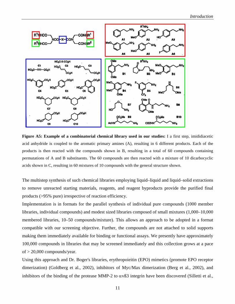
interactions (antagonists) (Fig. A5).
10
Introduction
Figure A5: Example of a combinatorial chemical library used in our studies: I a first step, imididiacetic
acid anhydride is coupled to the aromatic primary amines (A), resulting in 6 different products. Each of the
products is then reacted with the compounds shown in B, resulting in a total of 60 compounds containing
permutations of A and B substituents. The 60 compounds are then reacted with a mixture of 10 dicarbocyclic
acids shown in C, resulting in 60 mixtures of 10 compounds with the general structure shown.
The multistep synthesis of such chemical libraries employing liquid–liquid and liquid–solid extractions
to remove unreacted starting materials, reagents, and reagent byproducts provide the purified final
products (>95% pure) irrespective of reaction efficiency.
Implementation is in formats for the parallel synthesis of individual pure compounds (1000 member
libraries, individual compounds) and modest sized libraries composed of small mixtures (1,000–10,000
membered libraries, 10–50 compounds/mixture). This allows an approach to be adopted in a format
compatible with our screening objective. Further, the compounds are not attached to solid supports
making them immediately available for binding or functional assays. We presently have approximately
100,000 compounds in libraries that may be screened immediately and this collection grows at a pace
of > 20,000 compounds/year.
Using this approach and Dr. Boger's libraries, erythropoieitin (EPO) mimetics (promote EPO receptor
dimerization) (Goldberg et al., 2002), inhibitors of Myc/Max dimerization (Berg et al., 2002), and
inhibitors of the binding of the protease MMP-2 to αvß3 integrin have been discovered (Silletti et al.,
11
Introduction
2001; Boger et al., 2001). Based on the previous success with discovery and development of these
small molecule inhibitors of protein–protein interactions, we plan to identify effective in vitro and in
vivo inhibitors of viral infectivity from combinatorial chemical libraries
12
Materials and methods
B. MATERIALS AND METHODS B.1 Reagents and antibodies
Monoclonal antibodies (mAb) 33.1 and 83.6 (anti-LCMVGP2) have been described (Buchmeier et al.,
1981; Weber and Buchmeier, 1988). The rabbit anti-GFP polyclonal Ab was from Chemicon. FITC-
conjugated anti-rabbit IgG and phycoerythrin (PE)-conjugated anti-rat IgG were from Jackson
Immuno-Research (West Grove, PA), and the HRP-conjugated anti-mouse IgG was from Pierce
Chemical Co. (Rockford, IL). The Steady Glo® and Bright-Glo® luciferase assay systems were
obtained from Promega (Madison WI). Heparan sulfate and heparin were purchased from Sigma.
B.2 Cell lines
African green monkey kidney (Vero-E6) cells were maintained in Dulbecco’s modified Eagle medium
(DMEM) (Gibco BRL, Grand Island, NY) containing 10% fetal calf serum (HyClone, Logan, UT).
HEK293H cells were purchased from GIBCO BRL. For culturing as adherent cells, HEK293H were
kept in complete serum-containing medium: DMEM, 10 % (vol/vol) FBS, supplemented with
glutamine, and penicillin/streptomycin. For suspension cultures, cells were briefly trypsinized, washed
twice in DMEM and re-suspended in the chemically defined serum-free medium 293 SFMII (GIBCO).
The cell lines HeLa, A549 human lung carcinoma cells (ATCC CCL-185), and the packagic cell line
GP293® (Invitrogen) were cultured in DMEM, 10 % (vol/vol) FBS, supplemented with glutamine, and
penicillin/streptomycin. The glycosaminoglycan-deficient CHO cell line psgA-745 was obtained from
ATCC. PsgA-745 cells and control wild-type CHO cells were kept in Ham’s F12K medium
supplemented with 10 % (vol/vol) FBS and 1.5 g/l sodium bicarbonate.
B.3 Virus strains, purification, and quantification
Origin, passage and characteristics of LCMV ARM53b and clone-13 have been described elsewhere
(Dutko and Oldstone, 1983). Seed stocks of all viruses were prepared by growth in BHK-21 cells.
Purified virus stocks were produced and virus titers determined as described. LFV Josiah was grown in
Vero-E6 cells, polyethylene glycol-precipitated and γ-inactivated at the Center for Disease Control and
Prevention in Atlanta GA.
13
Materials and methods
Seed stocks of Pichinde were prepared by growth in BHK-21 cells. Purified virus stocks were
produced and titers determined as described (Dutko et al., 1983). Amapari, Parana, Oliveros, Latino,
Guanarito, Junin, Machupo, and LFV were grown in Vero-E6 cells in a BSL4 facility, polyethylene
glycol-precipitated and γ-inactivated as described (Elliot et al., 1982). Inactivation was verified by a
double blind passage of the inactivated viruses on Vero E6 cells followed by immunofluoresence
staining for detection of viral antigen. Pichinde virus was inactivated by UV irradiation as described
(Kunz et al., 2004). Inactivation was verified by plaque assay on Vero cells. The work with most of the
infectious viruses was done at biosafety level 3 (BSL-3), with the exception of LFV, Guanarito, Junin,
and Machupo viruses, which were handled in the BSL-4 laboratories at the Special Pathogens Branch,
and Pichinde, which was handled at BSL-2 at the Scripps Research Institute.
B.4 Detection of LCMVGP in ELISA
Purified viruses were coated in triplicate wells in 96-well EIA/RIA high-bond microtiter plates
(Corning) for 2 hours at 6 ºC and non-specific binding blocked with 1% (wt/vol) BSA/PBS. MAbs
83.6 (anti-LCMVGP2) was applied in 1: 100 dilution for 2 hours at 6 ºC and detected with peroxidase-
conjugated anti-mouse IgG (1: 1000) in a color reaction using ABTS (2,2’azino-bis(3-
ethylbenzthiazoline-6-sulfonic acid)) substrate. OD (405) was measured with an ELISA reader. For the
determination of specific binding, background binding to BSA was subtracted.
B.5 Immunoblotting
Proteins were denatured in hot (95 ºC) SDS-PAGE sample buffer: 2% (wt/vol) SDS, 50 mM Tris/HCl,
pH 6.8, 100 mM DTT. Proteins were separated by gel electrophoresis and transferred to nitrocellulose
using precast Novex® tris-glycine gels. After blocking in 5% (wt/vol) skim milk powder in PBS,
membranes were incubated with the primary antibody mouse monoclonal antibody M2 anti-flag in a
dilution of 1:1000 in 2% (wt/vol) skim milk powder, PBS for 12 hours at 6 ºC. After several washes in
PBS, 0.1% (wt/vol) Tween-20 (PBST), the secondary antibody, goat anti-mouse IgG coupled to
peroxidase was applied 1: 5000 in PBST for 1 hour at room temperature. Blots were developed by
enhanced chemiluminescence (ECL) using Super Signal West Pico ECL Substrate (Pierce) and signals
were recorded on autoradiographic film (Kodak, Rochester, N.Y.).
14
Materials and methods
B.6 Molecular biology techniques
B.6.1 Polymerase chain reaction (PCR)
PCR profile
Segment# duration (min) temperature (ºC) process 1 5 95 strand separation 2* 1 95 strand separation 3* 1 50 primer annealing 4* 1 72 extension 4 1 72 extension 5 for ever 4 cool down *segments 2-4 were repeated 16 times PCR reaction mixture Reagent final concentration µl PCR water - 39.5 10 x pfu/taq buffer 1 x 5 dNTPs 200 µM/nucleotid 1 primer 1 500 nM 1.25 primer 2 500 nM 1.25 DNA template 500 ng 1 Pfu/Taq-polymerase 2.5 U 1 B.6.2 Phenol chloroform extraction
Phenol chloroform extraction was performed to remove contaminating proteins from DNA. 50 μL of
phenol chloroform was added to the DNA solution. The mixture was vortexes on full speed for 1min
and then spun on full speed (14 krpm) for 1 min. The upper phase was transferred to a new
eppendorphtube and added 2.5 volumes of pure ethanol. The DNA precipitated at -70°C > 30 min (or
overnight), then spun for 15 min at 14 krpm in the cold room. Supernatant was removed and pellet
washed with 50 μL cold (-20°C) 75% ethanol. Ethanol was removed and the pellet air-dried. The pellet
was dissolved in PCR water (30 μL) at 37°C. The amount and quality of the DNA fragment was
checked by gel electrophoresis.
15
Materials and methods
B.6.3 Agarose gel electrophoresis
Agarose gel electrophoresis can be used to distinguish and separate DNA fragments of different sizes.
Small DNA fragments (<2.0 kilobases) are separated on gels with 1.0-2.0% agarose and larger DNA
fragments (2.0-10.0 kilobases) are separated on gels with 0.6-1.0% agarose. The DNA molecules will
run over the gel in the electrical field, migrating toward the anode. The speed of migration of the DNA
molecules depends on their size; therefore the small molecules will migrate faster than those of the
larger sizes because they more readily travel through the polymer matrix. To determine the size of the
separated DNA molecules, they are compared to a DNA marker fragments of known size run on the
same gel. Ethidium bromide is pre added to the gel and will interchelate between basepairs of the DNA
double helix. This will emit light (fluorescence) exposed to UV light providing a means to detect the
location of the DNA in the gel.
The agarose was dissolved in 100 ml 1x TAE buffer and heated to the boiling point in the microwave
oven. 1 drop of ethidium bromide was added to the agarose and the solution was pored into a gel mold
fitted with the appropriate comb. The gel was allowed to harden at room temperature. The comb was
removed and the gel was transfered to an electrophoresis chamber and covered with 1x TAE buffer.
DNA loading buffer was added to the DNA samples and they were loaded on the gel. A DNA marker
was added in a free well. The gel was run at 120 V. The DNA was visualized under UV-light.
B.6.4 Gel extraction
The QIAGEN Gel Extraction Kit (QIAquick Gel Extraction Kit) was used for extraction of DNA from
the gel.
The DNA fragment from the agarose gel was excised with a clean, sharp scalpel and weighed in a
colorless tube. 3 volumes of Buffer QG were added to 1 volume of gel and this was incubated at 50°C
for 10 min (until the gel slice had completely dissolved). 1 gel volume of isopropanol was added and
mixed with the sample. A QIAquick spin column was placed in a provided 2 ml collection tube. To
bind DNA, the sample was added to the QIAquick column, and centrifuge for 1 min. The flow-trough
was discarded and the QIAquick column was placed back in the same collection tube. For washing,
0.75 ml of Buffer PE was added to QIAquick column and centrifuged for 1min. The flow-trough was
discarded and the QIAquick column was centrifuged for an additional 1 min. The QIAquick column
was placed into a clean 1.5 ml microcentrifuge tube. To elute DNA, 50 μL of Buffer EB was added to
16
Materials and methods
the center of the QIAquick membrane, the column was standing for 1min, and then centrifuged for 1
min. For gel analyze of the extracted DNA, 1 volume of Loading Dye was added to 5 volumes of
purified DNA. The solution was mixed by pipetting up and down and then loaded to the gel.
B.6.5 Treatment of DNA fragments with calf intestinal phosphatase (CIP)
The removal of terminal phosphate greatly reduces re-ligation e.g. of vector DNA and is essential for
all vector DNA intended for use in ligation reactions. After completion of the restriction digest, calf
intestinal phosphatase (CIP) is added to the reaction. CIP is compatible wit all four NEB buffers.
1 µl of CIP (Promega) was added to each preparative vector digest and the reaction was mixed
thoroughly before it was incubated for 30 minutes at 37 ºC.
B.6.6 Ligation of DNA fragments with T4 DNA ligase
The DNA fragment and the vector DNA were separated in a preparative TAE gel. The DNA fragments
were recovered from the gel using QIAEXII gel extraction. DNA fragments recovered by QIAEX were
present in 20µl 1 mM Tris/HCl pH 8.0, which is ideal for subsequent ligations by T4 DNA ligase. For
each ligation reaction, a control-reaction that contained only the vector DNA was included to assess
the background of vector re-ligation.
Ligase reaction
Component µl DNA fragment (recovered by QUIAEX) 12
Vector DNA (recovered by QUIAEX) 5 10 x T4 DNA ligase buffer 2 T4 DNA ligase (NEB) 1 Reaction times: 2-16 h at 16ºC (water bath in cold-room)
B.6.7 Transformation of E.coli
The following protocol was used for the transformation of chemically competent strains of E.coli like
e.g. DH5-α: The competent bacteria were thawed on wet ice. For transformation, 50 μL bacteria were
needed, the rest of the competent bacteria were aliquoted in 50 μL aliquots that were snap frozen (i.e.
frozen quickly in a cold bath prepared with ethanol and dry ice). The 50 μL bacteria were mixed with 5
17
Materials and methods
μL of the ligation reaction and kept on ice for 30min and then put at 37°C for 30 seconds (heat shock).
The reaction was put on ice for one minute and added 1 ml of LB medium without antibiotic. The
reactions were shaken at 180 rpm for 40-60min at 37°C and then spun in microcentrifuge 14 krpm for
30 second. All LB medium was removed except of 50 μL of liquid so the bacteria could gently be re-
suspended. The bacteria suspention were plated on Amp/LB plates and kept at 37°C for 16-24 h.
B.6.8 Plasmid DNA Purification Using the QIAprep Spin Miniprep Kit
The QIAGEN Miniprep Kit is used for plasmid DNA purification from overnight cultures of E.coli
grown in LB medium. Circa 1 ml of each sample bacteria suspention was transfered to tubes, spun for
1min at 14 krpm. The supernatant was discarded and the pelleted bacterial cells were resuspended in
250 μL Buffer P1. 250 μL Buffer P2 was added to the tubes and mixed thoroughly by inverting the
tubes gently 4-6 times. 350 μL Buffer N3 was added to the tubes and mixed immediately and
thoroughly by inverting the tubes 4-6 times. The tubes were centrifuged for 10 min at 13 krpm in a
table-top microcentrifuge. The supernatant from this last step was applied to the QIAprep spin column
by decanting or pipetting. The columns were then centrifuged 60 s and the Flow-through were
discarded.
For washing, 0.75 ml Buffer PE was added to the QIAprep spin columns before they were centrifuged
for 60 s. The flow-through was discarded and the columns were centrifuge for an additional 1 min to
remove residual wash buffer. The QIAprep columns were placed in a clean 1.5 ml microcentrifuge
tubes. To elute DNA, 50 μL Buffer EB was added to the center of each QIAprep spin column. The
columns were standing for 1 min before centrifugation for 1 min.
B.7 Generation of pseudotyped retroviral vectors
The packaging cell line GP2-293® from BD Bioscience stably expresses the Moloney mouse leukemia
virus (MLV) derived gag and pol gene and allows packaging of any MMLV-based vector containing
the appropriate packaging signal.
Co-transfection of a MLV-derived retroviral genomic plasmid pLZRS-Luc-gfp and the GP
expression plasmid: GP2-293 cells were cultivated on plastic in T175 flasks to a confluency of 80%.
The cells were split 1:2 into T175 flasks coated with poly-L-lysine. Medium for maintenance and
18
Materials and methods
transfection: DMEM, 10% (v/v) FBS, Gln, P/S. Medium for virus production: DMEM, 10% (v/v) FBS,
Gln, P/S, 20 mM Hepes. Transfection was carried out according to Sonderegger et al., Curr. Prot. Cell
Biol. Unit 9.5. Briefly, 4 h prior to transfection, medium was removed and 40 ml of fresh medium was
added per flask. Solution A was prepared (2 ml 0.25 M CaCl2, 20 µg GP expression plasmid DNA
(QiaGen Maxi-prep), 20 µg MLV genomic plasmid pLZRS-Luc-gfp DNA (QiaGen Maxi-prep)).
Solution A was mixed into solution B (2 ml HBS) and incubated at room temperature for exactly 1
min. Transfection solution was added to cells and distributed evenly. Flasks were incubated at 37ºC,
5% CO2 for 4-12 h. Transfection mix was removed and cells were washed twice with medium.
Complete medium with 20 mM Hepes was added (25 ml/ flask) before the flasks were put back in the
incubator. The pseudotypes were harvested after 24 and 48 hours, cleared by low speed centrifugation
and frozen at –70ºC.
B.8 Infection of human cells with retroviral pseudotypes
Target cells: One day before the assay 2 x 10⁴ HeLa cells were plated per well in flat bottom M96
plates (total volume medium 200 µl) in DMEM, 10% FBS, Gln, non-essential amino acids, Pen/Strep.
The cells were circa 80% confluent at time of assay (after 16-24 hours).
Infection: Medium from target cells was removed, 100 µl pseudotypes was added to cells before they
were incubated for 1 h at 37ºC, 5% CO2. Mixture was removed and cells were washed twice with
DMEM, 10 mM Hepes without FBS. 100 µl complete medium containing 10 mM Hepes per well were
added and cells were incubated for 40-48 h at 37ºC, 5% CO2. The results were read by Steady-Glo®
luciferase assay.
B.9 Steady-Glo® luciferase assay
The Steady-Glo® luciferase assay is fully compatible with our medium (DMEM with 10 % FBS). The
presence of phenol red reduced the signal intensity by circa 30%. We will avoid this problem in the
future by using medium without phenol red.
The old medium was removed from the cells and replaced with 100 µl/well of fresh (37 ºC warm)
medium. 100 µl assay (room-temperature) reagent/well were added to the plates. After 5 minutes, the
cells were scratched off from the bottom of the plate and mix well with the solution (> 5 times up and
down pipetting). Content of wells were transferred into a white M96 plate suitable for
19
Materials and methods
chemiluminescence reader. Luminescence was measured in a 96-well plate luminometer (Bertold)
using WinGlow software.
B.10 Screening of chemical libraries: inhibition of the infection with retroviral pseudotypes by
compounds from combinatorial chemical libraries
Compound mixtures: The compound mixtures derived from combinatorial chemical libraries were
dissolved in DMSO, in a concentration of 5 mM. We had 10 µl per compound mixture. The libraries
were provided in 96 well plate formats. The pharmacologically active concentration in the primary
screening assay, ideally 50 µM, corresponded to a 100-fold dilution of the stocks.
Pseudotypes: Retroviral pseudotypes containing the GPs of different arenaviruses (LFV, LCMV,
Amapari, Guanarito, Junin, Machupo, and Vesicular stomatitis virus,) produced as described, were
used in this assay.
Target cells: One day before the assay 2 x 10⁴ HeLa cells were plated per well in flat bottom M96
plates (total volume medium 200 µl) in DMEM, 10% FBS, Gln, non-essential amino acids, Pen/Strep.
The cells were circa 80% confluent at time of assay (after 16-24 hours).
Blocking pseudotypes with compounds: For the screening assay, exact replicas of the plates were
done to avoid mix-up of samples. Empty wells of the compound plate were used for controls. 100 µl of
pseudotypes in OPTIMEM, 2% FBS, 10 mM Hepes were added per well of a M96 plate and 1 µl of
compound mixture were added per well and mixed briefly. For control wells DMSO only were added.
Plates were incubated for one hour on ice.
Infection: Medium from target cells were removed and 80 µl of pseudotypes /compound mixtures
were added to each well before plates were incubated for 1 h at 37ºC, 5% CO2. After incubation,
mixtures were removed and cells were washed once with DMEM, 20 mM Hepes without FBS. 100 µl
complete medium containing 20 mM Hepes were added per well and plates were incubated for 40-48 h
at 37ºC, 5% CO2. The results were read by Steady-Glo® luciferase assay.
20
Materials and methods
B.11 Target-specificity of compound mixtures
To assess the target-specificity of compound mixtures derived from combinatorial chemical libraries,
compound mixtures were tested for their ability to block infection of target cells with arenavirus
pseudotypes but not with pseudotypes containing the GP of the unrelated VSV. This was done by
using the assay described in B9.
B.12 Determination of the dose-response characteristics of selected candidate compounds
Cells: Human lung epithel A549 in DMEM/10% FBS/Gln/PS
One day before the assay 2 x 10⁴ cells were plated per well in flat bottom M96 plates (total volume
medium 200 µl). At the time of assay, the cells were circa 80% confluent (after 16-24 hours).
Blocking of LFV-PS/VSV-PS with increasing concentrations of candidate compounds:
Concentration µl LFVPS µl compound dilution 0 300 6 pure DMSO
1 µM 300 6 50 µM (1:100)
5 µM 300 5 500 µM (1: 10)
10 µM 300 6 500 µM (1:10)
20 µM 300 1.2 5 mM (stock)
50µM 300 3 5 mM (stock)
100 µM 300 6 5 mM (stock)
Infection in triplicates: Medium was removed from target cells, 90 µl of PS /compound mixtures
were added to cells and plates were incubated for 1 h at 37ºC, 5% CO2. After incubation, mixture was
removed and cells were washed once with DMEM, 10 mM Hepes without FBS. 100 µl complete
medium containing 10 mM Hepes was added per well and plates were incubated for 40-48 h at 37ºC,
5% CO2. The results were read by Steady-Glo® luciferase assay.
21
Materials and methods
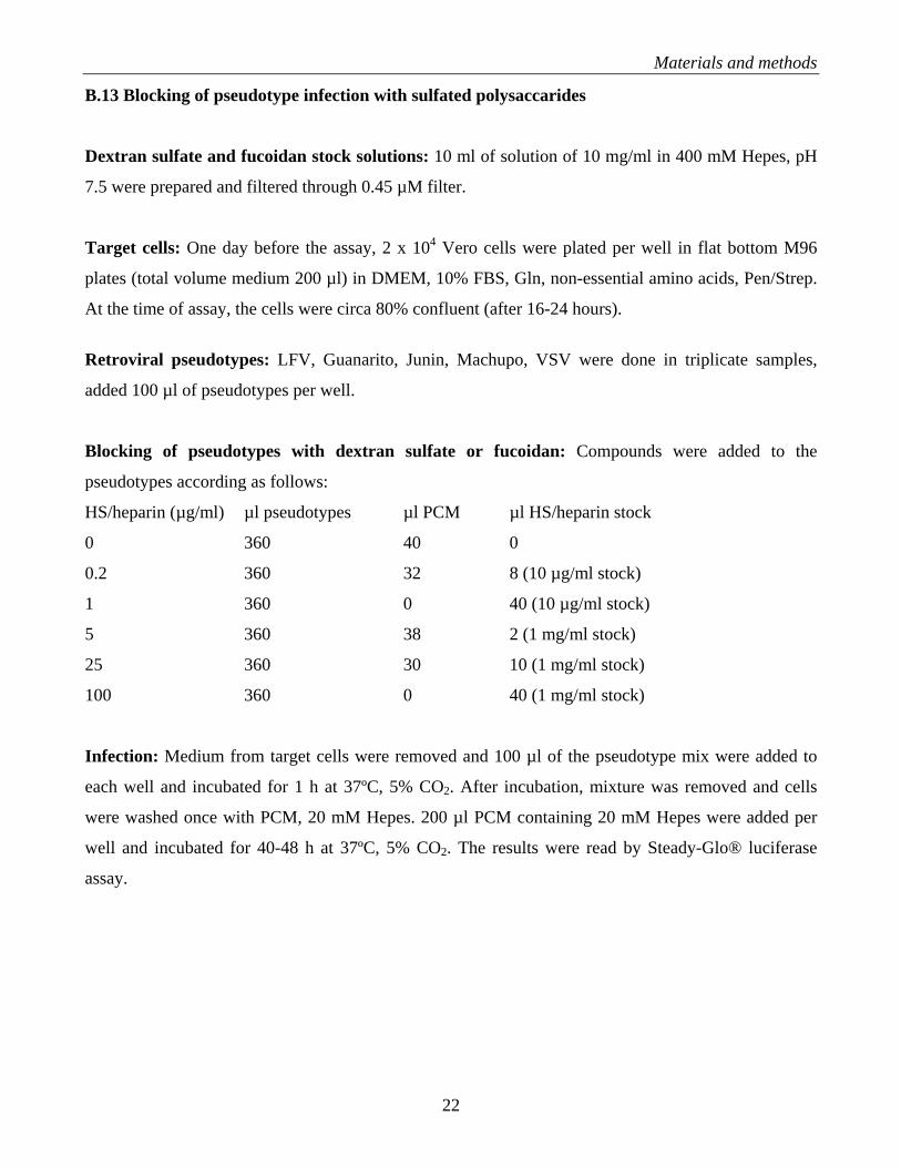
B.13 Blocking of pseudotype infection with sulfated polysaccarides
Dextran sulfate and fucoidan stock solutions: 10 ml of solution of 10 mg/ml in 400 mM Hepes, pH
7.5 were prepared and filtered through 0.45 µM filter.
Target cells: One day before the assay, 2 x 10⁴ Vero cells were plated per well in flat bottom M96
plates (total volume medium 200 µl) in DMEM, 10% FBS, Gln, non-essential amino acids, Pen/Strep.
At the time of assay, the cells were circa 80% confluent (after 16-24 hours).
Retroviral pseudotypes: LFV, Guanarito, Junin, Machupo, VSV were done in triplicate samples,
added 100 µl of pseudotypes per well.
Blocking of pseudotypes with dextran sulfate or fucoidan: Compounds were added to the
pseudotypes according as follows:
HS/heparin (µg/ml) µl pseudotypes µl PCM µl HS/heparin stock
0 360 40 0
0.2 360 32 8 (10 µg/ml stock)
1 360 0 40 (10 µg/ml stock)
5 360 38 2 (1 mg/ml stock)
25 360 30 10 (1 mg/ml stock)
100 360 0 40 (1 mg/ml stock)
Infection: Medium from target cells were removed and 100 µl of the pseudotype mix were added to
each well and incubated for 1 h at 37ºC, 5% CO2. After incubation, mixture was removed and cells
were washed once with PCM, 20 mM Hepes. 200 µl PCM containing 20 mM Hepes were added per
well and incubated for 40-48 h at 37ºC, 5% CO2. The results were read by Steady-Glo® luciferase
assay.
22
Materials and methods
B.14 Intracellular FACS staining for LCMV-NP using mAb 113
Preparation of a single cell suspension: The cells cultivated in 6 well plates were washed twice with
PBS. 1 ml/well of Trypsin-EDTA were added to the cells but not removed completely, leaving some
liquid left in each well. The plates were incubated at 37ºC for 5 minutes, the cells were detach by
slapping against the plate and re-suspend in 1ml/well 1% FBS/PBS. The cell-suspension were
transferred to 5 ml polystyrene tube and kept on ice. The tubes were spun 1600 rpm/4min/4oC and
supernatant was removed. Cells were re-suspend in 2 ml of 1% FBS/PBS. The tubes were again spun
1600 rpm/4min/4oC and supernatant was removed. The cells were re-suspend in 250 µl of 1%
FBS/PBS and kept on ice. 200 µl of each cell-suspensions were transferred into a M96 plate with
conical wells.
Fixation and permeabilization: The cells were spun down 1600 rpm/4min/4oC and supernatant were
removed. Cells were resuspended in 100 µl/well of 4% (w/v) PFA/PBS and kept at RT for 10 min. The
cells were washed twice with 1% (v/v) FBS/0.1% (w/v) saponin/PBS.
Wash procedure: The cells were spun down 1600 rpm/4min/4oC and re-suspend in 200 µl/well of 1%
(v/v) FBS/0.1% (w/v) saponin/PBS.
Intracellular staining with mAb 113 anti LCMVNP: mAb 113 was diluted 1: 100 in 1% (v/v)
FBS/0.1% (w/v) saponin/PBS. Cells were resuspended in 100 µl/well of Ab solution and incubated for
45 minutes on ice in the dark. Cells were wash twice with 1% (v/v) FBS/0.1% (w/v) saponin/PBS.
Detection with FITC-conjugated 2nd Ab: 2nd Ab anti-mouse IgG F(ab)2-FITC was diluted 1: 100 in
1% (v/v) FBS/0.1% (w/v) saponin/PBS. Cells were re-suspend in 100 µl/well of 2nd Ab and incubated
for 45 minutes on ice in the dark. After incubation, the cells were washed once with 1% (v/v)
FBS/0.1% (w/v) saponin/PBS and twice with 1% FBS/PBS. Cells were re-suspend in 200 µl/well PBS
and kept in the dark.
B.15 Detection of LCMV-NP by immunofluorescence staining
Cells: 104 Vero E6 cells were plated per well of a 8 well LabTek tissue chamber slides (Nunc) and
cultured over night.
23
Materials and methods
Infection with LCMV: LCMV was added at MOI of 0, 0.01, 0.1, and 1 in an inoculum of 250 µl
medium for 45 minutes at 37ºC, 5% CO2. After infection, cells were washed and 0.5 ml fresh medium
per well were added.
Fixation of cells: The culture medium was removed and 250 µl of 2% formaldehyde/0.1%
glutaraldehyde in PBS (room temperature) was added to each well for fixation of the cells at 37oC for
15 minutes in the dark. The formaldehyde fixative was aspirated and cells were washed twice with
PBS. PBS was removed after the second wash step and 500 µl PBS/1% (v/v) FCS were added and cells
incubated for 15 min at room temperature.
Immunofluorescence staining
Permeabilization of cells: After the blocking step (#2), 250 µl PBS/1% (v/v) FCS/0.1% (w/v) saponin
was added and cells incubated for 15 min at room temperature.
Incubation with primary antibody: Primary antibody: mAb 113 anti-LCMVNP 1: 200 in PBS/1%
(v/v) FCS/0.1% (w/v) saponin (250 µl/well) was added and cells incubated for 1h at room temperature.
Incubation with secondary antibody: The secondary antibody (goat anti mouse IgG FITC labeled)
was diluted 1:100 in PBS/1% (v/v) FCS/0.1% (w/v) saponin. Primary antibody solution was removed
and cells were washed 2 times with PBS. 200 µl/well secondary antibody in PBS/1% (v/v) FCS were
added and cells were incubated for 45 min at room temperature, protect from light. Secondary antibody
solution was removed and cells were washed 3 times with PBS.
Evaluation: Fluorescence microscope with the 5x objective was used for evaluation. All NP+ cells
were counted and cell clusters were scored as one infection event.
24
Results
C. RESULTS
C.1 Discovery of novel small molecule inhibitors of Lassa fever virus (LFV) infection from
combinatorial chemical libraries:
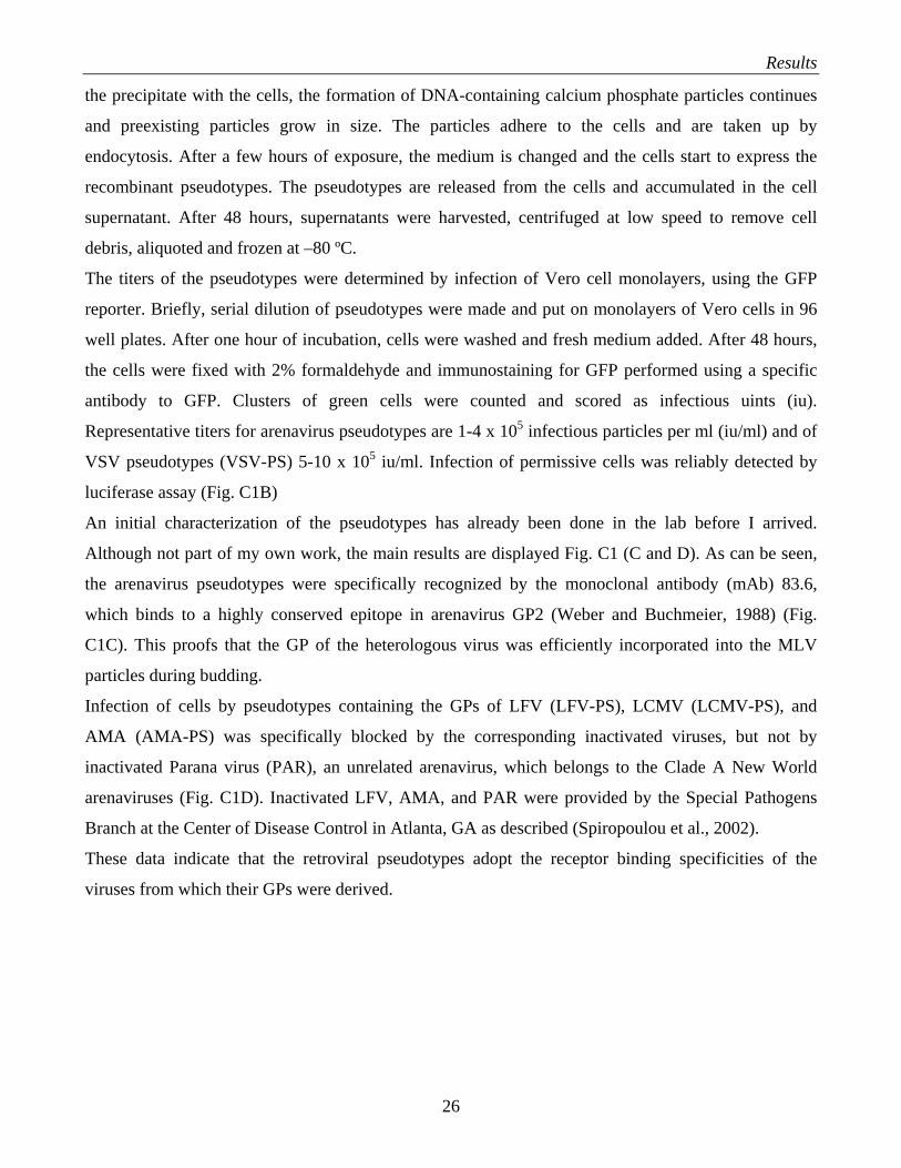
C.1.1 Production and characterization of retroviral pseudotypes
Since LFV is a BSL4 pathogen, studies with live virus are restricted to BSL4 laboratories. To discover
small molecule drugs that can block the infection of human cells with LFV, our laboratory has
generated retroviral vectors that contain the glycoprotein (GP) of LFV in their envelope. The principle
behind this is the fact that enveloped viruses can incorporate heterologous viral GPs into their lipid
membranes during budding. These so-called “pseudotypes” acquire the receptor specificity of the virus
from which the heterologous GP was derived.
Using the strategy outlined in Fig. C1A, we inserted the GPs of LFV strain Josiah and the LCMV
isolates ARM53b (ARM) and clone-13 (cl-13) into virions of recombinant Moloney leukemia virus
(MLV), which contain a luciferase and a green fluorescent protein (GFP) reporter gene. In addition, we
generated pseudotypes containing the GPs of the New World arenavirus Amapari (AMA) and of
vesicular stomatitis virus (VSV), which do not use α-DG as a receptor (Cao et al., 1998; Spiropoulou
et al., 2002).
The packaging cell line GP2-293-Luc® (BD Biosciences) stable expresses MLV gag and pol. The cells
were transfected with a plasmid expressing a packable MLV genome pLZRS-Luc-GFP and an
expression plasmid for LFVGP. pLZRS-Luc-GFP was developed by the laboratory of Dr. Gary Nabel
(Yang et al., 1998) and contains a luciferase transgene and a green fluorescence protein (GFP) reporter
encoded in a bicistronic mRNA. This guarantees a defined ratio of luciferase and GFP being expressed
in cells transduced with the recombinant retrovirus. For transfection, I used calcium phosphate, a
technique that results in high efficiencies of transfection. In this method, the formation of the DNA-
containing calcium phosphate particles is initiated under defined chemical conditions, in the absence of
cells or serum. The particle size is the most critical parameter regarding efficiency of transfection, that
is uptake of DNA-containing calcium phosphate particles by the cell. The main determinations of
particle size are calcium and phosphate concentration, the concentration of the DNA, size of the DNA
fragments involved, pH, temperature and time of incubation. After initial formation of the DNA-
containing calcium phosphate particles, the precipitate is added to the cells. During the incubation of
25
Results
the precipitate with the cells, the formation of DNA-containing calcium phosphate particles continues
and preexisting particles grow in size. The particles adhere to the cells and are taken up by
endocytosis. After a few hours of exposure, the medium is changed and the cells start to express the
recombinant pseudotypes. The pseudotypes are released from the cells and accumulated in the cell
supernatant. After 48 hours, supernatants were harvested, centrifuged at low speed to remove cell
debris, aliquoted and frozen at –80 ºC.
The titers of the pseudotypes were determined by infection of Vero cell monolayers, using the GFP
reporter. Briefly, serial dilution of pseudotypes were made and put on monolayers of Vero cells in 96
well plates. After one hour of incubation, cells were washed and fresh medium added. After 48 hours,
the cells were fixed with 2% formaldehyde and immunostaining for GFP performed using a specific
antibody to GFP. Clusters of green cells were counted and scored as infectious uints (iu).
Representative titers for arenavirus pseudotypes are 1-4 x 10⁵ infectious particles per ml (iu/ml) and of
VSV pseudotypes (VSV-PS) 5-10 x 105 iu/ml. Infection of permissive cells was reliably detected by
luciferase assay (Fig. C1B)
An initial characterization of the pseudotypes has already been done in the lab before I arrived.
Although not part of my own work, the main results are displayed Fig. C1 (C and D). As can be seen,
the arenavirus pseudotypes were specifically recognized by the monoclonal antibody (mAb) 83.6,
which binds to a highly conserved epitope in arenavirus GP2 (Weber and Buchmeier, 1988) (Fig.
C1C). This proofs that the GP of the heterologous virus was efficiently incorporated into the MLV
particles during budding.
Infection of cells by pseudotypes containing the GPs of LFV (LFV-PS), LCMV (LCMV-PS), and
AMA (AMA-PS) was specifically blocked by the corresponding inactivated viruses, but not by
inactivated Parana virus (PAR), an unrelated arenavirus, which belongs to the Clade A New World
arenaviruses (Fig. C1D). Inactivated LFV, AMA, and PAR were provided by the Special Pathogens
Branch at the Center of Disease Control in Atlanta, GA as described (Spiropoulou et al., 2002).
These data indicate that the retroviral pseudotypes adopt the receptor binding specificities of the
viruses from which their GPs were derived.
26
Results
Figure C1: Recombinant retroviral vectors pseudotyped with arenavirus GPs. (A) Production of
pseudotyped retroviruses: The packaging cell line GP2-293® (BD Biosciences) stable transfected with MLV
gag and pol, is co-transfected with a plasmid containing the packable MLV genome pLZRS-Luc-Gfp (Yang et
al., 1998), which contains a luciferase and a GFP reporter, and an expression plasmid for the heterologous GP
(pC-AVGP). Retroviral pseudotypes are released into the cell supernatant. Infection of target cells results in the
integration of the retroviral genome into the host cell DNA and can be quantified by detection of the luciferase
or GFP reporter. (B) Infection of cells with retroviral pseudotypes: Supernatants containing pseudotypes were
used to infect monolayers of Vero cells in 96 well plates. After 48 h, infection was quantified using the Steady-
Glo® luciferase reporter gene assay (Promega) in a Berthold 96 well-plate luminometer. Luminescence is
expressed as fold-increase over uninfected controls (n = 3 + SD). (C) Binding of mAb 83.6 to pseudotypes:
Equal amounts of concentrated pseudotypes containing the GPs of LCMV ARM53b (ARM), cl-13, LFV, AMA,
and VSV were immobilized in microtiter plates and probed with mAb 83.6 anti-LCMVGP2. Primary antibodies
were detected with POD- conjugated anti-mouse IgG in a color reaction using ABTS substrate. OD (405) was
measured using an ELISA reader (n = 3 + SD). (D) Blocking of arenavirus pseudotype infection by inactivated
viruses: HEK293 cells cultured in 96-well plates were blocked with inactivated LCMV (ARMi, cl-13i), LFV
(LFVi), AMA (AMAi), or PAR (Pari) at the indicated ratios of inactivated virus particles per cell. After
blocking for two hours at 4 ºC, cells were infected with the indicated pseudotypes at a multiplicity of infection
(MOI) = 1. Infection was assessed by luciferase assay as in (B) (n = 3, + SD).
27
Results
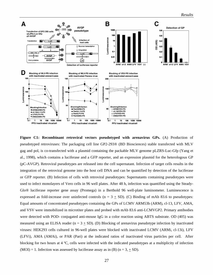
C.1.2 Screening of combinatorial chemical libraries
The Combinatorial chemical libraries were given to us by the laboratory of Dr. Dale L. Boger
(Department of Chemistry, Scripps). The libraries were provided in 96 well plates containing either
individual compounds or mixtures of 4-10 compounds in a concentration of 5 mM in DMSO. The
compounds/compound mixtures were screened for their ability to block infection of target cells by
retroviral pseudotypes that contain LFVGP in their envelope and a luciferase reporter gene.
For each library, two independent screenings were performed as shown in Fig C2 and only 90
compounds that showed >50% inhibition of infection in both independent screens were considered
candidates to be followed up. During my time in the laboratory, I have screened 13 complete libraries
with total > 10, 000 iminodiacetic acid-based and pyrrolidine-based peptidomimetic compounds.
Duplicate screening resulted in the identification of circa 4-5% compounds/mixtures that showed
reproducible reduced LFV-PS infection by > 50%.
Figure C2: Example of the high-throughput screening of a combinatorial chemical library for inhibitors
of LFV-PS infection. LFV-PS were pre-incubated with compound mixtures (concentration = 50 µM for
individual compounds and 5 µM for individual compounds in mixtures of 4-10) for 45 minutes and then added
to monolayers of HeLa cells in 96 well-plates. After one hour, the inoculum was removed, cells washed and
incubated for 48 h. Infection was quantified using the Steady-Glo® high sensitivity luciferase reporter gene
assay (Promega) in a Berthold® 96 well-plate luminometer. Data shown are two completely independent
experiments screening the same library (#19). Luminescence is expressed as fold-increase over background.
28
Results
A1-10, B1-10, C1-10, D1-10, E1-10, F1-10, and G1-10 (black bars) represent mixtures of seven different
compounds each. The samples labeled C (white bars) correspond to solvent only controls. 100% and 50% of
mean control values are indicated as horizontal lines. The mixtures B1-B9, which result in >50% inhibition of
LFV-PS infection in both screens (underlined), showed the best effect for inhibition.
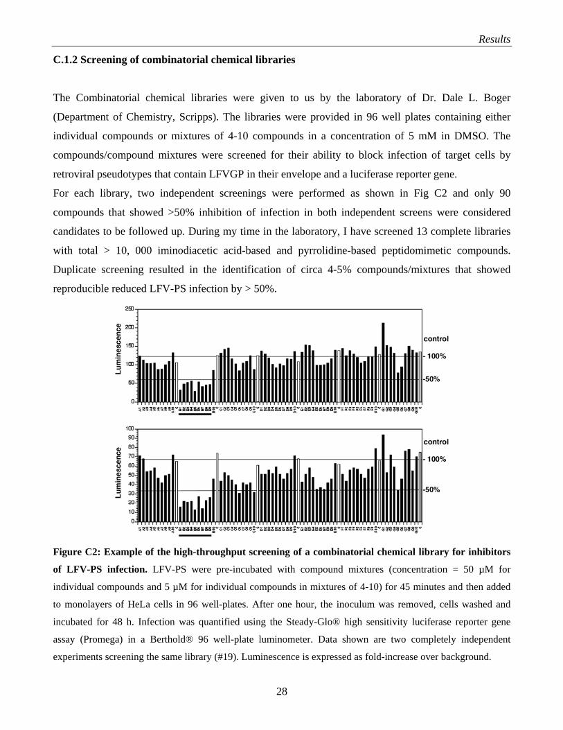
C.1.3 Determination of the target specificities of candidate compounds
Next, we wanted to select for compounds that specifically block LFVGP-mediated attachment and
entry but do not influence subsequent steps of our retroviral pseudotype-based assay. To this end, we
compared the effect of our 125 remaining candidate compounds/mixtures on infection with LFV-PS
with infection with pseudotypes containing the GP of vesicular stomatitis virus (VSV-PS). VSVGP is
structurally unrelated to LFVGP and binds to a different receptor. Our counter-selection is based on the
fact that compounds, which specifically block LFVGP-mediated infection, should not interfere with
VSV-PS infection while compounds that affect subsequent steps of the assay would reduce reporter
gene expression in both, LFV-PS and VSV-PS. Determination of target specificity (Fig. C3) revealed
that most compounds specifically reduced infection with LFV-PS but not VSV-PS.
Figure C3: Determination of target specificity of representative candidate compounds/mixtures that
showed significant blocking of LFV-PS infection in the screening assay: LFV-PS and VSV-PS were pre-
incubated with compound mixtures as in Fig. C2 and then added to monolayers of HeLa cells in 96 well-plates.
After one hour, the inoculum was removed, cells washed twice with DMEM and incubated with fresh medium
for 48 h. Infection was quantified using the Steady-Glo® high sensitivity luciferase reporter gene. Data shown
are triplicates (+/- SD). The samples labeled DMSO correspond to solvent controls.
29
Results
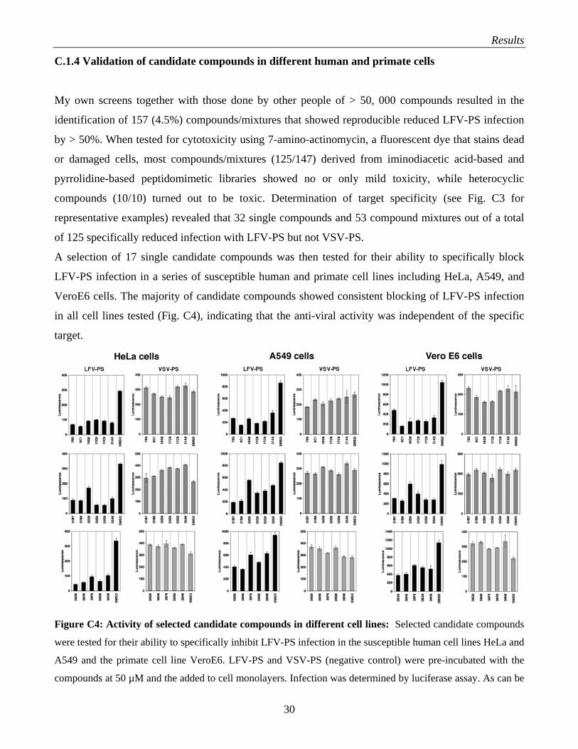
C.1.4 Validation of candidate compounds in different human and primate cells
My own screens together with those done by other people of > 50, 000 compounds resulted in the
identification of 157 (4.5%) compounds/mixtures that showed reproducible reduced LFV-PS infection
by > 50%. When tested for cytotoxicity using 7-amino-actinomycin, a fluorescent dye that stains dead
or damaged cells, most compounds/mixtures (125/147) derived from iminodiacetic acid-based and
pyrrolidine-based peptidomimetic libraries showed no or only mild toxicity, while heterocyclic
compounds (10/10) turned out to be toxic. Determination of target specificity (see Fig. C3 for
representative examples) revealed that 32 single compounds and 53 compound mixtures out of a total
of 125 specifically reduced infection with LFV-PS but not VSV-PS.
A selection of 17 single candidate compounds was then tested for their ability to specifically block
LFV-PS infection in a series of susceptible human and primate cell lines including HeLa, A549, and
VeroE6 cells. The majority of candidate compounds showed consistent blocking of LFV-PS infection
in all cell lines tested (Fig. C4), indicating that the anti-viral activity was independent of the specific
target.
Figure C4: Activity of selected candidate compounds in different cell lines: Selected candidate compounds
were tested for their ability to specifically inhibit LFV-PS infection in the susceptible human cell lines HeLa and
A549 and the primate cell line VeroE6. LFV-PS and VSV-PS (negative control) were pre-incubated with the
compounds at 50 µM and the added to cell monolayers. Infection was determined by luciferase assay. As can be
30
Results
seen, of the 17 compounds tested in the different cell lines only eight showed highly consistent activity in all cell
types: 7B5, 8C1, 16G8, 17C8, 17C9, 21A3, 31B7 and 31B8. The other ones, in particular compounds derived
from library #38 showed different activities in different cell lines. While all compounds were highly active in
HeLa cells, the cell type used for screening, some of the compounds showed only weak inhibition in A549 cells.
The reasons for these discrepancies are not fully understood, but a possible explanation will be given later on in
the discussion of my work.
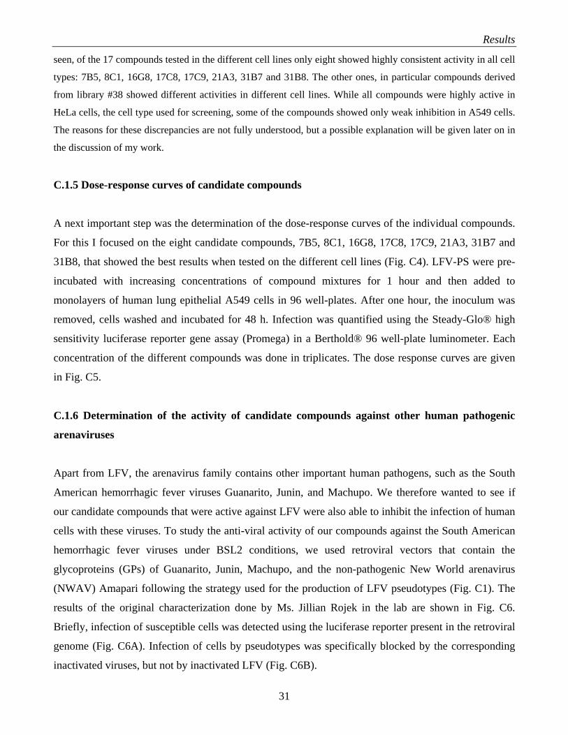
C.1.5 Dose-response curves of candidate compounds
A next important step was the determination of the dose-response curves of the individual compounds.
For this I focused on the eight candidate compounds, 7B5, 8C1, 16G8, 17C8, 17C9, 21A3, 31B7 and
31B8, that showed the best results when tested on the different cell lines (Fig. C4). LFV-PS were pre-
incubated with increasing concentrations of compound mixtures for 1 hour and then added to
monolayers of human lung epithelial A549 cells in 96 well-plates. After one hour, the inoculum was
removed, cells washed and incubated for 48 h. Infection was quantified using the Steady-Glo® high
sensitivity luciferase reporter gene assay (Promega) in a Berthold® 96 well-plate luminometer. Each
concentration of the different compounds was done in triplicates. The dose response curves are given
in Fig. C5.
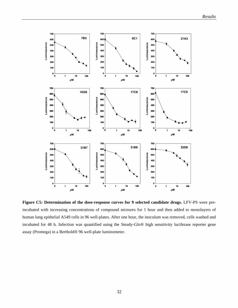
C.1.6 Determination of the activity of candidate compounds against other human pathogenic
arenaviruses
Apart from LFV, the arenavirus family contains other important human pathogens, such as the South
American hemorrhagic fever viruses Guanarito, Junin, and Machupo. We therefore wanted to see if
our candidate compounds that were active against LFV were also able to inhibit the infection of human
cells with these viruses. To study the anti-viral activity of our compounds against the South American
hemorrhagic fever viruses under BSL2 conditions, we used retroviral vectors that contain the
glycoproteins (GPs) of Guanarito, Junin, Machupo, and the non-pathogenic New World arenavirus
(NWAV) Amapari following the strategy used for the production of LFV pseudotypes (Fig. C1). The
results of the original characterization done by Ms. Jillian Rojek in the lab are shown in Fig. C6.
Briefly, infection of susceptible cells was detected using the luciferase reporter present in the retroviral
genome (Fig. C6A). Infection of cells by pseudotypes was specifically blocked by the corresponding
inactivated viruses, but not by inactivated LFV (Fig. C6B).
31
Results
Figure C5: Determination of the dose-response curves for 9 selected candidate drugs. LFV-PS were pre-
incubated with increasing concentrations of compound mixtures for 1 hour and then added to monolayers of
human lung epithelial A549 cells in 96 well-plates. After one hour, the inoculum was removed, cells washed and
incubated for 48 h. Infection was quantified using the Steady-Glo® high sensitivity luciferase reporter gene
assay (Promega) in a Berthold® 96 well-plate luminometer.
32
Results
Figure C6: Recombinant retroviral vectors pseudotyped with NWAV GPs. (A) Infection of cells with
retroviral pseudotypes: Undiluted supernatants of pseudotypes were added to monolayers of HeLa cells in 96
well-plates and infection quantified after 48 hours using luciferase reporter gene assay. Luminescence is
expressed as fold-increase over noninfected control cells (n = 3 + SD). (B) Blocking of New World arenavirus
GP pseudotypes (NWAV-PS) infection by inactivated viruses: HEK293 cells cultured in 96-well plates were
blocked with γ-inactivated Amapari (AMAi), Junin (JUNi), Guanarito (GUAi) or LFV (LFVi) at the indicated
ratios of inactivated virus particles/cell. After incubation for two hours at 4 ºC, cells were infected with either
NWAV pseudotypes or pseudotypes containing VSVGP (VSV-PS) at a multiplicity of infection (MOI) = 1.
Infection was assessed after 48 h by luciferase assay (n = 3, + SD).
The 17 selected candidate compounds were tested for their ability to block the infection of human cells
(A549) with retroviral pseudotypes containing the GPs of Guanarito, Junin, and Machupo. To ensure
the specificity of the observed reduction in infection by the candidate drugs, we used VSV pseudotypes
as a negative control. Interestingly, many compounds that showed activity against LFV pseudotype
infection were also active against the South American HF viruses (Fig. C7).
Many of the candidate compounds that showed specific activity against LFV-PS showed significant
activity against pseudotypes of Guanarito, Junin and Machupo. The responses to the compounds in the
different NWAV-PS are very similar. In particular the compounds 16G8, 17C8 and 17C9 showed
nearly full neutralizing potential against these pseudotypes. The NWAV-PS are all over a bit more
sensitive for neutralizing than the LFV-PS, otherwise the NWAV-PS respond more or less consistent
to how the LFV-PS respond to the compounds.
33
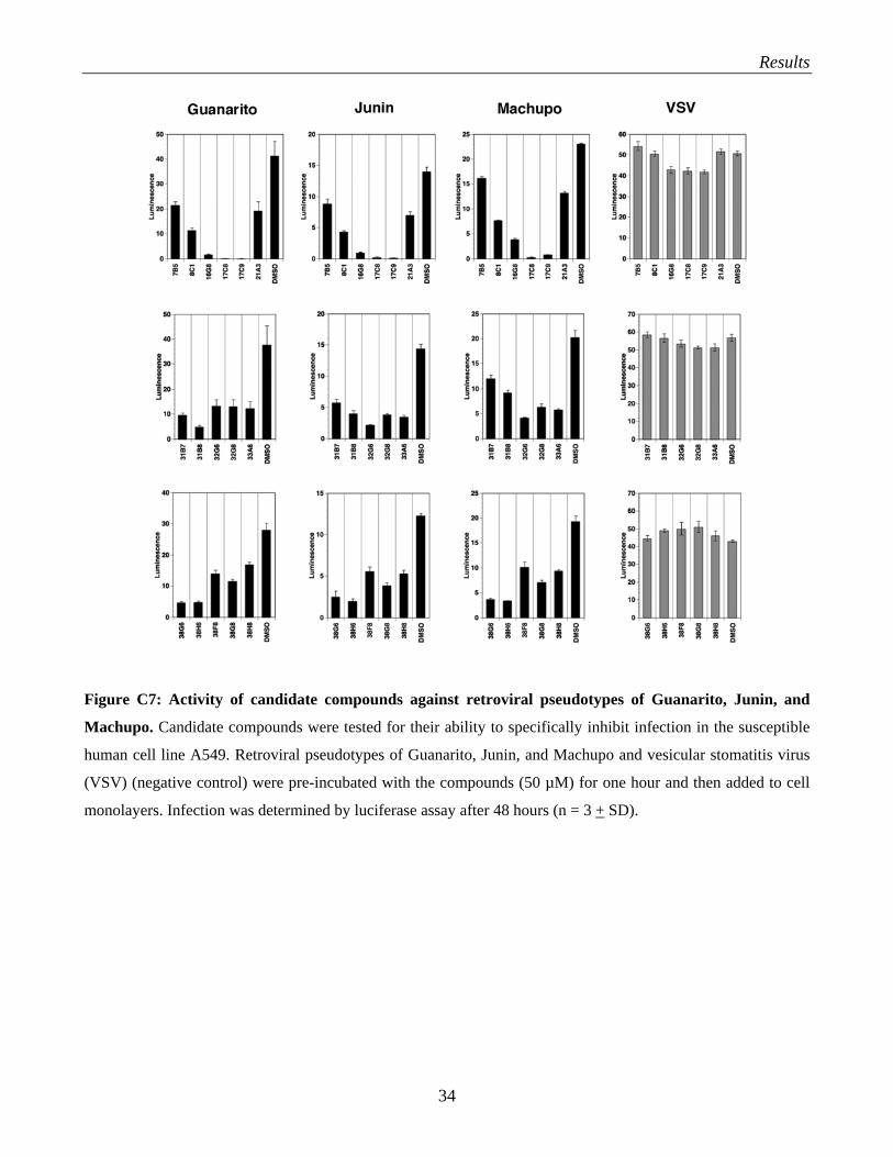
Results
Figure C7: Activity of candidate compounds against retroviral pseudotypes of Guanarito, Junin, and
Machupo. Candidate compounds were tested for their ability to specifically inhibit infection in the susceptible
human cell line A549. Retroviral pseudotypes of Guanarito, Junin, and Machupo and vesicular stomatitis virus
(VSV) (negative control) were pre-incubated with the compounds (50 µM) for one hour and then added to cell
monolayers. Infection was determined by luciferase assay after 48 hours (n = 3 + SD).
34
Results
C.2 Discovery of novel small molecule inhibitors of Lassa fever virus (LFV) infection from
natural product libraries:
During the past twenty years, several research groups have tested synthetic and natural compounds for
activity against arenaviruses and found some anti-viral activity. Trifluroperazine (TFP) and
chlorpromazine (CPZ), two pharmacologically active phenothiazine derivates, were found to be active
on the replication of the arenaviruses Junin, Tacaribe virus and Pichinde virus (Candurra et al., 1996).
Two myristic acid analogs (DL-2-hydroxymyristic acid (2OHM) and 13-oxamyristic acid (13OM)
showed to have effects on Junin virus replication (Cordo et al., 1999). Ribavirin triphosphate (RTP)
can interact with various viral RNA polymerases and inhibit virus replication (Parker, 2004). Synthetic
brassinosteriod ((22S,23S)-3ß-bromo-5α,22,23-trihydroxystimastan-6-one) has an antiviral mode of
action against Junin virus replication by preventing the synthesis of full length antigenomic RNA
(Castilla et al., 2005).
Since some of the compounds that show anti-viral activity like, hydroxymyristic acid, ribavirin
triphosphate, and brassinosteriods are natural products or derivatives thereof, we decided to perform a
broad screen of natural products for anti-viral activity. For this purpose, we used a natural product
library of 2, 000 defined and characterized natural compounds. The compounds were provided by the
Department of Chemistry at Scripps. The library was set up as 200 mixtures of 10 compounds each in
a concentration of 500 µM per compound in DMSO. In contrast to the combinatorial chemical libraries
used in our small molecule screens described above, the individual compounds present in the mixtures
of this library have no structural relatedness. The natural compounds are tested for inhibition for cell-
receptor binding and/or entry.
C.2.1 Screening of natural product libraries
Screening of natural product libraries was done in the same way as for the combinatorial chemical
libraries, but in addition to LFV-PS we tested the compounds for effect on Guanarito, Junin, and
Machupo pseudotypes. Initial screening of the three natural product libraries N1-N3 resulted in the
identification of a number of compound mixtures (with 10 compounds each) that significantly reduced
infection of human cells with either LFV or Machupo virus pseudotypes. To select for compounds that
block specific one of the respective psudotypes-mediated attachment and entry but do not influence
subsequent steps of our retroviral pseudotype-based assay, we compared the effect of our candidates
35
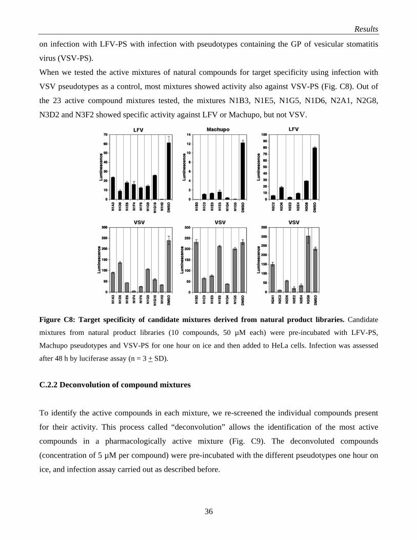
Results
on infection with LFV-PS with infection with pseudotypes containing the GP of vesicular stomatitis
virus (VSV-PS).
When we tested the active mixtures of natural compounds for target specificity using infection with
VSV pseudotypes as a control, most mixtures showed activity also against VSV-PS (Fig. C8). Out of
the 23 active compound mixtures tested, the mixtures N1B3, N1E5, N1G5, N1D6, N2A1, N2G8,
N3D2 and N3F2 showed specific activity against LFV or Machupo, but not VSV.
Figure C8: Target specificity of candidate mixtures derived from natural product libraries. Candidate
mixtures from natural product libraries (10 compounds, 50 µM each) were pre-incubated with LFV-PS,
Machupo pseudotypes and VSV-PS for one hour on ice and then added to HeLa cells. Infection was assessed
after 48 h by luciferase assay (n = 3 + SD).
C.2.2 Deconvolution of compound mixtures
To identify the active compounds in each mixture, we re-screened the individual compounds present
for their activity. This process called “deconvolution” allows the identification of the most active
compounds in a pharmacologically active mixture (Fig. C9). The deconvoluted compounds
(concentration of 5 µM per compound) were pre-incubated with the different pseudotypes one hour on
ice, and infection assay carried out as described before.
36
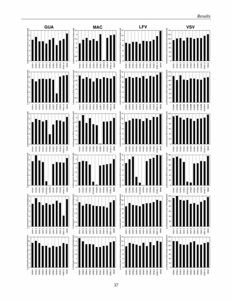
Results
37
Results
Figure C9: Deconvolution of candidate compound mixtures derived from natural product libraries.
Individual compounds (50 µM each) were preincubated with the indicated pseudotypes and then added to HeLa
cells. Infection was determined as in Fig. C2. Data shown are single measurements and represent one out of two
experiments. The candidate marked with * showed cytotoxicity on the cells.
The results clearly show which compounds are specific and which ones are not. Notably, most
compounds showed no activity when deconvoluted. In some mixtures, it was not possible to identify
individual compounds responsible for the observed anti-viral activity of the mixture. I other cases, one
or two compounds were found to have a significant effect while the rest did not. Notably, in one case,
we found that although the mixture showed specificity against arenaviruses but not VSV, two
individual compounds seem to have broader activity, also neutralizing VSV.
C.3 Evaluation of polyanionic compounds as anti-arenaviral drugs
Polyanionic compounds have been evaluated as very potent anti-viral drugs against arenaviruses, they
were found to be selective inhibitors, whereas the compounds were not inhibitory to cell growth.
Dextran sulfate, fucoidan and heparin were active at concentrations of 0.1-2.8 µg/ml and not cytotoxic
up to a concentration of 400 µg/ml. At concentrations below the cytotoxicity threshold (inhibition of
host cell DNA synthesis), the compounds inhibited the growth of Junin virus in Vero cells, as
measured at 24, 48 and 72 hours after infection. The mechanism of action of these compounds was
thought to be due to the inhibition of virus binding to the host cells (Andrei and de Clercq, 1990).
C.3.1 Selection of candidate polymers: heparin, HS, DS and fucoidan
Glycosaminoglycans (GAGs) are long unbranched polysaccharides containing a repeating disaccharide
unit. GAGs are highly negatively charged molecules, with extended conformation that imparts high
viscosity to the solution. GAGs are located primarly on the surface of the cell or in the extracellular
matrix. Many viruses utilize GAGs, predominantly heparan sulfate (HS) as receptor or co-receptors,
together with other molecules. Heparin and heparan sulfate (HS) are both GAGs of physiological
significance. Heparin is a modified (higher sulfated) version of HS and is more potent than HS.
Heparin is mainly found as a component of intacellular granules of mast cells lining the arteries of the
lungs, liver and skin. HS contains higher acetylated glucosamine than heparin and is mainly localized
at basement membranes as components of cell surfaces. If a virus uses HS as a receptor or co-receptor,
38
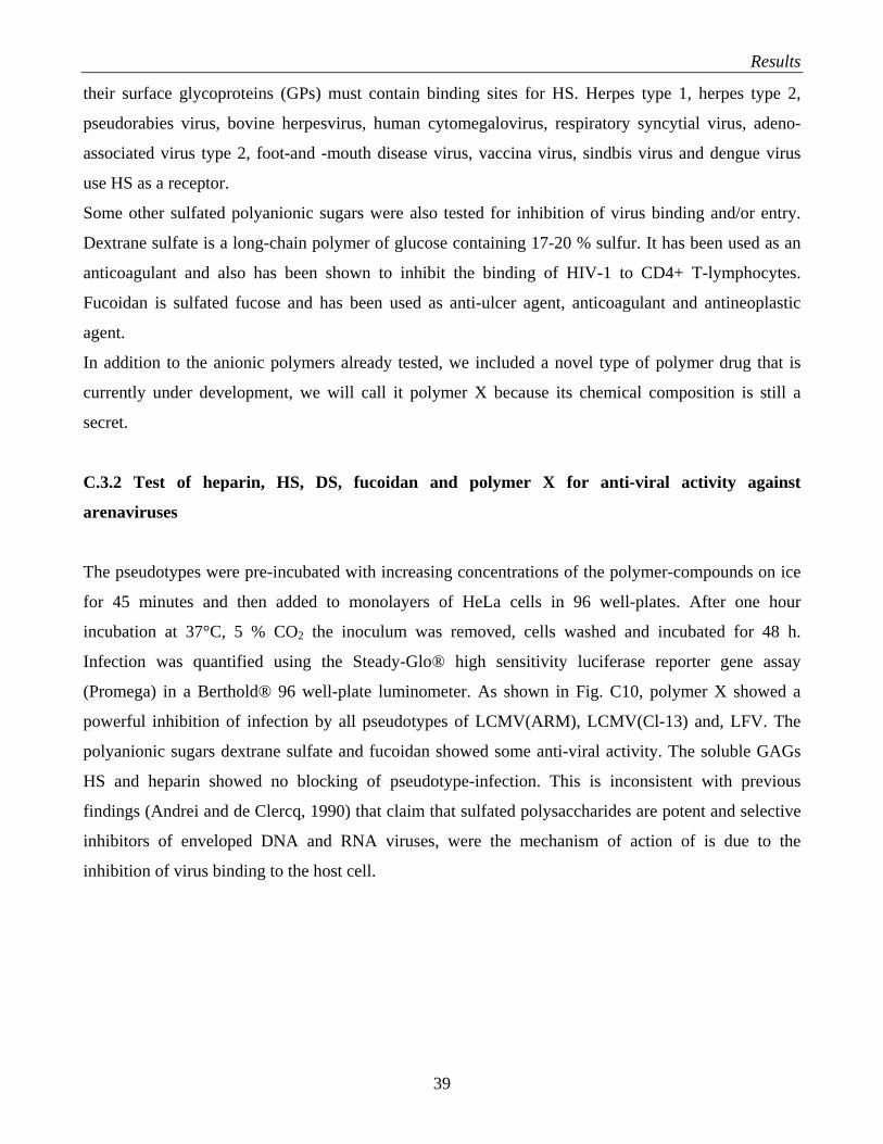
Results
their surface glycoproteins (GPs) must contain binding sites for HS. Herpes type 1, herpes type 2,
pseudorabies virus, bovine herpesvirus, human cytomegalovirus, respiratory syncytial virus, adeno-
associated virus type 2, foot-and -mouth disease virus, vaccina virus, sindbis virus and dengue virus
use HS as a receptor.
Some other sulfated polyanionic sugars were also tested for inhibition of virus binding and/or entry.
Dextrane sulfate is a long-chain polymer of glucose containing 17-20 % sulfur. It has been used as an
anticoagulant and also has been shown to inhibit the binding of HIV-1 to CD4+ T-lymphocytes.
Fucoidan is sulfated fucose and has been used as anti-ulcer agent, anticoagulant and antineoplastic
agent.
In addition to the anionic polymers already tested, we included a novel type of polymer drug that is
currently under development, we will call it polymer X because its chemical composition is still a
secret.
C.3.2 Test of heparin, HS, DS, fucoidan and polymer X for anti-viral activity against
arenaviruses
The pseudotypes were pre-incubated with increasing concentrations of the polymer-compounds on ice
for 45 minutes and then added to monolayers of HeLa cells in 96 well-plates. After one hour
incubation at 37°C, 5 % CO2 the inoculum was removed, cells washed and incubated for 48 h.
Infection was quantified using the Steady-Glo® high sensitivity luciferase reporter gene assay
(Promega) in a Berthold® 96 well-plate luminometer. As shown in Fig. C10, polymer X showed a
powerful inhibition of infection by all pseudotypes of LCMV(ARM), LCMV(Cl-13) and, LFV. The
polyanionic sugars dextrane sulfate and fucoidan showed some anti-viral activity. The soluble GAGs
HS and heparin showed no blocking of pseudotype-infection. This is inconsistent with previous
findings (Andrei and de Clercq, 1990) that claim that sulfated polysaccharides are potent and selective
inhibitors of enveloped DNA and RNA viruses, were the mechanism of action of is due to the
inhibition of virus binding to the host cell.
39
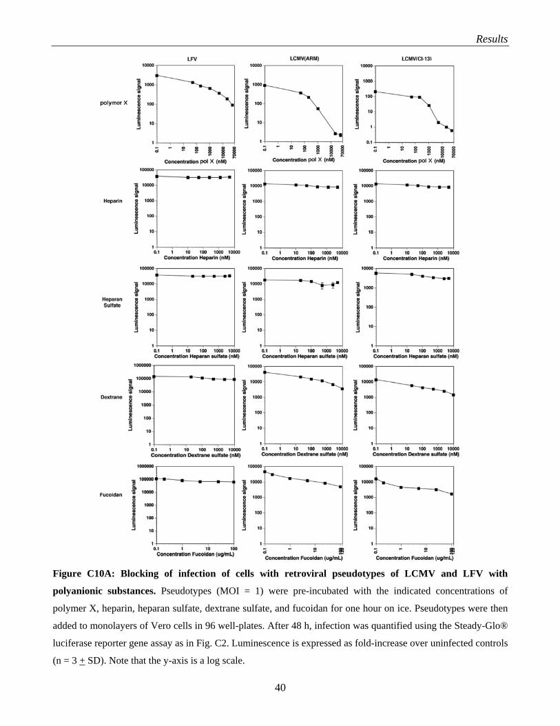
Results
Figure C10A: Blocking of infection of cells with retroviral pseudotypes of LCMV and LFV with
polyanionic substances. Pseudotypes (MOI = 1) were pre-incubated with the indicated concentrations of
polymer X, heparin, heparan sulfate, dextrane sulfate, and fucoidan for one hour on ice. Pseudotypes were then
added to monolayers of Vero cells in 96 well-plates. After 48 h, infection was quantified using the Steady-Glo®
luciferase reporter gene assay as in Fig. C2. Luminescence is expressed as fold-increase over uninfected controls
(n = 3 + SD). Note that the y-axis is a log scale.
40
Results
Figure C10B: Blocking of infection of cells with retroviral pseudotypes of Guanarito, Machupo, and
Junin with polyanionic substances. Pseudotypes (MOI = 1) were pre-incubated with the indicated
concentrations of polymer X, dextran sulfate, heparin, and fucoidan for one hour on ice. Pseudotypes were then
added to monolayers of Vero cells in 96 well-plates. After 48 h, infection was quantified as in Fig. C10A.
C.3.3 What is the role of HS and glycosaminoglycans in arenavirus infection?
To address the role of heparan sulfate in the infection of cells with arenaviruses, we made use of the
mutant CHO cell line pgsA-745 that is deficient in UDP-D-xylose: serine-1, 3-D xylosyltransferase
and lacks all glycosaminoglycans (Esko et al., 1985). PsgA-754 and wild-type CHOK1 cells were
infected with pseudotypes of LCMV (ARM53b, cl-13), LFV, Guanarito, Junin, and Machupo, as well
as VSV-pseudotypes that do not depend on glycosaminoglycans for infection. As shown in Fig. C11,
pgsA-745 that lack all glycosaminoglycans are more efficiently infected than normal CHOK1 cells,
41
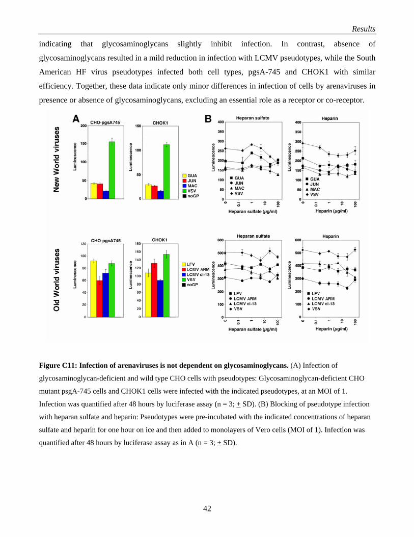
Results
indicating that glycosaminoglycans slightly inhibit infection. In contrast, absence of
glycosaminoglycans resulted in a mild reduction in infection with LCMV pseudotypes, while the South
American HF virus pseudotypes infected both cell types, pgsA-745 and CHOK1 with similar
efficiency. Together, these data indicate only minor differences in infection of cells by arenaviruses in
presence or absence of glycosaminoglycans, excluding an essential role as a receptor or co-receptor.
Figure C11: Infection of arenaviruses is not dependent on glycosaminoglycans. (A) Infection of
glycosaminoglycan-deficient and wild type CHO cells with pseudotypes: Glycosaminoglycan-deficient CHO
mutant psgA-745 cells and CHOK1 cells were infected with the indicated pseudotypes, at an MOI of 1.
Infection was quantified after 48 hours by luciferase assay (n = 3; + SD). (B) Blocking of pseudotype infection
with heparan sulfate and heparin: Pseudotypes were pre-incubated with the indicated concentrations of heparan
sulfate and heparin for one hour on ice and then added to monolayers of Vero cells (MOI of 1). Infection was
quantified after 48 hours by luciferase assay as in A (n = 3; + SD).
42
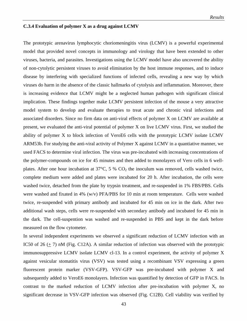
Results
C.3.4 Evaluation of polymer X as a drug against LCMV
The prototypic arenavirus lymphocytic choriomeningitis virus (LCMV) is a powerful experimental
model that provided novel concepts in immunology and virology that have been extended to other
viruses, bacteria, and parasites. Investigations using the LCMV model have also uncovered the ability
of non-cytolytic persistent viruses to avoid elimination by the host immune responses, and to induce
disease by interfering with specialized functions of infected cells, revealing a new way by which
viruses do harm in the absence of the classic hallmarks of cytolysis and inflammation. Moreover, there
is increasing evidence that LCMV might be a neglected human pathogen with significant clinical
implication. These findings together make LCMV persistent infection of the mouse a very attractive
model system to develop and evaluate therapies to treat acute and chronic viral infections and
associated disorders. Since no firm data on anti-viral effects of polymer X on LCMV are available at
present, we evaluated the anti-viral potential of polymer X on live LCMV virus. First, we studied the
ability of polymer X to block infection of VeroE6 cells with the prototypic LCMV isolate LCMV
ARM53b. For studying the anti-viral activity of Polymer X against LCMV in a quantiative manner, we
used FACS to determine viral infection. The virus was pre-incubated with increasing concentrations of
the polymer-compounds on ice for 45 minutes and then added to monolayers of Vero cells in 6 well-
plates. After one hour incubation at 37°C, 5 % CO2 the inoculum was removed, cells washed twice,
complete medium were added and plates were incubated for 20 h. After incubation, the cells were
washed twice, detached from the plate by trypsin treatment, and re-suspended in 1% FBS/PBS. Cells
were washed and fixated in 4% (w/v) PFA/PBS for 10 min at room temperature. Cells were washed
twice, re-suspended with primary antibody and incubated for 45 min on ice in the dark. After two
additional wash steps, cells were re-suspended with secondary antibody and incubated for 45 min in
the dark. The cell-suspention was washed and re-suspended in PBS and kept in the dark before
measured on the flow cytometer.
In several independent experiments we observed a significant reduction of LCMV infection with an
IC50 of 26 (+ 7) nM (Fig. C12A). A similar reduction of infection was observed with the prototypic
immunosuppressive LCMV isolate LCMV cl-13. In a control experiment, the activity of polymer X
against vesicular stomatitis virus (VSV) was tested using a recombinant VSV expressing a green
fluorescent protein marker (VSV-GFP). VSV-GFP was pre-incubated with polymer X and
subsequently added to VeroE6 monolayers. Infection was quantified by detection of GFP in FACS. In
contrast to the marked reduction of LCMV infection after pre-incubation with polymer X, no
significant decrease in VSV-GFP infection was observed (Fig. C12B). Cell viability was verified by
43
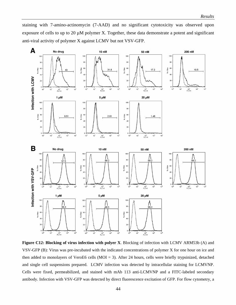
Results
staining with 7-amino-actinomycin (7-AAD) and no significant cytotoxicity was observed upon
exposure of cells to up to 20 µM polymer X. Together, these data demonstrate a potent and significant
anti-viral activity of polymer X against LCMV but not VSV-GFP.
Figure C12: Blocking of virus infection with polyer X. Blocking of infection with LCMV ARM53b (A) and
VSV-GFP (B): Virus was pre-incubated with the indicated concentrations of polymer X for one hour on ice and
then added to monolayers of VeroE6 cells (MOI = 3). After 24 hours, cells were briefly trypsinized, detached
and single cell suspensions prepared. LCMV infection was detected by intracellular staining for LCMVNP.
Cells were fixed, permeabilized, and stained with mAb 113 anti-LCMVNP and a FITC-labeled secondary
antibody. Infection with VSV-GFP was detected by direct fluorescence excitation of GFP. For flow cytometry, a
44
Results
FACSCalibur® flow cytometer was used. In histograms, the y-axis represents cell numbers and the x-axis
fluorescence intensity (FL1-H for FITC/GFP). The dotted line represents uninfected controls and the solid line
infected samples. Percentages of NP or GFP positive cells are given. One representative experiment out of three
is shown.
To determine the step in LCMV infection blocked by polymer X, the drug was added at different time
points relative to infection. LCMV ARM53b was used at MOI of 0.01, 0.1, and 1. Polymer X (2 µM)
was added exactly at the time of infection, 6 hours post infection, or 24 hours post infection. Infection
levels were assessed after a total infection period of 48 hours by determination of LCMVNP
expression using immunofluorescence staining with mAb 113 anti-LCMVNP (Fig. C13A) or FACS
(Fig. C13B). Vero cells were cultivated in 8 well LabTek chamber slides and infected with
LCMV(ARM53b) at MOI of 0.1. For one sample, LCMV were pre-incubated with the polymer X one
hour on ice before added to the cells. For the other samples, polymer X was added at different
timepoints after infection. After a total infection time of 48 hours, the medium/virus/polymer X
solution removed and cells fixed at 37°C for 15 min in the dark. Cells were washed twice and
incubated with PBS/1%(v/v) FCS for 15min at room temperature. The cells were so incubated 15 min
with PBS/1%(v/v) FCS/0.1% (w/v) saponin. Primary antibody mAb 113 anti-LCMVNP was added and
cells incubated for 1 hour at room temperature in the dark. Cells were washed twice and incubated with
a FITC-conjugated secondary antibody for 45 min at room temperature in the dark. The cells were so
washed three times before 1 drop of Vectashield were added to each fixing square and the cells
covered with a coverglass. Images were acquired using a fluorescence microscope.
As shown in Fig. C13, the data strongly suggest that the polymer drug blocks an early step in viral
infection. When added at the time of infection, polymer X efficiently blocked infection of LCMV at all
MOI tested, indicating fast inhibition of an early step in infection. Addition of polymer X after 6 or 24
hours resulted in marked decrease in the number of infected cells, indicating efficient blocking of cell-
to-cell spread. However, the similar levels of LCMVNP expression (mean immunofluorescence
intensities) in infected cells in presence and absence of polymer X indicate that the drug has little
effect on LCMVNP expression once infection has occurred (Fig. C13B).
Together, the data show that polymer X has a specific anti-viral activity against prototypic isolates of
LCMV. The drug efficiently blocks initial steps of virus infection and prevent cell-to-cell spreading of
virus. That means that polymer X is active in an early step of the virus lifecycle. The efficient
neutralization of the retroviral pseudotypes of LCMV indicates further that the viral GP is the main
drug target.
45
Results
Figure C13: Polymer X blocks infection and cell-to-cell spread of LCMV: (A) Detection of virus infection
and propagation by immunofluorescence. Monolayers of VeroE6 cells were infected with LCMV ARM53b at
MOI of 0.1 and polymer X added at the indicated time points. After 48 hours, cells were fixed, permeabilized
and stained with mAb 113 anti-LCMVNP using a FITC-conjugated secondary antibody and phalloidin-
rhodamine for counterstaining of F-actin filaments. (B) Monolayers of VeroE6 cells were infected with LCMV
ARM53b at MOI of 0.01, 0.1, and 1 and polymer X added at the indicated time points. After 48 hours, infection
46
Results
was determined by FACS staining for LCMVNP as in Fig. C12. The dotted line represents uninfected controls
and the solid line infected samples. Percentages of NP positive cells are given.
C.4 Production of recombinant Lassa fever virus glycoprotein
The glycoprotein (GP) of Lassa fever virus (LFV) serves as virus attachment protein to its receptor on
host cells. The GP precursor (GP-C) is post-translationally cleaved into GP1, which is implicated in
receptor binding and GP2 that is structurally similar to the fusion active membrane proximal portions
of the GPs of other enveloped viruses. In my studies I have identified a number of small molecule
compounds and polymer drugs that block infection of human cells by retroviral pseudotypes
containing the GP of LFV (LFVGP). Among the candidate drugs, compounds and substances that
target the viral GP are of greatest interest since they are able to target the free virus and will most
likely not interfere with crucial cell function, which will minimize their potential for adverse side
effects.
As pointed out in the introduction (section A.1.3), the viral GP at the surface of free LFV virions is in
its metastable pre-fusion state. These pre-fusion state GP trimers are the functional units of target cell
recognition, entry and fusion in arenaviruses. The production of a stable, trimeric form of LFVGP in
its pre-fusion state is therefore of paramount importance regarding the identification of anti-viral drugs
targeting the GP.
To prevent the transition from the (metastable) pre-fusion into the energetically more stable post-
fusion complex, the GP2 subunits containing the fusion machinery have to be locked into a stable
trimer that does not allow significant conformational changes. Recent attempts to express recombinant
influenza hemagglutinin (HA) derived from the influenza strain of the 1918 pandemic faced the
problem that the HA did not form trimers (Stevens et al., 2004). Among the potential trimerization
domains tested by Stevens and colleagues, the recently identified foldon domain derived from
bacteriophage T4 fibritin (Tao et al., 1997) turned out to be very efficient in the induction of trimer
formation. The foldon domain (Fig. C14), spans the C-terminal residues G457 to L483 of the
bacteriophage T4 fibritin and forms a trimeric complex of very high stability. Based on its properties
as a powerful driving force for trimerization, we will construct fusion proteins of the ectrodomain of
LFVGP and the foldon-domain (Fig. C15). These LFVGP-foldon constructs will be tested for trimer
formation and receptor binding. Trimers formed by the LFVGP-foldon fusion proteins may be
powerful tools for drug screening and for the investigation of the virus-cell interaction.
47
Results
Figure C14: Structure of the foldon domain of bacteriophage T4 fibritin (from Tao et al., 1997). (a) Stereo
diagram of the C-terminal foldon domain of a fibritin E subunit. The sidechains shown are those located in the
hydrophobic interior formed at the interface between the subunits. (b) Ribbon diagram of the foldon domain
looking along the trimer axis. (c) Hydrogen of the main chain formed within the foldon domain.
Figure C15: Design of a soluble trimeric form of LFVGP locked in its pre-fusion state. For details see text.
48
Results
C.4.1 Design of the LFVGP-foldon fusion protein
Layout of the foldon cassette:
The T4 fibritin foldon domain (GYIPEAPRDGQAYVRKDGEWVLLSTFL)
corresponds to residues G457 to L483 of the bacteriophage T4 fibritin (Tao et al., 1997).
The fusion of the hole sequence of LFVGP from M(1) to LGLV(431) derived from LFV Josiah with
the foldon domain will include a GGGS linker between the LFVGP and the foldon domain. In
addition, we will add a flag-tag (DYKDDDDK), a throbin cleavage site (LVPR/GS), and a six His tag
that allows purification of the soluble recombinant protein by Ni-NTA affinity chromatography.
Design of the LFVGP-foldon fusion protein: M(1)…LGLV(431)GGGSGYIPEAPRDGQAYVRKDGEWVLLSTFLDYKDDDDK LVPR/GS
HHHHHHStop
L G L V(431) 5’- TTG GGT CTA GTT GGC GGT GGA TCC GGC TAC ATC CCC GAG GCC CCG CGC GAC
GGC CAG GCC TAC GTG CGC AAG GAC GGC GAG TGG GTG CTG CTG TCC ACC TTC CTG
GAC TAC AAA GAC GAT GAC GAT AAA CTT GTC CCA CGT GGT TCT CAT CAC CAT CAC
CAT CAC TAG
Flag: 5’- GAC TAC AAA GAC GAT GAC GAT AAA-3’ D Y K D D D D K
Thrombin cleavage side: CTT GTC CCA CGT GGT TCT
L V P R / G S
A cDNA sequence containing the foldon cassette, followed by the flag-tag, thrombin cleavage site, and
His-tag has previously been made, subcloned into a basis vector (pBluescript), and verified by double
strand DNA sequencing. For the construction of the fusion proteins with LFVGP, the corresponding
sequence of LFVGP M(1)… LGLV(431) derived from LFV Josiah were amplified by PCR using the
cDNA of full-length LFVGP as a template as described in M&M.
The fragment from LFVGP pcAGGs cDNA was amplified in PCR with the forward primer at position
1130 and the backward primer LFEb that makes a blunt, phosphorylated 5’ end at position 1348. This
fragment contains a restriction-site, KpnI, at position 1164 that was used for cutting the fragment. The
49
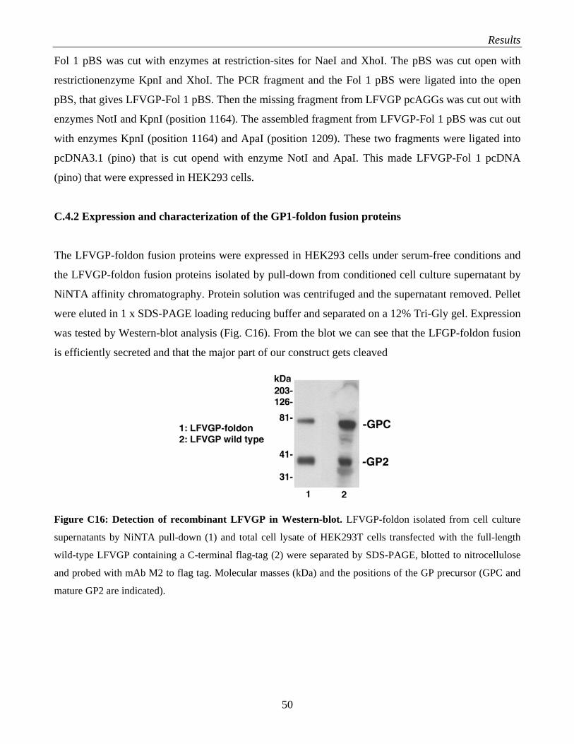
Results
Fol 1 pBS was cut with enzymes at restriction-sites for NaeI and XhoI. The pBS was cut open with
restrictionenzyme KpnI and XhoI. The PCR fragment and the Fol 1 pBS were ligated into the open
pBS, that gives LFVGP-Fol 1 pBS. Then the missing fragment from LFVGP pcAGGs was cut out with
enzymes NotI and KpnI (position 1164). The assembled fragment from LFVGP-Fol 1 pBS was cut out
with enzymes KpnI (position 1164) and ApaI (position 1209). These two fragments were ligated into
pcDNA3.1 (pino) that is cut opend with enzyme NotI and ApaI. This made LFVGP-Fol 1 pcDNA
(pino) that were expressed in HEK293 cells.
C.4.2 Expression and characterization of the GP1-foldon fusion proteins
The LFVGP-foldon fusion proteins were expressed in HEK293 cells under serum-free conditions and
the LFVGP-foldon fusion proteins isolated by pull-down from conditioned cell culture supernatant by
NiNTA affinity chromatography. Protein solution was centrifuged and the supernatant removed. Pellet
were eluted in 1 x SDS-PAGE loading reducing buffer and separated on a 12% Tri-Gly gel. Expression
was tested by Western-blot analysis (Fig. C16). From the blot we can see that the LFGP-foldon fusion
is efficiently secreted and that the major part of our construct gets cleaved
Figure C16: Detection of recombinant LFVGP in Western-blot. LFVGP-foldon isolated from cell culture
supernatants by NiNTA pull-down (1) and total cell lysate of HEK293T cells transfected with the full-length
wild-type LFVGP containing a C-terminal flag-tag (2) were separated by SDS-PAGE, blotted to nitrocellulose
and probed with mAb M2 to flag tag. Molecular masses (kDa) and the positions of the GP precursor (GPC and
mature GP2 are indicated).
50
Discussion
D. DISCUSSION
The goal with this study is to find a drug that can fight Lassa fever that affects an estimated 180
million individuals in Western Africa. There is neither an efficient cure nor an efficacious vaccine for
LFV. With over 200 000 infections per year and several thousand deaths, LFV represents a severe
threat for human health and a major humanitarian problem. Ribavirin is so far the only drug for
patients infected with LFV. Ribavirin does not neutralize free virus, but works in the late steps of the
virus lifecycle. The goal is to develop drugs that can act as “gatekeepers”, i.e. drugs that are able to
block the early step of the virus lifecycle, the virus-receptor binding and/or entry. To block the virus
binding and/or entry and then cell to cell spread, will give the infected patients immune system a better
chance in generating an efficient anti-viral immune response.
We tested small molecule derived from combinatorial chemical libraries to see if there was any
blocking of the virus-receptor interaction. We also tested sulfated polysaccharides like dextrane
sulfate, fucoidan, heparan sulfate and heparin that have showed anti-viral activity in studies already
done. In addition, we tested polymer X that is an anionic polymer that has showed activity against
HIV.
D.1 Discovery of novel small molecule inhibitors of Lassa fever virus (LFV) infection from
combinatorial chemical libraries and natural product libraries
To identify potential anti-viral compounds, small molecule inhibitors of protein interaction derived
from combinatorial chemical libraries were screened. This strategy has been used to identify several
physiologically active inhibitors of novel biochemically not yet characterized interactions. The
compounds/compounds mixtures were screened for their ability to block retroviral pseudotype
infection in target cells. Retroviral pseudotypes contain LFVGP in their envelope and a luciferace
reporter gene in their genome. The luciferase reporter gene makes the pseudotypeinfection easy to
detect. Since live LFV is a BSL4 pathogen, studies with virus are restricted to BSL4 laboratories. The
pseudotypes allow us to do our work under BSL2 conditions.
Many studies on receptor-ligand binding indicate that in most cases, small clusters of amino acid
recidues mediate the energetically most important interaction. Using Dr. Boger's libraries,
erythropoieitin (EPO) mimetics (promote EPO receptor dimerization) (Goldberg et al., 2002),
inhibitors of Myc/Max dimerization (Berg et al., 2002), inhibitors of androgen receptor binding to
DNA (Boger et al., 2000a), inhibitors of LEF-1/β-catenin dimerization (Boger et al., 2000b), and
51
Discussion
inhibitors of the binding of the protease MMP-2 to αvß3 integrin have been discovered (Silletti et al.,
2001; Boger et al., 2001). That makes us think that small molecules, interacting with the reseptor-
binding site for the virus, can block virus infection in cells.
We screened combinatorial libraries of small molecules that consist of imidodiacetic acid-based,
pyrrolidine-based peptidomimetic but also heterolytic compounds. The compounds were not attached
to solid supports making them immediately available for binding or functional assays. I tested 13
complete libraries. Each library had 80 wells that contained single compounds or mixtures of 4-10
compounds (that made the screening more effective). Totally this gave a number > 10,000 compounds.
Duplicate screening resulted in the identification of circa 4-5% compounds/mixtures that showed
reproducible reduced LFV-PS infection by > 50%. To test our candidate for specificity, we screened
the compounds for inhibition of VSV-pseudotype infection. VSVGP is structurally unrelated to
LFVGP and binds to a different receptor. If the compounds are specific, they should not affect the
infection of target cells by VSV-pseudotype. My screening together with previous screenings done in
the lab of another 31 libraries, corresponding to > 50, 000 compounds yielded 157 (4.5%)
compounds/mixtures that showed reproducible reduced LFV-PS infection. Determination of target
specificity revealed that 32 single compounds and 53 compound mixtures out of a total of 125
specifically reduced infection in LFV-PS but not VSV-PS. We found difference in specificity between
different libraries; some were very consistent, with most candidate compounds being specific for
inhibition of LFV-GP infection, when other compounds showed effect both against LFV-PS and VSV-
GP.
A selection of 17 individual compounds was then further characterized. The selection was made based
on the extent of inhibition, degree of specificity, and also chemical considerations, raised by our
chemist collaborators. The selected compounds were tested for inhibition of pseudotype infection in
different cell-lines (He-La, A549 and VeroE6) that showed that the candidate compounds had
consistent blocking of LFV-PS infection in all cell lines and were independent of the specific target.
The determination of the dose-response of the individual compounds was also tested on the different
cell lines. We found 8 candidate compounds to be particularly good selective inhibitors against LFV-
PS. Many of these candidate compounds were also active against the human pathogenic South
American HF viruses Guanarito, Junin and Machupo. The responses to the compounds in the different
NWAV-PS were very similar as in LFV-PS. Structurally our candidate compounds were hydrophobic
molecules with a high degree of aromaticity. Comparison of the most active structures revealed that an
overall tripartite symmetry was a common structural theme. Examples are shown in Fig. D1.
52
Discussion
Figure D1: Representative examples of candidate compounds
Our candidate drugs may block one or several of the following steps: virus-receptor binding, binding
of the viral GP to a yet unknown co-receptor, virus internalization by vesicles, and pH-dependent
fusion of the late endosome membrane with the viral membrane that releases the virus into the
cytoplasm (Fig. D2). More or less modified, these compounds are small and an oral administration of
the drug is therefore possible (as far as the stability and absorption in the GI-tract not cause any
problems). An oral route of administration would be of importance for better viability for the patients
when this does not need any specialized assistance.
D.2 Evaluation of polyanionic compounds as anti-arenaviral drugs
We tested polyanionic compounds as anti-arenaviral drugs because they have from earlier studies
showed antiviral activity against arenaviruses. They were found to be selective inhibitors, whereas the
compounds were not inhibitory to cell growth. In Graciela Andrei and Erik De Clercqs paper (1990) it
is claimed that the polyanionic compounds worked at the early step of infection of arenaviruses, by
inhibiting reseptor binding and/or fusion and entry. In general poly-anionic compounds have showed
activity against HIV-1 and other viruses. The anti-HIV-1 activity decreased with increasing the
hydrophobicity (Clercq et al, 1995). In our study, we tested heparin, heparin sulfate (HS), dextrane
sulfate and fucoidan. We selected those because many viruses utilize glycosaminoglycans,
predominantly HS as receptor or co-receptors, together with other molecules. If a virus use HS as a
receptor or co-receptor, their surface glycoproteins must contain binding sites for HS. By adding the
glycosaminoglycans to the viruses, the binding site for glycosaminoglycans will be occupied and the
virus will not be able to infect the target cell. Our results showed that dextrane sulfate and fucoidan
had some anti-viral activity. Heparin and HS did not show any significant anti-viral activity. The
53
Discussion
results did not come out as we expected them from the study already done (Andrei and de Clercq,
1990). The glycosaminoglycans did not show any significant anti-viral activity on the receptor
binding/fusion/entry stage. The glycosaminoglycans must therefore have a different mechanism of
action than Clercq et al claimed. The glycosaminoglycans antiviral-effect may have a mechanism
related to a later step in the virus life-cycle than the steps we tested (virus receptor
binding/fusion/entry).
We can suggest a general model for the (normally weak) anti-viral effect of polyanionic compounds
and the increased infection often seen after application of polycations. Increased infection after
application of polycations is caused of the decrease in repulsions between the viruses. Polycations are
of the opposite charge than the surface of the virus particles that are negatively charged (because of the
sugars attached). Polycations will therefore lower the surface energy of the viruses so they can easier
go together in clusters. A cluster of viruses will then be able to enter the cell through one receptor
instead of one single virus. We will then see an increased infection.
If we look at the different activities of heparin/HS vs. dextrane sulfate/fucoidan, and polymer X, it
seems like it is not only the charge of the polymer that is responsible for anti-viral activity. The
hydrophobicity of the compound is also of importance. The more hydrophobic the compound is, the
better anti-viral activity. Polymer X shows broad anti-viral activity. It shows nearly full inhibition of
infection of all the pseudotypes. Polymer X has the same charge as the polyanionic compounds, but is
more hydrophobic. Tested on real virus (LCMV and VSV), the polymer X shows specificity. Polymer
X acts at the step of infection. If it blocks the receptor-binding, fusion and/or entry is still unknown.
Since the polymer X act at the step of infection, it can inhibit cell-to-cell spread. The polymer X should
for that reason be used early in a virus-infection. Anyway, if cells are protected against re-infection in
an infected individual, the immune system will have a greater opportunity to clear the infection.
Polymer X is a huge molecule; it is too big to be absorbed in the GI-tract and must be administrated
i.v. When the mechanism of action is understood for polymer X and the small molecule drugs, it is a
possibility to combine the two drugs to make a better anti-viral effect.
D.3 Production of recombinant Lassa fever virus glycoprotein
Among the candidate drugs, the compounds and substrates that target the viral GP are of greatest
interest since they are able to target the free virus and will most likely not interfere with crucial cell
function, which will minimize their potential for adverse side effects. The viral GP at the surface of
free LFV virions is in its metastable pre-fusion state. These pre-fusion state GP trimers are the
54

Discussion
functional units of target cell recognition, entry and fusion in arenaviruses. The production of a stable,
trimeric form of LFVGP in its prefusion state is therefore important regarding the identification of
anti-viral drugs targeting the GP. It is critical to have the GP in its prefusion state, because this is the
state where the drugs have to be active (before infection of the cell). The conformation of the GP is
changed in its post-fusion state and the drugs may not recognize this conformation and the study will
then not be correct. To prevent the transition from the (metastable) pre-fusion into the energetically
more stable post-fusion complex, the GP2 subunits contain the fusion machinery have to be locked into
a stable trimer that does not allow significant conformational changes. These LFVGP-foldon
constructs will be tested for trimer formation and receptor-binding. Someone else has already done a
similar approach (Eschli et al., 2006) and that worked out well. Our approach worked out as expected.
We got expression of LFVGP-foldon and 80% of the constructs were cleaved. Preventing this cleavage
by mutate its cleavage site, can possible give the construct more stability.
D.4 Outlook
My work at Scripps resulted in the identification and first characterization of a number of candidate
small molecules that show specific anti-viral activity against pseudotypes of LFV and other human
pathogenic arenaviruses and in the demonstration of a potent anti-viral effect of a novel polymer drug
against LCMV. My results provide the basis for the following studies that will further characterize and
optimize our drug candidates.
D.4.1 Further validation of candidate compounds
Determination of the drug target: To avoid undesired side effects of our small molecule drugs, we
will select for compounds that efficiently block LFV infection by specifically targeting the viral GP
but not cellular receptor molecules. For this purpose, the recombinant soluble form of LFVGP, which I
made (Results, section C4) will be used.
Validation of activity of candidate drugs against live LFV: Activity of candidate drugs against live
LFV will be tested in a BSL4 facility by Dr. Christina Spiropoulou from the Special Pathogens Branch
of the Centers for Disease Control and Prevention (CDC) in Atlanta, GA. The Special Pathogens
Branch has also a well-established small animal model (guinea pig) for LFV infection for later in vivo
testing of candidate compounds.
55
Discussion
D.4.2 Determination of the mechanism of action of candidate drugs
Once we have identified specific compounds that block infection of human target cells with LFV in an
efficient manner, we will start to investigate the exact mechanism of their activity. The drugs could
target the following steps or combinations thereof: 1) the binding site(s) on LFVGP for its cellular
receptor(s), 2) the pre-fusion conformation of LFVGP as present at the virion surface, and 3) A
conformational intermediate of LFVGP occurring during pH-dependent membrane fusion (Fig. D2).
Figure D2: Potential steps in LFVGP-mediated infection that may be blocked by our drugs.
D.4.3 Extension of small molecule screening to other drug targets and other emerging pathogens
Although inhibition of virus-host cell binding and entry is a very promising anti-viral strategy for
highly pathogenic viruses, it is a good idea to aim at the development of high throughput screening
assays targeting other steps in virus multiplication. One attractive target for inhibitors is the proteolytic
processing of LFVGP. We and others have shown that the GPs of the Old World arenaviruses LFV and
LCMV are cleaved by the subtilase SKI-1/S1P and that processing of the GP is essential for viral
infectivity (Lenz et al., 2001; Beyer et al., 2003; Kunz et al., 2003). Since S1P protease is involved in
cholesterol metabolism of the host cell, a temporary block of S1P activity could be compensated by
dietary cholesterol supplementation. The S1P protease appears therefore as an attractive drug target to
interfere with the processing of LFVGP and thus virus multiplication. Another promising target for
interference with viral multiplication is the assembly of LFV virions. Since the presence of the viral
56
Discussion
GP is absolutely required for LFV particles to be infectious, GP incorporation during virus assembly at
the plasma membrane is another promising target for interference by anti-viral drugs. Based on the
incorporation of recombinant LFVGP in our pseudovirion system, one might try to establish an assay
system for the screening for small molecule drugs that prevent the formation of mature LFVGP whose
then can be incorporated into infections particles.
The identification of candidate small molecule anti-viral drugs against emerging viruses will
likely be of enormous benefit for the development of novel anti-viral therapeutics for these severe
human pathogens. Furthermore, the elucidation of their mechanism of action will shed light on
the molecular mechanism underlying virus-receptor binding, entry, and virus GP-mediated
membrane fusion of human pathogenic arenaviruses and thus identify novel drug targets and
contribute to subsequent rational drug design.
57
References
E. REFERENCES
Andrei, G., and De Clercq, E. (1990). Inhibitory effect of selected antiviral compounds on arenavirus
replication in vitro. Antiviral Res 14(4-5), 287-99.
Barresi, R., and Campbell, K. P. (2006). Dystroglycan: from biosynthesis to pathogenesis of human
disease. J Cell Sci. 119(Pt 2), 199-207.
Berg, T., Cohen, S. B., Desharnais, J., Sonderegger, C., Maslyar, D. J., Goldberg, J., Boger, D. L., and
Vogt, P. K. (2002). Small-molecule antagonists of Myc/Max dimerization inhibit Myc-induced
transformation of chicken embryo fibroblasts. Proc Natl Acad Sci U S A 99(6), 3830-5.
Beyer, W. R., Popplau, D., Garten, W., von Laer, D., and Lenz, O. (2003). Endoproteolytic processing
of the lymphocytic choriomeningitis virus glycoprotein by the subtilase SKI-1/S1P. J Virol 77(5),
2866-72.
Boger, D. L. (2003). Solution-phase synthesis of combinatorial libraries designed to modulate protein-
protein or protein-DNA interactions. Bioorg Med Chem 11(8), 1607-13.
Boger, D. L., Desharnais, J., and Capps, K. (2003). Solution-phase combinatorial libraries: modulating
cellular signaling by targeting protein-protein or protein-DNA interactions. Angew Chem Int Ed Engl
42(35), 4138-76.
Boger, D. L., Ducray, P., Chai, W., Jiang, W., and Goldberg, J. (1998). Higher order iminodiacetic
acid libraries for probing protein-protein interactions. Bioorg Med Chem Lett 8(17), 2339-44.
Boger, D. L., Goldberg, J., Satho, S., Ambroise, Y., Cohen, S. B., and Vogt, P. K. (2000). Non-amide
based combinatorial libraries derived from N-Boc-iminodiacetic acid: solution-pahse synthesis of a
150-membered piperazinone library with activity against LEF-1/beta-catenin mediated transcription.
Helv. Chim. Acta 83, 1825-1845.
58
References
Boger, D. L., Goldberg, J., Silletti, S., Kessler, T., and Cheresh, D. A. (2001). Identification of a novel
class of small-molecule antiangiogenic agents through the screening of combinatorial libraries which
function by inhibiting the binding and localization of proteinase MMP2 to integrin alpha(V)beta(3). J
Am Chem Soc 123(7), 1280-8.
Borrow, P., and Oldstone, M. B. (1994). Mechanism of lymphocytic choriomeningitis virus entry into
cells. Virology 198(1), 1-9.
Buchmeier, M. J., Bowen, M. D., and Peters, C. J. (2001). Arenaviridae: the virus and their replication.
Fourth ed. In “Fields Virology” (D. M. Knipe, and P. M. Howley, Eds.), Vol. 2, pp. 1635-1668. 2 vols.
Lippincott Williams & Wilkins, Philadelphia.
Buchmeier, M. J., Lewicki, H. A., Tomori, O., and Oldstone, M. B. (1981). Monoclonal antibodies to
lymphocytic choriomeningitis and pichinde viruses: generation, characterization, and cross-reactivity
with other arenaviruses. Virology 113(1), 73-85.
Burns, J. W., and Buchmeier, M. J. (1991). Protein-protein interactions in lymphocytic
choriomeningitis virus. Virology 183(2), 620-9.
Candurra, N. A., Maskin, L., and Damonte, E. B. (1996). Inhibition of arenavirus multiplication in
vitro by phenotiazines. Antiviral Res 31(3), 149-58.
Cao, W., Henry, M. D., Borrow, P., Yamada, H., Elder, J. H., Ravkov, E. V., Nichol, S. T., Compans,
R. W., Campbell, K. P., and Oldstone, M. B. (1998). Identification of alpha-dystroglycan as a receptor
for lymphocytic choriomeningitis virus and Lassa fever virus [see comments]. Science 282(5396),
2079-81.
Castilla, V., Larzabal, M., Sgalippa, N. A., Wachsman, M. B., and Coto, C. E. (2005). Antiviral mode
of action of a synthetic brassinosteroid against Junin virus replication. Antiviral Res. 68(2), 88-95.
Epub 2005 Sep 7.
Cordo, S. M., Candurra, N. A., and Damonte, E. B. (1999). Myristic acid analogs are inhibitors of
Junin virus replication. Microbes Infect 1(8), 609-14.
59
References
Dutko, F. J., and Oldstone, M. B. (1983). Genomic and biological variation among commonly used
lymphocytic choriomeningitis virus strains. J Gen Virol 64(Pt 8), 1689-98.
Elliott, L. H., McCormick, J. B., and Johnson, K. M. (1982). Inactivation of Lassa, Marburg, and Ebola
viruses by gamma irradiation. J Clin Microbiol 16(4), 704-8.
Esko, J. D., Stewart, T. E., and Taylor, W. H. (1985). Animal cell mutants defective in
glycosaminoglycan biosynthesis. Proc Natl Acad Sci U S A. 82(10), 3197-201.
Fisher-Hoch, S. P., and McCormick, J. B. (2004). Lassa fever vaccine. Expert Rev Vaccines 3(2), 189-
97.
Gallaher, W. R., DiSimone, C., and Buchmeier, M. J. (2001). The viral transmembrane superfamily:
possible divergence of Arenavirus and Filovirus glycoproteins from a common RNA virus ancestor.
BMC Microbiol 1(1), 1.
Garcia, C. C., Candurra, N. A., and Damonte, E. B. (2002). Mode of inactivation of arenaviruses by
disulfide-based compounds. Antiviral Res. 55(3), 437-46.
Geisbert, T. W., and Jahrling, P. B. (2004). Exotic emerging viral diseases: progress and challenges.
Nat Med 10(12 Suppl), S110-21.
Geisbert, T. W., Jones, S., Fritz, E. A., Shurtleff, A. C., Geisbert, J. B., Liebscher, R., Grolla, A.,
Stroher, U., Fernando, L., Daddario, K. M., Guttieri, M. C., Mothe, B. R., Larsen, T., Hensley, L. E.,
Jahrling, P. B., and Feldmann, H. (2005). Development of a new vaccine for the prevention of Lassa
fever. PLoS Med 2(6), e183.
Goldberg, J., Jin, Q., Ambroise, Y., Satoh, S., Desharnais, J., Capps, K., and Boger, D. L. (2002).
Erythropoietin mimetics derived from solution phase combinatorial libraries. J Am Chem Soc 124(4),
544-55.
60
References
Kilby, J. M., and Eron, J. J. (2003). Novel therapies based on mechanisms of HIV-1 cell entry. N Engl
J Med. 348(22), 2228-38.
Kunz, S., Edelmann, K. H., de la Torre, J.-C., Gorney, R., and Oldstone, M. B. A. (2003). Mechanisms
for lymphocytic choriomeningitis virus glycoprotein cleavage, transport, and incorporation into
virions. Virology 314 168-178..
Kunz, S., Sevilla, N., Rojek, J. M., and Oldstone, M. B. (2004). Use of alternative receptors different
than alpha-dystroglycan by selected isolates of lymphocytic choriomeningitis virus. Virology 325(2),
432-45.
Lenz, O., ter Meulen, J., Klenk, H. D., Seidah, N. G., and Garten, W. (2001). The Lassa virus
glycoprotein precursor GP-C is proteolytically processed by subtilase SKI-1/S1P. Proc Natl Acad Sci
U S A 98(22), 12701-5.
McCormick, J. B., and Fisher-Hoch, S. P. (2002). Lassa Fever. In “Arenaviruses I” (M. B. Oldstone,
Ed.), Vol. 262, pp. 75-110. Springer-Verlag, Berlin, Heidelberg, New York.
McCormick, J. B., King, I. J., Webb, P. A., Scribner, C. L., Craven, R. B., Johnson, K. M., Elliott, L.
H., and Belmont-Williams, R. (1986). Lassa fever. Effective therapy with ribavirin. N Engl J Med
314(1), 20-6.
Oldstone, M. B. (2002). Biology and pathogenesis of lymphocytic choriomeningitis virus infection. In
“Arenaviruses” (M. B. Oldstone, Ed.), Vol. 263, pp. 83-118.
Silletti, S., Kessler, T., Goldberg, J., Boger, D. L., and Cheresh, D. A. (2001). Disruption of matrix
metalloproteinase 2 binding to integrin alpha vbeta 3 by an organic molecule inhibits angiogenesis and
tumor growth in vivo. Proc Natl Acad Sci U S A 98(1), 119-24.
Spiropoulou, C. F., Kunz, S., Rollin, P. E., Campbell, K. P., and Oldstone, M. B. (2002). New World
arenavirus clade C, but not clade A and B viruses, utilizes alpha-dystroglycan as its major receptor. J
Virol 76(10), 5140-6.
61
References
Stevens, J., Corper, A. L., Basler, C. F., Taubenberger, J. K., Palese, P., and Wilson, I. A. (2004).
Structure of the uncleaved human H1 hemagglutinin from the extinct 1918 influenza virus. Science
303(5665), 1866-70.
Tao, Y., Strelkov, S. V., Mesyanzhinov, V. V., and Rossmann, M. G. (1997). Structure of
bacteriophage T4 fibritin: a segmented coiled coil and the role of the C-terminal domain. Structure
5(6), 789-98.
Weber, E. L., and Buchmeier, M. J. (1988). Fine mapping of a peptide sequence containing an
antigenic site conserved among arenaviruses. Virology 164(1), 30-8.
Yang, Z., Delgado, R., Xu, L., Todd, R. F., Nabel, E. G., Sanchez, A., and Nabel, G. J. (1998). Distinct
cellular interactions of secreted and transmembrane Ebola virus glycoproteins. Science 279(5353),
1034-7.
62