Embed Size (px)
Citation preview
DEVELOPMENT OF MULTIWALLED CARBON NANOTUBES REINFORCED HYDROXYAPATITE
by
Fatemeh Gholami
Thesis submitted in fulfillment of the
requirements for the degree of
Doctor of Philosophy
September 2014
ii
ACKNOWLEDGEMENT
I wish to express my profound gratitude to God Almighty for His protection and
infinite mercy from the beginning of my program till now. I would like to express my
special appreciation to my supervisor, Dr. Suzylawati Binti Ismail for her constant
guidance, patience, motivation, and great inspiration throughout the duration of my
research program. I would like to gratefully and sincerely thank my supervisor and co
supervisors, Assoc. Prof. Dr. Sharif Hussein Sharif zein, Dr. Tan Soon Huat and
Assoc Prof Dr Amin Malik Shah Abdul Majid for their help, encouragement, and
support through the research work. It is a great opportunity to have worked under
supervision of you.
I sincerely thank the management, most especially the Dean, Prof. Azlina Bt.
Harun @ Kamaruddin and all the academic staff and lab assistance of School of
Chemical Engineering, Universiti Sains Malaysia for granting me good environment
to carry out my research work. Furthermore, I greatly appreciate the Institute of
Postgraduate School, Universiti Sains Malaysia for its contribution for granting me
Graduate Assistantship during the course of study.
I would like to extend my sincere and deepest gratitude to all my adored friends.
Dr. Reza Mohammadpour, Dr Mohamed Khadeer Ahamed. B. and Dr. Moses A.
Olutoye for your kindness, help, concern, motivation and moral supports. I appreciate
all your efforts, my dear friends. Sincere appreciate to my perfect sister, Zahra
Gholami, for her support and encouragement along the difficult times of my work. She
was like a guardian angel around me to facilitate all the difficulties.
Last but not least, my deepest and most heart-felt gratitude to my beloved
mother, Mrs. Esmat Firouzeh and my adored father, Mr. Yahaya Gholami for their
endless love and support. I would like to dedicate this PhD thesis to them. To my

iii
wonderful sister, Elham, my kindness brother in low Mr. Ali Mansouri and my lovely
nephew, Mehrbod for their love and care. To my lovely brother Alireza for his love
and care and support. To those who indirectly contributed to this research, your
kindness means a lot to me. Thank you very much.
Fatemeh Gholami,
June 2014
iv
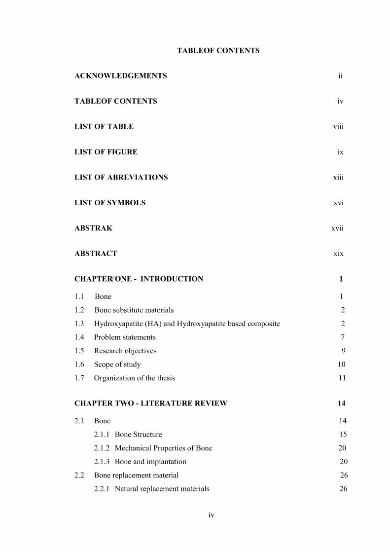
TABLEOF CONTENTS
ACKNOWLEDGEMENTS...................................................................................…ii
TABLEOF CONTENTS ........................................................................................... iv
LIST OF TABLE .................................................................................................... viii
LIST OF FIGURE .................................................................................................. iiix
LIST OF ABREVIATIONS ................................................................................... xiii
LIST OF SYMBOLS .............................................................................................. xvi
ABSTRAK .............................................................................................................. xvii
ABSTRACT ............................................................................................................. xix
CHAPTER ONE - INTRODUCTION…………… ………………………………1
1.1 Bone…………………………………………………………………….…… 1
1.2 Bone substitute materials……………………………………………….… 2
1.3 Hydroxyapatite (HA) and Hydroxyapatite based composite…… .……2
1.4 Problem statements…………………………………………………… ….… .7
1.5 Research objectives………………… …………………… ….……..... 9
1.6 Scope of study………………………………………………………….……...10
1.7 Organization of the thesis……………………………………….………….….11
CHAPTER TWO - LITERATURE REVIEW……… ……………….…….…..14
2.1 Bone……………………………………………………………….…….……..14
2.1.1 Bone Structure…………………………………………………..………15
2.1.2 Mechanical Properties of Bone…………………………………………20
2.1.3 Bone and implantation…………………………………...… ….….……20
2.2 Bone replacement material…………………………………………. …………26
2.2.1 Natural replacement materials………………………………… ………26
v
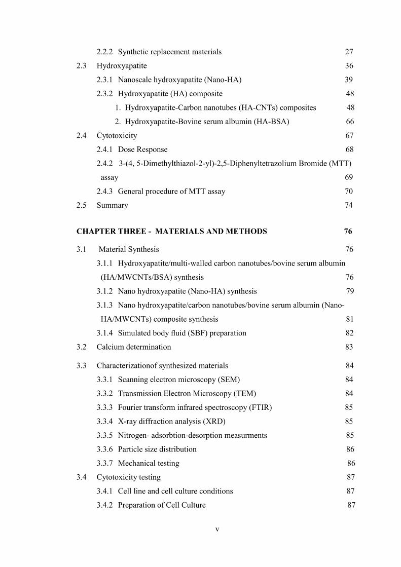
2.2.2 Synthetic replacement materials……………………………………….27
2.3 Hydroxyapatite………………………………………………………………..36
2.3.1 Nanoscale hydroxyapatite (Nano-HA)………………………………...39
2.3.2 Hydroxyapatite (HA) composite……………………………………….48
1. Hydroxyapatite-Carbon nanotubes (HA-CNTs) composites… ..48
2. Hydroxyapatite-Bovine serum albumin (HA-BSA)………… ...66
2.4 Cytotoxicity…………………………………………………………………...67
2.4.1 Dose Response…………………………………………………………68
2.4.2 3-(4, 5-Dimethylthiazol-2-yl)-2,5-Diphenyltetrazolium Bromide (MTT)
assay…………………………………………………………………... 69
2.4.3 General procedure of MTT assay……………………………………...70
2.5 Summary ……………………………………………………………...……...74
CHAPTER THREE - MATERIALS AND METHODS…… ………………...76
3.1 Material Synthesis………………..…………….……… …………….76
3.1.1 Hydroxyapatite/multi-walled carbon nanotubes/bovine serum albumin
(HA/MWCNTs/BSA) synthesis………………………………… ….76
3.1.2 Nano hydroxyapatite (Nano-HA) synthesis…………………… .. ...79
3.1.3 Nano hydroxyapatite/carbon nanotubes/bovine serum albumin (Nano-
HA/MWCNTs) composite synthesis………………………… … …..81
3.1.4 Simulated body fluid (SBF) preparation………………….………… .82
3.2 Calcium determination…………………….……………...…………… .83
3.3 Characterizationof synthesized materials………………………… …. 84
3.3.1 Scanning electron microscopy (SEM)…………………………… … 84
3.3.2 Transmission Electron Microscopy (TEM)……………………… … 84
3.3.3 Fourier transform infrared spectroscopy (FTIR)………………… … 85
3.3.4 X-ray diffraction analysis (XRD)……………………………… ……..85
3.3.5 Nitrogen- adsorbtion-desorption measurments …… ………….… ..85
3.3.6 Particle size distribution………………………………………………..86
3.3.7 Mechanical testing……………………………………………...………86
3.4 Cytotoxicity testing…………………………………………….………...……87
3.4.1 Cell line and cell culture conditions……………………………….…...87
3.4.2 Preparation of Cell Culture……………………………………….…….87
vi
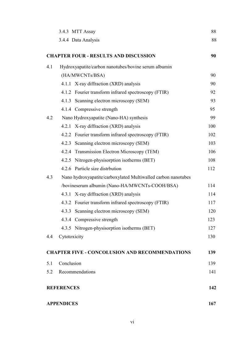
3.4.3 MTT Assay………………………………………………………….….88
3.4.4 Data Analysis…………………………………………………………...88
CHAPTER FOUR - RESULTS AND DISCUSSION…… … ……………… ..90
4.1 Hydroxyapatite/carbon nanotubes/bovine serum albumin
(HA/MWCNTs/BSA)……………… ………………………… …. .. 90
4.1.1 X-ray diffraction (XRD) analysis …………………… ….. 90
4.1.2 Fourier transform infrared spectroscopy (FTIR)……………. 92
4.1.3 Scanning electron microscopy (SEM)……………… ………. 93
4.1.4 Compressive strength………………………………………... 95
4.2 Nano Hydroxyapatite (Nano-HA) synthesis…………………………. 99
4.2.1 X-ray diffraction (XRD) analysis ……………………….. 100
4.2.2 Fourier transform infrared spectroscopy (FTIR)…………….. 102
4.2.3 Scanning electron microscopy (SEM) ……….. 103
4.2.4 Transmission Electron Microscopy (TEM)……. 106
4.2.5 Nitrogen-physisorption isotherms (BET)……………………. 108
4.2.6 Particle size distrbution……………………………………… 112
4.3 Nano hydroxyapatite/carboxylated Multiwalled carbon nanotubes
/bovineserum albumin (Nano-HA/MWCNTs-COOH/BSA)………... 114
4.3.1 X-ray diffraction (XRD) analysis ……………………… ..114
4.3.2 Fourier transform infrared spectroscopy (FTIR)…………….. 117
4.3.3 Scanning electron microscopy (SEM) ………………… …….120
4.3.4 Compressive strength……………………………………… .. .123
4.3.5 Nitrogen-physisorption isotherms (BET)……………… … .127
4.4 Cytotoxicity……………………………………………………… …… .130
CHAPTER FIVE - CONCOLUSION AND RECOMMENDATIONS … ..139
5.1 Conclusion……………………………………………………………… …..139
5.2 Recommendations…………………………………………………… ... 141
REFERENCES……………………………………………………………….. …..142
APPENDICES…………………………….…………………………………..… ..167

vii
Appendix A……………………………………………………….……………..….167
Appendix B……………………………………………………………..………..…168
viii
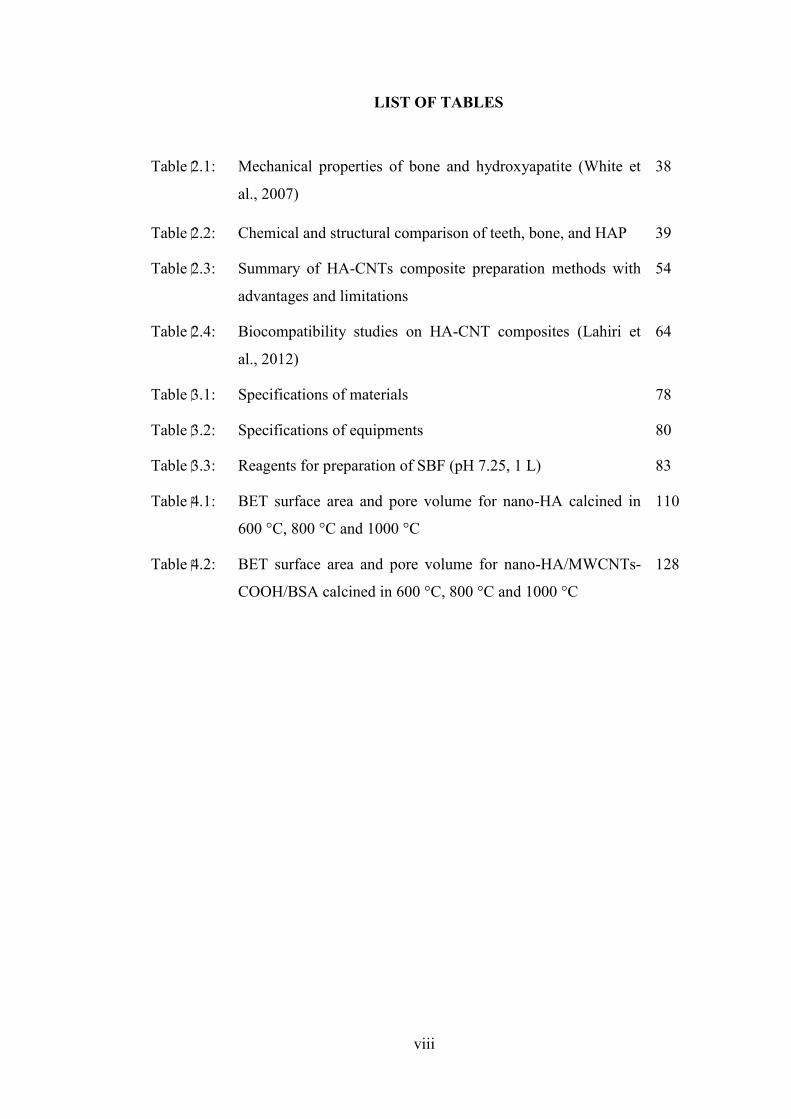
LIST OF TABLES
Table 2.1: Mechanical properties of bone and hydroxyapatite (White et
al., 2007)
38
Table 2.2: Chemical and structural comparison of teeth, bone, and HAP 39
Table 2.3: Summary of HA-CNTs composite preparation methods with
advantages and limitations
54
Table 2.4: Biocompatibility studies on HA-CNT composites (Lahiri et
al., 2012)
64
Table 3.1: Specifications of materials 78
Table 3.2: Specifications of equipments 80
Table 3.3: Reagents for preparation of SBF (pH 7.25, 1 L) 83
Table 4.1: BET surface area and pore volume for nano-HA calcined in
600 °C, 800 °C and 1000 °C
110
Table 4.2: BET surface area and pore volume for nano-HA/MWCNTs-
COOH/BSA calcined in 600 °C, 800 °C and 1000 °C
128
ix
LIST OF FIGURES
Figure 2.1: The hierarchical structure of bonen (Zhou and Lee, 2011) 15
Figure 2.2: Bone cells classified to three groups Osteoblast, Osteocyte,
and Osteoclast (Zhang, 1999)
18
Figure 2.3: Implant material requirement in orthopedic application 24
Figure 2.4: Hydroxyapatite Unit cell (White et al., 2007) 37
Figure 2.5: Overall production route for reaction of orthophosphoric acid
with calcium hydroxide (Munguía et al., 2005)
43
Figure 2.6: Overall production route for reaction of diammonium
hydrogen phosphate with calcium nitrate (Munguía et al.,
2005)
44
Figure 2.7: Molecular structure of (a) single-walled carbon nanotube and
(b) multi-walled carbon nanotube (Zhang et al., 2010b)
50
Figure 2.8: Different techniques of CNTs dispersion in HA matrix
(Lahiri et al., 2012)
56
Figure 2.9: Reduaction of (a) MTT salt to (b) Formazan 70
Figure 3.1: Overall research methodology flow diagram 77
Figure 4.1 X-ray diffraction patterns of (a) HA/MWCNTs-COOH/BSA,
(b) HA/MWCNTs-OH/BSA, (c) HA/MWCNTs/BSA, and
(d) HA/MWCNTs*/BSA
91
Figure 4.2: FTIR patterns of (a) HA/MWCNTs*/BSA, (b)
HA/MWCNTs/BSA, (c) HA/MWCNTs-OH/BSA and (d)
HA/MWCNTs-COOH/BSA
92
Figure 4.3: SEM micrographs of composites (a) HA/MWCNTs*/BSA,
(b) HA/MWCNTs/BSA, (c) HA/MWCNTs-OH/BSA and (d)
HA/MWCNTs-COOH/BSA
94
Figure 4.4: Compressive strength of HA/MWCNTs-COOH/BSA,
HA/MWCNTs-OH/BSA, HA/MWCNTs/BSA,
96
x
HA/MWCNTs*/BSA. Data are presented as the means ±2
standard
Figure 4.5: Compressive strength of HA/MWCNTs-COOH/BSA
composites following immersion in SBF for unsoaked, 7, 14,
21 and 28 days. Data are presented as the means72 standard
deviation (n=2)
98
Figure 4.6: X-ray diffraction patterns of HA powder synthesized at (a)
30 °C and (b) 60 °C
100
Figure 4.7: X-ray diffraction patterns of HA powder after calcination at
(a) 1000 °C, (b) 800 °C and (c) 600 °C
101
Figure 4.8: FTIR patterns of synthesized HA after calcination at various
temperatures of (a) 600 °C, (b) 800 °C, (c) 1000 °C
102
Figure 4.9: SEM micrographs of synthesized HA at different reaction
temperature (a) 30 °C, (b) 45 °C, (c) 60 °C
104
Figure 4.10: SEM micrographs of synthesized HA after calcination in (a)
1000 °C, (b) 800 °C, (c) 600 °C
106
Figure 4.11: TEM images of of synthesized HA after calcination in (a)
1000 °C, (b) 800 °C, (c) 600 °C
107
Figure 4.12: Nitrogen adsorption/desorption of nano-HA calcined at 600,
800 and 1000 °C
109
Figure 4.13: Pore size distribution of HA microsphere calcined at
different temperature (a) 600 °C ; (b) 800 °C and (c) 1000 °C
111
Figure 4.14: Particle size distribution of nano-HA prepared at 30 °C 112
Figure 4.15: Particle size distribution of nano-HA prepared at 45 °C 113
Figure 4.16: Particle size distribution of nano-HA prepared at 60 °C 113
Figure 4.17: X-ray diffraction patterns of Nano-HA/MWCNTs-
COOH/BSA composites after calcination at (a) 600 °C, (b)
800 °C and (c) 1000 °C
115
Figure 4.18: X-ray diffraction patterns of Nano-HA/MWCNTs- 116
xi
COOH/BSA composites following immersion in SBF for (a)
7 days, (b) 14 days, (c) 21 days, (d) 28 days
Figure 4.19: FTIR patterns of Nano-HA/MWCNTs-COOH/BSA calcined
at various temperatures (a) 600 °C, (b) 800 °C, (c) 1000 °C
118
Figure 4.20: FTIR patterns of Nano-HA/MWCNTs-COOH/BSA
composites following immersion in SBF for (a) 7 days, (b)
14 days, (c) 21 days and (d) 28 days
119
Figure 4.21: SEM micrographs of Nano-HA/MWCNTs-COOH/BSA after
calcination at (a) 600 °C, (b) 800 °C, (c) 1000 °C
120
Figure 4.22: SEM micrographs of Nano-HA/MWCNTs-COOH/BSA
composites following immersion in SBF for (a) 0 days, (b) 7
days, (c) 14 days, (d) 21 days and (e) 28 days
122
Figure 4.23: Compressive strength of Nano-HA/MWCNTs-COOH/BSA
composites following calcined at 600, 800 and 1000 °C
124
Figure 4.24: Compressive strength of Nano-HA/MWCNTs-COOH/BSA
composites following immersion in SBF for unsoaked, 7, 14,
21 and 28 days. Data are presented as the means ±2 standard
deviation
126
Figure 4.25: Nitrogen adsorption/desorption of Nano-HA/MWCNTs-
COOH/BSA calcined at 600 and 1000 °C
127
Figure 4.26: Pore size distribution of Nano-HA/MWCNTs-COOH/BSA
microsphere calcined at different temperature (a) 600 °C and
(b) 1000 °C
129
Figure 4.27: Influence of HA/MWCNTs/BSA composites on CCD-18Co
fibroblast metabolic activity as measured using MTT assay
(data are presented as the means ±2 standard deviations, n=3)
131
Figure 4.28: Influence of Nano-HA composites on CCD-18Co fibroblast
metabolic activity as measured using MTT assay (data are
presented as the means ±2 standard deviations, n=3)
134
Figure 4.29 Effect of developed composites on CCD-18Co fibroblast 137
xii
cells activity (A: Positive control, B: Negative control, C:
Nano-HA, D: Nano-HA/MWCNTs-COOH/BSA (0 days), E:
Nano-HA/MWCNTs-COOH/BSA (7 days), F: Nano-
HA/MWCNTs-COOH/BSA (14 days), G: Nano-
HA/MWCNTs-COOH/BSA (21 days), H: Nano-
HA/MWCNTs-COOH/BSA (28 days))
xiii
LIST OF ABREVIATIONS
MTT 3-(4, 5-dimethylthiazol-2-yl)-2,5-
diphenyltetrazolium bromide
NH4OH Ammonia
ACP Amorphous calcium phosphate
ANN Artificial neural network
BCP Biphasic calcium phosphat
BSA Bovine serum albumin
BET Brunauer–emmett–teller
Ca Calcium
CaCO3 Calcium carbonate
CaCl2 Calcium chloride
Ca(OH)2 Calcium hydroxide
Ca(NO3)2 Calcium nitrate
CaP Calcium phosphate
CPCs Calcium phosphate cements
CNTs Carbon nanotubes
MWCNTs-COOH Carboxylated multi-walled carbon
nanotubes
DAP Deficient hydroxyapatite
(NH4)2HPO4 Diammonium hydrogen phosphate
Ca(NO3)2·4H2O Diammonium hydrogen phosphate
(NH4)2HPO4 Diammonium phosphate
DCPA Dicalcium phosphate anhydrate
K2HPO4.3H2O Dipotassium phosphate anhydrate
xiv
DMEM Dulbecco’s modified eagle’s medium
FFBP Feed forward back propagation
FBS Fetal bovine serum
Ca10(PO4)6F Fluorapatite
FTIR Fourier transform infrared spectroscopy
HCl Hydrochloric acid
HA Hydroxyapatite
MWCNTs-OH Hydroxylate multi-walled carbon
nanotubes
MgCl2.6H2O Magnesium chloride hexahydrate
MWCNTs Multi-walled crbon nanotubes
CCD-18Co Normal human colon fibroblast cell line
OCP Octacalcium phosphate
H3PO4 Orthophosphoric acid
P Phosphor
PLlA Poly l-lactide
PLLA Poly(l-lactic acid)
PGA Polyglycolic acid
PLA Polylactic acid
KCl Potassium cloride
RBF Radial basis function
SEM Scanning electron microscopy
SBF Simulated body fluid
SWCNTs Single-walled cnts
NaCl Sodium cloride
SDS Sodium dodecyl sulphat
xv
Na2SO4 Sodium sulfate
SPS Spark plasma sintering
TTCP Tetracalcium phosphate
TEM Transmission electron microscopy
TCP Tricalcium phosphate
(CH2OH)3CNH2 Tris (hydroxyl-methyl) aminomethane
XRD X-ray diffractometer
α-TCP α-tri-Calcium phosphate
β-TCP β-tri-Calcium phosphate
xvi
LIST OF SYMBOLS
cm Centimetre
° Degree
°C Degree Celsius
°C/min Degree Celsius per minute
g
h
L
Gram
Hour
Litre
< Less than
m Meter
ml Millilitre
mm Millimetre
min Minute
> More than
nm
rpm
Nanometer
Revolutions per minute
% Percentage
s Second
T Temperature
wt % Weight percent
xvii
PEMBANGUNAN HIDROKSIAPATIT YANG DIPERKUATKAN DENGAN
TIUB-NANO KARBON
ABSTRAK
Persamaan komposisi kimia hidroksiapatit (HA) dengan fasa mineral tulang
dan keserasian biologinya yang sangat baik memenuhi keperluan bahan reka bentuk
untuk pembesaran dan pembaikan tulang. Walaubagaimanapun, aplikasi HA dalam
peranti menanggung beban adalah terhad disebabkan ciri mekanikal yang lemah.
Tiub-nano karbon (CNTs), dengan ciri kekukuhan dan kekuatannya yang baik,
mempunyai potensi besar dalam bidang kejuruteraan tisu. Kekukuhan dan kekuatan
CNTs, digabungkan dengan saiz yang kecil dan kawasan permukaan antara muka
yang besar, mencadangkan CNTs mempunyai potensi yang amat besar untuk
dijadikan ejen pengukuhan HA. Dalam kajian ini, komposit HA/MWCNTs/BSA
dengan ciri-ciri mekanik ditambah baik untuk digunakan sebagai bahan pengganti
tulang telah dibangunkan. Serbuk komposit HA/MWCNTs/BSA dengan pelbagai
jenis MWCNTs (MWCNTs-OH, MWCNTs-COOH, MWCNTs) telah berjaya
disediakan. Keputusan yang diperolehi menunjukkan bahawa komposit HA yang
disediakan dengan menggunakan MWCNTs-COOH dan BSA dengan daya
mampatan 29.75 MPa menunjukkan keputusan terbaik berbanding dengan komposit
HA yang disediakan dengan menggunakan MWCNTs yang lain. Komposit
HA/MWCNTs-COOH/BSA tersebut direndam dalam SBF selama 7 , 14, 21 dan 28
hari pada suhu 37 °C sebagai ujian �������� biologi bertujuan untuk menyiasat kesan
SBF terhadap ciri-ciri mekanikal komposit. Daya mampatan komposit telah menurun
dari 29.57 kepada 9.11 MPa selepas 28 hari direndam dalam SBF. Nano-HA dengan
saiz zarah antara 20 ke 25 nm telah berjaya dihasilkan dengan menggunakan kaedah
xviii
pemendakan larutan Ca(NO3)2.4H2O dan (NH4)2HPO4. Komposit Nano-
HA/MWCNTs-COOH telah berjaya dihasilkan menggunakan kaedah pemendakan.
Komposit tersebut dikalsin pada suhu 600, 800 dan 1000 °C. Kekuatan mampatan
komposit Nano-HA/MWCNTs-COOH selepas pengkalsinan pada suhu 600, 800 dan
1000 °C adalah masing-masing 27.01 , 27.78 dan 37.43 MPa. Kesan sitotoksik
sampel ujian dengan kepekatan yang berbeza telah dinilai dengan assay MTT
terhadap fibroblast kolon manusia normal. Pada kepekatan yang rendah, semua
komposit didapati tidak sitotoksik apabila dirawat kepada sel-sel fibroblast manusia
dan tidak menyebabkan kesan sitotoksik pada percambahan sel, manakala apabila
kepekatan sampel ditingkatkan, pengurangan daya maju sel dapat diperhatikan.
Komposit HA/MWCNTs-OH/BSA, HA/MWCNTs-COOH/BSA, dan
HA/MWCNTs/BSA menunjukkan kesan-kesan percambahan pada sel-sel. Sampel
yang digunakan dalam kajian ini tidak menunjukkan kesan toksik apabila
percambahan telah didorong. Tambahan pula, Nano-HA/MWCNTs-COOH/BSA
membayangkan kesan percambahan pada rangkaian sel CCD-18Co. Komposit
HA/MWCNTs-OH/BSA, HA/MWCNTs-COOH/BSA, HA/MWCNTs/BSA dan
Nano-HA/MWCNTs-COOH/BSA adalah antara pilihan yang baik untuk
membangunkan bahan-bahan ortopedik kerana komposit-komposit ini lulus
keseluruhan tapisan, dan kesan yang diingini telah ditunjukkan di dalam badan.
Tambahan pula, komposit-komposit ini boleh memasuki pasaran industri dengan
kadar kejayaan yang agak tinggi.
xix
DEVELOPMENT OF MULTIWALLED CARBON NANOTUBES
REINFORCED HYDROXYAPATITE
ABSTRACT
The similarity of the chemical composition of hydroxyapatite (HA) to the
mineral phase of bone and their excellent biocompatibility meets the requirement of
materials designed for bone repair and augmentation purposes. However, the
application of HA in load bearing devices is limited by its poor mechanical
properties. Carbon nanotubes (CNTs), with their outstanding stiffness and strength,
have good potential applications in tissue engineering. Their strength and stiffness,
combined with their small size and large interfacial surface area, suggest that they
may have great potential as a reinforcing agent for HA. In this study,
Hydroxyapatite/Multiwalled carbon nanotubes/Bovine serum albomin
(HA/MWCNTs/BSA) composites with highly improved mechanical properties for
use as bone replacement materials were developed. The HA/MWCNTs/BSA
composites powders with different types of MWCNTs (MWCNTs-OH, MWCNTs-
COOH, MWCNTs) were successfully prepared. The results show that the HA
composites which are prepared by using MWCNTs-COOH and BSA with
compressive strength of 29.75 MPa has the highest results in comparison with HA
composites which are prepared by using other types of MWCNTs. The
HA/MWCNTs-COOH/BSA composite immersed in SBF for 7, 14, 21 and 28 days in
37 °C as in vitro biological test in with the purpose to investigate the SBF effects on
mechanical properties. The compressive strength of immersed composites in SBF
was decreased from 29.57 to 9.11 MPa after 28 days immersing. The nano-HA with
particle size in the range 20 to 25 nm was successfully synthesized using
precipitation technique from Ca(NO3)2.4H2O and (NH4)2HPO4 solution. The Nano-
xx
HA/MWCNTs-COOH/BSA composites were successfully produced using
precipitation technique. The synthesized composites calcined at different
temperatures of 600, 800 and 1000 °C. The compressive strength of Nano-
HA/MWCNTs-COOH/BSA composites after calcination at 600, 800 and 1000 °C
are 27.01, 27.78 and 37.43 MPa respectively. The cytotoxic effect of the test samples
with different concentrations was evaluated by MTT assay against normal human
colon fibroblast. At low concentration, all developed composites were found to be
non-cytotoxic when treated to the human fibroblast cells and did not elicit cytotoxic
effects on cell proliferation, whereas when the concentration of samples was
increased, the reduction in cell viability was observed. The HA/MWCNTs-OH/BSA,
HA/MWCNTs-COOH/BSA, and HA/MWCNTs/BSA composites showed the
proliferative effects on the cells. The samples used in the present study did not show
toxic effects as proliferation was induced. Furthermore, the Nano-HA/MWCNTs-
COOH/BSA implies a cell proliferative effect on CCD-18Co cell line.
HA/MWCNTs-OH/BSA, HA/MWCNTs-COOH/BSA, HA/MWCNTs/BSA and
Nano-HA/MWCNTs-COOH/BSA composites are possibly a good choice to develop
orthopedic materials because these materials passed the entire filter, in which a
desirable effect was elicited in the body. Furthermore, these compounds can enter the
market industry with a possibly high success rate.
1
1 CHAPTER ONE
INTRODUCTION
Nowadays medical world is changing from treating injured organs of bodies by
long surgical operations to changing the damaged organ with synthesised implants.
Hard tissue and bone substitute materials are mostly synthesised using bioactive and
tough materials, which have chemical and structure similarity to the hard tissues.
Study of biomaterials includes the use of them in a new composite material with
improved properties, modification of the microstructure of the present biomaterials
and chemical synthesis of new biomaterials. Material scientists are currently working
on projects for the synthesis of biocompatible materials, which mimic the properties
of natural bone (Vallet-Regí et al., 2004).
Biomaterials are class of engineering materials, which can be used in animal
body, tissue replacement, reconstruction, and regeneration without any long-term
adverse effects. Among the different classes of biomaterials, bioceramics is one of
the important classes of available material used as human-body implants (Dorozhkin,
2010).
1.1 Bone
Bone is a natural composite material that contains 10 wt.% water, 20 wt.%
organic materials, and 70 wt.% minerals (Shi et al., 2006). The organic components
mostly comprise collagen fibrils and some cement-like substances, whereas the
inorganic mineral materials of bone are composed of calcium-deficient carbonate
substituted apatite comprising calcium (Ca2+) and phosphate (PO43-) ions, which have
similar structures to that of hydroxyapatite (HA, Ca10(PO4)6(OH)2) (Legeros et al.,
2
1993). According to its porosity, bone structure is classified into two categories:
compact (or cortical) bone and trabecular (or cancellous) bone. Porosity, density, and
molecular structure of bone affect its strength and mechanical properties (Rho et al.,
1998).
1.2 Bone substitute materials
Ideal bone replacement materials should be biocompatible, nontoxic,
osteoinductive and osteoconductive, stable, capable of long-term integration of
implant, and possess excellent mechanical properties (Horch et al., 2006; Kao and
Scott, 2007). Materials for body repair have been designed and used clinically since
1960s. Many different kinds of synthetic replacements materials have been
developed in the last decades as bone replacement materials. Synthetic materials
include ceramic-based materials such as calcium phosphate, calcium sulphate,
bioglass, polymers, ceramics and composites (Tadic and Epple, 2004; Tadic et al.,
2004; Horch et al., 2006; McAllister and Haghighat, 2007; Asti et al., 2008).
Composites are combinations of various materials, such as bioactive calcium
phosphates and polymers, which can be used as bone replacement materials with
improved mechanical properties (Boccaccini and Blaker, 2005, Chesnutt et al., 2009,
Xu et al., 2009).
1.3 Hydroxyapatite (HA) and Hydroxyapatite based composite
The composition of HA is similar to the mineral phase of natural bone. HA is
an great material for bone replacement, and has been used in clinical applications
such as dental and orthopedic applications in bulk form and as filler and coating for
more than three decades (Suchanek et al., 1998). However, the poor mechanical
3
properties of HA have prevented it from being used in major load-bearing devices.
The compressive strength of dense HA is four times greater than that of compact
bone, but it still has low tensile strength and fracture toughness.
The recent researches in biomaterials is focused on improving the weak points
of calcium phosphates and hydroxyapatite ceramics including their low
bioresorbability, surface area, bioreactivity, and the biological properties by
exploring the special advantages of nanotechnology (Bohner et al., 2012).
Nanotechnology has potential to synthesize the inorganic crystals with nano particle
size, characterized by a high bioresorbability and surface area, which increase the
bioreactivity of crystals. The trend is shifting toward nanotechnology to improve the
biological responses of HA, because nano-HA is a constituent of bone (Roveri et al.,
2010).
Nano-HA particles are similar to bone crystals, therefore using nano-HA is
considered to be very promising for use in in orthopedic application.
Nanotechnology offers an exclusive approach to overcome the weaknesses of several
conventional materials. Nanotechnology is include the almost all disciplines of
human life such as nanomedicine and nanofabrics. Compare to materials with micro
size particles, the nano-sized materials are suggest more improved performance,
because these materials have higher ratio of surface area to volume and unusual
chemical/electronic synergistic effects (Roveri et al., 2010).
Zhou et al. (2011) studied the important role of size and crystal morphology
control of nano HA particles for bone tissue engineering. Their review revealed that
4
developing the HA biomedical materials will benefit from nanotechnology
progresses.
Several methods have been used to synthesize HA. The HA was produced
through a hydrothermal process by exposing fluorapatite (Ca10(PO4)6F) to high
pressures and temperatures by Levitt at 1996s. Several methods, such as
hydrothermal exchange of coral, solid-state reaction between tricalcium phosphate
(TCP) and tetracalcium phosphate (TTCP) or between TCP and CaO, as well as wet
chemical methods including aqueous precipitation, hydrolysis, and sol–gel
techniques, have been used (Akao et al., 1981; Legeros 1993; Weng et al., 1998; Liu,
2001; Bezzi et al., 2003; Vijayalakshmi et al., 2006). Among the different methods
of HA synthesis, precipitation method is the most prevalent. It is a simple method
because of its ease of operation, tailoring composition, and scaling up for mass
production. Precipitation method is a wet chemical reaction between calcium
precursors (such as calcium nitrate (Ca(NO3)2), calcium carbonate (CaCO3), and
calcium hydroxide (Ca(OH)2) and phosphate precursors (such as diammonium
hydrogen phosphate ((NH4)2HPO4) and orthophosphoric acid (H3PO4)) under
controlled temperature and pH (Kweh et al., 1999; Munguía et al., 2005; Rhee, 2002;
Santos et al., 2004).
The properties of HA (including physical, physicochemical, mechanical, and
biological properties, as well as heat treatment behavior) and its preparation process
are well understood. After decades of research, poor mechanical properties of HA are
reportedly its main disadvantage (Hench, 1998; Kweh et al., 1999; Orlovskii et al.,
2002). Carbon nanotubes (CNTs) have outstanding mechanical properties and
5
chemical stability because of their cylindrical graphitic structure. Carbon is one of
the known basic foundations in the development of life on earth (Cheng et al., 2009).
Since the investigation of CNTs by Iijima in 1991, increasing research has focused
on the use of CNTs for reinforcement (Iijima in 1991; Gojny et al., 2003). Their
strength and stiffness, combined with their small size and large interfacial area,
suggest that they may have great potential as reinforcing agents for HA.
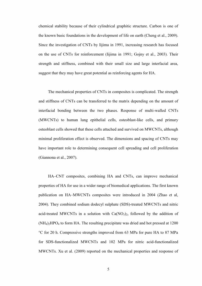
The mechanical properties of CNTs in composites is complicated. The strength
and stiffness of CNTs can be transferred to the matrix depending on the amount of
interfacial bonding between the two phases. Response of multi-walled CNTs
(MWCNTs) to human lung epithelial cells, osteoblast-like cells, and primary
osteoblast cells showed that these cells attached and survived on MWCNTs, although
minimal proliferation effect is observed. The dimensions and spacing of CNTs may
have important role to determining consequent cell spreading and cell proliferation
(Giannona et al., 2007).
HA–CNT composites, combining HA and CNTs, can improve mechanical
properties of HA for use in a wider range of biomedical applications. The first known
publication on HA–MWCNTs composites were introduced in 2004 (Zhao et al,
2004). They combined sodium dodecyl sulphate (SDS)-treated MWCNTs and nitric
acid-treated MWCNTs in a solution with Ca(NO3)2, followed by the addition of
(NH4)2HPO4 to form HA. The resulting precipitate was dried and hot pressed at 1200
°C for 20 h. Compressive strengths improved from 63 MPa for pure HA to 87 MPa
for SDS-functionalized MWCNTs and 102 MPa for nitric acid-functionalized
MWCNTs. Xu et al. (2009) reported on the mechanical properties and response of
6
osteoblast-like cells to HA–MWCNTs composites densified by spark plasma
sintering. HA powder and MWCNTs were dry mixed and then consolidated using
spark-plasma sintering at temperatures ranging from 900 °C to 1200 °C. Hardness
and Young’s modulus were evaluated, and osteoblast-like cells were cultured for two
and four days. The mechanical properties reportedly increased in hardness and
Young’s modulus compared with HA. They also reported an increase in cellular
response to the composites compared with HA alone (Xu et al., 2009).
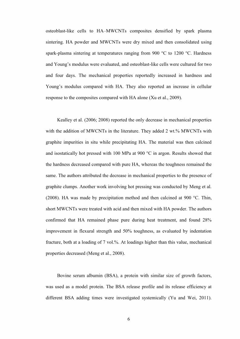
Kealley et al. (2006; 2008) reported the only decrease in mechanical properties
with the addition of MWCNTs in the literature. They added 2 wt.% MWCNTs with
graphite impurities in situ while precipitating HA. The material was then calcined
and isostatically hot pressed with 100 MPa at 900 °C in argon. Results showed that
the hardness decreased compared with pure HA, whereas the toughness remained the
same. The authors attributed the decrease in mechanical properties to the presence of
graphite clumps. Another work involving hot pressing was conducted by Meng et al.
(2008). HA was made by precipitation method and then calcined at 900 °C. Thin,
short MWCNTs were treated with acid and then mixed with HA powder. The authors
confirmed that HA remained phase pure during heat treatment, and found 28%
improvement in flexural strength and 50% toughness, as evaluated by indentation
fracture, both at a loading of 7 vol.%. At loadings higher than this value, mechanical
properties decreased (Meng et al., 2008).
Bovine serum albumin (BSA), a protein with similar size of growth factors,
was used as a model protein. The BSA release profile and its release efficiency at
different BSA adding times were investigated systemically (Yu and Wei, 2011).
7
Generally, BSA is used as a matrix or soft template to induce calcium carbonate
formation (Yao et al., 2009). The improvement in mechanical properties with the
addition of BSA can be explained by the fact that proper amounts of BSA can
promote CPC crystal growth (Burke et al., 2000; Hovarth et al., 2000; Sadollah et al.,
2012).
The addition of BSA can increase the compressive strength of calcium
phosphate composites (Chew et al., 2011; Low et al. 2011). The results demonstrated
that the addition of BSA and MWCNTs to the CPC caused an increment in the
compressive strength of the CPC composite from 1 MPa for pure CPC to more than
14 MPa for CPC/MWCNTs/BSA composite. This further improvement in the
mechanical properties might be due to the capability of BSA to promoting CPC
crystal growth (Boccaccini et al., 2007).
1.4 Problem statement
With the aging population, the requirement for biomaterials has increased
significantly, and more attention has been drawn to hard tissue research. The
significant attention on new and biocompatible bone graft materials is ascribed to
decades of bone graft usage to repair osseous defects causing from traumas and
diseases. More than 800,000 grafting procedures are performed annually (Tusli,
2004).
The development and improvement of synthetic materials for in vivo biological
applications is highly desirable as an alternative to allografts and autografts.
Hydroxyapatite (HA) has been extensively studied as a hard tissue and bone graft
8
biomaterial due to its crystallographic and biologically active characteristics
stemming from its chemical similarity to the mineral similarity to carbonated apatite
in teeth and bones. Synthetic HA is a biologically active calcium phosphate
bioceramic that is generally used for coating the orthopedic metal or utilized as bone
substitute material (Lange and Donath, 1989; Sadollah et al., 2012). While HA has
the ability to promote bone growth along its surface, its mechanical properties,
particularly its strength and toughness, are insufficient for major load-bearing
applications. Reinforcing HA with a second phase offers a possibility to overcome
these limitations.
An ideal reinforcing material would provide mechanical integrity to the
composite without negatively impacting its biological properties. Carbon nanotubes
(CNTs), considered one of the strongest and stiffest materials available, have
excellent potential to accomplish this.. Furthermore, the surface of the CNTs can be
functionalised to improve that interaction.
Already, CNTs have shown potential for reinforcing polymers, and to some
degree metals and ceramics, as well. Several publications have addressed HA-CNT
composites, specifically, but studies thus far have lacked a cohesive view of all
aspects of the composites, which encompass production, processing, densification
techniques, mechanical behaviour, and biological behaviour. Special attention must
be given to the biological impact of the CNTs presence, as current research is
conflicting as to whether CNTs might induce a cytotoxic response or perhaps even
improve cellular processes (Kealley et al., 2006; White et el., 2007). Regarding the
use of CNTs in bone replacement material, another concern is cytotoxicity. There are
9
several researches that focus on the harmful impact of carbon nanotubes on cell
proliferation and adhesive ability. CNTs have excellent mechanical properties and
some researches indicated that they are biocompatible materials in human body.
(MacDonald et al, 2005; Price et al., 2003; Hu et al., 2004).
Nanoscale hydroxyapatite (nano-HA) is the main component of mineral phase
of natural bone. Nano HA powders have greater surface area, thus it can exhibit the
improvement in sinterability and enhanced better densification and as a consequence,
improve mechanical properties (Legeros, 1993). Nano-HA is supposed to have better
bioactivity and biocompatibility and due to its small size and huge specific surface
area, they may have exclusive properties. Study by Webster et al. (2000) have
revealed that in comparison with micron sized ceramic materials, nano sized
materials demonstrated a significant increment in protein adsorption and osteoblast
adhesion. A large surface area is necessary for cell attachment and growth. The large
pore volume is also required to accommodate and subsequently deliver a cell mass
sufficient for tissue repair. The highly porous biomaterials also desirable for the easy
deffusion of nutrients to and waste products from the implant and for vascularization,
which are important requirements for the regeneration. This research is aimed at
synthesis of HA/MWCNTs/BSA, with improved mechanical properties using
MWCNTs as reinforcing material.
1.5 Research objectives
The aims of the research is to develop biocompatible composites with highly
improved mechanical properties for use as bone replacement materials. The
objectives will be achieved through the outlined steps as follows:
10
i. To investigate the mechanical strength of HA/MWCNTs composites with
different types of MWCNTs and BSA.
ii. To synthesis nano-HA/MWCNTs and study the mechanical strength of nano-
HA/MWCNTs/BSA composite for use as bone replacement materials.
iii. To investigate the cytotoxicity effect of developed composites via in vitro cell
culture.
1.6 Scope of study
This study covers the development of hydroxyapatite composites,
characterization of composites and cytotoxicity study. Calcium phosphate-based
biomaterials have revealed great performance without side effects on the in vitro or
in vivo. However, the final composites have relatively low compressive strength,
which limits their applicability in orthopedics. The MWCNTs have been chosen
because of their impressive properties, such as mechanical strength, stiffness and it is
suitable to apply in biological and biomedical systems. Furthermore, BSA has been
chosen because it can act as a promoter of HA crystal growth when bonded to a
surface. Thus, the addition of MWCNTs and BSA could lead to the improvement of
mechanical properties by modifying the properties of crystallites. The interfacial
bonding between HA, MWCNTs, and BSA is an important consideration for
composite materials.
HA has the ability to promote bone growth along its surface, but its mechanical
properties, especially compressive strength, is not sufficient for load-bearing
applications. Nanoscale HA with ultrafine structure and large surface area has been
11
synthesised for the next step of this research because it has a higher level of
biocompatibility compared with conventional HA.
In this study, the biological effects of the developed composites on fibroblast
cells are studied and the experimental results are compared with the predicted results
of ANN technique. The kinetics of nano-HA precipitation from aqueous solution at
the condition of high pH value in range between 10.5 to 11 investigated with purpose
of synthesis appropriate nano-HA for use as bone replacement materials.
Furthermore, the effect of different temperature on rate of conversion were studied.
Synthesized composites are characterized through surface area analysis, scanning
electron microscopy (SEM), surface and pore size measurements (Brunauer–
Emmett–Teller, BET), transmission electron microscopy (TEM), X-ray
diffractometer (XRD) and Fourier transform infrared spectroscopy (FTIR).
1.7 Organization of the thesis
This thesis is included five major chapters. Each chapter represents an integral
part of the main work and is sequentially arranged. The thesis is organized as
follows.
Chapter one provides an overview of the overall research project. The problem
statement is written after reviewing different types of bone replacement materials.
The research objectives, scope of study, and organization of the thesis are provided
in this chapter.
12
Chapter two presents a detailed review of literature on the properties,
characteristics, and production of calcium phosphate-based composites. It also
reviews relevant studies on MWCNTs, BSA, and the current status of MWCNTs as
reinforcement materials. The chapter also reviews an ANN method that can be used
as a potent tool to predict the cytotoxic properties of composites. Finally, a literature
review on the kinetic study of the precipitation reaction of HA synthesis is presented.
Chapter three focuses on the materials used and the experimental processes
which employed to synthesis of composites. This chapter also presents in detail the
experimental methodologies used in the synthesis of different composites and the
synthesis of nanoscale HA. It briefly describes the characterization of the
composites.
Chapter four presents and discusses the results obtained in this work. The first
section of this chapter contains results and discussion of the production of calcium
phosphate based composite, in vitro study of composites, and characterization of the
synthesized composite. The succeeding part includes the formation of
HA/MWCNTs/BSA using conventional HA and different types of MWCNTs,
followed by characterization of the composites. The third part of this chapter
contains a discussion of nano-HA synthesis through precipitation reaction and
preparation of HA/MWCNTs/BSA composite using nano-HA, followed by
characterization of composites. In last part, the cytotoxicity of the composites was
assessed with human fibroblasts cells (CCD-18 Co), which were inoculated and
exposed to different concentrations of different types of sample powder.

13
Chapter five presents a summary of the results of this research and offers
recommendations for future studies related to this research work.
14
2 CHAPTER TWO
LITERATURE REVIEW
The purpose of this chapter is to give a background relating to bone
replacement materials (particularly CPC and HA composites) and CNTs. Detailed
information relating to processing techniques and characterization; Heat treatment;
mechanical properties and biological responses of the composite materials; kinetic
study and mathematical analysis will be included in chapter 3 and 4. This literature
give an overview of the bone nature and different types of materials currently use or
under development that can replace bone and assist to repair it. The background on
CNTs, which chosen as reinforcing material for CPC and HA is also elaborated.
Furthermore, mechanical properties and cytotoxicity of composites are discussed.
2.1 Bone
Bone is an organ with critical functions in human physiology. Some of these
functions include protection, movement and support of other organs, blood
production, mineral storage and homeostasis, and blood pH regulation (Porter et al.,
2009). Bone is a complex natural organic–inorganic ceramic that consists of collagen
fibrils embedded with well-arrayed nano crystalline rod shaped inorganic material 25
nm to 50 nm in length (Poinern et al., 2009; Zhou and Lee, 2011). Bone stores 99%
of the total body calcium, whereas the remaining 1% circulates in the blood (Rho et
al., 1998).
15
2.1.1 Bone Structure
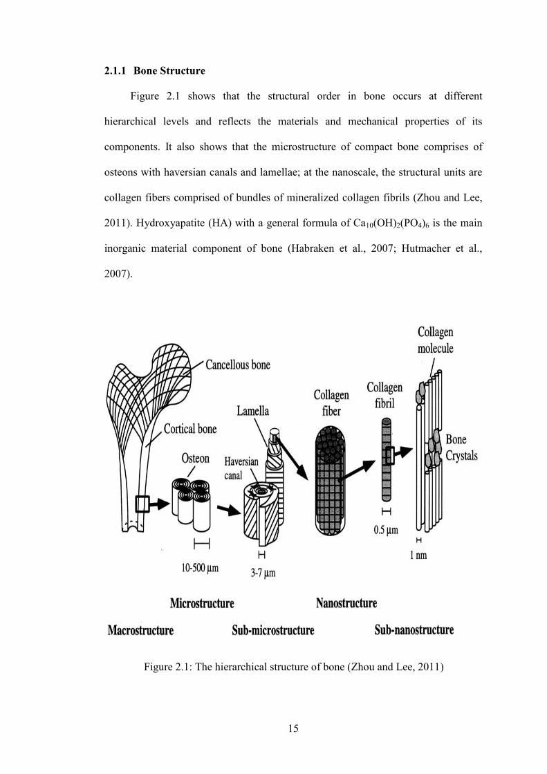
Figure 2.1 shows that the structural order in bone occurs at different
hierarchical levels and reflects the materials and mechanical properties of its
components. It also shows that the microstructure of compact bone comprises of
osteons with haversian canals and lamellae; at the nanoscale, the structural units are
collagen fibers comprised of bundles of mineralized collagen fibrils (Zhou and Lee,
2011). Hydroxyapatite (HA) with a general formula of Ca10(OH)2(PO4)6 is the main
inorganic material component of bone (Habraken et al., 2007; Hutmacher et al.,
2007).
Figure 2.1: The hierarchical structure of bone (Zhou and Lee, 2011)
16
Despite the diversity of external forms of bone, its internal structure is
relatively consistent. Bone structure is hierarchically organized (Weiner and Wagner,
1998; Hellmich and Ulm, 2002). Skeleton comprised from two hundred six bones
with different size, shape and weight. This complex internal and external structure is
responsible for 9 kg of adult human weight. Bones are rigid, living tissue, constantly
tearing down and rebuilding them, without this repair and reinforcement of even
minor weak spots, we would break bones on a regular basis. Bone serves multiple
functions, including mechanical, synthetic, and metabolic. Through mechanical
function, bone provides the frame work for the body; also provide protection for
internal soft tissue like brain, heart, liver and muscle. Bone and several associated
elements cartilage, connective tissue, vascular elements, and nervous components act
as fundamental organ generating movement force for the body (Gilbert, 2001).
Bone is multifunctional tissue and not only having protective function. The
spongy tissue inside interior cavity of long bones and interstices of cancellous bone
produces immature cells, called stem cells, which later develop to red blood cell.
Bone marrow produces other elements of the blood such as white blood cells and
platelets (Haywood, 2008).
In addition to mechanical function, bone serve metabolic role by reserving
minerals, fats, and acid-base balance. The bone stores 99% of the body's calcium and
85% of the phosphorus. The yellow bone marrow of long bones acts as storage of
fats. Bone balances acid, and base in blood, when acid production is increased, renal
net acid excretion, and kidney function is not enough to buffer the condition, bone
17
release of Ca2+ into the urine, balances the condition (Frost and Saunders-Smith,
2001; Lemann et al., 2003).
Bone structure is not uniformly solid material; it consists of both living tissue
and non-living substances. Generally, bone is categorized into two types: Cortical
bone, also known as compact or dense bone and Trabecular bone, also known as
cancellous or spongy bone, this classification is done base on porosity and the unit
microstructure. Cortical tissue give bone smooth, white, and solid appearance, its
porosity is 5-30%. Compact bone is account for 80% of the total bone mass of an
adult skeleton. Twenty percent of remaining of total bone mass is called trabecular
(cancellous or spongy). It is the porous or cellular structure, which fills the interior of
short bones and flat bones as well as in the inner part of bony tuberosities under
muscle attachments. Its porosity reaches to 30–90% (Wolstenholme et al., 1956;
Currey, 2002; Symposium, 2009).
Bone cell classified to three groups of cell osteoclast, osteocblast, and
osteocytes. Osteoclast cell are bone marrow originated cell line, they are leucocytes
related cell line. Osteocblast, is the differentiated osteogenic cells and is responsible
for the synthesis and mineralization of new bone therefore they called as bone-
forming cells (Green, 1994). Osteocytes are the inner cell and the most abundant cell
type in the bone, comprising more than 90% of all cells within the bone matrix or on
bone surfaces the inner cell of the bone. They derived from osteoblasts during the
formation of new bone. Osteocytes create network between bone cells, endothelial
cells, and hematopoietic cells (Link, 2013). Osteoclast dissolute mineralized matrix
of bone, and release the minerals, this process called bone resorption breaking up the
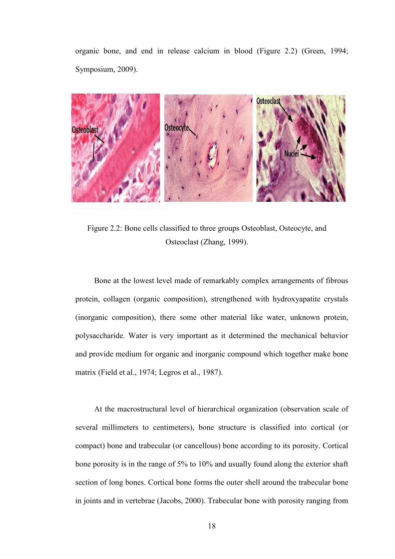
18
organic bone, and end in release calcium in blood (Figure 2.2) (Green, 1994;
Symposium, 2009).
Bone at the lowest level made of remarkably complex arrangements of fibrous
protein, collagen (organic composition), strengthened with hydroxyapatite crystals
(inorganic composition), there some other material like water, unknown protein,
polysaccharide. Water is very important as it determined the mechanical behavior
and provide medium for organic and inorganic compound which together make bone
matrix (Field et al., 1974; Legros et al., 1987).
At the macrostructural level of hierarchical organization (observation scale of
several millimeters to centimeters), bone structure is classified into cortical (or
compact) bone and trabecular (or cancellous) bone according to its porosity. Cortical
bone porosity is in the range of 5% to 10% and usually found along the exterior shaft
section of long bones. Cortical bone forms the outer shell around the trabecular bone
in joints and in vertebrae (Jacobs, 2000). Trabecular bone with porosity ranging from
Figure 2.2: Bone cells classified to three groups Osteoblast, Osteocyte, and
Osteoclast (Zhang, 1999).
19
75% to 95% is usually found in cubicoidal and flat bones (e.g., vertebrae and pelvis)
and in the end of long bones (e.g., femur). Trabecular bone accounts for
approximately 80% of the skeletal volume and 70% of the skeletal mass is cortical
bone. The microstructure of trabecular bone is composed of irregular, sinuous
convolutions of lamellae, and the microstructure of cortical bone is comprised of
regular, cylindrically shaped lamellae. Cortical bone is composed of osteons or
Haversian canals. The osteons are embedded in a matrix of lamellar bone known as
interstitial lamellae (Rho et al., 1998).
Lamellae are bands or layers of bone (3 µm to 7 µm thick) forming an
anisotropic matrix of mineral crystals and collagen fibers (Jacobs, 2000). Trabecular
packets and osteons are composed of lamellae and are attached to the bone matrix by
cement lines. At the molecular level (in the scale of ≤100 nm), three main important
materials exist, namely, biologic apatite crystals, collagens, and noncollagenous
organic proteins. Mature crystals are not needle shaped but plate shaped (Rho et al.,
1998; Weiner and Wagner, 1998). Plate-like biological apatite crystals of bone occur
within the isolated spaces within the collagen fibrils. This phenomenon limits the
possible primary growth of the mineral crystals and forces the crystals to be discrete
and discontinuous (Rho et al., 1998; Weiner and Wagner, 1998). The nanocrystalline
bone apatite has small but significant amounts of impurities, such as HPO4, Na, Mg,
citrate, carbonate, and K. Similar to the crystal of HA, the crystal of carbonate apatite
not only exhibits a hexagonal crystal structure but also produces X-ray diffraction
patterns (Weiner and Wagner, 1998). Weiner et al. (1999) proposed that their plate-
like structure is attributed to their growth by an octacalcium phosphate transition
20
phase. Octacalcium phosphate crystals are plate shaped and have a similar structure
to apatite, except for the presence of a hydrated layer.
2.1.2 Mechanical Properties of Bone
The mechanical properties of trabecular and cortical bones including
compressive strength, tensile strength, Young’s modulus, hardness, and fracture
toughness have been extensively studied. Differences between whole bones perhaps
explained by the variation in the structure and mechanical function of bones (Rho et
al., 1998). The mechanical properties of cortical bone (tibia, femur, and humerus) are
significantly influenced by the porosity, mineralization level, and organization of the
solid matrix. The mechanical properties of cortical bone vary between subjects,
although the density remains the same. In contrast to cortical bone, in trabecular
bone, the mechanical properties of the humerus, proximal tibia, and lumbar spine are
not different (Rho et al., 1995; Zhou and Lee, 2011).
The strength and mechanical properties of bone depend on bone density,
porosity, and molecular structure. Other factors, such as, humidity, mode of applied
load, direction of the applied load, and location of bone within the body, affect the
mechanical properties of bone (Rho et al., 1998).
2.1.3 Bone and implantation
Bones are relatively rigid structures and their shapes are closely related to their
functions. Bone metabolism is mainly controlled by the endocrine, immune, and
neurovascular systems, and its metabolism and response to internal and external
stimulations are still under assessment (Ratner et al., 2012). Disturbance to normal
21
function and metabolism of bone causes with any stimuli, ended in bone disease, and
need of different treatment. Long bones are most part of skeletal system to injury,
and internal or external fixation is a part of their treatment. Joint replacement is main
intervention where the bone is expected to host biomaterials. Response of the bone to
biomaterial arbitrates with the regeneration procedure. Materials implanted into the
bone, nevertheless, cause local and systemic biological responses even if they are
known to be inert (Ratner et al., 2012).
Biomaterials are the natural or synthetic material that is used to replace or
restore function of body tissue. Currently the wide ranges of materials used in
medicine and biotechnology. These materials are mainly divided into four classes;
polymers, metals, ceramics, and natural materials (including those from both plants
and animals). Different classed of this material can be mixed to develop composite
material with increased biocompatibility, efficacy in treatment; end result will be
calss fifth which call composite material. Examples of the fifth class of biomaterial
are silica-reinforced silicone rubber or carbon fiber- or hydroxyapatite particle-
reinforced (Ratner et al., 2012).
The application of exogenous material in treatment was started back in 1960s,
during World War II. Surgeons had used commercially available polymers and
metals, fabricating implants and components of medical devices from them, as
replacement for lost part in the body (Thomas, 2004). The first generations of use
biomaterial in implantation happen to be impressively effective; however there were
a few failure rejections due to infection. This achievement in medication made
surgeons to consider the physical, biological, and materials science and engineering,
22
where by the earliest interdisciplinary “bioengineering” collaborations were born.
These collaborative research teams not only focus to control the biomaterial
composition, quality, and purity, they also established the need for new materials
with new and special properties. This stimulated the development of many new
materials in the 1970s (Thomas, 2004; Ratner et al., 2012).
Biomaterials were specifically designed to be physically replaced the hard or
soft tissues, which suffer from a variety of destructive processes including fracture,
infection, and cancer that cause pain, disfigurement, or loss of function. Suture is one
of the oldest and literary the first generation of biomaterial used in wound healing
process. Back to 2000 b.c, the ancient Egyptians used linen to holding together the
edges of a wound or surgical incision. Later by developed technology synthetic
suture material like polymer (the most widely used) and some metal stainless steels
and tantalum were replace with it. The used biomaterial not summarized to wound
healing device also a wide range of biomaterials with multiplicity of properties are
used to produce ocular devices to correct functional deficiencies caused by disease,
age and ocular trauma (Lloyd et al., 2001).
Metals, ceramics and polymers used to replace defected part in the
cardiovascular, or circulatory system. The heart valves suffer from structural changes
that prevent the valve from either fully opening or fully closing and arteries,
particularly the coronary arteries and the vessels of the lower limbs, become blocked
by fatty deposits (atherosclerosis), all these can be successfully treated with implants.
Bacterial infections, dental caries (cavities), the demineralization and dissolution of
teeth associated with the metabolic activity in plaque, cause extensive damage to the
23
tooth and supporting gum, raise the need of tissue replacement. The need of tissue
replacement introduces another area for use of biomaterial. Therefore, verities of
material use to replace segment or entire teeth in their entirety.
One of the fastest growing areas for implant applications is devices for
controlled and targeted delivery of drugs. This area of implantation focus on the
synthesis, fabrication, and evaluation of biomaterials, including nanobiomaterials for
important applications in biomedicine. Drug delivery implantation emphasize the
development of novel biomaterials with exciting or improved physical, chemical, and
biological properties to control drug delivery and selectivity, and increased the
efficacy of drug.
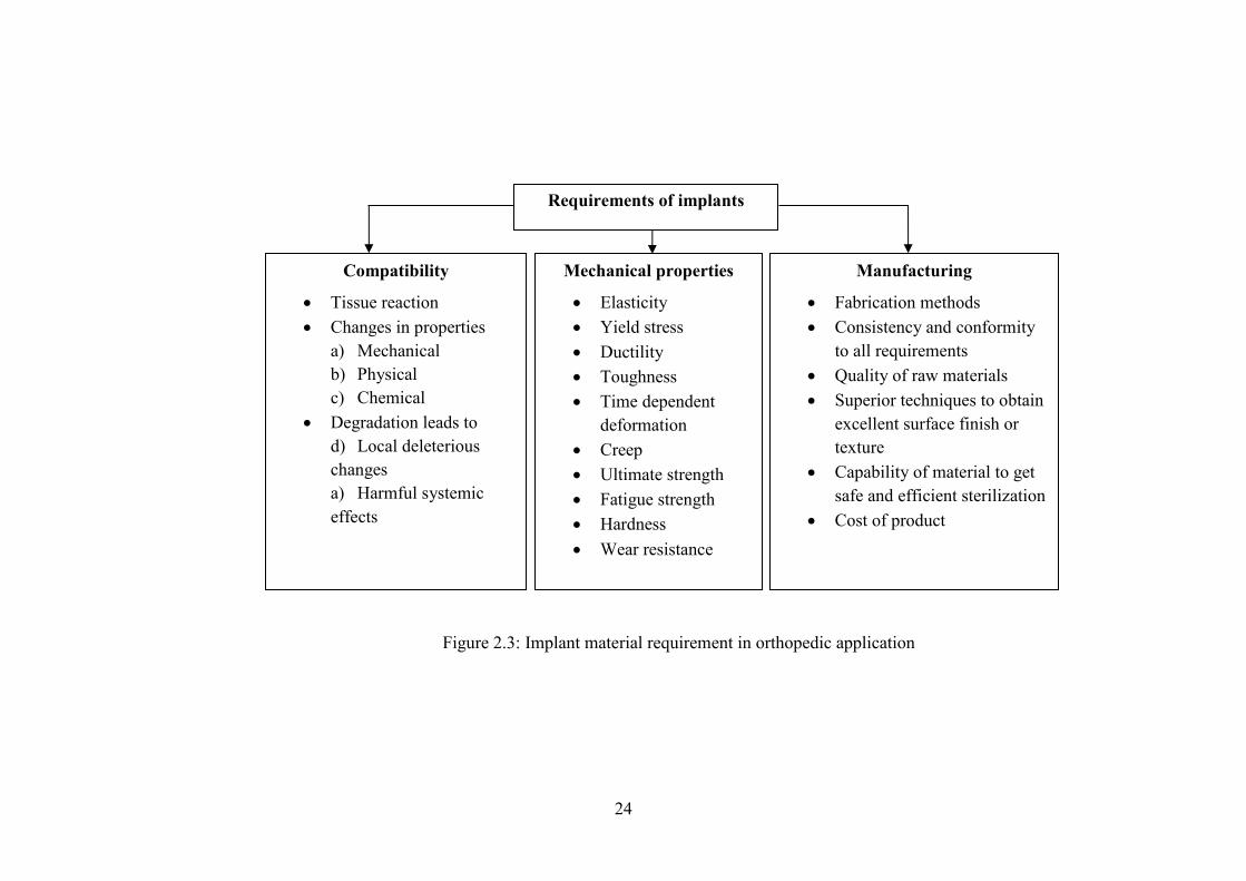
Orthopedics implant is the most dominant area for application biomaterials;
free movable joint in skeleton like hip, knee, shoulder, ankle, and elbow, affected by
chronic condition of osteoarthritis and rheumatoid arthritis. These chronic conditions
developed by genetic and environmental factors, aging joints, previous injuries, and
obesity affect the structure of synovial joints and induce considerable pain in joints
especially weight bearing joint and disturb the joint movement . The interruption in
ambulatory function quite devastating but it has been possible to replace these joints
with orthopedic implants products. Wide range of metals, polymers, ceramics, and
composites used within the manufacture of orthopedics implants the relief of pain
and restoration of mobility. There are many deal to be made before the material can
be produced and these summarized in Figure 2.3.
24
Manufacturing
Fabrication methods Consistency and conformity
to all requirements Quality of raw materials Superior techniques to obtain
excellent surface finish or texture
Capability of material to get safe and efficient sterilization
Cost of product
Requirements of implants
Compatibility
Tissue reaction Changes in properties
a) Mechanical b) Physical c) Chemical
Degradation leads to d) Local deleterious changes a) Harmful systemic effects
Mechanical properties
Elasticity Yield stress Ductility Toughness Time dependent
deformation Creep Ultimate strength Fatigue strength Hardness Wear resistance
Figure 2.3: Implant material requirement in orthopedic application