Embed Size (px)
Citation preview

Development of Fetal Monolayer Culture
Rat Intestine in Organ and
ANDREA QUARONI Section of Physiology, Division of Biological Sciences, Cornell University, Ithaca, New York 14853
ABSTRACT Maturation and differentiation of intestinal epithelial cells was demonstrated in segments of fetal rat small intestine, maintained for more than a month in suspension organ culture, by ultrastructural, biochemical, and immunological criteria. Over a 5-7 d period, fragments of fetal intestine evolved into globular structures covered with a single columnar epithelium ultrastructurally similar to suckling villus cells. Loose mesenchymal cells, cellular debris, and collagen were present inside the structures. After 6 d in culture, goblet cells, not present in the fetal intestine at day 18, were numerous and well developed. Intestinal endocrine cells were also observed. Immunofluorescence studies employing monoclonal antibodies specific for villus and crypt cells in vivo, and various enzyme assays, have demon- strated a level of differentiation and maturation of the cultured epithelial cells similar but not identical to that of suckling intestinal mucosa in vivo. Crypts and crypt cell markers were not observed in the the cultures. Addition of glucocorticoids to the culture medium resulted in the induction of sucrase-isomaltase but failed to promote most of the functional changes characteristic of the intestinal epithelium at weaning in vivo.
Epithelial cells were identified in explants derived from the organ cultures by their specific expression of intestinal cytokeratin. Differentiation-specific markers, present in the epithelial cells in primary cultures, were lost upon selection and subculturing of pure epithelial cell populations. These results suggest a requirement for mesenchymal and/or extracellular matrix components in the maintenance of the differentiated state of the epithelial cells.
The fetal intestinal organ cultures described here present significant advantages over tradi- tional organ and monolayer culture techniques for the study of the cellular and molecular interactions involved in the development and differentiation of the intestinal epithelium.
Much effort in recent years has been devoted to the develop- ment of in vitro culture systems which would allow a detailed investigation of the properties of the intestinal epithelial cells under relatively simple and well-defined experimental condi- tions, Various techniques have been described for the organ culture of adult intestinal fragments or explants (13, 34), but are limited in use by the rapid necrosis and degeneration of the epithelium. Better tissue preservation and longer survival times have been reported for rat and human colon (1) and fetal or embryonic rat (5) and chick (2-4) small intestine. A different approach, pursued successfully in a number of lab- oratories, is represented by monolayer cultures of epithelioid cells derived from rat small (19, 24-27) and large (37) intes- tine, and from human benign tumors (10), but these cells have not been shown to perform typical differentiated func- tions of villus cells in vivo.
Another approach to the problem of culturing differentiated intestinal epithelial cells is represented by the long-term sus- pension culture of fetal rat intestine (29, 35). Fetal intestinal organ cultures (FIOC) ~ differ from traditional organ culture systems in that the original architecture of the intestinal mucosa is lost within the first few days in culture, but they allow maintenance of morphologically and functionally stable differentiated intestinal cells in chemically defined medium for long periods of time. Normal development and differen- tiation in many epithelial systems appear to depend upon the interaction between epithelium and mesenchyme. In the case
1 Abbreviations used in thispaper: FCS, fetal calf serum; FIOC, fetal intestinal organ cultures; TBS-BSA, I mM CaCb, 5 mM KCI, 0.5 mM MgCI~, 0.136 M NaCI, 0.7 mM No2 HPO4, 25 mM Tris, 0.05% (wt/vol) NAN3, 0.2% (wt/vol) BSA, pH 7.4.
THE JOURNAL OF CELL BfOLOGY • VOLUMe 100 MAY 1985 1611 1622 1611 © The Rockefeller University Press - 0021-9525185/05[1611/12 $1.00
on April 13, 2019jcb.rupress.org Downloaded from http://doi.org/10.1083/jcb.100.5.1611Published Online: 1 May, 1985 | Supp Info:

of the intestine, experiments combining isolated epithelioid and fibroblastic cell populations, followed by transplantation into syngeneic hosts, have demonstrated the importance of epithelial-mesenchymal interactions for intestinal cell differ- entiation (1 l, 14, 20, 22). These interactions appear to be sufficiently preserved in the suspension cultures of fetal rat intestine, since normal differentiated functions develop and are expressed with time in culture.
In this paper, an ultrastructural, immunological, and bio- chemical evaluation of the differentiated state of the intestinal epithelial cells in the FIOC cultures is reported. Starting from a relatively immature, stratified epithelium of fetal rat intes- tine at day 18 of gestation, the epithelial cells in culture achieve a level of differentiation comparable but not identical to that of villus cells in suckling intestinal mucosa. In contrast, epithelial monolayer cultures derived from FIOC explants have been found unable to express differentiated functions after subculturing and establishment of pure epithelial popu- lations. These results are in accordance with previous studies suggesting a fundamental role for the intestinal mesenchyme, or mesenchyme-derived products, in the induction of intes- tinal cell differentiation.
MATERIALS AND METHODS
Materials: Sprague-Dawley rats (CD strain, of either sex, weighing 170- 175 g) and timed pregnant Sprague-Dawley rats (CD strain) were obtained from Charles River Breeding Laboratories, Inc. (Wilmington, MA). Dulbecco's mod- ified Eagle's medium with 4.5 g/l glucose (DME), Roswell Park Memorial Institute 1640 medium, irradiated fetal calf serum (FCS), penicillin-strepto- mycin mixture, and trypsin (2.5% in Hanks' balanced salt solution without calcium and magnesium) were obtained from M.A. Bioproducts (Walkersville, MD). Tris(hydroxymethyl)aminomethane (Tris), HEPES, phenylmethylsul- fonyl fluoride, aprotinin, leupeptin, and antipain were obtained from Sigma Chemical Co. (St. Louis, MO).
Membrane Purification: The luminal (brush border) membrane of intestinal epithelial ceils was purified from adult rat jejunum and from FIOC by the method of Kessler et al. (15). FIOC were washed three times with phosphate-buffered saline (PBS) before homogenization. A mixture of proteasc inhibitors (1 mM phenylmethylsulfonyl fluoride, 50 #g/ml leupeptin, 50 t~g/ ml antipain, 0.1 mg/ml aprotinin) was added to all buffers and solutions used for homogenization and membrane purification.
Immunoprecipitation and Characterization of Membrane Antigens: Membrane proteins of FIOC were labeled metabolically with [3H]lysine, and brush border membranes purified from adult rat jejunum were radiolabeled by reductive akylation with [~4C]folmaldehyde and sodium cyano- borohydride as described in the previous paper (30).
Solubilization of membrane proteins, binding to monoclonal antibodies- Sepharose 4B beads, SDS slab gel electrophoresis, two-dimensional slab gel electrophoresis, and visualization of radioactive bands by fluorography were performed as described in the previous paper (30).
Immunofluorescence Staining: Immunofluorescence staining of frozen sections of rat small intestine and FIOC was done using the double antibody fluorescence technique (30). Immunofluorescence staining of mono- layer cultures and FIOC explants was by the biotin-avidin method. Cells cultured on glass coverslips were washed three times with PBS, incubated for l h at 4*C with 1% formaldehyde in 100 mM sodium phosphate buffer (pH 7.4), washed three times with PBS, incubated for 1 h at 4"C with 100 mM glycine in PBS (pH 7.4), washed two times with PBS + 0.2% (wt/vol) bovine serum albumin (BSA). All subsequent incubations were performed at room temp. The cells were incubated for 30 min with mouse serum (controls) or monoclonal antibodies (ascites fluid) diluted 1:100 in PBS + 0.2% BSA, washed three times with PBS, incubated 30 rain with biotinylated anti-mouse IgG (H+L) immu- noglobulin (affinity purified, obtained from Vector Laboratories, Inc., Burlin- game, CA), 15 zg/ml in PBS + 0.2% BSA, washed three times with PBS, incubated 30 min with fluorescein isothiocyanate-labeled Avidin DCS (cell sorter grade, Vector Laboratories, Burlingame, CA), 10 ~g/ml in PBS + 0.2% BSA, washed three times with PBS, counterstained for 30 s with 0.01% (wt/ vol) Evans blue in PBS, washed two times with PBS, mounted.in glyceroI-PBS 9:1, and viewed in a Nikon Optiphot microscope equipped with epifluorescence
1612 THE IOURNAL OF CI-tL BIOLOGY - VOLUME 100, 1985
attachment. Cells to be stained with the monoclonal antibody BBC 3/48/2, specific for intestinal cytokeratin, after fxation with 1% formaldehyde and incubation with I00 mM glycine in PBS (pH 7.4) were washed twice with PBS and treated for 5 rain at -20"C with methanol, then for 3 rain at -20"C with acetone, washed with distilled water, and then with PBS. Following procedures were as described above. Fluorescence was excited with the output of an Osram HBO 100 lamp filtered with a Nikon B2 filter set (excitation filter 460-485 nm; dichroic mirror DM 510, eyepiece-side absorption filter 520-560 nm). Pictures were recorded with Kodak Ektachrome 400 film (Eastman Kodak Co., Rochester, NY). Black and white negatives of the color slides were obtained using Kodak Technical Pan Film 2415 or Panatomic-x film (Eastman Kodak Co.). The red fluorescence of the counterstain (Evans blue) was filtered out with a green filter (Tiffen #58).
Enzyme Assays: Sucrase, maltase, lactase, trehalase, glucoamylase, alkaline phosphatase, aminopeptidase, and "y-glutamyltransfemse activities in brush border membranes, purified from fetal intestinal mucosa and FIOC by the method of Kessler et al. (15) were determined as previously described ( 12, 25, 28). Results were expressed as mU of enzyme per milligram of protein; enzyme units were defined in all cases as the amount of enzyme transforming 1 umol of substrate per minute.
Electron Microscopy: Samples of FIOC for transmission electron microscopy were rinsed twice with PBS, fixed at room temperature for 1 h in 8 mM CaCI2, 2% paraformaldehyde-2.5% glutaraldehyde buffered with 0.1 M sodium cacodylate at pH 7.4, rinsed with the same buffer and postfixed in 1.3% osmium tetroxide buffered with 0.1 M collidine at pH 6.8. The samples were stained en bloc in a 1% solution of uranyl acetate in the same buffer for 1 h at 4"C, washed with collidine buffer, dehydrated in a graded series of alcohols, and embedded in a mixture of Polybed 812 and Araldite 6005. Sections with gray interference colors were cut on an LKB Ultrome III ultramicrotome (LKB Instruments, Inc., Gaithersburg, MD), stained with uranyl acetate and lead citrate, and examined in a Philips 300 transmission electron microscope. Samples for scanning electron microscopy were rinsed with PBS, fixed with 2% paraformaldehyde-2.5% g]utaraldehyde, postfixed in 1.3% osmium tetroxide, and dehydrated in a graded series of alcohols as described above. The samples were dried in a Polaron E 3000 Critical Point Drying Apparatus (Polaron Instruments Inc., Hatfield, PA) using CO2 as the transition fluid, attached to an aluminum stub using conductive paint, coated with gold using a Polaron E 5000 sputter coater, and examined in an AMRAY 1000 transmission electron microscope at 20 kV potential.
GeneraI CelI Culture Conditions: Monolayerculturesweregrown in plastic petri dishes (Lux, purchased from Miles Scientific Div., Naperville, IL) at 37"C in an atmosphere of 94% air, 6% CO2. Before being used for cell culture, glass coverslips were boiled in 50% nitric acid, rinsed extensively with distilled water, and autoclaved. Culture of the IEC-6, IEC- 17, and IEC- 18 cells, selection and subculturing of epithelioid cells with cloning cylinders (0.8-cm diameter), and other general cell culture procedures were as previously described (25). Growth rates (population doubling times), saturation densities, plating efficiencies, and ability of the cells to establish colonies in soft agar were determined as in our previous work (25).
Monoclonal Antibodies: The mouse monoclonal antibodies to in- testinal membrane proteins and intestinal cytokeratin used in this work have been prepared and characterized as described elsewhere (see Table II1 for list of antigen specificity of the antibodies): YBB 3/10, YBB 1/27, and CC 4/80 (30); BB 3/34 and BB 5/8 (12); BB 4/33, BB 4/35, BB 5/16, and BB 8/2 (28, 29, and Quaroni, A., submitted for publication); IEC 1/48, CC 4/11, CC 4/39, CC 4/91, BBC 1/35, BBC 3/88, BBC 3/90, BBC 3/91, YBB 1/57, YBB 2/61, YBB 2/54/3, and BBC 3/48/2 (Quaroni, A., submitted for publication).
Binding of Monoclonal Antibodies to Intestinal Cells in Monolayer Cultures: Cells were cultured in 35-mm diameter tissue culture dishes, with the exception of secondary cultures of cells from FIOC explants which were seeded and tested in the wells of 24-well Costar plates (Data Packaging Co., Cambridge, MA): all volumes referred to below were halved for assays done in Costar plates. When the cultures were confluent, the medium was aspirated and the cells were washed three times with I mM CaCI2, 5 mM KC1, 0.5 mM MgCI2, 0.136 M NaCI, 0.7 mM Na2 HPO4, 25 mM Tris, 0.05% NaN3 (pH 7.4) containing 0.2% (wt/vol) BSA (TBS-BSA), incubated for 90 min at 4"C with l ml mouse serum (controls) or monoclonal antibodies (ascites fluid) diluted 1:500 in TBS-BSA, washed three times with 4 ml TBS- BSA (5 min each time at 4"C), incubated for 90 min at 4"C with 1 ml TBS + 1% BSA containing l-l.5 x l0 s cpm of ~251-1abeled sheep anti-mouse lgG (H+L) F(ab')2 fragment (affinity purified, 7-9 uCi/~g, New England Nuclear, Boston, MA), washed three times with 4 ml TBS-BSA (5 min each time at 4"C), and then once with 4 ml of PBS. The cells were dissolved in 1 ml of 0. l N NaOH, neutralized with 1 M acetic acid, and counted with l0 ml Acquasol II (New England Nuclear, Boston, MA) in a Beckman LS 3800 liquid scintil- lation counter (Beckman Instruments, Inc., Pain Alto, CA). Parallel cultures

were used for cell counting, in triplicate. Data were expressed as cpm of ~2Sl bound per 106 cells. The background (cpm/106 cells bound to cultures incubated with mouse serum instead of monoclonal antibodies, diluted 1:500 in TBS- BSA) was in no case greater than 2,000 cpm/106 cells, and was subtracted from all the data included in Table I11.
Preparation of FIOC and FIOC Explants: FIOC were prepared and maintained in culture as previously described (29, 35) with minor modifi- cations outlined here. Two different culture conditions were used.
CULTURE IN SERUM-FREE MEDIUM: The culture medium was com- posed ofa l:l mixture of Dulbecco's modified Eagle medium (DME) with 4.5 g/l glucose, and Roswell Park Memorial Institute 1640 medium, supplemented with 10 mM HEPES, penicillin (50 U/ml), streptomycin (50 ug/ml), 2 mM glutamine, and 0.2% BSA (2x crystallized, Schwarz-Mann, Cambridge, MA). FIOC were cultured suspended in 15 ml complete culture medium/100-mm diameter tissue culture dishes. Half volume of medium was aspirated at 1- to 2-d intervals, and replaced with fresh medium.
CULTURE IN SERUM-CONTAINING MEDIUM: The culture medium was composed of a 1:1 mixture of DME and Roswell Park Memorial Institute 1640 medium, supplemented with 10 mM HEPES, 5% FCS, penicillin (50 U/ ml), streptomycin (50 t~g/ml), and 2 mM glutamine. FIOC were cultured suspended in 15 ml complete culture medium per 100-mm diameter tissue culture dishes. The surface of the dishes was covered with a Millipore LSWP- 090-25 filter (5.0 ~m pore size, Millipore/Continental Water Systems, Bedford, MA), sterilized by autoclaving. Half volume medium changes were performed at 1- to 2-d intervals. The presence of 5% FCS in the medium was beneficial in promoting long-term preservation of the epithelial cells and all the results included in this paper were obtained with FIOC cultured in the presence of serum. However, maturation and differentiation of the epithelial cells were also observed in FIOC cultured for 2-3 wk in serum-free medium, as judged on the basis of ultrastructural studies, immunofluorescence staining with monoclonal antibodies, and enzyme assays.
Hydrocortisone-supplemented media contained 250 ng/ml hydrocortisone- 2 l-succinate. Hydrocortisone (0.5 uM) added to the culture medium resulted in the induction of sucrase-isomaltase (Table 1) as well as a generalized increase in the specific activity (U/rag protein) of most other brush border enzymes tested, which may be an indication of a better functional preservation of the epithelial cells. The presence of FCS in the culture medium resulted in a marked tendency of the FIOC to attach to the surface of plastic tissue culture dishes, followed by cellular outgrowth (as described below for FIOC explants) and rapid degeneration of the fragments. It was therefore necessary to cover the surface of the dishes with a Millipore filter, to which the FIOC did not significantly attach.
FIOC explants were prepared as follows: FIOC, in culture for 7 or more d, were suspended in serum-containing medium and added to 60- or 100-ram
diameter tissue culture dishes, or 35-mm diameter dishes containing acid- washed sterile circular glass coverslip. Most of the medium was aspirated, leaving just enough to cover the surface of the dishes and keep the FIOC fragments moist. The dishes were placed overnight in the incubator. The following day, the proper volume of serum-containing complete medium (2, 4, and 10 ml, for 35-, 60-, and 100-mm diameter dishes, respectively) was added to the dishes. Thereafter, FIOC explants were cultured using standard cell culture techniques. Medium changes were performed at 3-d intervals.
RESULTS
Morphologic and Structural Development of Fetal Intestine in Culture
Shortly after being suspended in culture medium, the smaller intestinal fragments became everted, with the epithe- lium exposed to the medium; in the larger fragments, rudi- mentary villi became partially extruded at the two open ends (Fig. 1), and later coalesced to form cellular outgrowths which sealed the ends of the fragments. During the first week in culture, the surface of the FIOC became progressively covered with a single layer of epithelium originated by proliferation and migration of the cells present at the ends of the fragments. Frequent mitoses were observed in the epithelium, and most cells were found capable of incorporating radioactive thymi- dine into cellular DNA by autoradiography of histological sections. During the first 2-3 d in culture, a large amount of cellular debris was observed in the culture medium, and most of the ceils inside the fragments degenerated. After 7-8 d in culture, most fragments assumed a relatively stable structure and cellular organization, which was maintained up to a month in culture (the longest time considered in this study). The FIOC became approximately spherical or oblong bodies, with few cellular projections, almost completely covered with tightly packed polygonal cells characterized by a dense layer of surface microvilli. A few cells apparently being extruded from the epithelium and sloughed into the medium were observed by light and electron microscopy at all times in culture, but after the first week mitoses were rarely detected
FIGURE 1 Light microscopic morphology of segments of small intestine obtained from rat fetuses at day 18 of gestation, cultured in suspension in serum-containing medium. Arrows in a point to the open ends of the intestinal segments, which became sealed by cellular outgrowth at later times in culture. Pictures were taken after (a) 1 d, (b) 4 d, and (c) 8 d in culture. (a and b) x 50; (c) × 25.
QUARONI Development of Fetal Intestine in Culture 1613

and incorporation of radioactive thymidine into cellular DNA was very limited. Thus, the surface epithelium of the FIOC did not appear to undergo rapid renewal like the intestinal epithelium in vivo, although a limited cell proliferation could not be excluded. An important variable which was noted during this study was the key role played by the age of the rats from which the intestines were obtained in determining viability and function of the epithelial cells covering the FIOC. Fetuses older than day 19 of gestation resulted in progressively smaller yields of FIOC with morphologically well-preserved epithelial cells. This finding may be related to the rapid maturation of the fetal intestine after day 18 of gestation (17, 23, 30), and suggests that FIOC development in culture is correlated with the presence of a stratified epithelium in the starting tissue.
Ultrastructural Characteristics of the FIOC
The mucosa of the fetal rat intestine at the time it was placed in culture (day 18 of gestation) consists of stratified epithelium mixed with rudimentary villi lined by immature- appearing columnar epithelial cells (5). After 8 d in culture,
the FIOC were composed of a single layer of columnar epithelial cells at the surface (Fig. 2), a loose layer of appar- ently viable mesenchymal cells immediately underneath (Fig. 3 a), and a central region filled with fluid, degenerated cells, and extracellular matrix components. The surface epithelial cells were tall, with basaUy located nuclei, their apical surface was lined by tightly packed microvilli, and cytoplasmic proc- esses formed extensive interdigitations along the lateral and basal surfaces of the cells. Tight junctions were observed at the apical regions, and desmosomes were present at the lateral borders. The endocytic complex and giant lysosomal vacuoles (Fig. 3 b) typical of neonatal intestinal cells in vivo (16, 31) were often observed in the epithelial cells covering the FIOC. A well-defined, apparently continuous basal lamina was pres- ent immediately underneath the FIOC epithelium, following the contour of the basal surface membrane (Fig. 3 a). Cyto- plasmic processes projecting through gaps in the basal lamina to make contact with underlying mesenchymal cells, which are frequent in fetal and neonatal intestine (20), were not apparent. Numerous goblet cells filled with characteristic mucous granules (Fig. 2) and occasional enteroendocrine cells (Fig. 3 c) were observed.
FIGURE 2 Ultrastructural appearance of the surface epithelium of FIOC. Both columnar epithelial cells, which are covered with microvilli and similar to intestinal absorptive villus cells, and goblet cells that contain typical mucous granules are apparent. Bar, 2 ~m. x 5,000.
1614 THE JOURNAL OF CELL BIOLOGY . VOLUME 100, 1985

FIGURE :3 (a) Lower portion of the surface epithelial cells; a viable mesenchymal cell, underlying the epithelium, is visible in the lower part of the figure. A continuous basal lamina (arrows) follows the contour of the basal aspect of the epithelial cells. (b) Giant lysosmal vacuoles (GV) are apparent in these FIOC epithelial cells after 10 d in culture. (c) Endocrine epitheliaJ cell from an FIOC cuItured fn serum-containing medium for 14 d; typ[car secretory granules are seen in the cytoplasm. (a) Bar, 3 #m. x 10,400. (b) Bar, ] ~,m. x 8,200. (c) Bar, 2 #m x 15,000.
QUARONI Development of Fetal Intestine in Culture 161 5
Expression of Differentiation-specific Markers by FIOC Epithelium
The fetal rat intestinal epithelium at day 18 of gestation is known to lack most differentiation-specific marker enzymes typical of mature villus cells (17, 21, 23). This was also confirmed by immunofluorescence staining of frozen sections with a large panel of monoclonal antibodies to sucrase, ami- nopeptidase N, maltase, and alkaline phosphatase (data not shown). At day 17 of gestation, staining of the intestinal epithelium was observed only with the YBB 3/10 antibody (30) and with a series of monoclonal antibodies to intestinal cytokeratins; at day 18 of gestation weak staining of the luminal membrane of the epithelial cells was detected with antibodies (YBB 1/57 and YBB 2/61) specific for lactase. By day 19 of gestation, a faint staining was also observed with antibodies to alkaline phosphatase (BB 5/16) and aminopep- tidase (BB 4/33 and YBB 2/54/3); maltase was first detectable at day 21 of gestation. These results were confirmed by measurements of enzyme activities in purified brush border membranes from fetal rat intestine at days 18 to 21 of
gestation (Table I), which are similar or identical to previous reports by many groups (for review see references 17 and 23).
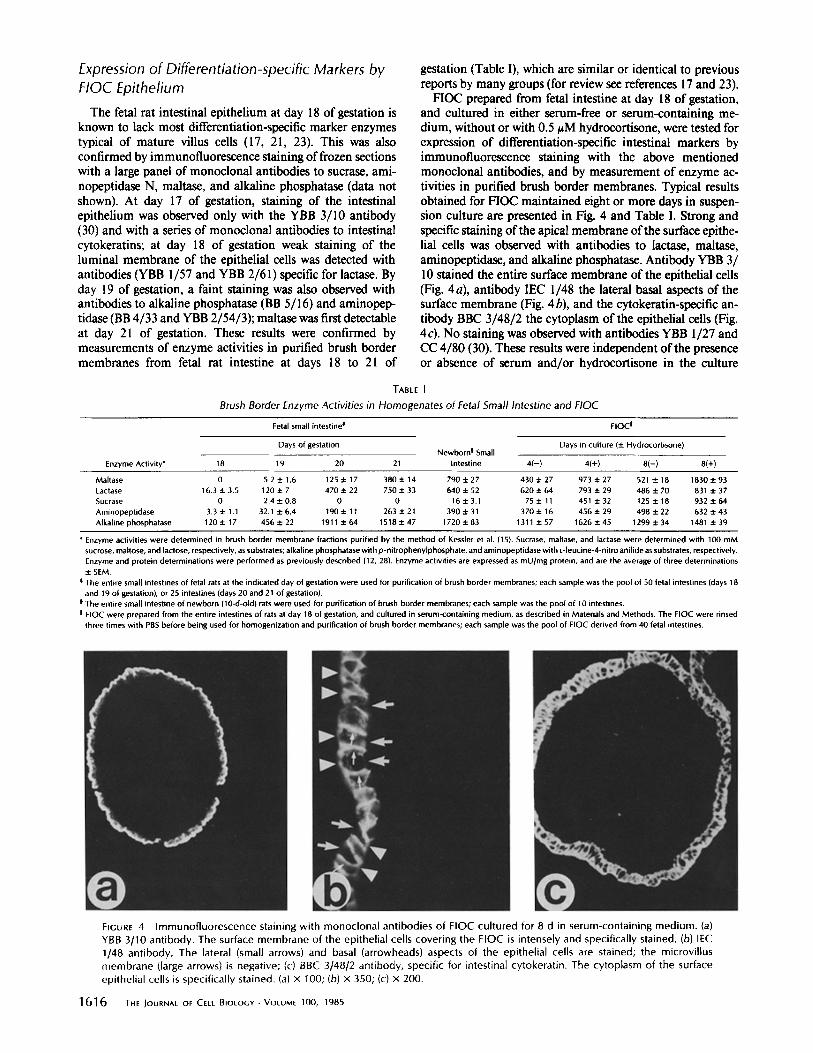
FIOC prepared from fetal intestine at day 18 of gestation, and cultured in either serum-free or serum-containing me- dium, without or with 0.5 tzM hydrocortisone, were tested for expression of differentiation-specific intestinal markers by immunofluorescence staining with the above mentioned monoclonal antibodies, and by measurement of enzyme ac- tivities in purified brush border membranes. Typical results obtained for FIOC maintained eight or more days in suspen- sion culture are presented in Fig. 4 and Table I. Strong and specific staining of the apical membrane of the surface epithe- lial cells was observed with antibodies to lactase, maltase, aminopeptidase, and alkaline phosphatase. Antibody YBB 3/ 10 stained the entire surface membrane of the epithelial cells (Fig. 4a), antibody IEC 1/48 the lateral basal aspects of the surface membrane (Fig. 4 b), and the cytokeratin-specific an- tibody BBC 3/48/2 the cytoplasm of the epithelial cells (Fig. 4c). No staining was observed with antibodies YBB 1/27 and CC 4/80 (30). These results were independent of the presence or absence of serum and/or hydrocortisone in the culture
TABLE I
Brush Border Enzyme Activities in Homogenates of Fetal Small Intestine and FIOC
Fetal small intestine ~ FIOC I
Days of gestation Days in culture (+ Hydrocortisone) Newborn ! Small
Enzyme Activity* 18 19 20 21 intestine 4(-) 4(+) 8(-) 8(+)
Maltase 0 5.2+1.6 125+17 380+14 790+-27 430+27 973+.27 5214-18 1830+.93 Lactase 16.3+3.5 120+7 470+22 750+.33 640+52 620+.64 793+.29 486+70 831+.37 Sucrase 0 2.4 + 0.8 0 0 16 + 3.1 75 +. 11 451 + 32 125 + 18 932 + 64 Aminopeptidase 3.3±1.1 32.1+.6.4 190+11 263+.21 390+.31 370+16 456+29 498+.22 632+.43 Alkaline phosphatase 120+.17 456+.22 1911+.64 1518+.47 1720+.83 1311+.57 1626+.45 1299+.34 1481+.39
* Enzyme activities were determined in brush border membrane fractions purified by the method of Kessler et al. (15). Sucrase, maltase, and lactase were determined with 100 mM sucrose, maltose, and lactose, respectively, as substrates; alkaline phosphatase with p -nitrophenylphosphate, and aminopeptidase with L-leucine-4-nitro anilide as substrates, respectively. Enzyme and protein determinations were performed as previously described (12, 28). Enzyme activities are expressed as mU/mg protein, and are the average of three determinations + SEM.
0 The entire small intestines of fetal rats at the indicated day of gestation were used for purification of brush border membranes; each sample was the pool of 50 fetal intestines (days 18 and 19 of gestation), or 25 intestines (days 20 and 21 of gestation).
| The entire small intestine of newborn (10-d-old) rats were used for purification of brush border membranes; each sample was the pool of lO intestines. I FIOC were prepared from the entire intestines of rats at day 18 of gestation, and cultured in serum-containing medium, as described in Materials and Methods. The FIOC were rinsed
three times with PBS before being used for homogenization and purification of brush border membranes; each sample was the pool of FIOC derived from 40 fetal intestines.
FIGURE 4 Immunofluorescence staining with monoclonal antibodies of FIOC cultured for 8 d in serum-containing medium. (a) YBB 3/10 antibody. The surface membrane of the epithelial cells covering the FIOC is intensely and specifically stained. (b) IEC 1/48 antibody. The lateral (small arrows) and basal (arrowheads) aspects of the epithelial cells are stained; the microvillus membrane (large arrows) is negative; (c) BBC 3148/2 antibody, specific for intestinal cytokeratin. The cytoplasm of the surface epithelial cells is specifically stained. (a) x 100; (b) x 350; (c) x 200.
1616 THE JOURNAL OF CELL BIOLOGY • VOLUME 100, 1985

FIGURE 5 (a] Identification by SDS slab gel electrophoresis (7.5% acrylamide gel) and fluorography of radioactively labeled membrane proteins specifically bound to monoclonal antibodies-Sepharose 4B beads. Brush border membranes were purified from FIOC cultured for 24 h in lysine and serum-free medium supplemented with 250 ng/ml hydrocortisone 21-succinate and L- [4,5-3H(N)]lysine, 100/~Ci/ml. Brush border membranes from adult rat intestine or from fetal (day 21 of gestation) intestine were radiolabeled by reductive alkylation with [14C]formaldehyde and sodium cyanoborohydride (30). Specifically bound membrane proteins from FIOC are present in lanes 2, 4, 6, 8, and 11; membrane proteins from adult rat brush borders are in lanes 3, 5, 7, 9, and 12. Lane 10, solubilized, labeled membrane proteins from brush borders of fetal rat intestine incubated with the monoclonal antibody BB 5/16. We used the following monoclonal antibodies: YBB 2/61, specific for lactase (lanes 2 and 3); BB 8/2, specific for maltase-glucoamylase (lanes 4 and 5); BB 4/33, specific for aminopeptidase (lanes 6 and 7); BB 5/16, specific for alkaline phosphatase (lanes 8, 9, and 10); BB 3/34, specific for sucrase-isomaltase (lanes I 1 and 12). Lane 1, [14C]-Iabeled molecular mass markers (from the top, respectively [see arrowheads]: myosin, 200 kD; phosphorylase b, 97.4 kD; bovine serum albumin, 69 kD; ~,-globulin heavy chain, 53 kD). (b) Two-dimensional electrophoresis of 14C-labeled alkaline phosphatase from fetal rat intestinal brush borders (day 21 of gestation) bound to monoclonal antibody BB 5/16 (same sample as in a, lane 10). (c) Two-dimensional electrophoresis of 3H-lysine-labeled alkaline phosphatase from FIOC brush border membranes, bound to monoclonal antibody BB 5/16 (same sample as in a, lane 8).

medium. In contrast, strong staining of the apical membrane of the epithelial cells with antibodies to sucrase-isomaltase was observed only with FIOC cultured in the presence of hydrocortisone. Long term culture of the FIOC (up to a month) produced no significant change in the staining pat- terns with monoclonal antibodies described above. In partic- ular, the YBB 3/10 antibody, which in vivo has been shown to stain only lower villus and crypt intestinal cells in rats older than 10-14 d, and only crypt cells in adult intestine (30), stained the entire epithelium covering the FIOC even after a month in culture. This observation argues against the devel- opment of functionally distinct subsets of FIOC epithelial ceils, similar to villus and crypt cells in vivo. The results of the enzyme assays (Table I) confirmed the observations made by immunofluorescence staining, and demonstrated a mat- uration and differentiation of the epithelial cells in the FIOC to a level comparable to that of newborn rat intestinal cells. A significant increase in the activity of most enzymes tested was observed between 4 and 8 d in culture. Addition of hydrocortisone to the culture medium resulted in a marked (six- to sevenfold) increase in the activity of sucrase, corre- sponding to the induction of this brush border enzyme by glucocorticoids previously demonstrated in vivo (6, 12); it also resulted in a smaller but significant increase in the activity of all other enzymes tested. However, immunofluorescence staining of FIOC frozen sections demonstrated no effect of hydrocortisone on YBB 1/27 and CC 4/80 antigens expres- sion (data not shown).
Continuous synthesis of brush border membrane enzymes in F1OC maintained for eight or more days in culture was demonstrated by metabolic labeling with [3H]lysine added to the culture medium over a 24-h period. Labeled, Triton X- 100 solubilized membrane proteins were purified with mono- clonal antibodies bound to Sepharose 4B and analyzed by SDS slab gel electrophoresis under reducing conditions. Lac- tase, maltase, and aminopeptidase synthesized by FIOC were found identical to the corresponding enzymes purified from adult intestinal brush border membranes (Fig. 5a, lanes 2-7). Sucrase-isomaltase (Fig. 5 a, lane 11) was present in the FIOC almost exclusively in the form of the high molecular weight precursor which has been previously shown to be split into the two subunits, after insertion into the brush border mem- brane, by pancreatic proteases present in the intestinal lumen (12). The alkaline phosphatase synthesized by the FIOC (Fig. 5 a, lane 8) lacked the high molecular weight component (Mr 130,000) present in fetal intestinal brush border membranes (Fig. 5a, lane 10) and had an apparent isoelectric point of 5.6, when analyzed by two-dimensional slab gel electropho- resis (Fig. 5c), much more acidic than the corresponding value ofpI 7.4, for the Mr 64,000 component of fetal alkaline phosphatase (Fig. 5b). A change in alkaline phosphatase isoenzymes during fetal and postnatal maturation of the in- testinal mucosa has already been demonstrated on the basis of differences in substrate specificity (l 7, 23).
FIOC Explants and Establishment of Monolayer Cultures of Intestinal Epithelial Cells
When FIOC were allowed to attach to the surface of tissue culture dishes or glass coverslips, cellular outgrowths were readily obtained (Fig. 6a). Both cells were epithelioid and fibroblastic morphology were apparent in the outgrowths, and apparently pure populations ofepithelioid cells (Fig. 6 b) could
1618 THE JOURNAL OF CELL BIOLOGY . VOLUME 100, 1985
FIGURE 6 Light microscopic morphology of a FIOC explant (a) and of a secondary epithelioid culture (b) selected with cloning cylin- ders. (a) x 50; (b) x 125.
be selected and subcultured using cloning cylinders as previ- ously described (25). The presence of cytokeratin tonofila- ments, detected with the monoclonal antibody BBC 3/48/2, represented a useful marker for both differentiated and un- differentiated intestinal epithelial cells in the explants. In the FIOC (Fig. 4c) and in the fetal intestine in vivo (data not shown), immunofluorescence staining with the BBC 3/48/2 antibody was shown to be specific for the intestinal epithe- lium. In the FIOC explants, areas containing cells with epi- thelioid morphology were stained fairly homogeneously with the BBC 3/48/2 antibody (Fig. 7a), while fibroblastic cells

were completely negative. In secondary cultures ofepithelioid cells selected with cloning cylinders, all the cells appeared to express cytokeratin tonofilaments (Fig. 7d). Upon further subculturing of epithelial populations selected from the FIOC explants, two cell lines of intestinal epithelial cells were estab- lished and designated IEC-19 and IEC-20, respectively. Both cell lines had the typical appearance of epithelioid cells in culture, with a centrally located nucleus, numerous microvilli over their surface membrane, and frequent pseudopods ex- tending from the cell borders to the surface of the culture dish and to neighboring cells. Their morphology, as well as their general growth characteristics (listed in Table II), are very
similar to those of the IEC-6, IEC-17, and IEC-18 cells pre- viously established from suckling small intestine (25-27). In both IEC-19 and IEC-20 cell lines at a low passage level, cytokeratin tonofilaments were detected by immunofluores- cence in the entire cell population, but in cultures from late passages (passages 10 or more) antigen expression was appar- ently heterogeneous (Fig. 7f). This finding may be an indi- cation for a dynamic regulation of cytokeratin filaments in older cultures, as described for clones of HeLa cells (18), overgrowth by morphologically indistinguishable nonepithe- lial cells, or progressive dedifferentiation and aging of the cultures.
FIGURES 7 Immunofluorescence staining of FIOC explants and epithelioid cells derived from them with the monoclonal antibodies BBC 3/48/2, specific for intestinal cytokeratin (a, d, and f), and YBB 3/10 (b, c, and e). FIOC explants and monolayer cells were cultured on glass coverslips, washed with PBS, fixed with 1% formaldehyde (cultures to be stained with the antibody BBC 3/48/2 were subsequently treated with methanol and acetone at -20°C), and stained with monoclonal antibodies (ascites form) diluted 1:100 in PBS + 0.2% BSA using the biotin-avidin method (see Materials and Methods for details of the procedure). (a-c) FIOC explants after 5 d in culture. In a, most of the epithelioid cells present in the outgrowth are stained for cytokeratin, but in b only cells close to the original FIOC fragment (F) are stained with the YBB 3/10 antibody; cells further away from the FIOC are totally negative. Arrows indicate the edges of the cellular outgrowth in b. c is a higher magnification view of an area of the explant shown in b close to the original FIOC. Both epithelioid and fibroblastic cells are present, but only the epithelioid cells are stained with the YBB 3/10 antibody. (d and e) Secondary cultures of epithelioid cells derived from a FIOC explant, and selected with a cloning cylinder. In d, all the cells in the field are stained for cytokeratin, demonstrating their origin from the epithelium covering the FIOC, but (as shown in e) staining with the YBB 3/10 antibody is limited to isolated or small groups of cells, suggesting a loss of ability to express differentiated functions in the epithelial cells upon subculturing. In f, we see IEC-19 cells at passage 22 stained for cytokeratin; both positive and negative cells are present. (a and b) × 50; (c) x 100; (d and e) x 75; (f) x 200.
Qu^RoNI Development of fetal Intestine in Culture 1 61 9

TABLE II
Culture Characteristics of Cell Lines Established from FIOC Ex- plants and Suckling Rat Intestine
Cells from Cells from suckling rat FIOC explants* intestines *
IEC-19 IEC-20 IEC-6 IEC-17 IEC-18
Saturation density 5.3 3.9 4.2 6.8 16.0 (cells x 10-41cm 2)
Plating efficiency (%)~ 7.1 6.8 2.5 2.9 5.4 Efficiency of colony N 0 0 0 0 0
formation in agar (%) Population doubl ing ~ 19 21 23 22 17
time (hours)
* Epithelioid cell lines established from FIOC explants as described in this paper; culture parameters were determined on cells between the 7th and 11th passage.
* Epithelioid cell lines established from 19-21-d-old rat intestine, and char- acterized as previously described (25, 26, 27); culture parameters were determined on cells between the 8th and 12th passage.
i 103 cells plated/100-mm dish; cultures were fixed after 2 wk from plating and stained with Giemsa's. Colonies of at least 10 cells were counted.
i 104 cells plated in 4 ml medium/60-mm dish. Dishes were examined after 2 wk in culture. Determined during the logarithmic phase of growth.
Expression of Differentiation-specific Antigens in FIOC Explants and Epithelial Monolayer Cultures Derived from Them
The ability of the epithelioid cells in the explants, and of the epithelial cell lines derived from them, to express intestinal differentiation-specific markers was investigated by immu- nofluorescence staining and with a radioimmunobinding as- say, using a large panel of monoclonal antibodies to intestinal cell surface components. In FIOC explants, staining with the antibodies YBB 3/10 (Fig. 7, b and c) and YBB 2/61 specific for lactase (not shown) was confined to areas populated by epithelial cells immediately surrounding the original FIOC fragments. This staining pattern was clearly in contrast with the staining of the entire population ofepithelioid cells present in the explants, obtained with the antibody to cytokeratin described above (Fig. 7 a). In secondary cultures of epithelial cells selected with cloning cylinders, no staining was observed with the antibody to lactase, and only isolated cells or small groups of cells were stained by the YBB 3/10 antibody (Fig. 7 e). The results obtained with the radioimmunobinding assay (Table III) allowed a quantitative evaluation of the results
TABLE III
Binding of Monoclonal Antibodies to Epithelioid Cells from FIOC Explants and IEC cells*
Cells from FIOC explants s Intestinal epithelioid cell lines ~
Monoclonal anti- Subculture level Antigen body* 1 2 3 I EC-6 IEC- 19 I EC-20
BBC 3/48/2 93,700 86,300 89,100 6,500 21,300 17,600 YBB 3/10 12,200 3,500 0 0 0 0 CC 4/80 0 0 0 0 0 0 YBB 1/27 0 0 0 0 0 0 IEC I148 121,400 112,700 117,100 93,400 121,600 145,300
Sucrase BB 3/34 0 0 0 0 0 0 BB 5/8 0 0 0 0 0 0 CC 4/I I 0 0 0 0 0 0 CC 4/39 0 0 0 0 0 0 CC 4/91 0 0 0 0 0 0 BBC 1/35 0 0 0 0 0 0
Ma]tase BB 8/2 3,100 2,200 2,600 1,100 1,500 2,100 BBC 3/88 2,700 2,000 1,700 1,800 1,300 1,400 BBC 3/90 1,300 1,400 1,100 1,200 1,400 1,200 BBC 3/91 3,400 2,000 2,300 1,900 1,700 2,200
Lactase YBB I157 0 0 0 0 0 0 YBB 2161 0 0 0 0 0 0
Aminopeptidase BB 4/33 76,100 53,600 57,900 51,100 72,200 39,400 YBB 2/54/3 42,300 30,700 32,400 27,600 29,900 19,500
Alkaline Phosphatase B8 4/35 0 0 0 0 0 0 BB 5116 0 0 0 0 0 0
* IEC cells and cells from FIOC explants at subculture level 2 and 3 were cultured in 35-mm dishes; cells from FIOC cultures at subculture level I were in wells of 24-well Costar plates. All cultures were rinsed with TBS-BSA, then incubated for 90 min at 4°C with mouse serum (controls) or monoclonal antibodies (ascites form) diluted 1:500 in TBS-BSA. After three washes with TBS-BSA, cultures were incubated for 90 min at 4°C with I x 106 cpm/35-mm dish (5 x I0 s cpm/Costar well) of 1251-1abeled sheep anti-mouse IgG (F[ab']2 fragment, affinity purified) in TBS-1% BSA. After three washes with TBS-BSA, cells were dissolved in 0.1 N NaOH, neutralized with I M acetic acid, and counted with Acquasol II in a Beckman LS 3800 liquid scintillation counter. Parallel cultures were used for cell counting, done in triplicate. Data are expressed as cpm of 1251 bound per 106 cells. The background (cpm/106 cells bound to cultures incubated with mouse serum instead of monoclonal antibodies) was subtracted from all the data included in this table. The background was in no case greater than 2,000 cpm/106 cells.
* All antibodies were mouse monoclonal antibodies used in ascites form. Their preparation and characterization has been described elsewhere (12, 28, 30, and Quaroni, A., submitted for publication).
t Cells with epithelioid morphology were selected from primary FIOC explants with cloning cylinders and serially subcultured to tissue culture dishes or to the wells of Costar plates as described in the text.
II Intestinal epithelioid cell lines established and characterized as previously described (IEC-6, reference 25), or as described in the text (IEC-19, IEC-20). Cells were used between the 6th and 9th passage.
1620 THE JOURNAL OF CELL BIOLOGY • VOLUME t00, 1985

obtained by immunofluorescence staining. Binding of the YBB 3/10 antibody was limited to secondary cultures of epithelial cells obtained from FIOC explants. As expected from the immunofluorescence data, none of the independ- ently derived monoclonal antibodies to lactase, maltase, al- kaline phosphatase, and/or sucrase-isomaltase tested showed significant binding (greater than twice background) to the cultures derived from the FIOC explants or to the established epithelial cell lines. However, the antibody IEC 1/48, which stains specifically the lateral-basal membrane of the epithelial cells in the FIOC (Fig. 4b) and in the intestine in vivo (29), and the two antibodies BB 4/33 and YBB 2/54/3, specific for aminopeptidase and staining the brush border membrane of the epithelial cells in the FIOC (not shown) and of the villus cells in vivo (28), did bind to all the intestinal epithelial cell cultures tested. These results confirm the origin of the IEC cells from the intestinal epithelium, but also demonstrate that they are essentially undifferentiated cells.
DISCUSSION
The suspension organ cultures of fetal rat intestine described in this paper present significant advantages over traditional organ culture techniques in the study of the maturation and differentiation of the intestinal epithelium in vitro. In culture, the FIOC retain stable morphological and functional charac- teristics over a long period of time, and their surface is populated by a single layer of intestinal epithelial cells express- ing most differentiated cell surface markers typical of villus cells in vivo. Most of the tissue and cellular changes associated with the development of the FIOC from the stratified, rela- tively undifferentiated intestinal epithelium present in the fetal intestine at day 18 of gestation (5) take place during the first few days in culture. Thus the FIOC present a reliable "end point" which can be reproduced in different prepara- tions, and may be used to study the influence of hormones and other factors on the maturation and differentiation of the intestinal epithelium.
A major aim of the present study was to describe in detail the state of development and differentiation of the epithelial cells lining the surface of the FIOC. In comparison with the tissue of origin, the FIOC present evidence of significant cytological differentiation. Ultrastructural studies have shown that the epithelial cells in the FIOC are characterized by a well-developed brush border, frequent interdigitating proc- esses at the lateral borders, tight junctions, and desmosomes (Figs. 2 and 3 a). The endocytic complex and the giant lyso- somal vacuoles (Fig. 3 b), typical of suckling intestinal epithe- lium in vivo (16, 31), were often observed in FIOC cultured in the absence of glucocorticoids. Goblet cells containing characteristic mucous granules, not present in the fetal intes- tine at day 18 of gestation, were numerous in the FIOC, and endocrine cells, although rare, were observed (Fig. 3c). A similar pattern of morphological and structural differentiation has been described by De Ritis et at. (5) for fetal rat intestine maintained in organ culture over a 2- to 3-d period. Crypts and identifiable crypt cells were not detected in the epithelium lining the surface of the FIOC, suggesting that functional development of the FIOC intestinal cells is, at least in part, arrested at an early suckling stage. However, direct contacts between epithelial and mesenchymal cells by means of epi- thelial cell processes projecting through gaps in the basal lamina, which are particularly abundant during early post- natal life (20), were not observed (Fig. 3 a).
Analysis of the expression of brush border enzymes and other cell surface markers in the FIOC provides a more detailed definition of the state of maturation and differentia- tion of their epithelial cells. The presence of typical brush border enzymes like lactase, aminopeptidase, alkaline phos- phatase, maltase, and sucrase in the epithelial cells of the FIOC after 4-8 d in culture, demonstrated both by immu- nofluorescence staining and with specific enzyme assays (Ta- ble I), is evidence for a marked maturation and differentiation of the epithelial cells. Most of these enzymes are undetectable in the original epithelium at day 18 of gestation (Table I). The specific activities of the enzymes determined in brush border membranes purified from the FIOC are comparable with those of villus cells in suckling intestine. This conclusion is also supported by the induction of sucrase-isomaltase in FIOC cultured in the presence of hydrocortisone, and by the switch in alkaline phosphatase isoenzymes recognized by the monoclonal antibody BB 5/16 (Fig. 5), from the fetal form to that characteristic of suckling intestine (17, 23, and Qua- roni, A., submitted for publication). However, not all the phenotypic changes associated with the development of the intestinal mucosa in vivo during late fetal and early postnatal life were observed in the FIOC. A homogeneous staining of the FIOC epithelial cells with the antibody YBB 3/10 was observed, and the YBB 1/27 and CC 4/80 antigens, specific for crypt cells during the suckling period (30), were not detected.
The presence of a relatively homogeneous, differentiated intestinal epithelium at the surface of the FIOC, and the ease with which explant cultures can be derived from them, al- lowed us to investigate maintenance and expression of differ- entiation-specific markers in monolayer cultures of intestinal cells. The presence ofcytokeratin tonofilaments, detected with the monoclonal antibody BBC 3/48/2, is a convenient marker for epithelial cells, independent from their state of differentia- tion (7-9, 18, 32, 33, 36). The antibody BBC 3/48/2 recog- nizes a cytokeratin component in IEC cells and intestinal villus cells which has a molecular weight of 42,000 (Quaroni, A., submitted for publication). Immunofluorescence staining of FIOC explants demonstrated that expression of most dif- ferentiation-specific markers in monolayer intestinal cells re- quires close proximity or possibly direct contact with other components of the FIOC fragments. Established cell lines of intestinal epithelial cells, while expressing epithelial- and in- testinal-specific markers (Table III) like cytokeratin, amino- peptidase, and the IEC 1/48 antigen, do not appear to express most brush border enzymes typical of villus cells in vivo and present in the FIOC, These results are in accordance with previous observations demonstrating a requirement for intes- tinal mesenchyme, or mesenchyme-derived products, for in- testinal cell differentiation (11, 14, 22). Of interest was the remarkable similarity in morphology, ultrastructure, growth properties (Table II), and antigen expression (Table III) be- tween the IEC-6 cells, established from suckling intestinal mucosa (25, 26) and the IEC-19 and IEC-20 cells, derived from the FIOC cultures.
The results obtained in this study demonstrate that the FIOC represent a valuable model system for the study in vitro of the factors involved in maturation and differentiation of the intestinal epithelium. Two critical periods have been identified for the morphologic and enzymatic development of rat small intestine in vivo (17, 23): (a) days 18-19 of gestation, characterized by a transition from a stratified to a
Qu^aoNI Development of Fetal Intestine in Culture 1621

simple epithelium, formation of the villi, and appearance of most brush border enzyme activities; and (b) weaning, at 20- 22 d after birth, characterized by the spontaneous appearance of sucrase-isomaltase, lengthening of the villi, marked in- creases in the rate of cell proliferation in the crypts and in the rate of cell migration. The intestinal epithelial cells in the FIOC appear to develop in vitro, starting from the relatively undifferentiated stratified epithelium present in fetal intestine at day 18 of gestation, according to an intrinsic program of maturation which includes mostly ultrastructural character- istics and brush border enzymes, and appears arrested at a pre-weaning stage. The results obtained in this study allow a distinction among various developmental events occurring almost contemporaneously in vivo, and suggest that factors and/or cellular interactions present in vivo but not available in culture under the conditions used in this study are impor- tant in the maturation and differentiation of the intestinal mucosa. The ease with which experimental conditions may be altered, and the possible effects observed in the FIOC, should represent a significant advantage for their identifica- tion and characterization in the future.
I gratefully acknowledge the technical assistance of Susanne Schulte, David Calnek, and Elaine Gershon Quaroni, and the secretarial assistance of Sue Hawk.
This work was supported by grants AM 32656 and AM 01214 from the National Institutes of Health, United States Public Health Service.
Received for publication 4 September 1984, and in revised form 21 January 1985.
REFERENCES
1 Aulrup, H., G. D. Stoner, F. Jackson, C. C~ Hams, A, K, M. Shamsuddin, L. A. Barrett, and B, F. Trump. 1978. Explant culture of rat colon: a model system for studying metabolism of chemical carcinogens. In Vitro 14:868-877.
2 Black, B. L.. and F. Moo& 1978. Alkalir~e phos~hatase and m a l ~ acitvity in the embryonic chick intestine in culture. Dev. Biol. 66:232-249.
3 Corradino, R. A. 1979. Embryonic chick intestine in organ culture: hydrocortisone and vitamin D-mediated processes. Arch. Biochem Biophys 192:302-310.
4. Corradino, R. A., C. S. Fullmer, and R. H. Wasserman, 1976. Embryonic chick intestine in organ culture: stimulation of calcium transport by exogenous vitamin D-induced calcium binding protein. Arch. Biochem. Biophys. 174:738-743.
5 De Ritis, G., Z. M. Falchuk, and J. S. Trier. 1975. Differentiation and maturation of cultured fetal rat jejunum. Dev. Biol. 45:304-317.
6. Doell, R. G., and N. Kretchmer. 1964. Intestinal invertase: precocious development of activity after injection of hydrocortisone. Science (Wash. DC). 143:4244.
7. Franke, W. W., B. Appelhans, E. Schmid, C. Freudenstein, M. Osborn, and K. Weber. 1979. The organization of cytokeratin filaments in the intestinal epithelium. Eur. Z Cell Biol. 19:255-268.
8. Franke, W. W., S. Winter, C. Grand, E. Schmid, D, L Schiller, and E. D. Jarasch. 1981, Isolation and characterization of desmosome-associated tonofilaments from rat intestinal brush border. Z Cell Biol. 90:116-127,
9 Franke. W. W,, D. L. Schiller, R. Moll, S. Winter, E, Schmid, I. Engelbrecht, H. Denk, R, Kaepler. and B. Platzer. 1981, Diversily of cylokeratins, Differentiation specific
expression of cytokeratin polypeptides in epithelial cells and tissues, ,L Mol. BioL 153:933-959.
10, Friedman, E. A., P. J. Higgins, M. Lipkin, H, Shinya, and A M, Gelb. 198l. Tissue culture of human epithelial cells from benign colonic tumors. In Vitro. 17:632-644.
11. Haffen, K., M. Kedinger, P. M. Simon, and F. Raul. 1981. Organogenetic potentialities of rat intestinal epithelioid cell cultures. Differentiation, 18:97-103.
12. Haun, H. P., A. Quaroni, and K. J. Isselbacher. 1980. Monoclonal antibodies to sucrase- isornaltase: probes for the study of postnatal development and biogenesis of the intestinal mierovinus membrane. Proc. Natl. Acad. Sci. USA. 77:6629-6633.
13. Kedinger, M., P. M. Simon, F. Real, J. F. Grenier~ and K. Haffen. 1980. The effect of dexamethasone on the development of rat intestinal brush border enzymes in organ culture, Dev. Biol. 74:9-21.
14. Kedinger, M., P. M. Simon, J. F. Grenier, and K. Haffen. 1981. Role of epithelial- meSenchymal interactions in the ontogenesis of intestinal brush-border enzymes. Dev. Biol 86:339-347.
15. Kessler, M. O. Aeuto, C. Storelli, H. Murer, M. Mi~ller. and G. Scmenza. 1978. A modified procedure for the rapid preparation of efficiently transporting vesicles from stool! intestinal brush border membranes. Bio¢him. Biophys. Acta. 506:136-154.
16. Knutton, S, A. R. Limbrick, and Z /3 . Robettson. 1974_ Regular structures in mem- branes. I, Membranes in the endocytic complex of ileal epithelial cells. Z Cell Biol 62:679--694,
17. Koldovsky, O, 1969. Development of the functions of the small intestine in mammals and man. S, Karger AG, Basel. 204 pp.
18. Lane, E. B., and M. W. Klymkowsky. 1982. Epithelial tonofilaments: investigating their form and function using monoclonal antibodies. Cold Spring Harbor Syrup. Quant. Biol. 46:387--402.
19. Lichtenberger, L., L. R. Miller, D. N. Erwin, and L. R. Johnson. 1973, Effect of pentagastrin on adult rat duodenal cells in culture. Gastroenterology. 65:242-251.
20. Mathan, M., J. A. Hermos, and J. S. Trier. 1972. Structural features of the epithelio- mesenchymal interface of rat duodenal mucosa during development..L Cell Biol. 52:577-588.
21. Montgomery, R. K., M. A. Sybicki, and R. J. Grand, 1981. Autonomous biochemical and morphological differentiation in fetal rat intestine transplanted at 17 and 20 days of gestation. Dev. Biol. 87:76-84.
22. Montgomery, R. K., H. M. Zinman, and B. T. Smith, 1983. Organntypic differentiation of trypsin-dissociated fetal rat intestine. Dev~ Biol 100:181-189,
23, Moog, F, 1979. The differentiation and redifferentiation of the intestinal epithelium and its brush border membrane. In Development of Mammalian Absorptive Processes. Ciba Foundation Symposium 70. K. Elliott and I. Whelan, editors. Exerpta Medica, Amster- dam. 31-44, Negrel, R., P. Rampal, J. L. Nano, C. Cavenel, and G, Ailhaod. t983. Establishment and characterization of an epithelial intestinal cell line from rat fetus. Exp. Cell Res. 143:427-437. Quaroni, A., J. Wands, R. L. Trelstad, and K. J. lsselbacher. 1979. Epithelioid cell cultures from rat small intestine. Characterization by morphologie and immunologic critena, J. Cell BioL 80:248-265. Quaroni, A., and R. J. May. 1980. Establishment and characterization of intestinal epithelial cell cultures. Methods Cell Biol, 21:403--427. Quaroni, A., and K. J. Isselbacher. 1981. Cytotoxic effects and metabolism of benzo- [a]pyrene and 7,12-dimethylbenzla]anthracene in duodenal and ileal epithelial cell cultures. J. NatL Cancer Inst. 67:1353-1362. Quaroni, A. 1984. Use of monoclonal antibodies in the study of intestinal structure and function. In Brush Border Membranes. Ciba Foundation Symposium 95. R. Porter and G M Collins, editors. Pitman Books Ltd., London. 113-131. Ouaroni. A. 1984, Identification and biogenesis of intestinal cell surface components. In Markers of Colonic Cell Differentiation, Progress in Cancer Research and Therapy, Vol. 29. S.R. Wolman and A. J. Mastromarino. editors. Raven Press, New York, 267- 293. Ouarnni. A, Crypt cell development in newborn rat small intestine, J CellBiol. 1601- 1610 Rodewald, g . 1973. Intestinal transport of antibodies in the newborn rat . / . Cell Biol. 58:189-211. Schmid, E,, D, L. Schiller. C. Grund, J. Stadler, and W. W. Franke. 1983. Tissue type- specific expression of intermediate filament proteins in a cultured epithelial cell line from bovine mammary gland. J. Cell BioL 96:37-50. Sun, T. T., C. Shih, and H. Green. 1979. Keratin cytoskeletons in epithelial cells of internal organs. Proc. Natl. Acad Sci. USA. 76:2813-2817 Trier, J. S. 1976. Organ-culture methods in the study of gastrointestinal-mucosal function and development. New Engl. J. Med. 295:150-155. Trading, R., A. Quaroni, and A. Walker. 1981. The use of fetal intestinal explants to study differentiation of the gut. Gastroenterology. 80:1305 Woodcock-Mitchell, J., R. Eichner, W. G. Nelson, and T. T. Sun. 1982. lmmunolocal- ization of keratin polypeptides in human epidermis using monoclonal antibodies. Z Cell Biol 95:580-588. Yeh, J. Y., and D. P. Chopra. 1980. Epithelial cell cultures from the colon of the suckling ral, In VitrOr 16:976--986.
24,
25.
26.
27.
28.
29,
30.
31.
32.
33.
34.
35.
36.
37.
1622 THE IOURNAL OF CELL BIOLOGY • VOLUME 100, 1985


![REVIEW Open Access Congenital Diaphragmatic Hernia · presence of the intestine in the thorax during late fetal development causes malrotation and/or malfixation [32] that can further](https://img.pdfslide.us/doc/110x75/605c44448983703723779de1/review-open-access-congenital-diaphragmatic-hernia-presence-of-the-intestine-in.jpg)