Embed Size (px)
Citation preview

Di
RA
a
ARRAA
KPCMpTC
1
dreahnqgdmpt[sltct
0h
Colloids and Surfaces B: Biointerfaces 97 (2012) 19– 26
Contents lists available at SciVerse ScienceDirect
Colloids and Surfaces B: Biointerfaces
jou rn al h om epage: www.elsev ier .com/ locate /co lsur fb
evelopment of diclofenac sodium loaded magnetic nanocarriers of pectinnteracted with chitosan for targeted and sustained drug delivery
aj Kumar Dutta ∗, Saurabh Sahunalytical Chemistry Laboratory, Department of Chemistry, Indian Institute of Technology Roorkee, Roorkee 247667, India
r t i c l e i n f o
rticle history:eceived 3 February 2012eceived in revised form 18 April 2012ccepted 20 April 2012vailable online 26 April 2012
eywords:ectinhitosan
a b s t r a c t
A novel spherical magnetic nanocarrier of 100–150 nm dimensions made of pectin interacted with chi-tosan (MPCh-DS0.05) resulted in 99.5% encapsulation efficiency of diclofenac sodium (DS) as a modeldrug. Similarly, magnetic nanocarrier made of only pectin crosslinked with Ca2+ (MPDS-0.05) resultedin only 60.6% encapsulation efficiency of DS. The increase in drug encapsulation efficiency (%) in MPCh-DS0.05 batch was due to synergistic drug encapsulation properties of pectin and chitosan. The structuraland morphological features of these magnetic nanocarriers were studied by X-ray diffractometry (XRD),Fourier transform infrared-spectrometry (FT-IR), thermogravimetry, electron microscopy and dynamiclight scattering (DLS) measurements. The magnetic properties were measured by vibrating sample mag-
agnetic nanocarrierH sensitivityargeted drug deliveryontrolled release
netometer (VSM) and superconducting quantum unit interference device measurements (SQUID). Thein vitro drug release was pH sensitive and exhibited sustained release sequentially in simulated gastricfluid (negligible release in 0–2 h), simulated intestinal fluid (∼69% release in 2–5 h), simulated colonicfluid (5–60 h) and also in phosphate buffer at pH 7.4 (0–48 h). The drug release profile in phosphatebuffer solution at pH 7.4 was in good agreement with swelling controlled mechanism on the basis ofKorsemeyer–Peppas model.
© 2012 Elsevier B.V. All rights reserved.
. Introduction
Non-steroidal anti-inflammatory drugs (NSAIDs), e.g.,iclofenac sodium (DS), are widely used in the treatment ofheumatoid disorders and other chronic inflammatory dis-ases [1]. It is characterized by short biological half-life (1–2 h),ttributed to a very rapid metabolism and elimination, and hasigh percentage capacity to bind with plasma protein [2,3]. Thisecessitates repeated daily dosing of drug which could conse-uently lead to severe dose-limiting side effects, including cardiac,astrointestinal, hepatic and renal adverse effects [4]. Novel drugelivery strategies are therefore in demand where an active drugolecule is potentially transported and delivered at a targeted
athological site and exhibit controlled/sustained drug releaseo achieve maximum therapeutic efficacy with minimal toxicity5–8]. In this regard, the concept of magnetic targeting by usinguperparamagnetic iron oxide nanoparticles (SPIONs) encapsu-ated in polymers is widely explored [9–11]. It was realized that
he therapeutic index of magnetic targeting drug delivery systemould be enhanced by using natural polymers, e.g., chitosan, due toheir better biocompatibility [12,13]. Further, reduction in the sizes∗ Corresponding author. Tel.: +91 0 1332 285280; fax: +91 0 1332 286202.E-mail address: [email protected] (R.K. Dutta).
927-7765/$ – see front matter © 2012 Elsevier B.V. All rights reserved.ttp://dx.doi.org/10.1016/j.colsurfb.2012.04.030
of these nanocarriers to the order of a few 100 nm is encouragingas they promote enhanced permeation and retention (EPR) effect[14].
Polysaccharides offer excellent matrix for drug loading andpH controlled release properties. Their biodegraded products arenon-toxic and are easily eliminated from human system. Fur-ther, non-toxic nature of SPIONs revealed from clinical phase-1study [15] was encouraging for their inclusion in the fabricationof magnetic targeting drug delivery system. Though pectin is alow cost polysaccharide and is widely used as a bulk matrix informulation of various drugs for colon specific delivery [16], butits use toward developing nanoscale drug carrier has been lim-ited. Moreover, magnetically functionalized pectin nanomaterialswere not reported until we optimized conditions for synthesisof 100–150 nm of calcium pectinate with magnetite nanoparti-cles (MNPs) encapsulated in it [17]. Instead of cross linking pectinwith Ca2+ ions, here we demonstrate the feasibility of cross-linking or network formation of pectin with chitosan, aimed toserve two purposes: (a) reduce the Ca2+ load in the human sys-tem and (b) to synergistically enhance loading of drug by bothpectin and chitosan. The purpose of this present work was to
develop an orally administered nanoscale targeted drug deliverysystem for colon site specific delivery. The targeting was aimedby external magnetic field which could be achieved by incorporat-ing SPIONs in the nanocarriers of pectin interacted with chitosan
2 rfaces
tpmamti(aDaK
2
2
GfwwgwwP4wohmaMt
2p
boptuplpwwptM[
stmi1hsb7o
0 R.K. Dutta, S. Sahu / Colloids and Su
o confine these magnetic nanocarriers at specific colon site forotential localized drug delivery. The structural, morphological andagnetic properties of the as-synthesized nanocarriers were char-
cterized by powder X-ray diffractometer (XRD), scanning electronicroscopy with energy dispersive X-ray analysis (SEM–EDAX),
ransmission electron microscopy (TEM), dynamic light scatter-ng (DLS) and zeta potential measurements, FT-IR, thermal analysisTGA), superconducting quantum unit interference device (SQUID)nd vibrating sample magnetometer (VSM). The in vitro release ofS was studied in simulated pH dependent gastrointestinal fluidnd in pH 7.4. The drug release mechanism has been studied usingorsemeyer–Peppas model.
. Materials and methods
.1. Material
Fe(NO3)3·9H2O, FeSO4·7H2O, liquid ammonia (Analyticalrade) were procured from Merck, India and were used without
urther purification. Pectin with 65–70% degree of esterificationas procured from Hi-Media Lab, India. Diclofenac sodium (DS)as procured from Sigma–Aldrich, GmbH, Germany and was also
ifted by Piramal Life Sciences Limited, Mumbai, India. Chitosanas purchased from Polysciences Inc., Warrington, PA (moleculareight ∼15,000 with minimum 85% degree of deacetylation).
epsin (activity of 800–2500 mL units/mg of protein) pancreatinNF (both of porcine origin) and pectinase (polygalacturonase)ere procured from Hi-Media Lab, India. Disodium hydrogen
rthophosphate, potassium dihydrogen orthophosphate, sodiumydroxide, sodium chloride, procured from Merck, India. Dialysisembrane made up of regenerated cellulose with MWCO of
bout 3500 Da was procured from Fisher Scientific Pittsburgh, PA.illipore water (resistivity of 18.1 M� cm at 25 ◦C) was used in all
he experiments.
.2. Preparation of simulated gastrointestinal fluid andhosphate buffer solution
The enzyme treated simulated gastric fluid (SGF) was preparedy dissolving 2.0 g NaCl in Millipore water followed by adding 3.2 gf purified pepsin (with an activity of 800–2500 mL units/mg ofrotein). About 80 mL of 0.1 M hydrochloric acid (HCl) was added tohe above solution and the volume was finally made up to 1000 mLsing Millipore water with pH adjusted to 1.2 as suggested in Britishharmacopeias 2009 [18]. Similarly, the enzyme treated simu-
ated intestinal fluid was prepared by dissolving 6.8 g of monobasicotassium dihydrogen phosphate (KH2PO4) in 250 mL of Milliporeater, to which 77.0 mL of 0.2 N NaOH was added and the volumeas made to 500 mL using Millipore water. Ten grams of purifiedancreatin (pancreatin 4 NF from porcine pancreas) was added tohe above solution and the volume was made up to 1000 mL using
illipore water, where the final pH was finally adjusted to 6.8 ± 0.118].
The simulated colonic fluid was prepared by mixing 2.5% (w/v)olution of pectinase (polygalacturonase) in phosphate buffer solu-ion at pH 5.5. The phosphate buffer solution was prepared by
ixing two solutions A and B. Solution A was prepared by dissolv-ng 13.61 g of potassium dihydrogen orthophosphate (KH2PO4) in
000 mL. Solution B was prepared by dissolving 35.81 g of disodiumydrogen orthophosphate (Na2HPO4) in 1000 mL. Then 96.4 mL ofolution A was mixed with 3.6 mL of solution B to obtain phosphateuffer of pH 5.5 [18]. Similarly, the phosphate buffer solution of pH.4 was prepared by mixing 250 mL of 0.2 M potassium dihydrogenrthophosphate (KH2PO4) to 393.4 mL of 0.1 M NaOH [18].B: Biointerfaces 97 (2012) 19– 26
2.3. Synthesis of magnetite-pectin-reinforced withchitosan-loaded with DS nanocarriers
Firstly, magnetite nanoparticles (MNPs) were prepared by co-precipitation of 2:1 molar ratio of Fe(NO3)3·9H2O and FeSO4·7H2Owith liquid ammonia [17,19]. The MNPs were centrifuged, washedseveral times and mixed with 0.4% (w/v) pectin dispersion at pH 4,as optimized by us for synthesizing magnetite encapsulated cal-cium pectinate nanostructures [17]. Then 0.01 M and 0.05 M ofdiclofenac sodium (DS) were respectively mixed with 25 mL ofvarious concentrations of chitosan solution in dilute acetic acid,as given in Table 1, and stirred. This mix was then added tothe dispersion of MNPs and pectin, stirred for 6 h to synthesizenanocarriers. The drug adhered on to the surface of the as syn-thesized nanocarriers, if any, was removed by dialysis. In general,the nanocarriers loaded with DS will be referred to as MPCh-DS,and in particular, MPCh-DS 0.01 and MPCh-DS 0.05 will refer to thebatches synthesized with 0.01 M and 0.05 M drug respectively. Inaddition, a batch of sample without drug, referred as MPCh wassynthesized using the optimum composition of pectin and chi-tosan, which was used as a control sample. Further, the nanocarriersof magnetite encapsulated in calcium pectinate and loaded with0.01 M DS was synthesized by mixing as-synthesized magnetitenanoparticles with 0.4% pectin solution and cross-linked with Ca2+
ions (given in Table 1(h)), by following the process developed byus [17].
2.4. Drug loading analysis in the synthesized MPCh-DSnanocarriers
The extent of encapsulation of drug in the as-developed mag-netic nanocarriers is referred to as drug encapsulation efficiency(%); and the amount of drug encapsulated per unit weight of thenanocarriers is referred as drug loading content (%), were analyzedby UV–vis spectroscopy. The total drug (W, mg) used in synthesis ofMPCh-DS nanocarriers can be thought to be partitioned in two frac-tions, i.e., free dissolved drug in the dispersion and drug loaded inthe nanocarriers. Therefore, amount of drug loaded = initial amountof the drug taken (X, mg) − amount of free dissolved drug (Y, mg).The concentration of the drug was measured by UV visible spec-troscopy at �max = 276 nm from the calibration curve plotted frommeasured absorbances against known concentrations of the drug.The concentration of free drug (mg/mL) in the dispersion wasdetermined by the bulk equilibrium reverse dialysis [20]. Further,drug loading content (wt%) in the nanocarriers was calculated as[amount of drug loaded in nanosphere (X − Y) in mg/weight of thenanocarriers (Z, mg)] × 100. Similarly, the drug encapsulation effi-ciency (%) in the nanocarriers is given as [(X − Y)/W)] × 100.
2.5. Characterization of MPCh-DS nanocarriers
The X-ray diffraction (XRD) measurements of the as synthe-sized batches of MPCh-DS, MNPs, MPCh and commercially procuredDS were performed with powder diffractometer (Bruker AXS D8Advance) operated at 40 kV using graphite monochromatized CuK˛
radiation source with a wavelength of 1.54 A in a wide angleregion 10–80◦ on a 2� scale. The morphology of the as-synthesizedMNPs and the MPCh-DS0.05 batch was studied by the transmis-sion electron microscopy (TEM) operated at 200 kV FEI Technai-G2
microscope and by the field emission scanning electron microscopy(beam resolution of 2 nm) with energy dispersive X-ray analyzer(FESEM–EDAX, FEI-Quanta 200F) operated at 20 kV. The sample for
TEM measurement was prepared by placing a diluted dispersion ofas-synthesized nanocarriers on a carbon coated 150 mesh coppergrid and dried at room temperature. For SEM studies, diluted dis-persion of nanocarriers were sprayed on a clean glass plate, dried in
R.K. Dutta, S. Sahu / Colloids and Surfaces B: Biointerfaces 97 (2012) 19– 26 21
Table 1Optimization of encapsulation efficiencies (%) and drug loading content (%) in the batches of MPCh-DS nanostructured materials with fixed concentration of MNPs.
Concentration of diclofenac sodium Chitosan (%, w/v) Pectin (%, w/v) Encapsulation efficiency (%) Drug loading content (wt%)
(a) 0.01 M 0.1% 0.4% 99.28 47.44 ± 1.94(b) 0.01 M 0.05% 0.4% 99.20 45.65 ± 1.90(c) 0.01 M 0.025% 0.4% 99.50 46.25 ± 1.80(d) 0.05 M 0.025% 0.4% 99.49 50.86 ± 1.92(e) 0.05 M 0.0125% 0.4% 97.56 50.20 ± 2.05(f) 0.05 M 0.00625% 0.4% 95.45 49.25 ± 1.85(g) 0.01 Ma Not added 0.4%b 60.6 28.97 ± 1.32(h) 0.05 Mb 0.025% 0.4% 92.74 68.65 ± 1.90(Control)
aCl2 s
a(NlmrpttipamnMplutaS3
2
Db(itoscf(osAfluhamadlctS
a Synthesis of this batch was performed by cross linking pectin with 0.8% (w/v) Cb control sample (nanocarrier without encapsulating with MNPs).
ir and then coated with thin layer of gold. Dynamic light scatteringDLS) experiments were performed by using the Malvern Zetasizerano ZS90 instrument with a 4 mW He–Ne laser (633 nm wave-
ength) and a detector at a fixed angle of 173◦. The DLS based sizeeasurements were carried out in triplicate at 25 ◦C by transfer-
ing 1 mL of dust free sample solution into four clear disposableolystyrene cell (Malvern). Furthermore, the zeta potentials (�) ofhe MPCh-DS0.05, chitosan, pectin, DS and MNPs were measured inriplicate at 25 ◦C by injecting 0.75 mL of dust free sample solutionnto disposable folded capillary cells (Malvern). The vibrating sam-le magnetometer (VSM, Princeton applied Research Model 155)nd superconducting quantum unit interference device (SQUID)agnetometer (MPMSXL, USA) were used to analyze the mag-
etic properties of the as synthesized MPCh-DS0.01, MPCh-DS0.05,PCh and MNPs. A known amount of lyophilized sample was
acked in a diamagnetic capsule and was inserted in a polyethy-ene straw as a sample holder. The magnetization measurementssing VSM were recorded from the hysteresis loop of M–H curve inhe range ±10,000 Oe at room temperature. The field cooled (FC)nd zero field cooled (ZFC) measurements were recorded usingQUID at an applied field of 50 Oe by scanning between 5 and00 K.
.6. In vitro drug release studies
The in vitro release of DS from the synthesized batch of MPCh-S0.05 was studied by transferring the nanocarriers into a dialysisag containing 5 mL of freshly prepared simulated gastric fluidSGF) at pH ∼1.2 containing 0.32% pepsin. The dialysis bag withts contents was transferred to 95 mL of the SGF without enzymeso simulate in vitro release studies for 2 h (0–2 h) at a temperaturef 37.0 ± 0.1 ◦C and at 100 rpm. This was followed by in vitro releasetudies for 3 h (2–5 h) in simulated intestinal fluid (SIF) at pH 6.8ontaining 1.0% (w/v) pancreatin. Finally the release study was per-ormed in simulated colonic fluid (SCF) at pH 5.5 containing 2.5%w/v) pectinase for 55 h (5–60 h). In the similar manner, the releasef DS from MPCh-DS0.05 batch was studied in phosphate bufferolution, at pH 7.4 for 48 h to mimic release condition of DS in blood.bout 1.5 mL of an aliquot was withdrawn from the respectiveuid at specific time intervals and was replaced with equal vol-me of respective fresh media to mimic the sink conditions of theuman body. The aliquot was centrifuged at 15,000 rpm for 15 minnd the supernatant liquid consisting of released drug was esti-ated with Shimadzu 1600 UV visible spectrophotometer against
ppropriate blank by recording the absorbance at � = 276 nm in
ifferent release media. All experiments were measured in trip-icate and drug release concentrations were determined from thealibration plot comprising absorbance of known drug concentra-ion in different release media (given as Supplementary Data as Fig.1a–d).
olution (MPDS).
2.7. Modeling of drug release
The drug release mechanism from the as-synthesized nanocar-riers were studied by Korsemeyer–Peppas equation [21], which canbe simplified as log Mt/M∞ × 100 = log (k × 100) + n log t; where(Mt/M∞) × 100 is the experimentally measured % cumulative drugrelease; M∞ corresponds to 100% drug release, Mt correspond todrug release at time (t), k is a constant incorporating structuraland geometric characteristics of material and n is the diffusionexponent characteristic of release mechanism [22]. For spheres,the value of n < 0.43 corresponds to drug release from the poly-mer matrix by Fickian diffusion [21,22]. Similarly, the value of ‘n’in the range of 0.43–0.85 indicates a combination of diffusion con-trolled and swelling controlled drug release. The ‘n’ > 0.85 indicatesswelling controlled drug release, attributed to polymer relaxationduring swelling [23].
3. Results and discussion
3.1. Evaluation of drug loading in nanocarriers
The drug encapsulation efficiencies (%) and the correspond-ing drug loading contents (%) in MPCh-DS0.01 and MPCh-DS0.05batches synthesized under different conditions are given in Table 1.A mix of 0.025% (w/v) chitosan, 0.4% (w/v) pectin was optimum,which resulted in to more than 99.5% drug encapsulation effi-ciency. The corresponding drug loading contents were measured as46.25 ± 1.80% and 50.86 ± 1.92% respectively for MPCh-DS0.01 andMPCh-DS0.05 batches. The % drug loading content correspondedto the drug dose expected to release from these nanocarriers.Notably, the drug encapsulation efficiency (%) in the nanocarriersmade of only calcium pectinate (i.e., without chitosan and mag-netite nanoparticles) was only 60.6%, and the corresponding % drugloading content was only 28.97 ± 1.32%. These results strongly indi-cated that the addition of small concentration of chitosan withpectin resulted in enhanced of drug encapsulation efficiency (%).This could be attributed to synergistic effect of high drug entrap-ment efficiency of chitosan [24] and those of pectin. In order toassess the effect of drug loading due to encapsulation of MNPs,a control nanocarrier synthesized by interacting pectin with chi-tosan (without magnetite nanoparticles) was studied. It revealed92.74% drug encapsulation efficiency, which was 8–9% less thanthose of MPCh-DS nanocarriers. It indicated a possible interactionof MNPs with drug molecules, which was however meager as com-pared to the drug molecules encapsulated in the network structure
of pectin and chitosan. Contrastingly, the drug loading content wasmeasured to be 68.65 ± 1.90%, given in Table 1(h), which was higherthan those of MPCh-DS nanocarriers. The higher drug loading con-tent, i.e., amount of loaded drug per unit weight of the nanocarrier,in the control sample was due to the absence of MNPs.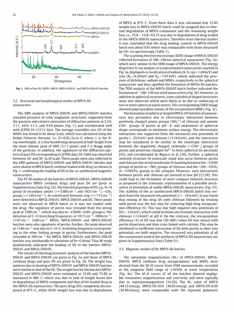
22 R.K. Dutta, S. Sahu / Colloids and Surfaces
20 40 60
(degrees)
22
0
31
1
40
0
51
1
44
0θ2
Inte
nsity (
A.U
.)
MNPs
MPCh-DS0.0 5
MPCh-DS0.0 1
MPCh
DS
3n
rt3wMDrtotbtwFn
nSggwwtpd1bairbqD
D(pwDmttp
ble remanance magnetization and coercivity and were typically
Fig. 1. XRD of the DS, MNPs, MPCh, MPCh-DS0.01, and MPCh-DS0.05 batches.
.2. Structural and morphological studies of MPCh-DSanocarriers
The XRD analysis of MPCh-DS0.05 and MPCh-DS0.01 batchesevealed presence of cubic magnetite structures, supported fromhe position and relative intensities of diffraction patterns at 2 2 0,
1 1, 4 0 0, 5 1 1, and 4 4 0 planes (Fig. 1), and corroborated wellith JCPDS 01-11111 data. The average crystallite size (D) of theNPs was found to be about 2 nm, which was calculated using theebye–Scherrer formula, i.e., D = 0.9�/�cos �, where � is the X-
ay wavelength, � is line broadening measured at half-height fromhe most intense peak of XRD (3 1 1 plane) and � is Bragg anglef the particles. In addition, the signatures of the diffraction pat-ern of pure DS corresponding to JCPDS data 39-1684 was observedetween 10◦ and 30◦ in 2� scale. These peaks were also reflected inhe XRD patterns of MPCh-DS0.05 and MPCh-DS0.01 batches andere absent in MPCh batch (without loaded with drug) as shown in
ig. 1, confirming the loading of DS in the as-synthesized magneticanocarriers.
The FT-IR studies of the batches of MPCh-DS0.01, MPCh-DS0.05anocarriers MPCh (without drug) and pure DS are given inupplementary Data (Fig. S2). The functional groups of DS, e.g., N Hroup of secondary amine (� = 3380 cm−1 and 1621 cm−1), CH2roup (� = 1453 cm−1), di-substituted benzene ring (� = 773 cm−1)ere detected in MPCh-DS0.01, MPCh-DS0.05 and DS. These peaksere not observed in MPCh batch as it was not loaded with
he drug. The signature of pectin was revealed from the strongeak at 1580 cm−1, which was due to COOH/ COO−group(s). Theetection of C O stretching frequencies at 1017 cm−1, 1094 cm−1,147 cm−1, 1290 cm−1 MPCh, MPCh-DS0.01 and MPCh-DS0.05atches were also signatures of pectin. Further, the peak observedt 1146 cm−1 was due to C O C stretching frequency correspond-ng to the ether linking groups in pectin. Furthermore, the peakecorded at 590 cm−1 for MPCh, MPCh-DS0.01 and MPCh-DS0.05atches was attributable to vibrations of Fe O bond. Thus IR studyualitatively indicated the loading of DS in the batches MPCh-S0.01 and MPCh-DS0.05.
The results of thermogravimetric analysis of the batches MPCh-S0.01 and MPCh-DS0.05 are given in Fig. 2a and those of MPCh
without drug) and pure DS are given in Fig. 2b. The weight lossatterns due to heating of MPCh-DS0.01 and MPCh-DS0.05 batchesere similar to that of the DS. The weight loss for the batches MPCh-S0.01 and MPCh-DS0.05 were estimated as 72.0% and 75.8% aseasured at 900 ◦C, which was due to sum of weight losses due
o degradation of MPCh component and that of the loaded drug inhe MPCh-DS nanocarriers. The pure drug (DS) completely decom-osed at 875 ◦C, while 29.8% weight loss was due to degradation
B: Biointerfaces 97 (2012) 19– 26
of MPCh at 875 ◦C. From these data it was calculated that 12.8%weight loss in MPCh-DS0.05 batch could be assigned due to ther-mal degradation of MPCh component and the remaining weightloss, i.e., 75.8 − 12.8 = 63.1% was due to degradation of drug loadedin the MPCh-DS0.05 nanocarriers. Therefore from thermal studiesit was concluded that the drug loading content in MPCh-DS0.05batch was about 63% which was comparable with those measuredby UV–vis spectroscopy (Table 1).
The scanning electron microscopy (SEM) image of MPCh-DS0.05reflected formation of 100–150 nm spherical nanocarrier (Fig. 3a),which were similar to the SEM image of MPCh-DS0.01. The energydispersive X-ray analysis of a representative nanocarrier, marked inFig. 3a, displayed co-localization of sodium (K-X-ray = 1.04 keV) andiron (K˛ = 6.39 keV and Kˇ = 7.07 keV), which indicated the pres-ence of diclofenac sodium and MNPs, respectively in the sphericalnanocarrier and thus signified the formation of MPCh-DS batches.The TEM analysis of the MPCh-DS0.05 batch further indicated theformation of ∼100–150 nm sized nanocarriers (Fig. 3b). However, inaddition to spherical structures, some cylindrical shaped structureswere also observed which were likely to be due to coalescing oftwo or more spherical nanocarriers. The corresponding SAED imageshowed polycrystalline nature of the encapsulated MNPs (Fig. 3c).
The formation of spherical shaped MPCh-DS batches of nanocar-riers was persuasive due to electrostatic interaction betweenpositively charged amino groups (NH3
+) of chitosan and anionicCOO− groups of pectin at pH ∼ 4, and attainment of sphericalshape corresponds to minimum surface energy. The electrostaticinteraction was supported from the measured zeta potentials ofpectin (−35.9 mV) and chitosan (+114.6 mV), and the interactionmay be considered to be similar to the ionotropic interactionbetween the negatively charged carboxylic ( COO−) groups ofpectin and positively charged Zn2+ to form spherical Zn-pectinate[25], and corroborated by Bigucci et al. [26]. Further, a sphericalnetwork structure of nanoscale could also occur between pectinand chitosan due to intramolecular H-bonding between the COOHgroups of pectin or NH2 groups of chitosan and the OH, OCH3or COOCH3 groups in the complex. However, such interactionsbetween pectin and chitosan are favored at low pH [27,28]. Thiscould lead to the formation of more compact network structureas proposed in the schematic representation of a possible mech-anism of formation of stable MPCh-DS0.05 nanocarriers (Fig. S3).The stability of the as synthesized MPCh-DS0.05 batch was evi-dent from the measured zeta potential (� = −18.6 mV). It was foundthat mixing of the drug DS with chitosan followed by treatingwith pectin was the key step for achieving high drug encapsula-tion efficiency (%). This was due high negative zeta potentials ofDS (−110 mV), which could facilitate electrostatic interaction withchitosan (+114 mV) at pH 4. On the contrary, the encapsulationefficiency (%) of DS was low (50–60%) when DS was mixed withpectin dispersion and then cross linked with calcium. This may beattributed to inefficient interaction of DS with pectin as their zetapotentials are both negative. The measured zeta potentials of allthe constituents used in the synthesis of MPCh-DS nanocarriers aregiven in Supplementary Data (Table S1).
3.3. Magnetic studies of the MPCh-DS batches
The saturation magnetization (Ms) of MPCh-DS0.01, MPCh-DS0.05, MPCh (without drug encapsulation) and MNPs werederived from the M–H curves from VSM measurements recordedin the magnetic field range of ±10 kOe at room temperature(Fig. 4a). The M–H curves of all the batches showed negligi-
due to superparamagnetism [19,29]. The Ms value of MPCh(44.15 emu/g), MPCh-DS-0.01 (36.63 emu/g) and MPCh-DS-0.05(34.40 emu/g) were comparable with the reported polymeric

R.K. Dutta, S. Sahu / Colloids and Surfaces B: Biointerfaces 97 (2012) 19– 26 23
0 200 40 0 600 80 0 1000
20
40
60
80
100
MPCh-D S-0 .01
MPCh-D S-0 .05
We
igh
t L
oss (
%)
o
0 250 500 750 1000
0
20
40
60
80
100
DS
MPCh
o
We
igh
t L
oss (
%)
(a) (b)
nd M
msipcmt
F1
Temperature ( C)
Fig. 2. (a) Thermogravimetric analysis (TGA) plot of MPCh-DS0.05 a
agnetic nanocarriers [12], but were lesser than that of the as-ynthesized MNPs (55.82 emu/g). The decrease in the Ms valuesn these batches were due to formation of magnetic dead layer byectin and/or chitosan at the domain boundary wall of MNPs, which
ould hinder the domain wall motion during application of theagnetic field. The reduced magnetization of iron oxide nanopar-icles due to polymer coating is known [30]. In addition to polymer,
ig. 3. (a) SEM–EDAX measurement of a representative MPCh-DS0.05 nanosphere of
00–150 nm, and (c) SAED image of MPCh-DS0.05 nanospheres exhibiting polycrystalline
Temperature ( C)
PCh-DS0.01 batches and (b) TGA plot of DS and MPCh nanocarriers.
MNPs are also likely to be coated/interacted with the drug to formmagnetic dead layer. This can be deduced from the decrease inthe measured Ms of the drug loaded batches for increase in thedrug concentration (Fig. 4a). The superparamagnetism of the batch
MPCh-DS0.05 was confirmed by recording field cooled (FC) andzero field cooled (ZFC) curves (Fig. 4b) at an external applied mag-netic field of 50 Oe from SQUID measurements. The FC-ZFC curves100–150 nm size, (b) TEM image showing morphology of nanospheres of sizes nature of MNPs in the nanospheres.

24 R.K. Dutta, S. Sahu / Colloids and Surfaces B: Biointerfaces 97 (2012) 19– 26
(a)
-10000 -500 0 0 50 00 10000
-60
-40
-20
0
20
40
60 MNPs
MPCh-DS0.05MPCh-DS0.01MPCh
M (
em
u/g
)
H(Oe)
(b)
0 50 100 150 200 250 300
4
6
8
10
12
M (
em
u/g
)
Temperature (K)
F ocarrib
dwp[ost
3
Dpgiacotth
Fg6a
ig. 4. (a) M–H curve of MNPs, MPCh, MPCh-DS0.01 and MPCh-DS0.05 batches of nanatch recorded at 50 Oe, measured by SQUID.
iverged at 112 K, characterized as blocking temperature (TB),hich corresponded to the transition from ferromagnetic to super-aramagnetic behavior and agreed well with reported literature29,31]. Therefore the magnetic studies confirmed the synthesisf magnetic nanocarriers which are superparamagnetic and pos-essed sufficiently high saturation magnetization, as required forargeted delivery by magnetic guiding.
.4. In vitro drug release studies
The sequential in vitro release of the drug DS from the MPCh-S0.05 batch in SGF containing pepsin at pH 1.2, SIF containingancreatin at pH 6.8 and in SCF containing pectinase at pH 5.5 isiven in Fig. 5. Negligible drug was released (0.02% of loaded drug)n SGF medium which was attributable to poor drug solubility incidic condition. In addition, the nanocarriers made of pectin andhitosan could shrink in acidic condition, as the carboxyl groupf pectin and the amine groups of chitosan could undergo pro-
onation and restrict the release of the drug loaded in it [28]. Onhe other hand, 69% of the drug was released in SIF in 3 h. Theigher pH in SIF is a favorable condition of swelling of pectin and00 15 30 45 60
0
20
40
60
80
100
SC
F
SIF
SG
F
% C
um
ula
tive
re
lea
se
Time (h )
ig. 5. Sequential in vitro release studies of DS from MPCh-DS0.05 batch in simulatedastric fluid (SGF pH 1.2) for 0–2 h, followed by simulated intestinal fluid (SIF pH.8) from 2–5 h and then in simulated colonic fluid (SCF pH 5.5) from 5 to 60 h, givens mean ± standard deviation from triplicate analysis.
ers measured by VSM at room temperature and (b) ZFC and FC curve of MPCh-DS0.05
form more porous material, and hence increases the rate of drugrelease from the matrix [32]. This could be attributed to repulsionbetween large number of negatively charged carboxylate groupsin pectin (pKa of 2.9–4.1), which increases at higher pH depend-ing on degree of esterification [33]. The swelling effect of the batchof MPCh-DS0.05 in aqueous medium was confirmed from dynamiclight scattering (DLS) measurement, which exhibited unimodal sizedistribution in the range of 230–520 nm with maximum intensityat ∼350 nm (Fig. 6). However, it was not possible to rule out theeffect of aggregation of spherical nanocarriers resulting into largeraverage size measured by DLS. The swelling effect is more persua-sive as the electrostatic interaction between pectin and chitosanbecomes weaker at higher pH as majority of the amino groups inchitosan are not protonated (pKa of chitosan = 6.3–7.0), leading torelease of the drug. Moreover, DS is a weak acid (pKa = 4.0), and ismore soluble at neutral pH [34], thus favored rapid release at pH6.8. The remaining drug (30%) was released in SCF (pH = 5.5) for aperiod of 55 h at a lower release rate. The lower drug release wasdue to lower pH of the medium, where the swelling effect of pectincould be less. Hence a pH dependent, sustained release of the drugfrom MPCh-DS0.05 in gastrointestinal system was observed. How-ever, the effective release of DS at colon specific site was less as alarger fraction of the drug was released in intestinal medium.
Further, the in vitro release of DS from MPCh-DS0.05 nanocarri-ers in phosphate buffer solution (pH 7.4) was studied to mimic therelease of drug in the pH condition of blood. A sustained releaseof about 97% of loaded DS was observed in 12 h (Fig. 7a). Notably,the release of DS was significantly more than the release in SIF,and it is attributable to pH dependent swelling effect as discussedabove. The drug release profile in phosphate buffer solution wasfitted with Korsemeyer–Peppas model [21] to gain an insight intoits release mechanism. The best results were obtained for the mod-eling study of 8 h of cumulative release of DS (88.9% release) fromMPCh-DS0.05 batch in pH 7.4, where a linear fit of R2 = 0.997 wasachieved (Fig. 7b). From the slope, ‘n’ was found to be 0.9065, whichconfirmed non-Fickian transport of drug release, attributable to theswelling effect of the host material (pectin reinforced chitosan)in the aqueous medium [21,23]. This corroborated the DLS mea-surement which revealed higher average size of the nanocarriersin higher pH dispersion. The value of k × 100 (where k is a con-stant related to the structural and geometric characteristic of thedevice) was found to be 13.33 and was in good agreement with the
reported values [23]. So it may be surmised that spherical magneticnanocarriers of 100–150 nm made of pectin and chitosan networkcould be used as a potential pH dependent, magnetically guideddrug delivery system. However, it would be interesting to explore
R.K. Dutta, S. Sahu / Colloids and Surfaces B: Biointerfaces 97 (2012) 19– 26 25
0
10
20
30
40
000100101
Volu
me (
%)
Size (d
Fig. 6. DLS measurement of MPCh-DS0.0
Fig. 7. (a) In vitro release studies of DS from MPCh-DS0.05 in phosphate buffer atpSp
tbeccr
4
wslT
[
[
[
[
H 7.4, for 48 h, given as mean ± standard deviation from triplicate analysis and (b)howing linear fit of in vitro release of DS up to 8 h from MPCh-DS0.05 batch inhosphate buffer solution (at pH 7.4), using Korsemeyer–Peppas equation.
he opportunity of imaging the therapeutic process at targeted sitey MNP based magnetic resonance imaging, which is reported toxhibit enhanced contrast [35]. In addition, further studies can bearried out on synthesis of similar nanocarriers loaded with anti-ancer drugs which essentially demands targeted drug delivery toeduce cytotoxicity in healthy cells and tissues.
. Conclusion
A novel magnetic nanocarrier was developed interacting pectin
ith chitosan, leading to synergistically enhanced drug encap-ulation efficiency (%) and subsequently revealed high drugoading content (wt%), suitable for sustained release therapy.he developed nanocarriers were suitable for EPR effect and
[
[
[
.nm)
5 nanocarriers in aqueous solution.
exhibited suitable saturation magnetization and superparamag-netism, which were important criteria for developing magneticallyguided targeted drug delivery. The in vitro release of the DS fromMPCh-DS0.05 nanocarrier in simulated gastrointestinal fluid waspH dependent and exhibited sustained release in colonic fluid.The in vitro release study at pH 7.4 (mimicking blood pH) alsoexhibited sustained release which satisfied non-Fickian releasebased on Korsemeyer–Peppas model and corresponded to swellingcontrolled mechanism. The swelling effect was evident from thedynamic light scattering (DLS) measurement. Overall, the as-developed pectin based magnetic nanocarrier could be used as apotential targeted drug delivery system.
Acknowledgements
One of the authors, SS is thankful to MHRD, India for providingfellowship. Authors also thank the staffs of Institute Instrumen-tation Centre and Centre of Nanotechnology, IIT Roorkee for theinstrumental facilities used in this project.
Appendix A. Supplementary data
Supplementary data associated with this article can befound, in the online version, at http://dx.doi.org/10.1016/j.colsurfb.2012.04.030.
References
[1] J. Zuckner, Am. J. Med. Suppl. 4B (1986) 39–42.[2] J.V. Willis, M.J. Kendall, R.M. Flinn, D.P. Thornhill, P.G. Welling, Eur. J. Clin.
Pharmacol. 16 (1979) 405–410.[3] T.D. Warner, I. Vojnovic, D. Bishop-Bailey, J.A. Mitchell, FASEB J. 20 (2006)
542–544.[4] J. Carson, W.M. Notis, E.S. Orris, N. Engl. J. Med. 323 (1990) 135–137.[5] R.V. Chari, Acc. Chem. Res. 41 (2008) 98–107.[6] M. Akbulut, S.M. D’Addio, M.E. Gindy, R.K. Prud’homme, Novel methods of tar-
geted drug delivery: the potential of multifunctional nanoparticles, Expert Rev.Clin. Pharmacol. 2 (2009) 265–282.
[7] S. Kim, J.Y. Kim, K.M. Huh, G. Acharya, K. Park, J. Control. Release 132 (2008)222–229.
[8] T. Fan, M. Li, X. Wu, M. Li, Y. Wu, Colloid Surf. B 88 (2011) 593–600.[9] P. Dames, B. Gleich, A. Flemmer, K. Hajek, N. Seidl, F. Wiekhorst, D. Eberbeck,
I. Bittmann, C. Bergemann, T. Weyh, L. Trahms, J. Rosenecker, C. Rudolph C.,Targeted delivery of magnetic aerosol droplets to the lung, Nat. Nanotechnol.2 (2007) 495–499.
10] M. Mahmoudi, S. Sant, B. Wang, S. Laurent, T. Sen, Superparamagnetic iron oxidenanoparticles (SPIONs): development, surface modification and applications inchemotherapy, Adv. Drug. Deliv. Rev. 63 (2011) 24–46.
11] N. Butescu, O. Jordan, P. Burdet, P. Stadelmann, A. Petri-Fink, H. Hofmann, E.Doelker, Eur. J. Pharm. Biopharm. 72 (2009) 529–538.
12] J.L. Arias, M. López-Viota, J. López-Viota, A.V. Delgado, Int. J. Pharm. 382 (2009)270–276.
13] T. Coviello, P. Matricardi, C. Marianecci, F. Alhaique, J. Control. Release 119(2007) 5–24.
14] H. Maeda, J. Wu, T. Sawa, Y. Matsumura, K. Hori, J. Control. Release 65 (2000)271–284.
15] A.S. Lübbe, C. Bergemann, W. Huhnt, T. Fricke, H. Riess, J.W. Brock, D. Huhn,Cancer Res. 56 (1996) 4694–4701.
16] S.A. Sande, Expert Opin. Drug Deliv. 2 (2005) 441–450.

2 rfaces
[[
[
[[
[
[
[
[
[
[[[
[
[
[
[
6 R.K. Dutta, S. Sahu / Colloids and Su
17] S. Sahu, R.K. Dutta, J. Magn. Magn. Mater. 323 (2011) 980–987.18] British Pharamacopoeia Commission Office (BP 2009). Published by the sta-
tionary office on behalf of the medicines and healthcare products regulatoryagency (MHRA), London, 2009.
19] M. Mikhaylova, D.K. Kim, N. Bobrysheva, M. Osmolowsky, V. Semenov, T.Tsakalakos, M. Muhammed M., Langmuir 20 (2004) 2472–2477.
20] S. Singh, M.S. Muthu, Nanomedicine 2 (2007) 233–240.21] R.W. Korsmeyer, R. Gurny, E. Doelker, P. Buri, N.A. Peppas, Int. J. Pharm. 15
(1983) 25–35.22] J. Siepmann, N.A. Peppas, Adv. Drug Deliv. Rev. 48 (2001)
139–157.23] Q. Wang, J. Zhang, A. Wang, Preparation and characterization of a novel pH-
sensitive chitosan-g-poly (acrylic acid)/attapulgite/sodium alginate composite
hydrogel bead for controlled release of diclofenac sodium, Carbohydr. Polym.78 (2009) 731–737.24] Y. Boonsongrit, A. Mitrevej, B.W. Mueller, Eur. J. Pharm. Biopharm. 62 (2006)267–274.
25] I. El Gibaly, Int. J. Pharm. 232 (2002) 199–211.
[
[
B: Biointerfaces 97 (2012) 19– 26
26] F. Bigucci, B. Luppi, T. Cerchiara, M. Sorrenti, G. Bettinetti, L. Rodriguez, V. Zecchi,Eur. J. Pharm. Sci. 35 (2008) 435–441.
27] O. Munjeri, J.H. Collett, J.T. Fell, J. Control. Release 46 (1997) 273–278.28] M. George, T.E. Abraham, J. Control. Release 114 (2006) 1–14.29] T.J. Daou, G. Pourroy, C.S. Bégin, J.M. Grenèche, B.C. Ulhaq, P. Legaré, P. Bern-
hardt, C. Leuvrey, G. Rogez, Chem. Mater. 18 (2006) 4399–4404.30] M.P. Morales, S.V. Verdaguer, M.I. Montero, C.J. Sterna, A. Roing, L. Casas, B.
Martinez, F. Sandiumenge, Chem. Mater. 11 (1999) 3058–3064.31] S. Si, A. Kotal, T.K. Mandal, S. Giri, H. Nakamura, T. Kohara, Chem. Mater. 16
(2004) 3489–3496.32] C.Y. Yu, H. Cao, X.C. Zhang, F.Z. Zhou, S.X. Cheng, X.Z. Zhang, R.X. Zhuo, Langmuir
25 (2009) 11720–11726.33] K. Ofori-Kwakye, J.T. Fell, Int. J. Pharm. 28 (2001) 139–145.
34] A. Llinas, J.C. Burley, K.J. Box, R.C. Glen, J.M. Goodman, J. Med. Chem. 50 (2007)979–983.35] L. Xiao, J. Li, D.F. Brougham, E.K. Fox, N. Feliu, A. Bushmelev, A. Schmidt, N.
Mertens, F. Kiessling, M. Valldor, B. Fadeel, S. Mathur, ACS Nano 5 (2011)6315–6324.