Embed Size (px)
Citation preview

DEVELOPMENT OF CONTROLLED RELEASE ANTIMICROBIAL FILMS
FROM LOW METHOXYL PECTIN
By
ROHINI MARATHE
A Thesis submitted to the
Graduate School-New Brunswick
Rutgers, The State University of New Jersey
in partial fulfillment of the requirements
for the degree of
Master of Science
Graduate Program in Food Science
written under the direction of
Professor Kit L. Yam
and approved by
New Brunswick, New Jersey
January, 2008

ii
ABSTRACT OF THE THESIS
Development of Controlled Release Antimicrobial Films
from Low Methoxyl Pectin
by ROHINI MARATHE
Thesis Director:
Professor Kit. L. Yam
Biopolymer-based controlled release antimicrobial packaging is an innovative
packaging that aids in controlled replenishment of antimicrobial compound at the food
surface, where it is required the most. Various biopolymers have been evaluated for
controlled release purposes; focus being mainly on their antimicrobial effectiveness.
However, low methoxyl pectin, with the potential to control release of active compounds
by forming different degrees of crosslinking, has not been exploited in antimicrobial
packaging and there is a lack of understanding of different variables affecting the release
properties of active compounds from pectin films.
The main objective of this thesis was to identify the key variables involved in the
development of pectin-based antimicrobial films by evaluation of the effect of different
variables such as composition variables (size of active compound and degree of calcium
crosslinking), process variables (pH of pectin, method of crosslinking), and
environmental variables on the release of antimicrobial compounds, especially nisin. A

iii
secondary objective was to demonstrate the concept that release of active compounds
could be altered by varying the degree of calcium crosslinking within the pectin matrix.
Antimicrobial compounds such as sodium benzoate, potassium sorbate and nisin
were chosen based on their different physical and chemical properties. Coomassie blue
dye was used as a model compound for proof of concept. Films were produced from LM
pectin by varying the DE of pectin and calcium concentration using the solution casting
method. Release of antimicrobial compounds into water as a food simulant was measured
by UV/Visible spectrophotometry or HPLC or agar diffusion assay, depending on the
antimicrobial compound being evaluated.
Degree of calcium crosslinking, pH of pectin slurry and method of calcium
crosslinking were identified as the key variables controlling release of nisin from pectin
films. Electrostatic attraction between pectin and nisin at pH above pKa of pectin was not
sufficiently strong to prevent the release of nisin. Release studies with dye-containing
pectin films demonstrated that variable release rates could be obtained by altering the
degree of calcium crosslinking.

iv
ACKNOWLEDGEMENTS
I would like to express my sincere gratitude to my advisor, Professor Kit L. Yam,
for his patience, support and guidance throughout this study. It has been a great
experience working under his guidance.
I would also like to thank my committee members Dr. Karen Schaich for her
valuable discussion about this research and Dr. Chikindas for his useful suggestions
related to the microbial study. I would also like to thank Dr. Qingrong Huang for taking
time to serve on my committee.
Special thanks to all my labmates for their assistance, discussion, friendship
through the years and to Jason from Dr. Chikindas’s lab for training me for conducting
the microbial assays for this project.
I would also like to thank the Dr. LinShu Liu, Dr. Tony Jin, Dr. Dave Coffin and
Dr. Marshal Fishman from ERRC, USDA for their suggestions and technical assistance. I
am very grateful to USDA, Department of Defense and the Pliant Corporation for
providing the funding and resources necessary for this project.
Finally, special thanks to my family members and friends for their constant
support and encouragement.

v
TABLE OF CONTENTS ABSTRACT OF THE THESIS .......................................................................................... ii
ACKNOWLEDGEMENTS............................................................................................... iv
TABLE OF CONTENTS.................................................................................................... v
LIST OF FIGURES .......................................................................................................... vii
LIST OF TABLES............................................................................................................. ix
1 INTRODUCTION ........................................................................................................ 1
1.1 Controlled Release Packaging ............................................................................. 1
1.1.1 Concept of Controlled Release Packaging (CRP) ................................... 1
1.1.2 Antimicrobial Packaging ......................................................................... 1
1.2 Edible/Biopolymer-Based Antimicrobial Packaging........................................... 6
1.2.1 Motivation for Biopolymer-Based Packaging ......................................... 6
1.2.2 Definition and Classification of Biopolymer-Based Packaging .............. 7
1.2.3 Typical Components ................................................................................ 7
1.2.4 Techniques for Manufacture of Edible Films .......................................... 8
1.3 Literature Review of Biopolymer-Based Antimicrobial Packaging.................... 9
1.3.1 Biopolymers............................................................................................. 9
1.3.2 Antimicrobial Compounds..................................................................... 12
1.4 Pectin – A Promising Biopolymer for CRP....................................................... 17
1.4.1 Structure of Pectin.................................................................................. 17
1.4.2 Degree of Esterification (DE) ................................................................ 17
1.4.3 Classification of Pectin .......................................................................... 18
1.4.4 Gelling Mechanism................................................................................ 19
1.4.5 Review of Pectin Usage in Controlled Drug Delivery........................... 21
1.4.6 Review of Pectin in Antimicrobial Packaging....................................... 22
1.4.7 Explanation of Terms and Concepts Related to Pectin and
Controlled Release ................................................................................. 22
1.5 Research Gaps and Opportunities...................................................................... 25
1.6 Objectives .......................................................................................................... 27
1.6.1 Overall Objective ................................................................................... 27

vi
1.6.2 Scope of Research.................................................................................. 27
1.6.3 Challenges in Development of Pectin Films.......................................... 28
1.6.4 Specific Objective.................................................................................. 31
1.7 Research Approach ............................................................................................ 32
1.7.1 Proof of Concept .................................................................................... 33
1.7.2 Research Approach to Study Pectin-Nisin Interaction .......................... 33
2 EXPERIMENTAL...................................................................................................... 35
2.1 Materials ............................................................................................................ 35
2.1.1 Pectin Selection...................................................................................... 35
2.1.2 Active Compounds................................................................................. 36
2.1.3 Plasticizer............................................................................................... 38
2.1.4 Crosslinking Agent ................................................................................ 38
2.1.5 Media Preparation for Inhibition Zone Assay ....................................... 38
2.1.6 Bacterial Culture for Inhibition Zone Assay.......................................... 39
2.2 Methods.............................................................................................................. 39
2.2.1 Preparation of Pectin Film ..................................................................... 39
2.2.2 Release Studies ...................................................................................... 40
2.2.3 Quantification of Active Compounds .................................................... 41
3 RESULTS AND DISCUSSION................................................................................. 46
3.1 Effect of Composition Variables on Release ..................................................... 46
3.1.1 Effect of Degree of Calcium Crosslinking............................................. 46
3.1.2 Effect of Size of Active Compound....................................................... 51
3.2 Preliminary Studies with Nisin Containing Pectin Films .................................. 58
3.3 Effect of Processing Variables on Release of Nisin .......................................... 60
3.3.1 Effect of Calcium Crosslinking Method ................................................ 60
3.3.2 Effect of pH of Pectin and Sequence of Nisin Addition on
Pectin-Nisin Interaction ......................................................................... 63
3.4 Effect of Environmental Variables .................................................................... 68
4 CONCLUSIONS ........................................................................................................ 70
5 FUTURE WORK........................................................................................................ 72
6 REFERENCES ........................................................................................................... 73
vii
LIST OF FIGURES Figure 1: Concept of Controlled Release Packaging (CRP)............................................... 1
Figure 2: Mode of Action of a Volatile Antimicrobial Compound .................................... 3
Figure 3: Mode of Action of a Non-Volatile Antimicrobial Compound ............................ 3
Figure 4: Immobilized Antimicrobial Packaging System................................................... 4
Figure 5: Structure of Nisin .............................................................................................. 13
Figure 6: Schematic Diagram for Pectin Backbone.......................................................... 17
Figure 7: General Structure of Low Methoxyl Pectin Chain............................................ 18
Figure 8: Schematic Diagram for Gelation in HM pectin................................................. 19
Figure 9: Schematic Diagram for Gelation in LM pectin ................................................. 21
Figure 10: Schematic Diagram to Illustrate Different Degrees of Calcium
Crosslinking .................................................................................................... 22
Figure 11: Different Ways to Manipulate Degree of Crosslinking................................... 24
Figure 12: Effect of Calcium Crosslinking on Swelling of LM Pectin Film.................... 24
Figure 13: Schematic Diagram to Illustrate Pectin-Nisin Interactions ............................. 29
Figure 14: Overview of Impact of pH on Interactions of Pectin with Nisin
and Calcium .................................................................................................... 30
Figure 15: Research Approach to Identify Key Variables for Controlled Release
LM Pectin Films ............................................................................................. 32
Figure 16: Research Approach to Investigate Preference for Pectin Interaction with
Calcium and Nisin........................................................................................... 34
Figure 17: Structure of Coomassie Brilliant Blue G – 250............................................... 36
Figure 18: Structures of Benzoic Acid and Sodium Benzoate ......................................... 37
Figure 19: Structures of Sorbic Acid and Potassium Sorbate........................................... 37
Figure 20: Calibration Curve for Coomassie Blue G – 250.............................................. 41
Figure 21: Calibration Curve for Nisin Estimation Using Agar Diffusion Assay............ 44
Figure 22: Calibration Curve for Nisin Estimation by HPLC .......................................... 45
Figure 23: Effect of Calcium Concentration on Release of Dye from LM 12
(DE 35) Pectin Films ...................................................................................... 47
Figure 24: Effect of Calcium Concentration on Release of Dye from LM-18
(DE 40) Pectin Films ...................................................................................... 48

viii
Figure 25: Effect of Calcium Concentration on Release of Dye from LM-22
(DE 50) Pectin Films ...................................................................................... 48
Figure 26: Summary of Effect of Crosslinking on Release of Active Compound
from LM Pectin Film ...................................................................................... 50
Figure 27: Decision Tree Diagram for Investigation of Benzoate Entrapment
in Pectin Films ................................................................................................ 52
Figure 28: Effect of Calcium Concentration on Release of Potassium Sorbate
from LM 12 (DE 35) and LM 18 (DE 40) Pectin Films................................. 56
Figure 29: Antimicrobial activity of nisin-containing pectin films .................................. 59
Figure 30: Effect of Calcium Crosslinking Method on Release of Nisin from
Pectin Film (Inhibition Zone Assay)............................................................... 61
Figure 31: Effect of Calcium Crosslinking Method on Release of Nisin from
Pectin Films .................................................................................................... 62
Figure 32: Effect of Sequence of Nisin Addition on Release of Nisin at pH<pKa
of Pectin .......................................................................................................... 64
Figure 33: Effect of Sequence of Nisin Addition on Release of Nisin at pH<pKa
of Pectin (Inhibition Zone Assay).................................................................. 65
Figure 34: Effect of Sequence of Nisin Addition on Release of Nisin at pH > pKa
of Pectin (Inhibition Zone Assay).................................................................. 66
Figure 35: Effect of Sequence of Nisin Addition on Release of Nisin at pH > pKa
of Pectin .......................................................................................................... 67

ix
LIST OF TABLES Table 1: Selected References for Polysaccharide-Based Antimicrobial Films .................. 9
Table 2: Review of Protein-Based Antimicrobial Films................................................... 11
Table 3: Specifications for LM Pectin from CP Kelco..................................................... 35
Table 4: Formula for Media Used in Inhibition Zone Assay............................................ 38
Table 5: Requirement of Calcium Chloride for Films with Different Calcium
Crosslinking. ...................................................................................................... 40
Table 6: Effect of DE of Pectin and Calcium Concentration on Release of Dye
from Film ........................................................................................................... 49
Table 7: Loading of Sodium Benzoate in Pectin Films - Theoretical vs.
Total Extractable................................................................................................ 51
Table 8: Effect of Calcium on Recovery of Sodium Benzoate from Pectin Films........... 53
Table 9: Recovery of Sodium Benzoate from Fresh Pectin Slurry................................... 54
Table 10: Recovery of Sodium Benzoate from Pectin Film............................................. 54
Table 11: Comparison of nisin estimated by HPLC and Agar Diffusion Assay .............. 69

1
1 INTRODUCTION
1.1 Controlled Release Packaging
1.1.1 Concept of Controlled Release Packaging (CRP)
Controlled Release Packaging, also referred to as CRP [1], is an innovative form of
active packaging where active compounds such as antimicrobials and antioxidants are
incorporated into a synthetic or natural polymer, and released from the polymer into food
in a controlled manner.
Active compound (antimicrobial, antioxidant)
CRP film (‘active’ layer)
Surface of food
Core of Food
Barrier film (Non-active layer)
Active compound (antimicrobial, antioxidant)
CRP film (‘active’ layer)
Surface of food
Core of Food
Barrier film (Non-active layer)
Figure 1: Concept of Controlled Release Packaging (CRP)
Illustrated in figure 1 is a typical CRP system, comprised of an inner active layer
that is laminated with an outer barrier layer. The active compound may be incorporated
into or coated onto the inner active layer and released continuously from the packaging
material into food at a desired rate to improve food safety and quality.
1.1.2 Antimicrobial Packaging
Antimicrobial packaging, one of the early developments in the field of CRP, is a
packaging system designed to release an antimicrobial compound on the food surface to
delay microbial growth. The antimicrobial activity may be due to incorporation of an

2
antimicrobial compound into the packaging polymer or due to antimicrobial property of
the polymer itself (e.g. chitosan) [2].
1.1.2.1 Motivation for Antimicrobial Packaging
Food companies all over the world are striving to minimize or delay food spoilage
in order to develop safe and high quality food products with a longer shelf life. Post-
process handling or moisture condensation on surface of refrigerated packaged foods are
two reasons for microbial contamination and spoilage on the surface of packaged foods
[3, 4]. Traditionally, antimicrobial compounds are added to entire food to inhibit or delay
microbial spoilage. But, the main disadvantage of this traditional method is that the
antimicrobial compound may not really be required in the interior part of food product,
resulting in an excessive use of ‘preservatives’. Growing consumer demand for minimum
or ‘zero’ use of preservatives has motivated researchers to develop alternate methods for
extending shelf life of foods with minimum use of food additives. Use of antimicrobial
sprays or dips [5] is one such option to deliver an antimicrobial compound at food
surface, and at the same time it avoids excessive use of the antimicrobial compound.
However, this method has some disadvantages such as – (i) it involves an additional
manufacturing step, (ii) may be messy, and (iii) loss of effectiveness of dip at surface
may occur due to diffusion of antimicrobial compound into food interior over time [6].
This is the main motivation for development of controlled release antimicrobial
packaging; wherein the packaging can be designed to replenish the required amount of
antimicrobial compound in a controlled manner at the food surface, where it is required
the most!
3
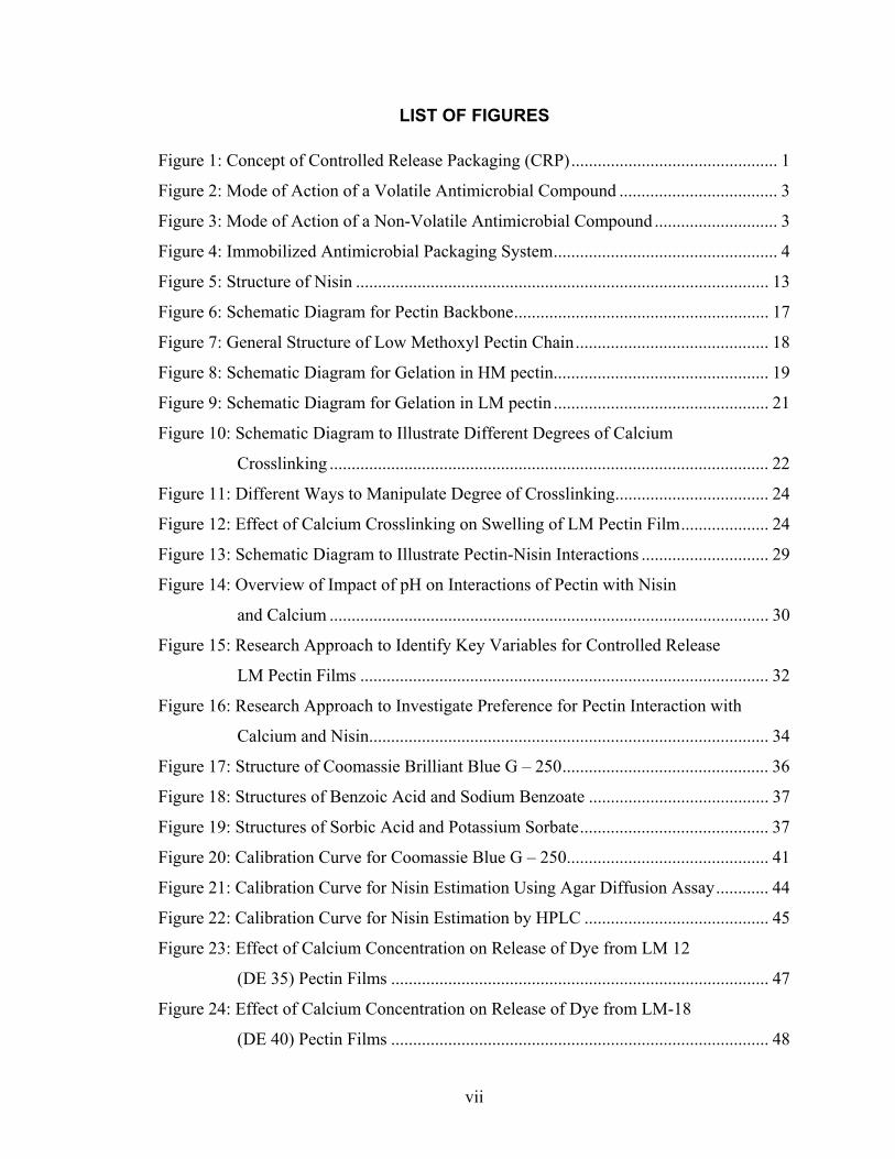
1.1.2.2 Classification Based on Mode of Action
An antimicrobial packaging system may function in two ways [7-9]:
(i) Migration/release – In this case the antimicrobial compound inhibits microbial growth
by migrating from packaging material onto the food surface. In case the released
compound is volatile it will first migrate to the headspace and then condense onto the
food surface (Figure 2). Non-volatile compounds diffuse directly from packaging
material into food matrix (Figure 3). A volatile compound is more effective than a non-
volatile compound for irregularly-shaped solid foods, where good contact between food
and packaging material is not possible. Non-volatile antimicrobial compounds can be
used effectively for semi-solid or liquid foods where excellent contact between food and
packaging allows migration of the compound.
Figure 2: Mode of Action of a Volatile Antimicrobial Compound
Figure 3: Mode of Action of a Non-Volatile Antimicrobial Compound
Antimicrobial compound migrated to headspace
Antimicrobial compound condensed onto food surface
Package headspace
Food contact ‘active’ layer
Food
Antimicrobial compound migrated to headspace
Antimicrobial compound condensed onto food surface
Package headspace
Food contact ‘active’ layer
Food
Antimicrobial compound migrated onto food surface
Food contact ‘active’ layer
Food
Antimicrobial compound migrated onto food surface
Food contact ‘active’ layer
Food

4
(ii) Immobilization – A non-food grade antimicrobial compound is immobilized on the
food contact surface and inhibits microbial growth at contact surface without migrating
from the packaging material (Figure 4). This mode requires very good contact between
food and packaging material to effectively inhibit microbial spoilage.
Figure 4: Immobilized Antimicrobial Packaging System
An indirect form of antimicrobial packaging is one in which the active compound
inhibits microbial growth by absorbing factors that are favorable for microbial growth
from the package; e.g. Oxygen or moisture absorbent sachets absorb oxygen or moisture,
respectively, from package to prevent mould growth.
1.1.2.3 Significance of Antimicrobial Release Rate
For an antimicrobial packaging system to be effective, two critical factors must be
considered while designing the system [8]. They are (i) rate of release of antimicrobial
compound from packaging onto food surface, and (ii) rate of growth of microorganism to
be inhibited. If the release rate of antimicrobial compound is slower than growth rate of
target microorganism, microorganisms will grow before the antimicrobial compound is
released. On the other hand, if the release rate of antimicrobial compound is faster than
the microbial growth rate, the entire antimicrobial compound from package will be
released too fast and the released antimicrobial agent will diffuse into food and will not
be available at the surface to inhibit microbial growth at the desired time. In this case the
Antimicrobial compound immobilized on packaging surface
Food contact layer of packaging film
Food
Antimicrobial compound immobilized on packaging surface
Food contact layer of packaging film
Food

5
packaging system will be ineffective. An ideal antimicrobial packaging system should
release the antimicrobial compound at a rate that matches the microbial growth rate. In
addition, at any given time, the concentration of antimicrobial compound maintained at
food surface should at least be equal to the minimum inhibitory concentration (m.i.c.)
necessary to inhibit the target microorganism [10].
1.1.2.4 Factors Influencing Release of an Antimicrobial Compound from CRP
Some of the major factors which govern the release of active compound from
packaging are [1, 8]:
(i) Size of antimicrobial compound: Mobility of an antimicrobial compound within the
polymer network depends on its size, provided there are no other chemical interactions
involved between the antimicrobial compound and the polymer chain. A small sized
compound will diffuse faster than a bigger compound through the same polymer matrix.
(ii) Compatibility between antimicrobial compound and packaging polymer: If the
antimicrobial compound binds with the packaging polymer i.e. they are compatible with
each other, then the antimicrobial compound may remain inside the polymer matrix and
this is not desirable for controlled release packaging. If the reverse is true, i.e. they are
not compatible, then it may pose problems while incorporating the antimicrobial
compound into the polymer matrix, or once incorporated, it may be released immediately,
thereby defeating the purpose of controlled release. Ideally, the antimicrobial compound
should be physically entrapped in the polymer matrix, to be released in a controlled
manner by modifying the polymer matrix. Some of the factors that may influence the
compatibility of antimicrobial compound and polymer matrix are polarity [11], pKa of
biopolymers such as pectin and chitosan [12], pKa of antimicrobial compounds,

6
especially nisin. The effect of pKa of biopolymer and antimicrobial compound on film
processing and controlled release properties is discussed in detail later in this chapter.
(iii) Extent of networking within polymer matrix: The effect of networking within a
polymer on the release of active compound has been exploited successfully for designing
controlled drug delivery applications [13-15] and is the focus of this thesis also. It is
discussed in detail later in this chapter.
1.2 Edible/Biopolymer-Based Antimicrobial Packaging
1.2.1 Motivation for Biopolymer-Based Packaging
The past few decades have witnessed an alarming increase in use of petrochemical-
based synthetic packaging materials because they are easily available in large quantities,
are cheap and possess mechanical properties suitable for commercial food packaging
applications. However, a major concern with use of synthetic polymers is their non-
biodegradable character, which poses a serious threat to the environment. Marketing of
eco-friendly packaging has increased consumer awareness about issues concerning
environmental pollution [16, 17]. Therefore, the food and packaging industries are jointly
focusing to develop alternate biodegradable packaging systems, including edible films
and coatings for food.
Use of biopolymer-based packaging is not novel; edible films and coatings have
been used for centuries [6, 18]. In China, a wax coating was used to delay moisture loss
from citrus fruits during the twelfth century. During the fifteenth century, Yuba, an edible
film made from soy milk was developed in Japan to help in food preservation [18].
Edible coatings to prevent shrinkage of meat were developed as early as the sixteenth

7
century. Further developments in use of edible coatings and films were seen in the
nineteenth and early twentieth century when sugar and chocolate coating was used to
prevent oxidation of nuts, oil-in water emulsion was coated on fruits and vegetables,
gelatin casings were developed for sausages, etc.
1.2.2 Definition and Classification of Biopolymer-Based Packaging
Biopolymer-based packaging is defined as packaging made from renewable
resources of agriculture or marine origin [17]. Edible packaging, as the name suggests, is
made from edible natural biopolymers and can be used as an integral part of food.
Biopolymers are categorized on the basis of their origin as:
• Natural – extracted from natural raw materials (e.g. starch, cellulose, protein,
alginate, pectin);
• Chemically synthesized polymers from bioderived monomers (e.g. polylactic acid
- PLA) [19];
• Microbial polymers (e.g. polyhydroxyalkanoates, polyhydroxybutyrates).
Though biopolymer-based packaging is environment friendly in comparison to
synthetic polymers, there are some challenges associated with respect to its mechanical
performance, commercial processing and cost [17].
In the past few decades, several films based on proteins, polysaccharides have been
developed and characterized for application in food packaging.
1.2.3 Typical Components
Edible films are typically comprised of either a protein or a polysaccharide or any
of the above in combination with lipids, a plasticizer and, if necessary, a cross-linking

8
agent. Proteins studied for edible packaging are whey protein, casein, corn zein, soy
protein, wheat gluten, gelatin, and collagen [17, 20]. Polysaccharide based films have
been developed using alginate, cellulose and cellulose derivatives, starch and starch
derivatives, chitosan, carrageenan [21]. Coatings based on lipids such as vegetable oils,
monoglycerides, acetylated glycerides, waxes such as paraffin wax, carnauba wax,
beeswax, are used alone or in combination with proteins or polysaccharides for different
purposes in food preservation [21]. Glycerol, sorbitol, polyethylene glycol (PEG) are
some of the commonly used plasticizers that help to improve the flexibility of edible
films [6]. Cross-linking agents such as calcium chloride (for alginate and low methoxyl
pectin films) [4], glutaraldehyde (for gelatin films) [22] are used in the formation of films
mentioned above to improve their mechanical strength, resistance to water, and other
barrier properties.
1.2.4 Techniques for Manufacture of Edible Films
Technique for making an edible film depends on the material used for the film.
Generally, films based on polysaccharides, except starch films, are formed by removal of
solvent [6]. In this method, the hydrocolloid is dispersed in a solvent such as ethanol,
water, to which plasticizer and crosslinking agent, if any, are added and the film-forming
solution is cast on a flat surface and solvent is removed during drying step to form a thin
film. In starch based films, film-making involves gelatinization of starch followed by
casting of film [2, 23]. Extrusion is another technique for making biopolymer films [24,
25]. Protein films are made by different techniques such as solvent evaporation (wheat
gluten, corn zein) or pH modification or thermal denaturation of protein (soy protein,

9
ovalbumin) or addition of crosslinking agent (whey protein) or gelation (gelatin),
followed by casting [26].
1.3 Literature Review of Biopolymer-Based Antimicrobial Packaging
1.3.1 Biopolymers
Several biopolymers have been investigated for the purpose of antimicrobial films;
selected references for the polysaccharide-based and protein-based films have been listed
in table 1 and 2, respectively.
Table 1: Selected References for Polysaccharide-Based Antimicrobial Films
Film
Antimicrobial Compound
Test Medium/ Food Test Method Reference
Carrageenan Potassium sorbate
Phosphate buffer Diffusion cell [27]
Carrageenan Sodium alginate
Nisin Lysozyme GFSE
Agar Inhibition zone assay [28]
Cellulose Pediocin Meat
Inhibition zone assay, effect on packed meat
[29]
Yam starch Chitosan Chitosan Culture
medium Shake flask method [2]
Cellulose based paper
Nisin Lacticin
Agar, Cheese/ham
Inhibition zone assay
[30]
Alginate Apple puree
Plant essential oils (oregano, cinnamaldehyde, etc.)
Agar Inhibition zone assay [31]
Chitosan Garlic oil, potassium sorbate, nisin
Agar Inhibition zone assay [32]
Konjac glucomannan Chitosan
Chitosan Nisin Agar Inhibition zone
assay [33, 34]

10
Sodium alginate Nisin Beef Spot assay [35]
Chitosan Acetic acid Propionic acid Lauric acid
Bologna Ham Pastrami
Bacterial enumeration of meat sample at timed intervals
[36]
Tapioca starch Potassium sorbate No study
Film characterization only
[23]
Yam starch Chitosan Chitosan Carrots
Bacterial enumeration of carrot samples at timed intervals
[37]
Alginate Garlic oil Agar Inhibition zone assay [38]
Alginate Potassium sorbate
Potassium sorbate solution
Release study with diffusion cell
[4]
Tapioca starch Nisin Agar, Culture medium
Inhibition zone assay and release studies in liquid culture medium
[39]
HPMC* - chitosan Nisin Agar Inhibition zone
assay [40]
Chitosan coating on paperboard Nisin
Distilled water, milk, orange juice
Release study with migration cell
[41]
Carrageenan Chitosan MC** HPMC*
Nisin Agar Inhibition zone assay [42]
MC*-HPMC** coating Nisin Peptone broth
Release study
[43]

11
Starch – potato, corn
Potassium sorbate Strawberry
Effect of coating on different properties of fruit, including microbial inhibition, but not "controlled release"
[44]
(* MC: Methyl cellulose; ** HPMC: Hydroxypropyl methyl cellulose)
Table 2: Review of Protein-Based Antimicrobial Films
Film Antimicrobial Compound
Test Medium/ Food
Test Method Reference
Corn zein Lysozyme/nisin Agar Inhibition zone assay [3]
Soy protein isolate Lysozyme/nisin Agar Inhibition zone
assay [3]
Whey protein isolate
Potassium sorbate Water-glycerol Release study [45]
Whey protein isolate
p-amino benzoic acid, sorbic acid Agar Inhibition zone
assay [46]
Whey protein isolate
Oregano, rosemary, garlic essential oils
Agar Inhibition zone assay [47]
Corn zein Wheat gluten Nisin Water
Agar
Release study Inhibition zone assay
[48]
Corn zein Lysozyme, Albumin proteins, EDTA
Water Release study [49]
Soy protein isolate Wheat gluten Egg albumen Whey protein isolate
Nisin Culture media Release study [50]
Corn zein Lysozyme Water Release study [51]
Whey protein isolate
p-amino benzoic acid, sorbic acid Hot dogs Surface
inhibition [52]

12
Whey protein isolate
p-amino benzoic acid, sorbic acid
Bologna Summer sausages
Surface inhibition [53]
Corn zein Sorbic acid Cooked sweet corn
Surface inhibition [54]
1.3.2 Antimicrobial Compounds
Nisin, potassium sorbate, lysozyme, essential oils such as oregano and garlic, and
chitosan are some of the antimicrobial compounds researched for use in antimicrobial
films; nisin being the most popular of them all.
1.3.2.1 Nisin and its Structure
Nisin is a small peptide produced by Lactococcus lactis subspecies lactis. It is
considered GRAS and is the only bacteriocin approved for use in foods [55], the
permitted limits varying across the globe. Commercially, nisin is available in the form of
Nisaplin®, a nisin ‘preparation’, which has only 2.5% nisin, the rest being lactose and
other milk solids.
Nisin is composed of 34 amino acids and has a molecular weight of 3510 Daltons.
The structure of nisin is shown in Figure 5 and was first reported by Gross and Morell in
1971 [56]. The presence of five internal thioether rings formed by lanthionine (Ala-S-
Ala) and β-methyllanthionine (Abu-S-Ala) groups is responsible for the conformation of
nisin. It is capable of forming dimers and oligomers [55]. It has a net positive charge due
to lysine and histidine amino acid residues, pKa of their side chains being 10.2 and 7
respectively [57]. It possesses an amphipathic nature due to hydrophobic groups at the N-
terminal and hydrophilic groups at the C-terminal. Solubility and thermal stability of
nisin solution varies with pH. At pH 2, it survives autoclaving conditions of 121 °C for

13
15 minutes, but at higher pH of about 6, heating at 121 °C for 3 min is sufficient to
destroy about 25-50% of the nisin [55]. Two major degradation products of nisin i.e.
nisin1-32 and (des-∆ Ala5)nisin1-32 have been identified and antimicrobial activity has
been characterized by Chan et al. [58]. In addition to loss of stability with increase in pH,
solubility also drops drastically with increase in pH, but is not of much importance
because of the low usage levels in food.
Figure 5: Structure of Nisin
1.3.2.2 Antimicrobial Activity
Nisin is a broad spectrum antimicrobial effective against Gram-positive bacteria,
most importantly, those belonging to the Bacillus and Clostridium genera. Nisin is not
only effective against vegetative cells, but it is also effective in inhibition of heat-resistant
spores such as Bacillus cereus, Clostridium botulinum. This makes it a very effective
antimicrobial for use in heat-treated foods where probability of spoilage due to surviving
spores is high [55]. Penetration of the C-terminal of nisin into the cell membrane causes
alteration of favorable pH conditions and consequently, rapid loss of cell metabolites
from the cell. Presence of polyvalent cations such as Ca2+, Mg2+ is thought to reduce the
H2N -
COOH
Ser
Ile
His
Val
Dha
Lys
Ile Dhb Ala
Ile
Dha
Leu
Ala Abu
Pro Gly
LysAla
S
S
Abu
Gly
AlaLeu
Met
Gly
Ala
SAs Met
Lys
Abu
AlaAbu
Ala
S
His
Ala
S
H2N -
COOH
Ser
Ile
His
Val
Dha
Lys
Ile Dhb Ala
Ile
Dha
Leu
Ala Abu
Pro Gly
LysAla
S
S
Abu
Gly
AlaLeu
Met
Gly
Ala
SAs Met
Lys
Abu
AlaAbu
Ala
S
His
Ala
S
Ile Dhb Ala
Ile
Dha
Leu
Ala Abu
Pro Gly
LysAla
S
S
Abu
Gly
AlaLeu
Met
Gly
Ala
SAs Met
Lys
Abu
AlaAbu
Ala
S
His
Ala
S
Abu: aminobutyric acid; Dha: dehydroalanine; Dhb: dehydrobutyrine (β-methyldehydroalanine)
Lanthionine ring β-methyllanthionine ring

14
antimicrobial effectiveness of nisin due to possible interaction of these ions with the
negatively charged sites of the cell membrane and making these membrane sites
unavailable for binding with nisin [59]. Nisin is effective against lactic acid bacteria,
responsible for spoilage of low pH foods (e.g. salad dressings); Listeria monocytogenes, a
pathogen; Brocothrix thermosphacta, responsible for spoilage in meat products. It is used
mainly in the preservation of processed cheese and has potential for use in several
products such as canned foods, juices, processed meat products, pasteurized liquid egg,
and salad dressings.
1.3.2.3 Review of Nisin Usage in Biopolymer-Based Antimicrobial Films
Nisin has been studied extensively in antimicrobial packaging. Lee et al. [60]
investigated the antimicrobial efficacy of a nisin-coated paper and found that even though
only 9% of the total incorporated nisin could migrate to the food, it was still able to
extend the shelf life of perishable foods such as milk cream and emulsions. Another
group studied an antimicrobial edible coating of nisin on hydroxypropyl methyl cellulose
[61]. Cha et al. [28] studied the effect of antimicrobial compounds such as nisin on the
mechanical properties of sodium alginate and κ-carrageenan based films and reported that
greater hydrophilic nature of sodium alginate films causes greater swelling than the
carrageenan films, and as a result more nisin is released, thereby producing bigger zones
of inhibition. Nisin immobilized on the surface of cellulose-based inserts and
polyethylene/polyamide pouches was able to extend the shelf life of ham and sliced
cheese when used in combination with modified atmosphere packaging [30]. Zhang et al.
[62] conducted an important experiment to compare the antimicrobial effectiveness of
different modes of delivery of nisin to a model system and found that a combination of

15
initial addition of nisin to the broth and slow release of nisin over a period of time was
more effective than either of the delivery modes alone. Mechanical properties and/or
antimicrobial activity of nisin-containing films made using biopolymers such as chitosan
[32], konjac glucomannan [33, 34], sodium caseinate [63], poly(lactic acid) - pectin
blends [64], hydroxypropyl methyl cellulose [40] have been evaluated. A study by Ko et
al. [50] on the effect of pH of film-forming solution and surface hydrophobicity of
protein films on antimicrobial activity of nisin showed that nisin was more effective
when incorporated into hydrophobic protein films at a low pH. Immobilization of nisin in
a calcium alginate gel was more effective than direct application of nisin on the surface
of beef carcass [35, 65]. In a recent study, activated beads of alginate, modified to form
covalent linkage with nisin, were not as effective as non-modified alginate-nisin solution
coating on beef surface for inhibition of Staphylococcus aureus in beef [66]. Varying the
molecular weight of hydroxypropyl methyl cellulose (HPMC) and methyl cellulose (MC)
failed to control the release of nisin from HPMC/MC coated on LPDE films [43]. Cha et
al. [42] compared the release of nisin from MC, HPMC, chitosan and carrageenan, either
used as a coating on PE films or in the form of heat-pressed films made using above
biopolymers in combination with PE pellets. It was found that in case of biopolymers
coated onto PE, nisin was released at a varying rate, gradually increasing over 10 hours
of the above mentioned study. However, in case of heat-pressed films, even after 5 days
of release study, amount of nisin released was equal to that released in the first 2 hours of
the release study.
16
1.3.2.4 Review of Potassium Sorbate in Biopolymer-Based Antimicrobial Films
Potassium sorbate is the second most studied antimicrobial compound in
biopolymer-based packaging. Selected research work, relevant to controlled release
applications, is mentioned in this section. The diffusivity of potassium sorbate in κ-
carrageenan films was studied as a function of temperature and pH of the receiving
solution and it was found that diffusivity was unaffected by pH of receiving solution and
was a function of only temperature [27]. In another study, water-glycerol model system
was used to study the diffusion of potassium sorbate from whey protein based films [45].
The proposed model describes the diffusion process as non-Fickian, where protein films
swell due to diffusion of solvent (water) into the film matrix, followed by migration of
potassium sorbate out of the swollen film matrix. The diffusion coefficients found using
this liquid model system may not hold true in case of a film in contact with a semi-solid
material. Zactiti et al. [4] studied the effect of degree of calcium crosslinking in alginate
films on the release of potassium sorbate. This is an important experiment for developing
controlled release films from pectin which is chemically similar to alginates and is
discussed in detail later. Release kinetics of potassium sorbate incorporated into tapioca
starch based edible films was studied in liquid media and semi-solid media and it was
found that 30 minutes were required to release all the potassium sorbate from film into
liquid media, whereas release into semi-solid media took slightly longer at 4 hours [67].

17
1.4 Pectin – A Promising Biopolymer for CRP
Pectin is a naturally occurring heterogeneous polysaccharide that has been used
extensively in the food industry as a gelling agent. In the past decade, it has been pursued
as a promising biopolymer for controlled drug delivery applications.
1.4.1 Structure of Pectin
Pectin is a methylated ester of polygalacturonic acid. The linear backbone of pectin,
also known as homogalacturonan backbone, is made of a sequence of α–(1→4) linked D-
galactopyranosyluronic acid units [68, 69]. This backbone is periodically interrupted by
α–(1→2) linked L-rhamnose residues. Side chains of neutral sugars such as arabinose,
xylose and galactose branch from the rhamnose portion of the chain. A schematic
representation [70] for the pectin backbone is shown below in Figure 6.
Figure 6: Schematic Diagram for Pectin Backbone
The galacturonic acid (GalA) residue may be partly esterified with methyl groups
or exist as an amide or simply as free carboxyl groups (Figure 7) [70].
1.4.2 Degree of Esterification (DE)
The ratio of GalA groups present as esters to the total number of GalA groups
present in a pectin chain is defined as the degree of esterification (DE). Another term,
G GO GO OR
O
G
O
G
R
O
S S
O G O
G = Galacturonic acid residues R = Rhamnose residues S = Neutral sugars
Hairy region
Smooth region

18
degree of amidation (DA) is similarly defined as the ratio of GalA groups in the form of
amides to the total GalA groups. The terms, DE and DA, together are known as degree of
substitution (DS) of the given pectin. Natural pectin is generally highly esterified with a
DE of 60-90%. Pectin with a specific DE can be produced by controlling the extent of
demethylation.
Figure 7: General Structure of Low Methoxyl Pectin Chain
1.4.3 Classification of Pectin
Pectin is classified on the basis of its degree of esterification (DE) as high methoxyl
(HM) with a DE > 50% or low methoxyl (LM) with a DE < 50%. LM pectin is further
classified as low methoxyl conventional (LMC), which has only ester and free carboxyl
groups, and low methoxyl amidated (LMA), which has amide groups in addition to the
R = -OH (to form a carboxyl group) R = -OCH3 (methyl ester) R = - NH2 (amide)
C O
OH
O
OH
H
H
HO
H
OH
OHH
C O
OH
O
OH
H
H
HO
H
OH
OHH
R
C O
OH
O
OH
H
H
HO
H
OH
OHH
C O
OH
O
OH
H
H
HO
H
OH
OHH
C O
OH
O
OH
H
H
HO
H
OH
OHH
C O
OH
O
OH
H
H
HO
H
OH
OHH
C O
OH
O
OH
H
H
HO
H
OH
OHH
C O
OH
O
OH
H
H
HO
H
OH
OHH
C O
OH
O
OH
H
H
HO
H
OH
OHH
R
C O
OH
O
OH
H
H
HO
H
OH
OHH
C O
OH
O
OH
H
H
HO
H
OH
OHH
C O
OH
O
OH
H
H
HO
H
OH
OHH
19
ester and carboxyl groups. In this thesis, pectin used is of the LMC type; hence only the
DE term is used henceforth.
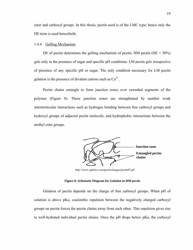
1.4.4 Gelling Mechanism
DE of pectin determines the gelling mechanism of pectin. HM pectin (DE > 50%)
gels only in the presence of sugar and specific pH conditions. LM pectin gels irrespective
of presence of any specific pH or sugar. The only condition necessary for LM pectin
gelation is the presence of divalent cations such as Ca2+.
Pectin chains entangle to form junction zones over extended segments of the
polymer (Figure 8). These junction zones are strengthened by number weak
intermolecular interactions such as hydrogen bonding between free carboxyl groups and
hydroxyl groups of adjacent pectin molecule, and hydrophobic interactions between the
methyl ester groups.
Figure 8: Schematic Diagram for Gelation in HM pectin
Gelation of pectin depends on the charge of free carboxyl groups. When pH of
solution is above pKa, coulombic repulsion between the negatively charged carboxyl
groups on pectin forces the pectin chains away from each other. This repulsion gives rise
to well-hydrated individual pectin chains. Once the pH drops below pKa, the carboxyl
Junction zone
Entangled pectin chains
http://www.cpkelco.com/pectin/images/pectin03.gif

20
groups lose their negative charge and pectin chains come closer due to decrease in
repulsion, resulting in a gel. This is the general gelling mechanism for HM pectin.
In case of LM pectin, gelation mainly follows the “egg-box” model (Figure 9)
(Grant et al., 1973, as cited in [68]). In this mechanism, two pectin chains, each in the
form of a helix, undergo side-by-side association to form a dimer in which the carboxyl
groups on adjacent pectin chains form ionic bonds with calcium ions. The ionic bonds
formed are stable when there are at least seven carboxyl groups on each pectin chain in
the dimer. Consecutive calcium crosslinks in a dimer result in the formation of a stable
junction zone and aggregation of several layers of dimers results in gel formation.
Presence of methyl esters in the pectin backbone interrupts the formation of junction
zones. Therefore, in LM pectin with a lower DE, with less number of methyl ester
groups, there is a better chance for formation junction zones with few calcium ions as
compared to LM pectin with higher DE. Gelation depends on factors such as molecular
weight, pH of pectin solution, presence of other groups such as acetyl and amide in the
pectin chain, temperature.

21
Figure 9: Schematic Diagram for Gelation in LM pectin
1.4.5 Review of Pectin Usage in Controlled Drug Delivery
Pectin is a hydrophilic polymer and this ability of pectin to form a hydrogel has
made it a potential candidate for applications related to oral and gastrointestinal tract drug
delivery. P. Sriamornsak [70] has reviewed research work investigating the use of pectin
for controlled drug delivery applications; some of them dating back to as early as 1981.
Controlled drug delivery using pectin either as a tablet component or coating [71-73]
based on the underlying principle of calcium cross-linking in the pectin matrix has been
studied. Calcium crosslinking delays the swelling of pectin matrix (when pectin is used to
coat the tablet) or erosion of tablet (when pectin is a component of the tablet), and these
in turn delay the release of drug from the tablet. A schematic diagram showing the effect
of crosslinking on swelling and hence release is shown later in section 1.4.7.2.
H OH
OH HH
H
OO H
O
COOH H OH
OH HH
H
OH
O
H OH
OH HH
HO
HO
COOCH3 H OH
OH HH
H
OH
O
H OH
OH HH
H
OO H
O
H OH
OH HH
H
OH
OOH HH
H
OH
O
H OH
OH H
HH
OCOOH
Ca
O
O O
O
O
O
O
Ca
O
C=O
C=O
O
CaCa
O
C=O
C=O
O
H OH
OH HH
H
OO H
O
COOH H OH
OH HH
H
OH
O
H OH
OH HH
HO
HO
COOCH3 H OH
OH HH
H
OH
O
H OH
OH HH
H
OO H
O
H OH
OH HH
H
OH
OOH HH
H
OH
O
H OH
OH H
HH
OCOOH
Ca
O
O O
O
O
O
O
Ca
O
C=O
C=O
O
CaCa
O
C=O
C=O
O
Aggregation of several dimers Formation of egg-box like dimer

22
1.4.6 Review of Pectin in Antimicrobial Packaging
Extruded edible films from pectin-starch blends have been studied by Fishman et
al. [24, 25], but without incorporation of antimicrobial compounds. Composite films of
PLA-pectin blends containing nisin [64] have also been studied and antimicrobial activity
of these films was evaluated. Overall, the above researchers have focused mainly on
studying the processing aspects and physical characteristics of these composite films but
not on controlling the release. Thus, there is plenty of scope for using pectin in controlled
release of antimicrobials such as nisin by employing the concept of calcium crosslinking
as used in controlled drug delivery.
1.4.7 Explanation of Terms and Concepts Related to Pectin and Controlled Release
1.4.7.1 Degree of Crosslinking
Figure 10: Schematic Diagram to Illustrate Different Degrees of Calcium Crosslinking
Degree of crosslinking in pectin films or coatings may be defined as the percent of
free carboxyl groups in a pectin molecule that are involved in the formation of ionic
bonds with calcium ions. The formation of these ionic bonds, also known as calcium
Ca
C=OO
C=OO
C=OO
OC=O
OC=O
OC=O
CaCa
C=OOH
C=OO
C=OOH
OHC=O
OC=O
OHC=O
Ca
100% Crosslinking
(all –COOH groups form ionic bonds with Ca2+ ions)
33% Crosslinking
(Only 1 of 3 –COOH groups form ionic bonds with Ca2+ ions)
Ca
C=OOC=OO
C=OOC=OO
C=OOC=OO
OC=OOC=OOC=O
OC=OOC=OOC=O
OC=OOC=OOC=O
CaCa
C=OOHC=OOH
C=OOC=OO
C=OOHC=OOH
OHC=OOHC=OOHC=O
OC=OOC=OOC=O
OHC=OOHC=OOHC=O
Ca
100% Crosslinking
(all –COOH groups form ionic bonds with Ca2+ ions)
33% Crosslinking
(Only 1 of 3 –COOH groups form ionic bonds with Ca2+ ions)
(a)
(b)

23
bridges or crosslinks, depends on the number of calcium ions made available to a pectin
molecule of a particular DE. Illustrated in Figure 10 above is a hypothetical situation
where two pectin chains, each with three free carboxyl groups, exist. If the amount of
calcium ions provided is in excess, such that all three pairs of carboxyl groups are
involved in the formation of crosslinks with calcium, then the degree of crosslinking
obtained is 100% (Figure 10a). If the amount of calcium ions provided is limited, such
that only one pair out of three pairs of carboxyl groups is involved in calcium
crosslinking, then only 33% degree of crosslinking is achieved (Figure 10b). The number
of free carboxyl groups depends on the DE of pectin used; lower the DE, higher the
number of free carboxyl groups in a given pectin molecule and therefore, higher is the
opportunity for the formation of calcium crosslinks.
Thus, degree of crosslinking can be manipulated by varying the degree of
esterification (DE) of LM pectin used or by varying the amount of calcium ions, as
shown in Figure 11. The amount of calcium ions will be referred to as calcium
concentration and expressed as mg of Ca per g of pectin. One way to manipulate the
degree of crosslinking (as explained earlier in figure 10a and 10b) is by varying the
calcium concentration, for the same DE of pectin (DE 1, Figure 11). Another approach is
to vary the DE of pectin used, keeping the calcium concentration constant. As mentioned
earlier in gelling mechanism of LM pectin (section 1.4.4), as the DE decreases, the
probability of calcium crosslinking increases than pectin with a higher DE and thus,
different degrees of crosslinking can be achieved. The other logical way is to vary the
number of pectin molecules by changing pectin concentration.

24
Figure 11: Different Ways to Manipulate Degree of Crosslinking
1.4.7.2 Degree of Swelling
Figure 12: Effect of Calcium Crosslinking on Swelling of LM Pectin Film
Degree of swelling is typically defined as the % increase in weight or thickness of a
crosslinked pectin film when suspended in a solvent like water. Swelling of film or tablet
coating takes place when solvent (simulant) diffuses into the pectin matrix. As the films
Low degree of crosslinking
High degree of crosslinking
TimeUnswollen coating / tablet
Swollen coating / tablet
Pectin chain
Simulant
Calcium Crosslink
Active compound
Low degree of crosslinking
High degree of crosslinking
TimeUnswollen coating / tablet
Swollen coating / tablet
Pectin chain
Simulant
Calcium Crosslink
Active compound
Degree of esterification (DE) Concentration in film
DE 1 DE 2
Ca (A mg)
LM Pectin
Calcium amount (mg per g of pectin)
Level X Level Y
Ca (B mg)
Ca (C mg)
Ca (D mg) Ca (X mg) Ca (Y mg)
Degree of esterification (DE) Concentration in film
DE 1 DE 2
Ca (A mg)
LM Pectin
Calcium amount (mg per g of pectin)
Level X Level Y
Ca (B mg)
Ca (C mg)
Ca (D mg) Ca (X mg) Ca (Y mg)

25
swells, mobility of active compound inside the pectin matrix increases and it diffuses to
the surface of film to be finally released into the simulant. It has been shown that extent
of crosslinking has a direct effect on swelling of pectin coating in tablets; degree of
swelling decreasing with increasing crosslinking. Illustrated above is a schematic
representation of the effect of calcium crosslinking on release of active compound or
drug from the pectin film or coating. The film/coating with more crosslinking takes
longer to swell and hence release is delayed. Though not novel, this is an important
concept that could be used to control the release of antimicrobial compounds from pectin
films. The range of release rates that can be obtained from films with small variations in
calcium crosslinking is yet to be determined.
1.5 Research Gaps and Opportunities
Based on the above literature review of antimicrobial edible films, some of the
research gaps identified are as follows:
• Most of the studies conducted in this field have focused on evaluation of
mechanical properties of antimicrobial films and their antimicrobial activity.
However, except for some research work [4, 72], not many attempts have been
made to control the release of antimicrobial compound by modifying the film
composition or properties.
• Release kinetics of antimicrobial compound from edible film has not been
investigated by many researchers; only antimicrobial efficacy of film, which is
also important, has been studied.

26
• Many biopolymers possess charged groups and have the potential to form films
with varying release rates; however, only a few of them such as alginate and
pectin have been utilized for controlled release antimicrobial films.
• The potential of pectin, a biopolymer, extensively studied in drug delivery
applications, has not been tapped in the area of antimicrobial edible films.
• Literature review of usage of nisin in biopolymers (section 1.3.2.3) indicates that
very little research has been conducted with pectin-nisin films, probably due to
the speculation that nisin, a positively charged peptide, might interact with
negatively charged pectin, and may not be released from the pectin matrix. This
presents an opportunity for study of release of nisin from pectin films.
• Nisin quantification is mostly done using the microbial assay. Since nisin forms
multimers and degradation products that may still retain antimicrobial activity
[58, 74], estimation of nisin (released from film) using the microbial assay may
not represent the intact nisin, but it may be combined antimicrobial activity of
nisin and/or its antimicrobially active derivatives. A correlation between chemical
estimation of nisin (e.g. by HPLC) and estimation using the microbial assay (e.g.
by agar diffusion assay) will provide information about stability of nisin when
present in the film and after being released into the environment.
The objective of this thesis was to address the above research gaps.
27
1.6 Objectives
1.6.1 Overall Objective
An overall objective of our group was to develop edible/biopolymer based CRP
films from low methoxyl (LM) pectin that can release an antimicrobial compound in a
controlled manner to enhance safety and shelf life of food products with short storage
periods. The biopolymer containing an active compound may be used as a single film in
contact with food or in combination with an outer biopolymer layer or as a coating on
another biopolymer. Fresh meats, cheese, fruits, packaged salads, etc. are the target food
applications for this innovative antimicrobial packaging film.
1.6.2 Scope of Research
Previous research on use of pectin in tablets for controlled drug delivery [71, 72]
and work done by Zactiti et al. [4] on effect of calcium crosslinking on potassium sorbate
permeability through alginate films have demonstrated that degree of crosslinking is an
important parameter governing the release characteristics of a tablet coating or alginate
film. These experiments indicate that it is possible to develop controlled release
packaging films from LM pectin with a wide range of release rates by simple variations
in the degree of calcium cross-linking within the film. A database of film formulations
possessing different release rates, generated from this study, will provide useful
information to produce a CRP film from LM pectin with a desired release property
simply by choosing the appropriate film formulation!

28
1.6.3 Challenges in Development of Pectin Films
Unlike synthetic polymers, pectin is a highly reactive biopolymer. Hence, in order
to conclude that crosslinking is the only factor affecting the release of an antimicrobial
compound from a LM pectin film, it is first necessary to develop an understanding about
the different variables affecting pectin films, be it processing parameters or type of
antimicrobial compound used or other environmental factors. As already mentioned,
there is scope for development of nisin-containing pectin films. However, like pectin,
nisin is also very reactive. Based on the understanding of chemical nature of pectin and
nisin, some of the challenges foreseen in the development of pectin-nisin films are
outlined below:
1.6.3.1 Effect of pH of Pectin Slurry on Pectin-Nisin Interaction
Nisin is positively charged at most pH conditions. Pectin has a pKa of about 3.5;
pKa varies with DE of pectin. Therefore, depending on pH of pectin slurry, two scenarios
(Figure 13) possible for pectin-nisin interaction [75] are stated below:
a) pH of pectin slurry is above the pKa of pectin: In this case, pectin in slurry will
be negatively charged and therefore, positively charged nisin will interact with the
negative sites (–COOH groups) of pectin. Due to this interaction, nisin may bind to the
pectin molecules and not be released from the film. Films containing nisin may still show
antimicrobial activity due to immobilization of nisin on the film surface.

29
Figure 13: Schematic Diagram to Illustrate Pectin-Nisin Interactions
b) pH of pectin slurry is below the pKa of pectin: In this case, the carboxyl groups
of pectin will be in the protonated form, i.e. without any charge, resulting in a repulsion
between pectin and nisin. As a result, nisin will not bind to the pectin molecules and can
be released from the film. However, maintaining the pH of pectin slurry below its pKa
will affect the ability to form crosslinks with calcium ions.
1.6.3.2 Effect of pH of Pectin Slurry on Calcium Crosslinking
a) pH of pectin slurry is above the pKa of pectin: In this case, pectin will be able to
form crosslinks with calcium and different degrees of calcium crosslinking can be
achieved.
COOH
COOH
COOH
COOCH3 COOCH3
COOH
COOH
COOH
NH3+
NH3+ NH3
+NH3+
Pectin
Nisin
COOHCOOH COOH COOH
pH < pKa, Pectin
COOH
COOH
COOH
COOCH3 COOCH3
COOH
COOH
COOH
NH3+
NH3+ NH3
+NH3+
Pectin
Nisin
COOHCOOH COOH COOH
pH < pKa, Pectin
Attraction between opposite charges may hamper release of nisin from film
Repulsion between like charges may cause early release of nisin from film
(a)
(b)
COO−
COO−
COO−
COOCH3 COOCH3
COO−
COO−
COO−
NH3+
NH3+ NH3
+NH3+
Pectin
Nisin
COOHCOOH COOH COOH
pKa, Pectin < pH
COO−
COO−
COO−
COOCH3 COOCH3
COO−
COO−
COO−
NH3+
NH3+ NH3
+NH3+
Pectin
Nisin
COOHCOOH COOH COOH
pKa, Pectin < pH

30
b) pH of pectin slurry is below the pKa of pectin: In this case, pectin will not be
able to form calcium crosslinks as effectively as in the case above. Entanglement of
pectin chains will be responsible for gelation and film formation. Here, the advantage of
being able to vary the release by varying the calcium crosslinking may not be realized to
the maximum extent.
Thus, pH of pectin slurry is very important in designing controlled release pectin
films containing nisin. The effect of pH of pectin on nisin interaction and calcium
crosslinking may be summarized as shown in schematic diagram (Figure 14).
Figure 14: Overview of Impact of pH on Interactions of Pectin with Nisin and Calcium
Condition most favorable for
controlled release of nisin
No pectin-nisin
interaction
Nisin stability better at
lower pH
Calcium crosslinking
not as effective
pH < pKa, pectin pH > pKa, pectin
Conditions suitable for release of
intact nisin
Pectin-nisin interaction possible
Nisin may not
be as stable
Calcium cross-linking
effective
Conditions not suitable for
release of intact nisin
pH, Pectin slurry
Condition not favorable for
controlled release of nisin

31
1.6.4 Specific Objective
The specific objective of this research was to identify key variables by evaluating
their effect on the release of antimicrobial compounds, especially nisin, from LM pectin
films.
Based on the challenges outlined in section 1.6.3, the variables to be studied were
categorized as follows:
1. Composition Variables:
• Size and chemical nature of active compounds: Here, three antimicrobial
compounds - sodium benzoate (low molecular weight and aromatic ring
compound), potassium sorbate (low molecular weight and straight chain
compound), and nisin (highly reactive peptide with large molecular weight) –
were chosen to evaluate the impact of their size on their release. Coomassie blue
dye (medium molecular weight compound with several rings) was used as a
model compound to prove the concept.
• Degree of calcium crosslinking: It is a product of two design variables - DE of
pectin and calcium concentration. Here, the purpose was to understand the impact
of the above design variables on release kinetics of the antimicrobial compounds
mentioned above.
2. Process Variables:
• pH of pectin: The aim of studying this process variable was to evaluate its effect
on the interaction of pectin with calcium and nisin and consequently, on the
release kinetics of nisin.

32
• Sequence of nisin addition: Here, the aim was to determine whether pectin
preferentially bound with calcium or nisin.
• Method of calcium crosslinking: Here, the purpose was to determine if release
was affected by the method of calcium crosslinking – single-step or two-step
crosslinking.
3. Environmental variable:
The main purpose of this experiment was to evaluate the effect of pH, both of
pectin slurry and food simulant used in release study, on the recovery of nisin from pectin
films. The other purpose was to compare the two methods used for nisin estimation - agar
diffusion assay and HPLC.
Understanding from this study will help to optimize design variables for
development of antimicrobial pectin films with variable release rates by simple variations
in the degree of calcium crosslinking within the film.
1.7 Research Approach
Figure 15: Research Approach to Identify Key Variables for Controlled Release LM Pectin Films
List variables (composition and process)
Determine release of active compound as a function of each variable
Establish impact of each variable on release property of film
Identify key variables based on extent of their impact on release

33
1.7.1 Proof of Concept
The research approach to attain the above goal is illustrated in the form of an
experimental design (Figure 15). The first step was to establish a proof of concept using a
model compound such as coomassie blue dye, the assumption being that its release was
not affected by any sort of chemical interactions with pectin. Other advantages of using a
dye for proof of concept were its fairly large size and ease of quantifying its release by a
UV-Vis spectrophotometer. Moreover, release of dye from film could be easily observed
visually.
Once proof of concept was demonstrated, the effect of key variables on release of
different antimicrobial compounds would be evaluated.
1.7.2 Research Approach to Study Pectin-Nisin Interaction
Pectin-nisin interaction could be avoided by saturating the –COOH groups of pectin
with calcium ions prior to addition of nisin. This may not be possible in films with lower
levels of calcium concentration. In such conditions, some negative sites may still be
available on pectin to bind with nisin and thus, either pectin-nisin interaction may take
place or pectin-calcium interaction may take place. It is worth investigating to find out
whether pectin preferably binds with any of the above. In order to seek this information,
the sequence of addition of nisin and calcium can be changed, and the effect of the
sequence of addition can be studied on the release of nisin.

34
Figure 16: Research Approach to Investigate Preference for Pectin Interaction with Calcium and Nisin
Nisin added to pectin slurry
first, followed by calcium
pH > pKa, pectin
pH, Pectin slurry
Nisin added to pectin slurry after calcium
has been added
Nisin added to pectin slurry
first, followed by calcium
Nisin added to pectin slurry after calcium
has been added
pH < pKa, pectin

35
2 EXPERIMENTAL
2.1 Materials
2.1.1 Pectin Selection
Commercial samples of low methoxyl pectin with varying DE were gifted by CP
Kelco, San Diego, CA, USA. Their specifications are shown in table below. All pectin
samples were used as is, without further purification.
Table 3: Specifications for LM Pectin from CP Kelco
Pectin Brand Name Pectin Type DE
(Degree of
Esterification)
DA
(Degree of
Amidation)
Suggested
Calcium
reactivity
GENU LM - 104 AS Partly amidated low ester 27 20 Low
GENU LM - 101 AS Partly amidated low ester 35 15 Low
GENU LM - 12 CG Low ester 35 0 High
GENU LM - 18 CG Low ester 40 0 Medium
GENU LM - 22 CG Low ester 50 0 High sugar products
GENU LM - 5 CS Low ester 7 0 High
Pectin grades mainly used for this study were LM-12 (DE 35), LM-18 (DE 40) and
LM-22 (DE 50). LM-5 was initially tried, but due to its extreme sensitivity to calcium, it
was very difficult to incorporate even small amounts of calcium without causing any
pregelation. The resultant films made using LM-5 pectin were very brittle and not
suitable for release studies.
36
2.1.2 Active Compounds
2.1.2.1 Dye - for Proof of Concept
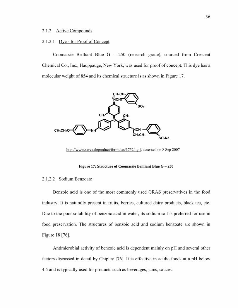
Coomassie Brilliant Blue G – 250 (research grade), sourced from Crescent
Chemical Co., Inc., Hauppauge, New York, was used for proof of concept. This dye has a
molecular weight of 854 and its chemical structure is as shown in Figure 17.
Figure 17: Structure of Coomassie Brilliant Blue G – 250
2.1.2.2 Sodium Benzoate
Benzoic acid is one of the most commonly used GRAS preservatives in the food
industry. It is naturally present in fruits, berries, cultured dairy products, black tea, etc.
Due to the poor solubility of benzoic acid in water, its sodium salt is preferred for use in
food preservation. The structures of benzoic acid and sodium benzoate are shown in
Figure 18 [76].
Antimicrobial activity of benzoic acid is dependent mainly on pH and several other
factors discussed in detail by Chipley [76]. It is effective in acidic foods at a pH below
4.5 and is typically used for products such as beverages, jams, sauces.
http://www.serva.deproduct/formulas/17524.gif, accessed on 8 Sep 2007
SO3Na
CH3CH2O NH
CCH3 CH3
NCHCH2CH3
+NCH2
SO3_
CH2CH3

37
Figure 18: Structures of Benzoic Acid and Sodium Benzoate
The minimum inhibitory concentrations (MIC) for selected bacteria, yeasts and
fungi have been listed by Chipley [76].
Sodium benzoate (>99% pure), in the form of a white powder, was purchased from
Acros Organics.
2.1.2.3 Potassium Sorbate
Sorbic acid is another GRAS preservative commonly used in the food industry.
Potassium salt is generally used in food due to the poor solubility of sorbic acid in water.
The structures of sorbic acid and potassium sorbate are shown in Figure 19 [77].
Figure 19: Structures of Sorbic Acid and Potassium Sorbate
It is mainly effective against yeasts and molds and is used typically used for
preservation of dairy products, baked goods, fruit and vegetable products, etc. [77].
Potassium sorbate (>99% pure), in the form of granules, was purchased from Sigma
Aldrich.
Sorbic acid Potassium sorbate
CH3-CH=CH-CH=CH-COOH CH3-CH=CH-CH=CH-COOK
C OH
O
C O- Na+
O
Benzoic acid Sodium benzoate

38
2.1.2.4 Nisin
Pure nisin (40 x 106 IU per gram) was a gift from Chr. Hansen (Milwaukee, WI,
USA). A stock solution (0.25% nisin) was prepared in acidic water (adjusted to pH 2 with
conc. HCl) to obtain a final antimicrobial activity of 105 IU/ml and stored at 4 °C.
2.1.3 Plasticizer
Glycerol, a commonly used plasticizer in the manufacture of edible films, was
purchased from Fisher Scientific.
2.1.4 Crosslinking Agent
Anhydrous calcium chloride, in the form of granules (4 mesh) was sourced from
Sargent-Welch.
2.1.5 Media Preparation for Inhibition Zone Assay
The formulations used for preparing media for the inhibition zone assay is given in
Table 4 below. All the ingredients were dispersed in deionized water and autoclaved at
121 °C for 15 minutes prior to use.
Table 4: Formula for Media Used in Inhibition Zone Assay
Ingredients TSBYE Nutrient Broth
Solid TSAYE Agar Soft TSAYE Agar
Tryptic soy broth with dextrose (Bacto)
30 g/L of water 30 g/L of water 30 g/L of water
Yeast extract powder (Bacto)
6 g/L of water 6 g/L of water 6 g/L of water
Granulated agar (Difco Laboratories, Detroit, MI, USA)
- 15 g/L of water 7 g/L of water

39
2.1.6 Bacterial Culture for Inhibition Zone Assay
An overnight culture of Micrococcus luteus ATCC 10420 was grown aerobically at
30 °C with agitation in TSBYE nutrient broth.
2.2 Methods
2.2.1 Preparation of Pectin Film
2.2.1.1 Single-Step Calcium Crosslinking
Aqueous slurry of pectin (4.56%, w/w) and glycerol (1.99%, w/w) was prepared by
continuously mixing dry pectin powder in a mixture of deionized water and glycerol for
almost an hour using a magnetic stirrer. Pectin slurry (35 g) was heated to 75 ºC while
being stirred continuously on a magnetic stirrer. To this continuously stirred hot pectin
slurry, 3 ml of either coomassie blue solution (0.0395%) or 1% potassium sorbate
solution or 1% sodium benzoate solution or 0.25% of pure nisin solution was added.
Crosslinking agent i.e. calcium chloride solution (0.7%), heated to the same temperature
as pectin slurry, was gradually added to the above slurry, being continuously stirred at 75
ºC, to finally obtain the film-forming solution. The amount of calcium chloride solution
added to the slurry varied with the desired degree of crosslinking and the values are given
in Table 5. The hot film forming solution was immediately degassed and 25 g of film
forming solution was poured into petri dishes (87mm diameter x 13 mm height) and
allowed to dry overnight at 30 ºC. The resultant film was peeled off from the petri dish
and equilibrated at 50% RH, 25 ºC for a day prior to using it for any tests.

40
Table 5: Requirement of Calcium Chloride for Films with Different Calcium Crosslinking.
Desired Calcium
Concentration in Film
(mg of Ca per g of pectin)
Pectin Slurry
(g)
Volume of
Calcium Chloride
Solution (0.7%) ml
Amount of
water (ml)
0 35 0 12
9 35 6 6 13.5 35 9 3 18 35 12 0
2.2.1.2 Two-Step Calcium Crosslinking
For very high levels of calcium crosslinking or when very low DE pectin was to be
used, it was difficult to make uniform films by the single-step crosslinking technique.
Almost instantaneous gelling and solidification of the gel caused difficulty in pouring the
gel to form uniform films. This imposed a practical limitation on the amount of calcium
that could be added and so, a two-step crosslinking process was carried out.
Here, pectin slurry was prepared as described in above section. In the first step, low
amount of calcium chloride was added to hot slurry, such that it was comfortable to pour
the gel into petri dishes. The gel was allowed to dry overnight into a film and then this
film was immersed in a known amount of calcium chloride solution and dried for 8-10
hours to obtain a higher degree of calcium crosslinking. The resultant film was rinsed 5
times, with 2 ml of deionized water each time; to remove any calcium or nisin adhered to
the surface.
2.2.2 Release Studies
Approximately 0.8 g of film pieces were cut into 2 cm × 2 cm square pieces and
immersed in 80 ml of deionized water in a 125 ml Erlenmeyer flask, which was shaken at

41
100 rpm and 23°C. Samples were withdrawn periodically and amount of active
compound released was quantified by methods described in section 2.2.3.
Note: In these release studies, 100% water was used instead of the FDA
recommended food simulants [78] like 10% ethanol because pectin precipitates in ethanol
and methanol, thereby it might affect the swelling property of pectin film.
2.2.3 Quantification of Active Compounds
2.2.3.1 Quantification of Dye by UV-Visible Spectrophotometry
Coomassie blue dye released from film into water was quantified by UV/Visible
spectrophotometry at 612 nm. A calibration standard curve for the quantification of dye
was generated (Figure 20) at 612 nm using a Varian Cary 50 UV-Vis spectrophotometer
(Palo Alto, CA, USA).
Figure 20: Calibration Curve for Coomassie Blue G – 250
Concentration of dye (µg/ml)
y = 0.0614x + 0.0108R2 = 0.9978
0.000
0.100
0.200
0.300
0.400
0.500
0.600
0.700
0.0 2.0 4.0 6.0 8.0 10.0 12.0
Abs
orba
nce
at 6
12 n
m
42
2.2.3.2 Quantification of Sodium Benzoate and Potassium Sorbate by HPLC
Benzoic and sorbic acids and their respective salts were analyzed using RP-HPLC
as per the standard protocol recommended by the USDA [79] and also used by
Buonocore et al. [80]. Both these compounds were analyzed using the same method; they
only differed in the wavelength of detection.
HPLC experiments were conducted on Shimadzu SCL-10Avp System (Kyoto,
Japan) equipped with a LC10ADVP solvent delivery unit, FCV-10ALvp Low Pressure
Gradient Unit, SIL-10Ai Autoinjector, and SPD-M10Avp Photodiode Array Detector.
Data were processed using Class VP EZ Start Version 7.3 data acquisition software.
A silica based C18 column, Cosmosil – code no. 379-72 (Waters, MA, USA), with
a particle diameter of 5 µm and dimensions of 250 mm × 4.6 mm I.D. was used for the
above quantification. 20 µl samples were injected.
The mobile phase for the above HPLC quantification was prepared by dissolving
0.68% (w/v) of potassium hydrogen phosphate in part of the water, followed by addition
of 0.1% (v/v) of concentrated phosphoric acid and 25% (v/v) of HPLC grade acetonitrile.
To this mixture, water was added to make up a mobile phase of water: acetonitrile in the
ratio 75:25. The flow rate of mobile phase was maintained at 1 ml/min, at ambient
temperature and wavelength of UV detection was set at 225 nm for benzoic acid and 260
nm for sorbic acid.
2.2.3.3 Microbial Assay for Nisin Activity
Antimicrobial activity of nisin released from film into water was assayed using the
agar diffusion assay [81, 82] . Briefly, solid TSAYE agar plates were prepared by

43
dispensing 20 ml of sterile media in sterile petri dishes. Sterile soft TSAYE agar was
melted in a microwave oven and tempered at about 45 °C. 4 ml of this agar was seeded
with 40 µl of an overnight broth culture on Micrococcus luteus ATCC 10420 and
dispensed over the solid agar mentioned above. Three wells of 6 mm diameter were
bored into each agar plate with a sterile glass tube. 50 µl of liquid sample, either standard
nisin solution or aliquot of samples collected at different time intervals during release
study were pipetted into each well. The agar plates were then stored in refrigerator for 24
hours to allow diffusion of nisin, followed by incubation at 30 °C for 24 hours during
which time clear inhibition zones were developed. Diameter of the inhibition zone
developed was measured using a ruler. A calibration (figure ) for standard nisin solutions
ranging from 12.5µg/ml (equivalent to 2.5 IU/ml) to 100 µg/ml (equivalent to 4000
IU/ml) was constructed by plotting width of inhibition zone developed vs. log of
corresponding nisin concentration. The amount of nisin in test solution was estimated by
comparing the width of inhibition zone with that of standard nisin solutions.
In order to estimate amount of nisin released when film was placed in contact with
soft agar, the previously described well diffusion assay was slightly modified. In this
case, a bigger well with a diameter of 1.6 cm was bored into the seeded agar plate and the
diameter of film sample placed on the seeded agar was also 1.6 cm. The agar plate with
film was stored in refrigerator for different time intervals, followed by incubation at 30
°C for 24 hours.

44
Figure 21: Calibration Curve for Nisin Estimation Using Agar Diffusion Assay
2.2.3.4 Quantification of Nisin by HPLC
Most common method for estimation of nisin is the inhibition zone assay
mentioned in section 1.2.3.3. However, a HPLC method has been developed by Liu and
Hansen [74].
Quantification of nisin released into water was made by adapting a modified
method developed by Buonocore et al. [80]. HPLC experiments were conducted on
Shimadzu SCL-10Avp System (Kyoto, Japan) mentioned in section 1.2.3.2.
A silica based C-18 reversed-phase column, ACE-5, with a particle diameter of 5
µm and dimensions of 150 mm × 4.6 mm I.D. was used for the above quantification.
Each column was preceded by a guard column. 50 µl samples were injected.
Mobile phase was comprised of water-acetonitrile, both containing 0.1% TFA. A
gradient of 20-60% acetonitrile over 20 minutes at a flow rate of 1 ml/min was
maintained. The wavelength of UV detection for nisin was set at 254 nm and nisin eluted
y = 3.7607x - 0.4937R2 = 0.9889
8
10
12
14
2.000 2.500 3.000 3.500 4.000
Wid
th o
f inh
ibiti
on z
one
(mm
)
Log of nisin concentration (IU/ml)

45
at 5.3 minutes. A calibration curve (Figure 22) was constructed for standard nisin
solutions ranging from 5 to 125 µg/ml. Standard nisin solutions were prepared using the
pH 2 water as a diluent in order to maintain nisin in the most stable form.
Figure 22: Calibration Curve for Nisin Estimation by HPLC
Peak
Are
a
y = 6354.3x - 10147R2 = 0.9996
0
200000
400000
600000
800000
1000000
0 20 40 60 80 100 120 140
Concentration of Nisin (µg/ml)

46
3 RESULTS AND DISCUSSION
3.1 Effect of Composition Variables on Release
As already mentioned, the main rationale to conduct an experiment for proof of
concept was to get a sense of whether the degree of calcium crosslinking, one of the main
composition variables was significantly effective in controlling the release of dye from
pectin films, provided no other variables such as interactions with pectin or degradation
of active compound were present. The dye chosen for proof of concept was stable,
moderately large and easy to quantify. Results obtained from proof of concept were
encouraging and provided a motive to study effect of other variables on release of active
compounds.
3.1.1 Effect of Degree of Calcium Crosslinking
3.1.1.1 Effect of Calcium Concentration on Release of Dye from Pectin Film
Calcium concentration, one of the two main design variables affecting the degree of
calcium crosslinking, was studied in this set of experiments. Pectin films containing dye
were prepared as per method described in section 2.2.1. All films were made using LM-
12 pectin (DE 35) and the amount of calcium in the films was varied to give a final
calcium concentration, expressed as mg of Ca per g of pectin, in the dry films at 9, 13.6,
18, and 28. A control film was also made without any calcium. Release studies were
conducted for each film and % of dye released was quantified for different time intervals,
as shown below in Figure 23.

47
Figure 23: Effect of Calcium Concentration on Release of Dye from LM 12 (DE 35) Pectin Films
Time taken to release all dye from films with 0, 9, 13.6, 18 mg Ca /g pectin was 1.5
hours, 7 hours, 48 hours, 120 hours, respectively (Figure 23). For film with 28 mg Ca/g
pectin, only 10% of dye was released after 7 days.
Similar release trend was observed in films made using LM-18 (DE 40) and LM-22
(DE 50) with different concentrations of calcium, as shown in Figure 24 and Figure 25,
respectively.
Frac
tiona
l rel
ease
of d
ye (%
)
Time (hours)
0
20
40
60
80
100
120
0 50 100 150 200
0 mg Ca/g pectin
9 mg Ca/g pectin
13.6 mg Ca/g pectin
18 mg Ca/g pectin
28 mg Ca/g pectin

48
Figure 24: Effect of Calcium Concentration on Release of Dye from LM-18 (DE 40) Pectin Films
Figure 25: Effect of Calcium Concentration on Release of Dye from LM-22 (DE 50) Pectin Films
0
20
40
60
80
100
120
0 20 40 60 80 100 120 140
0 mg Ca/g pectin
9 mg Ca/g pectin
18 mg Ca/g pectin
Time (hours)
Frac
tiona
l rel
ease
of d
ye (%
)
0
20
40
60
80
100
120
0 20 40 60 80
0 mg Ca per g pectin
9 mg Ca per g pectin
13.6 mg Ca per g pectin
18 mg Ca per g pectin
Frac
tiona
l rel
ease
of d
ye (%
)
Time (hours)

49
Results obtained from these release studies clearly demonstrated that the rate of
release of dye from film decreased significantly with a small increase in calcium
concentration.
3.1.1.2 Effect of DE of Pectin on Release of Dye from Pectin Film
Degree of esterification (DE) of pectin, the second design variable affecting the
degree of calcium crosslinking in the pectin film, was studied in this set of experiments.
Films were made using pectin with DE 35, 40 and 50 and for each DE calcium
concentration were varied as shown in grid below.
Table 6: Effect of DE of Pectin and Calcium Concentration on Release of Dye from Film
DE of Pectin Calcium Concentration in Film
(mg of Ca per g of pectin)
Time Taken for Release of All Dye
(hours)
35 (LM-12)
0 9
18 28
1.5 7
120 168 (~10% release)
40 (LM-18)
0 9
18
2 17
120
50 (LM-22)
0 9
18
1.5 4.5 48
Table 6 gives a comparison of dye release time for films made with different DE
pectins. For a film made with DE 50 pectin, at a calcium concentration of 18 mg Ca/g
pectin, the dye release time was 48 hours, whereas for a film made with DE 35 pectin and
same amount of calcium concentration, the total release time was 120 hours. This result
showed that with a decrease in the DE of LM pectin from 50 to 35, the dye release time
was extended from by 3 days. But, there was no significant difference in release rates for
35 DE and 40 DE films, indicating that in order to obtain a significant difference in
release rates, a wide range of DE of pectin would be necessary.

50
Overall, the rate of release of dye was influenced by degree of calcium crosslinking
within film, which was altered by varying the two design variables – calcium
concentration or DE of pectin used (Figure 11). Degree of crosslinking within film was
found to directly affect the rate of swelling and rate of dissolution of film in water, which
in turn was thought to affect the release of an active compound from the swollen films. It
is reasonable to say that the release of an active compound from such swellable films
follows non-Fickian diffusion and depends on the degree of swelling of film and rate of
dissolution of film in an aqueous food simulant. The sequence in which degree of
crosslinking affects release of active compound may be summarized as shown in Figure
26.
Figure 26: Summary of Effect of Crosslinking on Release of Active Compound from LM Pectin Film
Of the two design variables, it was clearly evident (Table 6) that a small change in
calcium concentration produced reasonable difference in release rates. However, the
same was not true in the case of DE of pectin, thereby indicating that calcium
concentration could be a more dominant variable among the two. Since commercial LM
DE of pectin
Release of dye from film
Degree of calcium crosslinking
Calcium Concentration
Rate of dissolution of filmDegree of swelling of film
DE of pectin
Release of dye from film
Degree of calcium crosslinking
Calcium Concentration
Rate of dissolution of filmDegree of swelling of film

51
pectin grades are available within a narrow range of DE, use of this design variable is
slightly restrictive for development of controlled release pectin films.
After proving the concept, the next step was to study the effect of other variables on
release of active compounds.
3.1.2 Effect of Size of Active Compound
3.1.2.1 Sodium Benzoate
Pectin films containing approximately 1% of sodium benzoate (on dry film) were
prepared and release tests were conducted as per method described in section 2.2.2 and
amount of sodium benzoate released into food simulant was estimated using HPLC
(section 2.2.3.2). The amount of benzoate that should be present in film theoretically was
calculated on the basis of material balance of sodium benzoate added to film forming
solution and weight of dry film obtained. Film was completely dissolved in water and
total benzoate content in film was estimated to compare the theoretical % loading with
the actual extractable amount (Table 7).
Table 7: Loading of Sodium Benzoate in Pectin Films - Theoretical vs. Total Extractable
Sample
LM 22 (DE 50)
LM 18 (DE 35)
mg of Ca per g
of film
% Total (added)
% Extractable of total added
% Total (added)
% Extractable of total added
0.0 1.30 11.22 0.930 8.00
9.0 1.16 11.67 1.229 5.80 13.5 1.18 12.43 1.177 2.91
18.0 1.16 10.94 1.196 0.36
The total extractable benzoate was only about 8 -12% of the amount originally
added. This meant that there was some kind of interaction between the rest of the

52
benzoate and pectin within the film during the release test with water as food simulant. A
detailed study was conducted to find out the reason for such poor recovery of benzoate
when films were dissolved in water.
3.1.2.1.1 Sodium Benzoate Entrapment in Pectin Films
Some of the probable reasons short listed were pH of film forming solution and
release test food simulant, solubility of sodium benzoate in film forming solution or food
simulant, thermal stability of sodium benzoate, interaction of sodium benzoate with
calcium ions, interaction of sodium benzoate with pectin, etc. A logical decision tree
method (Figure 27) was followed to rule out entrapment of benzoate in the different steps
involved in development of film and subsequent release study.
Figure 27: Decision Tree Diagram for Investigation of Benzoate Entrapment in Pectin Films
Check recovery of benzoate in calcium
No Interaction between calcium and benzoate
Yes
Check recovery of benzoate in fresh pectin
No
Retention of benzoate during film drying step
Retention of benzoate in pectin slurry
Check recovery of benzoate in film made
using above pectin slurry
No
Yes
Modify procedure for making film to avoid
entrapment of benzoate

53
Table 8: Effect of Calcium on Recovery of Sodium Benzoate from Pectin Films
Sample Expected Benzoate Concentration (µg/ml)
Observed Benzoate Concentration (µg/ml)
Benzoate in deionized water 118 121 123 116
Benzoate and calcium chloride in deionized water 118 121
LM 18 pectin slurry without calcium 24.6 18
LM 18 pectin slurry with calcium 24.6 20.5
The above results showed that benzoate could be recovered completely from
deionized water, irrespective of presence of calcium ions. In case of benzoate recovery
from pectin slurry, about 80% recovery was observed, irrespective of presence of calcium
ions. Thus, the possibility of poor recovery due to interaction of sodium benzoate and
calcium was ruled out. In fact, calcium dibenzoate is a known preservative approved for
food use.
The next step was to check recovery of benzoate in fresh pectin slurry. Calcium
was not added to the slurry in order to study effect of pectin only. pH of pectin slurry i.e.
the film forming solution was found to be about 3 and hence recovery of benzoate from
acidic pH solution was also checked to rule out any effect of pH. The results are shown in
table below.

54
Table 9: Recovery of Sodium Benzoate from Fresh Pectin Slurry
Sample
Expected Benzoate Concentration (µg/ml)
Observed Benzoate Concentration (µg/ml)
pH of Solution
Benzoate in deionized water 30 34 6.9
Benzoate in acidic water 30 31.87 3.04
1% pectin solution (LM 18) in same solution as above (containing 30 µg/ml benzoate)
30 30.86 3.09
Full recovery of sodium benzoate from fresh pectin slurry (1% solution), at an
acidic pH of 3, indicated that there was no interaction of benzoate with pectin till the step
of pectin slurry preparation. So, two films were made with the top and bottom portion of
the same pectin slurry used in the above estimation. These films were dissolved in water to
form a 1% solution and the benzoate content was estimated. The results obtained are given in
Table 10 below.
Table 10: Recovery of Sodium Benzoate from Pectin Film
Sample
Expected Benzoate Concentration
(µg/ml)
Observed Benzoate Concentration
(µg/ml)
pH of Solution
Benzoate in water 57.2 57 6.9
Benzoate in acidic water (same concentration as in film forming solution)
572 538 2.85
1% pectin solution (LM 18) containing 30 µg/ml benzoate 30 30.86 3.09
Film made with top portion ofabove pectin slurry 29 17.9 3.08
Film made with bottom portion of above pectin slurry 29 18.8 3.06
55
There was no problem with recovery of benzoate from fresh pectin slurry, but the
films made with the same pectin slurry showed only 50-60% recovery. This was in
confirmation with the first result (Table 7) in which only 10-12% of pectin could be
recovered. This indicated that there was definitely some interaction occurring between
pectin and sodium benzoate while the pectin slurry was being dried for 24 hours to form a
film. Higher recovery in above study could be attributed to lesser interaction between
benzoate and pectin due to difference in pectin content of films, 1% in above study
(Table 10) as compared to 3.3% in the regular films used in the first study (Table 7).
Thermal stability of benzoate in acidic conditions was also tested by heating a
solution of benzoate in deionized water, adjusted to a pH of 3, for same time-temperature
conditions as in the preparation of film forming solution. Full recovery was seen and thus
loss of benzoate due to heating was eliminated.
Some other tests were conducted, but the results obtained could not explain the
poor recovery of benzoate from pectin films. Thus, sodium benzoate, though small in
size, was not suitable for controlled release pectin films due to entrapment in pectin,
resulting in very poor release.
3.1.2.2 Potassium Sorbate
It was speculated that the benzene ring in benzoic acid could be responsible for π-
bonding, further leading to entrapment of benzoic acid in the pectin matrix. In order to
prove this theory of π-bonding due to benzene ring, potassium sorbate was chosen. It has
a pKa of 4.8, has similar molecular weight as sodium benzoate, but it is a straight chain
compound. So, it was expected to be released completely from pectin films.

56
Release studies were conducted with pectin films containing 1% potassium sorbate
(on a dry film). 0.8 g of film was suspended in 250 ml water and HPLC method was used
to quantify amount of sorbate released from film into water. The results are shown in
Figure 28 below.
Figure 28: Effect of Calcium Concentration on Release of Potassium Sorbate from LM 12 (DE 35) and LM 18 (DE 40) Pectin Films
The observations from above release study were as follows:
• Only 45-50% of added potassium sorbate could be totally extracted, even after
complete dissolution of the film in water. This observation was similar to that of
pectin films containing benzoate, but in case of sorbate, the % total extractable
was higher. Retention of potassium sorbate in pectin matrix has been reported by
Guilbert et al. [83]
Time (minutes)
Frac
tiona
l rel
ease
(%)
0
20
40
60
80
100
120
0 10 20 30 40
DE 40 with noCalcium
DE 40 with 18 mgCa/g pectin
DE 35 with 18 mgCa/g pectin

57
• Time taken to release the total extractable potassium sorbate (45-50% of added, as
mentioned above) from a film crosslinked with 18 mg Ca/g pectin was only 10
minutes longer than that of a film without any calcium. Such fast release rates
have also been reported for release studies with whey protein films and tapioca
starch films.
• Film made using LM 12 (DE 35) pectin and containing 18 mg Ca/g pectin took
more than 3 hours to begin swelling (observed visually) and started dissolving
after 7-8 hours, but all free potassium sorbate was released in less than 30 minutes
as shown in Figure 28 above. Thus, complete release of potassium sorbate even
before the film began to swell could mean that effect of degree of swelling on
release of active compounds was true only when the size of active compound was
above a certain threshold. This was in confirmation with the proof of concept
where same film compositions were used, but the active compound (dye) used
was atleast 7 times larger than potassium sorbate, big enough to be entrapped by
the crosslinks within the film.
The above experiment with potassium sorbate confirmed the importance of
choosing an active compound with an appropriate size such that it would not be released
too quickly. Hence, nisin, an important antimicrobial compound, fairly big in size was
considered for further development of antimicrobial pectin films. A preliminary study
was conducted with nisin-containing films to test whether nisin could diffuse through
pectin films; contrary to speculation that nisin would bind to pectin and hence could not
be released from pectin films.

58
3.2 Preliminary Studies with Nisin Containing Pectin Films
It was thought that nisin may not be released from pectin films due its interaction
with pectin. Hence, a preliminary study was conducted with nisin-containing films to find
out if the above speculation was true or nisin could be released from pectin films. Films
were made with LM 22 (DE 50) pectin using different protocols as given below:
• Control film – with 18 mg Ca/g pectin, without nisin
• Film 1 – In this case, slurry of pectin and calcium was made and known amount
of nisin solution was added directly to the above slurry. The slurry was then
allowed to dry overnight to form a film. Here, nisin (positively charged) may bind
preferentially with pectin instead of calcium binding with pectin and hence may
not release.
• Film 2 – In this case, slurry of only pectin and calcium was made, allowed to dry
overnight to form a film. The film was then suspended in a known amount of
nisin solution for 15 minutes to allow nisin to diffuse inside film. Here, the idea
was to saturate some of the negative sites on pectin with calcium ions so that
these negative sites would not be available to bind with nisin, thereby improving
its chances of release as compared to film 1.
• Film 3 – In this case, the two-step calcium crosslinking method was used to
saturate all the negative sites on pectin to form a film with maximum degree of
calcium crosslinking. The film thus developed was then suspended in nisin
solution for 15 minutes to allow nisin to diffuse inside film. Here, the assumption
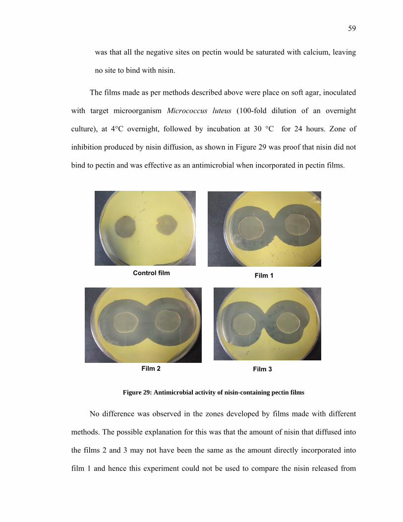
59
was that all the negative sites on pectin would be saturated with calcium, leaving
no site to bind with nisin.
The films made as per methods described above were place on soft agar, inoculated
with target microorganism Micrococcus luteus (100-fold dilution of an overnight
culture), at 4°C overnight, followed by incubation at 30 °C for 24 hours. Zone of
inhibition produced by nisin diffusion, as shown in Figure 29 was proof that nisin did not
bind to pectin and was effective as an antimicrobial when incorporated in pectin films.
Figure 29: Antimicrobial activity of nisin-containing pectin films
No difference was observed in the zones developed by films made with different
methods. The possible explanation for this was that the amount of nisin that diffused into
the films 2 and 3 may not have been the same as the amount directly incorporated into
film 1 and hence this experiment could not be used to compare the nisin released from
Control film Film 1
Film 2 Film 3

60
different films. However, this preliminary experiment gave an indication that nisin could
indeed be release from pectin films and proved the speculation that nisin would bind to
pectin wrong. It was however necessary to evaluate some of the processing variables to
understand the pectin-nisin system better.
3.3 Effect of Processing Variables on Release of Nisin
3.3.1 Effect of Calcium Crosslinking Method
In order to study this processing variable, three films were made with LM 12 pectin
(DE 35) at pH 3.2 as follows -
• Control film without calcium, with nisin (designated as ‘0’ in the microbial
assay).
• Single-step calcium crosslinked film containing 18 mg Ca per g of dry pectin, and
with nisin. All calcium was added to the pectin slurry, prior to film formation
(designated as ‘18’ in the microbial assay).
• Two-step calcium crosslinked film containing a total of 100 mg of Ca per g of dry
pectin, and with nisin (designated as ‘100’ in the microbial assay). Here, 18 mg
Ca per g of dry pectin was added to pectin slurry in the first step and the rest was
added after film was formed.
Release studies were conducted with these films in water in a flask and nisin
concentration of samples collected at different time intervals was estimated by well-
diffusion assay. The results are shown in Figure 30 and Figure 31 .

61
Figure 30: Effect of Calcium Crosslinking Method on Release of Nisin from Pectin Film (Inhibition Zone Assay)
8 hours 24 hours
6 hours 4 hours
2 hours0.5 hours

62
0
20
40
60
80
100
0 5 10 15 20 25
No calcium
Single-stepcrosslinking(18 mg Ca/gpectin)
Two-stepcrosslinking(100 mgCa/g pectin)
Figure 31: Effect of Calcium Crosslinking Method on Release of Nisin from Pectin Films
The film with 18 mg Ca/g pectin showed slower release of nisin as compared with
film without calcium for the first 8 hours after which maximum nisin was released. In
case of film containing 100 mg Ca/g pectin, made with the two-step crosslinking method,
it should have ideally shown a release rate slower than 18 mg Ca/g pectin film. However,
the opposite was observed, with the two-step film releasing maximum nisin in 3-4 hours,
similar to control film without any calcium. A probable reason for this reverse trend
could be attributed to the two-step process of crosslinking, which allowed enough time
for entrapped nisin to migrate to the surface, resulting in its immediate release. Thus, the
method of calcium crosslinking plays a role in the controlling the release of nisin.
The two-step method has been used in other studies and may not be the correct way
to make controlled release films.
Frac
tiona
l rel
ease
(%)
Time (hours)

63
3.3.2 Effect of pH of Pectin and Sequence of Nisin Addition on Pectin-Nisin Interaction
As explained previously in the research approach, pectin-nisin interaction may be
avoided by occupying the negative sites on pectin with calcium, so that few sites are
available to bind with nisin. In order to establish a certain preference for interaction of
pectin with either nisin or calcium, the sequence of addition of nisin was varied for two
pH conditions of pectin slurry as follows:
• pH of pectin slurry < pKa of pectin: For this condition slurry was used as is
because pH of pectin slurry for a 3.3% pectin concentration was already in the
range 2.6 - 2.8.
• pH of pectin slurry < pKa of pectin: For this condition, pH of pectin slurry was
adjusted to 3.85-3.95 using a 5% NaOH solution.
• For each pH condition, films were made with sequence of nisin addition as
follows -
• Control film without calcium, with nisin (designated a ‘0’ in the inhibition zone
assay images).
• Nisin added to pectin slurry first, followed by calcium chloride solution. This film
was designated as N1.
• Nisin added to pectin slurry after addition of calcium chloride solution. This film
was designated as N2.
In N1 and N2, calcium concentration of 18 mg Ca/g dry pectin was maintained.

64
Release studies were conducted with these films suspended in water in a flask and
nisin concentration of samples collected at different time intervals was estimated by well-
diffusion assay. The results for pH< pKa are shown in Figure 32 and Figure 33.
0
20
40
60
80
100
120
0 10 20 30 40 50 60
No calcium
Nisin addedfirst (N1)
Nisin addedsecond (N2)
Figure 32: Effect of Sequence of Nisin Addition on Release of Nisin at pH<pKa of Pectin
As seen in the images of inhibition zone assay (Figure 33), fractional release of
nisin through films N1 and N2 followed the same trend indicating no significant impact
of sequence of nisin addition on its release when pH of pectin slurry was below the pKa
of pectin. At pH<3, not much pectin-nisin interaction will occur, hence this result was
expected. However, the fractional release curve shows more than 100% release, which is
not possible. In order to find out a reason for higher release than expected, these same
samples from above study were analyzed again for nisin concentration using HPLC.
Surprisingly, no nisin could be detected in any of these samples. This could mean that
derivatives of nisin may have formed, which were not detectable on HPLC, but had
slightly better antimicrobial activity than nisin itself. It has been reported in literature that
Frac
tiona
l rel
ease
(%)
Time (hours)

65
nisin1-32 is formed in mildly acidic conditions [58] and this has slightly better
antimicrobial activity.
Figure 33: Effect of Sequence of Nisin Addition on Release of Nisin at pH<pKa of Pectin (Inhibition Zone Assay)
4 hours 6 hours
9 hours 24 hours
0.5 hours 2 hours

66
The results for pH > pKa are shown in Figure 34 and Figure 35.
Figure 34: Effect of Sequence of Nisin Addition on Release of Nisin at pH > pKa of Pectin (Inhibition Zone Assay)
0.5 hours 2 hours
4 hours 6 hours
9 hours 24 hours

67
0
20
40
60
80
100
120
0 20 40 60 80 100 120 140
No calcium
Nisin added first,calcium addedsecond
Calcium addedfirst, nisin addedsecond
Figure 35: Effect of Sequence of Nisin Addition on Release of Nisin at pH > pKa of Pectin
In this case, pH was around 4 at which calcium crosslinking occurs. At this pH,
films N1 and N2 did not dissolve for a week, whereas films N1 and N2 made at pH<pKa
dissolved completely in two days further confirming that calcium crosslinking was more
effective at this pH. Release of nisin was very low in films with 18 mg Ca/g pectin,
irrespective of the sequence of addition of nisin, at pH of pectin slurry > pKa of pectin.
The probable reasons for this low release could be:
• Nisin degraded in film forming step at given pH conditions, leaving only 20% of
intact nisin, all of which was released in 24 hours from the two films N1 and N2;
• Nisin was released, but degraded over five days in the deionized water which was
used as food simulant for the release study;
• More than 80% of nisin added to the films N1 and N2 was bound to the pectin,
resulting in release of remaining 20% nisin;
Frac
tiona
l rel
ease
(%)
Time (hours)

68
• Calcium crosslinking was effective in controlling the release of nisin, resulting in
a very slow release rate.
Between the control film and films N1 and N2, the processing conditions, pH,
release study conditions were same, except for the presence of calcium crosslinking in
films N1 and N2. So, the first 3 reasons should have been true for control film too.
However, that was not observed and control film was able to release almost 80% of
added nisin within a day. This means that calcium crosslinking was the main factor
preventing all nisin from being released.
Thus, this experiment indicated the following:
• Sequence of nisin addition was not significant for controlled release.
• At pH > pKa of pectin, calcium crosslinking was instrumental in preventing the
release of nisin. However, it could not be concluded that at calcium concentration
of 18 mg Ca/g pectin ‘controlled release’ was possible because the tight network
of calcium crosslinks did not allow any further release of nisin. Release tests at
lower levels of calcium concentration must be done to find out the extent of
release of nisin.
3.4 Effect of Environmental Variables
In order to evaluate the effect of pH of pectin slurry and food simulant on the
recovery of nisin, pectin films containing 18 mg Ca/g pectin were made as follows:
• pH of pectin slurry (pH=2.7) < pKa of pectin: release study with deionized water
at pH 2 and pH 5.

69
• pH of pectin slurry (pH=3.95) > pKa of pectin: release study with deionized water
at pH 2 and pH 5.
• The film pieces (0.4 g) were powdered and completely dissolved into water at pH
conditions mentioned and the solutions were analyzed for total nisin content using
agar diffusion assay and HPLC. Results were obtained as shown in
Table 11: Comparison of nisin estimated by HPLC and Agar Diffusion Assay
Preliminary results from HPLC and agar diffusion assays indicated that a
combination of high pH of food simulant and high pH of pectin slurry was not favorable
for nisin. 40% loss of nisin could have occurred at two stages – during film preparation
step due to high pH of pectin slurry and after release to high pH water. This agreed with
the previous result that showed 60-80% release of nisin from control film (without
calcium) made with pH 3.85 pectin slurry (Figure 35). Time and high pH, in combination
with calcium crosslinking, were probably responsible for low and delayed release of nisin
from films N1 and N2 at pH 3.85.
pH of Pectin Slurry
pH of Food
Simulant
pH of Solution After Complete
Dissolution of Film
% Recovery of Nisin as per
HPLC
% Recovery of Nisin as per Agar
Diffusion Assay
2.7 2 1.95 83.6 ± 5.1 120.5 ± 25.8
2.7 5 2.8 Not tested 102.2 ± 0
3.95 2 2.07 76.9 107.8 ± 45.8
3.95 5 4.08 57.4 ± 3.6 61.5 ± 19.6

70
4 CONCLUSIONS
The following were identified as key variables in the development of controlled
release LM pectin films:
• Degree of Calcium Crosslinking: This composition variable was effective in
controlling the release of an active compound, provided the size of active
compound was above a certain threshold, and there was no binding of active
compound to pectin molecules. Results obtained from proof of concept indicate
that calcium concentration was the more dominant variable among the two design
variables, DE of pectin and calcium concentration, responsible for different
degrees of crosslinking. However, a systematic design of experiment will have to
be conducted to further verify the above result.
• pH of Pectin Slurry: This process variable was as important as the degree of
calcium crosslinking because it affected the calcium crosslinking ability of pectin,
thereby affecting the release of active compound. Pectin slurry at a pH above the
pKa of pectin was found to be effective. An important observation was that at this
pH, electrostatic attraction between pectin and nisin was not strong enough to
prevent the release of nisin from pectin films. Hence, nisin could be incorporated
in controlled release pectin films.
• Method of Calcium Crosslinking: This was another important process variable for
nisin-containing pectin films; the two-step method of crosslinking not being the
preferred one as it allowed sufficient time for incorporated nisin to diffuse to the
surface during film formation, resulting in faster release from film.

71
Other variables such as sequence of nisin addition, pH of food simulant did not
affect the release properties of film to a great extent.
From the above results it can be concluded that for development of controlled
release pectin films containing nisin pectin slurry should be maintained at a pH above the
pKa of pectin and calcium crosslinking should be done by adding all calcium to the
pectin slurry in a single step; the sequence of adding calcium and nisin not being
important.

72
5 FUTURE WORK
• In this thesis, only one DE of pectin (DE 35) and one calcium concentration (18
mg Ca/g pectin) was used to study the effect of different variables on the release
of nisin. A detailed study of release of nisin from films made using a range of
calcium concentration and DE of pectin needs to be done to generate a database of
film formulations with a range of release rates. The set of experiments for this
study will be similar to the proof of concept study.
• Preliminary studies with nisin films placed on agar have shown that nisin is
released when in contact with moist medium. Further work may be done to
evaluate the performance of these films in a real food system. Nisin-containing
pectin film may be used as an absorbent pad in the tray holding deli meat slices,
the drip from the meat acting as a trigger for the films to swell and release nisin.
• Microscopic imaging could be another area of interest to find out the position of
nisin inside the pectin matrix. Nisin can be made to fluoresce by tagging it with its
antibody or by biotinization and then imaged using Confocal Laser microscopy
(personal communication with Dr. Peter Cooke of USDA, Wyndmoor,
Pennsylvania).

73
6 REFERENCES 1. LaCoste, A., et al., Advancing controlled release packaging through smart
blending. Packaging Technology and Science, 2005. 18: p. 77-87.
2. A. M. Durango, N.F.F.S., S. Benevides, J. Teixeira, M. Carvalho, C. Wobeto, N. J. Andrade,, Development and evaluation of an edible antimicrobial film based on yam starch and chitosan. Packaging Technology and Science, 2006. 19(1): p. 55-59.
3. Padgett, T., I.Y. Han, and P.L. Dawson, Incorporation of food-grade antimicrobial compounds into biodegradable packaging films. Journal of Food Protection, 1998. 61(10): p. 1330-1335.
4. Zactiti, E.M. and T.G. Kieckbusch, Potassium sorbate permeability in biodegradable alginate films: Effect of the antimicrobial agent concentration and crosslinking degree. Journal of Food Engineering, 2006. 77(3): p. 462-467.
5. Samelis, J., et al., Organic acids and their salts as dipping solutions to control Listeria monocytogenes inoculated following processing of sliced pork bologna stored at 4°C in vacuum packages. Journal of Food Protection, 2001. 64(11): p. 1772-1729.
6. Cagri, A., Z. Ustunol, and E.T. Ryser, Antimicrobial edible films and coatings. Journal of Food Protection, 2004. 67(4): p. 833-848.
7. Han, J.H., Antimicrobial food packaging. Food Technology, 2000. 54(3): p. 56-65.
8. Han, J.H., Antimicrobial food packaging, in Novel Food Packaging Techniques. 2003, Woodhead Publishing Ltd.: Cambridge, UK. p. 50-70.
9. Appendini, P. and J.H. Hotchkiss, Review of antimicrobial food packaging. Innovative Food Science & Emerging Technologies, 2002. 3(2): p. 113-126.
10. Han, J.H., Antimicrobial packaging systems, in Innovations in Food Packaging. 2005, Elsevier Academic press. p. 80-101.
11. Hamaguchi, P.Y., W. Weng, and M. Tanaka, Effect of pH on the formation of edible films made from the muscle proteins of Blue marlin (Makaira mazara). Food Chemistry, 2007. 100(3): p. 914-920.

74
12. Ofori-Kwakye, K. and J.T. Fell, Biphasic drug release: the permeability of films containing pectin, chitosan and HPMC. International Journal of Pharmaceutics, 2001. 226(1-2): p. 139-145.
13. Sriamornsak, P. and R.A. Kennedy, Development of polysaccharide gel-coated pellets for oral administration: 2. Calcium alginate. European Journal of Pharmaceutical Sciences, 2006. 29(2): p. 139-147.
14. Sriamornsak, P. and R.A. Kennedy, A novel gel formation method, microstructure and mechanical properties of calcium polysaccharide gel films. International Journal of Pharmaceutics, 2006. 323(1-2): p. 72-80.
15. Shu, X.Z., K.J. Zhu, and W. Song, Novel pH-sensitive citrate cross-linked chitosan film for drug controlled release. International Journal of Pharmaceutics, 2001. 212(1): p. 19-28.
16. Petersen, K., et al., Potential of biobased materials for food packaging. Trends in Food Science & Technology, 1999. 10(2): p. 52-68.
17. Cha, D.S. and M.S. Chinnan, Biopolymer-based antimicrobial packaging: A review. Critical Reviews in Food Science and Nutrition, 2004. 44(4): p. 223-237.
18. Debeaufort, F., J.-A. Quezada-Gallo, and A. Voilley, Edible films and coatings: Tomorrow's packagings: A review. Critical Reviews in Food Science and Nutrition, 1998. 38(4): p. 299-313.
19. Guilbert, S. and N. Gontard, Agro-polymers for edible and biodegradable films: Review of agricultural polymeric materials, physical and mechanical characteristics, in Innovations in Food Packaging, J.H. Han, Editor. 2005, Elsevier Academic Press. p. 263-276.
20. Krochta, J.M. and C.d. Mulder-Johnston, Edible and biodegradable polymer films: Challenges and opportunities. Food Technology (Chicago), 1997. 51(2): p. 61-74.
21. Gennadios, A., M.A. Hanna, and L.B. Kurth, Application of edible coatings on meats, poultry and seafoods: A review. Lebensmittel-Wissenschaft und-Technologie, 1997. 30(4): p. 337-350.
22. Bigi, A., et al., Mechanical and thermal properties of gelatin films at different degrees of glutaraldehyde crosslinking Biomaterials, 2001. 22(8): p. 763-768.

75
23. Fama, L., et al., Mechanical properties of tapioca-starch edible films containing sorbates. LWT - Food Science and Technology, 2005. 38(6): p. 631-639.
24. Fishman, M.L., et al., Extrusion of pectin/starch blends plasticized with glycerol. Carbohydrate Polymers, 2000. 41(4): p. 317-325.
25. Fishman, M.L., et al., Extrusion of pectin and glycerol with various combinations of orange albedo and starch. Carbohydrate Polymers, 2004. 57(4): p. 401-413.
26. Khwaldia, K., et al., Milk proteins for edible films and coatings. Critical Reviews in Food Science and Nutrition, 2004. 44(4): p. 239-251.
27. Choi, J.H., et al., Diffusivity of potassium sorbate in κ-carrageenan based antimicrobial film. Lebensmittel - Wissenschaft und Technologie 2005. 38 (4): p. 417 - 423.
28. Cha, D.S., et al., Antimicrobial films based on Na-alginate and κ-carrageenan. Lebensmittel-Wissenschaft und-Technologie, 2002. 35(8): p. 715-719.
29. Ming, X., et al., Bacteriocins applied to food packaging materials to inhibit Listeria monocytogenes on meats. Journal of Food Science, 1997. 62(2): p. 413-415.
30. Scannell, A.G.M., et al., Development of bioactive food packaging materials using immobilized bacteriocins Lacticin 3147 and Nisaplin. International Journal of Food Microbiology, 2000. 60(2-3): p. 241-249.
31. Rojas-Grau, M.A., et al., Effects of plant essential oils and oil compounds on mechanical, barrier and antimicrobial properties of alginate-apple puree edible films. Journal of Food Engineering, 2007. 81(3): p. 634-641.
32. Pranoto, Y., S.K. Rakshit, and V.M. Salokhe, Enhancing antimicrobial activity of chitosan films by incorporating garlic oil, potassium sorbate and nisin. LWT - Food Science and Technology, 2005. 38(8): p. 859-865.
33. Li, B., et al., Enhancing physical properties and antimicrobial activity of konjac glucomannan edible films by incorporating chitosan and nisin. Journal of Food Science, 2006. 71(3): p. C174-C178.
34. Li, B., et al., Preparation and performance evaluation of glucomannan-chitosan-nisin ternary antimicrobial blend film. Carbohydrate Polymers, 2006. 65(4): p. 488-494.

76
35. Cutter, C.N. and G.R. Siragusa, Growth of Brochothrix thermosphacta in ground beef following treatments with nisin in calcium alginate gels. Food Microbiology, 1997. 14(5): p. 425-430.
36. Ouattara, B., et al., Inhibition of surface spoilage bacteria in processed meats by application of antimicrobial films prepared with chitosan. International Journal of Food Microbiology, 2000. 62(1-2): p. 139-148.
37. Durango, A.M., N.F.F. Soares, and N.J. Andrade, Microbiological evaluation of an edible antimicrobial coating on minimally processed carrots. Food Control, 2006. 17(5): p. 336-341.
38. Pranoto, Y., V.M. Salokhe, and S.K. Rakshit, Physical and antibacterial properties of alginate-based edible film incorporated with garlic oil. Food Research International (Food of the 21st Century: Safety &Health), 2005. 38(3): p. 267-272.
39. Sanjurjo, K., et al., Study of the performance of nisin supported in edible films. Food Research International, 2006. 39(6): p. 749-754.
40. Sebti, I., et al., Water sensitivity, antimicrobial, and physicochemical analyses of edible films based on HPMC and/or chitosan. Journal of Agricultural and Food Chemistry, 2007. 55(3): p. 693-699.
41. Lee, C.H., et al., Wide-spectrum antimicrobial packaging materials incorporating nisin and chitosan in the coating. Packaging Technology and Science, 2003. 16(3): p. 99-106.
42. Cha, D.S., et al., Release of nisin from various heat-pressed and cast films. Lebensmittel-Wissenschaft und-Technologie, 2003. 36(2): p. 209-213.
43. Grower, J.L., K. Cooksey, and K. Getty, Release of nisin from methylcellulose-hydroxypropyl methylcellulose film formed on low-density polyethylene film. Journal of Food Science, 2004. 69(4): p. FMS107- FMS111.
44. Garcia, M.A., M.N. Martino, and N.E. Zaritzky, Plasticized starch-based coatings to improve strawberry (Fragaria x ananassa) quality and stability. Journal of Agricultural and Food Chemistry, 1998. 46(9): p. 3758-3767.
45. Ozdemir, M. and J.D. Floros, Analysis and modeling of potassium sorbate diffusion through edible whey protein films. Journal of Food Engineering, 2001. 47(2): p. 149-155.

77
46. Cagri, A., Z. Ustunol, and E.T. Ryser, Antimicrobial, mechanical, and moisture barrier properties of low pH whey protein-based edible films containing p-aminobenzoic or sorbic acids. Journal of Food Science, 2001. 66(6): p. 865-870.
47. Seydim, A.C. and G. Sarikus, Antimicrobial activity of whey protein based edible films incorporated with oregano, rosemary and garlic essential oils. Food Research International, 2006. 39(5): p. 639-644.
48. Teerakarn, A., et al., Nisin diffusion in protein films: Effects of film type and temperature. Journal of Food Science, 2002. 67(8): p. 3019-3025.
49. Gucbilmez, C.M., A. Yemenicioglu, and A. Arslanoglu, Antimicrobial and antioxidant activity of edible zein films incorporated with lysozyme, albumin proteins and disodium EDTA. Food Research International, 2007. 40(1): p. 80-91.
50. Ko, S., et al., Physical and chemical properties of edible films containing nisin and their action against Listeria monocytogenes. Journal of Food Science, 2001. 66(7): p. 1006-1011.
51. Mecitoglu, C., et al., Incorporation of partially purified hen egg white lysozyme into zein films for antimicrobial food packaging. Food Research International, 2006. 39(1): p. 12-21.
52. Cagri, A., et al., Inhibition of Listeria monocytogenes on hot dogs using antimicrobial whey protein-based edible casings. Journal of Food Science, 2003. 68(1): p. 291-299.
53. Cagri, A., Z. Ustunol, and E.T. Ryser, Inhibition of three pathogens on bologna and summer sausage using antimicrobial edible films. Journal of Food Science, 2002. 67(6): p. 2317-2324.
54. Carlin, F., et al., Utilization of zein coating and sorbic acid to reduce Listeria monocytogenes growth on cooked sweet corn. Journal of Food Science, 2001. 66(9): p. 1385-1389.
55. Thomas, L.V. and J. Delves-Broughton, Nisin, in Antimicrobials in Food, P.M. Davidson, J.N. Sofos, and A.L. Branen, Editors. 2005, Taylor & Francis Group. p. 237-274.
56. Gross, E. and J.L. Morell, Structure of nisin. Journal of the American Chemical Society, 1971. 93(18): p. 4634-4635.
78
57. Damodaran, S., Amino acids, peptides, and proteins, in Food Chemistry, O.R. Fennema, Editor. 1996, Marcel Dekker, Inc. p. 321-430.
58. Chan, W.C., et al., Isolation and characterisation of two degradation products derived from the peptide antibiotic nisin. FEBS Letters, 1989. 252(1-2): p. 29-36.
59. Abee, T., et al., Mode of action of nisin-Z against Listeria monocytogenes Scott-A grown at high and low-temperatures. Applied and Environmental Microbiology, 1994. 60(6): p. 1962-1968.
60. Ho Lee, C., et al., A coating for use as an antimicrobial and antioxidative packaging material incorporating nisin and α-tocopherol. Journal of Food Engineering, 2004. 62(4): p. 323-329.
61. Sebti, I. and V. Coma, Active edible polysaccharide coating and interactions between solution coating compounds. Carbohydrate Polymers, 2002. 49(2): p. 139-144.
62. Chi-Zhang, Y., K.L. Yam, and M.L. Chikindas, Effective control of Listeria monocytogenes by combination of nisin formulated and slowly released into a broth system. International Journal of Food Microbiology, 2004. 90(1): p. 15-22.
63. Kristo, E., K.P. Koutsoumanis, and C.G. Biliaderis, Thermal, mechanical and water vapor barrier properties of sodium caseinate films containing antimicrobials and their inhibitory action on Listeria monocytogenes. Food Hydrocolloids, 2008. 22(3): p. 373-386.
64. Liu, L.S., et al., Preparation of poly(lactic acid) and pectin composite films intended for applications in antimicrobial packaging. Journal of Applied Polymer Science, 2007. 106(2): p. 801-810.
65. Cutter, C. and G. Siragusa, Reduction of Brochothrix thermosphacta on beef surfaces following immobilization of nisin in calcium alginate gels. Letters in applied microbiology, 1996. 23: p. 9-12.
66. Millette, M., et al., Inhibition of Staphylococcus aureus on beef by nisin-containing modified alginate films and beads. Food Control, 2007. 18(7): p. 878-884.
67. Flores, S., et al., Physical properties of tapioca-starch edible films: Influence of filmmaking and potassium sorbate. Food Research International, 2007. 40(2): p. 257-265.

79
68. Walter, R.H., ed. The chemistry and technology of pectin. Food Science and Technology - A Series of Monographs, ed. S.L. Taylor. 1991, Academic Press, Inc.: San Diego, CA.
69. Wang, Q., J. Pagan, and J. Shi, eds. Pectin in fruits. Functional foods: Biochemical and processing aspects, ed. G. Mazza, J. Shi, and M.L. Maguer. Vol. 2. 2002, CRC Press. 263 - 310.
70. Sriamornsak, P., Chemistry of pectin and its pharmaceutical uses: A review. Silpakorn University International Journal, Vol. 3 (Number 1-2) 2003, 2003. 3: p. 206-228.
71. Wei, X., et al., Sigmoidal release of indomethacin from pectin matrix tablets: Effect of in situ crosslinking by calcium cations. International Journal of Pharmaceutics, 2006. 318(1-2): p. 132-138.
72. Sungthongjeen, S., et al., Studies on pectins as potential hydrogel matrices for controlled-release drug delivery. Drug Development and Industrial Pharmacy, 1999. 25(12): p. 1271-1276.
73. Ugurlu, T., et al., Colonic delivery of compression coated nisin tablets using pectin/HPMC polymer mixture. European Journal of Pharmaceutics and Biopharmaceutics, 2007. 67(1): p. 202-210.
74. Liu, W. and J.N. Hansen, Some chemical and physical properties of nisin, a small-protein antibiotic produced by Lactococcus lactis. Applied and Environmental Microbiology, 1990. 56(8): p. 2551-2558.
75. Takada, N. and P.E. Nelson, Pectin-protein interaction in tomato products. Journal of Food Science, 1983. 48(5): p. 1408-1411.
76. Chipley, J.R., Sodium benzoate and benzoic acid, in Antimicrobials in food, P.M. Davidson, J.N. Sofos, and A.L. Branen, Editors. 2005, CRC Press, Taylor & Francis Group. p. 11-48.
77. Stopforth, J.D., J.N. Sofos, and F.F. Busta, Sorbic acid and sorbates, in Antimicrobials in food, P.M. Davidson, J.N. Sofos, and A.L. Branen, Editors. 2005, CRC Press, Taylor & Francis Group. p. 49-90.
78. CFSAN [cited 2007, S., 10], Preparation of food contact notifications and food additive petitions for food contact substances: chemistry recommendations. 2002.

80
79. Determination of benzoic acid, sorbic acid, and methyl, ethyl, propyl, and butyl parabens by HPLC, USDA, Editor. 2004.
80. Buonocore, G.G., et al., A general approach to describe the antimicrobial agent release from highly swellable films intended for food packaging applications. Journal of Controlled Release, 2003. 90(1): p. 97-107.
81. Rogers, A. and T. Montville, Improved agar diffusion assay for nisin quantification. Food Biotechnology, 1991. 5(2): p. 161-168.
82. Tramer, J. and G.G. Fowler, Estimation of nisin in foods. Journal of the Science of Food and Agriculture, 1964. 15(8): p. 522-528.
83. Guilbert, S., N. Gontard, and L.G.M. Gorris, Prolongation of the shelf-life of perishable food products using biodegradable films and coatings. Lebensmittel-Wissenschaft und-Technologie, 1996. 29(1-2): p. 10-17.