Embed Size (px)
Citation preview
Environ. Sci. Technol. 1993, 27, 2870-2877
Development of an in Vifro Screening Test To Evaluate the in Vivo Bioaccessibility of Ingested Mine-Waste Lead
Michael V. Ruby,'gt Andy Davls,t Timothy E. Link,? Rosalind Schoof,* Rufus L. Chancy,§ Gary B. Freeman) and Paul Bergstromo
PTI Environmental Services, 2995 Baseline Road, Suite 202, Boulder, Colorado 80303, PTI Environmental Services, 15375 SE 30th Place, Suite 250, Bellevue, Washington 98007, Soil Microbial Systems Laboratory, USDA Agricultural Research Service, Building 3 18, BARC West, Beltsville, Maryland 20705, Battelle Columbus Laboratories, 505 King Avenue, Columbus, Ohio 43201, and ARCO, 555 Seventeenth Street, Denver, Colorado 80202
A screening-level in vitro test was developed to evaluate the relative solubility of ingested lead (Pb) from different mine wastes in the gastrointestinal (GI) tract. The in vitro method, modeled after assay methods for available iron from food, used a laboratory digestion procedure designed to reproduce GI tract chemistry and function. The i n vitro method was independently calibrated against a rabbit feeding study, demonstrating that only 1-6% of the total Pb in four mine-waste samples with disparate Pb mineralogy was bioaccessible. I n vitro method develop- ment tests indicated that H+ concentration and C1- complexation control dissolution of Pb-bearing minerals in the stomach and that both GI tract enzymes and organic acids are necessary to maintain Pb in the soluble form on entering the small intestine, The experimental results indicate that ingestion of Pb-bearing mine wastes results in limited Pb solubility and that the i n vitro test provides a screening-level estimate of the maximum available Pb from mine wastes.
Introduction When assessing risks associated with lead (Pb)-con-
taminated soils, one exposure pathway typically evaluated is soil ingestion by children. Standard procedures rec- ommended by the U.S. Environmental Protection Agency (EPA) for estimating soil Pb exposures using the Uptake Biokinetic Model assume that a typical child will ingest 100 mg of soil/day and that 30% of the ingested Pb will be absorbed into the systemic circulation (Le., will be bioavailable) (I). However, recently completed animal studies indicate that lead from mine waste is much less bioavailable than lead from other sources (2-4). Our previous research has demonstrated that Pb-bearing minerals dissolve slowly and incompletely in the gas- trointestinal (GI) tract, due to a variety of geochemical factors (5) resulting in Pb forms that are bioaccessible. The limited dissolution of Pb-bearing minerals in the GI tract is most likely responsible for the low bioavailability observed in the animal studies, because Pb absorption has been shown to occur from the soluble phase in the small intestine (6, 7).
The geochemical factors controlling dissolution of metals from mine waste include the mineral composition, the degree of encapsulation, and the presence of alteration rinds. Because the solubility of Pb from mine waste is
* Author to whom correspondence should be addressed. t PTI Environmental Services, Boulder. 8 PTI Environmental Services, Bellevue. f USDA Agricultural Research Service. 11 Battelle Columbus Laboratories. 0 ARCO.
2870 Envlron. Scl. Technol., Vol. 27, No. 13, 1993
Table I. Occurrence of Pb Minerals in Mine-Waste Samples
sample no. Pb phase BMW-1 BMW-2 BMW-3 BMW-4
anglesite (PbS04) ( % ) galena (PbS) (%) lead phosphatesa (%) manganese-lead
oxidesb ( % ) iron-lead oxidesc ( % ) iron-lead sulfatesd (% ) lead oxides [PbO and
Pb(0H)zl (%) lead metallime ( % ) lead silicates (PbSiOs
and PbSiO4) ( % ) lead barite
[(Ba-Pb)SO41 (%) cerussite (PbC03) (%)
59 4 26 4 7
31
2 29 2 24 1 3
2
3
3
10 1 5 31 39 7 26
6 39
17
2 11
Pb concn (mg/kg)f 3900 1030 5820 1790 1380 620 1180 420 As concn (mg/kg)g
Fe concn ( % )g 6.9 12.3 5.0 5.4 Mn concn (mg/kg)s 2200 2000 1100 1200 Zn concn (mg/kg)g 6040 NAi NA 1600 Ca concn (% )g 0.7 2.9 1.2 0.8 PHh 3.7 2.8 3.8 2.6
a Lead phosphate grainscontain variable compositions of Pb, Pod, soh, and halogens. Manganese-lead oxide particles contain more Mn than Pb, with variable compositions of constituents. Iron-lead oxide particles contain more Fe than Pb, with variable compositions of constituents. Iron-lead sulfate grains contain more Fe than Pb, with variable compositions of constituents. e Lead metallic3 contain elemental Pb with Fe, Mn, As, Cu, and Zn in varying proportions. f Determined by digestion (31) and atomic absorption spectroscopy (31). Determined by X-ray fluorescence. h Determined by the saturated paste method (32). NA, not analyzed.
constrained by the composition of the mineral assemblage, site-specific mineralogical data are necessary to charac- terize the availability of Pb (2, 5). Two dominant Pb mineral assemblages-sulfide/sulfate and oxide/phos- phate-have been identified in residential soils containing mine waste (8). The sulfide/sulfate assemblage consists of galena (PbS) grains altering to the oxidation product anglesite (PbS04) or enclosed in pyrite (FeS2) or silicate (SiO2) matrices, making them unavailable for alteration or dissolution. Precipitation ofjarosite [ K F ~ ~ ( S ~ M O H G I rinds on Pb-bearing particles was also observed and would retard Pb dissolution, both physically by reduction in the exposed surface area and chemically due to the insoluble nature of jarosite in acidic (pH < 4) media (9). The oxide/ phosphate assemblage consists of lead phosphates of varying compositions, lead ferromanganese oxides, iron- lead sulfates that are similar in composition to the mineral lead jarosite [PbFe&04)4(0H)121, and lead oxides [PbO and Pb(OH)2, Table I]. These Pb minerals occur in mine waste and soils as a complex set of alteration products
0013-936X/93/0927-2870$04.00/0 0 1993 American Chemlcal Soclety
Dow
nloa
ded
by U
NIV
ID
AH
O o
n A
ugus
t 27,
200
9 | h
ttp://
pubs
.acs
.org
P
ublic
atio
n D
ate:
Dec
embe
r 1,
199
3 | d
oi: 1
0.10
21/e
s000
49a0
30
that are weathering toward more stable lead phosphates. The geochemical controls on Pb dissolution from one of the mine-waste samples (BMW-1) used in this study have been discussed in detail in previous papers (5, 10).
The mass of Pb dissolved is also controlled by physi- ological factors, including gastric pH, the rate of Pb dissolution relative to the residence time of Pb-bearing mine waste in the GI tract, and by in vivo sorption and precipitation reactions that may limit dissolved Pb con- centrations. Gastric pH, which in humans ranges from a basal level of 1-2 to 4-6 after ingestion of food, with its attendant buffering capacity (11, 12), appears to be one of the most important physiological factors in determining the mass of Pb solubilized from a mine waste. Lead that enters the fluid phase in a fasting stomach (assumed pH = 1.3 and high chloride activity) will be present at approximately equimolar concentrations of Pb2+ and PbC1+ (13). Another critical factor is the residence time in the stomach. The gastric contents are emptied com- pletely into the small intestine within approximately 2 h in humans when fed various test meals (14).
On entering the duodenum, NaHC03 excreted with the pancreatic juices is mixed with the intestinal chyme, resulting in an increase in pH to approximately 7 in humans (15). Consequently, dissolved Pb concentrations are likely to decrease in the small intestine due to adsorption to mineral and food-particle surfaces with increasing pH and as a result of precipitation reactions (16). For the purpose of this discussion, the fluid or soluble phase within the small intestine is operationally defined as the fraction that can be separated from the solid fraction by relatively low- speed centrifugation, as defined in the methods section. Lead in the soluble small intestinal phase will be distrib- uted between fractions bound by proteins and enzymes (17,18); complexed by amino acids (19,20), low molecular weight carboxylic acids (211, and tannins and humic acids released from ingested soil and food, respectively (16); adsorbed to suspended particulates in the fluid phase; or present as free lead cations and hydroxide complexes. The distribution of Pb between these forms will control the solubility and, consequently, the mass available for absorption across the intestinal epithelium.
Because of the complexity of the geochemical and physiological factors controlling dissolution of lead from mine waste, it was determined that a rapid, inexpensive i n vitro method was needed to investigate interactions of the various factors and to compare the relative solubility of Pb from solid sources. The in vitro method for estimating Pb availability presented in this paper was modeled after an i n vitro assay method for available Fe in foodstuffs (22-26). The Fe availability method sim- ulates the stomach and small intestinal phases of digestion using solutions of specific pH that contain digestive enzymes (pepsin in the stomach, pancreatic enzymes and bile acids in the small intestine) mixed with the test substrate to reproduce GI tract function and chemistry.
The pH values of 1.3 and 7.0, selected for the gastric and small intestinal incubations, respectively, were based on measurements in fasting rabbit stomachs and small intestines. The strongly acidic stomach solution selected for the in vitro method is representative of a fasting child and maximizes Pb dissolution to provide an upper-bound estimate of available Pb that would apply to ingestion of small particles due to mouthing behavior by children several hours after food ingestion or under fasting con-
ditions. Selection of appropriate concentrations for di- gestive enzymes is problematic because concentrations are highly variable in the human system. The enzyme concentrations selected for the in vitro test were those used in refs 23 and 24 for in vitro estimation of Fe from food. The method developed herein included the addition of small quantities of organic acids that were determined to be present in the rabbit GI tract.
Gastric mixing rate (the rate a t which ingested material is mixed with fluid in the stomach due to peristalsis) will also affect mineral dissolution kinetics and influence the concentrations of Pb solubilized in the stomach. There- fore, mixing of the i n vitro flask contents employed a mixing rate designed to replicate in vivo mixing, as previously determined in Pb dissolution rate experiments (9). The test material mass (4 g) and fluid volume (40 mL) for the in vitro experiments were based on those found in the stomachs and small intestines of 33 New Zealand White rabbits. The rabbits had been dosed with 2.0 g of mine waste/kg of body weight and weighed, on average, 2.1 kg, resulting in 4.2 g of ingested mine waste (16.4 mg of Pb).
To test the validity of the in vitro model, an in vivo experiment was conducted to assess the dissolution of Pb from mine waste during passage through the GI tract of New Zealand White rabbits. A mine-waste dose of 2.0 g/kg of body weight was selected to represent the worst- case scenario of a child with pica-for-soil (e.g., a child who intentionally ingests soil), who may ingest up to 10 g of soil/day (27,281. Although differences in digestive anat- omy and function between humans and rabbits, such as coprophagy, biliary excretion, and development of Pb absorption mechanisms during growth, must be considered when assessing Pb bioavailability, the primary factors controlling Pb dissolution (Le., pH, mixing, and transit time) are comparable for humans and rabbits (5,11, 12, 14, 29). Therefore, the rabbit GI tract provides an appropriate model to assess Pb bioaccessibility from a mine waste on ingestion by a child. Values for several important parameters in the in vitro model were therefore based on observed values in rabbits, as well as humans.
Methods
Mine-Waste Collection and Characterization. The mine waste used for both the in vivo experiment and i n vitro method development (BMW-1) was blended using five mine-waste samples (waste rock and mine overburden material) collected from one mining site to achieve a desired Pb content (3900 mg/kg of Pb). Before blending, each of the five mine-waste samples was air-dried and sieved to <250 pm using a mechanical sieve shaker, because predominantly smaller particles adhere to children's hands and may be ingested (30). The remaining samples (BMW- 2-4) were composite samples collected from individual mine-waste piles and treated in an identical manner to BMW-1. Bulk Pb concentrations were determined for each sample by digestion in HN03/H202, followed by atomic absorption spectroscopy (31). Bulk arsenic (As), Fe, manganese (Mn), zinc (Zn), and calcium (Ca) con- centrations were determined by X-ray fluorescence. Sample pH was determined by the saturated paste method
Environ. Scl. Technol.,'Vol. 27, No. 13, 1993 2871
Dow
nloa
ded
by U
NIV
ID
AH
O o
n A
ugus
t 27,
200
9 | h
ttp://
pubs
.acs
.org
P
ublic
atio
n D
ate:
Dec
embe
r 1,
199
3 | d
oi: 1
0.10
21/e
s000
49a0
30
(32). Lead minerals in the mine-waste samples were identified by electron microprobe (JEOL 8600) at the Department of Geological Sciences, University of Colorado at Boulder (Table I). A description of microprobe methods, and more detailed descriptions of the Pb mineralogy, may be found in refs 5 and 8.
In Vivo Experiment. Thirty-six female New Zealand White rabbits, weighing an average of 2.1 kg at 12 weeks, were used in the in vivo study undertaken at Battelle Columbus Laboratories (Columbus, OH) and performed in compliance with Good Laboratory Practice Regulations (33). Animals were caged in individual stainless steel cages with mesh bottoms. To avoid interactions between the rabbit chow and the mine waste, all of the rabbits were fasted for 16 h prior to dosing and 4 h after. During nonfasting periods, Purina high-fiber rabbit chow was provided ad libitum. Deionized (DI) water was available a t all times. Twenty-seven rabbits were dosed with 2.0 f 0.02 g of BMW-1 (in gelatin capsules)/kg of body weight, and three rabbits each were killed at 0.5,1,1.5,3,6,8,16, 24, and 36 h after dosing. One group of three rabbits was given a mass of soluble Pb salts equal to that given to the mine-waste-dosed animals (14.3 mg of lead acetate [Pb- (CH3C02)~3HzO]/kg of body weight) and killed 1 h after dosing. Two groups of three rabbits eachserved as controls and were killed at 1 and 36 h after dosing.
Whole blood was collected from all rabbits via cardiac puncture after sedation with sodium pentobarbital. Sub- sequently, a lethal injection of pentobarbital was admin- istered, the stomach and intestines were exposed, and the region below the pyloric valve was clamped to prevent passage of the stomach contents into the duodenum. Measurements of pH were performed using an Orion portable pH meter in conjunction with a Ross semimicro pH electrode. The electrode was inserted through an incision in the stomach wall for measurement of pH in the fundic and pyloric regions of the stomach. These mea- surements were also taken in the duodenum and ileum by removing the small intestine and gently inserting the probe into either end of the segment. Large intestine measure- ments were made in the middle of the cecum.
Samples of the contents of the stomach, small intestine, and large intestine were collected from each animal in 50-mL Corning polypropylene centrifuge tubes and cen- trifuged at approximately 2100g for 25 min in a Beckman Model TJ6 centrifuge. Approximately 30-40 mL of stomach material was collected with a spoon through a slit in the stomach wall, and any remaining material was removed from the stomach with a spoon, collected into a disposable beaker, and weighed. The volume of material in the fasted rabbit stomachs was approximately 30 mL, while the volume in fed rabbit stomachs was approximately 60 mL. Entire small intestinal contents (10-20 mL) were extruded into single centrifuge tubes. Approximately 40 mL of large intestinal material was collected with a spoon through a slit in the middle portion of the organ, and the remaining material was collected with a spoon into a disposable beaker and weighed. The fluid phase was decanted, and reagent-grade "03 (17N) was used to acidify the fluid samples (1% v/v). Solid samples were dried to determine percent moisture and digested in "Od H202 (31). A Thermo Jarrel Ash, Video 11E atomic absorption spectrophotometer, equipped with a Model CTF 188 graphite furnace and Smith-Hieftje background correction, was used for analysis of samples with C2 mg/L
of Pb, and the same instrument, equipped for flame analysis, was used for samples with >2 mg/L of Pb. Organic acid analyses were performed by capillary elec- trophoresis at Galbraith Labs (Knoxville, TN).
Analytical quality assurance and quality control (QA/ QC) samples consisted of 1 in 20 samples analyzed as matrix spikes and duplicates, calibration verification, continuing calibration verification and blanks, and the use of the method of standard additions when recalcitrant matrices were encountered. Based on quality control limits for acceptable analytical results (34, all data were accurate and precise.
In Vitro Experiments. A mine-waste/solution ratio of 1:lO (4.0 f 0.01 g of mine waste and 40 mL of fluid) was selected for the in vitro method because each rabbit was dosed with 2 g of BMW-l/kg of body weight (average of 4.2 g of BMW-l/rabbit), and the 1 : lO solid-to-fluid ratio was the average ratio observed in the 36 rabbit stomachs and small intestines. The in vivo experiment was designed to minimize interactions between the mine waste and rabbit chow; however, because rabbits practice coprophagy (35), some interaction between mine waste and food is inevitable. Therefore, in vitro tests were conducted with both mine waste alone (in triplicate) and a mine-waste/ rabbit chow mixture (4.0 g of BMW-1,l.O g of rabbit chow, 40 mL of fluid), in duplicate. The in uitro test was also conducted with an equivalent mass of Pb as lead acetate, in both the presence and absence of Pb-free mine waste, to allow calculation of Pb solubility from BMW-1 relative to lead acetate. Subsequent to method development using BMW-1, three additional mine-waste samples (BMW-2- 4, Table I) were subjected to the in vitro test to examine Pb availability from mine waste with disparate Pb mineralogies.
A total of 40 mL of type I DI water was adjusted to pH 1.3 with HC1. Mine waste (4 g), pepsin (50 mg, activity of 800-2500 units/mg, Sigma Chemical Co.), and organic acids (acetate E0.5 mL1, citrate 10.5 81, lactate 10.42 mL1, and malate 10.5 gl) were added, and the flask was placed on a wrist-action shaker in a water bath at 37 "C, representative of GI tract temperature (23). The pH was checked at 10-min intervals, during the first half hour and at half-hour intervals thereafter, and measured volumes of 12N HC1 were added to maintain a pH of 1.3. Aliquots (2 mL) were removed from the reaction flask at 0.5, 1.0, 1.5, and 2.0 h after initiation of the reaction and centrifuged immediately at approximately 2100g for 25 min, and the liquid fraction was decanted for Pb analysis. After 2 h, the reaction vessel was titrated to pH 7.0 f 0.2 by the addition of a dialysis bag (8000 MWCO, Spectra/Por cellulose ester tubing) containing 3 mL of DI and NaHC03 of equivalent molarity to the calculated acidity (HC1) in the flask. The system required approximately 30 min to reach a pH of 7.0 f 0.2, after which the dialysis bag was removed and the contents were emptied into the reaction flask. Pancreatin (20 mg, activity equivalent to 4X U.S. Pharmacopeia specifications, Sigma Chemical Co.) and bile extract (70 mg) were added, and the flask was returned to the water bath. Two hours were allowed for the flask contents to reach equilibrium, the fluid volume was measured, and a 10-mL fluid sample was collected by centrifugation for Pb analysis. Fluids were analyzed for Pb using the methods and instrumentation described in the in vivo experimental section. GI tract enzymes and
2872 Envlron. Scl. Technol.. Vol. 27, No. 13, 1993
Dow
nloa
ded
by U
NIV
ID
AH
O o
n A
ugus
t 27,
200
9 | h
ttp://
pubs
.acs
.org
P
ublic
atio
n D
ate:
Dec
embe
r 1,
199
3 | d
oi: 1
0.10
21/e
s000
49a0
30
0 Table 11. Concentrations of Organic Acids in Rabbit GI Tract
lp
0 L L 0 I O 20 30 40
Time (hr)
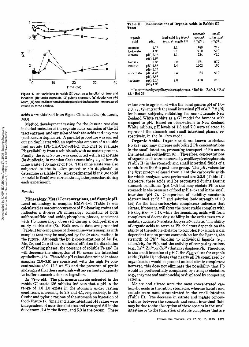
Figure 1. pH variations in rabbit GI tract as a function of time and location: (M) fundic stomach, (0) pyloric stomach, (A) duodenum, (4-1 ileum, (X) cecum. Error bars indicate standard deviation for the measured values in three rabbits.
acids were obtained from Sigma Chemical Co. (St. Louis, MO).
Method development testing for the i n vitro test also included omission of the organic acids, omission of the GI tract enzymes, and omission of both the acids and enzymes (each test in duplicate). A parallel procedure was carried out (in duplicate) with an equimolar amount of a soluble lead acetate [Pb(CH&Oz)y3HzO, 24.5 mgl to evaluate Pb availability from a soluble salt with no matrix present. Finally, the i n vitro test was conducted with lead acetate (in duplicate) in reaction flasks containing 4 g of low-Pb mine waste (100 mg/kg of Pb). This mine waste was also subjected to the in vitro procedure (in duplicate) to determine available Pb. An experimental blank (no solid material in flask) was carried through the procedure during each experiment.
Results
Mineralogy, Metal Concentrations, and Sample pH. Lead mineralogy in samples BMW-1-4 (Table I) was calculated as percent occurrence of Pb-bearing grains and indicates a diverse Pb mineralogy consisting of both sulfate/sulfide and oxide/phosphate phases, consistent with Pb mineralogy observed during a comprehensive study at this site (8). Bulk metals data are presented (Table I) for comparison of these mine-waste samples with samples that may be analyzed by the in vitro method in the future. Although the bulk concentrations of As, Fe, Mn, Zn, and Ca will have a minimal effect on the dissolution of Pb-bearing phases, the presence of soluble Fe and Ca will decrease the absorption of P b across the intestinal epithelium (16). The acidic pH values determined in these samples (2.6-3.8) are consistent with the high Fe con- centrations (5.0-12.3 wt %) and the presence of pyrite and suggest that these materials will have a limited capacity to buffer stomach acids on ingestion. In Vivo pH. The pH measurements collected in the
rabbit GI tracts (36 rabbits) indicate that a pH in the range of 1.0-1.3 exists in the stomach under fasting conditions, increasing to 2.8 and 4.1, respectively, in the fundic and pyloric regions of the stomach on ingestion of food (Figure 1). Small and large intestinal pH values were independent of nutritional status and averaged 6.9 in the duodenum, 7.4 in the ileum, and 5.9 in the cecum. These
organic acid
acetate butyrate citrate
lactate malate
lead-acid log Km: pK, ionic strength 1.0
4.7' 2.1 4.9c 2.1
pK1 3.1' 4.1
3.8' 2.0 pK1 2Ad 2.4
pK2 4.8'
stomach concd (mg/L)
189 e10 534
174 1302
small intestine"
(mg/L) 212 <lo e10
972 100
pK2 5.7d succinate DKI 4.2d 2.4 64 e10
PKZ 5Ad tartrate pK1 3.1' 2.6 <lo e10
pK2 4.8' a Determined by capillaryelectrophoresis. Ref 40. Ref 41. d Ref
42. e Ref 36.
values are in agreement with the basal gastric pH of 1.0- 2.0 (11,12) and with the small intestinal pH of 4.7-7.2 (15) for human subjects, validating the use of female New Zealand White rabbits as a GI model for humans with respect to pH. Based on observations in New Zealand White rabbits, pH levels of 1.3 and 7.0 were selected to represent the stomach and small intestinal phases, re- spectively, in the in vitro model.
Organic Acids. Organic acids are known to chelate Pb (21) and may increase solubilized Pb concentrations in the small intestine, promoting transport of Pb across the intestinal epithelium (6). Therefore, concentrations of organic acids were measured by capillary electrophoresis (Table 11) in the stomach and small intestinal fluids of a rabbit from the 6-h post-dose group. The pK, values for the first proton released from all of the carboxylic acids for which analyses were performed are 12.8 (Table 11); therefore, these acids will be protonated during fasting stomach conditions (pH 1-2) but may chelate Pb in the stomach in the presence of food (pH 4-6) and in the small intestine (pH 7). Comparison of stability constants (determined at 25 "C and solution ionic strength of 1.0 (36) for the lead carboxylate complexes) indicates that citrate, if present, will form the most stable complex with Pb (log KML = 4.11, while the remaining acids will form complexes of decreasing stability in the order tartrate > malate, succinate > acetate, butyrate > lactate. The ability of organic acids to serve as Pb chelators depends on the ability of the soluble chelator to complex Pb (which is pH dependent due to proton competition for the ligand), the strength of Pb2+ binding to individual ligands (e.g., selectivity for Pb), and the activity of competing cations (e.g., Ca2+, Zn2+, or Cu2+) that may displace Pb. Therefore, in the small intestine at pH 7, the KML values for organic acids (Table 11) indicate that nearly all Pb complexed by organic acids would be present as lead citrate complexes; however, this does not eliminate the possibility that Pb would be preferentially complexed by stronger chelators (e.g., enzymes and amino acids) or displaced by competing cations.
Malate and citrate were the most concentrated car- boxylic acids in the rabbit stomachs, whereas lactate and acetate were most concentrated in the small intestine (Table 11). The decrease in citrate and malate concen- trations between the stomach and small intestinal fluid may be due to the absorption of these species in the small intestine or to the formation of stable complexes that are
Envlron. Scl. Technol., Vol. 27, No. 13, 1993 2873
Dow
nloa
ded
by U
NIV
ID
AH
O o
n A
ugus
t 27,
200
9 | h
ttp://
pubs
.acs
.org
P
ublic
atio
n D
ate:
Dec
embe
r 1,
199
3 | d
oi: 1
0.10
21/e
s000
49a0
30
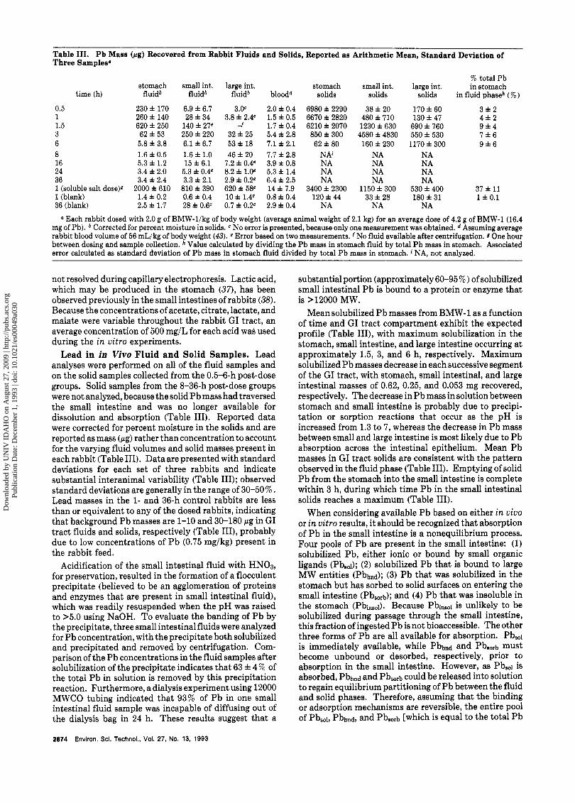
Table 111. Pb Mass (rg) Recovered from Rabbit Fluids and Solids, Reported as Arithmetic Mean, Standard Deviation of Three Samplesa
% total Pb stomach small int. large int. stomach small int. large int. in stomach
time (h) fluidb fluidb fluidb bloodd solids solids solids in fluid phaseh ( % )
0.5 1 1.5 3 6
230 f 170 6.9 f 6.7 3.OC 2.0 f 0.4 6980 f 2290 38 f 20 170 f 60 3 f 2 260 f 140 28 f 34 3.8 f 2.4e 1.5 f 0.5 6670 f 2820 480 f 710 130 f 47 4 f 2 620 f 250 140 f 27e f 1.7 f 0.4 6210 f 2070 1230 f 630 690 f 760 9 1 4 62 f 53 250 f 220 32 f 25 5.4 f 2.8 850 f 300 4580 f 4830 550 f 530 7 f 6 5.8 f 3.8 6.1 f 6.7 53 f 18 7.1 f 2.1 62 f 80 160 f 230 1170 f 300 9 f 6
8 1.6 f 0.5 1.6 f 1.0 46 f 20 7.7 f 2.8 NAi NA NA 16 5.3 f 1.2 15 f 6.1 7.2 f 0.4e 3.9 f 0.8 NA NA NA 24 3.4f 2.0 5.3 ~ ! = 0 . 4 ~ 8.2 f LOe 5.3f 1.4 NA NA NA 36 3.4 f 2.4 3.3 f 2.1 2.9f 0.2e 6.4f 2.5 NA NA NA 1 (soluble salt dose)g 2000 f 610 810 f 390 620 f 58e 14 f 7.9 3400 f 2300 1150 f 300 530 f 400 37 f 11 1 (blank) 1.4 f 0.2 0.6 f 0.4 10f 1.4e 0.8 f 0.4 120f 44 33 f 28 180 f 31 1 f 0.1 36 (blank) 2.5f 1.7 2 8 d ~ 0 . 6 ~ 0.7 f 0.2e 2.9f0.4 NA NA NA
a Each rabbit dosed with 2.0 g of BMW-l/kg of body weight (average animal weight of 2.1 kg) for an average dose of 4.2 g of BMW-1 (16.4 mg of Pb). Corrected for percent moisture in solids. No error is presented, because only one measurement was obtained. Assuming average rabbit blood volume of 56 mL/kg of body weight (43). e Error based on two measurements. f No fluid available after centrifugation. 8 One hour between dosing and sample collection. Value calculated by dividing the Pb mass in stomach fluid by total Pb mass in stomach. Associated error calculated as standard deviation of Pb mass in stomach fluid divided by total Pb mass in stomach. NA, not analyzed.
not resolved during capillary electrophoresis. Lactic acid, which may be produced in the stomach (37), has been observed previously in the small intestines of rabbits (38). Because the concentrations of acetate, citrate, lactate, and malate were variable throughout the rabbit GI tract, an average concentration of 500 mg/L for each acid was used during the i n vitro experiments.
Lead in in Vivo Fluid and Solid Samples. Lead analyses were performed on all of the fluid samples and on the solid samples collected from the 0.5-6-h post-dose groups. Solid samples from the 8-36-h post-dose groups were not analyzed, because the solid Pb mass had traversed the small intestine and was no longer available for dissolution and absorption (Table 111). Reported data were corrected for percent moisture in the solids and are reported as mass (pg) rather than concentration to account for the varying fluid volumes and solid masses present in each rabbit (Table 111). Data are presented with standard deviations for each set of three rabbits and indicate substantial interanimal variability (Table 111); observed standard deviations are generally in the range of 30-50 % . Lead masses in the 1- and 36-h control rabbits are less than or equivalent to any of the dosed rabbits, indicating that background Pb masses are 1-10 and 30-180 pg in GI tract fluids and solids, respectively (Table 1111, probably due to low concentrations of Pb (0.75 mg/kg) present in the rabbit feed.
Acidification of the small intestinal fluid with "03, for preservation, resulted in the formation of a flocculent precipitate (believed to be an agglomeration of proteins and enzymes that are present in small intestinal fluid), which was readily resuspended when the pH was raised to >5.0 using NaOH. To evaluate the banding of Pb by the precipitate, three small intestinal fluids were analyzed for Pb concentration, with the precipitate both solubilized and precipitated and removed by centrifugation. Com- parison of the Pb concentrations in the fluid samples after solubilization of the precipitate indicates that 63 f 4 % of the total Pb in solution is removed by this precipitation reaction. Furthermore, a dialysis experiment using 12000 MWCO tubing indicated that 93% of Pb in one small intestinal fluid sample was incapable of diffusing out of the dialysis bag in 24 h. These results suggest that a
substantial portion (approximately 60-95 9% ) of solubilized small intestinal Pb is bound to a protein or enzyme that is >12000 MW.
Mean solubilized Pb masses from BMW-1 as a function of time and GI tract compartment exhibit the expected profile (Table 111), with maximum solubilization in the stomach, small intestine, and large intestine occurring at approximately 1.5, 3, and 6 h, respectively. Maximum solubilized Pb masses decrease in each successive segment of the GI tract, with stomach, small intestinal, and large intestinal masses of 0.62, 0.25, and 0.053 mg recovered, respectively. The decrease in Pb mass in solution between stomach and small intestine is probably due to precipi- tation or sorption reactions that occur as the pH is increased from 1.3 to 7, whereas the decrease in Pb mass between small and large intestine is most likely due to Pb absorption across the intestinal epithelium. Mean Pb masses in GI tract solids are consistent with the pattern observed in the fluid phase (Table 111). Emptying of solid Pb from the stomach into the small intestine is complete within 3 h, during which time Pb in the small intestinal solids reaches a maximum (Table 111).
When considering available Pb based on either in vivo or i n vitro results, it should be recognized that absorption of Pb in the small intestine is a nonequilibrium process. Four pools of Pb are present in the small intestine: (1) solubilized Pb, either ionic or bound by small organic ligands (Pbsol); (2) solubilized Pb that is bound to large MW entities (Pbbnd); (3) Pb that was solubilized in the stomach but has sorbed to solid surfaces on entering the small intestine (Pb,,,b); and (4) Pb that was insoluble in the stomach (Pbinsol). Because Pbinsol is unlikely to be solubilized during passage through the small intestine, this fraction of ingested Pb is not bioaccessible. The other three forms of Pb are all available for absorption. Pbsoi is immediately available, while Pbbnd and Pbsorb must become unbound or desorbed, respectively, prior to absorption in the small intestine. However, as Pb,,i is absorbed, Pbbnd and Pbsorb could be released into solution to regain equilibrium partitioning of Pb between the fluid and solid phases. Therefore, assuming that the binding or adsorption mechanisms are reversible, the entire pool of Pbsol, Pbbnd, and Pbsorb [which is equal to the total Pb
2874 Environ. Scl. Technol., Vol. 27, No. 13, 1993
Dow
nloa
ded
by U
NIV
ID
AH
O o
n A
ugus
t 27,
200
9 | h
ttp://
pubs
.acs
.org
P
ublic
atio
n D
ate:
Dec
embe
r 1,
199
3 | d
oi: 1
0.10
21/e
s000
49a0
30
0.7 7 I
h -I 0.1 1 0 0 1 2 3 4 5
Time (hr)
Figure 2. Average Pb mass (mg) solubilized during the In vltro method development testing: (m) standard test, (+) rabbit chow present during standard test, (0) no acids present during test, (A) no enzymes present during test, (X) no enzymes or acids present during test.
in the stomach fluid phase, because surface adsorption of Pb will be negligible a t the acidic pH of the stomach (3911 is theoretically available for absorption, if the kinetics of Pb absorption across the intestinal epithelium are suffi- ciently rapid with respect to intestinal transit time and to the kinetics of Pb release or desorption. The mass of Pb present in the Pbb,,d and Pb,,bpoois and the relative release and desorption kinetics were not evaluated during this study. Consequently, the maximum mass of solubilized Pb in the stomach compared to the mass that is insoluble (e.g., the partitioning ratio from solid to fluid) represents a conservative (upper) estimate of available Pb.
Based on the above reasoning, calculation of in vivo Pb solubility from BMW-1 (mass of Pb solubilized divided by total Pb present in the stomach) shows that the solubilized Pb fraction increased from an initial 3 % to a maximum of 9% at the 1.5-h time point (Table 111). The limited Pb dissolution from mine waste is due to the low solubility of Pb-bearing minerals, kinetic dissolution limitations (IO), and encapsulation of Pb phases by inert matrices (e.g., silicates, pyrite, and jarosite) (5).
A total of 37% of the recovered Pb from Pb(0Ac)z is present in the stomach fluids after 1 h, suggesting that the balance is present in the stomach solids. It is unclear why the bulk of Pb from Pb(0Ac)z was present in the stomach solids. Blood-lead response 1 h after ingestion of Pb(0Ac)z was 9.4 times larger than from an equivalent mass of Pb contained in BMW-1, supporting maximum in vivo mine- waste-lead solubility, relative to Pb(OAc)z, of approxi- mately 10 % .
Lead in in Vitro Fluids. Reproducibility of the in vitro method was tested by conducting triplicate exper- iments using BMW-1, for which the maximum Pb mass solubilized (2-h sample) was 0.65 f 0.04 mg. The method development tests, consisting of the addition of rabbit chow, the omission of organic acids, the omission of GI tract enzymes, and the omission of both acids and enzymes (each performed in duplicate), resulted in maximum solubilized Pb masses a t the 2-h time point of 0.58 f 0.04, 0.55 f 0.01, 0.52 f 0.02, and 0.52 f 0.02 mg of Pb, respectively (Figure 2). The blank flasks were below the instrument detection limit (IDL, 0.10 mg/L) in each experiment. The method detection limits (IDL X dilution factor X volume in flask) in the mine-waste and method development flasks and the Pb(0Ac)Z flask were 0.020 and 0.60 mg, respectively.
The method development experiments indicate that the addit.ion of rabbit chow to the reaction flask causes a slight
decrease in the mass of Pb solubilized in the stomach from 0.65 f 0.04 to 0.58 f 0.04 mg, while the mass of Pb in the soluble phase during the small intestinal incubation was increasedslightlyfrom0.11 f 0.04 to0.18 f 0.03 mg (Figure 2). The reason for a decrease in stomach-solubilized Pb in the presence of rabbit chow is unknown, while the increase in intestinal solubilized Pb is most likely due to a solubilized component of rabbit chow, probably citrate, amino acids, or suspended organic matter, that is capable of binding Pb and retaining it in solution. The absence of either organic acids or GI tract enzymes, or both components together, results in a 20% decrease in max- imum stomach Pb solubility (0.65 f 0.04 to 0.52 f 0.02 mg), consistent with the observation that the presence of organic acids in solution may increase the dissolution of Pb-bearing phases (38). In addition, both acids and enzymes are necessary to retain Pb in solution during the small intestinal phase (Figure 2). These data suggest that organic acids and GI tract enzymes in the pH 7 environ- ment of the small intestine either bind Pb or inhibit the formation of Pb-bearing precipitates.
Based on the reasoning presented in the i n vivo results section, the fraction of available Pb i n vitro from BMW-1 (4 f 0.2 % ), determined by dividing the average dissolved Pb mass at 2.0 hr (0.65 mg) by the mass of Pb in the flask (15.6 mg) (Table IV), was in good agreement with the i n vivo result (9 f 4%). The comparison of in vivo and in vitro solubilized Pb masses (1.5 h for in vivo and 2.0 h for in uitro) were nearly identical: 0.62 versus 0.65 mg for the in vivo and in vitro systems, respectively (Tables 111 and IV). The overall rate of Pb dissolution in the stomach (the rate of change in solubilized Pb with time in Table I11 versus Table IV) was similar in the in vivo and in vitro systems, although the in vitro dissolution rate was more constant (less variability in the rate of Pb dissolution), indicating that dissolution kinetics are important in controlling Pb bioaccessibility from BMW-1.
The Pb(0Ac)z results indicate that, after correction for P b emanating from the blank mine waste (100 mg/kg of bulk Pb concentration resulted in 0.07 mg in the in vitro test), 68% of the Pb from Pb(0Ac)z is bioaccessible in vitro in the presence of a mine-waste matrix, while 76% is available when no matrix is present (Table IV). The discrepancy between available Pb from Pb(0Ac)z during in vitro (68 or 76 % , depending on the matrix present) and in vivo (37 % testing could be due to a variety of factors, including (1) pH differences between the systems, (2) lower concentrations of organic acids in vivo than used in vitro, or (3) incomplete recovery of ingested material during the in vivo study. Soluble Pb mass from Pb(OAc)z in the absence of mine waste decreased by a factor of 2 in the small intestinal simulation (Table IV), while in the presence of mine waste, soluble Pb decreased by a factor of 9. These results suggest that Pb absorption to the mine- waste surface may reduce Pb solubility in the small intestine.
The mine-waste samples BMW-2-4, consisting pre- dominantly of lead phosphates, manganese-lead oxides, iron-lead oxides, and iron-lead sulfates, also resulted in limited bioaccessible Pb when tested by the in vitro method (0.5-6 % , Table IV). BMW-2, which consists of ferro- manganese lead oxides and iron-lead sulfate, produced only0.5 % available Pb during the in vitro test and reached equilibrium dissolved P b concentration prior to collection of the initial sample (0.5 h, Table IV). BMW-3 and BMW-4
Environ. Sci. Technol., Vol. 27, No. 13, 1993 2875
Dow
nloa
ded
by U
NIV
ID
AH
O o
n A
ugus
t 27,
200
9 | h
ttp://
pubs
.acs
.org
P
ublic
atio
n D
ate:
Dec
embe
r 1,
199
3 | d
oi: 1
0.10
21/e
s000
49a0
30
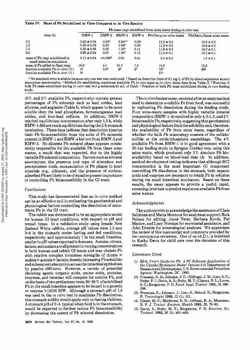
Table IV. Mass of P b Solubilized in Vitro Compared to in Vivo Results
Pb mass (mg) solubilized from mine waste during in uitro test time (h) BMW-1 BMW-2 BMW-3 BMW-4 Pb(OAc)z/no mine waste Pb(OAc)z/blank mine waste
0.5 0.43 f 0.04 0.02" 0.94"~~ 0.06n 11.0 f 0.2 10.6 f 0.2 1.0 0.50 i 0.02 0.02 Logb 0.09 11.9 f 0.2 10.4 f 0.3 1.5 0.59 f 0.06 0.02 1.30* 0.11 11.8 i 0.3 10.2 f 0.1 2.0 0.65 f 0.04 0.02 1.26* 0.15 11.4 f 0.1 10.2 f 0.1 mass of Pb (mg) solubilized in 0.11 f 0.04 <0.008d 0.03 0.04 5.3 f 0.2 1.0 f 0.1
small intestine simulation mass of Pb added to flask (mg) 15.6 4.1 23.3 7.2 15.6 15.6 fraction available Pb in uitro (%) 4d 0.5d 6d 2 d 16d 68d fraction available Pb in uiuoc ( % ) 9e 37e
" No standard error available because only one test was conducted. Based on detection limit of 0.1 mg/L of Pb by direct-aspiration atomic absorption spectrometry. c Method for establishing maximum available Pb in vivo same as in uitro, using data from Table 11. d Fraction of bulk Pb mass solubilized during in uitro test (4.0 g substrate/40 mL of fluid). e Fraction of bulk Pb mass solubilized during in uiuo feeding study.
(6% and 2 % available Pb, respectively) contain greater percentages of Pb minerals such as lead oxides, lead silicates, and anglesite (Table I), which appear to be more soluble than the lead phosphates, ferromanganese lead oxides, and iron-lead sulfates. In addition, BMW-3 reached equilibrium concentration after only 1.5 h, while BMW-4 did not reach equilibrium during the 2-h stomach incubation. These data indicate that dissolution kinetics limit Pb bioaccessibility from the suite of Pb minerals present in BMW-1 and BMW-4 but not from BMW-2 and BMW-3. No discrete Pb mineral phase appears consis- tently responsible for the available Pb from these mine wastes, a result that was expected, due to the highly variable Pb mineral composition. Factors such as mineral associations, the presence and type of alteration and precipitation rinds, encapsulation of Pb phases in inert, minerals (e.g., silicates), and the presence of surface- adsorbed Pb are likely to be of equal or greater importance in controlling Pb bioaccessibility in the GI tract.
Conclusions This study has demonstrated that an i n uitro method
can be an effective tool in evaluating the geochemical and physiological factors controlling the dissolution of mine- waste Pb in the GI tract.
The rabbit was determined to be an appropriate model for human GI tract conditions, with respect to pH and transit times. In a validation study using female New Zealand White rabbits, average pH values were 1.3 and 3-4 in the stomach under fasting and fed conditions, respectively, and approximately 7 in the small intestine, similar to pH values reported in humans. Acetate, citrate, lactate, and malate are all present in varying concentrations in both human and rabbit GI tracts and may chelate Pb with relative complex formation strengths of citrate > malate > acetate > lactate, thereby increasing Pb solubility and subsequent absorption across the intestinal epithelium by passive diffusion. However, a variety of potential chelating agents (organic acids, amino acids, proteins, enzymes, and tannins) will compete for soluble Pb, and on the basis of two preliminary tests, 60-95 ?6 of solubilized Pb in the small intestine appears to be bound to a protein or enzyme >12000 MW. Although a stomach pH of 1.3 was used in the i n uitro test to maximize Pb dissolution, this stomach acidity would apply only to fasting children. A stomach pH of 3-4, typical when food is in the stomach, would be expected to further reduce P b bioaccessibility by decreasing the extent of Pb mineral dissolution.
The in uitro beaker assay, modeled after an assay method used to determine available Fe from food, was successful in replicating Pb dissolution during the feeding study. Four mine-waste samples with highly variable mineral composition (BMW-1-4) resulted in only 4,0.5,6, and 2% bioaccessible Pb, respectively, suggesting that geochemical and physiological factors limit the solubility and, therefore, the availability of Pb from mine waste, regardless of whether the bulk Pb mineralogy consists of the sulfate/ sulfide or the oxide/phosphate assemblage. The 4% available Pb from BMW-1 is in good agreement with a 30-day feeding study in Sprague-Dawley rats, using this mine waste, which produced only 3% absolute Pb bio- availability based on blood-lead data (3). In addition, method development testing indicates that although HCl concentration is the most important GI component controlling Pb dissolution in the stomach, both organic acids and enzymes are necessary to retain Pb in solution during the small intestinal incubation. Based on these results, the assay appears to provide a useful, rapid, screening-level test to predict maximum available Pb from mine wastes.
Acknowledgments
The authors wish to acknowledge the assistance of Chris Sellstone and Maria Montour for analytical support; Rick Nelson for editing; Joyce Teter, Barbara Kyvik, Pat Madow, and Lucy Norman for secretarial assistance, and John Drexler for mineralogical analyses. We appreciate the review of this manuscript and comments provided by two anonymous reviewers. One of us (A.D.), is indebted to Kathy Davis for child care over the duration of this research.
Literature Cited EPA. User's Guide for Pb: A PC Software Application of the UptakejBiokinetic Model Version 5.0; Department of Research and Development, U.S. Environmental Protection Agency; Washington, DC, 1990. Freeman, G. B.; Johnson, J. D.; Killinger, J. M.; Liao, S. C.; Feder, P. I.; Davis, A. 0.; Ruby, M. V.; Chaney, R. L.; Lovre, S. C.; Bergstrom, P. D. Fund. Appl. Toxicol. 1992,19,388- 398. Freeman, G.; Johnson, J.; Liao, S.; Schoof, R.; Bergstrom, P. Toxicologist 1993, 13 (l), 301. Dieter, M. P.; Matthews, H. B.; Jeffcoat, R. A.; Museman, R. F. J. Toxicol, Environ. Health 1993, 39, 79-93. Davis, A,; Ruby, M. V.; Bergstrom, P. D. Environ. Sci. Technol. 1992,26 (3), 461-468.
2876 Environ. Sci. Technol., Vol. 27, No. 13, 1993
Dow
nloa
ded
by U
NIV
ID
AH
O o
n A
ugus
t 27,
200
9 | h
ttp://
pubs
.acs
.org
P
ublic
atio
n D
ate:
Dec
embe
r 1,
199
3 | d
oi: 1
0.10
21/e
s000
49a0
30
(6) Blair, J. A.; Coleman, I. L.; Hilburn, M. E. J. Physiol. 1979,
(7) Conrad, M. E.; Barton, J. C. Gastroenterology 1978, 74,
(8) Davis, A.;Drexler, J. W.; Ruby, M. V.; Nicholson, A. Environ. Sci. Technol. 1993, 27 (7 ) , 1415-1425.
(9) Vlek, P. L. G.; Blom, J. M.; Beek, J.; Lindsay, W. L. Soil Sci. SOC. Am. Proc. 1974, 38, 429.
(10) Ruby, M. V.; Davis, A. 0.; Kempton, J. H.; Drexler, J. W.; Bergstrom, P. D. Enuiron. Sci. Technol. 1992,26 (6), 1242- 1248.
(11) Malagelada, J. R.; Longstreth, G. F.; Summerskill, W. H. J.; Go, V. L. W. Gastroenterology 1976, 70, 203-210.
(12) Malagelada, J. R.; Go, V. L. W.; Summerskill, W. H. J. Dig. Dis. Sci. 1979, 24, 101-110.
(13) Hemphill, C. P.; Ruby, M. V.; Beck, B. D.; Davis, A.; Bergstrom, P. D. Chem. Speciation Bioavailability 1991,
(14) Hunt, J. N.; Spurrel, W. R. J. Physiol. 1951,113, 157-168. (15) Murthy, S. N. S.; Kostman, J.; Dinoso, V. P. Dig. Dis. Sci.
286,343-350.
731-740.
3 (3/4), 135-148.
1980,25, 289-294. (16) Chaney, R. L.; Mielke, H. W.; Sterret, S. B. Environ.
Geochem. Health 1989,11, 105-129. (17) Klotz, I. M.; Urquhart, J. M.; Fiess, H. A.J. Am. Chem. SOC.
(18) Richardt, G.; Federolf, G.; Habermann, E. Biochem. Phar-
(19) Li, N. C.; Manning, R. A. J. Am. Chem. SOC. 1955,77,5225-
(20) Quarterman, J.; Humphries, W. R.; Morrison, J. N.; Mor-
(21) Jugo, S.; Maljkovic, T.; Kostial, K. Toxicol. Appl. Phar-
(22) Narasinga Rao, B. S.; Proabhavati, T. Am. J. Clin. Nutr.
(23) Miller, D. D.; Schricker, B. R.; Rasmussen, R. R.; Van Campen, D. Am. J. Clin. Nutr. 1981,34, 2248-2256.
(24) Miller, D. D.; Schricker, B. R. In In Vitro Estimation of Food Iron Bioavailability; Kies, C., Ed.; ACS Symposium Series 203; American Chemical Society: Washington, DC,
(25) Reddy, M. B.; Chidambaram, M. V.; Fonseca, J.; Bates, G. W. Clin. Physiol. Biochem. 1986, 4, 78-86.
(26) Reddy, M. B.; Browder, E. J.; Bates, G. W. In Essential and Toxic Trace Elements in Human Health and Disease; Prasad, A., Ed.; Alan Lisa Inc.: New York, 1988.
(27) Calabrese, E. J.; Barnes, R.; Stanek, E. J., I I I ; Pastides, J.; Gilbert, C. E.; Veneman, P.; Wang, X.; Laszitity, A,; Kos-
1952, 74, 5537-5538.
macol. 1986,35 (8), 1331-1335.
5228.
rison, E. Environ. Res. 1980,23, 54-67.
macol. 1975,34, 259-263.
1978,31 , 169-175.
1982; pp 11-24.
tecki, P. T. Regul. Toxicol. Pharmacol. 1989,10,123-137. (28) Kimbrough, R. D.; Falk, H.: Stehr, P.; Fries, G. J. Toxicol.
Environ. Health 1984,14, 47-93. (29) Auvergne, A.; Bouyssou, T.; Pairet, M.; Bouillier-Oudot,
M.; Ruckebusch; Y.; Candau, M. Reprod. Nutr. Dev. 1987,
(30) Duggan, M. J.; Inskip, M. J.; Rundle, S. A,; Moorcroft, J. S. Atmos. Environ. 1985, 44, 65-79.
(31) EPA. Test Methods for Evaluating Solid Waste. Volume 1A: Laboratory Manual, PhysicallChemical Methods; Office of Solid Waste and Emergency Response, U.S. Environmental Protection Agency: Washington, DC, 1986,p 222.
(32) ASA. Methods for soil analysis. Part 2: Chemical and microbiological properties; Black, C. A., Ed.; American Society for Agronomy, Inc.: Madison, WI, 1965; pp 914.
(33) EPA. Toxic Substances Control Act, Good Laboratory Practice Standards; U.S. Environmental Protection Agency, 1989,40 CFR, Part 792.
(34) EPA. Laboratory Data Validation Functional Guidelines for Evaluating Inorganics Analyses; Hazardous Site Eval- uation Division, U.S. Environmental Protection Agency: Washington, DC, 1988, p 19.
27 (4), 755-768.
(35) Soave, 0.; Brand, C. D. Cornel1 Vet. 1991, 81, 359-364. (36) Martell, A. E.; Smith, R. M. In Critical Stability Constants;
(37) Argenzio, R. A.; Southworth, M. Am. J. Physiol. 1974,228
(38) Marty, J.; Raynaud, P. Arch. Sci. Physiol. 1966,20 (4), 515-
(39) Salim, R.; Cooksey, B. G. Plant Soil 1980,54, 399-417, (40) The Merck Index, 11th ed.; Budavari, S., Ed.; Merck and
Co., Inc.: Rahway, NJ, 1989. (41) Kemp, D. S.; Vellacio, F. In Organic Chemistry; Poulson,
S., Ed.; Worth Publishers, Inc.: New York, 1980 pp 327- 330.
(42) Streitwieser, A.; Heathcock, C. H. Inlntroduction to Organic Chemistry, 3rd ed.; Macmillan Publishing Co., New York, 1985; p 864.
Plenum Press: New York, 1977; Vol. 3.
(2), 454-460.
524.
(43) Armin, J.; Grant, R. T.; Pels, H.; Reeve, E. B. J. Physiol. 1952,116, 59-73.
Received for review May 3, 1993. Revised manuscript received August 3, 1993. Accepted August 11, 1993.'
Abstract published in Advance ACSAbstracts, October 1,1993.
Environ. Sci. Technol.. Vol. 27, No. 13, 1993 2877
Dow
nloa
ded
by U
NIV
ID
AH
O o
n A
ugus
t 27,
200
9 | h
ttp://
pubs
.acs
.org
P
ublic
atio
n D
ate:
Dec
embe
r 1,
199
3 | d
oi: 1
0.10
21/e
s000
49a0
30