Embed Size (px)
Citation preview

J. Agric. Food Chem. 1981, 29, 559-503
Development of a Radioimmunoassay for Parathion
559
Charles D. Ercegovich,' Rem0 P. Vallejo, Russell R. Gettig, Lynn Woods, Edward R. Bogus, and Ralph 0. Mumma*
Established methods in residue analysis of pesticides require extensive sample cleanup prior to quan- tification on relatively complex equipment. Radioimmunoassay (RIA) provides a simpler procedure with theoretically higher sensitivity and specificity necessitating only a minimum of sample cleanup. Parathion-specific antibodies were developed in rabbita by using a bovine serum albumin (BSA) conjugate wherein the reduced form of the pesticide was multiply bound to the protein via diazo bonds. An RIA method was developed by using either 3H- or 14C-labeled parathion as a tracer. The lower limit of detection was 4 ng of parathion in sample-free solutions and -10-20 ng in blood plasma and lettuce without any cleanup of the sample extract; this corresponds to 0.1 ppm of parathion.
Analyses of pesticides have been based mainly on con- ventional techniques such as chromatographic and color- imetric procedures. The development of immunological procedures and more specifically of radioimmunoassay (RIA) procedures presents the potential to analyze low concentrations of pesticides by combining the specificity of immunology with the sensitivity of radiochemistry. The theoretical ability of specific antibodies to recognize mi- crogram to subnanogram levels of pesticide antigens with a minimum of cleanup make RIA a method worthy of investigation (Ercegovich, 1971).
Parathion residues have been regularly quantified by gas-liquid chromatography (GLC) (Association of Official Agricultural Chemists, 19801, spectrophotometry (Asso- ciation of Official Agricultural Chemists, 1980), or po- larographic methods (Gajan, 1963). These methods involve time-consuming cleanup procedures with multiphase columns and several solvent manipulations.
Parathion was selected as an example of a pesticide for RIA development. Its aromatic nucleus, along with the relative stability of the thiophosphate moiety, is consonant with the required chemical derivatization necessary to synthesize antigenic conjugates. The continued worldwide use of parathion assures the applicability of any method developed. A preliminary report has been presented (Ercegovich et al., 1977). MATERIALS AND METHODS
Reagents and Equipment. Parathion, 98.5% purity, was obtained from American Cyanamid Co., Princeton, NJ. Bovine serum albumin, (crystallized, purified bovine al- bumin fraction V) and Freund's complete and incomplete adjuvant were purchased from Miles Laboratories, Inc., Elkhart, IN. Radioimmunoassay-grade charcoal and dextran were obtained from Schwarz/Mann, Orangeburg, NY. Handiflour liquid scintillation cocktail (Mallinckrodt, St. Louis, MO) was used for counting radioactivity.
Mass spectra were obtained on an AEI Model MS-902 mass spectrometer. Absorbances were measured on a Gilford spectrophotometer (Model 200). Radioactivity was quantified by using either a Mark 1, Nuclear Chicago, or Beckman LSsooO liquid scintillation spectrometer. NMR spectra were taken on a Varian A-60A NMR spectrometer.
Preparation of Reduced Parathion. Parathion (10.0 g, 34.4 mmol) was dissolved in 100 mL of diethyl ether and
Pesticide Research Laboratory and Graduate Study Center, Department of Entomology and Department of Chemistry, The Pennsylvania State University, University Park, Pennsylvania 16802.
'Deceased.
extracted with cold 1% Na2C03 (4 X 40 mL) to remove any phenol impurities. To the phenol-free ether solution was added 100 mL of 9:l acetic acid-concentrated HCL and 20 g of zinc powder. The yellow reaction mixture was stirred under reflux for 45 min, after which time the solids were filtered from the colorless solution and subsequently washed with CCL. The combined organic phase was then washed with 50 mL of distilled water and dried over an- hydrous sodium sulfate, and the solvent was removed under reduced pressure (aspirator). The product was identified as reduced parathion [O,O-diethyl 0 - ( p - aminophenyl) thiophosphate] on the basis of its proton NMR spectrum [(CCl,) 6 1.338 (t, 6, CH,), 3.998 (e, 2, NH2), 4.038 (q,2, CH2), 4.218 (9, 2, CH2), 6.4-7.08 (m, 4, aromatic)] and was subsequently used with purification.
Preparation of Antigen: BSA-Reduced Parathion. Reduced parathion (105 mg, 0.4 mmol) was dissolved in 50 mL of H20 containing 2 mmol of HCl. The solution was cooled in an ice bath, and a chilled 0.1 N NaNOz solution was added dropwise until a positive starch-iodide paper test was obtained. The mixture was stirred for 30 min and the excess nitrous acid was decomposed with urea. The reaction mixture was added to 500 mg of bovine serum albumin (BSA) and dissolved in 100 mL of borate buffer (pH 9), and the mixture stirred in an ice bath for 2 h. The bright orange colored reaction mixture was dialyzed against 2 changes of 4 L of distilled water daily for 5 days at 4 OC. The pH of the dialysis water was adjusted to neutrality to avoid precipitation of the conjugate. The dialyzed product was lyophilized and the orange protein conjugate stored under desiccation. The same procedure was applied in synthesizing a rabbit serum albumin (RSA) analogue of the conjugate for use in hapten specificity tests.
Characterization of the Antigen. The number of parathion residues conjugated to each BSA or RSA mol- ecule was obtained by determination of the phosphorus content of the conjugate (Chen et al., 1956). The exact location of the parathion residues was not determined, although tyrosine and lysine are the most likely sites for the diazonium linkage (Gelewitz et al., 1954).
Injection and Bleeding Schedules. New Zealand female rabbits, at least 12 weeks of age, were injected with 0.6 mg of conjugate homogenized in a mixture of 2 parts of Freund's complete adjuvant to 1 part of 0.9% saline, subcutaneously and intradermally in small aliquots in 30-40 sites on shaven areas of the back and flanks. Two weeks later, the animals were boosted with the same amount of antigen in the same manner as the initial in- jection. A second booster was likewise administered 2 weeks later. Subsequent booster injections were given every 2 weeks intravenously into the marginal ear vein by
@ 1981 American Chemical Society

560
using 2.0 mg of conjugate dissolved in 2.0 mL of saline. The rabbits were bled before immunization (control sera) and 8 day after each intravenous booster through the central ear artery.
Characterization of Antisera: Immunodiffusion. Established agar gel procedures (Ouchterlony, 1958; Campbell et at., 1970) were adapted with certain modifi- cations. Noble agar (2 g) was dissolved in 200 mL of borate saline buffer (pH 8.5) in a 500-mL beaker with heating. After the mixture was cooled to 50 "C, 4mL aliquots were pipetted into sterile 50 X 12 mm plastic Petri dishes and allowed to solidify. Agar plugs were removed by suction to form the desired pattern of wells. Undiluted antiserum (50 pL) was introduced into the central well. Solutions of BSA and BSA-reduced parathion conjugate (antigen) (50 WL each of 1 mg/mL) were pipetted into the peripheral wells. Each Petri dish was covered and placed inside a moisture chamber and stored at 4 "C. The reagents were allowed to diffuse in the agar for 24 h and excess reagents were washed out. Lines of precipitation were then exam- ined.
Characterization of Antisera: Passive Hem- agglutination Test. Tests to determine antisera spe- cificity were conducted according to standard procedures (Campbell et al., 1970) using tanned sheep red blood cells coated with the RSA analogue of the antigen. BSA and RSA were also adsorbed to tanned cells to determine an- tibody titer to these proteins. Control cells were prepared with saline instead of protein.
Preparation of Radiolabeled Parathion. Two ra- diolabeled parathion equivalents were separately employed as the tracer for the RIA procedure. The more extensively used tracer was ["Clethylparathion, 40 mCi/mmol, pur- chased from Amersham/Searle Corp. The other tracer was ring-labeled [3H]parathion, 300 mCi/mmol, synthesized by refluxing [3H]nitrophenol (Amersham/Searle Corp.) with 14 mg of diethylthiophosphoryl chloride (K & K, ICN Laboratories, Plainview, NY) in 10 mL of 2-butanone containing 10 mg of Na2C03 for 24 h. The resultant [3H]parathion was purified by preparative TLC on silica gel using benzene as the solvent and characterized by its mass spectrum. The tracer was characterized for specific activity by quantifying an amount on GLC and measuring the radioactivity associated with that amount by liquid scintillation counting.
Radioimmunoassay Procedures. Assays were per- formed in triplicate in 10 X 75 mm disposable glass tubes by sequential addition of 50 pL of sample or unlabeled standard diluted in PBS (pH 7.2), 200 pL of antiserum, and 650 pL of PBS (pH 7.2). The assay mixture was agitated in a vortex mixer and incubated for 1 h. After this initial incubation to allow free parathion to react with the antibodies, 100 p L of the radiolabeled tracer ( - 10 OOO dpm) in ethanol was added to the tubes. The contents were again mixed and then incubated for another 3 h. Separation of the bound and nonbound radioactivity was accomplished by the addition of 500 pL of a stock mixture of dextran-coated charcoal (DCC). The latter was pre- pared by mixing 10 g of RIA-grade charcoal and 250 mg of dextran in 100 mL of PBS (pH 7.2) with constant stirring during the procedure to prevent settling. The reaction mixture was agitated in a vortex mixer, incubated at 25 "C for 15 min, and then centrifugated at 1500g for 10 min. The supernatant, containing antibody-bound pesticide, was transferred by pipet to liquid scintillation vials for radioassay.
Standard curves were obtained by plotting BIB, X 100 (percent bound) vs. the amount of nonradiolabeled para-
J. Agric. Food Chem., VoI. 29, No. 3, 1981 Ercegovich et al.
thion present on logit-log paper. B represents the dpm bound by antiserum in tubes containing parathion inhib- itor (nonradiolabeled), and Bo represents the dpm bound by antiserum in tubes containing no parathion inhibitor. This procedure usually yields a linear standard curve.
Determination of Association Constants (K,) and Binding Capacities. Association constants were deter- mined by a modification of an established equilibrium dialysis method (Nisonoff and Pressman, 1958). Instead of examinination of the antigen-antibody association in equilibrium dialyses of a fixed amount of antiserum and varying concentrations of free hapten, the radioimmu- noassay procedure was employed. The amount of an- tiserum was kept constant and the concentrations of [3H]parathion were varied. Separation of bound radio- labeled parathion from free parathion was accomplished by the DCC method. After determination of the amounts of bound parathion, the same calculations described by Nisonoff and Pressman were applied to determine K, and binding capacities.
The optimal titer of the antiserum for use in competitive binding assays was determined by incubating a constant amount of [14C]parathion in serial dilutions of the an- tiserum. A dilution curve was derived by plotting the antiserum dilution against percent bound. The dilution of antiserum which bound 50% of total radioactivity was chosen as the operational dilution for the RIA procedure.
Antisera Specificity. The ability of antisera to dis- criminate between structurally related chemicals was tested by incubating a fixed amount of antiserum and tracer with varying amounts of test chemicals. The quantity of test chemical that caused a 50% reduction in the binding of tracer was extrapolated from curves derived by plotting the percent of radioactivity bound vs. con- centrations of test chemical.
Sample Preparation for Assay. Sera from coagulated human blood samples were collected by centrifugation at 2500 rpm for 10 min at room temperature. The sera ob- tained were fortified with the desired amount of non- radiolabeled parathion. The range of parathion concen- trations used was between 1 and lo00 mg/mL. Serial dilutions were made of each sample and 50-mL aliquots of these dilutions were used in assay procedures.
Crude tissue extracts of apple, carrots, celery, corn, cabbage, lettuce, endive, and parsley were obtained from first rinsing the plant samples with distilled water and then macerating the tissue in methanol in a blender. Each extract was filtered, dried over anhydrous sodium sulfate, concentrated in vacuo (aspirator), and then taken up in dimethyl sulfoxide (MGO) to an equivalent of 2 g of fresh plant tissue/mL. These extracts were fortified with var- ious amounts of parathion. Aliquots (50 pL) of the plant extract preparation were used per assay tube. RESULTS AND DISCUSSION Synthesis. A small nonimmunogenic molecule must
usually be conjugated to a large immunogenic molecule such as a protein to raise antibodies specific against the small molecule (hapten). The small molecule must possess a functionality by which it m be covalently bonded to the protein carrier. The reduction of the nitro group in par- athion to yield a reactive amino function allows the sub- sequent covalent linkage to BSA via a diazonium con- densation with appropriate amino acids in the chain. Microphosphorus determinations of the conjugate revealed 22 f 2 reduced-parathion residues per molecule of BSA.
A rabbit serum albumin conjugate of reduced parathion for use in testing antisera specificity was similarly s p - thesized to yield 20 f 2 residues per RSA molecule.

DeveloOmenl of a Radicimmunoassay fw Parathion
Table I. P d v e Hemarrdutination Test for Anti-BSA-Reduced Parathion Antiserum'
J. Agrk. FOodChem., Vol. 29, No. 3, I981 581
agglutination reaction antiserum RSA-reduced parathion RSA Wl tkNm agglutination reaction, dilution (0.25 me/mL) (0.24 ma/mL) dilution BSA (0.25 mg/mL)
100 +++ 200 +++ 400 800
1600 3 200 6 400 12 800 25600 51 200
+++ ++ ++ + * - - -
- 1000 +++ - 2 000 +++
4 000 8 000 16 000 32000 64 000 128 000 256 000 512 000
++t ++ ++ ++ ++ + f -
a (+)represents activity; (-) represents no activity.
Table 11. Inhibition of Pasaive Hemagglutination Reaction of Antioarathion Antiseruma
Figure 1. Doublediffusion pattern of the antiserum (S) against bovine serum albumin (A and B) and bovine serum albumin reduced parathion conjugate (C and D). h represents the haptenic spur.
Immunodiffusion. Antibodies specific to an antigen comprise a heterogeneous population differing in the region of recognition and in binding affinity. When serum con- taining these antibodies and the antigen are allowed to diffuse radially toward each other in a gel matrix, they will form a visible percipitin band at their optimal relative concentration or equivalence point (Ouchterlony, 1958). Precipitin bands (Figure 1) were observed from the reac- tion of the antiserum with BSA (wells A and B). Other bands were produced by the reaction between the antise- rum and the conjugate diffusing from wells C and D. Additional reaction between the antiserum and the con- jugate resulting in precipitin spurs projecting from all comers of the common fronts is attributed to the presence of antibodies directed against the haptenic moiety, para- thion. This confirms the presence of antibodies specific to the parathion hapten in addition to those specific to the carrier protein.
Passive Hemagglutination. The presence of antibo- dies specific to an antiserum can be confirmed when sera containing these antibodies cause the agglutination of red blood cells onto whose surface antigen had been absorbed. Progressive dilutions of the antiserum and the degree of agglutination they cause can be used to measure titer (Table I). In like manner, the titer of antibodies against the carrier molecule, BSA, can be determined. The titer, expressed as the reciprocal value of the highest dilution of antiserum that effected detectable agglutination, was 6400 against RSA-parathion and 256000 against BSA. No cross-reaction with RSA was evident at all antiserum di- lutions.
agglutination reaction free parathion with RSA-reduced concn, ng/mL parathion (0.25 mg/mL)
250 125 82.5 31.3 15.6 7.8 3.9 2.0 1.0
(+)represents activity;(-) represents no activity.
10 2 0 40 6080100 160
I/ANTISERA DILUTION
Figure 2. Antisera dilution curve8 for antisera prepared from three separate rabbits.
The specificity of antibodies to parathion was treated by hemagglutination inhibition. This demonstrated that free parathion inhibited agglutination associated with the reaction of antiserum and RSA-conjugated parathion ab- sorbed onto red blood cells (Table 11).
Determination of Association Constants (IC.) and Binding Capacity. Sera from various bleeding8 were tested according to the procedure described to determine association constants and binding capacities. The best antisera displayed association constants in binding ['HI- parathion of -1.5 X lo7 L/mol and binding capacities in the region of 2.0 X lo-' mol/L.
From the dilution curve (Figure 21, a working solution of 100 p L of antiserum in 100 pL of PBS (pH 7.2) was used in the RIA procedure.
Specificity. The reliability of any competitive binding assay depends on the specificity of the antibodies. The ability of the antiserum to discriminate between parathion and similarly strudured substances is evident in Table 111.

182 J. Agric. Food Chem., Vol. 29, No. 3, 1981
9 90- 3 0
8 5 0 -
o 0 -1
m
’z
Table 111. Nanograms of Pesticidal Materials Causing 50% Inhibition of Parathion Binding to Antiserum
Ercegovich et al.
I 1 “ ’ I
parathion 50 malathion 70000 paraoxon 1850 guthion 25 000 reduced parathion 275 imidan 20 000 p-nitrophenol > 10 000 diazinon > 10 000 methylparathion 15 000 carbaryl >10 000 sumithion > 10 000 1-naphthol > 10 000 sumioxon > 10 000
4 f O 2f0 5:O Ib ;O i 0 IAO 260 5;)O ’ PARATHION ( n g )
Figure 3. Amount of parathion (nanograms) vs. logit percent bound parathion by the antibody (standard curve for pure par- athion).
Only reduced parathion showed a significant cross-re- activity with the antiserum tested. Surprisingly reduced parathion’s inhibition of the antiserum was 5 times less than that of parathion even though the antiserum was generated against the BSA-reduced parathion conjugate. Paraoxon exhibited cross-reactivity, but to a much lesser degree.
Standard Curves. The applicability of the assay to the analysis of blood plasma and plant tissues was tested by determining inhibition levels by samples fortified with known amounts of parathion. The standard curves for these fortified samples can then be compared to a standard curve derived from parathion standard solutions free of sample material. The standard curve using sample-free solutions is presented in Figure 3. The standard curves for fortified human plasma (Figure 4) and lettuce tissues (Figure 5 ) both show the ability of antisera to detect as little as 10-20 ng of parathion, as compared to a lower limit of -4 ng for sample-free solutions. The decreased level of sensitivity with lettuce and blood plasma samples is attributable to the ability of the sample material to effect a small degree of nonspecific inhibition of antibodies. Other plant tissues gave similar results.
This radioimmunoassay developed for parathion dem- onstrates the applicability of immunoassays in pesticide analysis. The ability of the assay to detect 10-20 ng of parathion in crude samples is encouraging. The specificity of the antibody is shown in that only reduced parathion, the precursor to conjugation, exhibits significant cross- reactivity.
The titer and avidity of the antisera presented in this study is of moderate quality. Further improvements in sensitivity may be accomplished with conjugates employing different bridging groups. Other measures such as in- creasing the specific activity of the radiolabeled tracer and by using other isotopes may also be investigated. Mono- clonal generation of antibody also offers potential.
The possibilities of raising specific antibodies against pesticides such as DDT, the metabolite DDA, and mala-
10 2 0 30 40 60 SO 100 150200 300 PARATHION (ng)
Figure 4. Amount of parathion (nanograms) in human plasma v8. logit percent bound parathion by the antibody (standard curve for parathion in human plasma).
t- o s
I , 10 20 50 100 200 500 1000
PARATHION (ng)
Figure 5. Amount of parathion (nanograms) in lettuce vs. logit percent bound parathion by the antibody (standard curve for parathion in lettuce).
thion (Centeno et al., 1970; Haas and Guardia, 1968), some cyclodienes (Langone and Van Vunakis, 1975), and S- bioallethrin (Wing et al., 1978) point to increased devel- opment and application of immunochemical methods to pesticides analyses.
ACKNOWLEDGMENT This paper is dedicated in memory of the late Dr.
Charles D. Ercegovich, the original advisor for the work involved in this project.
LITERATURE CITED kssociation of Official Agricultural Chemists “Official Methods
of Analysis”, 13th ed.; AOAC: Washington, DC, 1980; pp 29.039-29.054, 29.160.
Campbell, D. H.; Garvey, J. J.; Cremer, N. E.; Sussdorf, D. H. “Methods in Immunology”, 2nd ed.; W. A. Benjamin: New York, 1970; pp 279-282.
Centeno, E. R.; Johnson, W. J.; Shehon, A. H. Znt. Arch. Allergy Appl. Zmmunol. 1970,37, 1-13.
Chen, P. S., Jr.; Toribara, T. Y.; Warner, H. Anal. Chem. 1956,
Ercegovich, C. D. Adu. Chem. Ser. 1971, No. 104, 162. Ercegovich, C. D.; Gettis, R. R.; Vallejo, R. P. “Abstracts of
Papers”, 174th National Meeting of the American Chemical Society, Chicago, IL, Aug 1977; American Chemical Society: Washington, DC, 1977; PEST 2.
Biophys. 1954,53,411-424.
28,1756-1758.
Gajan, R. E. J. Assoc. Off. Anal. Chem. 1963,45, 216-219. Gelewitz, E. W.; Riedeman, W. L.; Koltz, I. M. Arch. Biochem.
Haas, J. H.; Guardia, E. J. Proc. SOC. Exp. Biol. Med. 1968,129, 546-551.
J. Agric. Food Chem. 1981, 29, 563-567 503
Langone, J. J.; Van Vunakis, H. Res. Commun. Chem. Pathol.
Nisonoff, A.; Pressman, D. J. Immunol. 1958,80,417-428. Ouchterlony, 0. B o g . Allergy 1958, 5, 1-78. Wing, K. D.; Hammock, D. B.; Wustner, D. A. J. Agric. Food
Chem. 1978,26,1328-1333. Pharmacol. 1975,10,163-171.
Received for review August 25,1980. Accepted December 30,1980. Authorized for publication 88 paper no. 6062 in the Journal Series of the Pennsylvania Agricultural Experiment Station.
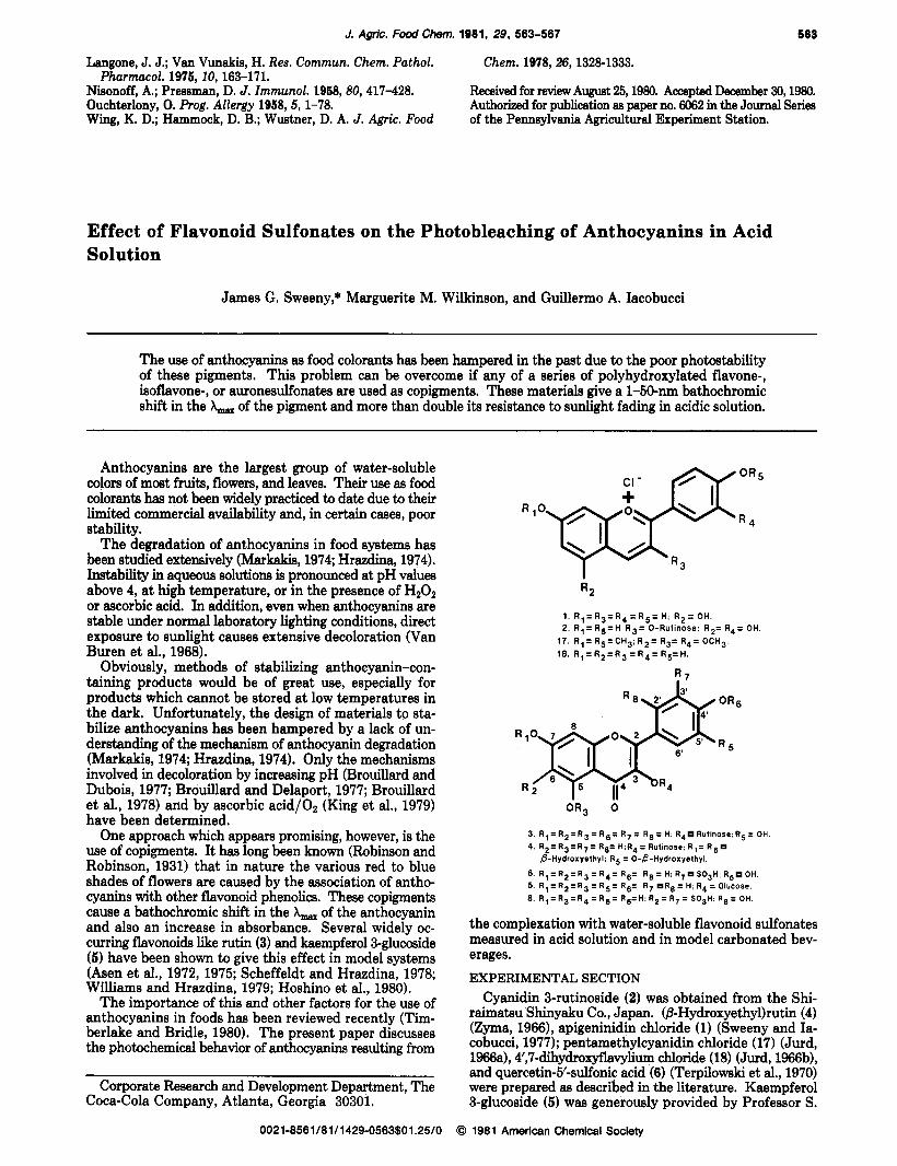
Effect of Flavonoid Sulfonates on the Photobleaching of Anthocyanins in Acid Solution
James G. Sweeny,* Marguerite M. Wilkinson, and Guillermo A, Iacobucci
The u8e of anthocyanins as food colorants has been hampered in the past due to the poor photostability of these pigments. This problem can be overcome if any of a series of polyhydroxylated flavone-, isoflavone-, or auronesulfonates are used as copigments. These materials give a 1-50-nm bathochromic shift in the A, of the pigment and more than double its resistance to sunlight fading in acidic solution.
Anthocyanins are the largest group of water-soluble colors of most fruits, flowers, and leaves. Their use as food colorants has not been widely practiced to date due to their limited commercial availability and, in certain cases, poor stability.
The degradation of anthocyanins in food systems has been studied extensively (Markakis, 1974; Hrazdina, 1974). Instability in aqueous solutions is pronounced at pH valuea above 4, a t high temperature, or in the presence of HzOz or ascorbic acid. In addition, even when anthocyanins are stable under normal laboratory lighting conditions, direct exposure to sunlight causes extensive decoloration (Van Buren et al., 1968).
Obviously, methods of stabilizing anthocyanin-con- taining producte would be of great use, especially for products which cannot be stored at low temperatures in the dark. Unfortunately, the design of materials to sta- bilize anthocyanins has been hampered by a lack of un- derstanding of the mechanism of anthocyanin degradation (Markakis, 1974; Hrazdina, 1974). Only the mechanisms involved in decoloration by increasing pH (Brouillard and Dubois, 1977; Brouillard and Delaport, 1977; Brouillard et al., 1978) and by ascorbic acid/Oz (King et al., 1979) have been determined.
One approach which appears promising, however, is the use of copigments. It has long been known (Robinson and Robinson, 1931) that in nature the various red to blue shades of flowers are caused by the association of antho- cyanins with other flavonoid phenolics. These copigments cause a bathochromic shift in the A, of the anthocyanin and also an increase in absorbance. Several widely oc- curring flavonoids like rutin (3) and kaempferol3-glucoside (5) have been shown to give this effect in model systems (Asen et al., 1972, 1975; Scheffeldt and Hrazdina, 1978; Williams and Hrazdina, 1979; Hoshino et al., 1980).
The importance of this and other factors for the use of anthocyanins in foods has been reviewed recently (Tim- berlake and Bridle, 1980). The present paper discusses the photochemical behavior of anthocyanins resulting from
Corporate Research and Development Department, The Coca-Cola Company, Atlanta, Georgia 30301.
1. R 1 = R 3 = R 4 = R 5 = H ; R 2 = O H . 2 . R l = R 5 = H ; R 3 = 0-Rutinose: R 2 = R 4 = OH.
17. R , = R S = C H 3 : R 2 = R 3 = R 4 = O C H 3 . 18. R 1 = R 2 = R 3 = R 4 = R 5 = H .
ORg 0
3 R 1 = R z = R 3 = R e = R 7 = R 8 = H. R 4 Rutinose.R5 = OH 4 R z = R 3 = R 7 = Re= H.R4 = Rutmore R ,= R 6
6 R , = R 2 = R 3 = R 4 = R6= R e = H. R7 S03H R 5 OH 5 R , = R 2 = R 3 = R 5 = R6= R, R e = H, R4 = Glucose 8 R l = R 3 = R 4 = R 5 = R6=H, R z = R 7 = S03H. R e = OH
8-Hydroxyethyl. R 5 = 0-8-Hydroxyethyl
the complexation with water-soluble flavonoid sulfonates measured in acid solution and in model carbonated bev- erages. EXPERIMENTAL SECTION
Cyanidin 3-rutinoside (2) was obtained from the Shi- raimatsu Shinyaku Co., Japan. (BHydroxyethy1)rutin (4) (Zyma, 1966), apigeninidin chloride (1) (Sweeny and Ia- cobucci, 1977); pentamethylcyanidin chloride (17) (Jurd, 1966a), 4’,7-dihydroxyflavylium chloride (18) (Jurd, 1966b), and quercetin-5’-sulfonic acid (6) (Terpilowski et al., 1970) were prepared as described in the literature. Kaempferol 3-glucoside (5) was generously provided by Professor S.
0021-8561/81/1429-0563$01.25/0 0 1981 American Chemical Society


![cAMP [125-I] Radioimmunoassay kit (Adenosine 3',5' cyclic ...€¦ · The basic principle of radioimmunoassay is the competition between a radioactive and a non-radioactive antigen](https://img.pdfslide.us/doc/110x75/60abd8dd139110551d199fa1/camp-125-i-radioimmunoassay-kit-adenosine-35-cyclic-the-basic-principle.jpg)
![Radioimmunoassay for a ,-Fetoprotein in the Serum of Rats1 · A radioimmunoassay for the detection of a ]-fetoprotein (AFP) in the sera of rats is described. The procedure is based](https://img.pdfslide.us/doc/110x75/5fc02c65ba3767624f46d2d7/radioimmunoassay-for-a-fetoprotein-in-the-serum-of-rats1-a-radioimmunoassay-for.jpg)