Embed Size (px)
Citation preview

G
V
DTa
PQ1
PRa
b
c
Ad
e
ARR1AA
KSRVFA
1
hbif1bt
0h
1
2
3
4
5
6
7
8
9
10
11
12
13
14
15
16
17
18
19
20
21
22
23
24
25
26
27
28
29
30
31
32
33
34
35
36
ARTICLE IN PRESS Model
IRMET 12317 1–7
Journal of Virological Methods xxx (2013) xxx– xxx
Contents lists available at ScienceDirect
Journal of Virological Methods
jou rn al hom ep age: www.elsev ier .com/ locate / jv i romet
evelopment and validation of a real-time two-step RT-qPCRaqMan® assay for quantitation of Sacbrood virus (SBV) and itspplication to a field survey of symptomatic honey bee colonies
hilippe Blancharda,∗, Sylvain Guillota, Karina Antùnezb, Hemma Köglbergerc,er Krygerd, Joachim R. de Mirandae, Stéphanie Francoa, Marie-Pierre Chauzata,ichard Thiérya, Magali Ribièrea
Anses, Sophia-Antipolis Laboratory, Bee Diseases Unit, BP 111, 06902 Sophia Antipolis, FranceLaboratorio de Microbiologia, Instituto de Investigaciones Biologicas Clemente Estable, Avenida Italia 3318, CP 11600 Montevideo, UruguayAGES, Institut für Saat- und Pflanzgut, Pflanzenschutzdienst und Bienen, Abteilung für Bienenkunde und Bienenschutz, Spargelfeldstrasse 191, 1220 Wien,ustriaUniversity of Aarhus, Faculty of Agricultural Sciences, Department of Integrated Pest Management, Forsogsvej 1, DK-4200 Slagelse, DenmarkDepartment of Ecology, Swedish University of Agricultural Sciences, 750-07 Uppsala, Sweden
rticle history:eceived 5 February 2013eceived in revised form3 September 2013ccepted 24 September 2013vailable online xxx
eywords:acbrood virus (SBV)eal-time RT-PCRalidationield surveypis mellifera
a b s t r a c t
Sacbrood virus (SBV) is the causal agent of a disease of honey bee larvae, resulting in failure to pupate andcausing death. The typical clinical symptom of SBV is an accumulation of SBV-rich fluid in swollen sub-cuticular pouches, forming the characteristic fluid-filled sac that gives its name to the disease. Outbreaksof the disease have been reported in different countries, affecting the development of the brood andcausing losses in honey bee colonies. Today, few data are available on the SBV viral load in the case ofovert disease in larvae, or for the behavioural changes of SBV-infected adult bees. A two-step real-timeRT-PCR assay, based on TaqMan® technology using a fluorescent probe (FAM-TAMRA) was thereforedeveloped to quantify Sacbrood virus in larvae, pupae and adult bees from symptomatic apiaries. Thisassay was first validated according to the recent XP-U47-600 standard issued by the French StandardsInstitute, where the reliability and the repeatability of the results and the performance of the assay wereconfirmed. The performance of the qPCR assay was validated over the 6 log range of the standard curve (i.e.from 102 to 108 copies per well) with a measurement uncertainty evaluated at 0.11 log10. The detection
and quantitation limits were established respectively at 50 copies and 100 copies of SBV genome, for atemplate volume of 5 �l of cDNA. The RT-qPCR assay was applied during a French SBV outbreak in 2012where larvae with typical SBV signs were collected, along with individuals without clinical signs. The SBVquantitation revealed that, in symptomatic larvae, the virus load was significantly higher than in sampleswithout clinical signs. Combining quantitation with clinical data, a threshold of SBV viral load related toan overt disease was proposed (1010 SBV genome copies per individual).37
38
39
40
41
42
43
. Introduction
Sacbrood is an infectious disease affecting the larvae of theoney bee causing characteristic clinical symptoms in developingroods. Larvae with sacbrood fail to pupate, and ecdysial fluid rich
n Sacbrood virus (SBV) accumulates beneath their unshed skin,orming the sac after which the disease is named (Bailey et al.,
Please cite this article in press as: Blanchard, P., et al., Development anquantitation of Sacbrood virus (SBV) and its application to a field survhttp://dx.doi.org/10.1016/j.jviromet.2013.09.012
964; Ball and Bailey, 1997). Sacbrood virus was the first honeyee virus to be identified (White, 1917) and the first for whichhe genome organisation and the complete nucleotide sequence
∗ Corresponding author. Tel.: +33 0492 948 769; fax: +33 0492 943 701.E-mail address: [email protected] (P. Blanchard).
166-0934/$ – see front matter © 2013 Published by Elsevier B.V.ttp://dx.doi.org/10.1016/j.jviromet.2013.09.012
44
45
46
© 2013 Published by Elsevier B.V.
were determined (Ghosh et al., 1999). It is a positive-sense single-stranded-RNA virus of the Picornavirales order. It belongs to theIflavirus genus, from the recently established family Iflaviridae (Kinget al., 2011).
SBV is widely distributed. It attacks Apis mellifera coloniesin every continent (Allen and Ball, 1996). Generally it has nomajor consequences for colony survival. However, sacbrood is amajor cause of colony mortality for Apis cerana colonies in Asia(Liu et al., 2010). The prevalence of this virus has been investi-gated in various countries, such as Great Britain (Bailey, 1967),
d validation of a real-time two-step RT-qPCR TaqMan® assay forey of symptomatic honey bee colonies. J. Virol. Methods (2013),
Australia (Dall, 1985), Austria, Germany, India, Nepal and SouthAfrica (Grabensteiner et al., 2001), France (Tentcheva et al., 2004),the USA (Chen et al., 2004), Uruguay (Antunez et al., 2006), Denmark(Nielsen et al., 2008) and Spain (Kukielka and Sanchez-Vizcaino,
47
48
49
50

ING Model
V
2 rologic
2taiagoGaa1he2r(Ses
tSFvai
R(gtebTfilTdcs
2
2
tv7dccisp4nFSSrw
wM
Q2
51
52
53
54
55
56
57
58
59
60
61
62
63
64
65
66
67
68
69
70
71
72
73
74
75
76
77
78
79
80
81
82
83
84
85
86
87
88
89
90
91
92
93
94
95
96
97
98
99
100
101
102
103
104
105
106
107
108
109
110
111
112
113
114
115
116
117
118
119
120
121
122
123
124
125
126
127
128
129
130
131
132
133
134
135
136
137
138
139
140
141
142
143
144
145
146
147
148
149
150
151
152
153
154
155
156
157
158
159
160
161
162
163
164
165
166
167
168
169
170
ARTICLEIRMET 12317 1–7
P. Blanchard et al. / Journal of Vi
009). Although it is primarily a disease of larvae, SBV also mul-iplies in pupae and adult bees where it provokes physiologicalnd behavioural changes that limit its transmission to larvae. Thisncludes an accelerated progression from brood tending to for-ging (Bailey and Ball, 1991), a degeneration of hypopharyngeallands (Du and Zhang, 1985), and a strong aversion to eatingr collecting pollen (Bailey and Fernando, 1972; Anderson andaicon, 1992). The behaviour of drones, which never eat pollen,ppears unaffected by SBV infection (Ball and Bailey, 1997). Someuthors have reported overt disease in brood (Allen and Ball,996; Grabensteiner et al., 2001; Nielsen et al., 2008), while othersave reported SBV detection without clinical symptoms (Tentchevat al., 2004; Antunez et al., 2006; Kukielka and Sanchez-Vizcaino,009). Although real-time RT-PCR detection methods have beenecently developed for the detection and the quantitation of SBVChantawannakul et al., 2006; Gauthier et al., 2007; Kukielka andanchez-Vizcaino, 2009; Locke et al., 2012; Yoo et al., 2012; Evisont al., 2012), few data are available on the SBV viral titres of larvaeamples presenting typical symptoms of SBV.
In this paper, a new real-time two-step RT-PCR based onhe TaqMan® technology to quantify SBV was described. ThisBV RT-qPCR was then validated according to the XP U47-600rench standard (as already described for chronic bee paralysisirus (CBPV) by Blanchard et al. (2012)). Finally, this method wasssessed on field samples collected from an outbreak of sacbroodn France in 2012.
The development of this quantitative assay for SBV genomicNA was performed using TaqMan® PCR with minor groove binderMGB) probe technology. A DNA probe with a conjugated MGBroup forms an extremely stable duplex with single-stranded DNAargets, allowing the use of shorter probes. The shorter probe lengthndows MGB probes with greater sensitivity and lower fluorescentackground compared to non-MGB probes (Kutyavin et al., 2000).he SBV RT-qPCR assay was validated for (i) the analytical speci-city, (ii) the PCR detection limit (DLPCR), (iii) the PCR quantitation
imit (QLPCR) and (iv) the linearity and efficiency of the qPCR assay.he method was then applied to assess the SBV genomic load inifferent categories of samples (larvae with or without typical SBVlinical symptoms, pupae and adult bees) coming from apiariesuffering from sacbrood.
. Materials and methods
.1. Real-time quantitative RT-PCR method
The SBV method development was based on the TaqMan®
wo-step real-time quantitative RT-PCR CBPV assay developed pre-iously by Blanchard et al. (2012) using the Applied Biosystems500 System (Applera). The SBV-specific primers and probe wereesigned using the Primer Express 3.0 software (Applera). Thehoice of primers and probe was constrained by absolute sequenceonservation across all the SBV sequences deposited in GenBank,ncluding the Thai, Chinese and Korean variants (Fig. 1). The cho-en primer set amplifies a 70 bp fragment located in the N-terminalart of the polyprotein region of the SBV genome: nucleotides34–503 according to the reference sequence (GenBank accessiono. AF092924, Ghosh et al., 1999). The forward primer is SBV-434: 5′-AACGTCCACTACACCGAAATGTC and the reverse primer isBV-R503: 5′-ACACTGCGCGTCTAACATTCC. The TaqMan® probe isBV-P460: 5′-TGATGAGAGTGGACGAAGA, labelled with the fluo-escent reporter dye FAM (6-carboxyfluorescein) at the 5′ end and
Please cite this article in press as: Blanchard, P., et al., Development anquantitation of Sacbrood virus (SBV) and its application to a field survhttp://dx.doi.org/10.1016/j.jviromet.2013.09.012
ith the non-fluorescent quencher (NFQ) with MGB at the 3′ end.The PCR is performed in duplicate in a MicroAmp optical 96-
ell reaction plate, containing 1× Taqman® Universal PCR Masterix with uracil-N-glycosylase (UNG – Applied Biosystems), 320 nM
PRESSal Methods xxx (2013) xxx– xxx
of each primer (SBV-F434 and SBV-R503), 200 nM of the qSBVprobe, 1× Exo IPC Mix and 1× Exo IPC DNA from IPC VICTM
Probe (Applied Biosystems). UNG reduces false-positive results bydestroying contaminating, dUTP-incorporated PCR products fromprevious runs. The Exo-IPC reagents are a pre-optimized blendof an Exogenous Internal Positive Control (IPC) DNA templateplus corresponding primers and VIC-based probe, designed to dis-tinguish true-negative results from false-negative results due toPCR inhibition. The VIC-based signal of the IPC amplification isdetected separately from the FAM-based signal of the SBV amplifi-cation. The reaction volume was completed with 5 �l of template,either 102–108 copies of the SBV plasmid DNA standard (describedbelow) or random-hexamer primed cDNAs, obtained as previouslydescribed in Blanchard et al. (2007), giving a total volume of 25 �l.The thermal cycling conditions are 2 min at 50 ◦C (active tempera-ture for UNG degradation of contaminating PCR-products), 10 minat 95 ◦C (activation of AmpliTaq Gold DNA Polymerase and inactiva-tion of UNG), followed by 40 cycles of denaturation at 95 ◦C for 15 sand annealing/extension at 60 ◦C for 1 min. Results are expressed asthe mean of the two replicates of each reaction. A biological positivecontrol is included in each run, obtained from homogenate of symp-tomatic larvae. Two negative controls are also included in each run:a blank RNA extraction negative control and a template-free qPCRnegative control, in which the sample is replaced by purified water.
2.2. Analytical specificity
Analytical specificity was assessed as described by Blanchardet al. (2012). The in silico analysis was performed by a Blast searchon the genome database with the 70-bp sequence of the SBV ampli-con. The experimental specificity was evaluated by exclusivity andinclusivity tests, testing the assay’s ability not to amplify any RNAthat is not SBV (exclusivity) and to positively detect all strains of SBV(inclusivity). The exclusivity test was performed on cDNAs obtainedfrom bee samples infected with other bee viruses, such as ABPV,CBPV, IAPV, BQCV or DWV. The inclusivity test was carried outon several SBV isolates from various geographic regions (Austria,Denmark, France, Sweden and Uruguay), coming from apiarieswith or without clinical symptoms and thus constituting a panelof samples that best represents SBV genetic diversity. The geneticdiversity among these isolates was assessed by a phylogenetic anal-ysis of the primer-free sequences of a 469 bp amplicon located inthe structural protein region, encompassing the 70 bp fragmentof the SBV assay, amplified by conventional PCR using primersSBV-1f and SBV-2r described by Grabensteiner et al. (2001). Thesequences were aligned to homologous SBV sequences obtainedfrom GenBank (Table 1) using CLUSTAL X (Thompson et al., 1997).Phylogenetic trees were constructed using the neighbour-joining(NJ), maximum likehood (ML) and maximum parsimony (MP)methods as implemented by the Phylowin program (Galtier et al.,1996). Statistical support for the branching pattern was provided bybootstrap analysis involving 500 replicates. Branches with less than50% bootstrap were omitted. The phylogenetic trees were drawnusing TreeView (Page, 1996).
2.3. Experimental scheme to assess the detection andquantitation limits of the SBV qPCR assay
The detection and quantitation limits of the SBV qPCR (DLPCRand QLPCR) were determined from a 4.44 kb plasmid obtained bycloning the 469 bp PCR fragment amplified from the polyproteinregion of SBV with primers SBV-1f and SBV-2r (Grabensteiner
d validation of a real-time two-step RT-qPCR TaqMan® assay forey of symptomatic honey bee colonies. J. Virol. Methods (2013),
et al., 2001) into a pCR® II TOPO® vector (Invitrogen). The plas-mid DNA concentration was determined by spectrophotometry(three trials on three independent dilutions) and converted to plas-mid copies per microliter. From this, stock solutions containing
171
172
173
174

ARTICLE IN PRESSG Model
VIRMET 12317 1–7
P. Blanchard et al. / Journal of Virological Methods xxx (2013) xxx– xxx 3
F t (Graq ervati
0sefmcqtfT
TO
175
176
177
178
179
180
181
182
183
184
185
186
187
188
189
190
ig. 1. Representation of the SBV genome with the location of the 429 bp fragmenPCR assay described in this paper and the extremely high degree of sequence cons
.2 × 102–0.2 × 10⁸ copies per �l were prepared through 10-folderial dilutions in TE buffer. These stock solutions were used tostablish the standard calibration curves for absolute quantitation,or calculating the assay performance parameters, and for esti-
ating the assay detection limits. The experimental scheme wasonducted as described by Blanchard et al. (2012). To determine the
Please cite this article in press as: Blanchard, P., et al., Development anquantitation of Sacbrood virus (SBV) and its application to a field survhttp://dx.doi.org/10.1016/j.jviromet.2013.09.012
PCR detection limit, three independent trials were performed onhree independent two-fold serial dilutions (200–6.25 DNA copiesor 5 �l of template), with eight replicates of each dilution level.he DLPCR was determined by the smallest number of nucleic
able 1rigin of the studied SBV isolates used for the phylogenetic analysis: isolate reference, sa
Isolatea Sample Origin
D12-2024/S1859 Larvae France
D12-2024/S1861 Larvae France
20100203/24 Larvae France
20100020/1 Larvae France
155/1030/1253 Bees Austria
1006 Bees Austria
1003 Bees Austria
KA 2 cDNAs Uruguay
JR6 cDNAs Sweden
BOX cDNAs Denmark
12024 Larvae/Pupae Denmark
Rothamstead Larvae United KinUK Larvae United KinAustria Larvae Austria
Germany 1 Larvae Germany
Germany 2 Larvae Germany
Germany 8 Larvae Germany
Sophia R80 Bees France
T73/05A Bees Denmark
India Larvae India
Nepal 1 Larvae Nepal
Nepal 4 Larvae Nepal
China Larvae China
CSBV/LN Larvae China
Korean Bees Korea
4 Bees Korea
11 Bees Korea
13 Bees Korea
a Isolate reference given by our colleagues.
bensteiner et al., 2001), the location of the 70 bp amplicon obtained with the SBVon for this assay, across all SBV genotypes.
acid targets given 95% of positive results. To assess the linearitydomain and the quantitation limit, three independent trials wereperformed on three independent 10-fold serial dilutions. Standardcurves were generated by linear regression analysis of the thresh-old cycle (CT) measured for each amplification vs. the log10 copynumber for each standard dilution. The measured quantity for each
d validation of a real-time two-step RT-qPCR TaqMan® assay forey of symptomatic honey bee colonies. J. Virol. Methods (2013),
dilution level was determined in retrospect by using the formula:log x = [CT − b]/a, where a is the slope, b is the Y-intercept, and xis the quantity. The obtained values were compared to the theo-retical quantities and provided, for each load level, the mean bias
mple type, country of origin, sequence label and GenBank Accession number.
Label GenBank accession no.
FRA 2 KC513752FRA 3 KC513753FRA 4 KC513754FRA 5 KC513755AUT 2 KC513758AUT 3 KC513759AUT 4 KC513760URY 1 KC513751SWE 1 KC513750DNK 1 KC513756DNK 2 KC513757
gdom GBR 1 AF092924gdom GBR 2 AF284616
AUT 1 AF284617DEU 1 AF284618DEU 2 AF284619DEU 3 AF284625FRA 1 AY152712DNK 3 EF570887IND 1 AF284626NPL 1 AF284627NPL 2 AF284629CHN 1 AF469603CHN 2 HM237361KOR 1 HQ322114KOR 2 HQ916827KOR 3 HQ916833KOR 4 HQ916834
191
192
193
194

ARTICLE IN PRESSG Model
VIRMET 12317 1–7
4 P. Blanchard et al. / Journal of Virological Methods xxx (2013) xxx– xxx
0.01
KOR3
KOR1
KOR2
KOR465
73
GE1
GE2
GE3
FR2
FR367
AU1
AU2
FR4
FR560
FR1
SW2
DK2
SW162
75
AU3
DK1
75
UR1
UK1
UK299
83
85
IND1
CH1
CH2
NEP1
NEP2
62
100URY1
75 DNK2
AUT4
Korean genotype
Asian genotype
European and South-American genotype
AUT4
DK3
DEU1
DEU2
DEU3
FRA2
FRA3
FRA2
FRA3
AUT1
AUT2
FRA4
FRA5
AUT2
FRA4
FRA5
FRA1
SWE1
DNK3
SWE1
DNK2
DNK1
AUT3AUT3
URY1
GBR2
GBR1
CHN1
CHN2
NPL1
NPL2
DNK1
F egmenr x (FRAn es <50
(mfwmTl
eh
195
196
197
198
199
200
201
202
203
204
205
206
207
208
209
ig. 2. Maximum likehood phylogenetic tree of SBV sequences, based on a 429 bp seference sequence AF092924). New sequences obtained in this study are in grey boode represents the bootstrap values as the result of 500 replicates. Bootstrap valu
mb). The standard deviation of the obtained values (SD) was deter-ined, and the uncertainty of the linearity was calculated using the
ormula ULINi = 2[√
SD2 + mb2]. The combined linearity uncertaintyas defined for the entire calibration range and given by the for-ula ULIN = |√�ULINi
2/k| where k is the number of dilution levels.he quantitation limit of the assay is then determined by the first
Please cite this article in press as: Blanchard, P., et al., Development anquantitation of Sacbrood virus (SBV) and its application to a field survhttp://dx.doi.org/10.1016/j.jviromet.2013.09.012
evel load of the calibration range.To evaluate the performance of the entire method (RNA
xtraction to qPCR analysis), the positive control, obtained byomogenisation of symptomatic larvae, was followed by means of
t of the N-terminal part of the SBV polyprotein (nucleotides 241–669 according to2, FRA3, FRA4, FRA5, AUT2, AUT3, URY1, SWE2, DNK1, DNK2). The number of each
% were omitted. The scale corresponds to the number of substitution per site.
a control chart. In order to be accepted, the quantitative result hadto be within the acceptability limits. These limits were defined bythe mean of the first ten values ± twice the standard deviation.
2.4. Application of the SBV RT-qPCR method on bee samples fromhoney bee colonies with SBV clinical symptoms
d validation of a real-time two-step RT-qPCR TaqMan® assay forey of symptomatic honey bee colonies. J. Virol. Methods (2013),
In 2012, clinical symptoms of SBV were observed in Frenchapiaries by the “Brigade Nationale d’Enquêtes Vétérinaires etPhytosanitaires” (BNEVP). Four apiaries located in three French
210
211
212

IN PRESSG Model
V
rological Methods xxx (2013) xxx– xxx 5
dAScp1hSft
2
wKs
3
3
7Dsii
AactaspFUtogwcp
3
tm8n5q
3l
w4peei
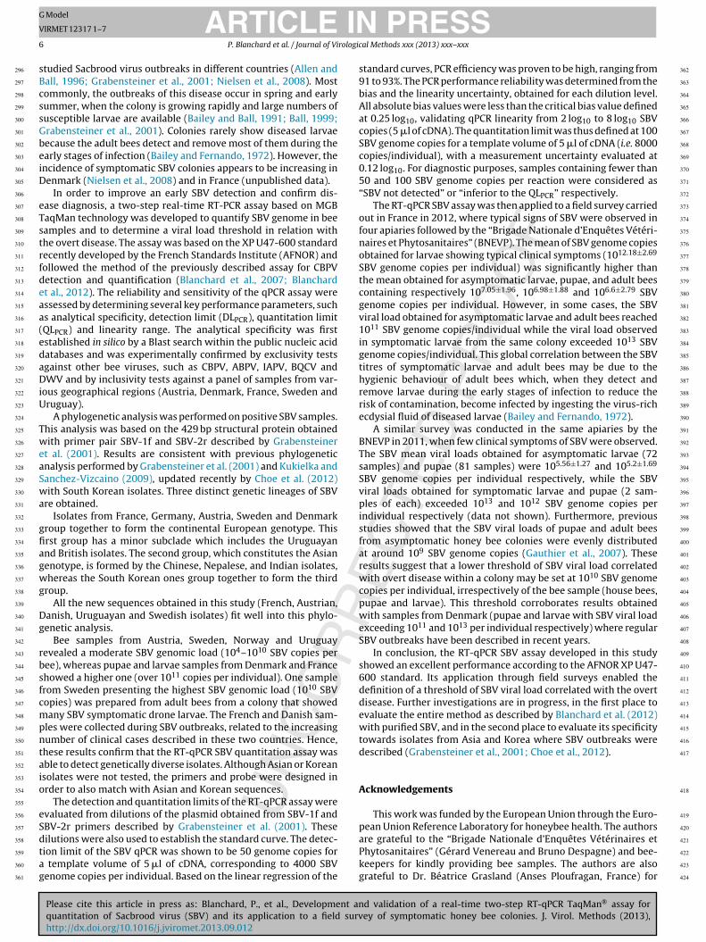
Fig. 3. Performance of linear regression for the SBV qPCR. Mean bias (mb) was deter-mined for each load level, bars represent the linearity uncertainty (ULINi) given by theformula: 2[
√SD2 + (mean bias)2] where SD is the standard deviation of the measured
values.
Fig. 4. Distribution of SBV viral load mean assessed by RT-qPCR assay in various cat-egories of samples from symptomatic hives: larvae with typical signs of SBV, larvae,
213
214
215
216
217
218
219
220
221
222
223
224
225
226
227
228
229
230
231
232
233
234
235
236
237
238
239
240
241
242
243
244
245
246
247
248
249
250
251
252
253
254
255
256
257
258
259
260
261
262
263
264
265
266
267
268
269
270
271
272
273
274
275
276
277
278
279
280
281
282
283
284
285
286
287
288
289
290
291
ARTICLEIRMET 12317 1–7
P. Blanchard et al. / Journal of Vi
epartments (Loiret, Lot et Garonne and Vendée) were visited inpril 2012. Three to five hives were sampled from each apiary.amples of bee larvae showing typical SBV clinical symptoms wereollected (12 samples of 2–5 larvae per hive), together with sam-les of asymptomatic larvae from the same hives (12 samples of5 larvae per hive), samples of pupae (12 samples of 10 pupae perive) and samples of in-house bees (12 samples of 10 bees per hive).ample processing, RNA extraction and cDNA synthesis were per-ormed as described in Blanchard et al. (2007). Five microliters ofhe cDNA were used as template for the qPCR.
.5. Statistical analysis
The SBV genomic loads obtained from the bee samplesere analysed by the Two-sample Kolmogorov–Smirnov and theruskal–Wallis non-parametric tests using the SYSTAT 9 computeroftware package (SPSS Inc.).
. Results
.1. Analytical specificity
In order to assess the specificity of the SBV assay, the chosen0 bp sequence was searched in silico by a Blast analysis of theNA public database. Only SBV sequences were identified by this
earch. Experimentally non significant amplification was detectedn cDNAs obtained from ABPV, CBPV, IAPV, BQCV and DWV, whichmplies an exclusive specificity.
The inclusivity test was carried out on different isolates fromustria, France, Denmark, Sweden and Uruguay. A phylogeneticnalysis was realised with isolates that gave a positive result inonventional PCR. SBV sequences from GenBank were included inhis study. All the methods used for the inclusivity test (NJ, MLnd MP) gave similar profiles. Results using the ML method arehown in Fig. 2. This analysis (realised on sequences of the SBVolyprotein region) revealed that all the European isolates (Austria,rance, Denmark and Sweden) are included in a group close to theK and Uruguay genotypes, whereas the Asian and Korean geno-
ypes are included in an another group (Fig. 2). Afterwards, samplesf bees, larvae, pupae or cDNA were tested by SBV qPCR. The SBVenomic loads observed in adult bees and cDNAs were moderate,ith 105–107 SBV copies per bee and 104–107 SBV copies per �l of
DNA. On the other hand, SBV genomic loads observed in larvae andupae samples were much higher, over 1011 copies per individual.
.2. Determination of the detection limit of the SBV qPCR assay
The detection limit of the SBV qPCR assay was determined usinghree independent 6-step, 2-fold dilutions series of the 4.44 kb plas-
id ranging from 200 to 6.25 DNA copies in 5 �l of template, with replicate reactions for each dilution series. The lowest number ofucleic acid targets detected in at least 95% of the 24 replicates was0 DNA copies (23 positive replicates). Therefore, the DLPCR of SBVPCR was determined as 50 genome copies per reaction.
.3. Determination of the linearity range and the quantitationimit of the SBV qPCR assay
The linearity range and quantitation limit of the SBV qPCR assayas studied using three independent 10-fold dilutions series of the
.44 kb plasmid ranging from 108 to 102 DNA copies in 5 �l of tem-
Please cite this article in press as: Blanchard, P., et al., Development anquantitation of Sacbrood virus (SBV) and its application to a field survhttp://dx.doi.org/10.1016/j.jviromet.2013.09.012
late. The results obtained for the three trials showed high PCRfficiency, ranging from 91 to 93%. The measured amount of SBV forach series and each dilution level was compared to the theoret-cally expected amounts, to calculate the individual absolute bias.
pupae and bees without clinical signs. The results are expressed as the mean SBVgenome copies per individual. Bars represent the standard deviations. (**Significantdifference at p < 0.01 within the various categories of samples).
Then, the mean bias, its standard deviation, and the linearity uncer-tainty were determined. As shown in Fig. 3, at each dilution level,the absolute bias value was less than the critical bias value (set at0.25 log10) validating the linearity over the entire calibration range.The combined linear uncertainty (ULIN) of the SBV qPCR was deter-mined to be 0.12 log10 within the range from 2 log10 to 8 log10 SBVcopies per reaction. These results validate the calibration range andprovide a quantitation limit of 100 SBV genome copies per reaction.
3.4. SBV quantitation in honey bee samples from symptomaticcolonies
Fig. 4 shows the mean SBV genomic loads obtained forSBV-symptomatic larvae, asymptomatic larvae, pupae, and adultbees from symptomatic colonies in four French apiaries. TheSBV genomic loads were 2.9 × 10⁷–2.1 × 10¹⁴ SBV copies persymptomatic larvae, 7 × 10⁴–3.3 × 10¹¹ SBV copies per asymp-tomatic larvae, 2.3 × 10⁴–1.1 × 10¹⁰ SBV copies per pupae and2.2 × 103–3.4 × 10¹¹ SBV copies per adult bee. The mean of theSBV genomic load observed in SBV-symptomatic larvae was signif-icantly higher than the mean of the genomic load observed in theother samples (p = 0.004). No significant difference in SBV genomicload was observed between the other sample types.
4. Discussion
Sacbrood virus is widely distributed throughout the world
d validation of a real-time two-step RT-qPCR TaqMan® assay forey of symptomatic honey bee colonies. J. Virol. Methods (2013),
and is considered to be one of the most common bee virusinfections. Unlike most of the other bee viruses, Sacbrood virusmay be reliably diagnosed by the distinctive and specific symp-toms of infection in larvae (Ball, 1996). Several previous works
292
293
294
295
ING Model
V
6 rologic
sBcssGbeiD
eTstrfdeaa(edaDiU
TweaSwa
gfiagwg
Dg
rbsfcmpntaio
eSdtag
296
297
298
299
300
301
302
303
304
305
306
307
308
309
310
311
312
313
314
315
316
317
318
319
320
321
322
323
324
325
326
327
328
329
330
331
332
333
334
335
336
337
338
339
340
341
342
343
344
345
346
347
348
349
350
351
352
353
354
355
356
357
358
359
360
361
362
363
364
365
366
367
368
369
370
371
372
373
374
375
376
377
378
379
380
381
382
383
384
385
386
387
388
389
390
391
392
393
394
395
396
397
398
399
400
401
402
403
404
405
406
407
408
409
410
411
412
413
414
415
416
417
418
419
420
ARTICLEIRMET 12317 1–7
P. Blanchard et al. / Journal of Vi
tudied Sacbrood virus outbreaks in different countries (Allen andall, 1996; Grabensteiner et al., 2001; Nielsen et al., 2008). Mostommonly, the outbreaks of this disease occur in spring and earlyummer, when the colony is growing rapidly and large numbers ofusceptible larvae are available (Bailey and Ball, 1991; Ball, 1999;rabensteiner et al., 2001). Colonies rarely show diseased larvaeecause the adult bees detect and remove most of them during thearly stages of infection (Bailey and Fernando, 1972). However, thencidence of symptomatic SBV colonies appears to be increasing inenmark (Nielsen et al., 2008) and in France (unpublished data).
In order to improve an early SBV detection and confirm dis-ase diagnosis, a two-step real-time RT-PCR assay based on MGBaqMan technology was developed to quantify SBV genome in beeamples and to determine a viral load threshold in relation withhe overt disease. The assay was based on the XP U47-600 standardecently developed by the French Standards Institute (AFNOR) andollowed the method of the previously described assay for CBPVetection and quantification (Blanchard et al., 2007; Blanchardt al., 2012). The reliability and sensitivity of the qPCR assay weressessed by determining several key performance parameters, suchs analytical specificity, detection limit (DLPCR), quantitation limitQLPCR) and linearity range. The analytical specificity was firststablished in silico by a Blast search within the public nucleic acidatabases and was experimentally confirmed by exclusivity testsgainst other bee viruses, such as CBPV, ABPV, IAPV, BQCV andWV and by inclusivity tests against a panel of samples from var-
ous geographical regions (Austria, Denmark, France, Sweden andruguay).
A phylogenetic analysis was performed on positive SBV samples.his analysis was based on the 429 bp structural protein obtainedith primer pair SBV-1f and SBV-2r described by Grabensteiner
t al. (2001). Results are consistent with previous phylogeneticnalysis performed by Grabensteiner et al. (2001) and Kukielka andanchez-Vizcaino (2009), updated recently by Choe et al. (2012)ith South Korean isolates. Three distinct genetic lineages of SBV
re obtained.Isolates from France, Germany, Austria, Sweden and Denmark
roup together to form the continental European genotype. Thisrst group has a minor subclade which includes the Uruguayannd British isolates. The second group, which constitutes the Asianenotype, is formed by the Chinese, Nepalese, and Indian isolates,hereas the South Korean ones group together to form the third
roup.All the new sequences obtained in this study (French, Austrian,
anish, Uruguayan and Swedish isolates) fit well into this phylo-enetic analysis.
Bee samples from Austria, Sweden, Norway and Uruguayevealed a moderate SBV genomic load (104–1010 SBV copies peree), whereas pupae and larvae samples from Denmark and Francehowed a higher one (over 1011 copies per individual). One samplerom Sweden presenting the highest SBV genomic load (1010 SBVopies) was prepared from adult bees from a colony that showedany SBV symptomatic drone larvae. The French and Danish sam-
les were collected during SBV outbreaks, related to the increasingumber of clinical cases described in these two countries. Hence,hese results confirm that the RT-qPCR SBV quantitation assay wasble to detect genetically diverse isolates. Although Asian or Koreansolates were not tested, the primers and probe were designed inrder to also match with Asian and Korean sequences.
The detection and quantitation limits of the RT-qPCR assay werevaluated from dilutions of the plasmid obtained from SBV-1f andBV-2r primers described by Grabensteiner et al. (2001). These
Please cite this article in press as: Blanchard, P., et al., Development anquantitation of Sacbrood virus (SBV) and its application to a field survhttp://dx.doi.org/10.1016/j.jviromet.2013.09.012
ilutions were also used to establish the standard curve. The detec-ion limit of the SBV qPCR was shown to be 50 genome copies for
template volume of 5 �l of cDNA, corresponding to 4000 SBVenome copies per individual. Based on the linear regression of the
PRESSal Methods xxx (2013) xxx– xxx
standard curves, PCR efficiency was proven to be high, ranging from91 to 93%. The PCR performance reliability was determined from thebias and the linearity uncertainty, obtained for each dilution level.All absolute bias values were less than the critical bias value definedat 0.25 log10, validating qPCR linearity from 2 log10 to 8 log10 SBVcopies (5 �l of cDNA). The quantitation limit was thus defined at 100SBV genome copies for a template volume of 5 �l of cDNA (i.e. 8000copies/individual), with a measurement uncertainty evaluated at0.12 log10. For diagnostic purposes, samples containing fewer than50 and 100 SBV genome copies per reaction were considered as“SBV not detected” or “inferior to the QLPCR” respectively.
The RT-qPCR SBV assay was then applied to a field survey carriedout in France in 2012, where typical signs of SBV were observed infour apiaries followed by the “Brigade Nationale d’Enquêtes Vétéri-naires et Phytosanitaires” (BNEVP). The mean of SBV genome copiesobtained for larvae showing typical clinical symptoms (1012.18±2.69
SBV genome copies per individual) was significantly higher thanthe mean obtained for asymptomatic larvae, pupae, and adult beescontaining respectively 107.05±1.96, 106.98±1.88 and 106.6±2.79 SBVgenome copies per individual. However, in some cases, the SBVviral load obtained for asymptomatic larvae and adult bees reached1011 SBV genome copies/individual while the viral load observedin symptomatic larvae from the same colony exceeded 1013 SBVgenome copies/individual. This global correlation between the SBVtitres of symptomatic larvae and adult bees may be due to thehygienic behaviour of adult bees which, when they detect andremove larvae during the early stages of infection to reduce therisk of contamination, become infected by ingesting the virus-richecdysial fluid of diseased larvae (Bailey and Fernando, 1972).
A similar survey was conducted in the same apiaries by theBNEVP in 2011, when few clinical symptoms of SBV were observed.The SBV mean viral loads obtained for asymptomatic larvae (72samples) and pupae (81 samples) were 105.56±1.27 and 105.2±1.69
SBV genome copies per individual respectively, while the SBVviral loads obtained for symptomatic larvae and pupae (2 sam-ples of each) exceeded 1013 and 1012 SBV genome copies perindividual respectively (data not shown). Furthermore, previousstudies showed that the SBV viral loads of pupae and adult beesfrom asymptomatic honey bee colonies were evenly distributedat around 109 SBV genome copies (Gauthier et al., 2007). Theseresults suggest that a lower threshold of SBV viral load correlatedwith overt disease within a colony may be set at 1010 SBV genomecopies per individual, irrespectively of the bee sample (house bees,pupae and larvae). This threshold corroborates results obtainedwith samples from Denmark (pupae and larvae with SBV viral loadexceeding 1011 and 1013 per individual respectively) where regularSBV outbreaks have been described in recent years.
In conclusion, the RT-qPCR SBV assay developed in this studyshowed an excellent performance according to the AFNOR XP U47-600 standard. Its application through field surveys enabled thedefinition of a threshold of SBV viral load correlated with the overtdisease. Further investigations are in progress, in the first place toevaluate the entire method as described by Blanchard et al. (2012)with purified SBV, and in the second place to evaluate its specificitytowards isolates from Asia and Korea where SBV outbreaks weredescribed (Grabensteiner et al., 2001; Choe et al., 2012).
Acknowledgements
This work was funded by the European Union through the Euro-pean Union Reference Laboratory for honeybee health. The authors
d validation of a real-time two-step RT-qPCR TaqMan® assay forey of symptomatic honey bee colonies. J. Virol. Methods (2013),
are grateful to the “Brigade Nationale d’Enquêtes Vétérinaires etPhytosanitaires” (Gérard Venereau and Bruno Despagne) and bee-keepers for kindly providing bee samples. The authors are alsograteful to Dr. Béatrice Grasland (Anses Ploufragan, France) for
421
422
423
424

ING Model
V
rologic
st
R
A
A
A
B
B
B
B
BB
B
B
B
C
C
C
D
D
ment aided by quality analysis tools. Nucl. Acids Res. 25, 4876–4882.
425
426
427
428
429
430
431
432
433
434
435
436
437
438
439
440
441
442
443
444
445
446
447
448
449
450
451
452
453
454
455
456
457
458
459
460
461
462
463
464
465
466
467
468
469
470
471
472
473
474
475
476
477
478
479
480
481
482
483
484
485
486
487
488
489
490
491
492
493
494
495
496
497
498
499
500
501
502
503
504
505
506
507
508
509
510
511
ARTICLEIRMET 12317 1–7
P. Blanchard et al. / Journal of Vi
tatistical analysis. The help of Mrs. Cristina Gastaldi in improvinghe English of the manuscript is also gratefully acknowledged.
eferences
llen, M., Ball, B.V., 1996. The incidence and world distribution of honey bee viruses.Bee World 77, 141–162.
nderson, D.L., Gaicon, H., 1992. Reduced pollen collection by honey bee(Hymenoptera: Apidae) colonies infected with Nosema apis and sacbrood virus.J. Econ. Entomol. 85, 47–51.
ntunez, K., D’Alessandro, B., Corbella, E., Ramallo, G., Zunino, P., 2006. Honeybeeviruses in Uruguay. J. Invertebr. Pathol. 93, 67–70.
ailey, L., 1967. The incidence of virus diseases in the honey bee. Ann. Appl. Biol. 60,43–48.
ailey, L., Fernando, E.F.W., 1972. Effects of sacbrood virus on adult honey-bees. Ann.Appl. Biol. 72, 27–35.
ailey, L., Gibbs, A.J., Woods, R.D., 1964. Sacbrood virus of the larval honey bee (Apismellifera Linneaus). Virology 23, 425–429.
ailey, L., Ball, B.V., 1991. Honey Bee Pathology, 2nd ed. Academic Press, London, pp.193.
all, B.V., 1996. Honey bee viruses: a cause for concern? Bee World 77, 117–119.all, B.V., 1999. Sacbrood. In: Colin, M.E., Ball, B.V., Kilani, M. (Eds.), Bee Disease
Diagnosis, Options Mediterannéennes. , pp. 91–96.all, B.V., Bailey, L., 1997. Viruses. In: Morse, R.A., Flottum, K. (Eds.), Honey Bee Pests,
Predators, & Diseases. A.I. Root Company, Medina, pp. 11–32.lanchard, P., Ribiere, M., Celle, O., Lallemand, P., Schurr, F., Olivier, V., Iscache, A.L.,
Faucon, J.P., 2007. Evaluation of a real-time two-step RT-PCR assay for quanti-tation of Chronic bee paralysis virus (CBPV) genome in experimentally-infectedbee tissues and in life stages of a symptomatic colony. J. Virol. Methods 141,7–13.
lanchard, P., Regnault, J., Schurr, F., Dubois, E., Ribiere, M., 2012. Intra-laboratoryvalidation of chronic bee paralysis virus quantitation using an accredited stan-dardised real-time quantitative RT-PCR method. J. Virol. Methods 180, 26–31.
hantawannakul, P., Ward, L., Boonham, N., Brown, M., 2006. A scientific note onthe detection of honeybee viruses using real-time PCR (TaqMan) in Varroa mitescollected from a Thai honeybee (Apis mellifera) apiary. J. Invertebr. Pathol. 91,69–73.
hen, Y., Zhao, Y., Hammond, J., Hsu, H.T., Evans, J., Feldlaufer, M., 2004. Multiplevirus infections in the honey bee and genome divergence of honey bee viruses.J. Invertebr. Pathol. 87, 84–93.
hoe, S.E., Nguyen, T.T.D., Hyun, B.H., Noh, J.H., Lee, H.S., Lee, C.H., Kang, S.W., 2012.Genetic and phylogenetic analysis of South Korean sacbrood virus isolates from
Please cite this article in press as: Blanchard, P., et al., Development anquantitation of Sacbrood virus (SBV) and its application to a field survhttp://dx.doi.org/10.1016/j.jviromet.2013.09.012
infected honey bees (Apis cerana). Vet. Microbiol. 157, 32–40.all, D.J., 1985. Inapparent infection of honey bee pupae by Kashmir and sacbrood
bee viruses in Australia. Ann. Appl. Biol. 106, 461–468.u, Z.-L., Zhang, Z.-B., 1985. Ultrastructural change in the hypopharyngeal glands of
worker bees (Apis cerana) infected with sacbrood virus. Zool. Res. 6, 155–162.
PRESSal Methods xxx (2013) xxx– xxx 7
Evison, S.E., Roberts, K.E., Laurenson, L., Pietravalle, S., Hui, J., Biesmeijer, J.C., Smith,J.E., Budge, G., Hughes, W.O., 2012. Pervasiveness of parasites in pollinators. PLoSONE 7 (1), e30641.
Galtier, N., Gouy, M., Gautier, C., 1996. SeaView and Phylo win, two graphic toolsfor sequence alignment and molecular phylogeny. Comput. Appl. Biosci. 12,543–548.
Gauthier, L., Tentcheva, D., Tournaire, M., Dainat, B., Cousserans, F., Colin, M.E.,Bergoin, M., 2007. Viral load estimation in asymptomatic honey bee coloniesusing the quantitative RT-PCR technique. Apidologie 38, 426–435.
Ghosh, R.C., Ball, B.V., Willcocks, M.M., Carter, M.J., 1999. The nucleotide sequenceof sacbrood virus of the honey bee: an insect picorna-like virus. J. Gen. Virol. 80,1541–1549.
Grabensteiner, E., Ritter, W., Carter, M.J., Davison, S., Pechhacker, H., Kolodziejek, J.,Boecking, O., Derakhshifar, I., Moosbeckhofer, R., Licek, E., Nowotny, N., 2001.Sacbrood virus of the honeybee (Apis mellifera): rapid identification and phylo-genetic analysis using reverse transcription-PCR. Clin. Diagn. Lab. Immunol. 8,93–104.
King, A.M.Q., Adams, M.J., Carstens, E.B., Lefkowitz, E.J., 2011. Virus Taxonomy:Classification and Nomenclature of Viruses: Ninth Report of the InternationalCommittee on Taxonomy of Viruses. Elsevier Academic Press, San Diego.
Kukielka, D., Sanchez-Vizcaino, J., 2009. One-step real-time quantitative PCR assaysfor the detection and field study of Sacbrood honeybee and Acute bee paralysisviruses. J. Virol. Methods 161, 240–246.
Kutyavin, I.V., Afonina, I.A., Mills, A., Gorn, V.V., Lukhtanov, E.A., Belousov, E.S., Singer,M.J., Walburger, D.K., Lokhov, S.G., Gall, A.A., Dempcy, R., Reed, M.W., Meyer,R.B., Hedgpeth, J., 2000. 3′-Minor groove binder-DNA probes increase sequencespecificity at PCR extension temperatures. Nucl. Acids Res. 28 (2), 655–661.
Liu, X., Zhang, Y., Yan, X., Han, R., 2010. Prevention of Chinese sacbrood virus infectionin Apis cerana using RNA interference. Curr. Microbiol. 61, 422–428.
Locke, B., Forsgren, E., Fries, I., de Miranda, J.R., 2012. Acaricide treatment affectsviral dynamics in Varroa destructor-infested honey bee colonies via both hostphysiology and mite control. Appl. Environ. Microbiol. 78, 227–235.
Nielsen, S.L., Nicolaisen, M., Kryger, P., 2008. Incidence of acute bee paralysis virus,black queen cell virus, chronic bee paralysis virus, deformed wing virus, Kash-mir bee virus and sacbrood virus in honey bees (Apis mellifera) in Denmark.Apidologie 39, 310–314.
Tentcheva, D., Gauthier, L., Zappulla, N., Dainat, B., Cousserans, F., Colin, M.E., Bergoin,M., 2004. Prevalence and seasonal variations of six bee viruses in Apis melliferaL. and Varroa destructor mite populations in France. Appl. Environ. Microbiol.70, 7185–7191.
Thompson, J.D., Gibson, T.J., Plewniak, F., Jeanmougin, F., Higgins, D.G., 1997. TheCLUSTAL X windows interface: flexible strategies for multiple sequence align-
d validation of a real-time two-step RT-qPCR TaqMan® assay forey of symptomatic honey bee colonies. J. Virol. Methods (2013),
White, G.F., 1917. Sacbrood. US Department of Agriculture Bulletin, pp. 431.Yoo, M.S., Thi, K.C.N., Van Nguyen, P., Han, S.H., Kwon, S.H., Yoon, B.S., 2012. Rapid
detection of sacbrood virus in honeybee using ultra-rapid real-time polymerasechain reaction. J. Virol. Methods 179, 195–200.
512
513
514
515