Embed Size (px)
Citation preview

See discussions, stats, and author profiles for this publication at: https://www.researchgate.net/publication/342335686
Significance of pro-angiogenic estrogen metabolites in normal follicular
development and follicular growth arrest in polycystic ovary syndrome
Article in Human Reproduction · June 2020
DOI: 10.1093/humrep/deaa098
CITATIONS
0READS
47
8 authors, including:
Some of the authors of this publication are also working on these related projects:
Modulation of TGFbeta signalling by hCG in human endometrial stromal cells: implications in embryo implantation and early placenta development View project
Study on the effect of DM and its moment of onset over maternal and fetal ovarian function. View project
Paulina Kohen
University of Chile
56 PUBLICATIONS 928 CITATIONS
SEE PROFILE
Claudio Villarroel
University of Chile
33 PUBLICATIONS 458 CITATIONS
SEE PROFILE
Alex Muñoz
University of Chile
18 PUBLICATIONS 415 CITATIONS
SEE PROFILE
Luigi Devoto
University of Chile
137 PUBLICATIONS 8,197 CITATIONS
SEE PROFILE
All content following this page was uploaded by Paulina Kohen on 05 July 2020.
The user has requested enhancement of the downloaded file.

Significance of pro-angiogenic estro-gen metabolites in normal folliculardevelopment and follicular growtharrest in polycystic ovary syndromeSoledad Henrıquez1,2, Paulina Kohen1, Xia Xu3,Claudio Villarroel1,4,Alex Mu~noz1,4, Ana Godoy1, Jerome F. Strauss III5, andLuigi Devoto1,4,*1Institute for Maternal and Child Research (IDIMI), Faculty of Medicine, University of Chile, Santiago, Chile2Institute of InterdisciplinaryResearch in Biomedical Sciences (I3CBSEK), Faculty of Health Sciences, SEK University, Santiago, Chile3Research Technology Program,Biomedical Research (formerly SAIC-Frederick), Inc., Frederick National Laboratory for Cancer Research, Frederick, MD,USA4Department of Obstetrics and Gynecology, Faculty of Medicine, San Borja-Arriaran Clinical Hospital, University of Chile, Santiago,Chile5Department of Obstetrics and Gynecology, School of Medicine, Virginia Commonwealth University, Richmond, VA, USA
*Correspondence address. Institute for Maternal and Child Research (IDIMI), Faculty of Medicine, University of Chile, Santa Rosa 1234, PC:8360160 Santiago, Chile. Tel: þ56-2-229770856; E-mail: [email protected]
Submitted on January 15, 2020; resubmitted on April 08, 2020; editorial decision on April 17, 2020
STUDY QUESTION: Do alterations in pro- and anti-angiogenic estrogen metabolites in follicular fluid (FF) contribute to the folliculargrowth arrest and anovulation associated with polycystic ovary syndrome (PCOS)?
SUMMARY ANSWER: FF of PCOS women with anovulation have reduced levels of pro-angiogenic estrogen metabolites (EMs) and vas-cular endothelial growth factor (VEGF) compared to that of fertile women with regular menstrual cycles, but exogenous gonadotropins in-crease the pro-angiogenic EMs and VEGF levels in PCOS women.
WHAT IS KNOWN ALREADY: PCOS is characterized by the arrest of follicular development that leads to chronic anovulation.Follicular arrest is generally associated with elevated plasma levels of luteinizing hormone (LH), androgens and anti-Mullerian hormone(AMH). There is also reduced angiogenesis in the follicles of PCOS women compared to those of normal cycling women. It is known thatangiogenesis is a critical factor during follicular development. We and other investigators have explored the role of EMs in ovarian angio-genesis, particularly in human corpus luteum function, showing that 4-hydroxyestrone (4-OHE1) and 16-ketoestradiol (16-kE2) havepro-angiogenic effects while 2-methoxyestradiol (2-ME2) and 2-methoxyestrone (2-ME1) have anti-angiogenic effects. Additionally, 2-hydroxyestradiol (2-OHE2), which is produced in the ovary, has proliferative and pro-angiogenic properties. We hypothesized that EMscould be involved in angiogenesis necessary for ovarian follicular development in fertile women, and that dysregulation of these factors maycontribute to follicular arrest in PCOS. The relationship between EMs, VEGF and AMH in the pathophysiology of follicular arrest in PCOShas not been previously studied at a follicular level in anovulatory women without ovulation induction.
STUDY DESIGN, SIZE, DURATION: This is a comparative experimental study of serum and FF collected from different sized follicles(antral <10 mm and dominant >16 mm) of women with and without ovarian stimulation. The study included women with regular menstrualcycles who were proven to be fertile (n¼ 20) and PCOS women with follicular arrest who were candidates for ovarian drilling (n¼ 17), aswell as other patients requiring ovarian stimulation, i.e. control women undergoing IVF for male factor infertility (n¼ 12) and PCOS womenundergoing IVF (n¼ 17). In vitro studies were carried out on granulosa-lutein cells (GCs) obtained from subsets of women undergoing IVF formale factor infertility (n¼ 6) and PCOS women undergoing IVF (n¼ 6). GCs were maintained in culture for up to 6 days.
PARTICIPANTS/MATERIALS, SETTING, METHODS: Intrafollicular estradiol, estrone and EMs concentrations were determined byhigh performance liquid chromatography–mass spectrometry. Testosterone in serum was measured by RIA, and LH, FSH and sexhormone-binding globulin in serum were measured with IRMA kits. AMH was determined in serum and FF by enzyme linked immunosor-bant assay (ELISA). VEGF levels were measured in FF and conditioned medium by ELISA. Conditioned medium were obtained from cul-tured GCs. The angiogenic potential was assessed by in vitro angiogenic assays.
VC The Author(s) 2020. Published by Oxford University Press on behalf of European Society of Human Reproduction and Embryology.All rights reserved. For permissions, please email: [email protected]
Human Reproduction, pp. 1–11, 2020doi:10.1093/humrep/deaa098
ORIGINAL ARTICLE Reproductive endocrinology
..
..
..
..
..
..
..
..
..
..
..
..
..
..
..
..
..
..
..
..
..
..
..
..
..
..
..
..
..
..
..
..
..
..
..
..
..
..
..
..
..
..
..
..
..
..
..
..
..
..
.
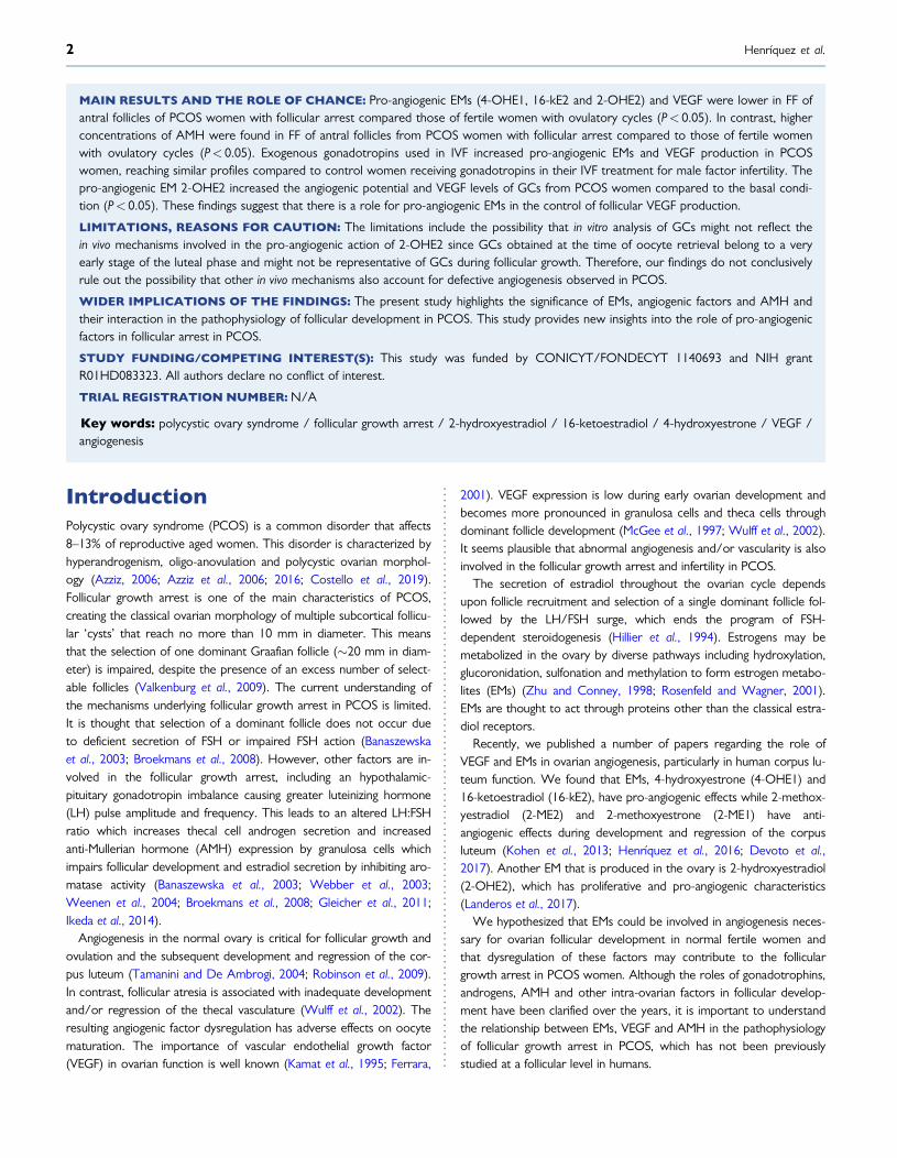
MAIN RESULTS AND THE ROLE OF CHANCE: Pro-angiogenic EMs (4-OHE1, 16-kE2 and 2-OHE2) and VEGF were lower in FF ofantral follicles of PCOS women with follicular arrest compared those of fertile women with ovulatory cycles (P< 0.05). In contrast, higherconcentrations of AMH were found in FF of antral follicles from PCOS women with follicular arrest compared to those of fertile womenwith ovulatory cycles (P< 0.05). Exogenous gonadotropins used in IVF increased pro-angiogenic EMs and VEGF production in PCOSwomen, reaching similar profiles compared to control women receiving gonadotropins in their IVF treatment for male factor infertility. Thepro-angiogenic EM 2-OHE2 increased the angiogenic potential and VEGF levels of GCs from PCOS women compared to the basal condi-tion (P< 0.05). These findings suggest that there is a role for pro-angiogenic EMs in the control of follicular VEGF production.
LIMITATIONS, REASONS FOR CAUTION: The limitations include the possibility that in vitro analysis of GCs might not reflect thein vivo mechanisms involved in the pro-angiogenic action of 2-OHE2 since GCs obtained at the time of oocyte retrieval belong to a veryearly stage of the luteal phase and might not be representative of GCs during follicular growth. Therefore, our findings do not conclusivelyrule out the possibility that other in vivo mechanisms also account for defective angiogenesis observed in PCOS.
WIDER IMPLICATIONS OF THE FINDINGS: The present study highlights the significance of EMs, angiogenic factors and AMH andtheir interaction in the pathophysiology of follicular development in PCOS. This study provides new insights into the role of pro-angiogenicfactors in follicular arrest in PCOS.
STUDY FUNDING/COMPETING INTEREST(S): This study was funded by CONICYT/FONDECYT 1140693 and NIH grantR01HD083323. All authors declare no conflict of interest.
TRIAL REGISTRATION NUMBER: N/A
Key words: polycystic ovary syndrome / follicular growth arrest / 2-hydroxyestradiol / 16-ketoestradiol / 4-hydroxyestrone / VEGF /angiogenesis
IntroductionPolycystic ovary syndrome (PCOS) is a common disorder that affects8–13% of reproductive aged women. This disorder is characterized byhyperandrogenism, oligo-anovulation and polycystic ovarian morphol-ogy (Azziz, 2006; Azziz et al., 2006; 2016; Costello et al., 2019).Follicular growth arrest is one of the main characteristics of PCOS,creating the classical ovarian morphology of multiple subcortical follicu-lar ‘cysts’ that reach no more than 10 mm in diameter. This meansthat the selection of one dominant Graafian follicle (�20 mm in diam-eter) is impaired, despite the presence of an excess number of select-able follicles (Valkenburg et al., 2009). The current understanding ofthe mechanisms underlying follicular growth arrest in PCOS is limited.It is thought that selection of a dominant follicle does not occur dueto deficient secretion of FSH or impaired FSH action (Banaszewskaet al., 2003; Broekmans et al., 2008). However, other factors are in-volved in the follicular growth arrest, including an hypothalamic-pituitary gonadotropin imbalance causing greater luteinizing hormone(LH) pulse amplitude and frequency. This leads to an altered LH:FSHratio which increases thecal cell androgen secretion and increasedanti-Mullerian hormone (AMH) expression by granulosa cells whichimpairs follicular development and estradiol secretion by inhibiting aro-matase activity (Banaszewska et al., 2003; Webber et al., 2003;Weenen et al., 2004; Broekmans et al., 2008; Gleicher et al., 2011;Ikeda et al., 2014).
Angiogenesis in the normal ovary is critical for follicular growth andovulation and the subsequent development and regression of the cor-pus luteum (Tamanini and De Ambrogi, 2004; Robinson et al., 2009).In contrast, follicular atresia is associated with inadequate developmentand/or regression of the thecal vasculature (Wulff et al., 2002). Theresulting angiogenic factor dysregulation has adverse effects on oocytematuration. The importance of vascular endothelial growth factor(VEGF) in ovarian function is well known (Kamat et al., 1995; Ferrara,
2001). VEGF expression is low during early ovarian development andbecomes more pronounced in granulosa cells and theca cells throughdominant follicle development (McGee et al., 1997; Wulff et al., 2002).It seems plausible that abnormal angiogenesis and/or vascularity is alsoinvolved in the follicular growth arrest and infertility in PCOS.
The secretion of estradiol throughout the ovarian cycle dependsupon follicle recruitment and selection of a single dominant follicle fol-lowed by the LH/FSH surge, which ends the program of FSH-dependent steroidogenesis (Hillier et al., 1994). Estrogens may bemetabolized in the ovary by diverse pathways including hydroxylation,glucoronidation, sulfonation and methylation to form estrogen metabo-lites (EMs) (Zhu and Conney, 1998; Rosenfeld and Wagner, 2001).EMs are thought to act through proteins other than the classical estra-diol receptors.
Recently, we published a number of papers regarding the role ofVEGF and EMs in ovarian angiogenesis, particularly in human corpus lu-teum function. We found that EMs, 4-hydroxyestrone (4-OHE1) and16-ketoestradiol (16-kE2), have pro-angiogenic effects while 2-methox-yestradiol (2-ME2) and 2-methoxyestrone (2-ME1) have anti-angiogenic effects during development and regression of the corpusluteum (Kohen et al., 2013; Henrıquez et al., 2016; Devoto et al.,2017). Another EM that is produced in the ovary is 2-hydroxyestradiol(2-OHE2), which has proliferative and pro-angiogenic characteristics(Landeros et al., 2017).
We hypothesized that EMs could be involved in angiogenesis neces-sary for ovarian follicular development in normal fertile women andthat dysregulation of these factors may contribute to the folliculargrowth arrest in PCOS women. Although the roles of gonadotrophins,androgens, AMH and other intra-ovarian factors in follicular develop-ment have been clarified over the years, it is important to understandthe relationship between EMs, VEGF and AMH in the pathophysiologyof follicular growth arrest in PCOS, which has not been previouslystudied at a follicular level in humans.
2 Henrıquez et al.

..
..
..
..
..
..
..
..
..
..
..
..
..
..
..
..
..
..
..
..
..
..
..
..
..
..
..
..
..
..
..
..
..
..
..
..
..
..
..
..
..
..
..
..
..
..
..
..
..
..
..
..
..
..
..
..
..
..
..
..
..
..
..
..
..
..
..
..
..
..
..
..
..
..
..
..
..
..
..
..
..
..
..
..
..
..
.Materials and methods
Human subjectsThis study was approved by the institutional review board of San Borja-Arriaran Clinical Hospital and Faculty of Medicine of the University ofChile. Written informed consent was obtained from all participants.
Four groups of patients were included in this study. The womenfrom Groups A and B did not undergo ovarian stimulation treatment,whereas women from Group C and D did receive treatment for ovar-ian stimulation. Further details are described below.
Women with regular menstrual cycles (A)Ovulatory women, aged 33–38 years with normal BMI (26–33 kg/m2),who were multiparous and did not use hormonal treatment for at least3 months prior to laparoscopy for benign gynecological disease (myoma,tubal ligation or hysterectomy) were included in Group A of the study.
Follicular development was monitored by serial vaginal ultrasounds(TVUs) that were performed at different times in the follicular phase.TVUs were performed until the follicle reached 10 mm (early follicularphase group) or 16 mm of diameter (late follicular phase group).Serum samples were collected on the same day as the surgical proce-dure. Follicular fluid (FF) was aspirated during the surgical procedurestransvaginally using ultrasonographic specific guides and Kitazato IVMneedles from antral (follicle <10 mm follicular diameter) and dominant(>16 mm follicular diameter) follicles.
PCOS women with follicular growth arrest (B)PCOS women presented with amenorrhea because of chronic anovu-lation, hyperandrogenism (biochemical or clinical) and ultrasonographicmorphology according to the Rotterdam criteria (Rotterdam ESHRE/ASRM-Sponsored PCOS Consensus Workshop Group, 2016). Theywere included in this group when there was no response to clomi-phene citrate (150 mg/day for 5 days) or Letrozol (5 mg/day for 5days). After 2–3 months without any hormonal treatment and ultra-sound monitoring of multiple follicles <10 mm, patients were invitedto undergo a drilling procedure. Before ovarian drilling, aspiration of FFwas performed under vaginal ultrasound guidance. Serum sampleswere collected at the same day as the ovarian drilling and FF wasobtained from antral follicles (<10 mm follicular diameter).
Study groups with ovarian stimulationControl women undergoing IVF for male factor infertility (C)Ovulatory women, aged 28–38 years, with a normal antral follicularcount (the number of follicles in both ovaries being >5 follicles and<12 follicles) and AMH levels (>1.2 ng/ml and <4.9 ng/ml)(Humaidan et al., 2016), and participating in our IVF program for malefactor infertility, were invited to donate their FF and granulosa cells forresearch. FF was obtained by transvaginal ultrasound aspiration fromantral follicles (<10 mm follicular diameter) and dominant (>16 mmfollicular diameter) follicles during the IVF procedure.
The ovarian stimulation protocol included recombinant FSH-hMG(Human Menopausal Gonadotrophin) adjusted according to age, BMI,antral follicle count (AFC) and AMH levels. Mean days of stimulationwere 11.2 § 0.6 days. The mean daily FSH dose was 236.7 § 52.4UI. The maximum daily FSH dose was 300 UI/daily. A fixed GnRH an-tagonist (Cetrotide 0.25 mg/day, Merck Serono) protocol was used.
The first dose was given on Day 6 of stimulation. The mean days ofantagonist administration were 6.3 § 0.7 days. A single dose of re-combinant hCG (Ovidrell 250 lg, Merck Serono) was used to triggerthe process of ovulation.
PCOS women undergoing IVF (D)PCOS patients who presented with oligo- or anovulation, hyperandro-genism (biochemical or clinical) and ultrasonographic ovarian morphol-ogy with a large number of arrested antral follicles <10 mm(Rotterdam criteria), were invited to donate their FF and granulosacells for research. The ovarian stimulation protocol included rFSH,GnRH antagonist and ovulation triggering with recombinant hCG. FFwas obtained by transvaginal ultrasound aspiration from antral (<10mm follicular diameter) and dominant (>16 mm follicular diameter)follicles during the IVF procedures.
Power analysis calculationsThe minimum number of patient samples required for the experimentswere determined by power analysis calculations. Sample size calcula-tions were performed based on the evidence indicating that the factordetermined in this study with the highest percentage of variability isAMH, with difference of the means of AMH in serum of 6.15 ng/mland SD 4.21 ( Stracquadanio et al., 2018 ). To detect this differencewith an a significance of 5% and a 1-b power of 88%, it was necessaryto recruit at least seven women per study group. Sample size calcula-tions were based on Garcıa-Garcıa et al. (2013).
Women with regular menstrual cycles (A)A total of 20 subjects were enrolled in this study group, 10 womencorresponding to the early follicular phase and 10 corresponding tothe late follicular phase. We obtained a serum sample to determineendocrine parameters and an adequate volume of FF to determineestrogens metabolites, AMH and VEGF levels. FF (0.5–0.8 ml) was col-lected from five to eight antral follicles of <10 mm per patient andfrom a single dominant follicles (0.7 ml).
PCOS women with follicular growth arrest (B)A total of 17 subjects were enrolled in this group. In 10 women withfollicular growth arrest, we obtained enough serum to determine en-docrine parameters and an adequate volume of FF to determine EMs,AMH and VEGF levels. From the remaining seven women, we onlyobtained adequate samples of serum and FF to determine AMH andVEGF levels. FF (0.5–0.8 ml) was collected from five to eight antral fol-licles of <10 mm per patient.
Control women undergoing IVF (C)From 12 women undergoing IVF for male factor infertility, we obtained 12samples of FF with adequate volume to determine estrogens metabolites,VEGF and AMH levels. In addition, to perform granulosa-lutein cell cultures,follicular aspirates were obtained from six women in this study group.
PCOS women undergoing IVF (D)From the total of 17 PCOS women undergoing IVF enrolled, weobtained 17 samples of FF with adequate volumes to determine estro-gens metabolites, VEGF and AMH levels. In addition, follicular aspirateswere obtained from six women of this study group for granulosa-luteincell cultures.
Reduced pro-angiogenic EMs in follicular arrest of PCOS 3
..
..
..
..
..
..
..
..
..
..
..
..
..
..
..
..
..
..
..
..
..
..
..
..
..
..
..
..
..
..
..
..
..
..
..
..
..
..
..
..
..
..
..
..
..
..
..
..
..
..
..
..
..
..
..
..
..
..
..
..
..
..
..
..
..
..
..
..
..
..
..
..
..
..
..
..
..
..
..
..
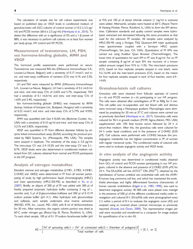
.The calculation of sample size for cell culture experiments was
based on published data on VEGF levels in conditioned medium ofgranulosa-lutein cell (GC) cultures of control woman (110.2§ 5.5 pg/ml) and PCOS woman (60.6§ 2.5 pg/ml) (Henrıquez et al., 2016). Todetect this difference with an a significance of 5% and a 1-b power of88%, it was necessary to perform cell cultures on at least four womenper study group, IVF control and IVF PCOS.
Measurement of testosterone, LH, FSH,sex hormone-binding globulin, AMH andVEGFThe hormonal profile assessments were performed on serum.Testosterone was measured RIA kits (DIAsource ImmunoAssays S.A.,Louvain-La-Neuve, Belgium) with a sensitivity of 0.17 nmol/l, and in-tra- and inter-assay coefficients of variation (CV) was 4.1% and 5.5%,respectively.
LH and FSH were measured by IRMA (DIAsource ImmunoAssaysS.A., Louvain-La-Neuve, Belgium). LH had a sensitivity of 0.2 mUI/ml,and intra- and inter-assay CVs of 2.65% and 5.7%, respectively. FSHhad a sensitivity of 0.1 mUI/ml, and intra- and inter-assay CVs of1.63% and 3.4%, respectively.
Sex hormone-binding globulin (SHBG) was measured by IRMA(Izotop, Institute of Isotopes Ltd., Budapest, Hungary) with a sensitivityof 0.22 nmol/l, and intra- and inter-assay CVs of 5.77% and 4.93%,respectively.
AMH was quantified with Gen II ELISA kits (Beckman Coulter, Inc.,USA) with a sensitivity of 0.10 ng/ml and intra- and inter-assay CVs of3.21% and 5.82%, respectively.
VEGF was quantified in FF from different diameter follicles by en-zyme linked immunosorbant assay (ELISA) according the protocol pro-vided by R&D Systems, Inc. (Minneapolis, MN, USA). The sampleswere assayed in duplicate. The sensitivity of the assay is 5.0 pg/ml.The intra-assay CV was 2.4–10.2% and the inter-assay CV was 5.1–8.7%. VEGF levels were also determined in conditioned medium col-lected from GC cultures obtained from normal and PCOS participantsin the IVF program.
Analysis of estrogen metabolitesEstradiol, estrone and estrogen metabolites (2-ME1, 2-ME2, 4-OHE1,2-OHE2 and 16KE2) were determined in FF from all women partici-pating of study by high performance liquid chromatography (HPLC)and mass spectrometry (HPLC-ESI-MS) as described in Xu et al.(2007). Briefly, an aliquot of 300 ml of FF was added with 300 ml offreshly prepared enzymatic hydrolysis buffer containing 2 mg of L-ascorbic acid, 5 ml of b-glucuronidase/sulfatase and 300 ml of 0.15 Msodium acetate buffer (pH 4.1). After hydrolysis with glucuronidaseand sulfatase, each sample underwent slow inverse extraction(RKVSD, ATR, Inc., Laurel, MD, USA) with 8 ml of dichloromethanefor 30 min. After extraction, the organic solvent portion was dried at60�C under nitrogen gas (Reacti-Vap III, Pierce, Rockford, IL, USA).To each dried sample, 100 ml of 0.1 M sodium bicarbonate buffer (pH
at 9.0) and 100 ml of dansyl chloride solution (1 mg/ml in acetone)were added. Afterwards, samples were heated at 60�C (Reacti-ThermIII Heating Module, Pierce, Rockford, IL, USA) to form the EM deriva-tives. Calibration standards and quality control samples were hydro-lyzed, extracted and derivatized following the same procedure as thatused for the unknown FF samples. All samples were analyzed byHPLC-ESI-MS using a Finnigan TSQ Quantum-AM triple quadrupolemass spectrometer coupled with a Surveyor HPLC system(ThermoFinnigan, San Jose, CA, USA). Quantitation of FF EMs wascarried out using Xcalibur Quan Browser (ThermoFinnigan). Thelower limit of quantitation for each EM in FF was 8 pg/ml. For an FFsample containing 8 pg/ml of each EM, the recovery of a knownadded amount ranged from 91% to 113%. The intra-batch precisions(CV), based on four replicate samples assayed in one batch, were4.2–16.4% and the inter-batch precisions (CV), based on the meansfor four replicate samples assayed in each of four batches, were 6.9–14.6%.
Granulosa-lutein cell culturesGranulosa cells were obtained from follicular aspirates of control(n¼ 6) and PCOS women (n¼ 6) participating in our IVF program.The cells were obtained after centrifugation of FF at 400g for 5 min.The cell pellet was re-suspended, and red blood cells and detrituswere removed using a Histopaque gradient. Macrophages were elimi-nated by preplating for 30 min at 37�C and cultures were carried outas previously described (Henrıquez et al., 2017). Granulosa cells werecultured for 96 h in growth medium (M199, Sigma-Aldrich, MO, USA)supplemented with 10% fetal bovine serum followed by 24 h inserum-free medium. After this, the granulosa cells were cultured for24 h under basal conditions and in the presence of 2-OHE2 (0.05mM). Cell cultures were performed with 2-OHE2 because this pro-angiogenic metabolite has the highest concentration in FF of womenwith regular menstrual cycles. The conditioned media of cultured cellswere used to evaluate angiogenic activity and VEGF levels.
In vitro analysis of the angiogenic activityAngiogenic activity was determined in conditioned media obtainedfrom GCs of control and PCOS women participating in our IVF pro-gram, cultured in the absence and presence of 2-OHE2 (0.05 lM) for24 h. The EA.hy926 cell line (ATCCVR CRL-2922TM), obtained by thehybridization of human umbilical vein endothelial cells with the A549/8 human lung carcinoma cell line, and which has maintained the endo-thelial phenotype and highly differentiated functions characteristic ofhuman vascular endothelium (Edgell et al., 1983, 1990), was used todetermine angiogenic activity; 40 000 cells were plated onto matrigelin the presence of 500 ml of the different conditioned media under in-vestigation and cultured 8 h. EA.hy926 cells were photographed every2 h within a period of 8 h to evaluate the angiogenic score (AS) andanalyzed using an inverted phase contrast microscope as previouslyreported (Aranda and Owen, 2009). Ten representative images perwell were recorded and transferred to a computer for image analysisfor quantification of an in vitro AS:
AS ¼ ðN� of sprouting cellsÞ1þ ðN� of connected cellsÞ2þ ðNumber of polygonsÞ3
Total number of cells
� �þ ½0; 1; or 2�
4 Henrıquez et al.
..
..
..
..
..
..
..
..
..
..
..
..
..
..
..
..
..
..
..
..
..
..
..
..
..
..
..
..
..
..
..
..
..
..
..
..
..
..
..Statistical analysisFF and serum values are presented as means § SEM. Data were ana-lyzed by one-way ANOVA followed by a Tukey’s multiple comparisontest. The statistical analysis of the angiogenic potential data fromgranulosa-lutein cell cultures was carried out using a non-parametricmethod: Kruskal–Wallis (one-way analysis of variance) followed by a
Dunn’s Multiple Comparison post hoc test. GraphPad-Prism Software4.0 (Inc., San Diego, CA, USA) was used to analyze data. Significancewas defined as P< 0.05. The data were analyzed for normality withthe Shapiro–Wilk test, P< 0.05.
Results
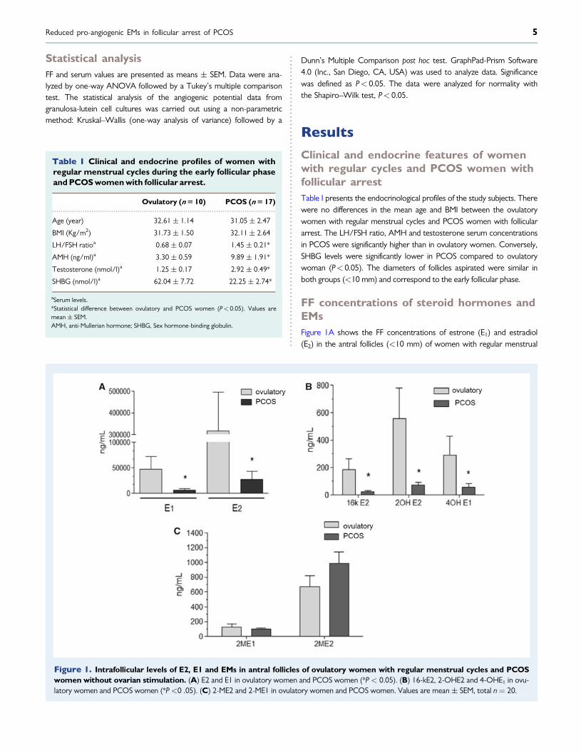
Clinical and endocrine features of womenwith regular cycles and PCOS women withfollicular arrestTable I presents the endocrinological profiles of the study subjects. Therewere no differences in the mean age and BMI between the ovulatorywomen with regular menstrual cycles and PCOS women with folliculararrest. The LH/FSH ratio, AMH and testosterone serum concentrationsin PCOS were significantly higher than in ovulatory women. Conversely,SHBG levels were significantly lower in PCOS compared to ovulatorywoman (P< 0.05). The diameters of follicles aspirated were similar inboth groups (<10 mm) and correspond to the early follicular phase.
FF concentrations of steroid hormones andEMsFigure 1A shows the FF concentrations of estrone (E1) and estradiol(E2) in the antral follicles (<10 mm) of women with regular menstrual
......................................................................................................
Table I Clinical and endocrine profiles of women withregular menstrual cycles during the early follicular phaseand PCOS women with follicular arrest.
Ovulatory (n 5 10) PCOS (n 5 17)
Age (year) 32.61 § 1.14 31.05 § 2.47
BMI (Kg/m2) 31.73 § 1.50 32.11 § 2.64
LH/FSH ratioa 0.68 § 0.07 1.45 § 0.21*
AMH (ng/ml)a 3.30 § 0.59 9.89 § 1.91*
Testosterone (nmol/l)a 1.25 § 0.17 2.92 § 0.49*
SHBG (nmol/l)a 62.04 § 7.72 22.25 § 2.74*
aSerum levels.*Statistical difference between ovulatory and PCOS women (P< 0.05). Values aremean § SEM.AMH, anti-Mullerian hormone; SHBG, Sex hormone-binding globulin.
Figure 1. Intrafollicular levels of E2, E1 and EMs in antral follicles of ovulatory women with regular menstrual cycles and PCOSwomen without ovarian stimulation. (A) E2 and E1 in ovulatory women and PCOS women (*P < 0.05). (B) 16-kE2, 2-OHE2 and 4-OHE1 in ovu-latory women and PCOS women (*P <0 .05). (C) 2-ME2 and 2-ME1 in ovulatory women and PCOS women. Values are mean§ SEM, total n ¼ 20.
Reduced pro-angiogenic EMs in follicular arrest of PCOS 5

..
..
..
..
..
..
..
..
..
..
..
..
..
..
..
..
..
..
..
..
..
..
..
..
..
..
..
..
..
..
..
..
..
..
..
..
..
..
..
.cycles and PCOS women with follicular arrest. The highest FF concen-trations of E1 and E2 were observed in ovulatory women compared toPCOS women (P< 0.001). Significantly lower levels of 2-OHE2, 4-OHE1 and 16-kE2 were detected in FF of antral follicles of PCOSwomen (P< 0.05) matched with similar size follicles of ovulatorywomen (Fig. 1B). There were no significant differences in the follicularlevels of 2-ME1 and 2-ME2 (Fig. 1C) between follicles from the twogroups of women. PCOS women who were undergoing ovarian stimu-lation had EMs levels similar to control women undergoing ovarianstimulation in our IVF program (Fig. 2A–C).
Intrafollicular levels of EMs, VEGF andAMH of ovulatory women with regularmenstrual cycles and PCOS women withfollicular arrestTable II presents the sum of FF pro-angiogenic EMs (16-ketoE2, 2-OHE2 and 4-OHE1), the sum of anti-angiogenic EMs (2-ME2 and 2-ME1) and ratio of
Ppro-angiogenic EMs/
Panti-angiogenic EMs in FF
from different diameter follicles from women with regular menstrualcycles and PCOS women with follicular arrest. The intrafollicular levelsof pro-angiogenic EMs were significantly higher in dominant and antralfollicles of ovulatory women compared with follicles of PCOS women(P< 0.05).
The AMH levels in FF were significantly higher in antral follicles(<10 mm) of PCOS women compared to antral and dominant follicles
(>16 mm) of ovulatory women (P< 0.05). The VEGF levels in FFwere significantly lower in antral follicles of PCOS women comparedwith antral and dominant follicles of ovulatory women (P< 0.05).
The sum of FF levels of anti-angiogenic EMs was significantly higherin dominant follicles compared with that in antral follicles of PCOS andovulatory women (P< 0.05). However no statistically significant differ-ences were seen between the sum of FF levels of anti-angiogenic EMsin antral follicles of ovulatory women compared to that of PCOSwomen. Interestingly, the ratio between
Ppro-angiogenic EMs andP
anti-angiogenic EMs was significantly lower in antral follicles ofPCOS women compared with antral and dominant follicles of ovula-tory women (P< 0.05).
The pro-angiogenic EMs levels were directly correlated with the lev-els of VEGF found in these follicles (Pearson’s r¼ 0.98; r2 ¼ 0.97;P< 0.0002) (Fig. 3A), while the AMH levels were inversely correlatedwith VEGF levels in antral follicles of PCOS women and in antral anddominant follicle of ovulatory women (Pearson’s r ¼ �0.98; r2 ¼0.96; P< 0.0005) (Fig. 3B).
Intrafollicular levels of AMH and VEGF ofPCOS women compared to controlsundergoing IVFTable III presents the FF levels of AMH and VEGF in follicles of differ-ent diameters from control and PCOS women undergoing ovarianstimulation for IVF. The highest FF concentrations of VEGF were
Figure 2. Intrafollicular levels of E2, E1, 16-kE2, 2-OHE2 and 4-OHE1 in antral follicle of control and PCOS women undergoingovarian stimulation for IVF. (A) E2 and E1 in control and PCOS women undergoing IVF. (B) 16-kE2, 2-OHE2 and 4-OHE1 in control and PCOSwomen undergoing IVF. (C) 2-ME2 and 2-ME1 in control and PCOS women undergoing IVF. Values are mean § SEM, PCOS n ¼ 17, control n ¼ 12.
6 Henrıquez et al.

..
..
..
..
..
.observed in dominant follicles of control and PCOS women comparedto antral follicles (P< 0.05). No significant differences were found be-tween FF AMH levels from follicles of different diameters between the
two study groups. Interestingly, the ovarian stimulation protocolreduces AMH levels compared to those detected in dominant folliclesand increases VEGF levels.
............................................................................................................................................................................................................................
Table II Intrafollicular levels of AMH, VEGF and estrogens metabolites (EMs) in antral and dominant follicle of normalwomen with spontaneous cycles and PCOS women with follicular arrest.
Ovulatory(n 5 10)
(antral follicle)
PCOS(n 5 10)
(antral follicle)
Ovulatory(n 5 10)
(dominant follicle)
Age (year) 32.6 § 1.14 29.6 § 3.0 33.1 § 2.0
AMH (ng/ml)* 213.2 § 28.0 546.3 § 16.7a 2.9 § 0.09b
VEGF (pg/ml)* 503.2 § 101.40 33.1 § 5.9a 6529.6 § 514.3b
PEMs pro-angiogenic (2-OHE2 þ 4-OHE1 þ 16-KE2) (ng/ml) 1099.7 § 128.1 441.9 § 31.8a 3658.5 § 278.0b
PEMs anti-angiogenic (2-ME2 þ 2-ME1) (ng/ml) 689.25 § 111.4 1239.3 § 177.7 3159.8 § 604.3b
PPro-angio/
PAnti-angio ratio 1.59 0.35a 1.15
Follicular diameter (mm) <10 <10 >16
aComparing the difference between antral follicles of ovulatory and PCOS women (P< 0.05). Values are mean § SEM.bComparing the difference between antral and dominant follicles of ovulatory women (P< 0.05). Values are mean § SEM.*AMH and VEGF determination correspond to n¼ 17 FF samples of PCOS.P
EMs, sum of estrogen metabolites; VEGF, vascular endothelial growth factor.
Figure 3. Linear regression between intrafollicular EMs, AMH and VEGF levels in different study groups. (A) Lineal regression be-tween REMs and VEGF. (B) Lineal regression between AMH and VEGF. REMs: sum of pro-angiogenic estrogen metabolites (2-OHE2, 4-OHE1 and16kE2).
Reduced pro-angiogenic EMs in follicular arrest of PCOS 7

..
..
..
..
..
..
..
.Effect of pro-angiogenic EMs on angiogenicactivity of GCsThe pro-angiogenic metabolite, 2-OHE2, significantly increased angio-genic activity of GCs of control and PCOS women compared to basalconditions (P< 0.05) (Fig. 4A), as assessed by formation of capillary
structures by the EA.hy926 cells. 2-OHE2 also significantly increasedVEGF secretion from GCs cultures of control and PCOS women com-pared to basal conditions (P< 0.05) (Fig. 4B). These findings documentthe role of pro-angiogenic EMs in the control of follicular VEGFproduction.
............................................................................................................................................................................................................................
Table III Intrafollicular levels of AMH and VEGF in antral and dominant follicles of control and PCOS women undergoingovarian stimulation for IVF.
Control IVF(n 5 12)
(antral follicle)
Control IVF(n 5 12)
(dominant follicle)
PCOS IVF(n 5 17)
(antral follicle)
PCOS IVF(n 5 17)
(dominant follicle)
Age (year) 30.2 § 1.3 30.2 § 1.3 32.2 § 1.3 32.2 § 1.3
AMH (ng/ml) 2.5 § 0.2 2.2 § 0.3 2.5 § 0.1 2.5 § 0.3
VEGF (pg/ml) 1080.7 § 66.8 3221.3 § 335.6a 1153.2 § 42.0 1902.3 § 50.0b
Follicular diameter (mm) <10 >16 <10 >16
Values are mean § SEM.aP < 0.05 dominant follicles versus antral follicles in control women.bP < 0.05 dominant follicle versus antral follicle in PCOS women.
Figure 4. Effect of 2-OHE2 on angiogenic activity of granulosa cell cultures from control and PCOS women undergoing IVF. (A)The graph represents the angiogenic potential quantification of PCOS and control GCs cultures stimulated with 2-OHE2. *P < 0.05 PCOS GCs plus2-OHE2 versus PCOS GCs in basal condition. (B) The graph represents the VEGF secretion of PCOS GCs cultures stimulated with 2-OHE2. *P <0.05 PCOS GCs plus 2-OHE2 versus PCOS GCs in basal condition. (C) Photomicrograph of the angiogenic assay. Values are mean § SEM, n ¼ 6.GCs, granulosa-lutein cells.
8 Henrıquez et al.

..
..
..
..
..
..
..
..
..
..
..
..
..
..
..
..
..
..
..
..
..
..
..
..
..
..
..
..
..
..
..
..
..
..
..
..
..
..
..
..
..
..
..
..
..
..
..
..
..
..
..
..
..
..
..
..
..
..
..
..
..
..
..
..
..
..
..
..
..
..
..
..
..
..
..
..
..
..
..
..
..
..
..
..
..
..
.DiscussionThe present study reveals for the first time physiological and patho-physiological intrafollicular levels of EMs, VEGF and AMH in PCOScompared to ovulatory women with a regular menstrual cycle withoutovulation stimulation, suggesting the participation of these factors innormal follicular development and follicular arrest in PCOS. Theresults show a diminished quantity of pro-angiogenic EMs (2-OHE2, 4-OHE1 and 16-kE2) in FF of antral PCOS follicles compared to FF fromantral follicles of ovulatory women. The reduction in levels of pro-angiogenic EMs is associated with lower VEGF levels in antral folliclesof PCOS women. Intrafollicular VEGF levels were higher in dominantfollicles of ovulatory women. Our observations are consistent withpreviously published data that showed lower levels of 2-hydroxyestro-gens in urine of PCOS women (Salih et al., 2007). On the other hand,no differences were found in the levels of anti-angiogenic EMs betweenantral follicles of PCOS and ovulatory women. However, the ratio ofpro-angiogenic EMs to anti-angiogenic EMs was significantly lower inPCOS compared to ovulatory women. These results are consistentwith a previous study that did not find genetic variation affecting theexpression of catechol-O-methyltransferase (COMT), an enzyme inthe pathway for the production of 2-methyoxyestradiol (2-ME2), ananti-angiogenic EM, in European-ancestry PCOS women (Hill et al.,2012). In conclusion, these data suggest that arrested follicles of PCOSproduce lower levels of pro-angiogenic EMs, leading to an anti-angiogenic environment that contributes to lower vascularity of thePCOS follicles.
The antral follicles of PCOS women have high intrafollicular AMHlevels compared to follicles of ovulatory women. AMH levels aremarkedly reduced in the dominant follicles of ovulatory women. AMHplays an inhibitory role in follicular development, preventing the pre-mature recruitment and maturation of follicles (Weenen et al., 2004).When follicles reach 7–9 mm in diameter, AMH expression is down-regulated and these follicles begin to respond to FSH and LH, leadingto increased estrogen production, follicular selection and ovulation.AMH secretion is maximal at the antral stage in human follicles, anddecreases in large follicle (Durlinger et al., 2002; Visser and Themmen,2005).
PCOS ovaries have a higher number of preantral and antral follicles,indicating that follicular growth arrest occurs when AMH production ishigh (Pellatt et al., 2007). Multiple studies have documented that se-rum AMH concentrations are elevated in PCOS women compared tonormally ovulating women (Laven et al., 2004). Moreover, other stud-ies have suggested that AMH levels reflect the severity of PCOS(Jacob et al., 2017). There are significant differences in AMH levelsamong anovulatory PCOS women with oligo-amenorrhea comparedwith PCOS ovulatory women (Das et al., 2008). In anovulatory PCOSwomen, hypersecretion of AMH by granulosa cells of mature small an-tral follicles could impair follicular growth by reducing FSH sensitivityand blocking the conversion of androgens into estrogens by inhibitingthe activity of aromatase, causing hyperandrogenism (Eldar-Geva et al.,2005; Piouka et al., 2009). Others found that follicular AMH levels arenegatively correlated with FSH concentrations, indicating that AMHlevels predict follicle responsiveness to FSH in ovulation inductioncycles (Dumesic et al., 2009). AMH influences transcription of genes ingranulosa cells through Smad proteins and regulates gene expressionto maintain primordial follicles in their arrested state (Visser, 2006).
The results obtained in the present work show an inverse relation-ship between intrafollicular levels of AMH and VEGF that suggests aninhibitory role of AMH on angiogenesis. In support of this observation,it has been shown that AMH downregulates TGF beta signaling path-ways leading to decreased cell differentiation and angiogenesis (Nilssonet al., 2007). Additionally, numerous publications show that FSH posi-tively regulates angiogenesis, stimulating HIF-1a expression and VEGFsecretion (Kuo et al., 2011; Stilley et al., 2014). This suggests that highAMH levels characteristic of PCOS, reduce sensitivity to FSH and aredetrimental to follicular angiogenesis, resulting in follicular growth ar-rest. Interestingly, the recombinant FSH doses used in ovulation induc-tion cycles in PCOS women stimulate angiogenesis and folliculargrowth.
Furthermore, our results from women undergoing IVF treatmentshow that ovarian stimulation increases the intrafollicular levels of pro-angiogenic EMs and VEGF, and reduces AMH levels in PCOS women,reaching levels found in FF from control women. Previously publisheddata showed that PCOS women with elevated AMH levels (66%) afterovulation induction are predisposed to ovarian hyperstimulation syn-drome (OHSS), while 16.5% of these women had normal AMH levelswith low risk of OHSS: a result that is in agreement with our data,since we did not observe ovarian hyperstimulation in our study group(Stracquadanio et al., 2018).
These data suggest that there is a gonadotropin-regulated intrafollic-ular reduction of pro-angiogenic EMs in PCOS with follicular arrest.The in vitro results of this study suggest that PCOS GCs have lowerangiogenic potential and VEGF levels due to the reduced concentra-tions of follicular pro-angiogenic EMs, and that these cells recover theirangiogenic capacity when incubated with a pro-angiogenic EMs (2-OHE2).
Altogether, the present study strongly suggests that there is reducedangiogenic potential accompanied by high levels of AMH in the PCOSfollicles that could explain, in part, the follicular growth arrest charac-teristic of this disorder. Notably, treatment with exogenous gonado-tropins during IVF improved the production of pro-angiogenic EMs andVEGF in PCOS women.
Authors’ rolesS.H. and P.K. contributed to the conceptual formulation of the work,designed experiments, conducted experiments and helped in writingthe manuscript. X.X. performed estrogen metabolite measurements.A.M. contributed to statistical and image analysis. A.G. and C.V. pro-vided cells and follicular fluid. J.F.S. interpreted results and participatedin writing the manuscript. L.D. contributed to the conceptual formula-tion of the work, designed experiments, provided cells, interpretedresults and participated in writing the manuscript. All authors approvedthe final version of the manuscript.
FundingThis work was supported by CONICYT/FONDECYT (FondoNacional de Desarrollo Cientifico y Tecnologico) grant N�1140693and NIH (National Institutes of Health) grant N�R01HD083323.
Reduced pro-angiogenic EMs in follicular arrest of PCOS 9

..
..
..
..
..
..
..
..
..
..
..
..
..
..
..
..
..
..
..
..
..
..
..
..
..
..
..
..
..
..
..
..
..
..
..
..
..
..
..
..
..
..
..
..
..
..
..
..
..
..
..
..
..
..
..
..
..
..
..
..
..
..
..
..
..
..
..
..
..
..
..
..
..
..
..
..
..
..
..
..
..
..
..
..
..
..
..Conflict of interestAll authors declare no conflict of interest.
ReferencesAranda E, Owen GI. A semi-quantitative assay to screen for angio-
genic compounds and compounds with angiogenic potential usingthe EA.hy926 endothelial cell line. Biol Res 2009;42:377–389.
Azziz R. Controversy in clinical endocrinology: diagnosis of polycysticovarian syndrome: the Rotterdam criteria are premature. J ClinEndocrinol Metab 2006;91:781–785.
Azziz R, Carmina E, Chen Z, Dunaif A, Laven JS, Legro RS, LiznevaD, Natterson-Horowtiz B, Teede HJ, Yildiz BO.. Polycystic ovarysyndrome. Nat Rev Dis Primers 2016;2:16057.
Azziz R, Carmina E, Dewailly D, Diamanti-Kandarakis E et al.;Androgen Excess Society. Positions statement: criteria for definingpolycystic ovary syndrome as a predominantly hyperandrogenicsyndrome: an Androgen Excess Society guideline. J Clin EndocrinolMetab 2006;91:4237–4245.
Banaszewska B, Spaczy�nski RZ, Pelesz M, Pawelczyk L. Incidence ofelevated LH/FSH ratio in polycystic ovary syndrome women withnormo- and hyperinsulinemia. Rocz Akad Med Bialymst 2003;48:131–134.
Broekmans F, Visser J, Laven J, Broer S, Themmen A, Fauser B. Anti-Mullerian hormone and ovarian dysfunction. Trends EndocrinolMetab 2008;19:340–347.
Costello MF, Misso ML, Balen A, Boyle J, Devoto L, Garad RM et al.Evidence summaries and recommendations from the internationalevidence-based guideline for the assessment and management ofpolycystic ovary syndrome: assessment and treatment of infertility.Hum Reprod Open 2019;1:1–24.
Das M, Gillott D, Saridogan E, Djahanbakhch O. AMH is increased infollicular fluid from unstimulated ovaries in women with polycysticovary syndrome. Hum Reprod 2008;23:2122–2126.
Devoto L, Henrıquez S, Kohen P, Strauss JF 3rd. The significance ofestradiol metabolites in human corpus luteum physiology. Steroids2017;123:50–54.
Dumesic DA, Lesnick TG, Stassart JP, Ball GD, Wong A, AbbottDH.Intrafollicular antimullerian hormone levels predict folliclerespon-siveness to follicle-stimulating hormone (FSH) in normoandrogeni-covulatory women undergoing gonadotropin releasing-hormoneanalog/recombinanthuman FSH therapy for in vitro fertilizationembryo transfer. Fertil Steril 2009;92:217–221.
Durlinger A, Visser J, Themmen A. Regulation of ovarian function:the role of anti-Mullerian hormone. Reproduction 2002;124:601–609.
Edgell CJ, Haizlip JE, Bagnell CR, Packenham JP, Harrison P,Wilbourn B, Madden VJ. Endothelium specific Weibel-Palade bod-ies in a continuous human cell line, EA.hy926. In Vitro Cell Dev Biol1990;26:1167–1172.
Edgell CJ, McDonald CC, Graham JB. Permanent cell line expressinghuman factor VIII-related antigen established by hybridization. ProcNatl Acad Sci USA 1983;80:3734–3737.
Eldar-Geva T, Margalioth E, Gal M, Ben-Chetrit A, Algur N, Zylber-Haran E et al. Serum anti-Mullerian hormone levels during
controlled ovarian hyperstimulation in women with polycystic ova-ries with and without hyperandrogenism. Hum Reprod 2005;20:1814–1819.
Ferrara N. Role of vascular endothelial growth factor in regulation ofphysiological angiogenesis. Am J Physiol Cell Physiol2001;280:1358-1366.
Garcıa-Garcıa JA, Reding-Bernal A, Lopez-Alvarenga JC. Sample sizecalculation in education medical research. Inv Ed Med 2013;2:217–224.
Gleicher N, Weghofer A, Barad DH. The role of androgens in folliclematuration and ovulation induction: friend or foe of infertility treat-ment? Reprod Biol Endocrinol 2011;9:116.
Henrıquez S, Kohen P, Mu~noz A, Godoy A, Orge A, Strauss JF III,Devoto L. In-vitro study of gonadotrophin signaling pathways inhuman granulosa cells in relation to progesterone receptor expres-sion. Reprod Biomed Online 2017;35:363–371.
Henrıquez S, Kohen P, Xu X, Veenstra TD, Mu~noz A, PalominoWA, Strauss JF 3rd, Devoto L. Estrogen metabolites in human cor-pus luteum physiology: differential effects on angiogenic activity.Fertil Steril 2016;106:230–237.
Hill LD, Ewens KG, Maher BS, York TP, Legro RS, Dunaif A, StraussJF 3rd. Catechol-O-methyltransferase (COMT) single nucleotidepolymorphisms and haplotypes are not major risk factors for poly-cystic ovary syndrome. Mol Cell Endocrinol 2012;350:72–77.
Hillier SG, Whitelaw PF, Smyth CD. Follicular oestrogen synthesis:the ‘two-cell, two-gonadotrophin’ model revisited. Mol CellEndocrinol 1994;100:51–54.
Humaidan P, Alviggi C, Fischer R, Esteves SC. The novel POSEIDONstratification of ‘Low prognosis patients in Assisted ReproductiveTechnology’ and its proposed marker of successful outcome.F1000Res 2016;5:1–8.
Ikeda K, Baba T, Morishita M, Honnma H, Endo T, Kiya T, Saito T.Long-term treatment with dehydroepiandrosterone may lead tofollicular atresia through interaction with anti-Mullerian hormone. JOvarian Res 2014;7:46.
Jacob S, Field H, Calder N, Picton H, Balen A, Barth J. Anti-Mullerianhormone reflects the severity of polycystic ovary syndrome. ClinEndocrinol (Oxf) 2017;86:395–400.
Kamat BR, Brown LF, Manseau EJ, Senger DR, Dvorak HF.Expression of vascular permeability factor/vascular endothelialgrowth factor by human granulosa and theca lutein cells. Role incorpus luteum development. Am J Pathol 1995;146:157–165.
Kohen P, Henrıquez S, Rojas C et al. 2-Methoxyestradiol in the hu-man corpus luteum throughout the luteal phase and its influenceon lutein cell steroidogenesis and angiogenic activity. Fertil Steril2013;100:1397–1404.
Kuo SW, Ke FC, Chang GD, Lee MT, Hwang JJ. Potential role offollicle-stimulating hormone (FSH) and transforming growth factor(TGFb1) in the regulation of ovarian angiogenesis. J Cell Physiol2011;226:1608–1619.
Landeros RV, Jobe SO, Aranda-Pino G, Lopez GE, Zheng J, MagnessR. Convergent ERK1/2, p38 and JNK mitogen activated proteinkinases (MAPKs) signalling mediate catecholoestradiol-inducedproliferation of ovine uterine artery endothelial cells. J Physiol 2017;595:4663–4676.
Laven J, Mulders A, Visser J, Themmen A, De Jong F, Fauser B. Anti-Mullerian hormone serum concentrations in normoovulatory and
10 Henrıquez et al.

..
..
..
..
..
..
..
..
..
..
..
..
..
..
..
..
..
..
..
..
..
..
..
..
..
..
..
..
..
..
..
..
..
..
..
..
..
..
..
..
..
..
..
..
..
..
..
..
..
.anovulatory women of reproductive age. J Clin Endocrinol Metab2004;89:318–323.
McGee E, Spears N, Minami S et al. Preantral ovarian follicles inserum-free culture: suppression of apoptosis after activation of thecyclic guanosine 3’,5’-monophosphate pathway and stimulation ofgrowth and differentiation by follicle-stimulating hormone.Endocrinology 1997;138:2417–2424.
Nilsson E, Rogers N, Skinner M. Actions of anti-Mullerian hormoneon the ovarian transcriptome to inhibit primordial to primary folli-cle transition. Reproduction 2007;134:209–221.
Pellatt L, Hanna L, Brincat M, Galea R, Brain H, Whitehead S, MasonH. Granulosa cell production of anti-Mullerian hormone is increasedin polycystic ovaries. J Clin Endocrinol Metab 2007;92:240–245.
Piouka A, Farmakiotis D, Katsikis I, Macut D, Gerou S, Panidis D.Anti-Mullerian hormone levels reflect severity of PCOS but are neg-atively influenced by obesity: relationship with increased luteinizinghormone levels. Am J Physiol Endocrinol Metab 2009;296:E238–E243.
Robinso RS, Woad KJ, Hammond AJ, Laird M, Hunter MG, MannGE. Angiogenesis and vascular function in the ovary. Reproduction2009;138:869–881.
Rosenfeld CS, Wagner JS, Roberts RM, Lubahn DB. Intraovarianactions of oestrogen. Reproduction 2001;122:215–226.
Rotterdam ESHRE/ASRM-Sponsored PCOS Consensus WorkshopGroup. Revised 2003 consensus on diagnostic criteria and long-term health risks related to polycystic ovary syndrome. Fertil Steril2004;81:19–25.
Salih S, Xu X, Veenstra TD, Duleba AJ, Fouad H, Nagamani M, Al-Hendy A. Lower levels of urinary 2-hydroxyestrogens in polycysticovary syndrome. J Clin Endocrinol Metab 2007;92:3285–3291.
Stilley JA, Guan R, Duffy DM, Segaloff DL. Signaling through FSHreceptors on human umbilical vein endothelial cells promotes an-giogenesis. J Clin Endocrinol Metab 2014;99:813–820.
Stracquadanio M. Ciotta L. Palumbo MA. Relationship between se-rum anti-Mullerian hormone and intrafollicular AMH levels inPCOS women. Gynecol Endocrinol 2018;3:223–228.
Tamanini C, De Ambrogi M. Angiogenesis in developing follicle andcorpus luteum. Reprod Domest Anim 2004;39:206–216.
Valkenburg O, Uitterlinden A, Piersma D, Hofman A, Themmen A,Fauser B, Laven JS. Genetic polymorphisms of GnRH and gonado-trophic hormone receptors affect the phenotype of polycysticovary syndrome. Hum Reprod 2009;24:2014–2022.
Visser J. Role of anti-Mullerian hormone in follicle recruitment andmaturation. J Gynecol Obstet Biol Reprod (Paris) 2006;35:2S30–2S34.
Visser JA, Themmen AP. Anti-Mullerian hormone and folliculogene-sis. Mol Cell Endocrinol 2005;234:81–86.
Webber L, Stubbs S, Stark J, Trew G, Margara R, Hardy K, Franks S.Formation and early development of follicles in the polycysticovary. Lancet 2003;362:1017–1021.
Weenen C, Laven JS, Von Bergh AR, Cranfield M, Groome NP,Visser JA et al. Anti-Mullerian hormone expression pattern in thehuman ovary: potential implications for initial and cyclic follicle re-cruitment. Mol Hum Reprod 2004;10:77–83.
Wulff C, Wilson H, Wiegand SJ, Rudge JS, Fraser HM. Prevention ofthecal angiogenesis, antral follicular growth, and ovulation in theprimate by treatment with vascular endothelial growth factor TrapR1R2. Endocrinology 2002;143:2797–2807.
Xu X, Roman JM, Issaq HJ, Keefer LK, Veenstra TD, Ziegler RG.Quantitative measurement of endogenous estrogens and estrogenmetabolites in human serum by liquid chromatography-tandemmass spectrometry. Anal Chem 2007;79:7813–7821.
Zhu BT, Conney AH. Functional role of estrogen metabolism in tar-get cells: review and perspectives. Carcinogenesis 1998;19:1–27.
Reduced pro-angiogenic EMs in follicular arrest of PCOS 11
View publication statsView publication stats


![Role of herbals in the management of polycystic ovarian ...PCOS by acting directly on ovarian androgen secretion andabnormal follicular development [5] and the use of insulin-sensitizing](https://img.pdfslide.us/doc/110x75/5ed75feda5b1445fe467ce70/role-of-herbals-in-the-management-of-polycystic-ovarian-pcos-by-acting-directly.jpg)