Embed Size (px)
Citation preview
RESEARCH ARTICLE Open Access
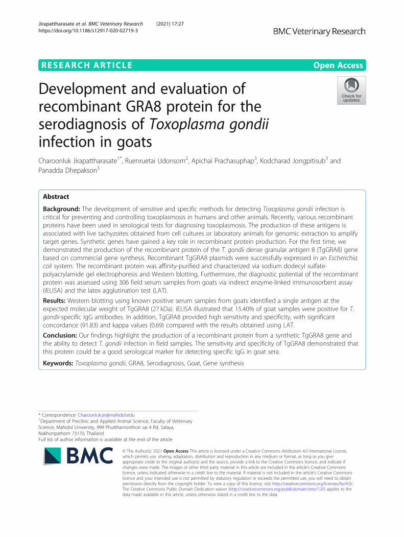
Development and evaluation ofrecombinant GRA8 protein for theserodiagnosis of Toxoplasma gondiiinfection in goatsCharoonluk Jirapattharasate1*, Ruenruetai Udonsom2, Apichai Prachasuphap3, Kodcharad Jongpitisub3 andPanadda Dhepakson3
Abstract
Background: The development of sensitive and specific methods for detecting Toxoplasma gondii infection iscritical for preventing and controlling toxoplasmosis in humans and other animals. Recently, various recombinantproteins have been used in serological tests for diagnosing toxoplasmosis. The production of these antigens isassociated with live tachyzoites obtained from cell cultures or laboratory animals for genomic extraction to amplifytarget genes. Synthetic genes have gained a key role in recombinant protein production. For the first time, wedemonstrated the production of the recombinant protein of the T. gondii dense granular antigen 8 (TgGRA8) genebased on commercial gene synthesis. Recombinant TgGRA8 plasmids were successfully expressed in an Escherichiacoli system. The recombinant protein was affinity-purified and characterized via sodium dodecyl sulfate-polyacrylamide gel electrophoresis and Western blotting. Furthermore, the diagnostic potential of the recombinantprotein was assessed using 306 field serum samples from goats via indirect enzyme-linked immunosorbent assay(iELISA) and the latex agglutination test (LAT).
Results: Western blotting using known positive serum samples from goats identified a single antigen at theexpected molecular weight of TgGRA8 (27 kDa). iELISA illustrated that 15.40% of goat samples were positive for T.gondii-specific IgG antibodies. In addition, TgGRA8 provided high sensitivity and specificity, with significantconcordance (91.83) and kappa values (0.69) compared with the results obtained using LAT.
Conclusion: Our findings highlight the production of a recombinant protein from a synthetic TgGRA8 gene andthe ability to detect T. gondii infection in field samples. The sensitivity and specificity of TgGRA8 demonstrated thatthis protein could be a good serological marker for detecting specific IgG in goat sera.
Keywords: Toxoplasma gondii, GRA8, Serodiagnosis, Goat, Gene synthesis
© The Author(s). 2021 Open Access This article is licensed under a Creative Commons Attribution 4.0 International License,which permits use, sharing, adaptation, distribution and reproduction in any medium or format, as long as you giveappropriate credit to the original author(s) and the source, provide a link to the Creative Commons licence, and indicate ifchanges were made. The images or other third party material in this article are included in the article's Creative Commonslicence, unless indicated otherwise in a credit line to the material. If material is not included in the article's Creative Commonslicence and your intended use is not permitted by statutory regulation or exceeds the permitted use, you will need to obtainpermission directly from the copyright holder. To view a copy of this licence, visit http://creativecommons.org/licenses/by/4.0/.The Creative Commons Public Domain Dedication waiver (http://creativecommons.org/publicdomain/zero/1.0/) applies to thedata made available in this article, unless otherwise stated in a credit line to the data.
* Correspondence: [email protected] of Preclinic and Applied Animal Science, Faculty of VeterinaryScience, Mahidol University, 999 Phutthamonthon sai 4 Rd, Salaya,Nakhonpathom 73170, ThailandFull list of author information is available at the end of the article
Jirapattharasate et al. BMC Veterinary Research (2021) 17:27 https://doi.org/10.1186/s12917-020-02719-3

BackgroundToxoplasmosis is caused by the protozoan parasite Toxo-plasma gondii, and this infection is widespread inhumans and animals, occurring in approximately 25–30% of the human population [1]. Most people infectedwith T. gondii are asymptomatic; however, fatal enceph-alitis caused by this protozoan can be observed in im-munocompromised patients [2]. Infection in humansand animals, as the intermediate hosts, occurs mainlythrough the ingestion of raw or undercooked meat con-taining viable tissue cysts or through exposure to soil,food, or water contaminated with oocysts passed in thefeces of infected cats or other felines [3]. Normally, farmanimals display no clinical symptoms, although T. gondiiinfection may induce abortion, leading to reproductivelosses in the livestock industry [4].Serological methods play a major role in the diagnosis
of T. gondii infection in humans and animals [5, 6]. Sev-eral serological tests have been developed using eitherlive tachyzoites or native soluble antigens; however, theyare expensive, laborious, and nonspecific [7]. Recently,recombinant T. gondii antigens were identified as goodcandidates for replacing native antigens because they areeasily produced in large volumes using standardizedmethods [8, 9]. Dense granule antigens (GRAs) of T.gondii are secreted in the parasitophorous vacuole (PV),and they are involved in survival and virulence of theparasite [10]. Several studies demonstrated the diagnos-tic potential of numerous GRAs such as GRA2 [11],GRA5 [12], GRA6 [13], and GRA7 [14, 15]. GRA8 is a38-kDa praline-rich (24%) protein that is released fromPVs shortly after invasion. GRA8 is a 269-amino acidpolypeptide with a terminal signal peptide, three degen-erate proline-rich repeats in the central region, and a po-tential transmembrane domain near the carboxy-terminal region [16]. Previous studies used the recom-binant GRA8 protein in specific IgM and IgG enzyme-linked immunosorbent assay (ELISA) in humans [17–19]. However, little information is available concerningthe use of recombinant GRA8 protein-based ELISA forthe serodiagnosis of T. gondii infection in animals.Regarding recombinant protein production in T. gon-
dii, the mRNA of target genes is extracted from livetachyzoites and recombinant plasmid is transformed tobacteria for protein expression. However, the transfer ofgene sequences between organisms may not be success-ful, leading to low level protein expression because ofdifferences in codon usage [20, 21]. Recently, syntheticgene synthesis has been used to design and create geneswithout an existing DNA template [22]. In addition,gene synthesis tools do not require access to a pathogen,thus preventing the exposure of research staff to harmfulliving parasites [23]. In this study, we used a synthesizedT. gondii GRA8 gene (designated TgGRA8) as a DNA
template for recombinant protein production. Further-more, the purified protein was used in specific IgG indir-ect ELISA (iELISA) in the diagnosis of T. gondii infectionusing goat sera. The latex agglutination test (LAT) wasused to validate the detection system in this study.
ResultsConstruction of the recombinant TgGRA8 plasmidsThe 582-bp GRA8 gene was PCR-amplified from syn-thetic TgGRA8 (Fig. 1). The PCR product was purifiedand double digested with NdeI and AgeI. The digestedproduct (25 ng/ml) was used for ligation. E. coli DH5α-competent cells were transformed with recombinantpET-21a vectors and cultured with 2XTY agar contain-ing ampicillin. Positive colonies were identified by col-ony PCR. Sequence analysis of the clone revealed 100%homology with the sequence of recombinant TgGRA8.
Fig. 1 Screening of the PCR product of the TgGRA8 gene. Lane M,100-bp DNA ladder; Lane 1, 582-bp PCR product after digestion withNdeI and AgeI
Jirapattharasate et al. BMC Veterinary Research (2021) 17:27 Page 2 of 9
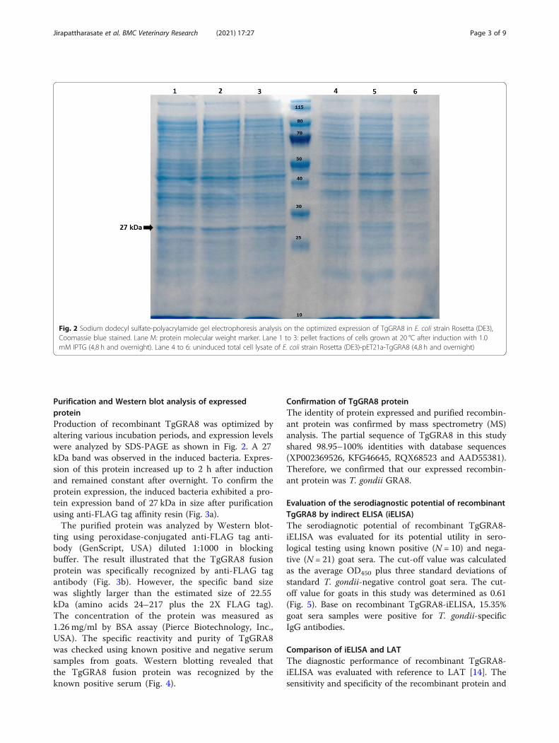
Purification and Western blot analysis of expressedproteinProduction of recombinant TgGRA8 was optimized byaltering various incubation periods, and expression levelswere analyzed by SDS-PAGE as shown in Fig. 2. A 27kDa band was observed in the induced bacteria. Expres-sion of this protein increased up to 2 h after inductionand remained constant after overnight. To confirm theprotein expression, the induced bacteria exhibited a pro-tein expression band of 27 kDa in size after purificationusing anti-FLAG tag affinity resin (Fig. 3a).The purified protein was analyzed by Western blot-
ting using peroxidase-conjugated anti-FLAG tag anti-body (GenScript, USA) diluted 1:1000 in blockingbuffer. The result illustrated that the TgGRA8 fusionprotein was specifically recognized by anti-FLAG tagantibody (Fig. 3b). However, the specific band sizewas slightly larger than the estimated size of 22.55kDa (amino acids 24–217 plus the 2X FLAG tag).The concentration of the protein was measured as1.26 mg/ml by BSA assay (Pierce Biotechnology, Inc.,USA). The specific reactivity and purity of TgGRA8was checked using known positive and negative serumsamples from goats. Western blotting revealed thatthe TgGRA8 fusion protein was recognized by theknown positive serum (Fig. 4).
Confirmation of TgGRA8 proteinThe identity of protein expressed and purified recombin-ant protein was confirmed by mass spectrometry (MS)analysis. The partial sequence of TgGRA8 in this studyshared 98.95–100% identities with database sequences(XP002369526, KFG46645, RQX68523 and AAD55381).Therefore, we confirmed that our expressed recombin-ant protein was T. gondii GRA8.
Evaluation of the serodiagnostic potential of recombinantTgGRA8 by indirect ELISA (iELISA)The serodiagnotic potential of recombinant TgGRA8-iELISA was evaluated for its potential utility in sero-logical testing using known positive (N = 10) and nega-tive (N = 21) goat sera. The cut-off value was calculatedas the average OD450 plus three standard deviations ofstandard T. gondii-negative control goat sera. The cut-off value for goats in this study was determined as 0.61(Fig. 5). Base on recombinant TgGRA8-iELISA, 15.35%goat sera samples were positive for T. gondii-specificIgG antibodies.
Comparison of iELISA and LATThe diagnostic performance of recombinant TgGRA8-iELISA was evaluated with reference to LAT [14]. Thesensitivity and specificity of the recombinant protein and
Fig. 2 Sodium dodecyl sulfate-polyacrylamide gel electrophoresis analysis on the optimized expression of TgGRA8 in E. coli strain Rosetta (DE3),Coomassie blue stained. Lane M: protein molecular weight marker. Lane 1 to 3: pellet fractions of cells grown at 20 °C after induction with 1.0mM IPTG (4,8 h and overnight). Lane 4 to 6: uninduced total cell lysate of E. coli strain Rosetta (DE3)-pET21a-TgGRA8 (4,8 h and overnight)
Jirapattharasate et al. BMC Veterinary Research (2021) 17:27 Page 3 of 9

kappa values at 95% confidence interval (95% CI) werecalculated. The seropositivity rate of goat samples inLAT was 17.0%. The sensitivity and specificity of LATfor the recombinant protein were 71.1 and 96.0%, re-spectively. Substantial agreement between the twomethods was indicated by κ = 0.69 (Table 1).
DiscussionRecently, recombinant DNA technology and syntheticDNA have played important roles in high-quality recom-binant antigenic protein production for the serologicaldiagnosis of T. gondii infection. Several recombinantproteins have been produced and applied for the detec-tion of T. gondii infection. These proteins include rhop-try proteins, matrix proteins, microneme proteins,surface antigens, and GRAs [8]. Among them, the GRAproteins have been considered potential diagnostic anti-gens and have been used to differentiate the stages of in-fection [24]. Generally, the method of recombinantprotein production requires cDNA extracted from livepathogens as a template to amplify target genes. How-ever, unsuccessful recombinant protein production using
natural gene sequences, including no or low expression,inclusion body formation, and protein inactivity, hasbeen described [25]. To overcome these problems, wedemonstrated the production of recombinant proteinfrom a synthetic TgGRA8 gene and tested the immuno-diagnostic potential of the produced protein via iELISA.Although codon optimization was used to optimize and
enhance protein expression in the present study, we failedto produce the recombinant protein using the full-lengthTgGRA8 gene. Previous studies described a transmem-brane region of the GRA8 gene encoding amino acids223–242 using bioinformatic prediction [16] and reportedthat the region can affect host cell growth and decreaseprotein yield [26]. Therefore, we selected the specific re-gion of the TgGRA8 protein based on the prediction oftransmembrane helices in proteins using an online pro-gram (http://www.cbs.dtu.dk/services/TMHMM/). Afterremoving the transmembrane region, the gene fragmentencoding amino acids 24–217 was used to express theprotein, and a specific 27-kDa band was observed on SDS-PAGE. Our result was similar to that of Babaie et al. [18],who designed and expressed a recombinant protein from
Fig. 3 a Sodium dodecyl sulfate-polyacrylamide gel electrophoresis analysis of expression of recombinant Toxoplasma gondii dense granularantigen 8 (TgGRA8) protein. Lane M, protein molecular weight marker; Lane 1, the soluble recombinant TgGRA8 protein was purified using anti-FLAG tag affinity resin. b Western blot analysis of purified recombinant TgGRA8. Lane M, protein molecular weight marker; Lane 1, the purified27-kDa TgGRA8 protein was detected using an anti-FLAG tag antibody
Jirapattharasate et al. BMC Veterinary Research (2021) 17:27 Page 4 of 9

the GRA8 gene fragment corresponding to amino acids23–169. However, a difference in size between the appar-ent band and the calculated molecular weight of TgGRA8(22.55 kDa) was observed in the present study. The pre-dicted protein encoded by the TgGRA8 gene featuredhigh proline content (54 amino acids). The presence of ex-cessive proline residues in proteins causes structural rigid-ity in the primary sequence, thereby decreasing theelectrophoretic mobility [16]. Regarding the yield ofTgGRA8, our production gained lower yields than themethod of Babaie and colleague [18]. Therefore, the at-tempt of using codon-optimisation DNA is not consideredadvantageous for recombinant GRA8 expression.Numerous GRA proteins, both single and combina-
tions of proteins, have been applied for the serodetectionof animal toxoplasmosis. In cats, a single GRA7 recom-binant protein [27] and a mixture of recombinantGRA2, GRA6, GRA7, and GRA15 [28] were used to de-termine the prevalence of T. gondii infection in Chinaand Japan, respectively. Moreover, recombinant GRA7
protein-based ELISA has been used in seroprevalencestudies of farm animals in Egypt [29] and Thailand [30].To date, only one study describing the use of TgGRA8together with recombinant GRA7 to detect specific IgGantibodies against the parasite was published in domesticturkeys [31].The potential utility of recombinant TgGRA8 protein
in serodiagnosis was assessed using known positive andnegative goat sera. The result of Western blotting indi-cated that the protein is a potential marker for detectingT. gondii infection in goats. A previous immunochemicalevaluation of TgGRA8 using ELISA recorded high re-activity for the recombinant protein using human sera[19], in line with the present result. A possible explan-ation for the high OD in this study could be the unspe-cific epitopes of this antigen in the amino-terminalregion [32].The infection rate in our study was lower than that of
27.9% in a previous report on goats in Satun province,Thailand [33]. The difference of the seroprevalence ratemay be attributable to the use of different serologicaldiagnosis techniques (iELISA and LAT) and differentsampling areas. The sensitivity and specificity obtainedusing recombinant TgGRA8 in this study indicated thatthe recombinant protein could be used as an antigen forserological tests of T. gondii. However, the use of recom-binant proteins for the serodetection of animal toxoplas-mosis may be affected by the immune system indifferent animal species. Therefore, the antibody re-sponse in various animals and the epitope structures ofrecombinant TgGRA8 should be confirmed.
ConclusionOur study produced a recombinant protein from a syn-thetic TgGRA8 gene. The sensitivity and specificity ofTgGRA8 demonstrated that this protein could be a goodserological marker for detecting specific IgG in goat sera.Commercial gene synthesis is an alternative tool to sup-port recombinant protein expression in the absence ofpathogen access.
MethodsGene synthesis of TgGRA8The complete GRA8 coding sequence (accession num-ber: TGME49_054720) was obtained from an onlinedatabase (http://ToxoDB.org). The TgGRA8 sequenceconsists of 810 nucleotides that encode a 269-amino acidprotein. A signal peptide (SPs) of GRA8 was determinedusing online program, SignalP 4.1 (http://www.cbs.dtu.dk/services/SignalP/). The results showed that smallfragments of amino acids 1–23 were expressed as a sig-nal sequence. Therefore, encoding amino acid 24–269was constructed and inserted into a pET-21a vector
Fig. 4 Western blot analysis. Purified proteins were separated via12% sodium dodecyl sulfate-polyacrylamide gel electrophoresis,transferred to nitrocellulose membranes, and then probed withknown positive and negative goat sera. a Lane M, protein molecularweight marker; Lane 1, strong reactivity with known positive serum;b Lane M, protein molecular weight marker; Lane 1, result fornegative serum
Jirapattharasate et al. BMC Veterinary Research (2021) 17:27 Page 5 of 9

using NdeI and XhoI as the cloning sites (General Bio-systems, USA).
Construction of recombinant TgGRA8The potential transmembrane regions (TMs) of TgGRA8were predicted by online server (http://www.cbs.dtu.dk/services/TMHMM/). The encoding amino acids 218–269 was transmembrane region. Therefore, only an anti-genic fragment of recombinant TgGRA8 encodingamino acids 24–217 was PCR-amplified (Fig. 6). Theprimers used for amplification of the sequence by PCRwas T7 promoter-(FW), 5′-TAA TACG ACT CAC TATAG-3′ (New England Biolabs, UK); and TgGRA8-RW,5′-AGT acc ggt GGT GGC GGT TGC CGG CTG-3′.The reverse primer was designed to contain the AgeI re-striction site. PCR was performed using PCR Q5® High-Fidelity DNA Polymerase (New England Biolabs) usingthe following program: 98 °C for 1 min, followed by 30cycles of 98 °C for 10 s, 58 °C for 20 s, and 72 °C for 20 s,and final extension at 72 °C for 2 min.The PCR amplicon was digested using NdeI and AgeI.
After digestion, the PCR product was ligated into themodified pET-21a vector harboring a C-terminal fusion
protein linker (GGGS) and 2X FLAG tag (DYKDDDDKDYKDDDDK) (General Biosystems, USA) and trans-formed into Escherichia coli DH5α-competent cells.Ten colonies were selected and expanded in overnight
cultures, and DNA was extracted using a QIAprep SpinMiniprep Kit (Qiagen, Germany). The insert of TgGRA8in the purified plasmid was sequenced using a Dye Ter-minator Cycle Sequencing Kit (Applied Biosystems,USA) and the 3500xL genetic analyzer (Applied Biosys-tems). The TgGRA8 sequences were determined usingBioedit version 7.2.5 (Tom Hall Ibis Biosciences, USA).
Expression of TgGRA8The recombinant TgGRA8 plasmids were transformedinto E. coli strain Rosetta (DE3) cells and cultivated in2XTY supplemented with 1% glucose and 200 ng/mlampicillin at 37 °C with shaking at 200 rpm. E. coli carry-ing recombinant TgGRA8 was measured at an optimaldensity at 600 nm (OD600) of 0.5 and induced withisopropyl-β-D-thiogalactopyranoside at a final concen-tration of 1 mM 20 °C for various incubation periods (2,4 and overnight) with shaking at 250 rpm. The inducedbacteria were harvested via centrifugation at 4400×g for
Table 1 Comparison of LAT and TgGRA8 recombinant protein-based iELISA for the detection IgG antibodies against Toxoplasmagondii infection
TgGRA8iELISA
LAT Sensitivity(95% CI)
Specificity(95% CI)
KappavaluePositive Negative Total
Positive 37 10 47 71.17 96.06 0.69
Negative 15 244 259 (56.72–82.45) (92.65–97.98)
Total 52 254 306
LAT Latex agglutination test, TgGRA8 T. gondii dense granular antigen 8, iELISA Indirect enzyme-linked immunosorbent assay, CI Confidence interval
Fig. 5 Antibody response to Toxoplasma gondii in field sera from goats using recombinant T. gondii dense granular antigen 8 protein-basedindirect enzyme-linked immunosorbent assays
Jirapattharasate et al. BMC Veterinary Research (2021) 17:27 Page 6 of 9

20min at 4 °C, and the bacterial pellet was resuspendedin 20ml of pre-chilled lysis buffer (150 mM NaCl, 50mM Tris-HCL [pH 9.5], 1% Triton X-100, 1 mM EDTA[pH 8.0], and 1% NP 40) and then incubated at 4 °C for30 min. After incubation, the bacterial cells were lysedvia sonication on ice for 10 min, and 1ml of 1× ProteaseInhibitor Cocktail was added (Promega, USA). TgGRA8expression was analyzed using 12% sodium dodecylsulfate-polyacrylamide gel electrophoresis (SDS-PAGE).
Protein purificationAnti-DYKDDDDK G1 affinity resin (GenScript, USA)was used for protein purification. The debris was centri-fuged at 10,000×g for 30 min at 4 °C, after which thesupernatant was transferred to a clean tube. The resinsuspension (600 μl) was loaded into an empty gravityflow column (Bio-Rad, USA) and washed with Tris-buffered saline (50 mM Tris-HCl, 150mM NaCl, pH7.4). Protein was eluted from the resin using alkalineelution buffer (0.1 M Tris, 0.5 M NaCl, pH 12.0) andneutralized with 1M HCl. The protein concentrationwas measured using NanoDrop ND-1000 UV/Vis spec-trophotometer (Thermo Fisher Scientific, USA).The eluted fractions were dialyzed using SnakeSkin
Dialysis Tubing, 10 kDa cut-off (Thermo Fisher Scien-tific) against phosphate-buffered saline (PBS, pH 7.2) at4 °C. The debris formed during dialysis was removed via
centrifugation at 10,000×g for 5 min at 4 °C, and theconcentration of the purified recombinant protein wasassayed using both SDS-PAGE and a Coomassie proteinassay reagent kit using BSA according to the manufac-turer’s protocol (Pierce Biotechnology, Inc., USA).To confirm the sequence of the recombinant protein,
the mass spectrometry (MS) analysis was carried out byProteomics Services Center, Faculty of Medical Technol-ogy, Mahidol University.
Western blottingFive micrograms of recombinant TgGRA8 was resolvedby 12% SDS-PAGE and then electrotransferred (Trans-blot, Bio-Rad) onto a nitrocellulose membrane (Milli-pore, USA). The membrane was washed three times withPBS, blocked with 5% skim milk, and then incubated at37 °C for 1 h with constant shaking. After incubation,the membrane was washed three times with PBS con-taining 0.01% Tween 20 (PBS-T) and rinsed with PBS.The TgGRA8 protein in nitrocellulose membrane wasprobed using antibody or known reference positive andnegative goat sera (diluted 1:250 in 5% skim milk) keptin our laboratory and incubated at 37 °C for 1 h withconstant shaking. The monoclonal antibody (mAb)against Flag-tag (GenScript, USA) was diluted 1:1000,while polyclonal mouse anti-goat immunoglobulin/HRP(Dako, Denmark) was diluted 1:2000 in blocking buffer.
Fig. 6 The complete nucleotide and amino acid sequence of T. gondii GRA8 (accession number: TGME49_054720). The expression region of GRA8in this study is delineated by shading (encoding a 194-residual peptide)
Jirapattharasate et al. BMC Veterinary Research (2021) 17:27 Page 7 of 9

After incubation, the membrane was washed three timeswith PBS-T. The protein band was developed accordingto peroxidase activity using 3,3′,5,5′-tetramethylbenzi-dine (KPL, Gaithersburg, MD, USA).
Goat serum samplesThe process of sample collection was reviewed and ap-proved by the Animal Care and Use Committee of theFaculty of Veterinary Science, Mahidol University,Thailand (Approval No. MUVS-2018-03-09). A total of306 serum samples were obtained from a goat farm inKanchanaburi province, Thailand. The goats were re-strained by holding the base of the horn and blood wascollected from the jugular vein and immediately trans-ferred into 10ml vacuum blood tubes without anticoagu-lant. The animals were not allowed returned to their cageuntil complete hemostasis has been achieved. All bloodsamples were kept in cooled box with ice pack and sent tothe laboratory at Faculty of Veterinary Science, MahidolUniversity. The sera were separated after sedimentation ofblood cells and stored at − 20 °C until examination.
IgG iELISAPurified recombinant TgGRA8 was diluted at a finalconcentration of 0.1 μg/ml in coating buffer (50 mM bi-carbonate, pH 9.6) and added to separate wells of theELISA plates (Nunc, Denmark). The coated plates wereincubated overnight at 4 °C. The next day, the plateswere washed five times with PBS-T and blocked with 5%PBS-skimmed milk (PBS-SM) for 1 h at 37 °C. Afterwashing with PBS-T, duplicate serum samples were di-luted 1:250 in PBS-SM, and 50 μl of diluted serum wereadded to each well. The plates were incubated at 37 °Cfor 1 h and washed with PBS-T five times. Specific IgGantibody was detected using horseradish-peroxidase-conjugated anti-goat IgG antibodies (Invitrogen, USA).The conjugate was diluted 1:5000 with PBS, and 50 μl ofdiluted conjugates were added. After incubation at 37 °Cfor 1 h, the plates were washed five times with PBS-T,and then 3,3′,5,5′-tetramethylbenzidine (Invitrogen,USA) was added to develop the color. After 15 min, thereaction was stopped by adding 50 μl of 0.1M HCl.OD450 was read using a microplate reader (modelELx808, Biotex, VT, USA).
LATThe negative and positive control sera were confirmedusing MAST® TOXOREAGENT (Mast Group, Liverpool,UK). Positive samples were considered when agglutin-ation was observed at a dilution of 1:32 or greater.
Statistical analysesThe results of iELISA and LAT were calculated usingonline software (http://vassarstats.net) to determine the
percentage of agreement, sensitivity, specificity, and thekappa values with 95% confidence intervals. Thestrength of agreement was graded as fair (κ = 0.21–0.40),moderate (κ = 0.41–0.60), and substantial (κ =0.61–0.80).
Supplementary InformationThe online version contains supplementary material available at https://doi.org/10.1186/s12917-020-02719-3.
Additional file 1.
Additional file 2.
AbbreviationELISA: Enzyme-linked immunosorbent assay; LAT: Latex agglutination test;PBS: Phosphate-buffered saline; PV: Parasitophorous vacuole
AcknowledgementsWe thank Dr. Sarawut Taksinoros and Dr. Raweenipa Tohkwankeaw, for theirsignificant contribution during sample collection, and veterinarians fromKanchanaburi provincial livestock office for their general support. The authorsthanks Dr. Phirom Prompiram for technical assistance. In addition, we wouldlike to express our appreciation to Asst. Prof. Dr., Onrapak Reamtong formass spectrometry analysis. The authors would like to thank Enago (https://www.enago.com) for the professional English language review.
Authors’ contributionsCJ conceived the present idea in this paper, artwork, perform data analysisand drafted the first manuscript. RU, AP, KJ carried out the laboratory work.PD approved the manuscript. All authors read and approved the final versionof the manuscript.
FundingThis research is supported by Mahidol University (grant number: A30/5261).The funding body has not participated in the research design, collection,analysis, interpretation of data and writing the manuscript.
Availability of data and materialsThe datasets used and/or analyzed during the current study are availablefrom the corresponding author on reasonable request.
Ethics approval and consent to participateThe process of sample collection was reviewed and approved by the AnimalCare and Use Committee of the Faculty of Veterinary Science, MahidolUniversity, Thailand (approval no. MUVS-2018-03-09). The animals were han-dled humanely in strict accordance with the requirements of the Animal Eth-ics Procedures and Guidelines of Institute of Animals for Scientific purposeDevelopment (IAD), Thailand. Before signing a consent, the owners of the se-lected farms were informed of the study and provided their approval forsampling of the goats. All samples collection process was conducted by localauthorities and veterinarians.
Consent for publicationNot applicable.
Competing interestsThe authors declare that they have no competing interests.
Author details1Department of Preclinic and Applied Animal Science, Faculty of VeterinaryScience, Mahidol University, 999 Phutthamonthon sai 4 Rd, Salaya,Nakhonpathom 73170, Thailand. 2Department of Protozoology, Faculty ofTropical Medicine, Mahidol University, 420/6 Ratchawithi Road, Ratchathewi,Bangkok 10400, Thailand. 3Department of Medical Sciences, Medical LifeSciences Institute, 88/7 Tiwanon Road, Talad Kwan Subdistrict, MuangDistrict, Nonthaburi 11000, Thailand.
Jirapattharasate et al. BMC Veterinary Research (2021) 17:27 Page 8 of 9

Received: 23 July 2020 Accepted: 9 December 2020
References1. Montoya JG, Liesenfeld O. Toxoplasmosis. Lancet. 2004;363:1965–76.2. Wang ZD, Huan-Huan Liu HH, Ma ZX, Ma HU, Li ZY, Yang ZB, Zhu XQ, Feng
B, Wei X, Liu Q. Toxoplasma gondii infection in immunocompromisedpatients: a systematic review and meta-analysis. Front Microbiol. 2017;8:389.
3. Dubey JP. Advances in the life cycle of Toxoplasma gondii. Int J Parasitol.1998;28:1019–24.
4. Stelzera S, Bassob W, Benavides Silvánc J, Ortega-Morad LM, Maksimova P,Gethmanna J, Conrathsa FJ, Schare G. Toxoplasma gondii infection andtoxoplasmosis in farm animals: risk factors and economic impact. FoodWaterborne Parasitol. 2019;12:e00037.
5. Zhang K, Lin G, Han Y, Li J. Serological diagnosis of toxoplasmosis andstandardization. Clin Chim Acta. 2016;461:83–9.
6. Khan AH, Noordin R. Serological and molecular rapid diagnostic tests forToxoplasma infection in humans and animals. Eur J Clin Microbiol Infect Dis.2020;39:19–30.
7. Dard C, Fricker-Hidalgo H, Brenier-Pinchart MP, Pelloux H. Relevance of andnew developments in serology for toxoplasmosis. Trends Parasitol. 2016;32:492–506.
8. Kotresha D, Noordin R. Recombinant proteins in the diagnosis oftoxoplasmosis. APMIS. 2010;118:529–42.
9. Holec-Gasior L. Toxoplasma gondii recombinant antigens as tools forserodiagnosis of human toxoplasmosis: current status of studies. ClinVaccine Immunol. 2013;20:1343–51.
10. Nam HW. GRA proteins of Toxoplasma gondii: maintenance of host-parasiteinteractions across the parasitophorous vacuolar membrane. Korean JParasitol. 2009;47:S29–37.
11. Ching XT, Lau YL, Fong MY, Nissapatorn V. Evaluation of Toxoplasma gondii-recombinant dense granular protein (GRA2) for serodiagnosis by westernblot. Parasitol Res. 2013;112:1229–36.
12. Ching XT, Lau YL, Fong MY, Nissapatorn V, Andiappan H. Recombinantdense granular protein (GRA5) for detection of human toxoplasmosis byWestern blot. Biomed Res Int. 2014;2014:690529.
13. Golkar M, Azadmanesh K, Khalili G, Khoshkholgh-Sima B, Babaie J, Mercier C,Brenier-Pinchart MP, Fricker-Hidalgo H, Pelloux H, Cesbron-Delauw MF.Serodiagnosis of recently acquired Toxoplasma gondii infection in pregnantwomen using enzyme-linked immunosorbent assays with a recombinantdense granule GRA6 protein. Diagn Microbiol Infect Dis. 2008;61:31–9.
14. Sadeghiani G, Zare M, Babaie J, Shokrgozar MA, Azadmanesh K, Fard-Esfahani P, Golkar M. Heterologous production of dense granule GRA7antigen of Toxoplasma gondii in Escherichia coli. Southeast Asian J. trop.Med. Public Health. 2009;40:692–700.
15. Terkawi MA, Kameyama K, Rasul NH, Xuan X, Nishikawa Y. Development ofan immunochromatographic assay based on dense granule protein 7 forserological detection of Toxoplasma gondii infection. Clin Vaccine Immunol.2013;20:596–601.
16. Carey KL, Donahue CG, Ward GE. Identification and molecularcharacterization of GRA8, a novel, proline-rich, dense granule protein ofToxoplasma gondii. Mol Biochem Parasitol. 2000;105:25–37.
17. Li S, Maine G, Suzuki Y, Araujo FG, Galvan G, Remington JS, Parmley S.Serodiagnosis of recently acquired Toxoplasma gondii infection with arecombinant antigen. J Clin Microbiol. 2000;38:179–84.
18. Babaie J, Miri M, Sadeghiani G, Zare M, Khalili G, Golkar M. Expression andsingle-step purification of GRA8 antigen of Toxoplasma gondii in Escherichiacoli. Avicenna J Med Biotechnol. 2011;3:67–77.
19. Costa JG, Duré AB. Immunochemical evaluation of two Toxoplasma gondiiGRA8 sequences to detect acute toxoplasmosis infection. Microb Pathog.2016a;100:229–36.
20. Gustafsson C, Govindarajan S, Minshull J. Codon bias and heterologousprotein expression. Trends Biotechnol. 2004;22:346–53.
21. Plotkin JB, Kudla G. Synonymous but not the same: the causes andconsequences of codon bias. Nat Rev Genet. 2011;12:32–42.
22. Kunjapur AM, Pfingstag P, Thompson NC. Gene synthesis allows biologiststo source genes from farther away in the tree of life. Nat Commun. 2018;9:4425.
23. Kobokovich A, West R, Montague M, Inglesby T, Gronvall GK. Strengtheningsecurity for gene synthesis: recommendations for governance. Health Secur.2019;17:419–29.
24. Mercier C, Cesbron-Delauw MF, Ferguson DJP. Dense granules of theinfectious stages of Toxoplasma gondii: their central role in the host/parasiterelationship. In: Soldati D, Ajioka J, editors. Toxoplasma: molecular andcellular biology. Norwich: Horizon Scientific Press; 2007. p. 475–92.
25. Rosano GL, Ceccarelli EA. Recombinant protein expression in Escherichia coli:advances and challenges. Front Microbiol. 2014;17:172.
26. Babaie J, Zare M, Sadeghiani G, Lorgard-Dezfuli M, Aghighi Z, Golkar M.Bacterial production of dense granule antigen GRA8 of Toxoplasma gondii.Iran Biomed J. 2009;13:145–51.
27. Cai Y, Wang Z, Li J, Li N, Wei F, Liu Q. Evaluation of an indirect Elisa usingrecombinant granule antigen Gra7 for serodiagnosis of Toxoplasma gondiiinfection in cats. J Parasitol. 2015;101:37–40.
28. Abdelbaset AE, Alhasan H, Salman D, Karram MH, Ellah Rushdi MA, Xuenan X,Igarashi M. Evaluation of recombinant antigens in combination and singleformula for diagnosis of feline toxoplasmosis. Exp Parasitol. 2017;172:1–4.
29. Fereig RM, Mahmoud HYAH, Mohamed SGA, AbouLaila MR, Abdel-WahabA, Osman SA, Zidan SA, El-Khodary SA, Mohamed AEA, Nishikawa Y.Seroprevalence and epidemiology of Toxoplasma gondii in farm animals indifferent regions of Egypt. Vet Parasitol Reg Stud Reports. 2016;3–4:1–6.
30. Udonsom R, Sukthana Y, Yoshifumi N, Fereig RM, Jirapattharasate C. Currentsituation of Neospora caninum and Toxoplasma gondii infection among beefcattle in Kanchanaburi, Ratchaburi and Nakhon Patom provinces, Thailand.Thai J Vet Med. 2018;48:403–9.
31. Koethe M, Pott S, Ludewig M, Bangoura B, Zöller B, Daugschies A, TenterAM, Spekker K, Bittame A, Mercier C, Fehlhaber K, Straubinger RK.Prevalence of specific IgG-antibodies against Toxoplasma gondii in domesticturkeys determined by kinetic ELISA based on recombinant GRA7 andGRA8. Vet Parasitol. 2011;180:179–90.
32. Costa JG, Duré AB. Effectiveness of two sequences of Toxoplasma gondiiSAG2 protein to differentiate toxoplasmosis infection stages by measuringIgG, IgA and IgM antibodies. Trop Biomed. 2016b;33:246–59.
33. Jittapalapong S, Sangvaranond A, Pinyopanuwat N, Chimnoi W, KhachaeramW, Koizumi S, Maruyama S. Seroprevalence of Toxoplasma gondii infectionin domestic goats in Satun Province, Thailand. Vet Parasitol. 2005;127:17–22.
Publisher’s NoteSpringer Nature remains neutral with regard to jurisdictional claims inpublished maps and institutional affiliations.
Jirapattharasate et al. BMC Veterinary Research (2021) 17:27 Page 9 of 9