Embed Size (px)
Citation preview
Development and characterization of segmented
polyurethanes based on L-amino acid based
chain extenders
A Thesis submitted for the degree of Doctor of Philosophy
By
Swati Sharma (M.Sc. Biochemistry)
Faculty of Life and Social Sciences Swinburne University of Technology
Melbourne
Australia
February 2016

i
Abstract
The acid catalysed Fischer and alkali metal salt based esterification reactions were used
to synthesise a series of diamine ester and dihydroxy ester compounds respectively from
various naturally occurring L-amino acids. L-leucine, L-isoleucine, L-phenylalanine, L-
tyrosine amino acid were used to synthesise diamine diester compounds whilst amine
group protected L-Z-serine, L-Z-threonine and L-Z-tyrosine amino acids were used to
synthesise novel dihydroxy diester compounds. For the dihydroxy ester compounds, in
particular, optimisation of the reaction temperature was done. The yield for both diamine
and dihydroxy ester synthesis were ~ 80 % and 85% respectively. Few of the synthesised
ester compounds were yellow colour oil and few of them were white solid in appearance.
The synthesised compounds were fully characterised by nuclear magnetic resonance,
infrared resonance spectroscopy and mass spectroscopy. The success of the reaction and
the purity of the synthesised compounds were confirmed by these techniques. The
intended purpose of the synthesis of these novel amino acid based ester compounds is to
use them as dihydroxy and diamine chain extender for polyurethane and polyurethane
urea synthesis.
A series of polyurethane and polyurethane urea based on amino acid based chain extender
(mentioned above) was synthesised. Polycaprolactone (Mw1000) act as polyol and 4,4-
methylenediphenyl diisocyanate act as diisocyanate component for polyurethane
synthesis. The control polyurethane synthesised was based on 1,4-butanediol as chain
extender with polycaprolactone as polyol and 4,4-methylenediphenyl diisocyanate as
diisocyanate component. Synthesised polymers were fully characterised for structural,
thermal, surface and mechanical properties. Obtained properties of the amino acid based
polyurethane/polyurethane urea were compared with the control polyurethane to evaluate
the effect of chain extender type and its structure onto the physiochemical properties of
the synthesised polymer. Synthesised amino acid based polyurethane/polyurethane urea
showed moderately high molecular weight with narrow polydispersity. The percent yield
of the polyurethane/polyurethane urea synthesis was ~ 70%. The amino acid based
polymers ranged from completely amorphous to semicrystalline polymer. The hard
segment was amorphous in all cases of the amino acid based polyurethane/polyurethane
urea. Phase mixed morphology was shown by amino acid based

ii
polyurethane/polyurethane urea as compared to phase segregated morphology observed
for control polyurethane. Mechanical properties were tested by obtaining stress strain
curve. The amino acid based polymers were weak elastomeric material with low tensile
strength and high extensibility as compared to control polyurethane. Non-linear structure
of chain extenders and weak hydrogen bonding might be one of the main reasons for low
mechanical properties. The amino acid based polyurethane/polyurethane urea were stable
up to high temperature. The polymer surface hydrophobicity was increased with the
incorporation of amino acid based chain extender.
The benzyloxycarbonyl protecting group of serine amino acid based polyurethane was
removed by the use of Hydrogen bromide/Acetic acid solution method. Reaction time
was optimised for the deprotection reaction. Nuclear magnetic resonance spectroscopic
analysis indicated that deprotection resulted in 62 mol % reduction in benzyloxycarbonyl
group content with minimal affecting polyurethane backbone.
Structure property relationship of the polymer was studied by using high molecular
weight (Mw 2,000) of polycaprolactone as soft segment. A series of polyurethane and
polyurethane urea were synthesised and characterised with polycaprolactone (Mw 2,000
Dalton) as soft segment and obtained properties were compared to
polyurethane/polyurethane urea series made with polycaprolactone (Mw 1,000 Dalton).
It was observed that as soft segment molecular weight increases, phase segregation,
percent crystallinity increases and glass transition of soft segment decreases. Increase in
phase segregation morphology has also helped to obtain improved mechanical properties
of the amino acid based polyurethane/polyurethane urea. Surface hydrophobicity
increases as soft segment molecular weight increases. However, thermal stability was
decreased due to phase segregated morphology.
At the end, the in vitro cytotoxicity response of the amino acid based
polyurethane/polyurethane urea was evaluated by LIVE/DEAD assay kit. Mouse
fibroblast cells were seeded on to the polyurethane/polyurethane urea films. The
polyurethane/polyurethane urea did not show any cytotoxic response and healthy cell
growth, attachment and proliferation was observed on polymer films which indicates that
these polyurethane/polyurethane urea may be useful for biomedical applications.

iii
Acknowledgements
On this great occasion of my thesis submission, first of all I am grateful to the almighty
God without whose blessings I would not have been able to complete this challenging
journey.
I would like to convey my sincere thanks to my supervisor Professor Ian Harding for his
valuable guidance, advice and availability whenever required during this PhD. I would
also like to thank Senior Research Scientist Roshan Mayadunne for providing me help
and guidance during my research work performed at CSIRO.
I would like to express my gratitude to Dr. Keith McLean, Dr. Thilak Gunatillake and Dr.
Tim Hughes from CSIRO for providing me encouragement, guidance and friendly advice
whenever I needed during my experimental work. I would like to pass on a big Thank
You to Dr. Akhil Gupta whose immense knowledge and support helped me to think out
of the box and do things in a different way to achieve the desired outcome. I really
appreciate the help of Dr. Roger Mulder from CSIRO for NMR analysis, Dr. James
Mardel from CSIRO for FTIR analysis and Veronica Glattauer from CSIRO for polymer
cell studies.
Last but not the least I would like to thank my husband Rahul Bhardwaj for showing great
faith in me. Thanks for being such a wonderful husband and always there to make me feel
stronger mentally and providing all love and support to me in fulfilling my motherhood
duties during thesis writing.

iv
Declaration
This thesis contains no material which has been accepted for the award of any other
degree or diploma, in any University, college or any other educational institute. To the
best of my knowledge, this thesis contains no material previously published or written by
another person except where due reference is made in the text of the thesis.
Signature of Candidate: ..................................................................... Date: ........................................................................

v
List of Conference Presentations
Following is a list of conference presentations from the work contained in this thesis.
Conference Presentations
Sharma S., Harding I., Mayadunne R. M., (January, 2009). Amino acid based
diamine diester as chain extender in polyurethane synthesis. 19th Annual
conference of the Australasian society for biomaterials and tissue engineering
(ASBTE) Sydney, Australia.
Sharma S., Harding I., Mayadunne R. M., (February, 2010). Novel complex
amino acid based diols as chain extenders in polyurethane synthesis. 20th Annual
society for biomaterials and tissue engineering (ASBTE) Queensland, Brisbane,
Australia.
Sharma S., Harding I., Mayadunne R. M., (October, 2010). Novel complex amino
acid based diols as chain extenders in polyurethane synthesis. Symposium on
biomaterials, Rutgers University, New Jersey, USA.
Sharma S., Harding I., Mayadunne R. M., (May, 2011). Synthesis and
characterization of amino acid based polyurethane. CSIRO advanced material
conference (CAM), 2011, Melbourne, Australia.

vi
Table of Contents Abstract .......................................................................................................................................... i
Acknowledgements ...................................................................................................................... iii
Declaration ................................................................................................................................... iv
List of Conference Presentations .................................................................................................. v
Table of Contents ......................................................................................................................... vi
Index of Figures .......................................................................................................................... xii
Index of Schemes ........................................................................................................................ xv
Index of Tables .......................................................................................................................... xvi
Abbreviations ........................................................................................................................... xviii
Chapter 1: Introduction ................................................................................................................ 1
1.1 Introduction ............................................................................................................................. 2
1.2 Research Hypothesis ............................................................................................................... 3
1.3 Layout of thesis ....................................................................................................................... 5
Chapter 2: Literature Review ........................................................................................................ 6
2.1 Introduction ............................................................................................................................. 7
2.1.1 Structure of polyurethane ............................................................................................. 7
2.1.2 Advantage of using polyurethane ................................................................................ 9
2.1.3 Role of polyurethane .................................................................................................... 9
2.2 Polyurethane history and development ................................................................................. 10
2.3 Components of polyurethane ................................................................................................ 12
2.3.1 Diisocyanate ............................................................................................................... 13
2.3.2 Types of diisocyanate................................................................................................. 13
2.3.3 Polyols ........................................................................................................................ 15
2.3.4 Chain extender ........................................................................................................... 16
2.3.5 Primary reaction of isocyanate group ................................................................................ 19
2.3.5.1. Reaction with polyol/hydroxyl group .................................................................... 20
2.3.5.2. Reaction with amines ............................................................................................. 20
2.3.5.3. Reaction with water ............................................................................................... 20
2.3.5.4. Secondary reaction of isocyanate group ................................................................ 21
2.4 Synthesis of segmented polyurethane ................................................................................... 21
2.5 Morphology in polyurethanes ............................................................................................... 23
2.6 Structure-property relationship of polyurethane ................................................................... 26
2.6.1 Soft segment................................................................................................................... 26

vii
2.6.2 Hard segment ................................................................................................................. 29
2.6.2.1 Chain extender ........................................................................................................ 29
2.6.2.2 Diisocyanate ............................................................................................................ 32
2.7 Hydrogen bonding ................................................................................................................ 34
2.8 Polyurethanes as biomaterials ............................................................................................... 36
2.9 Technical Approach .............................................................................................................. 40
Chapter 3: Synthesis and characterization of L-amino acids based ester compounds ................ 42
3.1 Introduction ........................................................................................................................... 43
3.1.1 Polyurethane Structure ............................................................................................... 43
3.1.2 Chain Extenders ......................................................................................................... 44
3.1.3 The Role of amino acid based chain extender in polyurethane biocompatibility properties............................................................................................................................. 45
3.1.4 Strategy to be used for the synthesis of amino acid based chain extenders ............... 47
3.2 Materials ............................................................................................................................... 49
3.3 Experimental Procedure ........................................................................................................ 49
3.3.1 Synthesis of diamine ester compounds .......................................................................... 49
3.3.1.1 Octane-1, 8-diyl bis (2-amino-4-methylpentanoate) (311) ..................................... 49
3.3.2 Synthesis of dihydroxy ester compounds ....................................................................... 50
3.3.2.1Butane-1,4-diylbis(2-(benzyloxycarbonylamino)-3-hydroxypropanoate) (318) ..... 50
3.4 Characterization .................................................................................................................... 51
3.4.1 Nuclear magnetic resonance spectroscopy ................................................................. 51
3.4.2 Fourier transform infrared spectroscopy .................................................................... 51
3.4.3 Mass Spectroscopy ..................................................................................................... 52
3.4.4 Thin layer chromatography ........................................................................................ 52
3.5 Results and Discussion ......................................................................................................... 52
3.5.1 Synthesis of L-amino acids based diamine ester compounds ........................................ 52
3.5.2 Characterization ............................................................................................................. 56
3.5.2.1 Octane-1, 8-diyl bis (2-amino-4-methylpentanoate) (311) ..................................... 57
3.5.2.1.1 1H-Nuclear Magnetic Resonance Analysis .......................................................... 57
3.5.2.1.3 Mass spectroscopy ............................................................................................... 63
3.5.2.2 Octane-1, 8-diyl bis (2-amino-3(4-hydroxyphenyl) propanoate) (314) .................. 63
3.5.2.2.1 1H-Nuclear Magnetic Resonance spectroscopy ................................................... 64
3.5.2.2.2 Fourier transform infrared spectroscopy .............................................................. 71
3.5.2.2.3 Mass spectroscopy ............................................................................................... 72
3.5.2.2.4 Other Compounds ................................................................................................ 72

viii
3.5.2 Synthesis of L-Z-amino acids based dihydroxy ester compounds ................................. 73
3.5.2.1 Reaction with caesium salt ...................................................................................... 73
3.5.2.2 Octane-1, 8-diyl bis (2-amino-4-methylpentanoate) – 318 ..................................... 74
3.5.2.3 Optimisation of reaction conditions ........................................................................ 77
3.5.2.4 Characterisation .......................................................................................................... 79
3.5.2.4.1Butane-1,4-diylbis(2-(benzyloxycarbonylamino)-3hydroxypropanoate)-318 ...... 79
3.5.2.4.1.1 1H-Nuclear Magnetic Resonance spectroscopy ................................................ 79
3.5.2.4.1.2 Fourier Transform Infrared spectroscopy ......................................................... 81
3.5.2.4.1.3 Mass Spectroscopy ............................................................................................ 82
3.5.2.4.2 Butane-1, 4-diylbis (2- (benzyloxycarbonylamino) 3(4hydroxyphenyl) ............. 83
propanoate) – (320) ............................................................................................................. 83
3.5.2.4.2.1 1H-Nuclear Magnetic Resonance Spectroscopy ................................................ 83
3.5.2.4.2.2 Fourier Transform Infrared Spectroscopy ......................................................... 85
3.5.2.4.2.3 Mass Spectroscopy ............................................................................................ 86
3.6 Conclusion ............................................................................................................................ 86
4.1 Introduction ........................................................................................................................... 89
4.2 Materials ............................................................................................................................... 92
4.3 Experimental Procedures ...................................................................................................... 92
4.3.1 Synthesis of polyurethane .............................................................................................. 92
4.3.2 Synthesis of control polyurethane .................................................................................. 94
4.3.3 Deprotection of Z-group ................................................................................................ 95
4.3.4 Synthesis of polyurethane urea ...................................................................................... 96
4.4 Characterization of polymers ................................................................................................ 98
4.4.1 Gel permeation chromatography .................................................................................... 98
4.4.2 Solubility ........................................................................................................................ 98
4.4.3 Nuclear magnetic resonance spectroscopy..................................................................... 99
4.4.4 Fourier transform infrared spectroscopy ........................................................................ 99
4.4.5 Thermal Characterization ............................................................................................... 99
4.4.6 Water contact angle measurement ............................................................................... 100
4.4.7 Compression moulding ................................................................................................ 100
4.4.8 Mechanical properties .................................................................................................. 101
4.5 Results and discussion ........................................................................................................ 101
4.5.1 Polyurethane synthesis reaction ................................................................................... 101
4.5.2 Characterization of polyurethane ................................................................................. 105
4.5.2.1 Gel Permeation Chromatography .......................................................................... 105

ix
4.5.2.2 Solubility ............................................................................................................... 106
4.5.2.3 1H Nuclear Magnetic Resonance Spectroscopy .................................................... 107
4.5.2.4 Fourier Transform Infrared Spectroscopy ............................................................. 109
4.5.2.5 Differential Scanning Calorimetry ........................................................................ 111
4.5.2.6 Thermogravemetric Analysis ................................................................................ 119
4.5.2.7 Water Contact Angle ............................................................................................. 120
4.5.2.8 Mechanical Properties ........................................................................................... 122
4.6 Deprotection of Z-group ..................................................................................................... 126
4.6.1 Introduction .................................................................................................................. 126
4.6.2 Deprotection reaction ................................................................................................... 127
4.6.3 Optimisation of deprotection reaction .......................................................................... 129
4.6.3.1 1H Nuclear Magnetic Resonance Spectroscopy .................................................... 129
4.6.3.2 Solubility ............................................................................................................... 131
4.6.3.3 Gel permeation chromatography ........................................................................... 131
4.6.3.4 Fourier Transform Infrared Spectroscopy ............................................................. 133
4.7 Polyurethane urea ............................................................................................................ 135
4.7.1 Polyurethane urea synthesis ..................................................................................... 135
4.7.2 Characterization of polyurethane ureas ........................................................................ 138
4.7.2.1 Gel Permeation Chromatography .......................................................................... 138
4.7.2.2 Solubility ............................................................................................................... 139
4.7.2.3 1H Nuclear Magnetic Resonance Spectroscopy .................................................... 140
4.7.2.4 Fourier Transform Infrared Spectroscopy ............................................................. 141
4.7.2.5 Thermal Analysis .................................................................................................. 143
4.7.2.6 Thermogravimetric Analysis ................................................................................. 148
4.7.2.7 Water Contact Angle ............................................................................................. 149
4.7.2.8 Mechanical properties ........................................................................................... 150
Chapter 5: Effect of soft segment molecular weight on the structure property relationship of polyurethanes (PUs) and polyurethane urea’s (PUUs) ............................................................. 155
5.1 Introduction ......................................................................................................................... 156
5.2 Materials ............................................................................................................................. 157
5.3 Experimental Procedure ...................................................................................................... 157
5.3.1 Synthesis of polyurethane ............................................................................................ 157
5.3.2 Synthesis of polyurethane urea .................................................................................... 158
5.4 Characterisation of polymers .............................................................................................. 159
5.4.1 Gel permeation chromatography .................................................................................. 159

x
5.4.2 Thermal characterization.............................................................................................. 160
5.4.3 Fourier transform infrared spectroscopy ...................................................................... 161
5.4.4 Water contact angle measurement ............................................................................... 161
5.4.5 Compression moulding ................................................................................................ 161
5.4.6 Mechanical properties .................................................................................................. 162
5.5 Result and Discussion ......................................................................................................... 162
5.5.1 Polyurethane synthesis ................................................................................................. 162
5.5.2 Characterization of Polyurethanes ............................................................................... 163
5.5.2.1 Gel permeation chromatography ........................................................................... 163
5.5.2.2 Differential scanning calorimetry ......................................................................... 164
5.5.2.3 Fourier Transform Infrared Spectroscopy ............................................................. 169
5.5.2.4 Thermogravemetric analysis ................................................................................. 173
5.5.2.5 Contact Angle ....................................................................................................... 175
5.5.2.6 Mechanical Properties ........................................................................................... 176
5.5.3 Polyurethane urea ......................................................................................................... 179
5.5.3.1 Polyurethane urea synthesis .................................................................................. 179
5.5.3.2 Characterization of polyurethane ureas ................................................................. 180
5.5.3.2.1 Gel permeation chromatography ........................................................................ 180
5.5.3.2.2 Differential scanning calorimetric...................................................................... 182
5.5.3.2.3 Fourier Transform Infrared Spectroscopy .......................................................... 187
5.5.3.2.4 Thermo Gravimetric Analysis ............................................................................ 192
5.5.3.2.5 Contact angle...................................................................................................... 195
5.5.3.2.6 Mechanical Properties ........................................................................................ 196
5.6 Conclusion .......................................................................................................................... 199
Chapter 6: Cells Cytotoxic studies on polyurethane and polyurethane urea films ................... 201
6.1 Introduction ......................................................................................................................... 202
6.2 Materials ............................................................................................................................. 205
6.3 Experimental Procedure ...................................................................................................... 205
6.4 Result and Discussion ......................................................................................................... 206
6.5 Conclusion .......................................................................................................................... 209
Chapter 7: Conclusion and future work .................................................................................... 210
7.1 Conclusion .......................................................................................................................... 211
7.2 Key contributions of research ............................................................................................. 214
7.3 Future Work ........................................................................................................................ 215
References ................................................................................................................................. 217

xi
Appendix A ............................................................................................................................... 254
Appendix B ............................................................................................................................... 257

xii
Index of Figures Figure 1 : Chemical structure of urethane within a polymer ............................................ 7
Figure 2 : Resonance structure of isocyanate group, R can be aliphatic or aromatic
group ....................................................................................................................... 13
Figure 3 : Chemical structure of common diisocyanate employed in polyurethane
synthesis .................................................................................................................. 14
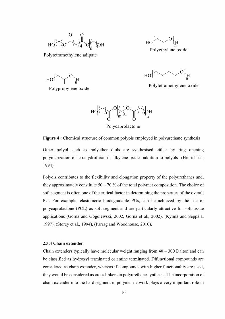
Figure 4 : Chemical structure of common polyols employed in polyurethane synthesis 16
Figure 5 : Chemical structure of common chain extenders employed in polyurethane
synthesis .................................................................................................................. 17
Figure 6 : Primary and secondary reactions of isocyanate group ................................... 19
Figure 7 : One step and two step (prepolymer) method of polymerization .................... 21
Figure 8 : Role of chain extender in polymer synthesis .................................................. 44
Figure 9 : Reaction of hydroxyl and amine group with the diisocyanate group ............. 45
Figure 10 : Chemical structure of L-amino acids used in the current research work ..... 48
Figure 11 : Structures of synthesised L-amino acids based diamine ester compounds .. 54
Figure 12 : General reaction mechanism of Fischer esterification reaction.................... 55
Figure 13 : 1H-NMR spectrum of 310 (spectrum C), 311 (spectrum B) is shown in
comparison with compound 309 (spectrum A) ....................................................... 58
Figure 14 : Zwitter ion form of L-leucine amino acid (301) ........................................... 60
Figure 15 : FTIR spectrum of 301 (spectrum A) and 311 (spectrum B)......................... 60
Figure 16 : 1H-NMR spectrum of compound 314 .......................................................... 65
Figure 17 : 1H-NMR spectrum of model compound 314-M .......................................... 67
Figure 18 : 2D-COSY- NMR of compound 314 ............................................................. 70
Figure 19 : FTIR spectrum of compound 314 ................................................................. 71
Figure 20 : Chemical structures of L-Z-amino acids based dihydroxy ester compounds
................................................................................................................................. 76
Figure 21 : 1H-NMR spectrum of compound 318 (spectrum C) and compared to 1H-
NMR spectrum of starting material 306 (spectrum B) and 317(spectrum A) ........ 80
Figure 22 : FTIR spectrum of compound 318 ................................................................. 82
Figure 23 : 1H-NMR spectrum of compound 320 .......................................................... 83
Figure 24 : FTIR spectrum of compound 320 ................................................................. 85

xiii
Figure 25 : Structure of polycaprolactone (PCL), 4,4- methylenediphenyl diisocyanate
(MDI) and 1,4-Butanediol (BDO) .......................................................................... 91
Figure 26 : Chemical structure of Series 1 PU .............................................................. 104
Figure 27 : GPC traces of Series 1 PU .......................................................................... 106
Figure 28 : 1H- NMR spectra of PCL-1-Z-Ser-PU ....................................................... 108
Figure 29 : FTIR spectra of PCL-1-Z-Ser-PU .............................................................. 110
Figure 30 : DSC thermograms for 1st and 2nd heating run of PCL-1-Z-Ser-PU ......... 113
Figure 31 : DSC thermograms for 1st and 2nd heating run of PCL-1-Z-Thr-PU ......... 114
Figure 32 : DSC thermograms for 1st and 2nd heating run of PCL-1-BDO- PU ......... 117
Figure 33 : TGA analysis of Series 1 PU ...................................................................... 119
Figure 34 : Tensile stress – strain curve of Series 1 PU................................................ 122
Figure 35 : Tensile stress- strain curve of PCL-1-BDO-PU ......................................... 123
Figure 36 : 1H-NMR spectra of the reaction mixture during deprotection of Z-group
from polyurethane: Spectra A for protected polymer, spectra B for deprotected
polymer after 10 minutes of reaction, Spectra C for deprotected polymer after 30
minutes of reaction ................................................................................................ 130
Figure 37 : GPC traces of protected and deprotected PCL-1-Z-Ser-PU ....................... 132
Figure 38 : FTIR absorption spectra of (A) Protected PCL-1-Z-Ser-PU (B) Deprotected
PCL-1-Ser-PU ....................................................................................................... 134
Figure 39 : The chemical structure of PCL-1-Leu-PUU showing urea and urethane
linkages ................................................................................................................. 135
Figure 40 : Chemical structure of Series 1 PUU ........................................................... 137
Figure 41 : GPC traces of Series 1 PUU ....................................................................... 138
Figure 42 : 1H-NMR spectra (Spectrum A) PCL-1-Leu-PUU, (Spectrum B) MDI and
(Spectrum C) PCL ................................................................................................. 140
Figure 43 : FTIR spectra of PCL-1-Leu-PUU .............................................................. 142
Figure 44 : DSC thermograms for 1st and 2nd heating run of PCL-1-Leu-PUU ......... 144
Figure 45 : DSC thermograms for 1st and 2nd heating run of PCL-1-Tyr- PUU ......... 146
Figure 46 : TGA analysis of Series 1 PUU ................................................................... 148
Figure 47 : The Stress strain curves for Series 1 PUU .................................................. 151
Figure 48 : DSC thermograms for the 1st and 2nd heating run of PCL-2-Z-Ser-PU ... 165
Figure 49 : DSC thermograms for the 1st and 2nd heating run of PCL-2-Z-Thr-PU ... 165

xiv
Figure 50 : FTIR absorption spectrum of polyurethane (A) PCL-1-Z-Ser-PU (B) PCL-2-
Z-Ser-PU ............................................................................................................... 170
Figure 51 : FTIR absorption spectra of carbonyl and N-H region of polyurethane (A)
PCL-1-Z-Ser-PU and (B) PCL-2-Z-Ser-PU ......................................................... 170
Figure 52 : Different types of phase morphology present in polyurethane structure.... 171
Figure 53 : TGA analysis of Series 1 PUs and Series 2 PUs ........................................ 174
Figure 54 : Tensile stress – strain curve for PCL-2-Z-Ser-PU...................................... 177
Figure 55 : Tensile stress – strain curve for PCL-2-Z-Thr-PU ..................................... 177
Figure 56 : DSC thermograms for the 1st and 2nd heating run of (A) PCL-2-Leu-PUU
and (B) PCL-2-Ileu-PUU ...................................................................................... 183
Figure 57 : DSC thermograms for the 1st and 2nd heating run of (C) PCL-2-Val-PUU
and (D) PCL-2-Tyr-PUU ...................................................................................... 184
Figure 58 : FTIR absorption spectra of polyurethane (A) PCL-1-Leu-PUU and (B) PCL-
2-Leu-PUU ............................................................................................................ 188
Figure 59 : FTIR absorption spectra of carbonyl and N-H region of polyurethane (A)
PCL-1-Leu-PUU and (B) PCL-2-Leu-PUU ......................................................... 188
Figure 60 : Hydrogen bonding interactions in polyurethane urea................................. 190
Figure 61 : TGA analysis of Series 2 PUUs ................................................................. 193
Figure 62 : TGA analysis of Series 1 PUUs ................................................................. 193
Figure 63: The Stress strain curves for Series 2 PUU ................................................... 197
Figure 64 : The Stress strain curves for PCL-2-Tyr-PUU ............................................ 197
Figure 65 : Fluorescence images of mouse fibroblast cells on Series 1 polymers surfaces
(A) 1,4- Butanediol (Control), (B) & (C) Series 1 PU, (D) TCPS control, (E) and
(F) Series 1 PUU (magnification x 200). Circle showing dead cells with red
fluorescence........................................................................................................... 206
Figure 66 : Fluorescence images of mouse fibroblast cells on Series 2 polymers surfaces
(A) PCL-1-BDO-PU (Control), (B) & (C) Series 2 PU, (D) TCPS control, (E) and
(F) Series 2 PUU (magnification x 200). Circle showing dead cells with red
fluorescence........................................................................................................... 207

xv
Index of Schemes
Scheme 1 : Reaction scheme for the preparation of polyurethane and polyurethane urea 8
Scheme 2 : Schematic representation of the synthesis of diamine ester compound 311 53
Scheme 3: Schematic representation of the synthesis of dihydroxy ester compound 318
................................................................................................................................. 75
Scheme 4 : Schematic representation of the synthesis of PCL-1-Z-Ser- PU ................ 102
Scheme 5 : Deprotection reaction to remove Z group from PCL-1-Z-Ser-PU ............. 128

xvi
Index of Tables
Table 1 : Biodegradable Polyurethane Elastomers ......................................................... 10
Table 2 : Peak integration data obtained for 1H-NMR spectrum of 311 ........................ 59
Table 3 : Selected FTIR frequencies (cm-1) of 301 and its ester derivative 311............ 62
Table 4 : Peak integration data obtained for 1H-NMR of compound 314 ...................... 66
Table 5 : Chemical shift (ppm) position of 1H-NMR spectrum of 314 and 314-M ....... 68
Table 6 : Optimisation of reaction temperature for alkali metal salt based reaction ...... 78
Table 7 : Peak integration data obtained for 1H-NMR spectrum of compound 318 ...... 81
Table 8 : Peak integration data obtained for 1H-NMR spectrum of compound 320 ...... 84
Table 9 : Abbreviations used for Series 1 PU ............................................................... 103
Table 10 : Composition of Series 1 PUs ....................................................................... 105
Table 11 : Molecular weights of Series 1 PU ............................................................... 105
Table 12 : FTIR peak assignment for PCL-1-Z-Ser-PU ............................................... 111
Table 13 : Thermal properties of Series 1 PUs ............................................................. 112
Table 14 : Water contact angle values for Series 1 PU ................................................. 121
Table 15 : Mechanical properties of Series 1 PU (mean ± SD, n = 6) .......................... 123
Table 16 : Composition of PCL-1-Z-Ser-PU for deprotection reaction ....................... 127
Table 17 : Molecular weight of PCL-1-Z-Ser-PU before and after deprotection reaction
............................................................................................................................... 133
Table 18 : Nomenclature for Series 1 PUU .................................................................. 136
Table 19 : Composition of Series 1 PUU ...................................................................... 137
Table 20 : Molecular weight of Series 1 PUU .............................................................. 139
Table 21 : FTIR peak assignment for PCL-1-Leu-PUU ............................................... 143
Table 22 : Thermal properties of Series 1 PUU ............................................................ 144
Table 23 : Contact angle values for Series 1 PUU ........................................................ 150
Table 24 : Mechanical properties of Series 1 PUU (mean ± SD, n = 6) ....................... 152
Table 25 : Polyurethanes abbreviations used for Series 2 PUs ..................................... 163
Table 26 : Comparison of molecular weight of Series 1 and 2 PUs ............................. 164
Table 27 : Thermal properties of Series 2 and 1 PUs ................................................... 166
Table 28 : Assignment of the major absorption bands in the FTIR spectra of the PCL-
MDI based PU(Mattia and Painter, 2007), (Yilgor and Yilgor, 2007) ................. 169
Table 29 : Contact angle values for Series 1 and 2 PU ................................................. 175

xvii
Table 30 : Mechanical Properties of Series 1 and 2 PUs (mean ±SD, n=6) ................. 178
Table 31 : Abbreviations used for PCL 2,000 based Series-2 PUUs ............................ 180
Table 32 : Molecular weight of Series 2 and Series 1 PUU.......................................... 181
Table 33 : Thermal properties of Series 2 PUUs and its comparison with Series 1 PUUs
............................................................................................................................... 184
Table 34 : Assignment of the major absorption bands in the FTIR spectrum of the MDI-
PCL based Polyurethane ....................................................................................... 189
Table 35 : Characteristic temperature on TGA curves for Series 1 and 2 PUUs .......... 194
Table 36 : Contact angle values for Series 1 and 2 PUUs (mean ± SD, n = 5)............. 196
Table 37 : Mechanical properties of Series 1 and 2 PUUs (mean ± SD, n = 6)............ 198
xviii
Abbreviations
PU Polyurethane
PUU Polyurethane urea
LDI Lysine diisocyanate
HDI Hexane diisocyanate
PCL Polycaprolactone
PEG Polyethylene glycol
MDI Methylenediphenyl diisocyanate
BDO Butanediol
NCO Isocyanate group
TDI Toluene diisocyanate
NDI Naphthalene diisocyanate
PPDI p-Phenylene diisocyanate
CHDI Cyclohexyl diisocyanate
BDI Butane diisocyanate
HMDI Hexamethylene diisocyanate
SPUU Segmented polyurethane urea
PTMO Polytetramethylene oxide
CL Caprolactone
DBTL Dibutyltin dilaurate
SS Soft segment
HS Hard segment
DMA Dynamic mechanical analysis
SAXS Small angle X-ray scattering
TEM Transmission electron microscopy
HSC Hard segment concentration
SEM Scanning electron microscopy
AFM Atomic force microscopy
PEO Polyethylene oxide
Da Dalton
WAXD Wide angle x-ray diffraction

xix
PTMG Polytetramethylene glycol
HUVECs Human umbilical vein endothelial cells
Gly-Leu Glycine-Leucine
Z Benzyloxycarbonyl
p-TsOH para-toluene sulphonic acid
MHz Mega hertz 1H-NMR Proton nuclear magnetic resonance spectroscopy 13C-NMR Carbon nuclear magnetic resonance spectroscopy
DMSO Dimethylsulphoxide
d6-DMSO deuterated Dimethylsulphoxide
ppm parts per million
EI Electron impact
PFK Perfluorokerosene
ESI Electrospray ionosation
FTIR Fourier transform infrared spectroscopy
TLC Thin layer chromatography
UV Ultra violet
IUPAC International union of pure and applied chemistry
Na2CO3 Sodium bicarbonate
CE Chain Extender
MS (ESI) Electrospray ionization mass spectroscopy
DTH Desaminotyrosyl hexyl ester
2-D-COSY Two dimensional correlation spectroscopy
N-Boc Tert-Butyloxycarbonyl
Na2CO3 Sodium carbonate
LI2CO3 Lithium carbonate
K2CO3 Potassium carbonate
Cs2CO3 Caesium carbonate
DMF Dimethylsulphoxide
CsOH Caesium hydroxide
COOH Carboxylic acid
NMP N-Methylpyrrolidone
Mw Weight average molecular weight

xx
Mn Number average molecular weight
GPC Gel permeation Chromatography
HPLC High performance liquid chromatography
LiBr Lithium Bromide
PI Polydispersity
DSC Differential scanning calorimetry
TGA Thermogravimetric analysis
ASTM American society for testing and materials
Tg Glass transition temperature
Tm Melting temperature
DMAc Dimethylacetamide
DCM Dichloromethane
THF Tetrahydrofuran
Xc Crystallinity
MPa Megapascal
SD Standard Deviation
HBr/HAc Hydrogen Bromide/Acetic acid
MDA Methylene dianiline
TCPS Tissue culture polystyrene
EthD Ethidium homodimer
AM Acetoxymethyl
DNA Deoxyribonucleic acid
PBS Phosphate Buffer Solution
CO2 Carbon dioxide
ECS Fetal calf serum
MEM Minimum essential medium

1
Chapter 1: Introduction

2
1.1 Introduction
In the last few decades, there has been significant interest and demand for the
development of biodegradable and biocompatible polymers for biomedical applications
such as tissue engineering (Ozdil and Aydin, 2014, Guo and Ma, 2014, Sobczak, 2015),
drug delivery (Wang and Wang, 2012), scaffold formation (Qizhi et al., 2013), and
biomedical devices (Lamba et al. (1998), Guelcher, 2008, Krishnan et al., 2014). There
are several important physiochemical properties required by the polymer to be used in
such areas, including biodegradability and biocompatibility as mentioned above, but also
good mechanical properties and easy polymer processing (Lanza et al., 1997, Medine et
al., 2012).
The class of polymer, “polyurethane”, refers to a surprisingly wide range of polymeric
materials available for use in the biomedical field. There are particular features of this
material which offer substantial advantages over other available polymers (Davis and
Mitchell, 2008, Shelke et al., 2014). The key advantage is the composition of the
polyurethane system, which allows materials to take on a range of different material
properties and degradation kinetics to suit various biomedical applications including
permanent implants and temporary biodegradable scaffolds (Gunatillake and Adhikari,
2003).
Segmented polyurethanes are synthesised from various polyols, diisocyanates and chain
extenders. These monomers can be structurally manipulated to achieve a wide range of
bio-material properties (Martina and Hutmacher, 2007). The use of polyurethanes as
biomaterials has been explored for various implants such as pacemakers and as vascular
grafts (Oertel, 1994). The main criterion for the selection of monomers depends on the
biocompatibility and non-toxicity of the degradation product (Marcos-Fernández et al.,
2006). Amino acid based polymers are developed and studied as biomaterials due to their
high biocompatibility and biodegradability. Several amino acid based polymers are used
for biomaterial applications (Bourke and Kohn, 2003). The use of amino acid as one of
the component for polyurethane synthesis will enhance the biocompatibility of the
polymer (Sarkar, 2007). Polyurethanes based on amino acids offer several advantages
3
including biocompatibility, biodegradability and a range of material properties which can
be tailored by changing the structure of the polymer (Skarja, 2001).
1.2 Research Hypothesis
The main research hypothesis tested was the synthesis of segmented polyurethanes by the
incorporation of novel amino acid based diester chain extenders with tuneable
physiochemical properties for potential use in biomedical applications, i.e. can these
segmented polyurethanes be successfully synthesised, are they biocompatible, and what
are their mechanical properties A further aim is to test those properties as a function of
the polymer structure, in particular the molecular weight of the polymer soft segment
monomer.
A number of steps will be taken to fulfil these goals, as follows:
1. The novel amino acid based diester chain extenders were synthesised and
characterised. The presence of amino acid as one of the components of the
polyurethane will increase its biocompatibility and its biodegradation, as amino
acids are naturally occurring biomolecules and these are already present in the
human body. The chances of obtaining toxic cell responses during in vitro studies
should then be decreased. The introduction of hydrolysable ester linkage through
chain extender will enhance the degradation property of the polyurethane
(Guelcher, 2008). Moreover, the incorporation of amino acid will also enhance
the enzyme mediated degradation of the polyurethane (Skarja and Woodhouse,
2001). Two series of L-amino acid chain extended polyurethanes and
polyurethane urea’s were to be synthesised. The diol used to link amino acid
groups and form chain extender is to be a simple, linear dihydroxy compound and
is selected based on its low toxicity (Kartvelishvili et al., 1997).
2. A series of polyurethanes and polyurethane urea’s were synthesised based on
Methylenediphenyl diisocyanate, Polycaprolactone and amino acid based chain
extenders. They will then be fully characterised for their resultant physiochemical
properties and compared to a control polyurethane based on 1,4-butanediol as the
4
chain extender, polycaprolactone is to be used as the soft segment as this polyol
is one of the biologically acceptable polyols reported in the literature (Chasin and
Langer, 1990), (Gunatillake and Adhikari, 2011). The aromatic diisocyanate 4,4-
methylenediphenyl diisocyanate (MDI) is used as the diisocyanate. MDI was
selected because of its high reactivity and better resultant mechanical properties,
largely due to its aromatic structure (Vermette, 2001). Toxic degradation products
from aromatic polyurethanes are reported in the literature (Szycher and Siciliano,
1991), providing a downside to the use of MDI. However whether the
concentration of these toxic products can reach physiologically significant levels
in vivo is still inconclusive and has not been resolved yet (Coury, 2004),
(Guelcher, 2008), (Blais, 1990). No evidence of toxicity of the resultant by
product (4,4-methylenedianiline (MDA) of MDI is reported (Liljensten et al.,
2002, Gisselfält et al., 2002). Moreover several MDI based polyurethanes are
currently used as biomedical implants (Gunatillake and Adhikari, 2011, Gisselfält
and Helgee, 2003).
3. Third, the effect of soft segment chemistry will be observed by using two different
molecular weights of the soft segment (a high value, 2000 Mw and a low value,
1000 Mw). The structure property relationship will then be observed via thermal,
surface and physical testing of the two polymer series formed. This will be
applied to both PU and PUUs.
4. Initial in vitro cell cytotoxicity study was performed on the synthesised polymers.
Mouse fibroblast cells will be used. Cells viability will be evaluated using a
standard LIVE/DEAD® assay kit. Tissue culture polystyrene (TCPS) will be used
as the positive control.
The final objective of this dissertation can be specified as follows:
1. Synthesis and characterisation of novel L-amino acid based diester chain
extenders.

5
2. Synthesis of polyurethanes and polyurethane ureas based on novel L-amino acid
based chain extenders. Structural, thermal, mechanical and surface properties of
the synthesised polymers compared to control polyurethane.
3. Study of the effect of the soft segment molecular weight on the structure property
relationship of amino acid based polyurethanes and polyurethane ureas.
4. Cytotoxicity screening of the synthesised polymers for preliminary evaluation of
polymer biocompatibility.
1.3 Layout of thesis
The rest of the thesis is divided into six chapters. Chapter 2 describes the existing
literature on polyurethane and its applications. It includes polyurethane history, chemical
reaction of diisocyanate group, structural morphology of segmented polyurethane and the
role of amino acid in polyurethane synthesis. In Chapter 3, the synthesis and
characterisation of novel amino acid based diester dihydroxy and diester diamine
compounds (which later acts as chain extender for polyurethane and polyurethane urea
synthesis) are described. A detailed characterisation of these chain extenders will show
their successful synthesis. The successful incorporation of amino acid based diester
dihydroxy and diester diamine compounds as chain extender into polyurethane and
polyurethane urea will be described in Chapter 4. This chapter will also include a detailed
structural, thermal and mechanical characterisation of the polymers. Chapter 5 includes
detailed analysis of the structure property relationship of the polyurethane and
polyurethane urea based on different molecular weights of the soft segment. A library of
polymers is developed and the effect of structural variation is examined in terms of
different physiochemical properties to understand the underlying principal of structure
property correlation. Chapter 6 includes a brief preliminary cytotoxicity screening
(LIVE/DEAD assay) of the synthesised polymers to determine any substantial general
cytotoxic response and Chapter 7 summarizes the research and discusses direction for
future work.

6
Chapter 2: Literature Review

7
2.1 Introduction
A vast number of biodegradable polymers have been synthesised in recent times and their
structure property relationship has been studied in detail (Vroman and Lan, 2009,
Dumitriu, 2002). Biodegradable polymers are now in great demand with increasing
applications in biomedical engineering such as drug delivery, medical devices, wound
dressing, and as scaffold for tissue engineering. To fulfil these new demands,
biodegradable polymers have been found to be encouraging candidates by characteristic
ability to manipulate their physio mechanical properties. This can be typically achieved
by regulating the ratio and nature of the starting material used for polymer synthesis.
There are several polymer classes available which are showing potential to be used as
biodegradable biomaterials for tissue engineering applications, but perhaps the most
promising are biodegradable polyurethanes (Kumar et al., 2001).
Polyurethane structures contains urethane linkages with in the polymer chains. The
urethane linkage is equivalent to carbamate linkage in organic chemistry (Hepburn,
1982). The structure of the urethane link is shown in Figure 1.
Figure 1 : Chemical structure of urethane within a polymer
2.1.1 Structure of polyurethane
Polyurethane elastomers are produced through the reaction of diisocyanate and polyol
and low molecular weight diols or diamines as chain extender. The chemical reaction
between an isocyanate group with hydroxyl group generate urethane linkage while,
reaction between isocyanate and diamine group produce characretstic urea linkage.
Polyurethane synthesis reaction has been employed to synthesise a range of thermoplastic
and thermoset polyurethanes in literature (Sobczak, 2015, Shi, 2004). Thermoplastic
polyurethanes are prepared by reacting three classic compounds: a diisocyanate, a

8
difunctional polyol and a dihydroxy or diamine chain extender. These monomers react to
form a linear, segmented copolymer consisting of alternating soft and hard segment
blocks, which are the characteristic feature of thermoplastic polyurethanes. The general
chemical structure of thermoplastic polyurethane produced using the prepolymer linking
method is shown in Scheme 1 “adapted from Gunatillake and Adhikari” (2011).
Scheme 1 : Reaction scheme for the preparation of polyurethane and polyurethane urea
.
The soft and hard segments are mostly thermodynamic incompatible, which leads to the
two phase morphology of the polymer. The soft segment domains are primarily composed
of polyols and are generally amorphous in nature, leading to the characteristic elastic
properties of the polymer. The hard segments are primarily composed of diisocyanate
chains linked with the chain extender and are generally glassy or semicrystalline in nature,
contribute to the characteristic mechanical properties of the polymer.

9
2.1.2 Advantage of using polyurethane
Polyurethane is a versatile and commercially important material, already in large
production. The urethane link is the main bond usually present within the polymer
backbone, but it is the flexibility of polyurethane structure to incorporate other functional
groups into the polymer network makes it more versatile as compared to other available
biomaterials. A range of different functional groups can be incorporated into the
polyurethane network such as ester, ether groups which can contribute to different range
of properties acquired by polyurethanes. Thus the polyurethane can form rigid hard
thermosetting materials as well as much softer elastomers (Lamba et al., 1998), (Wirpsza,
1993). Hence polyurethanes can be design to have specific properties of hardness,
abrasion, chemical resistance, mechanical and elastic properties and also other specific
tissue engineering properties such as blood and tissue compatibility (Biesman, 2002). It
has been reported in the literature (Gunatillake and Adhikari, 2011) that biocompatibility
and biodegradability are not the only characretstic required by material to act as an ideal
candidate for tissue engineering applications. Along with these properties, the material
needs to show optimum surface characretstic properties to encourage cells growth and
proliferation. It is expected that an ideal degradable biomaterial will possess good
mechanical and biological properties compliant with suitable degradation mechanism and
easy fabricating ability. Among the currently available synthetic polymers, polyurethane
offers various advantages in designing materials to fulfil these requirements. The
availability of a huge range of starting materials with an easy to follow two step
prepolymer synthetic method and opportunity to formulate the desired polymer with
targeted properties are the several benefits available with the use of polyurethanes
(Petrovic and Ferguson, 1991). Overall, the flexibility in polyurethane synthesis, along
with its processing and bio-friendly properties have made it a preferable choice over other
available synthetic polymers for biomedical applications.
2.1.3 Role of polyurethane
Polyurethane materials have a huge role in everyday materials, for example in the
construction industry, as coatings, adhesives and textiles, house hold furnishing, medical
devices (Saunders, 1964). These days, there is a high demand for polyurethane material
in the bioengineering field. But its role as biomaterial in medical field started in early

10
1960, when polyurethane was used for in situ bone fixation and polyurethane coatings
were applied to cardiovascular implants (Santerre et al., 2005, Soletti et al., 2011, Boretos
and Pierce, 1967). Bio-stable polyurethanes have been extensively investigated as long
term medical implants in the form of cardiac pacemakers and vascular grafts (Oledzka et
al., 2007, Zoltowska et al., 2014, Liu et al., 2012a, Thottappillil and Nair, 2015). Table 1
“adapted from Sobczak et.al.” (2015) shows some of the biomedical grade polyurethanes
that are commercially available and in use currently.
Table 1 : Biodegradable Polyurethane Elastomers
Product Name
Manufacturer /Supplier
Types of biodegradable polyurethane elastomers
Examples of applications
Actifit Orteq, Netherland BDI/PCL/BD; porous material
Meniscal repair
Artelon Artimplant, Sweden
Polyurethane urea; fibres, scaffold films, granules
Ligament fixation, bone scaffolds (odontology)
ChronoFlex AL
AdvanSource Biomaterials, USA
HMDI/polycarbonate/BD Various biomaterials
Degrapol AB Medica Spa, Italy
Porous foam Nerve and bone regeneration
Epidel Interface Biologics, Canada
Polyurethane-co-drug, PCL based PU
Catheter cuffs, antimicrobial materials
Lacthane Polganics, Netherland
BDI/polyethers/polyesters Wound and nasal dressing, surgical sealant
Novosorb Polynovo Biomaterials, Australia
Injectable gel, X-linked polymer, prepolymer
Porous nonporous monoliths orthopaedic
SynBioSys Octoplus, Netherland
BDI/PLGA/PEG/PCL Stent coatings, drug eluting microspheres
2.2 Polyurethane history and development
Until the 1920s and 1930s polymeric materials were largely based on natural or modified
natural materials. In 1937, Otto Bayer, of the Farben Industrie, Leverkusen, Germany,

11
performed and patented the pioneering work on synthetic polyurethane (Bayer, 1937).
Otto Bayer and colleagues took advantage of the known poly-addition reaction of
isocyanate and alcohol functional groups to develop a linear polymer possessing a large
number of urethane bonds and trademarked it as “Perlon U” and this proved as a huge
commercial success.
In early 1950’s, attempts to synthesise polyurethane elastomers was based on the use of
naphthalene-1, 5-diisocyanate (NDI) and resultant elastomer produces was very tacky and
gummy material. The reason for such appearance might be the use of bulky diisocyanate
(NDI) and lack of polymer melt stability. However the properties of these elastomers
were improved tremendously when short chain extenders were introduced into the
polymer network and also NDI was replaced by MDI (Oertel, 1994).
In the 1950’s, aromatic diisocyanate plays a huge part in the development of polyurethane
chemistry. Toluene diisocyanate (TDI) and methylenediphenyl diisocyanate (MDI) were
the two main aromatic diisocyanate used to develop polyurethane. In 1954, DuPont
chemists successfully synthesise the TDI based polyurethane fibre called “spandex”
followed by the synthesis of MDI based polyether urea with trade name of “Lycra”
(Dieterich et al., 1994).
In 1957, a new family of polyurethane was made available by Schollenberger et.al.
(1958). He introduced a new “cross linked” thermoplastic polyurethane elastomer. This
was based on MDI, adipic acid and 1,4-butanediol (BDO). The polymer showed useful
properties such as high elasticity, extensibility and solubility. Nevertheless, the structure
property relationship of the synthesised polyurethanes was still not well understood.
Cooper et.al. (1966) was arguably the first to gain a good understanding in this area. They
reported that the excellent elastomeric properties of a thermoplastic polyurethane is the
result of phase separated hard and soft segment morphology. During the 1960s, MDI
become the raw material of choice for the rapidly evolving applications of polyurethane
(Joshi, 2009).

12
In addition to elastomers, polyurethanes can also be produced as foams (rigid and
flexible), adhesives, binders, coatings, and paints. Because of their unique properties,
polyurethanes have found a wide variety of applications in commercial areas such as the
automotive, furniture, construction, seating, exterior panels, structural foam, furniture,
house-hold electrical materials, and refrigerator insulation (Saunders, 1964), (Wright and
Cumming, 1969). These days polyurethanes especially biodegradable version have made
their way to biomedical applications. Polyurethanes are now synthesised in custom made
manner to fulfil specific medical applications.
2.3 Components of polyurethane
The three main components used for polyurethane synthesis are: a diisocyanate, a polyol
and a dihydroxy or diamine chain extender (Ortel, 1994). These three monomers react to
form a linear, segmented polyurethane made up of alternating hard and soft segments.
The chemical reaction between the isocyanate group and the hydroxyl or amine group
produces urethane and urea groups respectively. Polyurethane morphology is highly
affected by the choice and structural composition and ratio of the reactants, synthesis
method and processing conditions. A variety of materials are available to produce
polyurethanes of desired physiochemical properties. In general, the resultant properties
of the final polyurethane can be projected based on the choice of specific components
(Lamba et al., 1998), (Pinchuk, 1995), (Hepburn, 1991). For example, aliphatic reactants
have more molecular flexibility than cycloaliphatic groups, followed by aromatic groups.
As a result, polyurethanes having aromatic group in their backbone shows enhanced
mechanical properties, while aliphatic polyurethanes are generally produce softer and
weaker materials. Similar to the role of molecular flexibility in the polyurethane, presence
of asymmetric or symmetric reactants along with bulky side groups also prevent the
alignment of polymer segments which in turn leads to reduced tensile strength
(Caracciolo et al., 2009). Additionally, hydrogen-bond forming groups in polyurethane
structure promotes phase segregation and hence improved mechanical properties.
Overall, it shows that a clear understanding of the reactant chemistry help in designing
the polyurethane with property required for specific application (Parrag, 2010).

13
2.3.1 Diisocyanate
The diisocyanate is the central component in polyurethane synthesis since the
diisocyanate/hydroxyl reaction produces the characteristic urethane group that defines the
polymer. The isocyanate group (NCO) is highly reactive, especially with nucleophile
agents. The reactivity of the NCO group is determined by the electrophilic characteristic
of the central carbon atom of the cumulated bond. The electron structure of the isocyanate
group can be described using its resonance structure, as shown in Figure 2.
Figure 2 : Resonance structure of isocyanate group, R can be aliphatic or aromatic group
The fourth structure in Figure 2 forms only in the case of aromatic isocyanates, where the
aromatic ring can stabilize the negative charge of the nitrogen atom. The formation of this
resonance structure explains the higher reactivity of aromatic isocyanates compared to
aliphatic isocyanates (Bagdi, 2010).
The resonance structure illustrates that the NCO group can react with either electron
donors or electron acceptors. Most reactions of isocyanate involve addition at the N=C
double bond. Aromatic isocyanates are generally more reactive than aliphatic, as the
electron withdrawing nature of the benzene ring makes the isocyanate carbon more
susceptible to nucleophilic attack. The presence of bulky side groups in the ortho position
on aromatic isocyanate, or branched or bulky substituent on aliphatic molecules will
sterically hinder the approach of electron donors and reduce the rate of the reaction.
2.3.2 Types of diisocyanate
Aromatic and aliphatic diisocyanate are utilised to attain special properties desired in the
final product. For example, aliphatic diisocyanate based polyurethane produce light stable
polymer and aromatic diisocyanate based polyurethane will undergo photo-degradation,

14
if exposed for long enough, (Wang, 1998). The incorporation of aromatic diisocyanate in
the polymer network produces a stiffer polymer chain with a higher melting point as
compared to aliphatic isocyanates. In general, aliphatic diisocyanate based polyurethane
shows weaker mechanical properties as compared to polyurethanes based on aromatic
diisocyanate (Skarja, 2001). The commonly used and commercially available aromatic
and aliphatic diisocyanates for synthesizing polyurethanes are shown in Figure 3.
Figure 3 : Chemical structure of common diisocyanate employed in polyurethane
synthesis
The molecular rigidity of the aromatic structure of the aromatic diisocyanate might be a
reason for better hard segment polymer chain packaging which in turn strengthen the hard
segment interaction through pi-electrons (Pinchuk, 1995), (Skarja, 2001). The two most
commonly used aromatic diisocyanates in polyurethane synthesis are toluene
diisocyanate (TDI) and methylenediphenyl diisocyanate (MDI). Out of two, MDI has
N C O
NCO1,5-Naphthalene diisocyanate
NCON C O
2,6-Tolylene diisocyanate
NCON C O
4,4-Methylene diphenyl diisocyanate
N C ONCO
1,6-Hexamethylene diisocyanate(MDI)
(TDI)(NDI)
(HDI)
NCO
N C O
2,4-Tolylene diisocyanate
N NCO
C O
Methylene bis(p-cyclohexyl isocyanate)(H12MDI)
HCCH2
OCN C
CH2CH2CH2OCN
OO
H2C CH3
Lysine diisocyanate (LDI)

15
superior reactivity and provides better mechanical properties to the synthesised polymer.
(Zia et al., 2009, Szycher, 1999). Other aromatic diisocyanates available are p-phenylene
diisocyanate (PPDI) and 1,5-naphtalene diisocyanate (NDI) (Randall and Lee, 2002).
For the development of biodegradable polyurethane, aliphatic diisocyanates are
recommended over aromatic diisocyanates. Since the use of aromatic diisocyanate can
lead to the release of toxic and carcinogenic by-products upon degradation (Kavlock et
al., 2007, Szycher and Siciliano, 1991). The first commercially produced aliphatic
diisocyanate was 1,6-hexamethylene diisocyanate (HDI). Several others aliphatic
diisocyanates are now in commercial use, including methylene bis(p-cyclohexyl
isocyanate) (H12MDI), cyclohexyl diisocyanate (CHDI), and 1,4-butanediisocyanate
(BDI) (Lutz and Börner, 2008). Recently, a lysine amino acid based diisocyanate (LDI)
i.e. 2,6-DiisocyantoEthylCaproate, has been used to develop biodegradable
polyurethanes. It degrades into less toxic (and possibly non-toxic) by-products and hence
is the preferred choices for use in the development of biodegradable and biocompatible
polyurethanes (Zhang et al., 2000).
2.3.3 Polyols
Polyols generally result in the soft segment of biodegradable polyurethanes. Polyols are
generally low molecular weight hydroxyl terminated polymers. Different types of polyols
has been used to synthesise polyurethane such as polyesters, polyethers, hydrocarbon
polymers and/or polydimethylsiloxanes. Figure 4 shows the chemical structure of some
common soft segments polyols used for biodegradable polyurethanes synthesis.
Esterification reaction between acid and diol is carried out to produce polyesters. One of
the most commonly used polyester polyol used for biodegradable polyurethane synthesis
is polycaprolactone (PCL). It has been widely used to make biocompatible polyurethane
due to its high biodegradability property (Heijkants et al., 2005) along with a low melting
point of approximately 60°C and a glass transition temperature of -60°C (Liu et al., 2006).
16
Figure 4 : Chemical structure of common polyols employed in polyurethane synthesis
Other polyol such as polyether diols are synthesised either by ring opening
polymerization of tetrahydrofuran or alkylene oxides addition to polyols (Hinrichsen,
1994).
Polyols contributes to the flexibility and elongation property of the polyurethanes and,
they approximately constitute 50 – 70 % of the total polymer composition. The choice of
soft segment is often one of the critical factor in determining the properties of the overall
PU. For example, elastomeric biodegradable PUs, can be achieved by the use of
polycaprolactone (PCL) as soft segment and are particularly attractive for soft tissue
applications (Gorna and Gogolewski, 2002, Gorna et al., 2002), (Kylmä and Seppälä,
1997), (Storey et al., 1994), (Parrag and Woodhouse, 2010).
2.3.4 Chain extender
Chain extenders typically have molecular weight ranging from 40 – 300 Dalton and can
be classified as hydroxyl terminated or amine terminated. Difunctional compounds are
considered as chain extender, whereas if compounds with higher functionality are used,
they would be considered as cross linkers in polyurethane synthesis. The incorporation of
chain extender into the hard segment in polymer network plays a very important role in

17
polyurethane structure and hence to its properties. For an example, polyurethane based
on chain extender permits hard segment segregation and hence improves the physical
properties of the resultant polymer along with an increase in the hard-segment glass
transition temperature (Tg) (Wang, 1998). However, in case of no use of chain extender,
polyurethane is formed by directly reacting diisocyanate and polyol. Generally it has very
poor physical properties (low tensile strength) and often does not exhibit micro-phase
separation. This shows the importance of using chain extender and how it influences the
overall property of the synthesised polyurethane.
A number of di and poly-functional active hydrogen reactants are used as chain extenders,
branching agents or cross linkers in the synthesis of polyurethanes. Zhang et.al. (2006)
define the significance of chain extender as “The role of the chain extender is to produce
an ‘extended’ sequence in the copolymer consisting of alternating chain extender and
diisocyanate. These extended sequences, generally hard segments, and act both as fillers
and as physical crosslink sites increasing mechanical strength in the polymer”
The most important chain extenders for polyurethane synthesis are aliphatic and aromatic
diols and diamines (Hollinger, 2012). In general, polyurethanes produced with aromatic
chain extenders and diamine based chain extenders have better physical properties as
compare to polyurethane based on aliphatic and diol based chain extender. This is due to
the formation of better crystalline hard segment domain in polymer structure which
further enhances the mechanical properties of the polyurethane. The chemical structures
of commonly used chain extenders are shown in Figure 5.
Figure 5 : Chemical structure of common chain extenders employed in polyurethane
synthesis

18
Typical examples of diols reported in the literature and used as chain extender are 1,2-
ethanediol, dipropylene diols. Conventional chain extenders such as 1,4-butanediol
(BDO) and 1,6-hexanediol are used in preparing most biodegradable polyurethane
formulation (Lamba et al., 1998), (Gunatillake and Adhikari, 2011). Aliphatic diamines
used as chain extender include 1,2-ethanediamine, propylene diamine, hexylene diamine.
1,4-butanediamine (putrescine) is a naturally occurring compound and is used as chain
extender in biodegradable polyurethane due to its low reactivity (Guan et al., 2002).
Cycloaliphatic diamines include isophorone diamine, 1,4-cyclohexyl diamine along with
aromatic diamines are often used for the synthesis of polyurethanes (Frisch and
Kordomenos, 1985). Overall, it can be concluded that the major influence of chain
extender structure on polyurethane properties is attributed to its effect on hard segment
ordering, which, in turn, affects the polyurethane crystallinity and mechanical properties.
Generally short chain, compact and symmetrical molecules favours better ordering of the
hard segment.
Several research groups (Guan et al., 2004, Guan and Wagner, 2005, Guan et al., 2002),
(Skarja and Woodhouse, 2000, Skarja, 1998, Elliott et al., 2002), (Marcos-Fernández et
al., 2006) have developed naturally occurring amino acid based novel chain extenders for
the synthesis of biodegradable polyurethane synthesis. The introduction of these amino
acid based chain extender also contribute towards a method of introducing degradable
chemical moieties into the hard segment of polyurethanes. Amino acids and short
peptides can easily be incorporated as chain extenders in the form of dihydroxy and
diamine compounds (Lipatova et al., 1983), (Guan et al., 2005), (Marcos-Fernández et
al., 2006), (Sarkar et al., 2009), (Skarja, 2001), (Parrag and Woodhouse, 2010). Using
this approach allows the choice of hard segment for achieving specific physical and
mechanical properties while still promoting overall polymer degradation. As a result, PUs
can be developed that are susceptible to enzyme degradation and also promote cell
attachment and proliferation. The incorporation of these amino acid based chain extender
into the polyurethane provides an opportunity to design and develop synthetic bio-
mimetic PUs with modified physical, chemical, mechanical and degradation properties
(Parrag and Woodhouse, 2010).

19
2.3.5 Primary reaction of isocyanate group
Isocyanate (NCO) group is highly reactive in nature and can promotes reactions with
other functional groups which further enhances the diversity of polyurethane copolymers.
Examples of the primary and secondary reactions of isocyanate group with different
functional groups are shown in Figure 6.
Figure 6 : Primary and secondary reactions of isocyanate group
All of these reactions can occur during the synthesis of polyurethane (Hepburn, 1991),
(Hood, 2012).
20
2.3.5.1. Reaction with polyol/hydroxyl group The reaction of isocyanate with a polyol results in urethane bond formation. This is one
of the primary reaction occur during polyurethane synthesis. This is an exothermic
reaction and highly influenced by the structure of both reactants. Especially, aliphatic
polyols with primary hydroxyl end groups are the most reactive followed by secondary
alcohol or polyol and least reactive are phenols.
2.3.5.2. Reaction with amines Reaction of isocyanate with primary amines, at room temperature and in the absence of
catalyst, is about 100 to 1000 times faster than the reaction with primary alcohol. The
reactivity of amines increases with the basicity of the amine, and aliphatic amine reacts
much faster than aromatic amines. In the case of aromatic amines, steric hindrance and
electron withdrawing substituent reduces the reactivity toward isocyanate.
2.3.5.3. Reaction with water One of the most common side reactions of the NCO group is with any water in the reaction
mixture. Water is hard to completely remove from the reaction as it involves hygroscopic
materials such as dimethylformamide (DMF) or dimethylacetamide (DMAc) as solvent.
Even trace amounts of water present in the synthetic reaction can significantly affect the
stoichiometric value of the NCO group and hence affect the final polyurethane
composition and properties. The main product of the reaction with water is a substituted
carbamic acid, which breaks down into an amine and carbon dioxide. The amine then
reacts with further isocyanate to yield the substituted urea. In order to prevent NCO
reaction with water, care must be taken to dry the reactant and solvents. Catalyst choice
is also critical in preventing NCO reaction with water.
There are two main type of catalyst are used in polyurethane synthesis: tertiary amines
and organometallic species. The most common organometallic catalyst is tin based, e.g.
dibutyltin dilaurate (DBTL) or stannous octoate (Joshi, 2009). These catalysts are widely
used in polyurethane synthesis as they are readily soluble in reaction solvent, are not very
volatile and have a low odour. Tertiary amines, unlike organometallic catalysts, increase
the reaction rate of NCO with amines, hydroxyl group and water. To minimise the effect
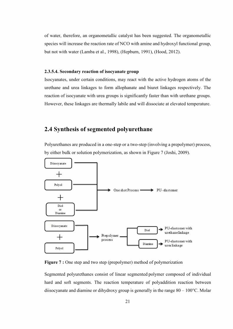
21
of water, therefore, an organometallic catalyst has been suggested. The organometallic
species will increase the reaction rate of NCO with amine and hydroxyl functional group,
but not with water (Lamba et al., 1998), (Hepburn, 1991), (Hood, 2012).
2.3.5.4. Secondary reaction of isocyanate group Isocyanates, under certain conditions, may react with the active hydrogen atoms of the
urethane and urea linkages to form allophanate and biuret linkages respectively. The
reaction of isocyanate with urea groups is significantly faster than with urethane groups.
However, these linkages are thermally labile and will dissociate at elevated temperature.
2.4 Synthesis of segmented polyurethane
Polyurethanes are produced in a one-step or a two-step (involving a prepolymer) process,
by either bulk or solution polymerization, as shown in Figure 7 (Joshi, 2009).
Figure 7 : One step and two step (prepolymer) method of polymerization
Segmented polyurethanes consist of linear segmented polymer composed of individual
hard and soft segments. The reaction temperature of polyaddition reaction between
diisocyanate and diamine or dihydroxy group is generally in the range 80 – 100°C. Molar
22
ratio of isocyanate and polyol or hydroxyl group is very critical in obtaining high
molecular weight polyurethane. It is suggested that while formulating a polyurethane
structure, this ratio should maintain at one and even a slight deviation in the stoichiometry
of the reacting groups can limit the final polymer molecular weight (Król, 2007),
(Hepburn, 1991).
In the one-shot method of polyurethane synthesis which is normally used for bulk
polymer synthesis, all the reaction components (such as polyol, isocyanate and chain
extender) are simultaneously mixed vigorously at the same time, and allowed to
polymerise. The resulting polymer has a random distribution of monomer units along the
polymer chain. The reaction is highly exothermic and is very quick.
Two step or prepolymer method produce alternating or block co-polyurethanes (Scheme
1, Section 2.1.1). First step includes the reaction of diisocyanate with polyol to form
prepolymer via urethane linkage. In the second step, a low molecular weight chain
extender is added to the reaction mixture and it would link the prepolymer segments,
yielding high molecular weight polymer. If terminal isocyanate groups from the
prepolymer react with terminal amine groups from the diamine chain extender, a urea
linkage is formed, and the end product is a polyurethane urea. Alternatively, if a diol chain
extender is used, additional urethane functional groups are formed leading to
polyurethane urea. The two step solution polymerization pathway has been reported by
several researchers (Gunatillake and Adhikari, 2011), (Lamba et al., 1998), (Randall and
Lee, 2002, Liu et al., 2012b)
Solvent is commonly used as reaction medium in two step polyurethane synthesis
method. The role of the solvent is to provide a non-reactive medium in which the all the
starting materials and resulting polymer are all soluble. Polar aprotic solvents such as N,
N-dimethylacetamide (DMAc), dimethylformamide (DMF), dimethylsulphoxide
(DMSO), and N-methyl pyrrolidine (NMP) are commonly used for solution
polymerization reactions. Usually polyurethanes are prepared with the use of catalysts
such as dibutyl tin dilaurate (DBTL) and stannous octaoate (Engels et al., 2013).
Temperature of polymerisation reaction ranges from 80°C – 100°C and anhydrous

23
conditions are recommended to minimise the side reactions during polymerisation
reaction (Blackwell and Gardner, 1979).
The method used for synthesis of polyurethane has a marked influence on morphology as
well as the physical properties of the resulting polymer. One step method of polymer
synthesis produces a polyurethane network with random distribution of monomer units
along the polymer chains. While two step method generate a more regular structured
polymer. It seems that in two step method, prepolymer formed in first step are well
connected with each other via chain extender providing a regular and repeated hard and
soft segment sequence in polyurethane structure. Hence the overall size distribution of
polymer is narrow which may impart better mechanical properties to the polyurethane
(Joshi, 2009).
2.5 Morphology in polyurethanes
Segmented polyurethanes are composed of alternating hard and soft blocks and depend
on the incompatibility of these two segments, polymer phase morphology arise. In
specific circumstances, soft and hard segment of polyurethane can segregate into phase
separated morphology or these two segment can mix with each other to form phase mixed
morphology. Hence the compatibility and incompatibility between these two segments
define the elastomeric polymer properties. Generally the soft segments present in the
polymer structure are in rubbery state and have their glass transition temperature (Tg)
below the temperature of use. Hard segment melting temperatures (Tm) are above normal
usage temperatures and hence the hard segments are rigid, glassy and/or crystalline
(Marcos-Fernández et al., 2006). Overall the factors which might affect phase
morphology in polyurethanes includes hard and soft segment polarity, length and
structure of each segment, their tendency to crystallize, hydrogen bonding interactions
between each segment and overall polymer composition (Hood, 2012).
For segmented polyurethanes, increased phase segregation generally results in improved
mechanical properties. Hard segments act as physical crosslink sites and soft segments

24
adding some flexibility to the polymer (Foks et al., 1990). The elastic nature of segmented
polyurethanes is a consequence of the thermodynamic incompatibility that serves as the
driving force for phase separation (Cooper and Tobolsky, 1966). Hence, the design of
elastic polyurethanes therefore requires appropriate chemistry to promote phase
segregation (Wang and Hsieh, 1997), (Smith, 1977), (Parrag, 2010) .
A lot of research has been done in the past to understand the morphology of segmented
polyurethane (Camberlin and Pascault, 1984). Morphology of polymer can be studied at
three different levels, based on the size of the structure present in the polymer network.
The smallest structural level is the molecular structure followed by domain structure
which has a dimension of ~ 50-1000 Å and act as second level of polymer morphology
in polyurethane structure. The third level of morphology is characterised by the spherical
semicrystalline spherulites where the size is generally in the micron range (Petrovic and
Ferguson, 1991). A lot of information can be collected from each structural level by
investigating them with different characterisation techniques such as nuclear magnetic
resonance spectroscopy (NMR), Fourier transform infrared spectroscopy (FTIR), small
angle X-ray scattering (SAXS), electron microscopy and polarized light microscopy. A
collective study of different morphology levels can deliver detail information about the
polymer block sequence, its distribution, phase volume fraction, size, shape and
orientation of each segment. Each level of morphology provides detail information about
the structure of polyurethane. For example, molecular structure provides detail
information about the polymer block sequence or its distribution. A second level of
polymer morphology determines phase volume fraction, size, shape, orientation as a
function of segment content and sample history. Characterisation of all structural levels
can be complemented by thermal analysis methods such as differential scanning
calorimetric (DSC) and dynamic mechanical analysis (DMA) to render additional, if
somewhat less direct, information on the domain structure (Wang, 1998).
Bonart et.al. (1969) and Clough et.al. (1968) provided the first evidence for the formation
of two phase structure in segmented polyurethane. But due to lack of sufficient data from
small angle X-ray scattering (SAXS) techniques, it was not further confirmed the
presence of two phase morphology in polyurethane. However, the first direct observation

25
of the presence of two phase morphology in segmented polyurethane was obtained by
Koutsky et.al. (1970). Koutsky and co-workers used electron microscopy technique to
study the commercially available polyether and polyester urethane with 38% hard
segment and showed that the sizes of the hard domains ranged from 30 to 100 Å.
Later on, extensive work was carried by couple of research group such as Thomas et al.
(1987), Chen-Tsai et.al. (1986), and Serrano et.al. (1987) to define the morphology of
TDI/BDO based polyurethanes with varying hard segment concentration. They have used
transmission electron microscopy (TEM) and SAXS techniques to demonstrate that this
polymer exhibit two phase morphology. It could not, however, be assumed that complete
phase separation occurred. In fact, there was evidence that appreciable hydrogen bonding
existed between the hard and soft segments, which implied incomplete phase separation.
Following this, Li and co-workers (1988) validate that at low hard segment concentration,
the hard segment phase was dispersed in a matrix of soft segment, in the form of cylinders
or spheroids and at very high hard segment content, isolated soft segments were
embedded in a hard segment matrix. They used polybutadiene based polyurethane
(MDI/BDO) and SEM and SAXS data to study the morphology of segmented
polyurethane. Similarly, Estes et.al. (1971) also proposed a two phase model for
polyurethane morphology with semicontinuous and interpenetrating domains.
Wilkes et.al. (1974) has noticed the appearance of large supermolecular structures in the
form of spherulite in segmented polyurethane. Their observation was further confirmed
by Chang and Thomas (1979), who also proposed spherulite morphology when studying
PCL/MDI/BDO polyurethanes with varying soft segment concentration. They report that
by increasing the hard segment concentration, the size of spherulite also increases
Crystallization is also one of the important property shown by segmented polyurethane.
Percent crystallinity present in the hard and soft segment can provide additional
information on the morphology of polyurethanes. The crystalline form of the hard
segments depends on their structure, as well as on the crystallization conditions (Petrovic
and Ferguson, 1991). More recent models for the obtained crystallization properties of

26
segmented polyurethane have been proposed by several researchers including Hernandez
et.al. (2003, 2008). However, in the past, a simple model for the packing arrangement
of MDI/BDO hard segments was proposed by Bonart et.al.(1969, 1974). Following the
trend, Blackwell and co-workers (1979, 1981, 1982, 1984) also proposed a more complex
model for the chain conformation and packing of MDI/BDO hard segment of
polyurethane. They proposed a planar zigzag conformation for 1,4-butanediol (BDO) and
V-shape for MDI units with hydrogen bonding present within the urethane groups. All of
these studies were based on different characterisation techniques such as SAXS, FTIR,
DSC and atomic force spectroscopy (AFM). Despite these efforts, the exact nature of
micro-phase structure within polyurethanes has yet to be elucidated.
Overall it was observed that morphology of segmented polyurethane influence the final
properties of polymer. Hence a control over the morphology is essential in order to obtain
the desired product properties. A profound knowledge of morphology is thus vital to
understanding structure-property relationships (Noshay and McGrath, 1977).
2.6 Structure-property relationship of polyurethane
Polyurethane properties are strongly affected by the structure of the chain segments.
Several excellent papers have been published dealing with structure-property relationship
(Schollenberger and Dinbergs, 1973), (Schneider et al., 1975), (Saunders, 1964, Seefried
et al., 1975), (Heikens et al., 1968), (Morbitzer and Hespe, 1972), (Wang, 1998). The
property differences caused by structure variations such as the type of diisocyanate,
polyol and the nature of chain extender can be classified as “soft segment effective” and
“hard segment effective”. The role of these hard and soft segment in the structure property
relationship for polyurethanes is described here in detail.
2.6.1 Soft segment Typically, soft segments (SS) with average molecular weights of 1,000 – 5,000 Dalton
are used for synthesis of thermoplastic polyurethanes. Different types of polyol have been
used as the soft segment. The most frequently used soft segment for polyurethane

27
synthesis are polyether and polyester polyols. Polyester based polyurethane generally
show superior tensile strength as compared to polyether based polymers (Yen and Cheng,
1994). This is often attribute to the strong hydrogen bonding between N-H and C=O of
ester group as compare to the weak urethane N-H– ether oxygen bond. Another reason
might be the high tendency of polyester to crystalize. However, polyether based
polyurethanes show a high degree of phase segregation than polyester due to greater
incompatibility between polyether soft segment and polar hard segment (Prisacariu,
2011), (Paik Sung and Schneider, 1978), (Buckley et al., 2010). Polyether urethane shows
high degree of hydrolytic susceptibility than that of ester linkage (Dieterich et al., 1994).
Among the commonly used polyether’s, polytetramethyleneoxide (PTMO) gives
elastomers the best physical properties. This result is believed to be caused by the
regularity of the PTMO structure, which permits crystallisation upon extension. However,
for polyesters, polycaprolactone (PCL) is the preferred soft segment for biomedical grade
polyurethane, since it is considered non-toxic and has been approved for use in
biodegradable medical devices (Engelberg and Kohn, 1991).
In general, increasing SS length creates the potential for increased strain-at-break.
Polyester-based PUs show higher tensile strengths then their polyether-based PUs. Strain
induced crystallization of soft segment leads to a self-reinforcing effect, which enhances
the modulus of the polymer (Wilkes and Wildnauer, 1975), (Morbitzer and Hespe, 1972).
Yeh et.al.(2003) studied soft segment structure development during deformation of a
segmented polyurethane-urea elastomer. Stress induced crystallization of PTMO as a soft
segment was observed with increasing strain up to 300 – 500%.
Korley et.al.(2006) studied the effect of crystallinity in soft segment on overall polymer
properties. The research group has investigated the morphology and mechanical
behaviour of polyurethanes based on polyethylene oxide (PEO) and polyethyleneoxide-
polypropyleneoxide-polyethyleneoxide (PEO-PPO-PEO) as soft segments with varying
hard segment content. The study demonstrate that soft segment imparts flexibility into
the polymer structure along with ordered soft segment also acts a reinforce filler during
deformation process, improving the overall toughness of the material.

28
Difference in molecular weight of the soft segment also affects the properties of
polyurethane. A study about the effect of increasing molecular weight on polymer
morphology has been reported in literature (Velankar and Cooper, 1998). For this study,
polyester based soft segment with different molecular weights (Mw- 830, 1250, 2000 and
3000 Dalton) but with constant composition were used to synthesise a series of
polyurethane. Characterisation was done by using techniques such as differential
scanning calorimetry (DSC), small angle x-ray scattering (SAXS) and rheology. The
research group demonstrated that polyurethane based on low molecular weight soft
segment were amorphous in nature while polymer based on high molecular weight soft
segment showed phase segregation morphology. Similar studies of the effect of PCL
molecular weight on polyurethane properties was reported by Hejikants et.al.(2005). The
PCL molecular weights were in the range of 750 to 2800 Da and polyurethanes were
synthesised using two step methods without the use of catalyst. The tensile strength
usually increased from 38.7 MPa for PCL750 to 55 MPa for PCL1900, while the
elongation at break increased from 870% to 1173%. Polyurethanes based on PCL
molecular weight 1600 Da and lower exhibited crystalline urethane and amorphous PCL
phase with some dispersed in hard segment. In polyurethane with PCL molecular weight
higher than 1600 Da, an additional SS crystalline phase was observed. It was observed
that as molecular weight of PCL increases, tensile strength and elongation at break for
polyurethane increases. These studies show that materials with improved mechanical
properties can be designed by the appropriate choice of softs segment molecular weight.
Bogart et.al. (1983) have studied the structure property relationship of MDI-PCL-BDO
based PUs. Soft segment molecular weights of 830 and 2,000 Dalton were used. They
reported an increased tendency for the soft segments to crystallise as the molecular weight
of those soft segments increased. Kloss et.al. (2002) reported a similar observation for
PCL based polyurethanes. Wang et.al. (2003) has also described the effect of PCL
molecular weight on the degree of crystallinity. WAXD studies showed that PCL 530
based polyurethane were amorphous in nature and high molecular weight PCL based
polymer were semicrystalline in nature.
29
Overall, based on the reports stated in literature it can be concluded that the softs segment
highly influences the overall properties of polyurethane which includes mechanical,
hydrolytic, phase segregation and crystalline behaviour of polymer.
2.6.2 Hard segment The hard segment (HS) is composed of chain extender and diisocyanate. Both
components contribute to the structure - property relationship of the polyurethane. The
effect of the chain extender on material properties depends on the type and structure of
chain extender used. This is particularly relevant to this thesis, since the synthesis and
incorporation of novel amino acid based chain extenders into polyurethane is one of the
main feature of this thesis. It is therefore very beneficial to discuss the effect of chain
extenders on the structure property relationship of polyurethanes.
2.6.2.1 Chain extender Several reports have been described by various authors about the effect of chain extender
(CE) on morphology and overall properties of polyurethane (Hong et al., 1992),
(Blackwell et al., 1982), (Pandya et al., 1988). Chain extender length, structure and its
functionality can influence hard segment (HS) packing and crystallinity (Vlajic et al.,
1990, Petrović et al., 1998, Liaw, 1997).
When diamine is used as the chain extender, (urea bond is formed) better physical
properties are obtained than if a diol based chain extender (urethane bond is formed) is
used. Due to the greater basicity of amine, diamine reacts more rapidly with a given
diisocyanate than diol. Therefore diamines may be preferred, particularly in systems
employing low reactivity diisocyanates (e.g. aliphatic examples). The urea group
generated by the NH2/NCO reaction enables the development of a highly cohesive
hydrogen bonding network within the hard segment. This is due to the greater hydrogen
bonding potential for the urea group in comparison to urethane. This network acts to
increase phase separation as well as increasing hard domain cohesion, both of which lead
to improved physical properties (Gogolewski, 1989), (Takahara et al., 1985).

30
Yen et.al. (2003) observed a high mechanical properties for diamine chain extended PUs
in comparison to BDO chain extended PUs, probably because of greater hydrogen
bonding exist between the hard segment and hence improves the mechanical properties
of the synthesised polyurethane. Other studies reported in literature (Paik Sung et al.,
1980a, Adhikari et al., 2000, Wang and Cooper, 1983, Ahn et al., 1994) also demonstrated
the influence on polymer properties from the choice of urea over urethane groups and
reports the similar findings of enhanced mechanical properties of PU. Structure of chain
extender also influence the end properties of the PUs. Polyurethanes based on aromatic
chain extenders are usually stiffer (higher modulus) than those PUs based on aliphatic
chain extender. It might be due to reduced hard segment chain mobility and increased
inter-chain interaction (Blackwell and Gardner, 1979).
It has been reported in the literature (Bonart et al., 1974) that the number of carbon atoms
present in the backbone of chain extender can affect the polymer properties. Gisselfalt
et.al. (2003) has reported that use of aliphatic chain extender with an even number of
carbon atom present in chain extender has improved the mechanical properties of
polyurethane as compared to when odd number of carbon atom based chain extender was
used. This is believed to result from the formation of a hydrogen bonded network in the
minimum energy molecular conformation, which promotes a high level of hard segment
interaction and phase separation (Blackwell et al., 1982).
The structure of chain extender has a pronounced effect on polyurethane properties. The
non-linear chain extender tends to reduce the tensile properties of the synthesised
polyurethane as compare to the when corresponding aliphatic linear chain extender based
PUs were tested. The low mechanical properties might be due to poor hard segment
packing (weak hydrogen bonding) due to bulky pendant group present as irregular
structure in the backbone of synthesised polyurethane. The steric hindrance due to
irregular arrangement of hard segment chains prevents hard segment domain formation.
This would lead to incomplete phase separation which in turn effect the mechanical
properties of the polyurethane (Hepburn 1993). Bae et.al. (1999) studied the effect of
chain extender structure on the mechanical behaviour of polyurethanes. They synthesised
two series of MDI-PTMG based polyurethane containing linear and non-linear chain
31
extenders. The Dynamic mechanical analysis (DMA) of both the series of polymers were
studied and compared to observe the effect of the chemical structure of chain extender on
PUs. Linear chain extenders such as 1,3-propanedol, 1,4-butanediol (BDO), 1,5-
pentanediol; non-linear chain extenders such as 1,4-pentanediol, 2,5-hexanediol; and
cyclic chain extenders such as 1,4-cyclohexanediol, and 1,4-cyclohexanedimethanol were
used. 1,4-butanediol (BDO) has been used as the control aliphatic linear chain extender.
Polyurethane’s (PUs) based on non-linear chain extenders showed higher soft segment
glass transition temperatures (Tg) and lower young’s modulus as compared to control
BDO based PUs (Asefnejad et al., 2011 ) and melting temperature of hard segment
increases from 10C to 170C with the use of linear chain extender. BDO based PU
showed higher young’s modulus value which corresponds to the presence of well-ordered
hard segment. Similar trends were also observed for soft segment Tg when cyclic chain
extenders were used.
Vlajic et.al. (1990) studied the effect of chain extender structure and hard segment content
on the properties of the polymer. Elastomers from symmetrical and rigid difunctional
chain extenders were shown to have better mechanical and physical properties than cross
linked polymers. The influence of chain extender length on copolymer properties was
investigated by Pandya et.al.(1988). The Tg of polyurethane based on the homologous
series of saturated diol as chain extenders decreases from lower to higher members i.e. Tg
of hexanediol (used as chain extender) is lower than that of ethane diol. Tg is found to
increases from butanediol to butynediol extended polymer. i.e. unsaturation in the chain
extender increases Tg (Jayashree, 1999), (Nair et al., 2010). Chain extenders based on
amino acids have also been developed to enhance enzyme mediated degradation of
polyurethane (Skarja, 2001). The role of these chain extenders in rendering synthetic
polyurethanes to become more bio-friendly will be discussed in Section 2.8.
In general, it was observed that polyurethanes prepared from non-linear chain extenders
have non-crystalline hard segment domains with a low degree of order which result in
poor mechanical properties (low young’s modulus).

32
2.6.2.2 Diisocyanate Diisocyanate is the major component of the hard segments (HS) in polyurethanes. The
hard domains generally contain diisocyanate and chain extender units (McBane et al.,
2007), (Gogolewski, 1989). Cyclic and aliphatic diisocyanates impart different
characteristics to the polyurethane. HS structure and its distribution within the polymer
network strongly influence polymer morphology and thermal behaviour (Paul et al., 1999,
Pandya et al., 1986), (Schneider et al., 1975).
As discussed earlier, Cooper et.al. (1966) reported that “the versatile properties of PU are
attributable to the micro-phase separated structure that these materials present”. The
effect of the chemical structure of diisocyanate has a strong influence on the micro-phase
property of polyurethane. PUs based on aromatic MDI and 1,4-butanediol, form a zigzag
structure in which the benzene rings of MDI are arranged at right angles to each other
(Blackwell et al., 1984). Schneider and co-workers (1975) reported that 2,6-toluene
diisocyanate (2,6-TDI) based polyurethanes are more hydrogen bonded and hence more
phase separated than 2,4-toluene diisocyanate (2,4-TDI) based PU. Differential scanning
calorimetric (DSC) and Fourier transform infrared spectroscopy (FTIR) techniques were
used in this study. Li and co-workers (1993) has also evaluated the effect of use of
aliphatic (HDI) and aromatic diisocyanate (MDI) on polymer overall properties. They
used SAXS and DSC techniques to conclude that PU based on HDI had higher mobility
as compared to MDI based PU. The morphological difference between aromatic
diisocyanate (MDI) and aliphatic diisocyanate (HDI) was also investigated by Fernandez
Borja et.al. (2008). DSC and FTIR results concluded that MDI based PUs are less phase
segregated as compared to HDI based PUs.
In another study, Song et.al. (1996) synthesized PUs based on aliphatic polyester and
three aromatic diisocyanates MDI, XDI and TDI. Their aim was to study the effect of
isocyanate geometry on the crystallinity and physical properties. They found from DSC
and SAXS data that the PUs crystalline behaviour depends on the degree of the efficient
packaging of the molecules in the diisocyanate structure. It means, an ordered packaging
of the molecules in diisocyanate would lead to high crystalline structure of the hard
segment of the PUs. Hence, they have reported the decreasing order of crystallinity of the

33
hard segment based on the different diisocyanate present in the polyurethane
MDI>XDI>TDI. The results revealed that the higher order of hard segment (HS), higher
will be the crystallinity and higher will be the thermal stability of the soft segment. The
crystalline behaviour of HS leads to the higher degradation temperature. Similar
observations about the relationship of crystallinity to type of diisocyanate to polymer
thermal and mechanical properties was also reported by Zia et.al. (2009).
Different characterisation techniques were used by Bogart et.al. (1983) to study the
structure property relation of PCL-BDO based PU. Two different types of diisocyanate
i.e. MDI and CHDI were used for the study. High level of hard segment crystallinity was
observed in MDI based PU as compared to CHDI based PU which was amorphous in
nature. DSC results showed that as hard segment content increases, the hard segment Tm
increases and longer the hard segment better will be the phase separation. Mechanical
properties were also observed by DMA and stress strain curve. They have reported that
as hard segment percentage increases, young modulus increases and ultimate elongation
decreases in both the cases. Overall it can be concluded that, increase in hard segment
content increases the hard segment crystallinity, increases hard segment melting
temperature and also increased phase segregation morphology. A similar observation for
the effect of domain size on tensile properties has been suggested by Smith et.al.(1977).
Abouzahr et.al. (1982,) also studied the effect of hard segment (HS) content on MDI
based PUs. Different percentage of HS was used to synthesise PUs and based on the
properties observed related to percent hard segment, PUs were divided into four category.
It was observed that at very low HS percentages, polymer was poorly phase separated and
weak elastomeric properties were obtained. At very high hard segment content, soft
segment was dispersed within the hard segment and polymer behaved as a brittle plastic.
However, at moderate level, polymer was phase separated with higher modulus and
extensibility. Similar observations were made by Prisacariu et.al. (2010), on PUs based
on crystallisable HS.
Yilgor et.al.(2007) studied the influence of hydrogen bonding and diisocyanate symmetry
on micro-phase morphology on non-chain extended, polyether urethanes and polyether
34
ureas. Polyurethanes based on symmetrical diisocyanates, such as 1,4-phenylene
diisocyanate (PPDI), 1,6-hexamethylene diisocyanate (HDI) and 1,4-cyclohexyl
diisocyanate (CHDI), all showed phase segregated morphology, and PUs based on
unsymmetrical diisocyanates such as bis(4-isocyanatocyclohexyl)methane (HMDI), 2,6-
and 2,4-toluene diisocyanate (TDI), and 1,3-phenylene diisocyanate (MPDI) did not show
any micro-phase morphology (at room temperature). Their study shows the important role
of hard segment symmetry on the morphology of polyurethanes (Joshi, 2009).
In general, the selection of diisocyanate and chain extender is made on a complementary
basis to generate maximum hard segment interaction and cohesion. Therefore, if an
aliphatic or unsymmetrical diisocyanate is employed, the use of diamine chain extender
can serve to improve the ultimate material properties in comparison to dihydroxy (diol)
based chain extenders. Conversely, if a symmetric aromatic diisocyanate is used, a
dihydroxy or a branched diamine chain extender may be employed while retaining
adequate mechanical properties.
2.7 Hydrogen bonding
Polyurethanes are extensively hydrogen bonded. A variety of hydrogen bonds are
possible. Degree of phase separation can provide an indication of the level of hydrogen
bonding present in the polyurethane (Miller et al., 1990), (Lee et al., 1987), (Seymour and
Cooper, 1973), (Srichatrapimuk and Cooper, 1978). Phase separation favours inter-
urethane, inter-urea or urethane urea hydrogen bonding while phase mixing shows less
hydrogen bonding. Fourier transform infrared (FTIR), is a commonly used spectral
technique to study the hydrogen-bonding properties of polyurethane (Born and Hespe,
1985), (Koberstein et al., 1986), (Coleman et al., 1986), (Coleman et al., 1988),
(Harthcock, 1989), (Paik Sung et al., 1980a), (Bonart et al., 1974). Polyether urethane has
fewer hydrogen bonds (40%) as compared to polyester urethane of the same hard segment
content. Increasing the hard segment content will increase hydrogen bonding in
polyurethane. At room temperature, approximately 90% of the N-H groups in the hard
segment of a typical polyurethane are hydrogen bonded (Seymour and Cooper, 1973).
However, the balance between hard and soft segment hydrogen bonding will also depend

35
on the particular hard and soft segment chemistry used, and the accompanying phase
separation.
The primary force for micro-phase separation is believed to be the strong intermolecular
interaction (hydrogen bonding) between urethane units. Thus, qualitative information of
phase separation can be extracted from hydrogen bonding between the two phases. If
hydrogen bonds exist only within the hard segment domains, phase separation occurs to
a greater extent. By contrast, if they can be formed between the soft and hard segments,
the interphase hydrogen bonding enhances the degree of phase mixing (Joshi, 2009),
(Wang, 1998).
The extent of hydrogen bonding can be affected by structure and composition of the
polyurethane as well as temperature. It has been reported in the literature (Lamba et al.,
1998) that hydrogen bonding will start to dissociate as the temperature increases and this
process can be accelerated by the glass transition of the hard segments. Thermal annealing
of polyurethane can lead to the rearrangement of hydrogen bonds (Ishihara et al., 1974).
Thermal FTIR studies by McKiernan et.al. (2002) showed the existence and high
concentrations (≈75%) of hydrogen bonding present in aliphatic polyurethane even in the
melt. They also reported that hydrogen bonding controls the crystallization, packing, and
morphology of polyurethanes. FTIR has been used as a sensitive tool for the
characterization of hydrogen bonding (Lee et al., 1987), (Senich and MacKnight, 1980),
(Sung and Hu, 1981), (Tang et al., 1995), (Teo et al., 1997), (Painter et al., 1991).
Valuable information can be obtained from the N-H and C=O stretching vibrations of an
FTIR spectrum of polyurethane which appear ~3200-3400 and 1600-1700cm-1
respectively (Seymour and Cooper, 1973), (Sung and Schneider, 1975), (Nakayama et
al., 1969), (Luo et al., 1996), (Brunette et al., 1981), (Sung and Schneider, 1977). In
summary, it can be concluded that the extent of inter segmental as well as interphase
hydrogen bonding play an important role to modify the physical and mechanical
properties of the polyurethane. (Fernández d'Arlas et al., 2008).

36
2.8 Polyurethanes as biomaterials
Segmented polyurethanes represent an important class of synthetic polymers for potential
tissue engineering and other biomedicine applications. Using non-toxic soft segment
polyols, hard segment chain extenders allows the development of a whole new family of
biodegradable polymers which may exhibit diverse properties and thus may be suited to
a wide array of applications, but most importantly are biocompatible and often
biodegradable. Technically, it may be impossible to develop truly biocompatible
polymers (Williams, 2014) however cell compatibility has been widely used in the
literature to be synonymous with “biocompatible” and will be continue to be the
convention used here, knowing that the distinction may need to be made in future.
A significant advantage of working with PUs, in comparison to other biomaterials, is that
flexibility in the chemistry used in the synthesis process allows the development of
polymers with diverse physical, chemical, mechanical, and degradation properties. A
priori knowledge of the particular properties needed for site specific applications enables
researchers to tailor the PU properties to meet those requirements. Moreover, the ability
to incorporate specific amino acid and peptide sequences into the backbone structure of
the polymer confers unique biological functionality to these synthetic materials. The
formation of tailor made synthetic polymers, with the ability to adjust their material
properties whilst maintaining biocompatibility, could have important implications in, for
example, the development of ideal biomaterial scaffolds for tissue engineering.
Degradable polyurethanes can be synthesised by introducing hydrolysable linkages (most
commonly esters) into the polymer backbone in a variety of ways. The most common
approach has been the use of degradable soft segment polyester diols such as polylactic
acid or polycaprolactone. This approach has the advantage of employing well
characterised and accepted polyesters. Thus the chances of producing toxic degradation
products upon polymer degradation can be reduced or omitted (Vermette, 2001).
Alternatively, to further enhance the degradation process, hydrolysable linkages may be
inserted via chain extenders, leading to degradable hard segments, which otherwise are

37
the slowest segments to degrade in polyurethane. This approach is less common, however
naturally occurring amino acid or peptide based chain extenders with hydrolysable ester
linkages have been synthesised and incorporated into polyurethanes. The introduction of
biodegradable chain extenders into the hard segment is considered as one of the most
convincing method to design polyurethane with easily degradable hard segment
(Gunatillake and Adhikari, 2011), (Pulapura and Kohn, 1992). The idea of incorporating
amino acid as one of the monomeric units in the form of a chain extender has three
attractive features (Skarja and Woodhouse, 1998):
1. Non-toxic products would be released upon polymer degradation (although the
study of polymer degradation and characterisation of the degradation products
was outside the scope of this work).
2. Enzyme mediated degradation can be tailored into the polymer based on the
known amino acid based enzyme profile at the site of application (although,
again, the study of enzyme mediated degradation was outside the scope of this
work).
3. The side chain functional group (hydroxyl and amine groups) of different amino
acids can be used to generate a pendant group on the polymer backbone. Such
pendant groups may then be used as reactive sites, for example as a drug carrier.
As mentioned above, modification of the chain extender is one of the strategies used by
researchers to enhance the hard segment degradation rate. The incorporation of chain
extenders based on amino acids has been explored by several research groups (Rockwood
et al., 2007), (Liu et al., 2012b), (Elliott et al., 2002, Rechichi et al., 2008, Parrag and
Woodhouse, 2010) to develop PUs for soft tissue engineering. For example, Gu et.al.
(2015) has reported the synthesis of biodegradable polyurethane urea (PUU). Lysine
amino acid based Poly-lysine-PCL oligomer was used as the soft segment. Overall
polymer degradability was improved by introducing poly-lysine oligomer into the main
chain of the PUUs. The results of cells viability tests indicated that the PUUs showed
good biocompatibility with endothelial cells and the thermostability and hydrophilicity
of PUUs increased with increased poly-lysine oligomer content.

38
In other studies, Wang et.al.(2014) has reported the synthesis of polyurethane urea based
on a novel tripeptide as chain extender. Phenylalanine-lysine ethyl ester-phenylalanine
(PLP) was used as chain extender and provide site for trypsin cleavage. PEG, HDI was
used as polyol and diisocyanate. In a similar way, a series of L-amino acid based
polyurethane urea was developed by Chan-Chan et.al.(2013). L-amino acid such as L-
arginine, L-glycine and L-aspartic acid was used as the chain extender, PCL 2000 (Mw)
was used as polyol and H12MDI was used as the diisocyanate. The polymers showed good
mechanical properties. Lu et.al.(2012) has illustrated the synthesis of polyurethane urea
based on cysteine amino acid. Cell compatibility results indicated that the synthesised
polymer performs better than controls. The evolving aim of these amino acid based chain
extender synthesis is to produce biodegradable, and more importantly, biocompatible,
polyurethanes predominantly for use in biomedical applications
Rechichi et.al.(2008) synthesised phenylalanine amino acid based chain extender and
prepared a series of PUU. PCL or PCL-PEG-PCL act as soft segment and MLDI act as
diisocyanate component. Cell studies indicated that polyurethanes were non-toxic and
encourage cell adhesion and proliferation. In another study, Fromstein et.al.(2002)
investigated the effect of blending the amino acid based PUUs on polymer properties such
as degradation rate to assess their suitability for biomedical engineering. The dipeptide
Gly-Leu has also been introduced as part of the chain extender by reacting with
cyclohexanedimethanol (Parrag and Woodhouse, 2010). The incorporation of growth
factors to improve cell growth has also been explored with biodegradable polyurethanes.
Based on the studies reported in literature, it seems promising that PUs/PUUs based on
amino acids, amino acid derivatives and peptides can act as potential biomaterials for soft
tissue engineering applications.
The flexibility available in polyurethane chemistry to tailor physio chemical, mechanical
and biological properties have made this class of synthetic polymer to act as potential
biomaterial for biomedical applications. Currently, there is a need to develop
biodegradable polyurethanes which can fulfil the requirement of an ideal biomaterial with
improved biocompatibility and controlled biodegradation to address the materials needed
in tissue engineered products and therapies. Hence a good understanding of the
relationship of the molecular structure of polyurethanes with mechanical,

39
biocompatibility and degradation properties plays a vital role in designing biodegradable
polyurethane for soft tissue applications. A number of research studies are reported in
literature (Thilak Gunatillake, 2011), where either amino acid or peptides have been used
to synthesise custom made chain extenders which have later been used for PU/PUU
synthesis. The effect of these newly developed custom made amino acid based chain
extender on PUs/PUU physiological properties is very well documented in literature. For
example, Skarja et.al.(1998) have prepared polyurethanes based on two different soft
segments (PCL, PEO) with phenylalanine diester chain extender. Enzyme mediated
erosion of L-phenylalanine based PUs was also demonstrated by Ciardelli et.al.(2004).
In another study, Kavlock et.al.(2007, 2013) have developed a family of biodegradable
PUUs based on BDI, PCL, and tyramine-1,4-diisocyanatobutane-tyramine or its tyrosine
analogue as chain extender. The phenyl group in these chain extenders are expected to
impart rigidity similar to PU with MDI based hard segments. The new PUUs supported
in-vitro attachment and proliferation of viable MG-63 human osteoblast like cells. Sarkar
et.al.(2009) has illustrated the synthesis of L–tyrosine based chain extender and evaluated
its effect on structure property relationship of PUs when this amino acid based chain
extender was incorporated in PU. Similarly, Fernandez Borja et.al.(2008) investigated the
synthesis of a series of biodegradable non-toxic polyurethane urea based on L-lysine or
L-ornithine ethyl esters as chain extender. Mori et.al.(1985) and Sosa et.al.(1985) have
also reported the synthesis of L-serine amino acid based chain extenders and later used
them for segmented biodegradable polyurethane synthesis. As a part of evaluating the
significance of use of amino acid based chain extender in PU/PUUs synthesis, Guan et.al.
(2005) prepared a family of polyurethane urea based on either PCL or triblock copolymer
PCL-PEG-PCL as the soft segment. BDI was used as the hard segment and the peptide
Ala-Ala-Lys, act as the chain extender. Synthesised polymers were flexible. Endothelial
cells adhesion was >140% of tissue culture polystyrene on PU surfaces. Following this
trend, Parrag et.al.(2010) has exemplified the synthesis of biodegradable PUU based on
chain extenders containing di-peptide Gly-Leu for fibroblast compatibility. Mouse
embryonic fibroblasts cells were successfully cultured up to 28 days. Many studies
(Harris et al., 2006) report on fibroblast compatibility with biodegradable PUs having
different chemical structure
40
Whilst these reports provide excellent progress towards the final goal of tailor made,
biodegradable and biocompatible polyurethanes and polyurethane urea, there remains
much to do before a systematic understanding of the structure function of biomedical
grade polymers is fully known. A major goal of this thesis, then, is to systematically
synthesise a series of L-amino acid based chain extenders, incorporate them into
polyurethane and polyurethane urea, and thereby formulate a systematic series of
potentially unique polymers which can be tested for structure function relationships.
2.9 Technical Approach
As discussed in Section 2.8, different amino acids have been used as chain extenders in
the synthesis of biocompatible and biodegradable polymers, however relatively little
work has been reported on amino acids (as chain extender) for polyurethane synthesis.
There is thus a need to continue generating a library of polyurethanes based on different
L-amino acid based chain extenders. This includes the synthesis and characterisation of
different L-amino acids which can be the basis of chain extender, and then incorporation
of these into polyurethanes. Characterisation of the polymer to evaluate the effect of
incorporated amino acid on the structure property relationship of polyurethane then
follows. Keeping this project goal in mind, the following technical approach will be
employed:
The approach followed is to develop segmented polyurethanes based on L-amino acids.
L-amino acid based dihydroxy and diamine diester compounds are synthesised and later
used as chain extenders for segmented polyurethane, and polyurethane urea synthesis.
PCL will be used as the soft segment and MDI will be used as the diisocyanate. MDI was
used because of its high reactivity. It is recognised that MDI is often not ideal for
obtaining a genuinely biodegradable polyurethane because of reported toxic degradation
products (Marcos-Fernández et al., 2006), (Vermette, 2001). However, as these chain
extenders are synthesised for the first time, the highly reactive diisocyanate was chosen
in order to achieve a high molecular weight of the synthesised polyurethane. The resultant
polymers will be screened for obvious signs of bio-incompatibility with the philosophy
that if they are incompatible, then the knowledge of structure-property gained through
synthesis and testing of novel polymers will be of benefit in the general scientific sense,

41
and if they are compatible then future testing can be used to determine their ultimate
development as biomedical polymers.
Following synthesis, the polymers are to be characterised in detail, using different
techniques such as nuclear magnetic resonance spectroscopy (NMR), Fourier transform
infrared spectroscopy (FTIR), differential scanning calorimetry (DSC) and tensile testing.
The effect of soft segment molecular weight will be investigated and an evaluation (albeit
a preliminary one) of cell cytotoxicity of the synthesised material will be reported.
42
Chapter 3: Synthesis and characterization of
L-amino acids based ester compounds

43
3.1 Introduction
Polyurethanes represent a large family of polymeric materials with an enormous diversity
of chemical composition and properties. These properties have contributed to their
widespread applications in many field and their use in a range of commodity products
(Saunders, 1964, Vermette, 2001) particularly in biomedical application areas such as
cardiovascular (Gogolewski et al., 1987, Gogolewski and Pennings, 1983), artificial
organs (Zdrahala and Zdrahala, 1999), tissue replacement (Lelah and Cooper, 1986) and
augmentation (Lamba et al., 1998, Gorna and Gogolewski, 2006).
3.1.1 Polyurethane Structure
Compared to many industrially prepared polymers, polyurethanes possess complex
chemical structures, typically arising from three components - a diisocyanate, a polyol,
and a chain extender. These three parts in the polyurethane structure enables one to create
a virtually infinite number of materials with various physicochemical and mechanical
characteristics. However, although there are three components, polyurethane polymer
structures are frequently referred to as two-phase structures in which the soft segment and
hard segment domains are present in polymer structure. The hard segment is composed
of the diisocyanate and the chain extender, while the soft segment is made up of the polyol
moieties used. For this reason, polyurethanes are often referred to as segmented block
copolymers. This particular molecular architecture, as well as the collective intrinsic
properties of each ingredient, contributes to the unique characteristics of the polyurethane
when compared to other homo polymers (Changhong, 2006, Hepburn, 1982).
As reported in the literature, the most common formulation for biodegradable
polyurethane synthesis is based on the use of hexane diisocyanate (HDI) (Woo et al.,
2000), or lysine diisocyanate (LDI) (Han et al., 2011, Hassan et al., 2006, Storey et al.,
1994) as the diisocyanate, and a combination of polyethylene glycol (PEG) (Sreenivasan,
1991, Yeganeh et al., 2005), and/or polycaprolactone (PCL) (Bruin et al., 1990, Storey et
al., 1994, Gorna et al., 2002, Wang et al., 2003) as the polyol segment. The popularity of
these components is probably due to their world-wide acceptance as biocompatible and
biodegradable for polyurethane synthesis. There is, however, a lot of variation observed

44
in literature about the choice and use of novel chemicals/compounds to act as
monomers/starting material or chain extenders for biodegradable polyurethane synthesis.
Specifically, in case of chain extenders, the choice could be synthetic or naturally
occurring compounds and the choice of starting material for the chain extender depends
on the properties and application of the resultant polyurethane (Jayakumar et al., 2006),
(Tatai et al., 2007).
3.1.2 Chain Extenders
The chain extender generally forms a crucial part of the hard segment of polyurethane
and by incorporating different types of chain extenders into polyurethane, one can easily
manipulate the structural and mechanical properties of that polyurethane to better tailor
the material to its future applications (Hepburn, 1991, Nair and Laurencin, 2007, Zhang
et al., 2006). Chain extenders are low molecular weight difunctional species that react
with diisocyanate to form a linear extended structure in the polymer. As the name
suggests, chain extenders are normally used to extend polymeric chains by joining two
existing polymer chains (Gunatillake et al., 2003). The role of chain extenders in
polyurethane synthesis is shown in Figure 8. The polymer chains linked in this way need
not be of the same molecular weight giving rise to diverse and complex final polymer
structure.
Figure 8 : Role of chain extender in polymer synthesis
The functional groups that are typically used in chain extenders for polyurethane
synthesis are hydroxyl and amine. The popularity of these functional groups is mainly
due to their excellent reactivity with diisocyanate functionality, with each functional
group reacting to produce two structurally distinct species. Figure 9 shows the reaction

45
of the two different functional group (OH/NH2) with diisocyanate group. The hydroxyl
group reacts with diisocyanate to form a urethane linkage and the amine group reacts to
form a urea link. Based on the type of bond present, the resultant polymer is often referred
as polyurethane (PU) or polyurethane urea (PUU) (Perrin et al., 1997).
Figure 9 : Reaction of hydroxyl and amine group with the diisocyanate group
There are several commercially available aliphatic dihydroxy and diamine compounds
such as 1,4-butanediol (Hong et al., 2007), 1,4-butanediamine (Guan et al., 2004) that
have been used as chain extenders for PU/PUU synthesis. Chain extenders can contribute
to the overall physiochemical properties of polymer, so are also often custom made to suit
the final polymer properties and the applications.
3.1.3 The Role of amino acid based chain extender in polyurethane biocompatibility
properties
As mentioned earlier in section 3.1.2, chain extenders can be synthesised from naturally
occurring molecules or can be chemically designed to fulfil the end-polymer
requirements. One such requirement which has been attracting considerable recent
interest is the design and development of novel biocompatible and biodegradable
polymers for tissue engineering applications (Gunatillake and Adhikari, 2003, Jagur-
Grodzinski, 2006). Amino acid based diisocyanates such as LDI and other biocompatible
monomers such as HDI, PEG, and PCL have been used as starting materials to produce
such biodegradable polyurethanes (Lakshmi and Cato, 2006) . To further enhance the
biocompatibility of the resultant polyurethane, there is an increase in the use of
biocompatible chain extenders such as those synthesised from naturally occurring

46
compounds, e.g. L-amino acids and peptides. This approach will also help to improve the
biodegradability of the polymer. Thus a bio-compatibility related shift has been observed
from using the conventional (1,4-butanediol) compounds to naturally occurring and
synthetically modified molecules as chain extenders or as a part of polyurethane
composition. For example, L-amino acids (Skarja, 1998, Sarkar et al., 2009, Marcos-
Fernández et al., 2006), peptides (Parrag and Woodhouse, 2010, Silva et al., 2003),
monosaccharide or oligosaccharide such as glucose (Zetterlund et al., 1997), sucrose
(Jhurry and Deffieux, 1997), polysaccharide chitin (Silva et al., 2003, Atef El-Sayed et
al., 2010), cellulose (Hanada et al., 2001), starch (Kim et al., 2007, Kendaganna Swamy
and Siddaramaiah, 2003, Alfani et al., 1998), alginate (Yun et al., 2007) and amino diol
(Li et al., 2002, Biemond et al., 2011) amongst others, have been used for polyurethane
synthesis.
L-amino acid based chain extenders synthesis will be the focus of this thesis and have
been reported previously in the literature (Chapter 2, section 2.8). For example, Kastrava
et al. (1999) has reported on the use of straight chain aliphatic diols such as 1, 4-
butanediol, 1, 3-propanediol and 1, 6-hexanediol and esterified with L-amino acids such
as L-valine, L-leucine, L-isoleucine, norleucine, L-phenylalanine and L-methionine
amino acid to synthesize amino acid based diamine ester compounds and later use them
as chain extenders in the preparation of polyester amides and polyurethanes
(Kartvelishvili et al., 1997, Katsarava et al., 1999, Kartvelishvili et al., 1996), Skarja et.al.
(2000) have recently illustrated the synthesis and the use of amino acid based diamine
diester as chain extenders for the preparation of biodegradable polyurethanes. Guelcher
et al. (2005) has reported the synthesis of tyrosine and its derivative tyramine based
polyurethane synthesis. Amino alcohol (2-amino-1-ethanol) based chain extenders have
been used to formulate biodegradable polyurethane (Caracciolo et al., 2008). Sarkar
et.al.(2008) ; and others (Marcos-Fernández et al., 2006) have reported the synthesis of
L-tyrosine, L-lysine and L-ornithine amino acid based chain extenders respectively and
later used them for segmented biodegradable polyurethane synthesis. More recently,
Parrag et.al.(2010) has exemplified the synthesis of a peptide based chain extender and
its incorporation into polyurethane.

47
Following on from this literature trend (as discussed in Chapter 2, section 2.8), this
chapter details the synthesis and characterisation of L-amino acid based diamine and L-
Z-amino acid based dihydroxy ester compounds, some of which are novel. These
compounds will then be used as chain extenders for polyurethane synthesis and this will
be discussed in the following chapter 4.
3.1.4 Strategy to be used for the synthesis of amino acid based chain extenders
In this chapter, the reaction scheme devised by Huang et.al. (1979) and Skarja et.al.
(2000) was followed to synthesise amino acid based diamine ester compounds which
were later used as chain extender for polyurethane urea synthesis. The reaction scheme is
originally based on the Fischer esterification reaction which includes the reaction of an
acid with alcohol to form ester bond and water is released as by product. The reaction
scheme is based on the use of L-amino acid as acid and an aliphatic dihydroxy compound
as alcohol to form the ester compound. A Lewis acid is used as a catalyst for the reaction.
In this chapter, a series of ester derivatives are synthesised based on different L-amino
acids. For example, L-leucine, L-valine, L-isoleucine, L-tyrosine L-phenylalanine amino
acids are used during esterification reaction and reacted with an aliphatic dihydroxy
compound (1,8-octanediol) to synthesise different amino acid based diamine ester series.
L-Z-amino acids were used to synthesis dihydroxy ester compounds which were later
used as chain extenders for polyurethane synthesis. The amine (NH2) group of L-amino
acid was protected by a benzyloxycarbonyl group (Z). The reaction was carried out by
reacting the caesium salt of L-Z-amino acid with an alkyl halide to synthesise dihydroxy
ester compounds. This method has been used in the past for solid state peptide synthesis
(Gisin, 1973). L-Z-serine, L-Z-threonine and L-Z-tyrosine amino acids were used, in this
chapter, to synthesis a novel series of dihydroxy ester based compounds for later use as
chain extenders.
The main objective of the work discussed in this chapter is to describe the methods of
synthesis and characterization of L-amino acid based diamine/dihydroxy ester
compounds. In developing these compounds and then incorporating them as chain
extenders into PU and PUU, ester functionality and naturally occurring amino acids will

48
be incorporated into the polymer (PU/PUU) backbone, making them more susceptible to
both hydrolytic and amino acid specific enzymatic degradation (Shah et al., 2009) for
future work.
However this thesis is mainly focused on the synthesis of amino acid based ester
compounds (with diamine and dihydroxyl functional groups) and to incorporate them as
chain extender in polyurethane synthesis. Later on, their effect on the physio mechanical
properties of the synthesised polymer will be studied. The chemical structures of the L-
amino acids and L-Z-amino acids to be used for the synthesis of the L-amino acid based
ester compounds described in this chapter, are shown in Figure 10.
Figure 10 : Chemical structure of L-amino acids used in the current research work
The uniqueness of this part of research work is the innovation of the L-amino acid based
ester compounds. These L-amino acid based diamine esters and dihydroxy ester
compounds are synthesised for the first time and later used (for the first time), as chain
extenders, in polyurethane and polyurethane urea synthesis (see chapter 4). L-amino acids
such as phenylalanine (Skarja and Woodhouse, 2002) tyrosine (Sarkar et al., 2008),
ornithine (Guelcher et al., 2005), and serine (Sosa et al., 1985, Sarkar et al., 2009) have
been used to synthesise chain extenders in the past and used for polyurethane synthesis.
However, to the best of this author’s knowledge, the novel synthesised amino acid based

49
ester compounds reported in this chapter have not been reported before. The novel
dihydroxy ester compounds synthesised using the above mentioned L-Z-amino acids are
also reported here for the first time.
3.2 Materials
Chemicals such as L-valine, L-leucine, L-isoleucine, L-phenylalanine, L-tyrosine,
Benzyloxycarbonyl (Z)-L-serine, Z-L-threonine, Z-L-tyrosine and 1,8-octanediol, 1,4-
diiodobutane, hydrogen bromide/acetic acid solution, were obtained from sigma Aldrich,
USA and used as received. Monohydrate p-toluene sulphonic acid (p-TsOH) and caesium
carbonate were obtained from Merck, Sydney. Benzene as reaction solvent was obtained
from sigma Aldrich, USA. All other solvents such as ethanol, chloroform and chemicals
used for purification methods were of analytical grade and were obtained from sigma
Aldrich, USA. Deionised water was used for all purposes. Unless stated otherwise, all
reactions were carried out in oven dried glassware and under nitrogen atmosphere.
Chemical reaction temperatures were controlled using an IKA brand magnetic
temperature modulator.
3.3 Experimental Procedure
The following section describes in detail of the experimental procedure followed and
characterization techniques used for the synthesis of L-amino acid based diamine and
dihydroxy ester compounds.
3.3.1 Synthesis of diamine ester compounds
3.3.1.1 Octane-1, 8-diyl bis (2-amino-4-methylpentanoate) (311) 1, 8-octanediol (309) (4.02 g, 0.0275 mol) was dissolved in 50 mL of benzene. L-leucine
(301) (9.53 g, 0.057 mol) was added to the reaction mixture following the addition of p-
toluene sulphonic acid (13.06 g, 0.0687 mol). After the addition, the reaction mixture was
allowed to stir under reflux for 24 hr at 110°C. The collection of water as side product
using a Dean Stark tube favoured the forward reaction. The mixture was cooled to room
temperature and the solvent removed under reduced pressure. Crude product in the form

50
of tosylate salt was obtained and dried under vacuum for 24 hours. The crude product was
purified by washing three times with 60°C distilled water and anhydrous ethanol. The
white tosylate salt of L-leucine amino acid (310) was dried under vacuum at 60°C for 48
hours. The tosylate salt of L-leucine (310) was converted to free diamine ester following
the purification scheme reported by Skarja et.al. (2000). The tosylate salt was added to
the distilled water at 90°C with continuous stirring following the slow addition of molar
excess of sodium bicarbonate to the solution. The diamine diester compound (311) (which
is not soluble in water) appeared as an oil and floated to the top of the aqueous layer. The
oil was extracted with chloroform (5x100 mL) using a separating funnel. The organic
layer (oil dissolved in chloroform) was washed with brine and dried over sodium sulphate,
filtered and solvent removed under reduced pressure. A light yellow colour oil was
obtained as product (311) with 80% yield.
Based on the synthetic method describe above for compound 311, a series of different L-
amino acids (302, 303, 304, 305, Figure 10) were converted to their diamine ester
derivatives and referred as compound 312, 313, 314 and 315 respectively with 80% yield
in all cases. The chemical structure of the synthesised compounds is shown in Figure 11.
The detail characterization results for compound 312, 313, 314 and 315 are reported in
Appendix A at the end of the thesis.
3.3.2 Synthesis of dihydroxy ester compounds
3.3.2.1Butane-1,4-diylbis(2-(benzyloxycarbonylamino)-3-hydroxypropanoate)
(318) In the first step, an aqueous solution of L-Z-serine amino acid (306) (1.0 g, 4.18 x 10⁻³
mol) in 12 mL of absolute ethanol and 6 mL of distilled water was titrated to neutral pH
7.0 by drop wise addition of 15 mL of 20% (w/v) caesium carbonate solution. Stirring
was continued throughout the whole procedure. After the titration, the solvent (ethanol
and water) was evaporated under reduced pressure and compound caesium salt of L-Z-
serine amino acid (316) was obtained as a white solid material.
In the second step, to a solution of 316 (12.0g, 0.032 mol) in 20 mL DMF under inert
atmosphere at 50°C, 1,4-iodobutane (317) (2.5 g, 8.066 x 10⁻⁶ mol) was added using a
51
syringe into the reaction mixture. After the addition, the solution was allowed to stir for
3 hours. The reaction mixture was cooled to room temperature. The crude product was
obtained by adding 10 mL of water to the reaction mixture and the aqueous layer extracted
with dichloromethane (3 x 10 mL). The combined organic solution was washed with 100
mL water, dried with sodium sulphate, filtered and the solvent removed under reduced
pressure. The residue was chromatographed (hexane: acetone, 6:4 ratio) to obtain the
product 318 as colourless solid with 85% yield.
Based on the synthetic method describe above for compound 318, two other L-amino
acids (307, 308) are converted to their corresponding dihydroxy ester derivatives and
referred as compound 319, 320 with 85% yield in all cases. The chemical structure of the
synthesised compounds is shown in Figure 20. The detail characterization results for
compounds 319 and 320 are reported in Appendix A at the end of the thesis.
3.4 Characterization 3.4.1 Nuclear magnetic resonance spectroscopy
Unless otherwise specified, all 1H and 13C Nuclear magnetic resonance spectroscopy
(NMR) spectra were recorded at room temperature (25°C) using a Bruker Av400
spectrometer at 400 MHz and 100.6 MHz respectively, or a Bruker Av200 spectrometer
at 200 MHz and 50 MHz respectively. Chemical shifts (δ) are measured in parts per
million (ppm) using known solvent (deuterated dimethylsulphoxide, d6-DMSO) chemical
shifts as an internal reference standard (2.49ppm for 1H-NMR and 39.5ppm for 13C-NMR
for the (DMSO-d6). 1H-NMR spectroscopic data are recorded as follows: chemical shift
(δH) (relative integral, multiplicity) whereby multiplicity is defined as: s for singlet; d for
doublet; t for triplet; q for quartet or quintet; m for multiplet, or combinations thereof.
3.4.2 Fourier transform infrared spectroscopy
Fourier transform infrared spectroscopy (FTIR) spectra were obtained using a
ThermoNicolet 6700 spectrometer using a SmartATR (attenuated total reflectance)
attachment fitted with a diamond window. The sample was analysed (32 scans) at 25ºC
in the transmission mode over 4000-700 cm-1.

52
3.4.3 Mass Spectroscopy Electrospray mass spectra were recorded on a Thermo scientific Q-Exactive FTMS
instrument using electrospray ionization (ESI) employing 35 eV cone voltage, employing
lock spray and sodium iodide as a reference sample.
3.4.4 Thin layer chromatography Thin layer chromatography (TLC) was performed using 0.25 mm thick plates precoated
with Merck Kieselgel 60 F254 silica gel, and visualized under ultra violet (UV) light
(254nm and 365 nm). All organic compounds were named according to the IUPAC
guidelines.
3.5 Results and Discussion
3.5.1 Synthesis of L-amino acids based diamine ester compounds The Fischer esterification reaction was used to synthesise diamine ester compounds. The
Fischer (sometimes called Fischer-Speier) esterification is a condensation reaction first
described by Emil Fischer and Arthur Speier in 1895 (Lamba et al., 1998). It is an example
of a nucleophillic acyl substitution reaction based on the electrophilicity of the carbonyl
carbon atom and nucleophilicity of the alcohol. Basically, 2 molecules of L-amino acid
was reacted with 1 molecule of linear dihydroxy compound in the presence of excess p-
Toluene sulphonic acid to form diamine diester compound. p-Toluene sulphonic acid (p-
TsOH) was used to catalyse the esterification reaction. Scheme 2 depicts the synthetic
pathway of diamine ester compound 311 based on compound 301 and 309. Benzene was
used as solvent and the reaction was carried out under reflux conditions at 110°C for 24
hours.

53
Scheme 2 : Schematic representation of the synthesis of diamine ester compound 311
The acid catalyst plays two important roles in this reaction, as described below, and to
fulfil its role, a small excess of acid catalyst monohydrate p-toluene sulphonic acid (p-
TsOH) was used.
Firstly, it acts as a catalyst for the esterification reaction by increasing the electrophilicity
of the carbonyl oxygen of the amino acid and thus enhancing nucleophilic attack by the
alcohol.
Secondly, it protonates the amine group of the amino acid to form a tosylate salt which
prevents that group from participating in side reactions which could lead to unwanted by-
products and decreased yields.
Water is released as a side product of the reaction and thus the reaction was driven to
completion by trapping the released water in a Dean - Stark tube. Crude product in the
form of tosylate salt 310 was obtained by evaporating benzene under reduced pressure
and purified by following the purification scheme developed by Skarja et.al. (2000).
In brief, the crude product 310 was dried under vacuum and dissolved in distilled water
at 90°C. Neutralisation of the acid was performed in order to obtain the tosylate free
diamine diester compound 311. Sodium bicarbonate (Na2CO3) was used as the base to

54
neutralise the crude product 310. Release of gas, presumably carbon dioxide, and a
change in pH of the solution was observed during titration.
Sodium bicarbonate breaks the ionic interactions between p-toluene sulphonic acid and
the amine group of the amino acid. This results in the formation of a water soluble sodium
salt of p-toluene sulphonic acid. The water insoluble diester diamine appears as oil on the
surface of aqueous layer. The purified diamine ester product 311 was obtained through
organic solvent extraction as described in the experimental procedure section 3.3.
Based on the synthetic pathway described in scheme 2, L-amino acids 302, 303, 304, and
305 were esterified using compound 309 as linking diol to synthesise a series of diamine
ester compounds, specifically 312, 313, 314 and 315 respectively. The chemical structure
of the resultant diamine ester compounds is shown in Figure 11. The pure compounds
311, 312, and 313 were a light yellow oil in appearance whilst compounds 314 and 315
were colourless solids. In all cases, the yield was ~80% and the products were stored in a
desiccator until further use.
Figure 11 : Structures of synthesised L-amino acids based diamine ester compounds
311 312
313
314
315

55
The detailed reaction mechanism of the Fischer esterification is shown in Figure 12
(Lamba, 1998).
The reaction mechanism involves several steps:
1. Proton transfer from the acid catalyst (p-TsOH) to the carbonyl oxygen of the amino
acid increasing the electrophilicity of carbonyl carbon atom.
2. The carbonyl carbon is attacked by the nucleophillic oxygen atom of the alcohol.
3. Proton transfer from the oxonium ion to a second molecule of the alcohol gives an
activated complex.
4. Protonation of one of the hydroxyl group of activated complex gives a new oxonium
ion.
5. Loss of water from this oxonium ion and subsequent deprotonation gives the ester.
Figure 12 : General reaction mechanism of Fischer esterification reaction

56
As mentioned in section 3.1.4, synthesis of diamine diester compounds using naturally
occurring L-amino acids has been previously reported. For example, Huang et.al. (1979)
used ethylene glycol as the linking diol and L-phenylalanine as amino acid. Skarja et.al.
(1998) used 1,4-cyclohexane dimethanol as the linking diol and phenylalanine as the
amino acid, and also used the combination of 1,6-hexanediol with Z-lysine amino acid to
synthesise diamine ester compounds. They reported the use of these compounds as chain
extenders for polyurethane synthesis (Skarja and Woodhouse, 2000). However, the
diamine ester compounds reported in the literature as of today differ in chemical structure
to those reported in this thesis based on the choice of the linking diol (1,8-octane diol,
309) and its combination with L-amino acid for the esterification reaction.
3.5.2 Characterization To authenticate the structure of the synthesised diamine ester compounds, 1H and 13C-
nuclear magnetic resonance (NMR) spectra were obtained. Fourier transform infrared
Spectroscopy (FTIR) and Mass spectroscopy (MS) were another techniques used to
confirm the synthesis of targeted compounds. NMR peak assignments were based on the
spectra of the constituent starting materials, in comparison to reference spectrum of
similar molecules and evaluation of integrated peak area. Peak assignments from 1H-
NMR and 13C-NMR were used to show the successful synthesis of targeted compounds
(or otherwise). Unless otherwise stated, deuterated dimethylsulphoxide (d6-DMSO) was
used as solvent for NMR spectroscopy. Conclusive proof of successful synthesis is,
however, not always clear due to the following:
The presence of similar chemical environments for different protons and carbon
atoms within the structures, resulting in considerable overlap of peaks making
assignment a difficult task.
Due to the difference in the solubilities of the synthesised ester compounds with
respect to their corresponding L-amino acids, the 1H-NMR of L-amino acids could
not be obtained in d6-DMSO (solvent for NMR spectroscopy) and hence L-amino
acids 1H-NMR could not be used to analysed final product.
Nevertheless, Fourier Transform Infrared Spectroscopy (FTIR) and Mass Spectroscopy
(MS) were performed and were adequate to prove the structure of all synthesised

57
compounds (i.e. those for which 1H-NMR was challenging). Furthermore, 1H-NMR
spectra for the starting material (309) and crude tosylate form of the final compound
(soluble in d6-DMSO) were able to be performed and were consistent with the predicted
structure. The use of a model compound and its 1H-NMR spectrum to further provide the
evidence for the synthesis of targeted ester derivatives is also reported. Model Compound
of known composition was used as a control to confirm the structure of the amino acid
based ester compounds via 1H-NMR spectroscopy.
To avoid the repetition of text, only one aliphatic diamine ester compound 311 and one
aromatic diamine ester compound 314 are described here in detail and only 1H-NMR and
FTIR and MS characterization results are discussed here in detail and 13C-NMR data for
311 and 314 are given in Appendix A at the end of thesis. Compounds 311 and 314 were
chosen at random for full description here. The characterization results (1H-NMR, 13C-
NMR, FTIR, MS) for the rest of the synthesised final compounds (312, 313 and 315) are
also reported in Appendix A.
3.5.2.1 Octane-1, 8-diyl bis (2-amino-4-methylpentanoate) (311)
3.5.2.1.1 1H-Nuclear Magnetic Resonance Analysis In Figure 13, the 1H-NMR spectrum of the purified compound 311 (spectrum B) was
compared with the spectrum of the starting material 309 (spectrum A) and to the tosylate
form 310 (spectrum C).
The multiplet centred at 4.0ppm in spectrum B, (shown in the inset) is attributed to the
terminal methylene protons (a) of 309 which are adjacent to ester functionality. Since
these protons are close to the reactive site, they may be used to evaluate the success of
the esterification reaction. It was observed that these terminal methylene protons yield a
multiplet centred at 3.3ppm in spectrum A (309) when these protons are present next to
the reactive hydroxyl group. As expected, the esterification reaction results in the shifting
of the peak for the terminal methylene protons of 309 from 3.3ppm (spectrum A) to
4.0ppm (spectrum B) due to the presence of the ester group which has a greater
deshielding effect in comparison to the hydroxyl group in 309. Spectrum B in Figure 13

58
clearly shows the presence of multiplet (shown in inset) at 4.0ppm and this shift has
clearly indicated the synthesis of ester linkage.
δ/ppm (d6 DMSO)
Figure 13 : 1H-NMR spectrum of 310 (spectrum C), 311 (spectrum B) is shown in
comparison with compound 309 (spectrum A)
A similar observation was reported by Skarja et.al. (1998) during the synthesis of 305
based diamine diester compound. The six terminal methyl protons (i, h) appear as a
quartet at 0.9ppm in spectrum B, 311. Due to multiple overlap, the protons (b, c, d, f, g)
of 311 (spectrum B) appear between 1.4ppm to 1.7ppm, the overall peak integration for
these 9 protons (b, c, d, f, g) was performed and compared to the peak integration of the
other assigned protons. The complete removal of acid catalyst p-toluene sulphonic acid
(p-TsOH) from 311 (spectrum B) was confirmed by the disappearance of p-TsOH proton
signals in the NMR spectrum of 311. For example, the four aromatic protons (k,j) and

59
three methyl protons (l) of p-TsOH present in spectrum C (310) at 7.2ppm, 7.5ppm and
at 2.3ppm respectively were absent in the spectrum of pure 311 (spectrum B). Peak
integration data for 1H-NMR spectrum of 311 is shown in Table 2 and reference to NMR
tables confirmed the assignment of protons for 311.
Table 2 : Peak integration data obtained for 1H-NMR spectrum of 311
Proton Chemical Shift (ppm)
Peak area
No of Protons (Stoichiometric Prediction)
Area/Proton
i and h 0.8-0.9 6.00 6 3.0 b, f and g 1.5-1.8 4.81 5 0.962 e 3.2 1.06 1 1.06 a 4.0 1.99 2 0.995 c and d 1.3-1.4 5.06 4 1.26
The 1H-NMR spectrum of 311 displays all the expected structural features of the
targeted compound and this demonstrates successful formation of ester linkage and
hence expected diamine ester compound 311. However NMR is not necessarily used
to quantify purity, however the lack of peaks for 301 would suggest that any starting
material present must be less than 1% of the final product (311). Similarly, there are
no other peaks unaccounted for which would indicate significant side-product
formation. The final product (311) is therefore estimated at better than 99% pure.
3.5.2.1.2 Fourier transform Infrared spectroscopy
Fourier transform infrared spectroscopy (FTIR) analysis was performed to determine
the presence of specific functional groups such as ester (C=O) and free amine (NH2)
in compound 311. The starting material, L-leucine amino acid (301) also contains
similar groups and hence to distinguish the carboxyl (C=O) peak present from either
ester (314) or acid (301) group, the FTIR of both the compounds were compared and
discussed in detail. This analysis further supports the absence of starting material,
301 in pure synthesised 311. As mentioned earlier, it was not possible to demonstrate
this in regards to the corresponding amino acid (301) through 1H-NMR spectroscopy,
due to NMR solvent solubility issues.

60
It is generally know that L-amino acid (301 in this case) exists in its zwitter ion form
in the solid state. The zwitter ion form of 301 is shown in Figure 14.
Figure 14 : Zwitter ion form of L-leucine amino acid (301)
The carboxyl group exists as the carboxylate anion, -CO2- and the amine group exists
as the ammonium ion -NH3+. It is anticipated that the peaks associated with these
groups will appear at different positions as compared to acid (COOH) and amine
(NH2) group present in the compound 311. The IR spectra of 301 and 311 are
collectively shown in Figure 15.
Figure 15 : FTIR spectrum of 301 (spectrum A) and 311 (spectrum B)
The discussion mainly includes the carboxyl and amine group peak position in 301
and 311. The FTIR spectrum A shown in Figure 15 is for L-Leucine amino acid (301)

61
and spectrum B is for 311. The selected FTIR peak positions for both 301 and 311
are recorded in Table 3. It has been reported in the literature (Wade, 1987) that when
ionization occurs in the carboxyl group (COO- ) of amino acids, resonance is possible
between the two C-O bonds. In consequence, the characteristic carbonyl (C=O)
absorption (1730 – 1755 cm1 region) vanishes and is replaced by two bands between
1600 - 1550cm-1 and between 1400-1300 cm1, which corresponds to the anti–
symmetrical and symmetrical vibrations of the COO- structure. Similar observations
were recorded when the 301 and 311 IR spectra are compared. Compound 301 shows
characteristic peak for COO- structure in the region of 1600 cm-1. The peak observed
at 1511, 1576 and 1605cm-1 corresponds to the asymmetric stretching of COO-,
υAs(CO2-) and 1405cm-1 corresponds to symmetric stretching of carboxylate anion
υs(CO2-).
The lack of signals in the region 1730 – 1755cm-1 (characteristic of C=O stretching
vibrations) demonstrate that 301 has lost the acidic carbonyl group in favour of a
carboxylate ion COO- group. In the case of pure 301 (FTIR spectrum A), we can see
the characteristic NH3+ stretching vibrations (broad band) at 3042cm-1 υ (NH3
+); the
asymmetric/symmetric N-H bending of NH3+ group appears at 1605cm-1 δAS(N-H),
and at 1511 cm-1, δS(N-H) respectively. Out of plane vibrations for COO- group γ
(COO-) appears at 838, 848 cm-1. However, a weak peak at 2617 and 2569cm-1
appears in the FTIR of 301 and this corresponds to the acid, COOH, group of the
amino acid. Sometimes a very small percentage of carboxylate anion, (COO-) groups
are also present as acid groups (COOH). The conversion of COO- to COOH is very
fast and thus although the peak for COOH appears in the IR, the main species present
is the carboxylate anion COO-. Similar observations for FTIR data of 301 have been
reported in the literature (Grosan et al., 2012), (Façanha Filho et al., 2008).
In the case of 311, it can be seen that the vibrational frequencies of both N-H and
COO- group have changed. Specifically, the symmetrical and asymmetrical N-H
bending (1605-1511 cm-1) from the zwitter ion structure of L-Leucine (301) have
disappeared. Instead, a strong band appears at 1600 cm-1, which is due to δ (N-H)

62
Table 3 : Selected FTIR frequencies (cm-1) of 301 and its ester derivative 311
302 Assignment 311 Assignment 1405 Symmetrical COO- stretch:
υs(COO-) 1385, 1170 Symmetrical
COO- stretch: υs(COO-)
1511 Symmetrical NH3+
bending: δs(N-H) - -
1511, 1576, 1605
Asymmetrical COO- stretch: υAS(CO2
-) 1730 Asymmetrical
COO- stretch: υAS(CO2
-) 1605 Asymmetrical NH3
+ bending: δAS(N-H)
- -
- - 1600, 3376, 3305
NH2 bending: δ (N-H)
3042 NH3+ stretching: υ (NH3
+ )
2958, 2865, 2979, 1384, 1405, 1385, 1469, 1453
Aliphatic C-H group 2857, 2930, 2953, 1467, 1403
Aliphatic C-H group
923 C-C group 963 C-C group 1082, 1033 C-N group 1082, 1033 C-N group 838, 848 γ(COO-) -
Vibration mode: ν = stretching, δ = bending, ω = wagging, s= symmetrical, AS= asymmetrical
vibration from the amine NH2, moiety. Additional bands at 3376, 3305cm-1 are
characteristic of the primary amine functional group in 311.
A frequency shift is also seen case of the υs (CO2-) band and the υas(CO2-) band. A
very distinctive sharp peak at 1730cm-1 for C=O of an ester group and stretching of
C-O of an ester at 1170cm-1 was observed in 311. These changes clearly confirm that
the L-Leucine amino acid (301) COO- group has been converted into ester group in
311. The absence of COOH/COO- peaks in the FTIR of 311 also demonstrate the
purity of the synthesised compound 311.

63
Overall it was observed that both the carboxyl and amine groups are present but in slightly
different positions for compounds 301 and 311. It can thus be concluded that the
synthesised compound (311) is pure and not contaminated with the residual starting
material (301). The different form of carboxyl group relating to each compound was well
established by FTIR spectra. The conversion of acid into ester group was well
demonstrated. In conclusion, we argue that 301 has been fully converted into the ester
derivative 311.
3.5.2.1.3 Mass spectroscopy Additional evidence for the success of the synthetic reaction and purified product was
provided by mass spectroscopy (MS (ESI)) data. The calculated molecular weight of the
compound 311 (C20H40N2O4) was 372.5432 and obtained MS based relative molecular
weight of compound 311 was m/z 373.5431 [M+H] + with molecular ion. The accurate
isotopic mass of the molecular ion (1H) present is 1 and hence, the subtraction of
molecular weight of molecular ion from the relative molecular weight of expected
compound, provides an indication that the accurate molecular weight (372.5432) of the
targeted product (311) was achieved. Hence, it proves that the synthesised product was
pure.
In conclusion, based on 1H NMR, FTIR and MS data, compound 311 was synthesised
successfully. The yield of the reaction was 80% and the compound was pure (> 99%).
3.5.2.2 Octane-1, 8-diyl bis (2-amino-3(4-hydroxyphenyl) propanoate) (314) Compound 304 has been used extensively for polymer synthesis. The phenol ring present
in the side chain of 304 can provide better mechanical properties to the polymer when
used as chain extender as compared to the use of aliphatic chain extender for polymer
synthesis. For example, Sarkar et.al. (2009) and Gupta et.al. (2004) have reported the
synthesis of a tyrosine based desaminotyrosyl tyrosine hexyl ester (DTH) compound and
have used it as a chain extender for polyurethane synthesis. A tyrosine based polymer
called pseudo (polyaminoacid), has also been studied and described, in detail, by Bourke
and Kohn et.al. (2003). Following a similar trend, in this thesis, L-tyrosine amino acid

64
(304) was used to make a diamine ester derivative (314) and later used as chain extender
for polyurethane synthesis. The reaction scheme 2 is followed to synthesise 314 and the
novel chemical structure of 314 is shown in Figure 11.
3.5.2.2.1 1H-Nuclear Magnetic Resonance spectroscopy The 1H-NMR spectrum of the diamine ester 314 was obtained in d6-DMSO (NMR
solvent) and is shown in Figure 16. The pre-cursor 304 (L-tyrosine) was insoluble in d6-
DMSO and hence 304 1H-NMR spectrum cannot be used for comparison with the 1H-
NMR spectrum of the diamine ester compound 314 to further confirm the synthesis of
314. As mentioned earlier (section 3.5.2), L-tyrosine amino acid (304) is only soluble in
D2O and the synthesised diamine ester compound (314) is sparingly soluble in D2O.
Hence a model compound, based on aromatic amino acid (L-phenyl alanine), was
therefore used to obtain 1H-NMR in d6-DMSO spectra and used to compare with the 1H-
NMR (d6-DMSO) spectra of the targeted 314. The commercially available compound
used was L-phenylalanine ethyl ester HCl salt (314-M), an ethyl ester derivative of L-
phenyl alanine amino acid. Although compound 314 is based on L-tyrosine amino acid
(304), hence an L-tyrosine ethyl ester would have been ideal for the comparison purpose
but it was unavailable at the time of 314 synthesis. L-phenylalanine (305) is very similar
in chemical structure to L-tyrosine amino acids (304) except the former has a benzene
group in its side chain and 304 has a phenyl group in its side chain.
This can be easily accounted for when comparing the two structures. 314-M was easily
available and was also soluble in d6-DMSO, hence was used as the model compound for
314 to examine the appearance of amino acid protons peaks in 314-M 1H-NMR spectrum
with respect to amino acid based proton peaks appeared in the synthesised compound 314
spectrum. d6-DMSO was used as solvent for 314 and 314-M for 1H-NMR spectra. The 1H-NMR spectrum of the diamine ester 314 is shown in Figure 16 and 1H-NMR spectrum
of the 314-M is shown in Figure 17 and describe later in this section. The terminal
methylene protons (a), next to the hydroxyl group, of 309 appear at position 3.3ppm
(spectrum A of Figure 13). This signal for these protons shifts to 3.9ppm in the 1H-NMR
spectrum of 314 (Figure 16). The change in shift is due to the deshielding effect of ester
bond formation.

65
Furthermore, there is no peak observed for the presence of terminal methylene proton of
1,8-octanediol (309) at 3.3ppm in the 1H-NMR spectrum of compound 314,
demonstrating the absence of the starting material 309. The proton (e) present on the
chiral carbon atom and next to the amine group of amino acid appears at position 3.4ppm
in Figure 16.
δ/ppm (d6- DMSO)
Figure 16 : 1H-NMR spectrum of compound 314
As observed in the 1H-NMR spectrum of 309 (shown in Figure 13), the methylene protons
(b, c, d) of the aliphatic dihydroxy appear at 1.4ppm and 1.2ppm and these protons appear
at the same position in the 1H-NMR spectrum of 314. The presence of two doublet signals
for the four aromatic 304 protons (g, h) at 6.6ppm, 6.9ppm and a quartet signal for the
two benzyl methylene protons (f) at 2.7ppm are further evidence to confirm the synthesis
of the 314. The ratio of the area of the peak for terminal methylene protons (a) of 309 to
the area of peak for the chiral carbon proton (e) of 304 was 2:1 respectively, as expected.

66
Peak integration data for 1H-NMR spectrum of 314 is shown in Table 4 and reference to
NMR tables confirmed the assignment of protons for 314. This supports the hypothesis
that the synthesised targeted diamine ester product 314 was pure and without traces of the
starting material (304, 309). Due to multiple overlaps between c and d, the overall peak
integration for these 4 protons were performed and compared to the integration values for
the other assigned protons.
Table 4 : Peak integration data obtained for 1H-NMR of compound 314
The signals observed in the 1H-NMR spectrum of 314 are in full agreement with the
predicted structure of 314. Thus the successful synthesis of 314 was concluded.
Model compound 314 – M
Figure 17 shows the 1H-NMR spectrum of 314-M. The 1H-NMR spectrum of 314-M was
obtained in d6-DMSO which was the same solvent used to obtain the 314 1H-NMR
spectrum.
Proton Chemical Shift (ppm)
Peak area No of Protons (Stoichiometric Prediction)
Area/Proton
a 3.9 1.95 2 0.97b 1.6 1.98 2 0.99c and d 1.2 4 4 1e 3.4 0.97 1 0.97f 2.7 1.95 2 0.97g 6.6 1.93 2 0.96h 6.9 1.94 2 0.97

67
δ/ppm (d6 -DMSO)
Figure 17 : 1H-NMR spectrum of model compound 314-M
Both compounds are very similar in structure and are present in the same environment
(d6-DMSO), however it would still expect some chemical shift of the analogous protons
a, e, f because of the salt form of 314-M and the absence of phenol group in 314-M as
compared to the diamine structure and phenol group present in 314. It is therefore more
focussed on the position of methylene protons (a) related to ethyl ester in 314-M and to
1, 8-octanediol (309) in 314. The methylene protons (a) are present next to an ester group
in the synthesised compound 314 and 314-M and present next to a hydroxyl group in the
starting compound 309 and hence provide a key assignment to follow the confirmation of
314 synthesis.
It was expected that these protons (a) should appear at the same position in both the
targeted (314) and model compound (314-M) due to similar neighbour environment, but
in a different position for the starting material 309. Another proton to follow in 314-M
spectrum is the proton present on the chiral carbon atom (e). This proton is present next
to a carboxyl group in the stating material (304) and next to an ester group in the final
compound 314 as well as the model compound 314-M.

68
A comparison of the 1H-NMR spectrum of commercially available 314-M with the
synthesised 314 shows, for example, that the methylene protons (a) in both the
compounds are present very close to each other (3.9ppm for 314 and 4.0ppm for 314-M)
consistent with successful synthesis of 314. The small change in position of the methylene
proton (a) in 314-M is probably due to the presence of the methyl group next to this
proton in 314-M. Another important proton to look at is the methine proton (e) present
on a chiral carbon atom.
As mention above due to the different environment present next to this proton in 314 and
314-M, this proton appeared at 4.2ppm in 314-M and at 3.4ppm in 314 spectra. In general,
by comparing the 1H-NMR spectra of 314-M to 314, all expected protons at a position
quite close to that expected, were observed. Other examples include the quartet signal
was observed for two benzyl methylene protons (f) in 314-M at 3.0 – 3.2ppm compared
to 2.7ppm in 314, a slight shift in frequency, but consistent with the slight differences in
structure between the two compounds and consistent with the same number of such
protons in both compounds.
In a similar way, benzyl methylene protons (g, h) appear as two doublet signals at 6.6 –
6.9ppm for the four aromatic protons in 314 and the same protons (g) appear at 7.3ppm
in the 314-M spectrum. The slight change in chemical frequency is probably due to the
presence of the phenol group in 314 as compared to benzene group present in 314-M. The
Table 5 shows the comparison of the peak position of protons appeared in 314 and 314-
M 1H-NMR spectra.
Table 5 : Chemical shift (ppm) position of 1H-NMR spectrum of 314 and 314-M
Proton Compound a b e f g h Chemical Shift (ppm)
314 3.9 1.6 3.4 2.7 6.6 6.9 314 - M 4.0 1.1 4.2 3.0 – 3.3 7.3 -
Overall, we can conclude from comparing the 1H-NMR spectra of 314 and 314-M, that
the 1H-NMR spectrum of 314 (Figure 16) displays all the expected structural features of
the targeted compound and this confirms the formation of 314. To further support 314

69
synthesis, we have also reported the 2-D COSY NMR spectrum of 314 in the following
section to show the expected proton coupling in the synthesised compound.
Two dimensional correlation spectroscopy (2-D-COSY)
In the structure of 314 (Figure 18), we expect to observe coupling between the protons
on the four equivalent methylene groups (a, b, c, d) of 309 and between the protons at the
methine proton (e) and the benzyl methylene protons (f) of 304. Figure 18 shows the 2-
D-COSY spectrum of compound 314. The first thing to note about the spectrum is that
the 1H-NMR spectrum of the compound being studied is plotted along both the horizontal
and vertical axes, and each axis is calibrated according to the chemical shift values (in
parts per million, ppm). The COSY spectrum shows distinct spots on a diagonal extending
from the upper right corner of the spectrum down to the lower left corner. By extending
vertical and horizontal lines from each spot on the diagonal, we can easily see that each
spot on the diagonal corresponds with the same peak on each coordinate axis. Lines have
been drawn to help identify the correlation. The diagonal peaks serve only as reference
points. The important peaks in the spectrum are the off-diagonal peaks.
By extending a horizontal line from the spot at 1.2ppm (which is labelled c, d in Figure
18 and corresponds to the original methylene protons of 309 (Figure 16). This horizontal
line eventually encounters an off-diagonal spot (slightly to the left of the same spot in the
COSY spectrum) that corresponds to another methylene proton (b) of the original
compound 309 at 1.4ppm (b). A vertical line drawn from this off diagonal spot intersects
the spot on the diagonal that corresponds to the same methylene proton (b). The presence
of this off-diagonal spot, which correlates the methylene proton spot (c, d) and the
methylene proton spot (b), confirms that these two protons arc coupled to each other as
would have expected. A similar result would have been obtained by drawing a vertical
line from the 1.4ppm spot (b) and a horizontal line from the 4.0ppm spot (a). The two
lines would have intersected at the second off-diagonal spot (at the lower right of the
COSY spectrum) showing the correlation between the two protons.

70
Figure 18 : 2D-COSY- NMR of compound 314
In a similar way, the benzyl methylene protons (f) of the original 304 compound (present
at 2.7ppm) are coupled with the methine proton (e) of 304 and the two aromatic protons
(g, h) also show coupling on the spectrum at the left side of lower end of spectrum. The
vertical and horizontal lines described in this analysis are drawn on the COSY spectrum
in Figure 18. The COSY spectrum shows faint other spots, beside the ones discussed
previously. These extra spots have much lower intensities than the principal spots on the
plot. The COSY method can sometime detect interactions between nuclei that are several
atoms apart but still close together spatially to produce off diagonal spots. These minor
spots, along with spots due to the solvent (d6-DMSO) and moisture present in the solvent,
were ignored for the purpose of compound identification.
From interpretation of the COSY spectrum for 314, it has been demonstrated that all the
expected proton coupling was present, further demonstrating the formation of the targeted
314. Hence by analysing the COSY spectrum of 314, the proton couplings observed is in
full agreement with the predicted structure of synthesised compound. This was further

71
supported by 1H-NMR spectra of 314 and 314-M compound. Thus the successful
synthesis of 314 was concluded.
3.5.2.2.2 Fourier transform infrared spectroscopy
The Fourier transform infrared (FTIR) spectrum of compound 314 is shown in Figure 19.
The 2400 – 3600cm-1 region is shown in inset.
Figure 19 : FTIR spectrum of compound 314
FTIR analysis was performed to determine the presence of amine, ester and phenol groups
in the synthesised compound 314. The FTIR spectrum shows a strong peak at 1722cm-1
which is a characteristic feature of carbonyl (C=O) ester linkage. The primary amine
(NH2), aliphatic and aromatic C-H group area is shown in the inset of Figure 19. The
broad double peak at 3287, 3399cm-1 is attributed to the primary amine (NH2) functional
group and may overlap a small hydroxyl peak. The presence of two sharp peaks for the
primary amine also indicates the successful generation of free amine from its tosylate salt.
It is reported in the literature that tosylate salts yield a broad peak at 2900cm-1 (Wade,
1987, Skarja and Woodhouse, 2000) which was absent in the current FTIR spectrum of
314. The FTIR peaks for the aromatic C-H of the phenol group were present at 2679,
2849 cm-1, as shown in the inset in Figure 19. In addition, the aromatic C=C group appears
at 1459, 1516, 1595, 1613cm-1 position.
72
The peak at 2596cm-1 is due to the aliphatic C-H bond consigned to the aliphatic C-H of
compound 314. The C-O stretch for the ester bond was found at 1043, 1185, 1258 cm-1.
The distinctive peak for the p-substituted phenyl ring was observed at 820 cm-1. Overall,
it was observed that the spectral characteristic of 314 was found to be in full agreement
with the expected FTIR peaks. Thus, the formation of pure 314 was concluded.
3.5.2.2.3 Mass spectroscopy The calculated molecular weight of the compound 314 (C26H36N2O6) was 472.5791 and
obtained MS (ESI)) based relative molecular weight of compound 314 was m/z 473.5790
[M+H] + with molecular ion. The accurate isotopic mass of the molecular ion (H+) present
is 1 and hence, the subtraction of molecular weight of molecular ion from the relative
molecular weight of expected compound, provides an indication that the accurate
molecular weight (472.5791) of the targeted product 314 was achieved. Hence it proves
that the synthesised product was pure.
Overall, it can be conclude that with the supporting evidences obtained from 1H-NMR,
FTIR and MS, the compound 314 was pure > 99% and synthesised successfully with 80%
yield.
3.5.2.2.4 Other Compounds In total, 5 different L-amino acid based diamine esters were synthesised using the Fischer
esterification reaction (scheme 2). Compounds 311 and 314 were described in detail in
section 3.5.1. The remaining synthesised compounds 312, 313, and 315 were the diamine
ester derivatives of 302, 303 and 305 amino acid respectively. These products were also
analysed by 1H-NMR and 13C-NMR, FTIR and MS and details are reported in Appendix
A. The chemical structures of all the synthesised diamine ester compounds are shown in
Figure 11. The synthesised compounds were purified and found to be pure and affording
80% yield in all cases. The aim of the synthesis of these compounds was to use them as
chain extender for polyurethane urea synthesis which is described in the next chapter.
Here one point needs to be mentioned that out of the above synthesised products,
compound 315 was not used further as chain extender for polyurethane urea synthesis.
The reason is that this compound is based on L-phenylalanine amino acid and as many
73
detailed research works has been reported in literature (Skarja, 2001), (Huang et al., 1979)
based on the synthesis of L-phenylalanine based chain extender and its incorporation into
polyurethane urea as chain extender. Hence it was felt that it is not worth pursuing this
compound further for polymer synthesis.
3.5.2 Synthesis of L-Z-amino acids based dihydroxy ester compounds
3.5.2.1 Reaction with caesium salt
The reaction of L-Z-amino acid caesium carboxylate salts with alkyl halide is a very
useful reaction for the preparation of L-Z-amino acid based carboxylic esters. Alkali
metal carbonates represent one of the most important and widely used classes of weak
base in organic synthesis. Often, lithium carbonate (LiCO3), sodium carbonate (Na2CO3,)
or potassium carbonate (K2CO3) is used, however caesium carbonate (Cs2CO3) has
become the first preference as it is less expensive and the reaction is easily carried out
under mild conditions and with high yield (Kellogg, 2006 ).
The first synthetic development using Cs2CO3 were made in the early 1970's by Gisin et.
al. (1973) when preparing Merrifield-resins (chloromethylated polystyrene) for solid
phase peptide synthesis. Merrifield resin is named after its inventor, Robert Bruce
Merrifield (1984 winner of the Nobel Prize in Chemistry), and is used in peptide synthesis
via the solid phase peptide synthesis method. The first step in this method is to attach the
N-amino acid, via a benzyl ester linkage, to the resin and is achieved by the reaction of
chloromethylated polystyrene resin with the caesium salt of N-Boc (tert-
Butyloxycarbonyl) protected amino acid. The caesium salt of Boc-amino acid is more
compatible and easier to attach to the polymer resin under mild conditions at room
temperature as compared to other sodium, lithium or potassium salts of the amino acid. It
is reported in the literature that no racemisation reaction and no deprotection of amino
group was observed during esterification reaction. These advantages of using Cs2CO3 has
made it a first choice to use for esterification of sensitive compounds such as L-Z-amino
acids and peptides. Gisin and Wang et.al.(1977) has reported the use of Cs2CO3 for the
esterification of suitably protected amino acid or peptide groups under neutral conditions
at room temperature. The reaction was easily carried out and yields were generally very

74
high (Gisin, 1973). The reported success of Gisin et.al. (1977 and 1973), and others
(Sharma and Pasha, 1997) with caesium carbonate and alkyl halide based esterification
reaction is the basis behind using this method in this thesis to esterify the L-Z-amino acids
to synthesise novel dihydroxy ester compounds.
L-Z-serine (306), L-Z-threonine (307), L-Z-tyrosine amino acid (308) were converted to
their corresponding dihydroxy ester derivative such as 318, 319, 320 using the above
mentioned caesium salt based esterification reaction. The chemical structure of L-Z-
amino acid is shown in Figure 10 and the chemical structure of synthesised compounds
is shown in Figure 20. The amine (NH2) group of L-amino acid used in the following
reaction is protected by a benzyloxycarbonyl group (Z) and the carboxyl group is used to
form ester linkage.
3.5.2.2 Octane-1, 8-diyl bis (2-amino-4-methylpentanoate) – 318
The synthetic pathway for the synthesis of 318 is shown in Scheme 3. The
difunctionalisation reaction contains two steps:
In the first step, L-Z-Serine amino acid (306) was converted to its carboxylate salt 316
using caesium carbonate, Cs2CO3. This was achieved by neutralising the acidic solution
(ethanol/water mixture, 2:1) of L-Z-Serine amino acid to neutral pH using an aqueous
solution of caesium carbonate (weak base). This step activates the carboxyl group of L-
Z-Serine amino acid by converting it into the caesium salt of L-Z-Serine amino acid. After
titration, the solvent (ethanol/water) was evaporated under reduced pressure and a white
solid material, presumed to be the caesium salt of the amino acid (316), was obtained and
stored in a desiccator for the next step. The caesium salt of the amino acid was made in
bulk and stored for use in the next step of the reaction.
In the second step, 316 was reacted with 1,4-diiodobutane (317) under an inert
atmosphere at the optimised temperature of 50°C for 3 hours. Dimethylformamide (DMF)
was used as reaction solvent. The crude product was obtained by solvent extraction with
dichloromethane (DCM) from the reaction mixture.

75
Scheme 3: Schematic representation of the synthesis of dihydroxy ester compound 318
Thin layer chromatography (TLC) of the obtained crude product was done with hexane:
acetone (6:4) as mobile phase. The TLC plates were examined under ultra violet (UV)
light. Two spots (instead of the expected single spot for the pure compound, 318) were
observed for the final crude product mixture obtained in step 2. It was assumed that one
spot was for the expected diamine ester compound (318) and the second spot represents
an impurity. The two compounds were eventually separated by column chromatography
using the same hexane: acetone (6:4) as the mobile phase and pure compound 318 was
obtained. The TLC of the pure compound 318 obtained after column chromatography
showed a single spot on TLC plates and hence confirmed the purity of synthesised
compound. No further purification was undertaken. The percent yield obtained for 318
was 85%.
In the present study, the synthetic route reported in scheme 3 was used for the
esterification of compounds 307, and 308 to their corresponding dihydroxy ester
compounds 319, 320 respectively. The optimised reaction conditions (temperature and
time) were used. The chemical structure of the novel synthesised compound 318, 319 and
320 is shown in Figure 20.

76
Figure 20 : Chemical structures of L-Z-amino acids based dihydroxy ester compounds
Despite the frequent use of caesium carbonate based methodology in organic synthesis, the
actual role of caesium ion in esterification reaction has not, to date, been fully confirmed.
It is reported in the ester synthesis literature that a high yield and a faster reaction was
observed when caesium carbonate (Cs2CO3) was used as compared to other available alkali
metal salts such as lithium carbonate (Li2CO3), sodium carbonate (Na2CO3) or potassium
carbonate (K2CO3) (Strijtveen and Kellogg, 1986), (Dijkstra et al., 1987). Several proposed
mechanism regarding the role of caesium have been suggested. For example, Gisin et.al.
(1973) and Kruizinga et.al. (1981) (Kruizinga and Kellogg, 1981) argue that rate of
esterification depends on the size of the cation used to generate the carboxylate anion of the
amino acid. The larger the size of the cation, the greater the dissociation in polar solvents
such as DMF, and thus the greater the nucleophilic activity of the carboxylate ion to
displace the halide ion (iodine in the current case) and the greater the reaction rate. Since

77
caesium ion (Cs+) is the largest ion in size as compare to Na+, Li+, K+ ion and hence
practically, it would generate the fastest reaction.
3.5.2.3 Optimisation of reaction conditions
Reaction Temperature and Reaction Time
Room temperature (25°C) was the first choice to carry out caesium carbonate and alkyl
halide based esterification reaction as 25°C was the temperature used initially by Gisin and
Wang et.al. (1977) for solid phase peptide synthesis and amino acid/peptide ester synthesis
respectively. The reaction was monitored by TLC with hexane: acetone (6:4) solvent
mixture as the mobile phase. It was observed that at room temperature, after 24 hours, the
yield of the desired 318 compound was only 50%, with the partially functionalised mono
ester product (25%) and unreacted starting material such as 306 and 317 present as
significant impurity (25%). The reaction temperature was increased to 30°C, and then in
10°C interval from 30°C to 70°C. It was observed, initially, that as the temperature
increased, the desired product 318 appeared as the main product and impurities such as 306,
317 started to disappear. The time for completion of the reaction also gradually decreased
as the temperature increased. For example, the time for reaction to complete was 3 hours at
50°C. However, at higher temperatures (60°C and above), degradation products began to
appear. As seen on TLC plates, new spots started to appear. No attempt was made to identify
these newly emerged impurities; however they were clearly not the desired dihydroxy ester
product as seen on TLC plates nor the partially functionalized monohydroxy ester product,
or any of the starting materials. The minimum reaction time (3 hour) and max yield (85 %)
was obtained at 50°C and hence 50°C was chosen as the optimum temperature and 3 hours
as optimum time for this reaction. The result of the reaction temperature and time
optimisation is shown in Table 6.
Since the L-Z-amino acids based chain extenders reported in this thesis are novel, there are
no literature values of optimization to directly compare against. However, Sharma et.al.
(1997) has reported the synthesis of Boc protected amino acid esters by the use of caesium
carbonate and Merrifield resin and also optimised the reaction conditions such as the
temperature, time and solvent. Their research group has reported that at room temperature,
esterification took 8 hours to complete whilst it only took 3 hours at 50°C. This result is

78
consistent with the current observation reported in this thesis in terms of reaction
temperature and time optimisation studies and this was then used as standard optimum
reaction temperature and time for all the L-Z-amino acid based dihydroxy ester compounds
synthesised in this thesis.
Table 6 : Optimisation of reaction temperature for alkali metal salt based reaction
Temperature (°C)
Time (hours)
Dihydroxy Ester (final product) Yield (%)
Monohydroxy Ester product (impurity) Yield (%)
Starting material (impurity) Yield (%)
25 24 50 25 25 30 12 60 20 20 40 6 70 20 10 50 3 85 15 ND 60 2 60 unknown impurities
started to appear ND
70 2 60 unknown impurities started to appear
ND
ND- Not detected
Another parameter of importance when carrying out this reaction is the anhydrous
reaction condition. This reaction is moisture sensitive and small quantities of moisture in
either the reaction solvent (DMF) or in the amino acid based caesium salt can significantly
and detrimentally impact on the reaction yield. The moisture (H+ and OH- ions) present
in the second step of the reaction can lead to the conversion of the carboxylate ion (active
form, nucleophile) to the inactive carboxylic acid (COOH) and the caesium ion could be
converted into caesium hydroxide (CsOH). These two species (COOH/CsOH) are then
very difficult to remove from the reaction mixture in step 2 of the esterification reaction
(Scheme 3). As reported in the literature (Sharma and Pasha, 1997), and experienced in
the current work, it is recommended that this reaction should be carried out under a
nitrogen atmosphere in order to avoid moisture contamination in the reaction.
To summarise, unless otherwise stated, the reaction temperature for the synthesis of
compound 318 was 50°C for 3 hours and reaction was carried out under inert nitrogen
atmosphere with 85% yield.

79
3.5.2.4 Characterisation 1H-NMR, 13C-NMR, FTIR and mass spectroscopy (MS) data were obtained to confirm
the synthesis and purity of final compounds. As shown in Figure 10, the chemical
structure of 306 and 307 amino acid are quite similar, hence their respective dihydroxy
ester based compounds namely 318 and 319 are also structurally similar. To avoid
repetition, therefore, only one aliphatic ester compound 318 and one aromatic ester
compound 320 are described in detail, and only 1H-NMR, FTIR and MS results of these
two compounds, are discussed here in detail. 13C-NMR data for 318 and 320 can be
obtained from Appendix A. Analysis of the compound 319 with full characterisation data
is also reported in Appendix A at the end of thesis.
3.5.2.4.1Butane-1,4-diylbis(2-(benzyloxycarbonylamino)-3hydroxypropanoate)-318
3.5.2.4.1.1 1H-Nuclear Magnetic Resonance spectroscopy
In Figure 21, 1H-Nuclear Magnetic Resonance (1H-NMR) spectra of 317 (spectrum A),
306 (spectrum B) and 318 (spectrum C) are shown. d6-DMSO was used as the NMR
solvent. The 1H NMR spectra shown in Figure 21 can be interpreted using peak position
of the terminal methylene proton (a) of 317 and the alpha proton (a) of 306. These protons
are in close proximity to the ester bond in the final compound 318, thus the shift in the
peak of these protons will be observed in 1H-NMR of 318.
The changes in the signals associated with these protons can be used to follow the
esterification reaction. The terminal methylene proton (a) next to iodine group appears as
triplet at 3.1ppm in the 1H-NMR spectrum A of 317. The signals for these protons (a)
have moved to 4.0ppm (spectrum C) in final compound 318. A similar observation was
detected in the case of the alpha proton (a) of 306 present next to a carboxylic acid.

80
δ/ppm (d6 -DMSO)
Figure 21 : 1H-NMR spectrum of compound 318 (spectrum C) and compared to 1H-NMR
spectrum of starting material 306 (spectrum B) and 317(spectrum A)
The alpha proton (a) signal appears as a multiplet at 4.0ppm in the 306 spectrum B. The
signal for same alpha proton (c) has moved downfield to 4.2ppm in the spectrum C of
318. The ester bond has a greater deshielding effect than either the iodine group (317) or
the carboxylic group (306) and hence these protons have moved down field in the 1H-
NMR spectrum 318.
The methylene protons (d) present in the side chain of 318 are seen as a triplet at 3.6ppm
in spectrum C. The peak area ratio for the terminal proton of 317 (a) to the alpha proton
(c) of 306 and to the protons in the side chain of 306 (d) was as expected as 2:1:2
respectively. The ratio provides evidence in support of the synthesis of 318.

81
The signals for aromatic protons (g) and methylene protons (f) of the benzyloxycarbonyl
group (Z) group of 318 were observed correspondingly as a doublet at 7.3ppm and a
singlet at 5.0ppm, also consistent with the synthesis of the targeted compound 318. The
methylene proton (b) of 317 appears as a triplet at 1.6ppm in the spectrum C of 318. The
N-H proton (e) of carbamate (Z) group of 318 appears as a multiplet at 4.9ppm in
spectrum C. Peak integration data obtained from 1H-NMR spectrum (Figure 21) for
compound 318 is shown in Table 7.
Table 7 : Peak integration data obtained for 1H-NMR spectrum of compound 318
Proton Chemical Shift (ppm)
Peak Area
No of Protons (Stoichiometric Prediction)
Area/Proton
g 7.3 5.02 5 1.04 f 5.1 2.00 2 1.00 e 4.9 0.97 1 0.97 c 4.1 1.05 1 1.05 d 3.6 2.00 2 1.00 a 4 2.00 2 1.00 b 1.6 2.00 2 1.00
Comparison of peak area/proton ratio indicates close agreement with the predicted
protons for 318 and thus the formation of 318 was concluded.
3.5.2.4.1.2 Fourier Transform Infrared spectroscopy
Fourier Transform Infrared Spectroscopy (FTIR) was used to provide further evidence
for the confirmation of the structure of compound 318, and the spectrum region from 630
– 1830cm-1 is shown in Figure 22. The 2700 – 3700cm-1 region of FTIR is shown as an
inset in Figure 22. A typical broad peak at position 3492cm-1 (shown in inset) was
observed for the primary hydroxyl (O-H) group present in compound 318. N-H stretches
for the amine (NH2) group are shown at position 3337 – 3356cm-1. Broad peaks in the
region of 3059 – 2947cm-1 correspond to aliphatic and aromatic C – H stretches. A
characteristic ester (C=O) peak (keto) was detected as a strong band at 1737cm-1. The
carbonyl peak (C=O) for the Z-group urethane link of 318 was observed at 1684 and 1669
cm-1.

82
Figure 22 : FTIR spectrum of compound 318
The C-O and C-N stretches for ester and urethane linkages were observed at 1190 and
1220cm-1 respectively. A very strong distinctive peak for the mono substitution phenyl
ring within the Z-group of 318 was present at position 697, and 752 cm-1. All the major
functional groups expected in 318 were demonstrated by FTIR spectrum and thus the
spectrum is consistent with the formation of compound 318.
3.5.2.4.1.3 Mass Spectroscopy
Additional evidence for the success of the synthetic reaction and purified product was
provided by mass spectroscopy (MS(ESI)) data. The calculated molecular weight of the
compound 318 (C26H32N2O10) was 532.5415 and obtained MS based relative molecular
weight of compound 318 was m/z 571.5413 [M+K] +with molecular ion. The accurate
isotopic mass of the molecular ion (K+) present is 39.0 and hence, the subtraction of
70
75
80
85
90
95
100
630 930 1230 1530 1830
90
91
92
93
94
95
96
97
98
2700 2900 3100 3300 3500 3700
Wavenumbers (cm-1)
Tran
smitt
ance
(%)
Wavenumbers (cm-1)
Tran
smitt
ance
(%)
2700 – 3700 cm-1 region 3492 O-H
33563337 N-H
30593032
29732931
Aliphatic, aromaticC-H
17371669, 1684
1540,1525
aromatic1355, 1334
ester1220, 1190urethane
1056, 1031
ester
752, 697Mono subst.Phenyl ring
esterurethane
318

83
molecular weight of molecular ion from the relative molecular weight of expected
compound, provides an indication that the accurate molecular weight (532.5415) of the
targeted product 318, was achieved. Hence it supports the observation that the synthesised
product is pure.
In conclusion, FTIR data have shown the presence of all major functional groups present
in compound 318, complementing the earlier observations from NMR analysis.
Additional evidence for the success of synthesised pure > 99% compound 318 was
provided by mass spectroscopy. Hence it can conclude that the target compound 318 was
synthesised successfully with 85% yield.
3.5.2.4.2 Butane-1, 4-diylbis (2- (benzyloxycarbonylamino) 3(4hydroxyphenyl)
propanoate) – (320)
3.5.2.4.2.1 1H-Nuclear Magnetic Resonance Spectroscopy
The 1H-NMR spectrum was obtained for compound 320 and is shown in Figure 23.
δ/ppm (d6 DMSO)
Figure 23 : 1H-NMR spectrum of compound 320
a
bc
d
ef
g
h
320
h
dmso
d
gc
bef
h2o
a

84
The shift of the alpha proton (c) of 308 and the terminal methylene protons (g) of 317
was the key to the assignment of the structure for the product 320. These protons are
present next to the ester linkage in the final compound 320 hence the shift in these protons
signal should demonstrate the successful synthesis of 320 (and the absence of any signal
at the original position should indicate purity). The 1H-NMR spectrum signals for c, g
proton are shown in the inset in Figure 23. As shown in Figure 23, the alpha proton (c) of
308 appears as a multiplet at 4.1ppm and the terminal methylene protons (g) of 317
appears as a broad multiplet at 3.9ppm (spectrum A of Figure 21). The downfield shift of
the proton signal was due to the deshielding effect of the ester linkage.
The benzyl methylene protons (d) in the side chain of 320 appear as a quartet at 2.8ppm.
The four phenol protons (e, f) of the amino acid side chain of 308 appear as two doublet
signals at 6.6 and 7.0ppm respectively. The five benzyl aromatic protons (a) of the Z-
group of 308 appear as a multiplet at 7.5ppm in 1H-NMR of 320. The methylene protons
(b) of the Z-group appear as a singlet at 5.0ppm. The methylene protons (h) of 317
appears as triplet at 1.6ppm in 320 1H-NMR spectrum.
Note that the continued presence of the Z-group protons in the 1H-NMR spectrum of 320
as shown in Figure 23 demonstrates the stability of Z-group during the esterification
reaction. Peak integration data obtained from 1H-NMR spectrum for 320 is shown in
Table 8.
Table 8 : Peak integration data obtained for 1H-NMR spectrum of compound 320
Proton Chemical Shift (ppm)
Peak area
No of Protons (Stoichiometric Prediction)
Area/Proton
d 2.8 2.00 2 1.00 b 5.0 1.79 2 0.895 c and g 4.1 and 3.9 2.84 3 0.948 e 6.6 1.81 2 0.905 f 7.0 1.82 2 0.91 h 1.6 1.75 2 0.878 a 7.5 4.49 5 0.898

85
Comparison of peak area/proton ratio indicates close agreement with the predicted
protons for 320 and hence this confirms the formation of the dihydroxy ester compound
320.
3.5.2.4.2.2 Fourier Transform Infrared Spectroscopy
To further support the structure of synthesised 320, FTIR spectrum was obtained and is
shown in Figure 24. The 2500 - 4000cm-1 region of FTIR spectrum is shown (in inset) in
Figure 24.
A typical broad double centred peak arises at 3287 and 3399cm-1 corresponding to O-H
and N-H groups in the structure of 320. Aromatic (C=C) and aliphatic (C-H) carbon atoms
from the side chain of 320 and Z-group of 320 appear at 2849, 3004, 30063 cm-1. The
characteristic carbonyl peak (C=O) of the ester group appears at 1722 cm-1. Ester bond
C-O and urethane bond C-N stretches are shown at 1031, 1056, 1190, 1220 cm1.
Figure 24 : FTIR spectrum of compound 320

86
Aliphatic and aromatic C-C, C=C and C-H peaks were observed at 2849, 3004, 3063cm1.
A sharp peak for mono substituted phenyl ring is shown at 824 cm-1. The peak for amine
group as N-H stretch is appeared at 3399cm-1. The presence of FTIR peaks for specific
functional groups such as ester, hydroxyl, and urethane further supports the synthesis of
320. All the major functional groups expected in 320 were demonstrated by FTIR
spectrum and thus the spectrum is consistent with the observation of the formation of
compound 320.
3.5.2.4.2.3 Mass Spectroscopy
Additional evidence for the success of the synthetic reaction and purified product (320)
was provided by mass spectroscopy (MS (ESI)) data. The calculated molecular weight of
the compound 320 (C38H40N2O10) was 684.9812 and obtained MS based relative
molecular weight of compound 320 was m/z 707.8812 [M+ Na] +with molecular ion. The
accurate isotopic mass of the molecular ion (Na+) present is 22.9 and hence, the
subtraction of molecular weight of molecular ion from the relative molecular weight of
expected compound, provides an indication that the accurate molecular weight
(684.9812) of the targeted product 320, was achieved. Hence it supports the observation
that the synthesised product is relatively pure.
In conclusion, absence of peaks for the starting materials in both 1H-NMR and FTIR
shows that starting material is not present in the synthesised final compound. Overall, the
synthesised compound 320 was found to be relatively pure by NMR, FTIR and MS
characterization affording 85% yield.
3.6 Conclusion
This chapter was devoted to the synthesis and characterization of L and L-Z-amino acid
based dihydroxy and diamine ester compounds. Two different series of ester based
compounds were synthesised. For the first series, Fischer esterification was utilised to
synthesise L-amino acid based diamine ester compounds with yields of approximately
80% and good purity in all cases. For this acid catalysed reaction, L-phenylalanine, L-
leucine, L-isoleucine, L-valine and L-tyrosine, were converted to their corresponding

87
diamine ester compounds with 1, 8-octanediol as the linking diol. The novelty of the
current research work, however, lies in the use of the straight chain aliphatic linking diol
-1, 8-octanediol, producing a diamine ester as reported in the current thesis.
For the second series, alkali metal salt based reaction was used to synthesise novel L-Z-
amino acid based dihydroxy ester compounds. The method has been reported in the
literature where it was used for solid phase peptide synthesis. Caesium carbonate was
used as the alkali metal salt and the N-group protected L-amino acids, L-Z-serine, L-Z-
threonine and L-Z-tyrosine, were used to synthesise the novel dihydroxy ester
compounds. The L-Z-amino acid based ester compounds reported in this thesis are
synthesised for the first time. The reaction conditions, such as temperature and reaction
time, were optimised and a purification scheme was developed to obtain the pure product.
The progress of the reaction was followed by thin layer chromatography and the yield of
the reaction was 85% in all cases.
The synthesised compounds for each series were characterised by 1H-NMR, 13C-NMR,
FTIR and MS spectroscopy. Following purification the synthesised compounds were
confirmed to be pure by the absence of starting material peaks in each of the above
characterisations.
The synthesised compounds possessed varied functional groups (hydroxyl and amine
group), which further provide opportunity to modify them for tailored applications, or to
use them as chain extenders for polyurethane synthesis. The formation of difunctional
compounds (dihydroxy and diamine) are of great importance when dealing with polymers
especially polyurethane. Their use as a chain extender for polyurethane synthesis will be
demonstrated in the following chapter 4.
88
Chapter 4: Synthesis and characterization of
amino acid based polyurethane (PU) and
polyurethane urea (PUU)

89
4.1 Introduction
Polyurethanes are an important class of synthetic polymers widely investigated for use in
medical implants due to their excellent physio mechanical properties and good
biocompatibility. In recent times, amino acid based polyurethanes have been developed
to investigate the influence of amino acids to improve cell compatibility as well as
biodegradability. Amino acids can be incorporated into the polyurethane structure as the
chain extender (Zhang et al., 2000). Various amino acids based chain extenders have been
used for polyurethane synthesis and are reported in the literature (Guelcher et al., 2005),
(Marcos-Fernández et al., 2006). A detailed literature review on the synthesis and
incorporation of amino acid based chain extenders into polyurethane and polyurethane
urea was reported in Chapter 2 (section 2.8).
The key objective of incorporating an amino acid component into polyurethane is to first
enhance the biocompatibility and biodegradability of the polymer for in vivo applications.
Second, this will also provide an opportunity to tailor the physio mechanical properties
of the synthesised polymer. L-amino acid based polyurethane (PU) and polyurethane urea
(PUU) are synthesised and described in this chapter. The amino acid based ester
compounds reported in Chapter 3 were used as chain extenders (CE). A two-step method,
involving synthesis of a prepolymer, was used to synthesise the polymers. It is reported
in the literature that the two step method gives good control of polymer architecture (Król,
2007).
A series of PUs and PUUs were thus synthesised using 4,4-methylenediphenyl
diisocyanate (MDI), Polycaprolactone (PCL) and amino acid based chain extenders (CE).
The use of amino acid based CEs were considered advantageous for a number of reasons,
as discussed in Chapter 2. In brief, the advantages are:
1. To promote the release of non-toxic degradation product during polyurethane
degradation process
2. To facilitate the enzymatic degradation subsequent to in vivo implantation.

90
3. To introduce the hydrolysable ester linkages into the hard segment of the
polyurethane to enhance especially the polymer hard segment degradation
process.
Despite of these advantages, the newly synthesised amino acid based dihydroxy and
diamine chain extenders are significantly higher in molecular weight (Mw) and the
chemical structures are more complex (non-linear) with bulky pendant groups which will
have a significant effect on the polyurethane properties and morphology.
To test the above, the properties of amino acid based PU and PUUs are compared to a
control PU based on the conventional linear chain extender, 1,4-Butanediol (BDO). BDO
is a commercially available aliphatic dihydroxy compound and has been previously used
as a chain extender for polyurethane synthesis (Caracciolo et al., 2008), (Miller et al.,
1985), (Lee and Tsai, 2000).
It was anticipated that the mechanical properties of the amino acid based PUs and PUUs
may be different from conventional BDO based PU. A detailed investigation of their
physical and chemical properties is therefore necessary to understand the potential of
these materials for use as biodegradable scaffolds and other applications where
biodegradable polymers are sought. Previously there has been some work reported in this
area where amino acid based chain extenders had been incorporated into PU and PUUs
(for detail, see Chapter 2). For example, Kartvelishvili et.al. (1997) has described
degradable polyurethane synthesis containing amino acid based diamine diester
compounds similar to those reported in Chapter 3 (Figure 11) However, Kartvelishvili
et.al. Incorporated these compounds into non-segmented polyurethanes, which usually
have significantly different mechanical properties (i.e. stiff and hard), in comparison to
segmented polyurethane which is soft and flexible as reported in this thesis. Segmented
PUs consists of ’soft’ and ‘hard’ segments derived, respectively, from polyol and
diisocyanate/chain extender combinations. Due to the relative incompatibility of these
two segments for each other, segmented PUs exhibit a two phase morphology. Skarja
et.al. (1998) have also synthesised amino acid based segmented polyurethane with a view
to enhance the enzymatic degradation of the hard segment. These chain extenders were

91
diamine rather than the new dihydroxy variant used in this thesis. Newly synthesised
amino acid based diamine chain extenders are also used in this thesis.
In this chapter, a range of synthesised amino acid based diamine diester compounds such
as 311, 312, 313, 314 and dihydroxy diester compounds such as 318, 319, 320 have been
incorporated as chain extender into PUUs and PUs respectively. The synthesis and
characterisation of these compounds is reported in Chapter 3. The polymers (PUs/PUUs)
reported in this chapter are synthesised for the first time as novel synthesised amino acid
based chain extenders were used for their synthesis.
The chemical structure of PCL, MDI used to make PUs and PUUs reported in this chapter
are shown in Figure 25. 1,4-butanediol (BDO) was used as chain extender for control PU
synthesis and its structure is shown in Figure 25. The chemical structures of novel amino
acid based chain extender used for PUs and PUUs synthesis were shown in Figure 20 and
11 respectively, in Chapter 3.
Figure 25 : Structure of polycaprolactone (PCL), 4,4- methylenediphenyl diisocyanate
(MDI) and 1,4-Butanediol (BDO)
The main aim of this chapter is to synthesise and fully characterise a series of PUs and
PUUs based on the novel amino acid based chain extenders. A library of polymers is
developed to study the effect of chain extenders on the physio mechanical properties of
the materials and compared with control PU based on BDO as chain extender. This will
hopefully be useful for future biomaterial applications for example tissue engineering.
92
4.2 Materials
Polycaprolactone (PCL) as polyol (Mw 1057) was dried at 60°C for 48 hr under vacuum
(0.1 torr) prior to use and 4,4-methylenediphenyl diisocyanate (MDI), was used as
received from Sigma Aldrich. Since MDI can react with moisture in the air, pure MDI
was distributed into small dry bottles and fresh MDI was used for each reaction. 1,4-
butanediol (BDO) was stored under nitrogen atmosphere. BDO was vacuum dried at 60°C
each time before use. Anhydrous DMF (<50ppm water) was obtained from sigma
Aldrich. The solvents were analytical grade and were used as received. Deionised water
was used for all purposes. Stannous octaoate (Aldrich) was obtained from Sigma Aldrich
and kept moisture free and used as received. All other solvents and chemicals used for
purification methods were of analytical grade. Unless stated otherwise, all reactions were
carried out in oven dried glassware. An inert atmosphere of nitrogen was used while
working with anhydrous solvents. Chemical reaction temperatures were controlled using
an IKA brand magnetic temperature modulator.
4.3 Experimental Procedures
4.3.1 Synthesis of polyurethane Polycaprolactone (PCL, Mw-1057) was used as polyol and 4,4-methylenediphenyl
diisocyanate (MDI) was used as diisocyanate for polymer synthesis. The amino acid
based dihydroxy diester compounds (318, 319, 320) synthesised and reported in chapter
3 are used as chain extender here. 1,4-Butanediol (BDO) was used as control chain
extender for control PU synthesis while PCL (1057) and MDI remain the same as polyol
and diisocyanate component respectively for control PU. A two step or prepolymer
method was adopted for the synthesis of polyurethane. Stannous octaoate was used as
catalyst and N,N-dimethylformamide (DMF) was used as solvent for the polymerization.
The series of PUs based on PCL 1057 molecular weight is referred to as Series-1-PU
where 1 represent the ~ 1000 Mw of PCL. The polymerisation reaction was carried out
under a completely dry environment under inert (dry N2 was used) atmosphere. PUs were
synthesised with equivalent hard and soft segments (50 weight.% each) in all the cases.
93
The synthesis of polyurethane based on 318 (Z-Serine amino acid based dihydroxy chain
extender) as chain extender is described here as an example.
Polymerization was conducted in a 250 mL, 3 neck round bottom flask equipped with
magnetic stirrer, nitrogen gas inlet and outlet fitted to the two necks of the flask, and an
additional funnel fitted to the third neck. PCL (25.02g / 0.023 mol) was added to the flask.
Reaction solvent, anhydrous DMF (50 mL) was added to the flask using a syringe. 0.05
mol% (2-3 drops) of stannous octaoate as catalyst was added to the reaction mixture under
dry and inert atmosphere with continuous stirring. MDI (12.57g / 0.050 mol) was charged
to the reaction flask at room temperature. The temperature was increased to 80°C and the
reaction was allowed to proceed for 3 hours at this temperature, and slowly cooled to
room temperature (25°C) with continuous stirring. The temperature of the reaction was
carefully maintained within the range of ± 3°C. Chain extension step was then carried out
by simultaneous addition of Z-serine amino acid based dihydroxy chain extender (318)
(13.0g / 0.024 mol) to the prepolymer solution under vigorous stirring. Typically, 318
was dissolved in a minimum amount of anhydrous DMF (10 mL) and was added drop
wise to the reaction mixture via a syringe to react with the NCO terminated pre-polymer.
The NCO/OH ratio was maintained at 1.05.
The temperature of the reaction was then gradually increased to 80°C and stirred for
another 12 hours at the same temperature. The temperature of the reaction was controlled
within the range of ± 3°C. After 12 hours, the reaction was quenched by pouring the
reaction mixture into a cold concentrated aqueous solution of sodium chloride. At this
point, solid polyurethane polymer precipitates out from the reaction mixture. The final
polymer was filtered out and washed with distilled water. The washing was repeated at
least three times to remove any impurities and unreacted material. The polymer was dried
under vacuum at 40°C for 48 hours. The yield of the synthesised polymer was 70%. The
synthesised polyurethanes were stored in sealed plastic bags for the purpose of
characterisation and future experiments. Synthesised PU will be referred as Series 1 PU
which includes PCL-1-Z-Ser-PU which represent PU synthesised on Z-serine based
dihydroxy chain extender (318) and 1 represent the ~ 1000 Mw of PCL.

94
Following the similar prepolymer synthesis protocol, 319 (Z-Threonine amino acid based
dihydroxy diester) compound was incorporated into PU as chain extender. Prepolymer
method followed is illustrated in Scheme 4. 70% yield was obtained from the
polymerisation reaction. PU synthesised in this series will be referred as Series 1 PU,
where PCL-1-Z-Thr-PU represent a PU based on Z-threonine based dihydroxy chain
extender (319) and 1 indicate to ~1000 Mw of PCL. Composition of Series 1 PU is
descried in Table 10 and chemical structure of Series 1 PU is shown in Figure 26.
An important point related to the use of compound 320 as chain extender needs to be
mentioned. PU obtained based on 320 as chain extender was of very low molecular weight
(~ 12,000) polymer as compared to PU based on 318 and 319 as chain extender.
Synthesised PU was very tacky to handle and could not be processed further for
characterisation. The exact reason for this is not known. But it might be that compound
320 has phenol groups available for chain extension step in polymerisation reaction. The
phenol groups might not be as reactive as compared to primary and secondary alcohol
present in 318 and 319 respectively. In general, the OH group of phenol should be more
reactive but perhaps the static hindrance effect due to the presence of bulky groups has
restricted the active participation of phenol in the chain extension step of PU synthesis
and hence the polymer did not propagate to produce high molecular weight PU. Due to
time limitation, no further work was done in order to obtain high molecular weight
polymer based on 320. The obtained polymer was not suitable for further characterisation
and it was felt that the synthesis of PU based on 320 needs to be terminated at this stage
of research work and can be looked into near future. Based on the situation, it was decided
that only 318 and 319 chain extender based PU data should be included and discussed in
the thesis.
4.3.2 Synthesis of control polyurethane
The same two step solution polymerization method was used for the preparation of control
polyurethane containing equivalent hard and soft segments (approximately 50 weight. %
each). The prepolymer method followed is illustrated in Scheme 4. PCL (Mw~1057) act
as polyol and MDI act as diisocyanate. 1,4-butanediol (BDO) was used as chain extender
for control PU synthesis. Briefly, polymerizations were carried out in 250 mL, three-neck,

95
round bottom flask equipped with magnetic stirrer. Flask was fitted with a nitrogen gas
inlet, outlet and an addition funnel. PCL (25.02g / 0.023 mol) was added to the flask
followed by anhydrous DMF (50 mL) as solvent. 0.05 mol% of stannous octaoate was
added to the PCL solution as catalyst in the round bottom flask. MDI (20.9g / 0.083 mol)
was added to the flask under constant stirring at room temperature. Reaction temperature
was increased to 80°C and reaction was allowed to proceed for 3 hours and then slowly
cooled to room temperature (25°C) with continuous stirring. The temperature of the
reaction was carefully maintained within the range of ± 3°C. Chain extension step was
then carried out by simultaneous addition of 1,4-butanediol (5.05g / 0.056 mol), to the
prepolymer solution under vigorous stirring. The temperature of the reaction was then
gradually increased to 80°C and stirred for another 12 hours at the same temperature.
After 12 hours, the reaction was quenched by pouring the reaction mixture into a cold
concentrated aqueous solution of sodium chloride. Solid polyurethane polymer
precipitates out from the reaction mixture. The final polymer was filtered out and washed
with distilled water. The washing was repeated at least three times to remove the
impurities and unreacted starting material. The polymer was dried under vacuum at 40°C
for 48 hours. The synthesised polyurethanes were stored in sealed plastic bags for the
purpose of characterisation and future experiments. Yield of the reaction was 70%. The
synthesised polyurethane will be referred as Series 1 PU where PCL-BDO-PU represent
the PU based on 1,4-butanediol as chain extender and 1 refers to the molecular weight of
PCL~1000 used for PU synthesis. The abbreviations used for Series 1 PU is explained in
Table 9. The chemical structure of Series 1 PUs is shown in Figure 26 and the molar ratio
of the reactants used for the synthesis of Series 1 PU is shown in Table 10.
4.3.3 Deprotection of Z-group Preliminary experiment was carried out in an attempt to remove the benzyloxycarbonyl
(Z) group from PCL-1-Z-Ser-PU with minimal polymer backbone degradation. The
acidolysis method reported in literature (Xie et al., 2007) was followed to remove the Z
group from PU backbone. Z group deprotection experiment was carried out only on PCL-
1-Z-Ser-PU of Series 1 PU. As mentioned earlier, that it was just an initial work to explore
the possibility of removing the Z group from these newly synthesised PU. PCL-1-Z-Ser-
PU was choose randomly. For the Z group deprotection experiment, a new batch of PCL-

96
1-Z-Ser-PU was synthesised based on two step prepolymer method. PCL (Mw1057) act
as soft segment and MDI act as diisocyanate. Compound 318 act as chain extender.
Reaction conditions remain the same as mentioned in experimental procedure 4.3.
Isocyanate index of 1.05 was used with 50 weight percentage hard segment. The
quantities of the reactant used for the synthesis of fresh PCL-1-Z-Ser-PU for deprotection
of Z group experiment is described in Table 16.
The deprotection reaction was carried out in 250 mL round bottom flask equipped with
magnetic stirrer. PCL-1-Z-Ser-PU (2gm) was dissolved in 20 mL of dichloromethane
(DCM) and added to the flask. An ice bath was used under the flask to maintain 0°C
temperature of the reaction mixture. A 5 mL solution of 25% HBr/HAc was added drop
wise with the help of syringe into the flask with continuous stirring. The reaction mixture
was stirred vigorously at 0°C for 30 minutes. Reaction time was optimised and reaction
progress was monitored by 1H-NMR analysis. The reaction mixture was concentrated
under reduced pressure to remove the acid and solvent dichloromethane. Finally, the
reaction mixture was poured into a large amount of diethyl ether to precipitate the
polymer. The polymer was filtered and dried under vacuum at room temperature. The
yield of the reaction was 70%. The obtained deprotected polymer was stored in a sealed
plastic bag and referred as PCL-1-Ser-PU.
4.3.4 Synthesis of polyurethane urea The method of synthesis of polyurethane urea is similar to PU synthesis as described in
section 4.3.1. The same two step solution polymerization method was used for the
preparation of polyurethane urea containing equivalent hard and soft segments
(approximately 50 weight. % each). Stannous octaoate was used as catalyst and
anhydrous DMF act as reaction solvent. Amino acid based diamine compounds (311, 312,
313, 314) act as chain extenders to synthesise PUU. Detail synthesis of these compound
is reported in chapter 3. Polycaprolactone (PCL, Mw-1057) was used as polyol and
aromatic diisocyanate, 4,4- methylenediphenyl diisocyanate (MDI) was used as
diisocyanate for polymer synthesis. PUUs were synthesised with equivalent hard and soft
segments (50 weight.% each) in all the cases The synthesis of polyurethane based on 311

97
(L-leucine amino acid based diamine chain extender) as chain extender is described here
as an example.
Briefly, Polymerization was conducted in a 250 mL, 3 neck round bottom flask equipped
with magnetic stirrer, nitrogen gas inlet and outlet fitted to the two necks of the flask, and
an additional funnel fitted to the third neck. PCL (25.03g / 0.023 mol) was added to the
flask. Reaction solvent, anhydrous DMF (50 mL) was added to the flask using a syringe.
0.05 mol (2-3 drops) of stannous octaoate as catalyst was added to the reaction mixture
under dry and inert atmosphere with continuous stirring. MDI (14.21g / 0.056 mol) was
charged to the reaction flask at room temperature. The temperature was increased to 80°C
and the reaction was allowed to proceed for 3 hours at this temperature, and slowly cooled
to room temperature (25°C) with continuous stirring. The temperature of the reaction was
carefully maintained within the range of ± 3°C. Chain extension step was then carried out
by simultaneous addition of L-leucine amino acid based diamine chain extender (311)
(11.42g / 0.030 mol) to the prepolymer solution under vigorous stirring. Typically, 311
was dissolved in a minimum amount of anhydrous DMF (10 mL) and was added drop
wise to the reaction mixture via a syringe to react with the NCO terminated pre-polymer.
The NCO/OH ratio was maintained at 1.05. The temperature of the reaction was then
gradually increased to 80°C and stirred for another 12 hours at the same temperature. The
temperature of the reaction was controlled within the range of ± 3°C. After 12 hours, the
reaction was quenched by pouring the reaction mixture into a cold concentrated aqueous
solution of sodium chloride. At this point, solid polyurethane polymer precipitates out
from the reaction mixture. The final polymer was filtered out and washed with distilled
water. The washing was repeated at least three times to remove any impurities and
unreacted material. The polymer was dried under vacuum at 40°C for 48 hours. The yield
of the synthesised polymer was 70%. The synthesised polyurethane urea were stored in
sealed plastic bags for the purpose of characterisation and future experiments.
Synthesised PUU will be referred as Series 1 PUU which includes PCL-1-Leu-PUU
which represent PUU synthesised on L-Leucine amino acid based diamine chain extender
(311) and 1 represent the ~ 1000 Mw of PCL. The net yield of the polymerisation reaction
was ~70% (in all cases). The chemical composition of the Series 1 PUUs is shown in
Table 19 and chemical structure of all the PUUs based on amino acid based diamine
compounds are shown in Figure 40.
98
An important point related to the use of compound 315 as diamine chain extender for
Series 1 PUU synthesis needs to discuss here. Compound 315 is based on L-phenyl
alanine amino acid. This amino acid has been extensively used in the literature (Skarja
and Woodhouse, 1998) in regards to synthesis of diamine chain extender and later
incorporate it in PUU synthesis. Hence it was felt that, instead of making PUU based on
this diamine compound, we should be focused on the synthesis of PUU based on rest of
the diamine chain extenders. Hence, compound 315 was not used for Series 1 PUU
synthesis.
4.4 Characterization of polymers
The synthesised polymers were characterized extensively by various techniques to
determine the structure and understand the basic properties of the polymers. The
characterization studies include structural, thermal, surface and mechanical
characterization.
4.4.1 Gel permeation chromatography The molecular weights of polymers were determined by Gel Permeation Chromatography
(GPC). A Waters 515 HPLC pump attached to Waters 590 equipped with a refractive
index detector (a Waters 410 differential refractometer) which uses dimethyl formamide
(DMF) as solvent (containing Lithium Bromide (LiBr), 0.05 M). The flow rate of the
mobile phase was maintained at 1mL/min and the operating temperature was 30°C.
Polyurethane sample was dissolved in DMF (approx. 2 mg/mL) and filtered through a 0.5
m (MFS Advantech) syringe filter and injected (50 µl) into an HPLC pump. Calibration
of the apparatus was routinely performed with monodisperse polystyrene as standard.
Data was analysed using Empower Pro software to determine the average number
molecular weight (Mn), weight average molecular weight (Mw) and polydispersity
(Mw/Mn) of the polymer. All molecular weights reported here are in terms of polystyrene
standard.
4.4.2 Solubility The solubility of polymers checked in different solvents at 25ºC. The concentration of the
sample was 0.5 gm/10 mL solvent.

99
4.4.3 Nuclear magnetic resonance spectroscopy Unless otherwise specified, all 1H NMR spectra were recorded at room temperature
(25°C) using a Bruker Av400 spectrometer at 400 MHz or a Bruker Av200 spectrometer
at 200 MHz. Chemical shifts (δ) are measured in parts per million (ppm) using known
solvent chemical shifts as an internal reference standard (2.49ppm for 1H-NMR for the
(d6-DMSO-). 1H-NMR spectroscopic data are recorded as follows: chemical shift (δH)
(relative integral, multiplicity) whereby multiplicity is defined as: s for singlet; d for
doublet; t for triplet; q for quartet or quintet; m for multiplet, or combinations thereof.
4.4.4 Fourier transform infrared spectroscopy FTIR spectra were obtained using a Thermo Nicolet 6700 spectrometer using a
SmartATR (attenuated total reflectance) attachment fitted with a diamond window. The
sample was analysed (32 scans) at 25ºC in the transmission mode over 4000-700 cm-1.
4.4.5 Thermal Characterization The thermal behaviour of the PUs and PUUs was characterised by Differential Scanning
Calorimetry (DSC) and Thermogravemetric Analysis (TGA). The DSC was performed
using a DSC30 (Mettler Toledo) instrument. For the DSC measurements, the samples
were vacuum dried for 48 hours at room temperature in the presence of phosphorous
pentaoxide. A specimen of approximately 5 mg was cut from compression moulded sheet
and was encapsulated in an aluminium pan. Samples were scanned in alternating DSC
mode from -50°C to 200°C at a rate of 10°C min-1 under nitrogen atmosphere. A three
stage heating and cooling method was used in which the sample was first heated from -
50°C to 200°C and then quenched immediately to -50°C and heated again to 200°C. First
heating run provides information about any crystallinity present in the soft segment which
might disappear in the second heating run due to the rapid cooling of the polymer sample.
DSC thermograms were analysed using Mettler: STAR EV.9.00 software to determine
polyurethane glass transition (Tg) and melting temperature (Tm). Melting point (Tm) was
taken as the peak temperature of the observed endothermic transition, whereas glass
transition temperature (Tg) was measured at the half width of the transition after plotting
tangents on the curve. The percent crystallinity (% Xc) of the polymer was calculated

100
according to the melting peak area of the DSC graph by assuming that a perfect PCL
crystal has a melting enthalpy of 139.5 J/g [Enthalpy was determined using the values
(ΔHm / ΔHm100) where Δ Hm100 is Δ Hm is enthalpy of melting temperature of 100%
crystalline PCL] (Pitt et al., 1990). Thermo-gravimetric analysis (TGA) was performed
with a Mettler Toledo TGA/STDA851 from 0°C to 800°C under nitrogen at a rate of 20°C
min -1.TGA graphs were analysed using Mettler: STAR EV.9.00 software. The specimens
were placed in a ceramic pan and an average of 10 mg of solid sample was used for the
experiments.
4.4.6 Water contact angle measurement Water contact angles were measured using a contact angle meter (KSV CAM200
Instruments Ltd.). Thin films of polyurethane were prepared on thoroughly cleaned and
dried glass slides by spin coating of 5 wt% solution of polymer in DMF. The films were
dried initially at room temperature for 24 hours followed by vacuum drying at 50°C for
another 48 hours to remove the residual solvents. Static water contact angles were
measured by depositing a drop of 2 µl of deionised water (Millipore, Elix 5, 15 MΩ) at
room temperature (25°C) and then a picture of the surface was taken through a camera
(Sony). The contact angle was measured manually on the picture. To verify the contact
angle reproducibility, an average of five readings (± standard deviation) from different
parts of the each film was taken.
4.4.7 Compression moulding Compression moulding was performed on a hydraulic press (with a thermostat and water-
cooling capability) to obtain polymer films of the synthesised polyurethanes. The polymer
was cut into small pieces using clean tin snips and pressed into a 1 mm thick plaque at a
temperature above the melting point of the polymer (typically). The temperature was held
constant for a period of 5 minutes before cooling in a standard manner under the flow of
cold water. A standard mould was used which consisted of a rectangular cavity 100 mm
x 60 mm x 1 mm deep cut into a metal plate. Teflon fabric sheet was used on both sides
of mould to prevent adhesion of the polymer to the metal. The press used was a model
12-10-1T Wabash hydraulic press. In order to avoid unnecessary degradation of the
material, the polyurethanes were not subjected to annealing. Polymer films thickness was
101
measured with digital callipers. The polymer films were stored in plastic bags in a
desiccator prior to characterization.
4.4.8 Mechanical properties The mechanical properties of the polyurethane films were measured using an Instron
model 4468 universal testing machine according to the ASTM D-882 method. The
compression moulded plaques were cut into thin strips with a straight section of 40 mm
x 5 mm x 0.4 mm. A 100 N load cell was used with a crosshead speed of 100 mm/min at
room temperature. At least seven replicate measurements were taken and averaged (±
standard deviation). Strong pneumatic grips were used to prevent slippage. Data was
analysed using Blue hill v.2.5 software.
4.5 Results and discussion
4.5.1 Polyurethane synthesis reaction The new amino acid based dihydroxy compounds discussed in Chapter 3 were used as
chain extenders to prepare a series of polyurethane. The method used in this thesis for
polyurethane synthesis is known as the solution based two-step or “prepolymer method”.
The general reaction scheme for the synthesis of Z-Serine amino acid containing
dihydroxy (318) chain extender based PU by the two-step method is shown in Scheme 4.
The first step is known as the end-capping step and is associated with the reaction of
diisocyanate with polyol to form the prepolymer. The second step is known as the chain
extension step, where the chain extender is added to the prepolymer to attain high
molecular weight of polymer. The polymerisation reaction follows a step growth
polymerisation mechanism. The step growth method allows better control of the reaction
as well as copolymer structure than that in the one-step process and was selected as the
principal method to synthesise polyurethanes in this study.

102
Scheme 4 : Schematic representation of the synthesis of PCL-1-Z-Ser- PU
Laboratory or experimental synthesis of segmented polyurethanes is generally performed
via solution polymerization (Lamba et al., 1998). The solvent provides a non-reactive
medium in which the diisocyanate, polyol, chain extender and the resulting polymer are
soluble (Gogolewski, 1989). The choice of solvent may affect the effectiveness of the
catalyst (Lamba et al., 1998). Anhydrous Dimethylformamide (DMF) was used as solvent
and stannous octaoate was used as catalyst for both PU and PUU synthesis in this thesis.
The detailed protocol for the synthesis of PU can be obtained from the experimental
section (4.3). MDI was used as the diisocyanate. An aromatic diisocyanate (MDI) was
chosen because it is widely used in the synthesis of polyurethane due to its high reactivity
(Fernández d'Arlas et al., 2008). As mentioned before, novel synthesised amino acid

103
based chain extenders were used for the first time here. To obtain high molecular weights
of polyurethanes based on these novel chain extenders, it was felt that a highly reactive
diisocyanate (MDI) was needed. Aliphatic diisocyanates are normally preferred for
formulating biodegradable polyurethane, however they are relatively low in reactivity as
compared to their aromatic analogues. PCL of molecular weight 1057 was chosen as the
polyol because of its known biodegradability (Abedalwafa et al., 2013). The potentially
degradable L-amino acid based novel dihydroxy compounds (318, 319) were used as
chain extenders and were incorporated into polyurethane. The polyurethane series based
on PCL 1057 is referred to as Series 1 PU.
The polymer abbreviations used in this thesis is based on the type and molecular weight
of the chain extender and soft segment used for polymer synthesis. The abbreviations
used for Series 1 PUs are shown in Table 9. For example, PCL-1-Z-Ser-PU refers to
polyurethane consisting of 1057 molecular weight of PCL, and chain extended with L-Z-
Serine based dihydroxy chain extender (318, see Chapter 3). Z is the benzyloxycarbonyl
group, an amine protecting group present in the chain extender which provides a pendant
group in the final PU structure (Scheme 4).
Table 9 : Abbreviations used for Series 1 PU
Polyol Molecular weight
Diisocyanate Chain extender (Dihydroxy)
Polyurethane (PU) representative codes
Poly
capr
olac
tone
(P
CL-
1)
(Mw
=105
7)
4,4-
met
hyle
nedi
phen
yl
Diis
ocya
nate
(MD
I) L-Z-Serine dihydroxy ester
(318) PCL-1-Z-Ser-PU
L-Z-Threonine dihydroxy ester (319) PCL-1-Z-Thr- PU
1,4-Butanediol (BDO) PCL-1-BDO- PU (control PU)
An isocyanate index (molar ratio of NCO to OH) of 1.05 was used for the PU synthesis
in order to ensure a more complete reaction leading to a high enough molecular weight
so that the properties of the material could be evaluated without molecular weight
limitations. The amino acid based PUs obtained were yellowish white rubbery solids

104
whilst the control BDO based PU was white and opaque in appearance. The polymers
were stored in air tight sealed plastic bags. It was observed that amino acid based PUs
were very soft and elastic compared with the strong and tough BDO based PU. The
percent yield of the polymers synthesised was ~70% in each case. The loss in yield is
largely due to loss of low molecular weight polymer during the precipitation process to
recover the final polymer from the solvent. These low molecular weight polymers were
formed due to the side reactions and are unwanted. The chemical structures of Series 1
PUs are shown in Figure 26.
Figure 26 : Chemical structure of Series 1 PU
All PUs contained 0.05 % stannous octaoate as catalyst, an isocyanate index of 1.05, with
50 weight % hard segment. All PUs described in this and following chapters contained
MDI as diisocyanate component. The quantities of starting material used for the synthesis
of Series 1 PUs are given in Table 10.
=MDI

105
Table 10 : Composition of Series 1 PUs
PU PCL-1-Z-Ser-PU PCL-1-Thr-PU PCL-1-BDO-PU MDI (Mw) 250.2 MDI mass (g) 12.57 12.34 20.9 MDI (Moles) 0.05 0.049 0.083 PCL (Mw) 1057 PCL mass (g) 25.02 25.08 25.02 PCL moles 0.023 0.023 0.023 CE (Mw) 532.3 560.5 90.12 CE mass (g) 13 13.16 5.05 CE moles 0.024 0.023 0.056
4.5.2 Characterization of polyurethane
4.5.2.1 Gel Permeation Chromatography
The molecular weights of Series 1 PUs were measured by gel permeation
chromatography. GPC traces for Series 1 PUs are shown in Figure 27 and molecular
weights are shown in Table 11.
Table 11 : Molecular weights of Series 1 PU
Polyurethane (PU)
Number Average Molecular Weight (Mn) (Dalton)
Weight Average Molecular Weight (Mw) (Dalton)
Polydispersity Index
PCL-1-Z-Ser- PU 6.67 x 10^4 12.76 x 10^4 1.91 PCL-1-Z-Thr- PU 5.86 x 10^4 8.70 x 10^4 1.48 PCL-1-BDO- PU 6.60 x 10^4 16.94 x 10^4 2.56
Series 1 PUs molecular weight are comparable to those reported for amino acid based
PUs in the literature (Guelcher et al., 2005), (Sarkar et al., 2009), (Parrag and Woodhouse,
2010). GPC data also indicates a relatively low polydispersity for Series 1 PUs as
compared to literature (Bil et al., 2009) values reported for polyurethane synthesis
showing the distribution of molecular weight was not broad and was acceptable for PU
synthesis.

106
Figure 27 : GPC traces of Series 1 PU
GPC shows the polymerisation reaction was controlled to form moderately high
molecular weight PUs. It is reported in literature, the effect of increasing molecular
weight above 25,000 Dalton has little effect on the mechanical properties of PUs
(Hepburn, 1991), and all polymers synthesised here were above this value. The GPC data,
therefore, indicates that the molecular weights and polydispersity of all Series 1 PUs were
sufficient to indicate that good mechanical properties should result.
4.5.2.2 Solubility
The solubility of Series 1 PUs was tested in various organic solvents at room temperature.
The results indicate that Series 1 PUs are soluble in polar aprotic solvents such as
Dimethylsulphoxide (DMSO), Dimethylacetamide (DMAc), N-Methyl pyrrolidine
(NMP), Dichloromethane (DCM) but are insoluble in water and protic solvents such as
acetone, ethanol, methanol, ethyl acetate. They were also all partially soluble in
Tetrahydrofuran (THF). The insolubility of Series 1 PUs in most of the organic solvents
is due to the existence of branched structure in PU which perhaps limit the hydrogen

107
bonding and hence less solubility. The use of isocyanate excess (1.05) in formulating PUs
may also reduce polymer solubility due to the possibility of allophanate linkage formation
(Okuto, 1966), (Chao and Tian, 2002).
A similar organic solvent solubility result for tyrosine amino acid based PU has been
reported in the literature (Sarkar, 2007) and it was concluded that amino acid based PUs
are soluble in most organic solvents but insoluble in protic solvents. The Series 1 PUs
solubility results were thus consistent with previous literature findings. The solubility
feature indicates that the synthesised PUs are soluble for practical purpose such as
polymer processing.
4.5.2.3 1H Nuclear Magnetic Resonance Spectroscopy
Since synthesised segmented PUs are insoluble in chloroform, deuterated
dimethylsulphoxide (d6-DMSO) was chosen as the solvent for 1H-NMR spectroscopy. To
avoid repetition, only the PCL-1-Z-Ser-PU 1H-NMR spectrum is described here in detail
and is shown in Figure 28.
The aromatic protons (11, 13) of MDI appeared at 7.32 and 7.09ppm (m, -C6H4-in MDI),
respectively. The methylene proton (12) from MDI was assigned to the peak at 3.85ppm.
The PCL diol associated protons were also present in the 1H-NMR of PU. For example,
the six methyl (CH3) protons (2, 3) of neopentyl glycol (used for the ring opening of
caprolactone to make polycaprolactone (PCL) appeared at 0.875 and 0.195ppm.
Methylene protons (CH2) of PCL (6, 6’, 7,7’, 8,8’) and Z-serine based chain extenders
(19) were present at 1.1-1.5ppm. At 2.24ppm terminal methylene (CH2) protons (5, 5’)
present next to the carbonyl (C=O) bond of PCL were observed as a multiplet.

108
δ/ppm (d6-DMSO)
Figure 28 : 1H- NMR spectra of PCL-1-Z-Ser-PU
At 3.7 – 4.0ppm, the methylene protons of PCL (9, 9’, 1, 4) and of chain extender (14,
15) were observed. The methylene proton (14) next to the hydroxyl group of the chain
extender which was involved in the urethane linkage has shifted from position 3.6ppm
(spectrum C, Figure 21, Chapter 3) to 4.4ppm due to the change in the environment of
protons from hydroxyl to urethane linkage. The 1H-NMR spectra of Z-Serine amino acid
based dihydroxy chain extender was reported in Figure 21 of Chapter 3. The alpha proton
of amino acid (15) appeared at position 4.10ppm in the 1H-NMR of the chain extender.
This proton has shifted to 4.19ppm in the NMR of PU (Figure 28). The shift of the (14,
15) peaks demonstrate the change in the environment of these chain extender protons
from being next to a hydroxyl group in the starting material to being next to a urethane
link in the final polyurethane. The benzyl methylene protons (16) of Z-group of amino
acid appeared at position 5.02ppm. The aromatic protons (17) of Z-group of amino acid
were observed at 7.0 – 7.3ppm as multiplet.

109
The presence of protons related to the Z-serine based chain extender confirms that the
amino acid based chain extender has been successfully incorporated into the polymer.
The assignment of chemical shifts in the 1H-NMR spectrum of PCL-1-Z-Ser-PU displays
all the expected structural features of the synthesised polymer and this verifies the
molecular structure and the successful formation of the expected PU. The peak
assignments from 1H-NMR show that all the three components (PCL, MDI, Chain
extender) are present in the polymer. However, due to the presence of similar chemical
environments for particular protons, there is considerable overlap of the peaks, making
the assignment a difficult task. In general, the presence of characteristic peaks indicates
that the polymer is composed of the PCL along with MDI and Z-Serine based chain
extender. There is clear evidence of urethane linkages formed during polymerisation
Some unassigned peaks do appear in the spectra, presumably corresponding to material
formed by side reactions and from the unreacted materials/solvents. The intensity of such
peaks is considerably lower than the assigned peaks, however, indicating the polymer is
of reasonable purity.
Similar 1H- NMR spectroscopy assignments for the PCL-1-Z-Thr-PU and PCL-1-BDO-
PU were observed and are reported in Appendices B and this further confirms the
formation of the Series 1 PUs.
4.5.2.4 Fourier Transform Infrared Spectroscopy
FTIR spectra of PCL-1-Z-Ser-PU is shown in Figure 29 and FTIR peaks are reported in
Table 12. To avoid repetition, only the PCL-1-Z-Ser-PU FTIR spectrum is described in
detail here.
The absorption bands around 3345, 3336cm-1 were assigned to urethane N-H bond, peaks
observed at 2907, 2867, 2948cm-1 represent aliphatic and aromatic C-C/C=C groups in
the PU backbone and in the pendant group. The ester bond carbonyl group (C=O)
appeared as a strong peak at 1731cm-1. Skarja et.al. (1998) has also described the ester
carbonyl peak at 1725cm-1 for PCL based PU. A band present at 1532cm-1 appeared to be
for (C=C) in benzene rings of the MDI and Z group.

110
Figure 29 : FTIR spectra of PCL-1-Z-Ser-PU
Sarkar et.al. (2009) has reported the appearance of C=C peak at 1620cm-1 due to aromatic
benzene ring structure in tyrosine – PCL based PU supporting the current observation of
aromatic carbon atom peaks at 1532cm-1. A very strong band at 1220cm-1, 1167cm-1 was
assigned to δ (N-H) + v (C-N) urethane stretching. Peaks at 1066cm-1 and 1083cm-1
correspond to v (C-O-C) ester group of soft segment and v (CO-C) of hard segment stretch
which confirms the urethane linkage formation.
A weak band at 697cm-1 indicates the presence of the mono substituted phenyl ring of the
Z group on the chain extender. Thus the existence of bands for N-H, C=O of urethane
linkage and ester link indicates that expected polymerisation reaction has taken place.
Similar observations were recorded by Fernandez Borja et.al. (2008) for MDI based PU.

111
Table 12 : FTIR peak assignment for PCL-1-Z-Ser-PU
=stretching mode, a =asymmetric stretching, s=symmetric stretching, = in-plane bending or
scissoring,=in-plane bending or rocking
In summary, the FTIR spectrum for PCL-1-Z-Ser-PU was found to be in full agreement
with the expected polymer structure. Thus the formation of PCL-1-Z-Ser-PU was
concluded. Similar FTIR observations for the structural identity of PCL-1-Z-Thr-PU and
PCL-1-BDO-PU are reported in Appendix B at the end of thesis. Hence the successful
formation of Series 1 PUs was concluded.
4.5.2.5 Differential Scanning Calorimetry
Differential scanning calorimetric (DSC) analysis of the polymers provides important
information regarding the morphology of the PU structure. The relative compatibility of
soft and hard segments in PUs governs the morphology. A relatively incompatible
segment leads to phase separated morphology whereas more compatible systems produce
Wave number (cm-1) Assignment
3345, 3336
(N-H) urethane (H-Bonded)
2907, 2867 a (C-H) in CH2
2857, 2948 s (C-H) in CH2
1731 (C=O) urethane non Bonded
1699 (C=O) urethane H- Bonded
1532
(C-C) aromatic ring
1220, 1167 (C-N) + (N-H)
1412 (C-C) aromatic ring
1083, 1066 (C-O-C) in hard segment, (O=C-O-C) in soft segment
697 Mono substituted phenyl, Z group
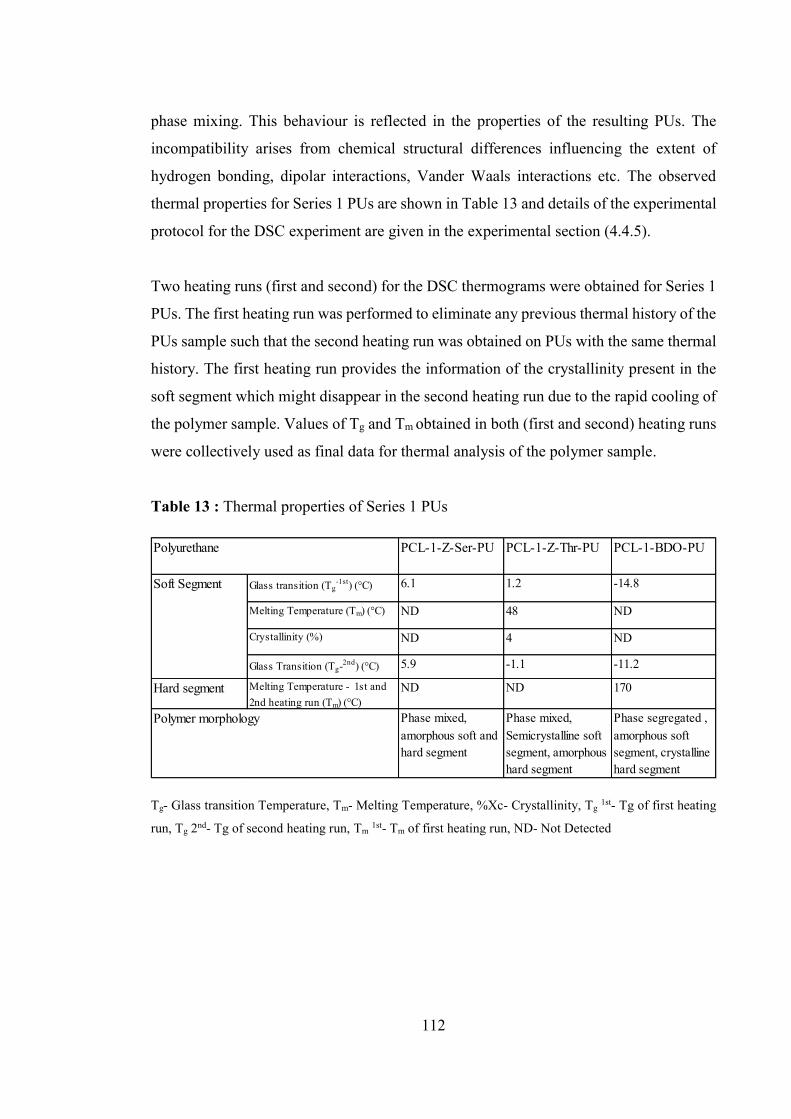
112
phase mixing. This behaviour is reflected in the properties of the resulting PUs. The
incompatibility arises from chemical structural differences influencing the extent of
hydrogen bonding, dipolar interactions, Vander Waals interactions etc. The observed
thermal properties for Series 1 PUs are shown in Table 13 and details of the experimental
protocol for the DSC experiment are given in the experimental section (4.4.5).
Two heating runs (first and second) for the DSC thermograms were obtained for Series 1
PUs. The first heating run was performed to eliminate any previous thermal history of the
PUs sample such that the second heating run was obtained on PUs with the same thermal
history. The first heating run provides the information of the crystallinity present in the
soft segment which might disappear in the second heating run due to the rapid cooling of
the polymer sample. Values of Tg and Tm obtained in both (first and second) heating runs
were collectively used as final data for thermal analysis of the polymer sample.
Table 13 : Thermal properties of Series 1 PUs
Tg- Glass transition Temperature, Tm- Melting Temperature, %Xc- Crystallinity, Tg 1st- Tg of first heating
run, Tg 2nd- Tg of second heating run, Tm 1st- Tm of first heating run, ND- Not Detected
PCL-1-Z-Ser-PU PCL-1-Z-Thr-PU PCL-1-BDO-PU
Glass transition (Tg-1st) (°C) 6.1 1.2 -14.8
Melting Temperature (Tm) (°C) ND 48 ND
Crystallinity (%) ND 4 ND
Glass Transition (Tg-2nd) (°C) 5.9 -1.1 -11.2
Hard segment Melting Temperature - 1st and 2nd heating run (Tm) (°C)
ND ND 170
Phase mixed, amorphous soft and hard segment
Phase mixed, Semicrystalline soft segment, amorphous hard segment
Phase segregated , amorphous soft segment, crystalline hard segment
Polymer morphology
Polyurethane
Soft Segment

113
PCL-1-Z-Ser-PU - The DSC thermograms for the 1st and 2nd heating runs of PCL-1-Z-
Ser-PU are shown in Figure 30.
Figure 30 : DSC thermograms for 1st and 2nd heating run of PCL-1-Z-Ser-PU
1st Heating Run - The soft segment glass transition, (Tg) for 1st heating run for PCL-1-Z-
Ser-PU was observed at 6.1°C. No soft segment melt endotherm (Tm) was observed. For
the hard segment, no Tg or Tm was observed during the first heating run of the polymer.
Amorphous nature for the soft and hard segment was observed.
2nd Heating Run – The soft segment Tg was observed at 5.9°C and no soft segment melting
endotherm Tm was observed. For the hard segment, no Tg or Tm was observed for the 2nd
heating run. Thermal transitions of the polymer, shows that soft and hard segments are
amorphous in nature.
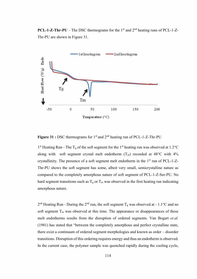
114
PCL-1-Z-Thr-PU – The DSC thermograms for the 1st and 2nd heating runs of PCL-1-Z-
Thr-PU are shown in Figure 31.
Figure 31 : DSC thermograms for 1st and 2nd heating run of PCL-1-Z-Thr-PU
1st Heating Run - The Tg of the soft segment for the 1st heating run was observed at 1.2°C
along with soft segment crystal melt endotherm (Tm) recorded at 48°C with 4%
crystallinity. The presence of a soft segment melt endotherm in the 1st run of PCL-1-Z-
Thr-PU shows the soft segment has some, albeit very small, semicrystalline nature as
compared to the completely amorphous nature of soft segment of PCL-1-Z-Ser-PU. No
hard segment transitions such as Tg or Tm was observed in the first heating run indicating
amorphous nature.
2nd Heating Run - During the 2nd run, the soft segment Tg was observed at - 1.1°C and no
soft segment Tm was observed at this time. The appearance or disappearances of these
melt endotherms results from the disruption of ordered segments. Van Bogart et.al.
(1981) has stated that “between the completely amorphous and perfect crystalline state,
there exist a continuum of ordered segment morphologies and known as order – disorder
transitions. Disruption of this ordering requires energy and thus an endotherm is observed.
In the current case, the polymer sample was quenched rapidly during the cooling cycle,
115
the polymer chains did not get sufficient time to form any crystal structure and hence did
not show any soft segment Tm in the second heating run. However, no hard segment Tg
or Tm was observed for the 2nd run of PCL-1-Z-Thr-PU. Based on these DSC
observations, hard segment are predominantly amorphous in nature and soft segment is
semicrystalline in nature.
Both the polymers, PCL-1-Z-Ser-PU and PCL-1-Z-Thr-PU show soft segment Tg values
below body temperature indicating they will exhibit elastomeric properties at body
temperature (37°C). However, the observed soft segment Tg values for both the PUs
remain substantial higher than the Tg of pure homopolymer soft segment, PCL (- 62°C)
(Bogdanov et al., 1999) indicating that a certain degree of phase mixing is present in these
PUs. In other words, there must be some hard segments present within the soft-segment
phase and these hard segments place restriction to the movement of chains. Camberlin
et.al. (1983) has proposed that Tg value can be affected by these chain end restrictions i.e.
Tg value can increase by up to 4°C for a soft segment Tg . Hence, a high value of Tg
obtained in current case indicate that there must be a significant amount of phase mixing
present in the synthesised PUs. Similar observations for the increase in PCL based soft
segment Tg due to phase mixing morphology have been recorded in the literature (Li et
al., 1994).
Unlike the soft segment, the hard segment glass transition is usually poorly resolved by
DSC. It is reported in the literature (Joshi, 2009) that this happens primarily due to the
following reasons:
(1) The sequence length of the hard segment is often broadly distributed thereby
resulting in a broad Tg. Due to the chemistry involved in the PU synthesis, it is
difficult to achieve control over the hard segment length distribution.
(2) The change in the heat capacity of the PU hard segment is often small, making it
difficult to observe.
The absence of hard segment Tg and Tm for the PUs investigated here, illustrate that their
hard segments are largely amorphous in nature and cannot form a crystalline structure.
This is presumably due to:

116
(1) The short chain length distribution of the hard segment and
(2) The unsymmetrical structure of the chain extender, which prevents any ordering of
the hard segments through hydrogen bonding.
It has been reported in the literature that hard segment should be of sufficient length to
form the crystals and should be symmetrical in nature, to allow chains to come together
at distances close enough to have effective hydrogen bonding (Pergal et al., 2011). For
example, more than three MDI units are required to form stable enough hard segment
crystals to show a Tm in MDI based PUs (Wang et al., 1994). A lack of symmetry in the
hard segment due to the presence of the bulky pendant Z groups of the serine and
threonine based chain extender is another reason for lack of crystallinity. The disruption
caused by the pendant group while packing the hard segment creates more free volume
within the polymer chains which further results in loose packing of the hard segment and
hence the absence of a hard segment Tm. It has been reported in the literature (Parrag and
Woodhouse, 2010) that if the chain extender and the diisocyanate are not symmetrical in
structure and have bulky pendant groups, they will not form an ordered structure through
hydrogen bonding. Similar results have been documented in the other reports for the
amorphous hard segment of PU due to the non-symmetrical structure of chain extender
(Won et al., 1998), (Skarja, 2001), (Hepburn, 1991).
It can be concluded that PCL-1-Z-Ser-PU contains both amorphous soft and hard
segments whilst PCL-1-Z-Thr-PU shows a very weekly semicrystalline soft segment (4%
crystallinity) and an amorphous hard segment. Both polymers show phase mixed
morphology. Given that L-Z-serine and L-Z-threonine amino acid based PU are
synthesized here for the first time, it is difficult to compare these observed properties
directly with literature results.
PCL-1-BDO-PU
The DSC thermograms for the 1st and 2nd heating run of the control PCL-1-BDO-PU are
shown in Figure 32. Hepburn (1991) has reported that PUs containing non-linear chain
extenders are usually more phase mixed, when compared to those containing linear chain

117
extenders such as the BDO based PU used here. The control PCL-1-BDO-PU displays
phase segregated morphology, as shown in Figure 32, which is more consistent with
Bagdi et.al. (2009) who reports that MDI-BDO based hard segments tend to form phase
segregation morphology through strong hydrogen bonding.
Figure 32 : DSC thermograms for 1st and 2nd heating run of PCL-1-BDO- PU
1st Heating Run - The Tg of the soft segment for the first heating run was observed at -
14.8 °C and no soft segment Tm was observed. For the hard segment, no Tg was observed
whilst a Tm was obtained at 170°C. The observed Tm was not a sharp melting endotherm
as expected for BDO based PUs, presumably because it is a mixture of several hard
segment transitions, poorly resolved in DSC. Further detailed analysis of DSC, such as
modulated DSC, might be useful in understanding this transition, but was considered
beyond the scope of this work.
Broadly speaking, the symmetrical structure (no pendant group) of BDO as a chain
extender has helped the MDI based hard segments to form a densely packed regular
structure, bound via H-bonding, and thus shows a hard segment melt endotherm (Tm),
albeit not a sharp one. This transition can be attributed to the melting of the hard segment
crystalline domain. Similar results are reported by Chen et.al. (1997) and Caracciolo et.
al. (2008) based on MDI-BDO as a hard segment for their synthesised PU.

118
Two other thermal transitions were also observed at 50°C and 80°C during the first
heating run. Both transitions are weakly defined and broad. It is proposed in literature
that these endotherms are morphological in origin. As proposed by Seymour and Cooper
(1973), these transitions represents disordering of the hard segment with relatively short
and long range order of hard segment. Short range order represents the interaction
between the soft segment and hard segment that contributes to the phase mixing behaviour
of the PU. Long range order represents ‘unspecified’ interactions within the hard segment
domains. The broad nature of the two peaks shows the wide distribution of the hard
segment lengths.
2nd Heating Run - During the second run, the Tg of the soft segment appeared at -11.1°C
which is close to the Tg observed in the first run (-14.8°C) and no soft segment Tm was
observed. It is worth mentioning that the Tg for both first and second heating run for
control PU is lower than the Tg of the novel amino acid based PUs (Table 13) and remains
much higher than Tg of the pure homopolymer. The low Tg value for BDO based PU
shows the phase segregated morphology of the control PU as compare to phase mixed
behaviour of amino acid based PU.
For the 2nd heating run, the hard segment melt endotherm (Tm) appears again at the same
temperature of 170°C, showing the crystalline nature of the hard segment. Hence it was
concluded that the BDO based PU shows amorphous soft segment and crystalline hard
segment with phase segregated morphology.
Amino acid based PUs, are synthesized for the first time, hence it would be difficult to
compare the observed thermal properties of these PUs to the PUs reported in the literature
based on MDI-PCL as hard and soft segment. However, in general, it was observed that
the novel amino acid based PUs are more phase mixed with semicrystalline soft segment
(PCL-Z-thr-PU) and amorphous hard segment as compared to the control BDO based PU
which shows phase segregated morphology with amorphous soft segment and crystalline
hard segment.

119
4.5.2.6 Thermogravemetric Analysis
Thermogravemetric analysis (TGA) is a suitable method to evaluate the thermal stability
of polymers, including PU elastomers (Hepburn, 1991). The TGA data for Series 1 PU is
shown in Figure 33.
Figure 33 : TGA analysis of Series 1 PU
The TGA analysis shows that amino acid based PUs are more thermally stable compared
to the control BDO based PU. The onset of degradation for PCL-1-Z-Ser-PU and PCL-
1-Z-Thr-PU is at ~ 350°C and is essentially complete by ~ 550°C. For the control PCL-
1-BDO-PU, the onset of degradation is ~ 270°C and is complete by ~ 470°C. The PUs
exhibit a two-step degradation processes presumably reflective of the two phase (hard
and soft segment) structure of the PU. It is reported in the literature (Gupta and Adhikari,
2003) that the thermal degradation of the PU starts in the hard segment first then follows
into the soft segment. The hard segment degradation involves dissociation of urethane
and urea bonds presents in the hard segment.
The result indicates that amino acid based PUs are thermally stable up to a high
temperature and the reason may be:
(1) A higher level of aromatic components.
(2) Mixed phase morphology.

120
The increase in thermal stability of amino acid based PU is probably due mainly to the
presence of more aromatic components compared to that of the control PU. Similar
observations for higher thermal stability of MDI and aromatic chain extender based PU
have been noted in the past. For example, Azzam et.al.(2007) has been reported that the
aromatic and heterocyclic moieties present in the polymer main chain or as side group
can contribute to the increased thermal stability of PU. The aromatic group present in the
MDI and Z group of the chain extender present in the hard segment of amino acid based
PUs provide additional intermolecular attraction through delocalised π bond interaction
(Dieterich et al., 1994) with neighbouring polymer chains having similar aromatic MDI
and Z groups.
Antipova et.al. (1970) has reported that the degree of phase segregation or segmental
mixing has a significant effect on the thermal stability of PU, with a mutual stabilization
effect present between the soft and hard segment of phase mixed morphology. DSC
analysis indicates phase mixing behaviour of amino acid based PUs and these polymers
also shows high thermal stability as compared with the control PU which shows phase
segregation morphology. This observation supports the fact that during heating at high
temperature, especially under nitrogen atmosphere, interurethane hydrogen bonding
dissociate and after the dissociation, more phase mixing favours the thermal stability of
PUs. A similar correlation between the phase mixed morphology and higher thermal
stability of PTMG/PEG-MDI based PU has been reported by Wang et.al. (1997).
In summary, amino acid based PUs in Series 1 are more thermally stable than the control
PU. This could be an advantage in thermal processing of these polymers as they offer a
broader temperature range for processing.
4.5.2.7 Water Contact Angle
Contact angle are commonly used to describe surface wettability (Dou et al., 2006). Water
contact angle was measured in order to obtain an idea of the relative polarity of the amino
acid based PU surface. Contact angle values for Series 1PU are reported in Table 14.

121
Table 14 : Water contact angle values for Series 1 PU
Polyurethane Contact Angle (ɵ) PCL-1-Z-Ser-PU 72° ± 4 PCL-1-Z-Thr-PU 77° ± 3 PCL-1-BDO-PU 68° ± 2
There is a very slight increase in contact angle values from PCL-1-Z-Ser-PU to PCL-1-
Z-Thr-PU, however the difference is minor. Both amino acid based PUs exhibited a
higher contact angle than the control PCL-1-BDO PU, but again the difference is minor,
although the difference between the threonine based PU and the control PU is more
obvious. In all cases, the contact angle indicates a relatively hydrophobic surface with
the two amino acid, non-linear chain extender based, PUs being slightly more
hydrophobic than the control.
Similar observation have been reported in the literature (Guelcher et al., 2005) showing
relatively hydrophilic contact angles for linear chain extenders where non-linear chain
extenders are slightly more hydrophobic. For example, Skarja et.al.(1998) has reported
contact angle value of 71° and Sarkar et.al. (2009)as reported the contact angle value of
75° for non-linear chain extender based polyurethane.
The small increase in hydrophobicity shown in Table 14 may be due to:
1. Incomplete phase separation between hard and soft segments leading to fewer and
smaller dipole formation(Fernández d'Arlas et al., 2008), and
2. The hydrophobic nature of the PCL soft segment (Kavlock et al., 2007)
All these factors, the hydrophobic nature of PCL, MDI, non-linear structure of chain
extender and the presence of bulky aromatic pendant Z group have made the surface of
the amino acid based PU more hydrophobic in nature as compared to BDO based PU.
In summary, the novel amino acid based PUs are slightly more hydrophobic in nature
than the control PU and may therefore be good alternatives where slightly more water
repellent properties are required.

122
4.5.2.8 Mechanical Properties
Tensile properties of the PUs can be affected by different factors such as polymer overall
molecular weight, polymer morphology, hard and soft segment length and their
crystallinity. The stress strain graphs of the amino acid based Series 1 PUs are shown in
Figure 34 and for Series 1 control PUs are shown in Figure 35. The mechanical properties
are shown in Table 15 and compared to the control PU. It is clear that the Series 1 amino
acid based PUs are weak materials with low mechanical strength. Specifically, they have
low modulus of elasticity, low tensile strength and high percentage of elongation during
tensile testing.
Figure 34 : Tensile stress – strain curve of Series 1 PU
The stress-strain curve shows a significantly long region of plastic deformation, unlike
conventional PUs. Both the amino acid based PUs are soft materials with near identical
elongation at break values. In contrast, the control PCL-1-BDO-PU behaved as a strong
and stiff polymer with a high value of modulus and tensile strength but with low percent
of elongation. Weak mechanical properties shown by amino acid based PU might be due
to the less ordered hard segment domain observed in amino acid based Series 1 PUs.

123
Figure 35 : Tensile stress- strain curve of PCL-1-BDO-PU
Since the amino acid chain extenders have a higher molecular weight than BDO, there
are fewer numbers of chain extender units incorporated into the hard segment of the PUs.
This leads to fewer urethane linkages in the hard segment as compared to the control PU,
which in turn leads to a less ordered hard segment domain and thus a relatively more
amorphous material.
Table 15 : Mechanical properties of Series 1 PU (mean ± SD, n = 6)
Polyurethane (PU)
Modulus of elasticity (MPa)
Ultimate tensile strength (MPa)
Elongation at break (%)
PCL-1-Z-Ser-PU 6.2 ± 2 4.8 ± 1 625.8 ± 35 PCL-1-Z-Thr-PU 4.2 ± 1 10.9 ± 6 660 ± 59 PCL-1-BDO-PU 52.8 ± 22 32.9 ± 12 234 ± 18
It is reported in the literature (Lamba et al., 1998) that phase morphology plays a vital
role in determining the mechanical properties of the synthesised polymer, with
amorphous materials prone to show weaker mechanical properties. As seen via DSC
(Table 13), amino acid based PUs particularly PCL-1-Z-Ser-PU show phase mixed
morphology in contrast to phase segregated morphology shown by control PU.
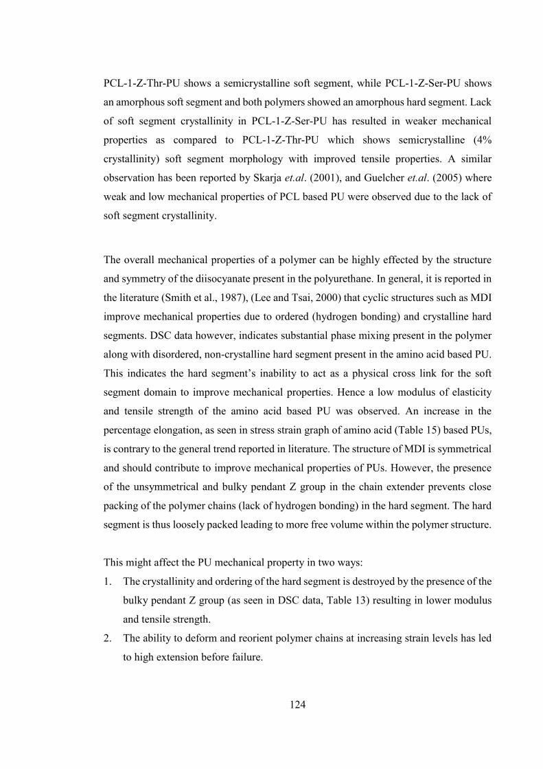
124
PCL-1-Z-Thr-PU shows a semicrystalline soft segment, while PCL-1-Z-Ser-PU shows
an amorphous soft segment and both polymers showed an amorphous hard segment. Lack
of soft segment crystallinity in PCL-1-Z-Ser-PU has resulted in weaker mechanical
properties as compared to PCL-1-Z-Thr-PU which shows semicrystalline (4%
crystallinity) soft segment morphology with improved tensile properties. A similar
observation has been reported by Skarja et.al. (2001), and Guelcher et.al. (2005) where
weak and low mechanical properties of PCL based PU were observed due to the lack of
soft segment crystallinity.
The overall mechanical properties of a polymer can be highly effected by the structure
and symmetry of the diisocyanate present in the polyurethane. In general, it is reported in
the literature (Smith et al., 1987), (Lee and Tsai, 2000) that cyclic structures such as MDI
improve mechanical properties due to ordered (hydrogen bonding) and crystalline hard
segments. DSC data however, indicates substantial phase mixing present in the polymer
along with disordered, non-crystalline hard segment present in the amino acid based PU.
This indicates the hard segment’s inability to act as a physical cross link for the soft
segment domain to improve mechanical properties. Hence a low modulus of elasticity
and tensile strength of the amino acid based PU was observed. An increase in the
percentage elongation, as seen in stress strain graph of amino acid (Table 15) based PUs,
is contrary to the general trend reported in literature. The structure of MDI is symmetrical
and should contribute to improve mechanical properties of PUs. However, the presence
of the unsymmetrical and bulky pendant Z group in the chain extender prevents close
packing of the polymer chains (lack of hydrogen bonding) in the hard segment. The hard
segment is thus loosely packed leading to more free volume within the polymer structure.
This might affect the PU mechanical property in two ways:
1. The crystallinity and ordering of the hard segment is destroyed by the presence of the
bulky pendant Z group (as seen in DSC data, Table 13) resulting in lower modulus
and tensile strength.
2. The ability to deform and reorient polymer chains at increasing strain levels has led
to high extension before failure.

125
A similar observation has been reported by Sarkar et.al. (2008) where a tyrosine amino
acid based chain extender was used for PU synthesis. The bulky side chains of the tyrosine
amino acid prevented the close packing of the hard segment of PU and hence affected the
overall mechanical properties of the synthesised PU.
The control BDO based PU, behaves as a stiff and strong material with high modulus and
tensile strength. It is reported in the literature (Patterson et al., 1999), (Joshi, 2009) that
MDI-BDO hard segment result in very strong hydrogen bonding that helps to attain
improved mechanical properties. The mechanical properties are also depend on the phase
morphology of the polymer. It was observed in the DSC, that the control PU polymer was
segregated into two separated phases showing both a Tg of the soft segment and a Tm of
the hard segment. The hard segment is crystalline in nature and the soft segment was
present in a viscoelastic state. In phase segregated morphology, the hard segment may act
as a physical cross link for the soft segment domains and therefore act as a reinforcing
filler (McBane et al., 2007). When the polymer is pulled during mechanical testing, the
polymer chains align with each other, making it very difficult to stretch. Hence the high
modulus and tensile strength was obtained, as seen in control PU and due to the stiffness
of the polymeric chains, it cannot be stretched for long and hence a low value of
percentage elongation was observed. The other reason for enhanced mechanical
properties of BDO-PU is the symmetrical structure and low molecular weight of the chain
extender. BDO as a linear chain extender, does not have any pendant group in its structure
to disturb the hard segment packaging which has result in very ordered structural packing
of the hard segment due to strong hydrogen bonding which in turn shows improved
mechanical properties.
It can be concluded that amino acid based PUs behave as mechanically weak materials
with high extension to failure and low tensile strength and modulus compared to the
control PU. These results show the importance of the chain extender structure and effect
of polymer morphology on mechanical properties of PUs confirming the requirement of
a short chain symmetrical molecule as a chain extender to achieve good mechanical
property.

126
4.6 Deprotection of Z-group
4.6.1 Introduction
Deprotection of the Z-group from polymer backbone may be useful for future polymer
applications. For example, the resultant free amine group could be used to covalently
attach any given drug or biologically active compound, such as a polysaccharide or
peptide, to the PU making the polymer more biocompatible. Incorporation of the charged
groups (NH3+) into the PU backbone could produce material known as a PU ionomer
which has been shown to have improved blood compatibility (Jinhuang et al., 1989).
Several investigations on drug delivery systems based on polymers with pendant
functional groups have been reported in literature (in't Veld et al., 1992), (Fiétier et al.,
1990), (Zhou and Kohn, 1990). Skarja et.al. (2001) have also synthesised PU with amine
groups as pendant groups on the PU backbone.
The research group (Xie et al., 2007) has reported the hydrogenation method for the
deprotection reaction of the Z group from the PU backbone, resulting in the regeneration
of NH3+ group as pendant group. The synthesis of deprotected PU was not the main focus
and it was not further characterised. Detailed characterisation and application of
deprotected PU can be considered as future work and some preliminary experiments are
carried out here.
An initial experiment to demonstrate the removal of the Z group from the PCL-1-Z-Ser-
PU is described in this section 4.3.3. The aim of the experiment is to demonstrate the
possibility of deprotection of the Z group from the synthesised PU without affecting the
PU backbone and leaving a functional pendant amine group at the polymer backbone. A
new batch of PCL-1-Z-Ser-PU was synthesised, based on the two step prepolymer
method as described in section 4.3. Polymer reaction conditions remained the same as
describe in section 4.3 and the quantities of the reactants used for the PU synthesis are
given in Table 16. The polymer contained 0.05 % stannous octoate as catalyst, an
isocyanate index of 1.05 was used, and 50 weight % hard segment was present in PU.

127
Table 16 : Composition of PCL-1-Z-Ser-PU for deprotection reaction
Reagent Mass (g) Molecular Weight Moles PCL 2.5 1057 0.00236 MDI 1.25 250.25 0.00499 CE (318) 1.3 532.31 0.00244
318 - Octane-1,8-diyl bis (2-amino-4-methylpentanoate)
CE – Chain extender
4.6.2 Deprotection reaction
An acidolysis method, using hydrogen bromide and acetic acid solution (HBr/HAc), was
utilised to remove the Z group from the PU backbone. This method has been reported in
the literature (Deng et al., 2007), (Katchalski et al., 1948), (Ben-Ishai and Berger, 1952),
(Anderson et al., 1952) (Hernández and Klok, 2003) and results in cleavage of the
carbamate (urethane) linkage and regeneration of the free amine group. An alternative
has been reported in the literature involving hydrogenation of the Z- group from the
polymer backbone using palladium/charcoal carbon as catalyst (Skarja, 2001). The
hydrogenation method, however, has been reported as time consuming, complete
elimination of the Z- group cannot be achieved and the complete separation of the residual
palladium activated charcoal is very difficult from the final product. Xie et.al. (2007) has
reported the use of HBr/HAc method to remove the Z group from the backbone of PCL-
PEG-HDI based PU. The HBr/HAc based method is fast and easy to use, and more likely
to be complete, so was the chosen method here.
The general reaction for deprotection of the Z group from PCL-1-Z-Ser-PU is shown in
Scheme 5. At the end, a free amine results as a salt with bromine ion (NH3+ Br-). The
deprotected PU will be denoted as PCL-1-Ser-PU. Following deprotection, the resultant
polymer was stored in a desiccator until further use.

128
Scheme 5 : Deprotection reaction to remove Z group from PCL-1-Z-Ser-PU
The yield of the deprotection reaction was 70% and this did not vary by more than 5% on
repetition of the experiment. The deprotected PU exhibits polycationic character due to
its pendant NH3+ cation. The acidolysis method attacks the carbamate (urethane) linkage
present between the pendant Z group and NH group of the amino acid and results in the
release of benzyl alcohol and carbon dioxide as side products. The NH group of the chain
extender is converted into the free NH3+ group. PU also contains a number of urethane
linkages in the polymer backbone, so it was anticipated that backbone cleavage may occur
during deprotection, leading to a loss in polymer molecular weight and subsequent
modification of polymer properties. Deprotection reaction conditions such as reaction
time were therefore first optimised in an attempt to promote deprotection but limit
polymer backbone cleavage. The detailed method of the deprotection reaction is given in
the experimental section 4.3.3.

129
4.6.3 Optimisation of deprotection reaction For the optimisation of the deprotection reaction, the 1H-NMR spectrum of the
deprotection reaction mixture was obtained at different time intervals. This monitors the
progress of the reaction in order to attain the minimum time required for the deprotection
of Z group from the PU backbone. It was observed that long exposure of the polymer
reaction mixture to HBr/HAc solution causes degradation of the urethane linkage present
in the polymer backbone. This results in polymer degradation and loss of polymer
molecular weight. Details of the degradation reaction are beyond the scope of this thesis.
However it is relevant to report the observations here as it indicates that optimisation is
required.
4.6.3.1 1H Nuclear Magnetic Resonance Spectroscopy Aliquots of reaction mixture were taken at 10 minutes and 30 minutes time interval from
the start of the deprotection reaction. 1H-NMR spectroscopy was performed and
compared with the protected PU 1H NMR. The collective 1H- NMR spectrum of protected
PU (zero reaction time) and deprotection reaction mixtures at 10 minute and at 30 minutes
are shown in Figure 36. It was observed that the proton peaks obtained in Spectrum A at
7.39ppm (due to protons of aromatic ring of the Z group) and at 5.15ppm (due to benzyl
methylene proton of Z group) were reduced in intensity in the Spectrum B and C. The
reduction in peak height related to protons associated with the Z group demonstrates a
clear indication of the progress of the deprotection reaction. Calculations were done on
the basis of Z proton (present at 5.15ppm) peak height ratio by keeping PCL peak (present
at 2.2ppm) constant to monitor the percent reduction of Z group after 10 and 30 minutes.
It was observed that 62% reduction in Z protons peak was observed after 10 minutes and
a further 11% reduction in peak height ratio was observed after 30 minutes. This
observation further supports the fact that Z group based protons were removed during the
deprotection reaction. Similar observation for 1H-NMR spectroscopy was recorded by
Xie et.al. (2007) where the HBr/HAc method was utilised to remove the Z group at the
backbone of PCL-PEG-HDI based PU.
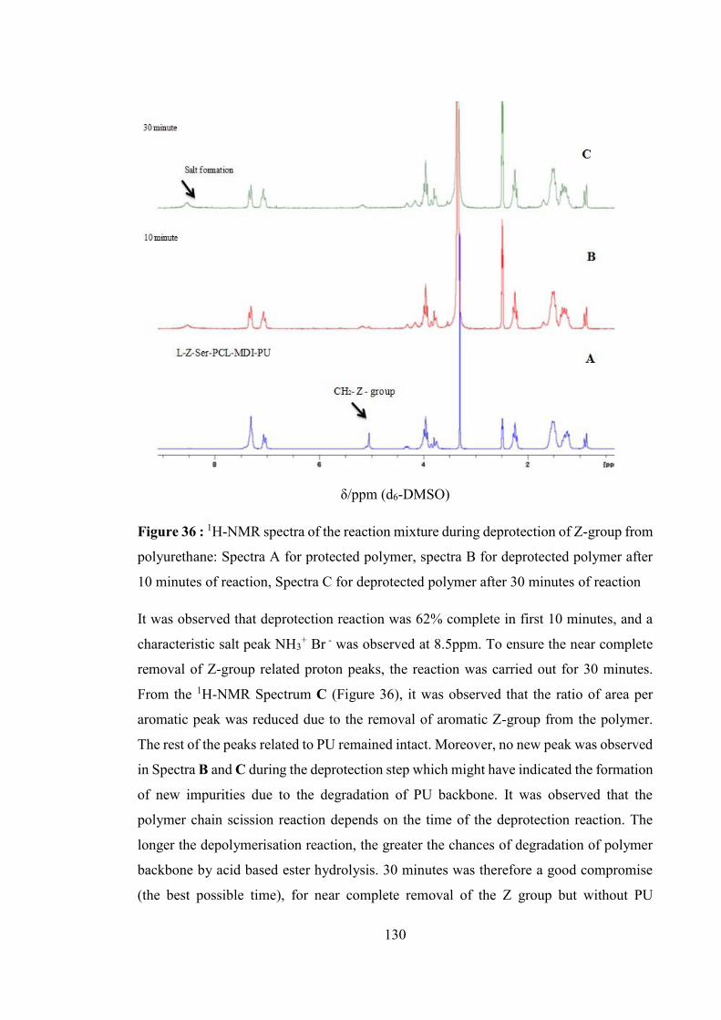
130
δ/ppm (d6-DMSO)
Figure 36 : 1H-NMR spectra of the reaction mixture during deprotection of Z-group from
polyurethane: Spectra A for protected polymer, spectra B for deprotected polymer after
10 minutes of reaction, Spectra C for deprotected polymer after 30 minutes of reaction
It was observed that deprotection reaction was 62% complete in first 10 minutes, and a
characteristic salt peak NH3+ Br - was observed at 8.5ppm. To ensure the near complete
removal of Z-group related proton peaks, the reaction was carried out for 30 minutes.
From the 1H-NMR Spectrum C (Figure 36), it was observed that the ratio of area per
aromatic peak was reduced due to the removal of aromatic Z-group from the polymer.
The rest of the peaks related to PU remained intact. Moreover, no new peak was observed
in Spectra B and C during the deprotection step which might have indicated the formation
of new impurities due to the degradation of PU backbone. It was observed that the
polymer chain scission reaction depends on the time of the deprotection reaction. The
longer the depolymerisation reaction, the greater the chances of degradation of polymer
backbone by acid based ester hydrolysis. 30 minutes was therefore a good compromise
(the best possible time), for near complete removal of the Z group but without PU

131
backbone scission. The current optimisation reaction time (30 minutes) is consistent with
the time used in literature (Xie et al., 2007).
In conclusion, 30 minutes reaction time was sufficient to remove most of the Z group
from PU backbone but not so long as to promote the unwanted scission of the PU chain.
The deprotected polymer will be referred as PCL-1-L-Ser-PU.
4.6.3.2 Solubility
The deprotected polymer, PCL-1-Ser-PU, was found to be soluble in a range of different
solvents such as water, acetone, methanol, ethanol, ethyl acetate and tetrahedron furan
(THF) solvent. It is worth noticing that the protected PU was insoluble in the above
mentioned solvents, providing further evidence that the deprotection reaction was
successful. The cationic nature of the deprotected PU has increased the solubility of the
PU probably via hydrogen bonding. The deprotected PU was also soluble in
Dimethylformamide (DMF), Dimethylsulphoxide (DMSO) and N- Methyl pyrrolidine
(NMP) indicating solubility in both polar aprotic and protic solvents. This shows that the
unprotected PU solubility and hydrophilicity has increased as compared to the protected
PU.
4.6.3.3 Gel permeation chromatography
The effect of the deprotection reaction on polymer molecular weight was investigated by
gel permeation chromatography (GPC). The molecular weight of protected and
deprotected PU was determined by GPC and compared. Figure 37 shows the GPC traces
of deprotected and protected PCL-1-Z-Ser-PU and Table 17 shows the molecular weight
of PU before and after deprotection reaction.

132
Figure 37 : GPC traces of protected and deprotected PCL-1-Z-Ser-PU
The Z group based urethane group was expected to be prone to hydrolysis due to the
proximity of the phenyl group to the urethane linkage. Similar benzyl esters are removed
by hydrogenolysis in literature (Skarja, 2001). The Z group is similar in nature to benzylic
ester and is known to be particularly susceptible to hydrolysis as compared to backbone
urethanes groups which were expected to be much less reactive.
The weight average molecular weight (Mw) of the original polymer used for the
deprotection reaction was 5.07 x 10⁴ Dalton and after deprotection there was only a minor
change in the weight average molecular weight (Mw) of polymer (4.98 x 10⁴ Dalton) was
observed. The polymer molecular weight and its distribution did not significantly change
indicating that polymer chains were intact and little altered.
The minor changes observed in the Mw of deprotected polymer shows the loss of
molecular weight from the removed Z group from PU and the data is consistent with the
molecular weights (The Z group should account for approximately 1000 molecular
weight and there was a loss of approximately 800 molecular weight after its removal).

133
Table 17 : Molecular weight of PCL-1-Z-Ser-PU before and after deprotection reaction
Polyurethane (PU)
Weight average molecular weight (Mw) (Dalton)
Number average molecular weight (Mn) (Dalton)
Poly Dispersity Index (PDI)
PCL-1-Z-Ser-PU (Protected PU)
5.07 x 10⁴ 2.95 x 10⁴ 1.71
PCL-1-Ser-PU (Unprotected PU)
4.98 x 10⁴ 2.93 x 10⁴ 1.70
In summary, the molecular weight and polydispersity of the deprotected PU did not
change significantly from the protected PU and hence it can be concluded that the PU
chains were intact and polymer backbone cleavage did not happen during the deprotection
step.
4.6.3.4 Fourier Transform Infrared Spectroscopy
Fourier transform infrared spectroscopy (FTIR) was performed on PCL-1-Z-Ser-PU
(protected) and PCL-1-Ser-PU (deprotected). The comparison was hoped to further
illustrate the complete removal of the Z-group from the protected PU, formation of a salt
peak in the deprotected polymer and to exclude the possibility of scission of the polymer
backbone during the deprotection reaction. The FTIR spectra of deprotected PU
(Spectrum B) is compared to the protected PU (Spectrum A) and shown in Figure 38.
The FTIR spectra of protected PU (Spectrum A) shows characteristic peaks at 3345,
3336cm-1 (N-H), 1731cm-1 (C=O) and 1120, 1167cm-1 for the C-N bond of Z and urethane
group. In spectrum B of deprotected PU, a change in the peak area of 3100 – 3600cm-1
region was observed which corresponds to a change in the N-H region of PU. The peak
area and shape for the N-H group (3345, 3336 cm-1) region in deprotected polymer was
changed. A broad N-H peak was observed at 3480cm-1 (Spectrum B) which illustrates the
formation of salt (ammonium bromide, NH3+Br-) at the polymer backbone. In the FTIR
spectrum B, the aromatic C-H out of plane bending absorption peaks observed at 749cm-
1 and 697cm-1 due the Z- benzyl group disappeared. Similar FTIR observation for the Z
group peak removal has been reported by Deng et.al. (2007) and Won et.al. (1998) where
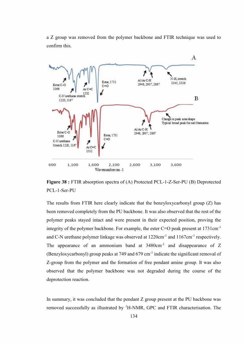
134
a Z group was removed from the polymer backbone and FTIR technique was used to
confirm this.
Figure 38 : FTIR absorption spectra of (A) Protected PCL-1-Z-Ser-PU (B) Deprotected
PCL-1-Ser-PU
The results from FTIR here clearly indicate that the benzyloxycarbonyl group (Z) has
been removed completely from the PU backbone. It was also observed that the rest of the
polymer peaks stayed intact and were present in their expected position, proving the
integrity of the polymer backbone. For example, the ester C=O peak present at 1731cm-1
and C-N urethane polymer linkage was observed at 1220cm-1 and 1167cm-1 respectively.
The appearance of an ammonium band at 3480cm-1 and disappearance of Z
(Benzyloxycarbonyl) group peaks at 749 and 679 cm-1 indicate the significant removal of
Z-group from the polymer and the formation of free pendant amine group. It was also
observed that the polymer backbone was not degraded during the course of the
deprotection reaction.
In summary, it was concluded that the pendant Z group present at the PU backbone was
removed successfully as illustrated by 1H-NMR, GPC and FTIR characterisation. The
135
formation of NH3+ as a pendant group shows the potential of the synthesised novel amino
acid based PU to be used in applications where the presence of a cationic group may be
advantageous, for example in delivery systems for gene based therapeutics. Future work
may include a physiochemical characterisation of the deprotected PU to further test its
biomedical applications.
4.7 Polyurethane urea
4.7.1 Polyurethane urea synthesis
A series of polyurethane ureas (PUUs) were prepared using the two step prepolymer
method. The incorporation of diamine chain extender into the polymer results in the
formation of a urea linkage with the NCO group in the hard segment of the polymer. PCL
(Mw 1057) was used as the polyol, MDI was used as the diisocyanate and the novel
synthesised compounds 311, 312, 313 and 314 were used as diamine chain extenders for
PUU synthesis. The general chemical structure of the leucine diamine chain extender
(311) based PUU as an exemplar, is shown in Figure 39 where the urethane and urea
linkages are highlighted.
Figure 39 : The chemical structure of PCL-1-Leu-PUU showing urea and urethane
linkages
The synthesised series of PUU based on PCL 1057 (Mw) is referred as Series 1 PUU.
The synthesis, characterisation and chemical structure of the novel amino acid based
diamine chain extenders were detailed in Chapter 3. The prepolymer method for PUU
synthesis is described in detail in the experimental section (4.3).
The resultant polymers were slightly yellow in colour, opaque and rubbery in appearance.
The final polymer was stored in a sealed plastic bag for future research work. The net
yield of the polymerisation reaction was ~70% (in all cases) and the result was

136
reproducible within range of ± 5%. A loss of low molecular weight species during solvent
precipitation may account, in part, for the lower yield of the purified polymer.
The chemical structure of all the novel synthesised PUU is shown in Figure 40 and Table
18 shows the abbreviations followed for PUUs reported in this chapter. The nomenclature
is based on the type and molecular weight of the soft segment and chain extender.
Table 18 : Nomenclature for Series 1 PUU
Polyol Molecular Weight
Diisocyanate Chain extender (Diamine)
Polyurethane urea (PUU) representative codes
Poly
capr
olac
tone
(P
CL-
1) (M
w=1
057)
4,4-
met
hyle
ne d
iphe
nyl
diis
ocya
nate
(MD
I)
L-Leucine diamine ester (311)
PCL-1-Leu-PUU
L-Isoleucine diamine ester (312)
PCL-1-Ileu-PUU
L-Valine diamine ester (313)
PCL-1-Val- PUU
L-Tyrosine diamine ester (314)
PCL-1-Tyr- -PUU
For example, PCL-1-Leu-PUU refers to PUU consisting of 1057 molecular weight of
PCL and with L-Leucine based diamine chain extender (311, chapter 3). All the PUUs
described in this and the following chapters contained MDI as the diisocyanate
component and follow this nomenclature.
The quantities of starting material used in the synthesis of Series 1 PUUs are given in
Table 19. All synthesised PUUs contained 0.05% stannous octoate as catalyst, had an
isocyanate index of 1.05, with 50 weight% hard segment.

137
Figure 40 : Chemical structure of Series 1 PUU
Table 19 : Composition of Series 1 PUU
PUU PCL-1-Leu-PUU
PCL-1-Ileu-PUU
PCL-1-Val-PUU
PCL-1-Tyr-PUU
MDI (Mw) 250.2 MDI mass (g) 14.21 14.4 14.6 13.1 MDI moles 0.056 0.057 0.058 0.052 PCL (Mw) 1057 PCL mass (g) 25.03 25.02 25.02 25.03 PCL moles 0.023 0.023 0.023 0.023 CE (Mw) 372.5 358.5 344.4 472.5 CE mass (g) 11.42 11.25 11.07 12.48 CE moles 0.03 0.031 0.032 0.026

138
4.7.2 Characterization of polyurethane ureas
4.7.2.1 Gel Permeation Chromatography
The measured average molecular weight (Mw), number average molecular weight (Mn)
and polydispersity index of Series 1 PUUs is shown in Table 20 and GPC traces are shown
in Figure 41. All PUU molecular weights reported in this thesis are polystyrene equivalent
molecular weights
A typical GPC chromatograph of Series 1 PUUs shows a uni modal peak (Figure 41).
Series 1 PUUs synthesised to a relatively high molecular weight, typical for experimental
amino acid based polyurethanes reported in the literature (Skarja, 1998), (Marcos-
Fernández et al., 2006), (Kavlock et al., 2007), (Fromstein and Woodhouse, 2002). PCL-
1-Leu-PUU shows the highest of number average molecular weight and the aromatic
PCL-1-Tyr-PUU shows the lowest of the number average molecular weight for Series 1
PUUs.
Figure 41 : GPC traces of Series 1 PUU
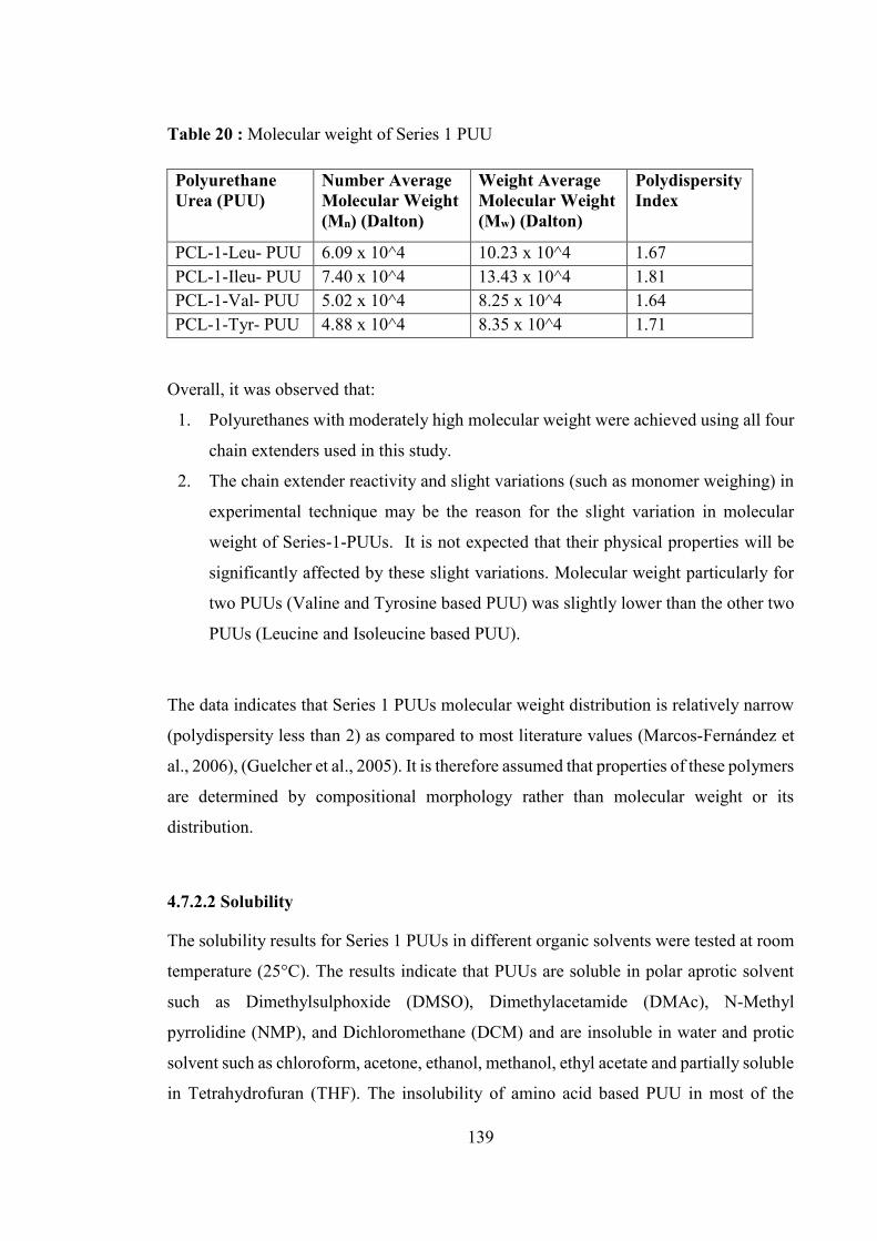
139
Table 20 : Molecular weight of Series 1 PUU
Polyurethane Urea (PUU)
Number Average Molecular Weight (Mn) (Dalton)
Weight Average Molecular Weight (Mw) (Dalton)
Polydispersity Index
PCL-1-Leu- PUU 6.09 x 10^4 10.23 x 10^4 1.67 PCL-1-Ileu- PUU 7.40 x 10^4 13.43 x 10^4 1.81 PCL-1-Val- PUU 5.02 x 10^4 8.25 x 10^4 1.64 PCL-1-Tyr- PUU 4.88 x 10^4 8.35 x 10^4 1.71
Overall, it was observed that:
1. Polyurethanes with moderately high molecular weight were achieved using all four
chain extenders used in this study.
2. The chain extender reactivity and slight variations (such as monomer weighing) in
experimental technique may be the reason for the slight variation in molecular
weight of Series-1-PUUs. It is not expected that their physical properties will be
significantly affected by these slight variations. Molecular weight particularly for
two PUUs (Valine and Tyrosine based PUU) was slightly lower than the other two
PUUs (Leucine and Isoleucine based PUU).
The data indicates that Series 1 PUUs molecular weight distribution is relatively narrow
(polydispersity less than 2) as compared to most literature values (Marcos-Fernández et
al., 2006), (Guelcher et al., 2005). It is therefore assumed that properties of these polymers
are determined by compositional morphology rather than molecular weight or its
distribution.
4.7.2.2 Solubility
The solubility results for Series 1 PUUs in different organic solvents were tested at room
temperature (25°C). The results indicate that PUUs are soluble in polar aprotic solvent
such as Dimethylsulphoxide (DMSO), Dimethylacetamide (DMAc), N-Methyl
pyrrolidine (NMP), and Dichloromethane (DCM) and are insoluble in water and protic
solvent such as chloroform, acetone, ethanol, methanol, ethyl acetate and partially soluble
in Tetrahydrofuran (THF). The insolubility of amino acid based PUU in most of the

140
organic solvents is probably due to the high molecular weight of the polymers. It has also
been reported in the literature (Okuto, 1966), (Chao and Tian, 2002) that the insolubility
of PU/PUUs in organic solvents might be attributed to the allophanate linkage formation
when the isocyanate functionality is used in excess, as is the case here (NCO/OH - 1.05)
for Series 1 PUUs.
4.7.2.3 1H Nuclear Magnetic Resonance Spectroscopy
To avoid repetition of 1H-NMR spectrum and its peak assignment for all the PUUs, only
leucine based PCL-1-Leu-PUU 1H-NMR spectra is describe here in detail and is shown
in Figure 42. The choice of PCL-1-Leu-PUU for full description was random. The 1H-
NMR spectrum of the rest of Series 1 PUUs are reported in the Appendix B. Note that
DMSO-d6 was chosen as the NMR solvent.
PCL-1-Leu-PUU
The 1H-NMR spectrum for PCL-1-Leu-PUU is shown in Figure 42 along with the 1H-
NMR spectrum of the starting materials PCL and MDI.
δ/ppm (DMSO-d6)
Figure 42 : 1H-NMR spectra (Spectrum A) PCL-1-Leu-PUU, (Spectrum B) MDI and
(Spectrum C) PCL

141
In spectrum A, the peak present at 0.90 – 0.95ppm represents the terminal methyl (CH3)
protons of leucine diamine chain extender (311) and appears as a doublet. Peaks in the
region of 1.4 – 1.6ppm are related to the diastereotopic hydrogen of methylene (CH2),
protons of PCL and diamine chain extender, and appear as a multiplet. The six methylene
protons of neopentyl glycol (used for the ring opening of caprolactone to make PCL)
appear at 0.87 and 0.19ppm. Methylene protons (CH2) of PCL are present at 1.1 – 1.5
ppm. At 2.2ppm, terminal methylene protons (CH2) present next to the carbonyl bond of
PCL are present as a multiplet. The protons of CH2 of the MDI moiety and CH2 of the
urethane linkage appear in the region of 3.8 – 4.2ppm respectively. The aromatic protons
of MDI appear in the region of 7.1 – 7.4ppm. Similar observations were recorded by
Rafiemanzelat, et.al. (2010) for 1H-NMR spectrum of poly ether urethane urea having
MDI as diisocyanate and leucine based cyclopeptide as chain extender. Due to the
presence of multiple peaks in the polymer spectrum, it is sometimes difficult to analyse
each peak.
The presence of major protons associated with urethane and urea linkages are present to
confirm the PUU synthesis. Thus the successful synthesis of PCL-1-Leu-PUU can be
concluded from the 1H-NMR data
The 1H-NMR spectroscopy peaks related to the PCL-1-Ileu- PUU, PCL-1-Val- PUU ,
PCL-1-Tyr-PUU are reported in Appendix B and display all the expected structural
features of the targeted polymer. This demonstrates the successful formation of Series 1
PUUs.
4.7.2.4 Fourier Transform Infrared Spectroscopy
Fourier Transform Infrared Spectroscopy (FTIR) analysis was performed to determine
the presence of specific functional groups in the PUU particularly ester, urethane and urea
groups. To avoid repetition, only the PCL-1-Leu-PUU, FTIR data is discussed here, with
the main FTIR peaks reported in Table 21. FTIR spectrum of PCL-1-Leu-PUUis shown
in Figure 43. FTIR data for the rest of the Series 1 PUU is reported in the Appendix B.

142
A strong peak at 1735cm-1 can be attributed to the carbonyl group (C=O) of urethane
linkage indicating successful synthesis of the polymer. The broader shoulder around 3330
cm-1 is indicative of hydrogen bonded N-H stretching. It is worth noting the absence of
the 2260 cm-1 band that is characteristic of the unreacted isocyanate group.
Stretching vibrations of the ester groups are assigned to the 1060, 1160, 1239cm-1 bands.
The peak at 1536cm-1 is characteristic of the carbonyl stretching vibration, which is very
intense. The urea carbonyl (hydrogen bonded) peak around 1640 – 1629cm-1 indicates
the presence of the urea group. The characteristic bands of ester and urethane free
carbonyl overlap between 1735 and 1710cm-1.
The absorption peaks at 2854 and 2924cm-1 correspond to symmetric and asymmetric
CH2 groups respectively. Bands at 1458, 1420, 1394 and 1365cm-1 correspond to various
modes of CH2 vibrations.
Figure 43 : FTIR spectra of PCL-1-Leu-PUU

143
Table 21 : FTIR peak assignment for PCL-1-Leu-PUU
Wavenumber (cm -1) Assignment
3339, 3330
(N-H) (urethane H-Bonded), (N-H) (urea H-Bonded)
3191, 3125
(C-C) aromatic ring
2924, 2854
a (C-H) in CH2, s (C-H)in CH2
1735
(C=O) urethane non-bonded
1645
(C=O) urea H-Bonded
1597
(C-C) aromatic ring
1530
(C-N) + (N-H) Amide II
1448 - 1365
(C-C) aromatic ring
1239,1060
(C-N) + (N-H)
1067, 1311
(C-O-C) in hard segment, (O=C-O-C) in soft segment
=stretching mode, a =asymmetric stretching, s =symmetric stretching, = in-plane bending,
=in-plane bending or rocking
In summary, it was observed that the FTIR spectral characteristic of the synthesised
polyurethane PCL-1-Leu-PUU was found to be in full agreement with the expected
structure. Thus the formation of PUU was concluded. Similar FTIR observations for the
structural identity of PCL-1-Ileu-PUU, PCL-1-Val-PUU and PCL-1-Tyr-PUU are
reported in the Appendix B. In all cases, successful formation of Series 1 PUUs was
concluded.
4.7.2.5 Thermal Analysis
The Differential scanning calorimetry (DSC) thermograms of Series 1 PUUs were
obtained and to avoid repetition, only the aliphatic leucine amino acid based PCL-1-Leu-
PUU and aromatic tyrosine amino acid based PCL-1-Tyr-PUU DSC thermograms are
described in detail here. DSC results for Series 1 PUUs are shown in Table 22.

144
Table 22 : Thermal properties of Series 1 PUU
Tg- Glass transition Temperature, Tm- Melting Temperature, %Xc- Crystallinity, Tg 1st- Tg of first heating
run, Tg 2nd- Tg of second heating run, Tm 1st- Tm of first heating run, ND- Not Detected
PCL-1-Leu-PUU
The DSC thermogram for the 1st and 2nd heating run of PCL-1-Leu-PUU is shown in
Figure 44.
Figure 44 : DSC thermograms for 1st and 2nd heating run of PCL-1-Leu-PUU
PCL-1-Leu-PUU PCL-1-Ileu-PUU PCL-1-Val-PUU PCL-1-Tyr-PUU
Glass transition (Tg-1st) (°C) -6.5 -3 -7.2 13.2
Melting Temperature (Tm-1st heating ) (°C) 47 47 46.5 NDCrystallinity (%) 5 5 5 ND
Glass Transition (Tg-2nd ) (°C) -10.5 -15.1 -7 13.3Tm & Cystallinity (%) 5 5 5 ND
Phase mixed,SS= Semicrystalline, HS= Amorphous
Phase mixed,SS= semicrystalline, HS= Amorphous
Phase mixed, SS= semicrystalline, HS= Amorphous
Phase mixed, SS= AmorphousHS=Amorphous
Polymer Morphology
Soft Segment
Polyurethane Urea

145
1st Heating Run - The soft segment glass transition (Tg) observed in the first heating run
for PCL-1-Leu-PUU was at -6.5°C which is much higher than the Tg of pure PCL (-60°C)
(Bogdanov et al., 1999). During first heating run, the polymer shows soft segment crystal
melt endotherm (Tm) at 47°C with crystallinity of 5% indicates semicrystalline nature of
the soft segment. The increase in soft segment Tg as compare to respective homopolymer
(PCL) Tg might be due to the mixing of the hard segment chains with soft segment chains
of the polymer which decreases soft segment chain flexibility and mobility and
consequently increases Tg of the soft segment. It is reported in literature (Gunatillake and
Adhikari, 2011) that the presence of short chain hard segments units in soft segments can
increase the Tg of the soft segment. The another reason for higher Tg might be due to the
presence of less perfect crystals of PCL in the polymer soft segment which is again due
to soft and hard segment phase mixing behaviour.
Based on the above observations it can be concluded that phase mixing has occurred in
the synthesised PUU. However, the leucine based diamine chain extender introduces a
urea group into the polymer, which is more polar than the urethane groups introduced by
dihydroxy chain extender and this can promote phase separation through increased urea
based hard segment interchain interactions (Oprea, 2002) (Azzam et al., 2007). It seems
like that the disruptive effect of the pendant aliphatic side chains of the chain extender
present in the hard segment may have offset the phase separation phenomena due to the
urea group and hence polymer shows phase mixed behaviour. It is reported in literature
(Hepburn, 1982, Bae et al., 1999) that polyurethanes that contain non-linear or side chain
containing chain extenders are usually more phase mixed, when compare to those
containing linear chain extenders. No hard segment Tg or Tm was observed for PCL-1-
Leu-PUU for the first heating run indicate amorphous nature of the hard segment.
2nd Heating Run - The soft segment Tg observed during second heating run was at -10.5°C
which is close to Tg observed in first heating run. But the soft segment Tm was not
observed during the second heating run indicate amorphous nature of the soft segment.
The reason might be that the enough time was not provided to PUU chains to relax
properly. Once PUU gets heated during first heating run, the polymer gets cool down very
rapidly and the polymer chains has not get enough time to relax and organise. That’s why
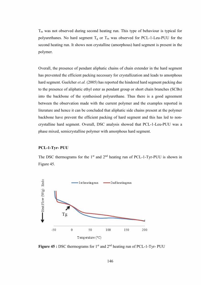
146
Tm was not observed during second heating run. This type of behaviour is typical for
polyurethanes. No hard segment Tg or Tm was observed for PCL-1-Leu-PUU for the
second heating run. It shows non crystalline (amorphous) hard segment is present in the
polymer.
Overall, the presence of pendant aliphatic chains of chain extender in the hard segment
has prevented the efficient packing necessary for crystallization and leads to amorphous
hard segment. Guelcher et.al. (2005) has reported the hindered hard segment packing due
to the presence of aliphatic ethyl ester as pendant group or short chain branches (SCBs)
into the backbone of the synthesised polyurethane. Thus there is a good agreement
between the observation made with the current polymer and the examples reported in
literature and hence it can be concluded that aliphatic side chains present at the polymer
backbone have prevent the efficient packing of hard segment and this has led to non-
crystalline hard segment. Overall, DSC analysis showed that PCL-1-Leu-PUU was a
phase mixed, semicrystalline polymer with amorphous hard segment.
PCL-1-Tyr- PUU
The DSC thermograms for the 1st and 2nd heating run of PCL-1-Tyr-PUU is shown in
Figure 45.
Figure 45 : DSC thermograms for 1st and 2nd heating run of PCL-1-Tyr- PUU

147
1st Heating Run - The soft segment glass transition temperature (Tg) for the first heating
run was observed at 13.2°C, which is substantially higher than Tg of pure homopolymer
PCL (-60°C). It has been reported in the literature (Umare and Chandure, 2008),
(Changhong et al., 2008), (Vermette, 2001) that the interactions between the soft and hard
segment can influence the glass transition temperature of the polymer.
DSC shows phase mixing behaviour between the hard and soft segment of the polymer.
The observed soft segment glass transition temperature is lower than the body
temperature 37ºC, indicating that PUUs can maintain elasticity at body temperature.
Similar observation has been recorded by Zhang (2006) where MDI-PCL based
polyurethane was synthesised. No soft segment crystal melt endotherm temperature (Tm)
was observed for PCL-1-Tyr-PUU during the 1st heating run showing the amorphous
nature of the soft segment as compared to semi crystalline nature of PCL-1-Leu-PUU.
2nd Heating Run – The soft segment Tg was observed at 13.3°C which is quite close to
the 1st heating run soft segment Tg. No Tm was observed for the soft segment showing
amorphous nature. In a same way, hard segment also did not show any thermal transitions
such as Tg or Tm indicating amorphous nature. The absence of a melting endotherm (Tm)
for hard segment based on MDI based PU was also reported by Changhong et.al. (2008),
(Lamba et al., 1998). The absence of hard segment transition might be due to the presence
of the bulky phenyl group of chain extender as pendant group which can disturb the
packing of the hard segment to form any crystals and hence does not show a crystal melt
endotherm (Tm). Skarja et.al. (1998) and Parrag et.al. (2010) have reported the similar
observation of the presence of non crystalline hard segment due to the presence of bulky
phenyl pendant group which has prevented the ordered structural arrangement of the hard
segment and this has led to amorphous hard segment.
Overall, it was concluded that aromatic amino acid based PCL-1-Tyr-PUU shows phase
mixed morphology with amorphous hard and soft segment. In general, it was observed
that hard segment showed the non crystalline behaviour in Series 1 PUUs. This might be
due to the presence of pendant group in the chain extender which have resulted in
disordered hard segment arrangement.

148
4.7.2.6 Thermogravimetric Analysis
Thermogravimetric curves provide information about the decomposition mechanism of
various materials. Thermogravimetric Analysis (TGA) traces illustrating the polymer
weight loss as a function of temperature for Series 1 PUUs is shown in Figure 46.
Figure 46 : TGA analysis of Series 1 PUU
All the aliphatic amino acid based PUUs showed the onset of degradation at temperature
between 250 and 280°C. Weight loss in the first degradation step occurs mainly due to
depolycondensation of urethane groups. The second step shows the degradation of the
soft segment of PUUs which occurs between 300 and 420°C. Up to 80% degradation
occurs at this stage. For the aromatic amino acid based PCL-1-Tyr-PUU, the onset of
degradation starts at 285°C and completes by 435°C (the highest of the series 1 PUUs).
Valine, leucine and isoleucine based PUU shows a similar thermal degradation trend,
whilst the aromatic tyrosine based PCL-1-Tyr-PUU exhibits a slightly broader slope. This
might be due to the presence of phenyl group in the hard segment of the PUU. These
aromatic groups maybe involved in non-covalent π - π interaction with each other
(Hinrichsen, 1994) or with the benzyl group of the MDI present in the hard segment.
Phenyl groups are reported to strengthen inter chain hard segments and thus provides

149
tyrosine based PUU more thermal stability as compared to other aliphatic chain extender
based PUU. Indeed, Guelcher et.al. (2005) has reported the higher melting temperature
of aromatic tyramine and tyrosine amino acid based PU due to phenyl group inter chain
attraction.
Sarkar et.al. (2009) reports the onset of degradation at 300°C for a tyrosine based HDI-
PCL-PU. It demonstrating that the observed TGA results, here, are consistent with
literature. For the control PCL-1-BDO-PU (Figure 33), the onset of degradation is around
270°C and is complete by 470°C. It shows that control PU is more thermally stable as
compare to Series 1 PUUs. Although the observed difference in the degradation
temperature in insignificant. However it is also reported in the literature that the urea
linkage is not thermally stable at high temperature and is considered as a weak link for
polyurea thermal degradation studies (Yilgör et al., 2000). It might be the reason for
obtaining the low thermal stability of Series 1PUUs as compared to control BDO-PU
which lacks urea linkage.
Fernandez et.al. (2006) has also reported the low thermal stability of diamine chain
extender based PCL-PUUs. Ornthine and lysine amino acids were used to make diamine
chain extender. They reported the onset of thermal degradation temperature for ornithine
based PUUs as 195 to 218°C and for lysine based PUUs, 172 to 215°C. Both are less
than the current Series 1 PUUs, indicating that the Series 1PUUs, here, exhibit improved
thermal stability. Tyrosine based PUUs also show improved thermal stability as compared
to aliphatic chain extender based PUUs in Series 1.
In summary, it was observed that Series 1 PUUs have shown a potential range of thermal
stability which might be useful for thermal processing of these polymers.
4.7.2.7 Water Contact Angle
Static contact angle measurements of the materials was obtained at room temperature
(25°C) in order to obtain an indication of the relative polarity of the polymer surface.
Deionised water was used and five different contact angle readings were taken at different
places on each polymer film. It was observed that synthesised PCL-MDI-based

150
polyurethane urea have high contact angle value, indicating lower surface polarity which
is consistent with literature (Fernández d'Arlas et al., 2008). The value of the water contact
angle for Series 1 PUUs was in the range from 82° to 94° and is shown in Table 23.
Table 23 : Contact angle values for Series 1 PUU
Polyurethane Urea (PUU) Contact Angle (ɵ) PCL-1-Leu-PUU 83 ± 2 PCL-1-IsoLeu-PUU 82 ± 2 PCL-1-Val-PUU 85 ± 1 PCL-1-Tyr-PUU 94 ± 3
The obtained PUUs can be considered somewhat hydrophobic. This behaviour is due to
the very low surface energy of PCL, which is expected to migrate to the surface. The
hydrophobic nature of the hard segment (aromatic diisocyanate (MDI)) and high
hydrocarbon content of the chain extender also contributes to the high value of contact
angle for the synthesised PUUs. The aromatic amino acid based PCL-1-Tyr-PUU shows
the highest water contact angle (94°±3) (i.e. hydrophobic), probably due to the presence
of the benzene ring in the side chain of the tyrosine amino acid. It is reported in the
literature (Sarkar, 2007) (Guan et al., 2004, Guan and Wagner, 2005) that the water
contact angle value of up to 75° for tyrosine amino acid based PCL-HDI polyurethane
considered as hydrophobic polymer surface. In summary, it was observed that the
incorporation of the novel amino acid based chain extenders into the PUUs has made the
polymer surfaces more hydrophobic.
4.7.2.8 Mechanical properties
The stress strain curves for Series 1 PUUs are shown in Figure 47 and the values are
shown in Table 24. The results show that the PUUs have high extension to failure,
exhibiting very low modulus and tensile strength. Tyrosine based PUU had the highest
tensile strength in Series 1 PUUs.
Aliphatic amino acid based PUUs containing leucine, isoleucine and valine amino acid
display very similar mechanical properties, such as high elongation to failure and low
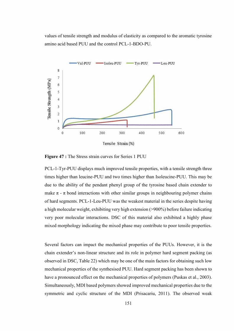
151
values of tensile strength and modulus of elasticity as compared to the aromatic tyrosine
amino acid based PUU and the control PCL-1-BDO-PU.
Figure 47 : The Stress strain curves for Series 1 PUU
PCL-1-Tyr-PUU displays much improved tensile properties, with a tensile strength three
times higher than leucine-PUU and two times higher than Isoleucine-PUU. This may be
due to the ability of the pendant phenyl group of the tyrosine based chain extender to
make π - π bond interactions with other similar groups in neighbouring polymer chains
of hard segments. PCL-1-Leu-PUU was the weakest material in the series despite having
a high molecular weight, exhibiting very high extension (>900%) before failure indicating
very poor molecular interactions. DSC of this material also exhibited a highly phase
mixed morphology indicating the mixed phase may contribute to poor tensile properties.
Several factors can impact the mechanical properties of the PUUs. However, it is the
chain extender’s non-linear structure and its role in polymer hard segment packing (as
observed in DSC, Table 22) which may be one of the main factors for obtaining such low
mechanical properties of the synthesised PUU. Hard segment packing has been shown to
have a pronounced effect on the mechanical properties of polymers (Puskas et al., 2003).
Simultaneously, MDI based polymers showed improved mechanical properties due to the
symmetric and cyclic structure of the MDI (Prisacariu, 2011). The observed weak

152
mechanical property for Series 1 PUUs based on MDI does not match with this literature
finding. DSC scans revealed hard segment was not packed efficiently due to bulky side
chains of the chain extender which may have restrict the hard segment mobility and
reduce its packing to observe any thermal transitions. This shows that the mechanical
properties or the synthesised PUUs are mainly governed by the soft segment and the hard
segment does not act as physical cross linker to improve the tensile properties.
Table 24 : Mechanical properties of Series 1 PUU (mean ± SD, n = 6)
Polyurethane Urea (PUU)
Modulus of Elasticity (MPa)
Tensile Strength (MPa)
Elongation at break (%)
PCL-1-Leu-PUU *
4.3 ± 1 0.52 ± 0 982.2 ± 11 PCL-1-Ileu-PUU 5.1 ± 1 1.1 ± 0 337.0 ± 46 PCL-1-Val-PUU 7.2 ± 2 2.0 ± 1 563.7 ± 22 PCL-1-Tyr-PUU 11.6 ± 8 7.3± 3 421.7 ± 58 PCL-1-BDO-PU 52.8 ± 22 32.9 ± 12 234 ± 18
*Sample did not break during tensile testing.
Moreover, the presence of a pendant aliphatic carbon atom chain on the chain extender
probably leads to lose packing of the hard segment and hence the loss of crystallinity and
disordered packing (less hydrogen-bonding) (Teo et al., 1997) leading to poor mechanical
properties. Parrag et.al. (2010) has also reported the low mechanical properties of
phenylalanine based PU due to a pendant bulky aromatic side chain of phenylalanine
amino acid based chain extender present in the hard segment. Similarly Sarkar et.al.
(2007) has reported observations of obtaining low mechanical properties for aromatic
tyrosine based PUs
The observed weak mechanical properties of synthesised PUU as compared to BDO
based PU are further supported by DSC data as phase mixed morphology was observed
for Series 1 PUUs. When the polymer was stretched during tensile testing, the ability of
loosely packed hard segments to deform and reorient at increasing strain could lead to
high elongation properties.
In summary, the importance of phase morphology and role of chain extender chemical
structure on phase morphology was demonstrated with weak mechanical properties

153
obtained for the Series 1 PUUs. PUUs based on novel amino acid based chain extenders
showed significantly lower mechanical properties as compared to BDO based PU, due to
the non-linear chemical structure of the amino acid based chain extenders used for PUU
synthesis. As compared to the control BDO, the synthesised chain extenders are non-
linear with bulky pendant groups, and hence form very weak H-bonding interactions with
other polymeric chains which in turns lower the tensile properties of the material. The
effect of this weak interaction was reflected in attaining poor mechanical properties of
the synthesised polymers. The synthesised polymers behaved as low modulus highly
extendable materials with very poor tensile strength.
4.8 Conclusion
This Chapter was focused on the synthesis and characterisation of Series 1 PU and Series
1 PUU based on novel L-amino acid based chain extenders. It was found that the
synthesised PUs and PUUs were light yellow colour in appearance with low modulus and
highly extendable materials as compared to the hard and strong BDO based polyurethane.
The Series 1 PUs and Series 1 PUUs all show moderately high molecular weights with
relatively narrow polydispersity. The polymers were soluble in polar aprotic solvents with
potential to use solvent based processing techniques. DSC analysis of synthesised
polymers showed a phase mixed morphology with an amorphous hard segment. The
synthesised polymers were thermally stable up to ~285°C. Series 1 PUs exhibited high
thermal stability due to the presence of aromatic moieties within the hard segment derived
from the new chain extenders. Mechanical properties as observed by stress strain curves,
were weak as compared to the BDO based PU. Amino acid based PUs and PUUs
exhibited properties of high extensibility and low tensile strength. Overall the amino acid
based Series 1 PUs and Series 1 PUUs exhibited weak mechanical properties as compared
to the control PU. The lack of hydrogen bonding within the hard segment and the non-
linear structure of the novel chain extender may be the possible reasons for the low
mechanical properties observed. The surface hydrophobicity, as measured by water
contact angle, increased with incorporation of the novel amino acid based chain extenders
indicating their potential for use as implant material in future biomedical applications.

154
The results in this chapter show that the composition of the polymer plays a crucial role
in determining the properties of the resultant polymeric material, thus future work could
focus on optimisation of formulation to give improved mechanical properties. The
polyurethanes synthesised in this work may be suitable for applications such as scaffolds
for soft tissue regeneration, drug delivery systems and other applications where high
mechanical strength is not required.
Since the aim of the experimental work was to create a range of polyurethanes that exhibit
different properties (physical, thermal and mechanical), it is logical to investigate the
effect of changing the chemical composition of the soft segment, which has been kept
constant in this study (PCL) and which will be discussed in the following chapter.

155
Chapter 5: Effect of soft segment molecular weight on the structure property relationship of polyurethanes (PUs) and polyurethane urea’s (PUUs)

156
5.1 Introduction
The structure property relationship of polyurethanes prepared from conventional chain
extenders, diisocyanate and polyols, are very well documented in the literature
(Gunatillake et al., 2003). The amino acid based chain extenders reported in this thesis
are, however, novel and detailed understanding of their influence on polyurethane
properties is important to explore. In chapter 4, the effect of (amino acid based) chain
extender structure on PU and PUU properties was investigated, keeping the polyol type,
molecular weight and the diisocyanate constant. In this chapter, an investigation to study
the effect of the polyol molecular weight will be discussed.
A series of PUs and PUUs were prepared using polycaprolactone (PCL) of molecular
weight 2,000 Daltons and their properties were compared with those prepared using PCL
of molecular weight 1,000 Dalton (Chapter 4). This molecular weight (Mw) range
represents that generally used for polyurethane formulations (Król, 2007). The synthesis
and characterisation of the chain extenders used here was described in detail in chapter 3
and the synthesis and characterisation of polyurethanes (PUs) and polyurethane ureas
(PUUs) based on 1,000 Dalton (Mw) PCL were described in chapter 4. The synthesis of
the 2,000 Dalton (Mw) PCL based PUs and PUUs followed the same procedure. The
diisocyanate used was 4,4’-methylenediphenyl diisocyanate (MDI) and the weight
percent of hard to soft segment was kept constant at 50 weight %. Since PUs and PUUs
exhibit different structure property relationships due to their functional group differences,
it is reasonable to assume that the use of a different molecular weight soft segment (PCL
2,000 Dalton) may impact the physio mechanical properties of the two classes of
polymers in different ways. This work aims to describe the effect of soft segment
structural variation on the properties of amino acid based PUs and PUUs with a long term
goal of helping to assess their suitability for biomedical applications.

157
5.2 Materials
Polycaprolactone (PCL) as polyol (Mw 2,000) was dried at 60°C for 48 hr under vacuum
(0.1 torr) prior to use and 4,4-methylenediphenyl diisocyanate (MDI), was used as
received from Sigma Aldrich, USA. Since MDI can react with moisture in the air, pure
MDI was distributed into small dry bottles and fresh MDI was used for each reaction.
Anhydrous DMF was obtained from sigma Aldrich, USA. The solvents were analytical
grade and were used as received. Deionised water was used for all purposes. Stannous
octaoate (Aldrich) was obtained from Sigma Aldrich and kept moisture free and used as
received. All other solvents and chemicals used for purification methods were of
analytical grade. Unless stated otherwise, all reactions were carried out in oven dried
glassware. An inert atmosphere of nitrogen was used while working with anhydrous
solvents. Chemical reaction temperatures were controlled using an IKA brand magnetic
temperature modulator.
5.3 Experimental Procedure
5.3.1 Synthesis of polyurethane Polycaprolactone (PCL, Mw-2,000) was used as polyol and 4,4-methylenediphenyl
diisocyanate (MDI) was used as diisocyanate for polymer synthesis. The amino acid
based dihydroxy diester compounds (318, 319) synthesised and reported in chapter 3 are
used as chain extender here. Synthesised PUs will be referred as Series 2 PU where 2
refers to the 2,000 molecular weight of the PCL. A two step or prepolymer method was
adopted for the synthesis of polyurethane and described in detail in chapter 4. Prepolymer
method followed is illustrated in Scheme 4, chapter 4. Stannous octaoate was used as
catalyst and N,N-dimethylformamide (DMF) was used as solvent for the polymerization.
The polymerisation reaction was carried out under a completely dry environment under
inert (dry N2 was used) atmosphere. PUs were synthesised with equivalent hard and soft
segments (50 weight % each) in all the cases. In brief, the method of synthesis of Series
2 PU is described below.

158
Polymerization was conducted in a 250 mL, 3 neck round bottom flask equipped with
magnetic stirrer, nitrogen gas inlet and outlet fitted to the two necks of the flask, and an
additional funnel fitted to the third neck. PCL was added to the flask. Reaction solvent,
anhydrous DMF was added to the flask using a syringe. Few drops of stannous octaoate
as catalyst was added to the reaction mixture under dry and inert atmosphere with
continuous stirring. MDI was charged to the reaction flask at room temperature. The
temperature was increased to 80°C and the reaction was allowed to proceed for 3 hours
at this temperature, and slowly cooled to room temperature (25°C) with continuous
stirring. The temperature of the reaction was carefully maintained within the range of ±
3°C. Chain extension step was then carried out by simultaneous addition of amino acid
based dihydroxy chain extender to the prepolymer solution under vigorous stirring. The
NCO/OH ratio was maintained at 1.05. The temperature of the reaction was then
gradually increased to 80°C and stirred for another 12 hours at the same temperature.
After 12 hours, the reaction was quenched by pouring the reaction mixture into a cold
concentrated aqueous solution of sodium chloride. At this point, solid polyurethane
polymer precipitates out from the reaction mixture. The final polymer was filtered out
and washed with distilled water. The polymer was dried under vacuum at 40°C for 48
hours. Synthesised PUs will be referred as Series 2 PU where 2 refers to the 2,000
molecular weight of the PCL. In total, two PUs were synthesised based on two (318 and
319) amino acid based dihydroxy chain extender. The net yield of the polymerisation
reaction was ~70% (in all cases for Series 2 PU). The polyurethane synthesised were
stored in a sealed plastic bag for the purpose of characterisation and future experiments.
5.3.2 Synthesis of polyurethane urea The method of synthesis of polyurethane urea is similar to PU synthesis as described in
section 5.3.1. The same two step solution polymerization method was used for the
preparation of PUU containing equivalent hard and soft segments (approximately 50
weight % each). Stannous octaoate was used as catalyst and anhydrous DMF act as
reaction solvent. Amino acid based diamine compounds (311, 312, 313, 314) act as chain
extenders to synthesise PUUs. The reaction conditions and procedure was the same as for
Series 2 PU (Section 5.3.1). Polycaprolactone (PCL, Mw-2,000) was used as polyol and
aromatic diisocyanate 4,4-methylenediphenyl diisocyanate (MDI) was used as
diisocyanate for polymer synthesis. Synthesised PUUs will be referred as Series 2 PUU

159
where 2 refers to the 2,000 molecular weight of the PCL. The method of the synthesis of
Series 2 PUUs is described as follow.
Briefly, Polymerization was conducted in a 250 mL, 3 neck round bottom flask equipped
with magnetic stirrer, nitrogen gas inlet and outlet fitted to the two necks of the flask, and
an additional funnel fitted to the third neck. PCL (Mw 2,000 Dalton) and reaction solvent
(DMF) was added to the flask. Few drops of stannous octaoate as catalyst was added to
the reaction mixture under dry and inert atmosphere with continuous stirring. Following
that, MDI was charged to the reaction flask at room temperature. The temperature was
increased to 80°C and the reaction was allowed to proceed for 3 hours at this temperature,
and slowly cooled to room temperature with continuous stirring. Chain extension step
was then carried out by simultaneous addition of amino acid based diamine chain extender
to the prepolymer solution under vigorous stirring. The NCO/OH ratio was maintained at
1.05. The temperature of the reaction was then gradually increased to 80°C and stirred
for another 12 hours at the same temperature and then, the reaction was quenched by
pouring the reaction mixture into a cold concentrated aqueous solution of sodium
chloride. At this point, solid polyurethane urea polymer precipitates out from the reaction
mixture. The final polymer was filtered out and washed with distilled water. The polymer
was dried under vacuum at 40°C for 48 hours. Synthesised PUUs will be referred as Series
2 PUU where 2 refers to the 2,000 molecular weight of the PCL. In total 4 PUUs were
synthesised based on four (311, 312, 313, 314) amino acid based diamine chain extender.
The net yield of the polymerisation reaction was ~70% (in all cases for Series 2 PUU).
The polyurethane urea synthesised were stored in a sealed plastic bag for the purpose of
characterisation and future experiments.
5.4 Characterisation of polymers
5.4.1 Gel permeation chromatography The molecular weights of polymers were determined by Gel Permeation Chromatography
(GPC). A Waters 515 HPLC pump attached to Waters 590 equipped with a refractive
index detector (a Waters 410 differential refractometer) which uses dimethyl formamide
(DMF) as solvent (containing Lithium Bromide (LiBr), 0.05 M). The flow rate of the

160
mobile phase was maintained at 1mL/min and the operating temperature was 30°C.
Polyurethane sample was dissolved in DMF (approx. 2 mg/mL) and filtered through a 0.5
m (MFS Advantech) syringe filter and injected (50 µl) into an HPLC pump. Calibration
of the apparatus was routinely performed with monodisperse polystyrene as standard.
Data was analysed using Empower Pro software to determine the average number
molecular weight (Mn), weight average molecular weight (Mw) and polydispersity
(Mw/Mn) of the polymer. All molecular weights reported here are in terms of polystyrene
standard.
5.4.2 Thermal characterization The thermal behaviour of the PUs and PUUs was characterised by Differential Scanning
Calorimetry (DSC) and Thermogravemetric Analysis (TGA). The DSC was performed
using a DSC30 (Mettler Toledo) instrument. For the DSC measurements, the samples
were vacuum dried for 48 hours at room temperature in the presence of phosphorous
pentaoxide. A specimen of approximately 5 mg was cut from compression moulded sheet
and was encapsulated in an aluminium pan. Samples were scanned in alternating DSC
mode from -50°C to 200°C at a rate of 10 C min-1 under nitrogen atmosphere. A three
stage heating and cooling method was used in which the sample was first heated from -
50°C to 200°C and then quenched immediately to -50°C and heated again to 200°C. First
heating run provides information about any crystallinity present in the soft segment which
might disappear in the second heating run due to the rapid cooling of the polymer sample.
DSC thermograms were analysed using Mettler: STAR EV.9.00 software to determine
polyurethane glass transition (Tg) and melting temperature (Tm). Melting point (Tm) was
taken as the peak temperature of the observed endothermic transition, whereas glass
transition temperature (Tg) was measured at the half width of the transition after plotting
tangents on the curve. The percent crystallinity (% Xc) of the polymer was calculated
according to the melting peak area of the DSC graph by assuming that a perfect PCL
crystal has a melting enthalpy of 139.5 J/g [Enthalpy was determined using the values
(ΔHm / ΔHm100) where Δ Hm100 is Δ Hm is enthalpy of melting temperature of 100%
crystalline PCL] (Pitt et al., 1990). Thermo-gravimetric analysis (TGA) was performed
with a Mettler Toledo TGA/STDA851 from 0°C to 800°C under nitrogen at a rate of 20°C
min -1. TGA graphs were analysed using Mettler: STAR EV.9.00 software. The
161
specimens were placed in a ceramic pan and an average of 10 mg of solid sample was
used for the experiments.
5.4.3 Fourier transform infrared spectroscopy
Fourier Transform Infrared Spectroscopy (FTIR) spectra were obtained using a
ThermoNicolet 6700 spectrometer using a SmartATR (attenuated total reflectance)
attachment fitted with a diamond window. The sample was analysed (32 scans) at 25ºC
in the transmission mode over 4000-700 cm-1.
5.4.4 Water contact angle measurement
Water contact angles were measured using a contact angle meter (KSV CAM200
Instruments Ltd.). Thin films of polyurethane were prepared on thoroughly cleaned and
dried glass slides by spin coating of 5 wt% solution of polymer in DMF. The films were
dried initially at room temperature for 24 hours followed by vacuum drying at 50°C for
another 48 hours to remove the residual solvents. Static water contact angles were
measured by depositing a drop of 2 µl of deionised water (Millipore, Elix 5, 15 MΩ) at
room temperature (25 C) and then a picture of the surface was taken through a camera
(Sony). The contact angle was measured manually on the picture. To verify the contact
angle reproducibility, an average of five readings (± standard deviation) from different
parts of the each film was taken.
5.4.5 Compression moulding
Compression moulding was performed on a hydraulic press (with a thermostat and water-
cooling capability) to obtain polymer films of the synthesised polyurethanes. The polymer
was cut into small pieces using clean tin snips and pressed into a 1 mm thick plaque at a
temperature above the melting point of the polymer (typically). The temperature was held
constant for a period of 5 minutes before cooling in a standard manner under the flow of
cold water. A standard mould was used which consisted of a rectangular cavity 100 mm
x 60 mm x 1 mm deep cut into a metal plate. Teflon fabric sheet was used on both sides
of mould to prevent adhesion of the polymer to the metal. The press used was a model
12-10-1T Wabash hydraulic press. In order to avoid unnecessary degradation of the
material, the polyurethanes were not subjected to annealing. Polymer films thickness was

162
measured with digital callipers. The polymer films were stored in plastic bags in a
desiccator prior to characterization.
5.4.6 Mechanical properties
The mechanical properties of the polyurethane films were measured using an Instron
model 4468 universal testing machine according to the ASTM D-882 method. The
compression moulded plaques were cut into thin strips with a straight section of 40 mm
x 5 mm x 0.4 mm. A 100 N load cell was used with a crosshead speed of 100 mm/min at
room temperature. At least seven replicate measurements were taken and averaged (±
standard deviation). Strong pneumatic grips were used to prevent slippage. Data was
analysed using Blue hill v.2.5 software.
5.5 Result and Discussion
5.5.1 Polyurethane synthesis
A series of amino acid based polyurethanes (PUs) was synthesised using the two-step
method described in the experimental section 5.3. Here, polycaprolactone (PCL) of
molecular weight 2,000 Dalton was used as the soft segment and the series is thus referred
as Series 2 PU. 4,4-methylenediphenyl diisocyanate (MDI) was used as diisocyanate. Z-
serine and Z-threonine amino acid based diester dihydroxy compounds (318 and 319,
chapter 3) were used as the chain extender. The polymer abbreviation used is based on
the type and molecular weight of chain extender and polyol used respectively. For
example, PCL-2-Z-Ser-PU, refers to PU consisting of 2,000 Dalton molecular weight
PCL, chain extended with an L-Z-Serine based dihydroxy chain extender (318, chapter
3), Z is a benzyloxycarbonyl group; an amine group protecting agent present in the chain
extender and acting as a pendant group in the PU structure. Table 25 shows the
abbreviations used for Series 2 PUs in this chapter.
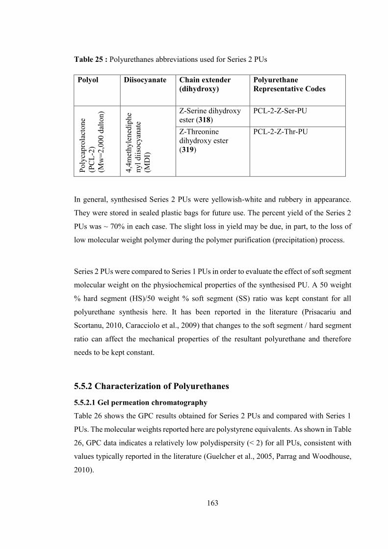
163
Table 25 : Polyurethanes abbreviations used for Series 2 PUs
Polyol Diisocyanate Chain extender (dihydroxy)
Polyurethane Representative Codes
Poly
capr
olac
tone
(P
CL-
2)
(Mw
=2,0
00 d
alto
n)
4,4m
ethy
lene
diph
eny
l diis
ocya
nate
(M
DI)
Z-Serine dihydroxy ester (318)
PCL-2-Z-Ser-PU
Z-Threonine dihydroxy ester (319)
PCL-2-Z-Thr-PU
In general, synthesised Series 2 PUs were yellowish-white and rubbery in appearance.
They were stored in sealed plastic bags for future use. The percent yield of the Series 2
PUs was ~ 70% in each case. The slight loss in yield may be due, in part, to the loss of
low molecular weight polymer during the polymer purification (precipitation) process.
Series 2 PUs were compared to Series 1 PUs in order to evaluate the effect of soft segment
molecular weight on the physiochemical properties of the synthesised PU. A 50 weight
% hard segment (HS)/50 weight % soft segment (SS) ratio was kept constant for all
polyurethane synthesis here. It has been reported in the literature (Prisacariu and
Scortanu, 2010, Caracciolo et al., 2009) that changes to the soft segment / hard segment
ratio can affect the mechanical properties of the resultant polyurethane and therefore
needs to be kept constant.
5.5.2 Characterization of Polyurethanes
5.5.2.1 Gel permeation chromatography
Table 26 shows the GPC results obtained for Series 2 PUs and compared with Series 1
PUs. The molecular weights reported here are polystyrene equivalents. As shown in Table
26, GPC data indicates a relatively low polydispersity (< 2) for all PUs, consistent with
values typically reported in the literature (Guelcher et al., 2005, Parrag and Woodhouse,
2010).
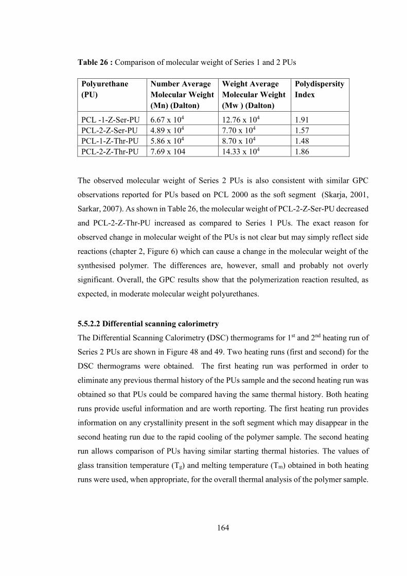
164
Table 26 : Comparison of molecular weight of Series 1 and 2 PUs
Polyurethane (PU)
Number Average Molecular Weight (Mn) (Dalton)
Weight Average Molecular Weight (Mw ) (Dalton)
Polydispersity Index
PCL -1-Z-Ser-PU 6.67 x 10⁴ 12.76 x 10⁴ 1.91 PCL-2-Z-Ser-PU 4.89 x 10⁴ 7.70 x 10⁴ 1.57 PCL-1-Z-Thr-PU 5.86 x 10⁴ 8.70 x 10⁴ 1.48 PCL-2-Z-Thr-PU 7.69 x 10⁴ 14.33 x 10⁴ 1.86
The observed molecular weight of Series 2 PUs is also consistent with similar GPC
observations reported for PUs based on PCL 2000 as the soft segment (Skarja, 2001,
Sarkar, 2007). As shown in Table 26, the molecular weight of PCL-2-Z-Ser-PU decreased
and PCL-2-Z-Thr-PU increased as compared to Series 1 PUs. The exact reason for
observed change in molecular weight of the PUs is not clear but may simply reflect side
reactions (chapter 2, Figure 6) which can cause a change in the molecular weight of the
synthesised polymer. The differences are, however, small and probably not overly
significant. Overall, the GPC results show that the polymerization reaction resulted, as
expected, in moderate molecular weight polyurethanes.
5.5.2.2 Differential scanning calorimetry The Differential Scanning Calorimetry (DSC) thermograms for 1st and 2nd heating run of
Series 2 PUs are shown in Figure 48 and 49. Two heating runs (first and second) for the
DSC thermograms were obtained. The first heating run was performed in order to
eliminate any previous thermal history of the PUs sample and the second heating run was
obtained so that PUs could be compared having the same thermal history. Both heating
runs provide useful information and are worth reporting. The first heating run provides
information on any crystallinity present in the soft segment which may disappear in the
second heating run due to the rapid cooling of the polymer sample. The second heating
run allows comparison of PUs having similar starting thermal histories. The values of
glass transition temperature (Tg) and melting temperature (Tm) obtained in both heating
runs were used, when appropriate, for the overall thermal analysis of the polymer sample.

165
The thermal properties obtained via DSC for Series 2 PUs are shown in Table 27. The
DSC result for Series 1 PUs are also shown in Table 27 for easy comparison. Series 1
PUs were originally shown and discussed in detail in section 4.5.2.5 of chapter 4. The
DSC analysis shows a significant effect of increasing the soft segment (PCL) molecular
weight on the thermal properties of PUs.
Figure 48 : DSC thermograms for the 1st and 2nd heating run of PCL-2-Z-Ser-PU
Figure 49 : DSC thermograms for the 1st and 2nd heating run of PCL-2-Z-Thr-PU
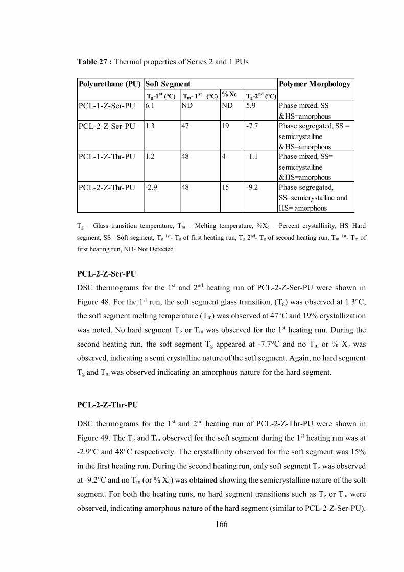
166
Table 27 : Thermal properties of Series 2 and 1 PUs
Tg – Glass transition temperature, Tm – Melting temperature, %Xc – Percent crystallinity, HS=Hard
segment, SS= Soft segment, Tg 1st- Tg of first heating run, Tg 2nd- Tg of second heating run, Tm 1st- Tm of
first heating run, ND- Not Detected
PCL-2-Z-Ser-PU
DSC thermograms for the 1st and 2nd heating run of PCL-2-Z-Ser-PU were shown in
Figure 48. For the 1st run, the soft segment glass transition, (Tg) was observed at 1.3°C,
the soft segment melting temperature (Tm) was observed at 47°C and 19% crystallization
was noted. No hard segment Tg or Tm was observed for the 1st heating run. During the
second heating run, the soft segment Tg appeared at -7.7°C and no Tm or % Xc was
observed, indicating a semi crystalline nature of the soft segment. Again, no hard segment
Tg and Tm was observed indicating an amorphous nature for the hard segment.
PCL-2-Z-Thr-PU
DSC thermograms for the 1st and 2nd heating run of PCL-2-Z-Thr-PU were shown in
Figure 49. The Tg and Tm observed for the soft segment during the 1st heating run was at
-2.9°C and 48°C respectively. The crystallinity observed for the soft segment was 15%
in the first heating run. During the second heating run, only soft segment Tg was observed
at -9.2°C and no Tm (or % Xc) was obtained showing the semicrystalline nature of the soft
segment. For both the heating runs, no hard segment transitions such as Tg or Tm were
observed, indicating amorphous nature of the hard segment (similar to PCL-2-Z-Ser-PU).
Tg-1st (°C) Tm- 1st (°C) % Xc Tg-2nd (°C)PCL-1-Z-Ser-PU 6.1 ND ND 5.9 Phase mixed, SS
&HS=amorphousPCL-2-Z-Ser-PU 1.3 47 19 -7.7 Phase segregated, SS =
semicrystalline &HS=amorphous
PCL-1-Z-Thr-PU 1.2 48 4 -1.1 Phase mixed, SS= semicrystalline &HS=amorphous
PCL-2-Z-Thr-PU -2.9 48 15 -9.2 Phase segregated, SS=semicrystalline and HS= amorphous
Polyurethane (PU) Polymer MorphologySoft Segment

167
The DSC thermograms for Series 1 PUs (PCL-1-Z-Ser-PU and PCL-1-Z-Thr-PU) were
shown in chapter 4, Figure 30 and 31 respectively. On comparing the DSC results of
Series 2 with Series 1 PUs, it can be observed that the soft segment Tg decreases with
increasing soft segment (PCL) molecular weight. This shift is indicative of differences of
the morphology within the polymers, with a stronger phase separation in Series 2 PUs
compared to the more phase mixed morphology shown by Series 1 PUs. Similar results
have been reported previously (Seefried et al., 1975, Gorna et al., 2002, Garrett et al.,
2003, Gisselfält and Helgee, 2003) in regards to decreases in soft segment Tg with
increasing soft segment molecular weight. The more the soft domains are contaminated
with dissolved hard segments (as in the phase mixing morphology shown by Series 1
PUs), the higher will be the soft segment Tg. Similar observations for the soft segment
morphology in relation to soft segment molecular weight have also been reported in the
literature (Hu et al., 1982, Paik Sung et al., 1980b, Van Bogart et al., 1981).
Series 2 PUs also show soft segment crystallization (% Xc), which increases with
increasing PCL molecular weight as compared to Series 1 PUs. For example, 19% Xc was
observed for PCL-2-Z-Ser-PU and 0% Xc was observed for PCL-1-Z-Ser-PU. Similarly,
15% Xc was observed for PCL-2-Z-Thr-PU and only 4% Xc was shown by PCL-1-Z-Thr-
PU. A similar observation was recorded by Fernandez et al. (2006) with PCL based PUs.
No hard segment Tg or Tm was observed for either Series 1 or Series 2 PU. This is
consistent with both series having an amorphous hard segment domain. Usually, as
reported in the literature (Camberlin and Pascault, 1984, Yilgör et al., 1982, Joshi, 2009),
in the segmented PUs, change in heat capacity of the amorphous hard blocks is often too
small to be observed. Possible reasons for obtaining non-crystalline hard segment of the
PUs might be:
1. Disruption of hard segment packing caused by a non-linear chain extender with
bulky pendant Z group may inhibit polymer chain packing in the hard segment
resulting in the absence of hard segment Tg or Tm. A similar observation was
reported by Zhang et al. (2006) for PUs having their hard segment based on MDI
and using a non-linear chain extender.

168
2. The length of the hard segment unit is not long enough to make an ordered
structure and does not, therefore, exhibit a Tm. It has been reported in the literature
that MDI based hard segments need at least 2-3 hard segment units in a sequence
to form an ordered enough structure to show any Tm.
Based on the DSC results shown in Table 27, the following observations can be made for
Series 2 PUs:
1. In the first heating run, Series 2 PUs showed a soft segment glass transition
(Tg) and melting endotherm (Tm) characteristic of PCL based soft segment
with percent crystallinity of 19% (PCL-2-Z-Ser-PU) and 15% (PCL-2-Z-Thr-
PU).
2. In the second heating run, the soft segment Tm disappeared due to rapid
cooling of the polymer sample before the second run was completed.
3. In the second heating run, Series 2 PUs exhibited a soft segment Tg with no
other notable soft segment thermal transitions observed indicating that the soft
segment of Series 2 PUs are largely semi crystalline in nature.
4. No hard segment thermal transitions (Tg/Tm) were observed in the first or
second heating run indicating that hard segments are non-crystalline or
amorphous in nature.
The overall thermal characteristic features observed by comparing DSC result of Series
1 and 2 PUs indicate that:
1. PUs formed with higher molecular weight soft segments are more phase
separated. This means longer soft segment length allowed high degree of phase
separation resulting in lower soft segment Tg.
2. As PCL molecular weight increases, soft segment glass transition (Tg) decreases.
3. The soft segment crystallinity increases as PCL length increases.
4. The presence of a non-linear and bulky chain extender tends to destroy the ordered
packing of the hard segment which results in amorphous hard segments having no
hard segment Tg or Tm.

169
5.5.2.3 Fourier Transform Infrared Spectroscopy
The DSC results show clear differences in morphology between Series 1 and Series 2
PUs. This is further investigated in this section using Fourier Transform Infrared
Spectroscopy (FTIR). FTIR can reveal information about hydrogen bonding within the
hard and soft segments respectively. Here only the serine amino acid based Series 1 and
2 PUs (PCL-1-Z-Ser-PU and PCL-2-Z-Ser-PU) are reported in detail. The threonine
amino acid based Series 1 and 2 PUs were similar and to avoid repetition of text, threonine
amino acid based polymers FTIR data is not discussed here. The FTIR spectrum of PCL-
1-Z-Ser-PU and PCL-2-Z-Ser-PU is shown in Figure 50 and the carbonyl, C=O (approx.
1600 – 1800 cm-1) stretching region and amine, N-H (approx. 3100 -3500 cm-1) region of
the PCL-1-Z-Ser-PU and PCL-2-Z-Ser-PU is shown separately in Figure 51 and
important FTIR absorption bands for MDI-PCL based polyurethane are reported in Table
28.
Table 28 : Assignment of the major absorption bands in the FTIR spectra of the PCL-
MDI based PU(Mattia and Painter, 2007), (Yilgor and Yilgor, 2007)
Frequency (cm-1)
Assignment Frequency (cm-1)
Assignment
~3440 ν(NH),free 1598 ν(C=C),benzene ring
3340 (NH) H-bonded, urethane 1528, 1508, 1530 δ (N-H) + ν(C-N), amide II
2940, 2863, 2862
νa(CH2), νsCH2), νs(CH2) 1464, 1370, 1365 δa(C-H) in CH3, ω(C-H) in CH2, δs(C-H) in CH3
1730 ν(C=O),free urethane , ester carbonyl (soft segment)
1240,1202,1219, 1191
δ (N-H) + ν(C-N), amide III
1710 ν(C=O),H-bonded urethane 1216, 1065,1063, 1046, 1016
ν(CO-O-C)
=stretching mode, a =asymmetric stretching, s=symmetric stretching, = in-plane bending or
scissoring,=in-plane bending or rocking, ω = wagging

170
Figure 50 : FTIR absorption spectrum of polyurethane (A) PCL-1-Z-Ser-PU (B) PCL-2-
Z-Ser-PU
Figure 51 : FTIR absorption spectra of carbonyl and N-H region of polyurethane (A)
PCL-1-Z-Ser-PU and (B) PCL-2-Z-Ser-PU
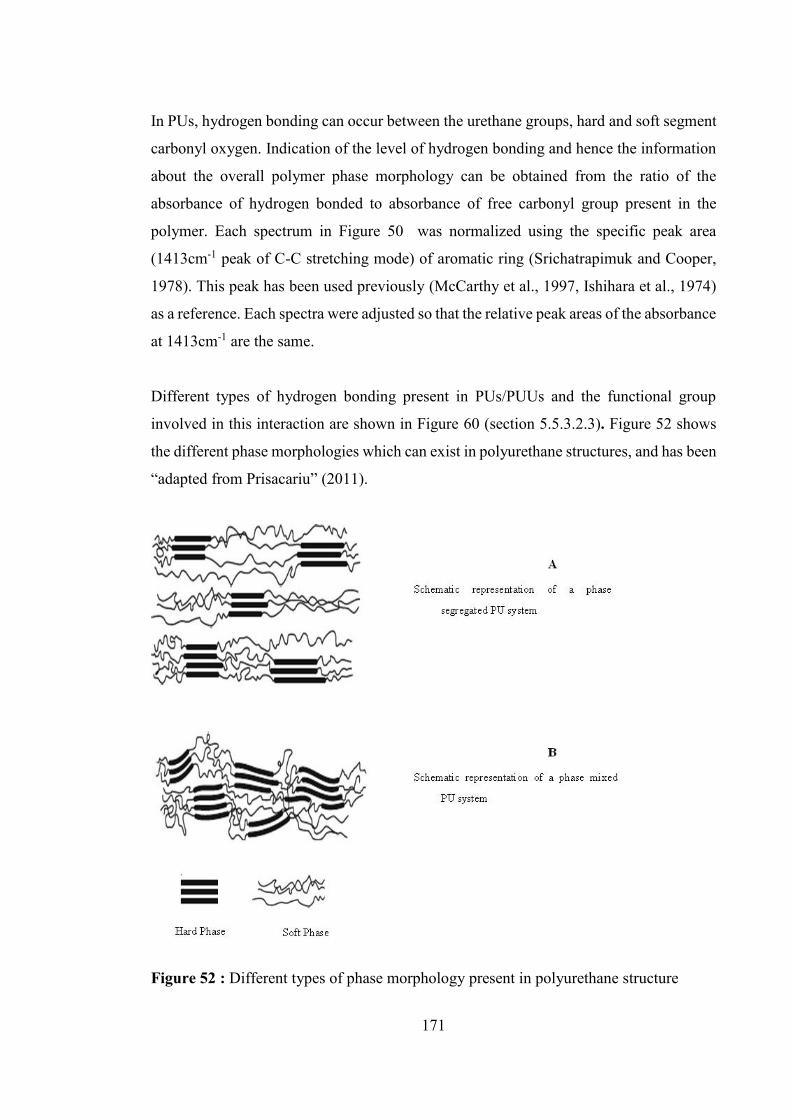
171
In PUs, hydrogen bonding can occur between the urethane groups, hard and soft segment
carbonyl oxygen. Indication of the level of hydrogen bonding and hence the information
about the overall polymer phase morphology can be obtained from the ratio of the
absorbance of hydrogen bonded to absorbance of free carbonyl group present in the
polymer. Each spectrum in Figure 50 was normalized using the specific peak area
(1413cm-1 peak of C-C stretching mode) of aromatic ring (Srichatrapimuk and Cooper,
1978). This peak has been used previously (McCarthy et al., 1997, Ishihara et al., 1974)
as a reference. Each spectra were adjusted so that the relative peak areas of the absorbance
at 1413cm-1 are the same.
Different types of hydrogen bonding present in PUs/PUUs and the functional group
involved in this interaction are shown in Figure 60 (section 5.5.3.2.3). Figure 52 shows
the different phase morphologies which can exist in polyurethane structures, and has been
“adapted from Prisacariu” (2011).
Figure 52 : Different types of phase morphology present in polyurethane structure

172
As shown in Figure 50, the characteristic carbonyl group peak for PCL based PU is
present at 1719cm-1 (spectrum B, Figure 50, PCL-2-Z-Ser-PU) and at 1726cm-1 (spectrum
A, Figure 50, PCL-1-Z-Ser-PU) representing the ester carbonyl (C=O) group of PCL.
This peak is very strong and dominating and hence it has overlapped the peaks due to free
urethane carbonyl group which otherwise would have appeared at 1730 cm-1. In Figure
51, the appearance of an additional peak at 1709cm-1 in spectrum A, Series 1 PU is
indicative of the presence of H- bonded carbonyl group in low molecular weight based
PU. This indicates that with lower molecular weight of PCL, there is certain degree of
phase mixing present. This peak (1709cm-1 ) is either absent or merged with the strong
ester carbonyl absorbance at 1719cm-1 in Series 2-PU in Figure 50.
For PCL based PU, significant phase mixed morphology is observed with the low
molecular weight soft segment, which is consistent with literature findings (Petrovic and
Ferguson, 1991, Wang and Cooper, 1983). The phenomena of phase mixing behaviour is
further supported by the appearance of asymmetric stretch (Figure 50) at 1216cm-1 and
1065cm-1 (CO-O-C) in Series 1 PUs which is less intense for Series 2 PUs. Similar
observation for FTIR studies with different molecular weight of PCL based polyurethane
are recorded in the literature (Sarkar et al., 2008). This indicates that the ester oxygen of
carbonyl group of PCL forms an H-bonded structure with urethane linkages to give rise
to asymmetric C-O stretch indicative of a higher degree of phase mixed behaviour.
The carbonyl stretching region (1600 – 1800 cm-1) is dominated by an strong soft segment
ester carbonyl band located at 1726cm-1 (spectrum A) to 1719cm-1 (spectrum B). It has
been reported in the literature (Rao, 1963, Sarkar, 2007) that the presence of a high
percentage of soft segment crystallinity can shift the frequency of the ester C=O peak
towards a lower frequency. A similar observation was noted here as well when the soft
segment crystallinity was found to shift the main PCL ester band from 1726cm-1 (Series
1 PU) to a lower frequency at 1719cm-1 (Series 2 PUs), (Figure 51). This shift
characterizes the more crystalline nature of the high molecular weight of soft segment
(Skarja and Woodhouse, 2000). The observation is in agreement with the DSC results
(Table 27) which also support the semicrystalline behaviour of high molecular weight
PCL, along with phase segregated morphology.
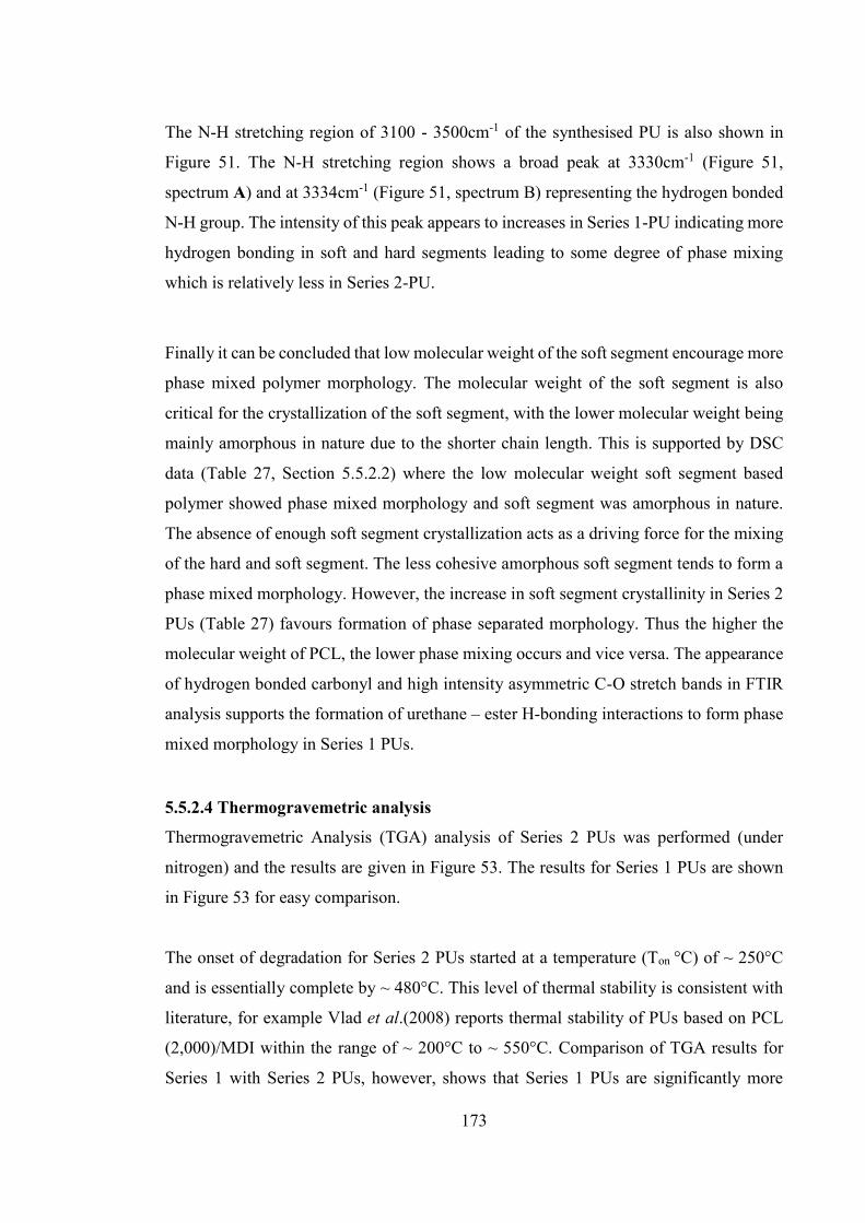
173
The N-H stretching region of 3100 - 3500cm-1 of the synthesised PU is also shown in
Figure 51. The N-H stretching region shows a broad peak at 3330cm-1 (Figure 51,
spectrum A) and at 3334cm-1 (Figure 51, spectrum B) representing the hydrogen bonded
N-H group. The intensity of this peak appears to increases in Series 1-PU indicating more
hydrogen bonding in soft and hard segments leading to some degree of phase mixing
which is relatively less in Series 2-PU.
Finally it can be concluded that low molecular weight of the soft segment encourage more
phase mixed polymer morphology. The molecular weight of the soft segment is also
critical for the crystallization of the soft segment, with the lower molecular weight being
mainly amorphous in nature due to the shorter chain length. This is supported by DSC
data (Table 27, Section 5.5.2.2) where the low molecular weight soft segment based
polymer showed phase mixed morphology and soft segment was amorphous in nature.
The absence of enough soft segment crystallization acts as a driving force for the mixing
of the hard and soft segment. The less cohesive amorphous soft segment tends to form a
phase mixed morphology. However, the increase in soft segment crystallinity in Series 2
PUs (Table 27) favours formation of phase separated morphology. Thus the higher the
molecular weight of PCL, the lower phase mixing occurs and vice versa. The appearance
of hydrogen bonded carbonyl and high intensity asymmetric C-O stretch bands in FTIR
analysis supports the formation of urethane – ester H-bonding interactions to form phase
mixed morphology in Series 1 PUs.
5.5.2.4 Thermogravemetric analysis
Thermogravemetric Analysis (TGA) analysis of Series 2 PUs was performed (under
nitrogen) and the results are given in Figure 53. The results for Series 1 PUs are shown
in Figure 53 for easy comparison.
The onset of degradation for Series 2 PUs started at a temperature (Ton °C) of ~ 250°C
and is essentially complete by ~ 480°C. This level of thermal stability is consistent with
literature, for example Vlad et al.(2008) reports thermal stability of PUs based on PCL
(2,000)/MDI within the range of ~ 200°C to ~ 550°C. Comparison of TGA results for
Series 1 with Series 2 PUs, however, shows that Series 1 PUs are significantly more

174
thermally stable. The onset temperatures (Ton °C) of degradation for Series 1 PUs were ~
350°C whereas, for Series 2 PUs they were ~250°C. This difference may be attributed to
different phase morphology observed between the materials in the two series. It was
indicated during discussion of the DSC results (Section 5.5.2.2) that Series 1 PUs exhibit
phase mixed morphology (Section 4.5.2.5, chapter 4) whilst Series 2 PUs exhibit phase
segregated behaviour (Table 27).
Figure 53 : TGA analysis of Series 1 PUs and Series 2 PUs
Increased thermal stability through phase mixed morphology may be established via the
formation of hydrogen bonds between the soft and hard segments. It has been previously
postulated (Wang and Hsieh, 1997) that the thermal stability of PUs can be highly
stimulated by the degree of phase separation/phase mixed morphology and thus there is
a mutual stabilization effect present between the soft and hard segment leading to
improved thermal stability of the material.
Antipova et.al. (1970), has stated that “the urethane group can behave as an antioxidant
having a stabilizing effect on the soft segment. Therefore the presence of hard segments
within the soft segments can increase the stability of soft segment at high temperature,
while the soft segment may have a protective function on the hard segment and hence

175
increase the thermal stability of segmented PU. This can be interpreted as a mutual
stabilization effect of phase mixed segmented PUs”. The low thermal stability of Series
2 PUs, as compared to Series 1 PUs, is presumably due to less H-bonding between hard
and soft segment as both are present as separate domains which in turn leads to early
thermal degradation. Even though the thermal stability of Series 2 PUs is lower than
Series 1 PUs, they still can be considered thermally stable based on their thermal
degradation limit (> 300° C), and therefore have potential use in, for example, biomedical
applications.
Overall, the effect of soft segment (PCL) molecular weight on thermal degradation can
be seen during thermogravimetric analysis. It was observed that as the molecular weight
of the soft segment increases, phase segregated morphology dominates, making the
polymer less thermally stable. It is probably due to poor intramolecular interactions such
as hydrogen bonding. Presumably, this will place a limit to the amount of soft segment
(PCL) content which is advisable for such polymers. An optimisation process was
considered outside the scope of this thesis.
5.5.2.5 Contact Angle
Advancing contact angle measurements of the polymers with water were performed in
order to study their relative hydrophobicity. Static contact angle results for Series 2 PUs
are given in Table 29. The results for Series I PUs are also shown in the Table 29 for easy
comparison.
Table 29 : Contact angle values for Series 1 and 2 PU
Polyurethane (PU) Series 1 PU Series 2 PU
PCL-L-Z-Ser-MDI-PU 72° ± 4 75° ± 3
PCL-L-Z-Thr-MDI-PU 77° ± 3 80° ± 2
The data in Table 29 shows that the contact angle increases slightly as PCL molecular
weight increases. It was noted during DSC experiments that phase separation increases
as PCL molecular weight increases, and hence soft segment crystallinity also increases.
176
Thus increasing phase separation may be expected to generate more pure PCL surface
regions. Since PCL is hydrophobic compared to the polar hard segments, increasing its
content at the surface would be expected to increase the contact angle, as observed. The
effect, as expected, is however very slight and indicates data which is consistent with an
expected increase in contact angle rather than proof that it genuinely does. It has been
reported in the literature (Skarja, 2001, Sarkar, 2007) that high contact angle with
increasing PCL molecular weight results from an increase in the phase separation of the
polymer, which in turns increases as the molecular weight of PCL increases. The contact
angle values obtained for Series 1 and 2 PUs are close to literature findings. For example,
Vlad et.al.(2010) has reported contact angle values up to 89° based on MDI-PCL-PU and
Skarja et.al.(2001) has reported a contact angle value of 71° for PCL 1250-Z-lysine amino
acid based polyurethane. Increase in contact angle value with increasing PCL molecular
weight is also reported by Marija et.al.(2011).
Overall it can be concluded that there is a very slight increase in contact angle with
increasing PCL molecular weight, consistent with an increases in phase segregation of
the polymer.
5.5.2.6 Mechanical Properties
The stress strain curves for Series 2 PUs are shown in Figure 54 and 55. The mechanical
properties of Series 2 PUs are summarised, and compared with Series 1, in Table 30. In
general, it was observed that Series 1 and 2 PUs show poor tensile properties as compared
to literature findings (Sarkar, 2007, Marcos-Fernández et al., 2006, Guelcher et al., 2005)
for other similar types of polyurethanes. These polymers can therefore be described as
relatively weak material.

177
Figure 54 : Tensile stress – strain curve for PCL-2-Z-Ser-PU
Series 2 PUs based polymers were hard and stiff with high values of modulus of elasticity
and low values of percent elongation and tensile strength. Series 1 PUs behaved as more
flexible materials, with higher values of tensile strength and percent elongation and lower
values of modulus of elasticity. The molecular weights (Table 26) of Series 1 and 2 PUs
were significantly different and this presumably is a major contributor to the variation in
their tensile properties.
Figure 55 : Tensile stress – strain curve for PCL-2-Z-Thr-PU
The mechanical properties observed for Series 2 PUs (Table 30) indicate that the modulus
of elasticity increases and elongation at break and ultimate tensile strength decreases with
increasing molecular weight of soft segment. The elongation at break values for both the
series, as shown in Table 30, might suggest an elastic material but this is somewhat
misleading. None of the materials, and particularly the Series 1 PUs, exhibited any

178
elastomeric behaviour but instead behave similar to “chewing gum” with a high percent
of elongation. It was observed, following tensile testing, that the polymer did not return
to its original shape. Hence it would be inappropriate to call them elastomers. They are
more correctly described as flexible, stretchy – chewing gum type materials. The reason
for this type of behaviour is not fully known.
Table 30 : Mechanical Properties of Series 1 and 2 PUs (mean ±SD, n=6)
Series 1 and 2 PU Modulus of
Elasticity (MPa)
Ultimate tensile
strength (MPa)
Elongation at
break (%) PCL-1-Z-Ser-PU 6.2 ± 2 4.8 ± 1 625.8 ± 35 PCL-2-Z-Ser-PU 362.4 ± 89.1 2.5 ± 0.5 32.1 ± 4.7 PCL-1-Z-Thr-PU 4.2 ± 1 10.9 ± 6 660 ± 59 PCL-2-Z-Thr-PU 22.6 ± 6.2 0.61 ± 0.2 454.1 ± 49.7
The tensile strength of the PUs can be affected by several factors including hard and soft
segment cohesion energy, packing of macromolecules, phase morphology and chain
extender structure (Gunatillake et al., 2003). The stress-strain curves for Series 1 and 2
PUs show that these factors have resulted in relatively weak materials. Additional factors
may also play significant roles in determining tensile properties for Series 2 PUs. Firstly,
the absence of hard segment chains packing as ordered structure reduces the tendency for
the hard segment to act as a reinforcing filler or physical crosslinking site, and this results
in poor mechanical properties for the polymer in both the series. Secondly, the presence
of soft segment (PCL) crystallization at 2,000 Dalton molecular weight may have
overruled any effect on the overall polymer tensile strength and stiffness due to disordered
hard segment. Soft segment crystal structures may act as physical cross-linkers in a
manner similar to that normally ascribed to the hard segment for Series 2 PUs which has
shown somewhat improved tensile properties as compared to Series 1 PUs. As was noted
in the DSC data (Table 27), increasing the PCL molecular weight leads to increasing soft
segment crystallinity and thus apparently improving tensile properties such as modulus
of elasticity in Series 2PUs. It has been reported in literature (Hepburn, 1982, Lamba et
al., 1998, Van Bogart et al., 1983) that the crystallizable material tends to show better
mechanical properties as compare to non-crystallizable material. Yen et.al.(1994) have
also reported that enhanced tensile properties depends on the soft segment crystallinity

179
The role of soft segment crystallinity in mechanical properties of PU is described in the
literature by Bogart et.al.(1983).
Overall, with the exception of PCL-1-Z-Thr-PU, all materials have poor tensile strength
throughout both series. The reason for obtaining such weak materials might be the lack
of hard segment crystallization in both series due to the presence of non-linear chain
extender containing a pendant Z group. It is possibly the bulkiness of the chain extender
which prevents hard segment domain formation and also decreases hydrogen bonding.
Hence there is a reduction in ability of the hard segments to act as reinforcement fillers
or as physical cross linking sites, that impacts the overall mechanical properties.
Therefore, the PUs containing non-linear chain extenders seem to have quite different
overall morphology to those with linear chain extenders. Bae et.al. (1999) for example,
found that when non-linear chain extenders were used, the hard segments did not produce
an ordered structure due to the bulkiness of the chain extender. Hence the PU prepared
with non-linear chain extenders may have non-crystalline hard segment domains (as seen
in DSC Table 27) with less degree of order and resulting in weak mechanical properties.
A substantial difference was observed in tensile properties for Series 2 PUs (hard and
stiff) as compared to Series 1 PUs (soft and flexible). As a result of an amorphous hard
segment, the tensile properties of the synthesised PUs are controlled by the soft segment
rather than the hard segment. In addition, the structure of the non-linear chain extender
has shown to impact the overall mechanical properties. The observed tensile properties
of the synthesised polymer showed the importance of ordered hard segment structure and
soft segment crystallization in determining the mechanical properties of the polymer.
5.5.3 Polyurethane urea
5.5.3.1 Polyurethane urea synthesis
A series of amino acid based polyurethane ureas (PUUs) were synthesised based on the
two-step method described in the experimental section 5.3.2. Polycaprolactone (PCL) of
molecular weight 2,000 Dalton was used as the soft segment and the resultant series of
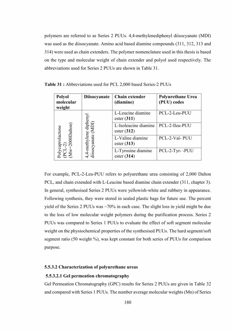
180
polymers are referred to as Series 2 PUUs. 4,4-methylenediphenyl diisocyanate (MDI)
was used as the diisocyanate. Amino acid based diamine compounds (311, 312, 313 and
314) were used as chain extenders. The polymer nomenclature used in this thesis is based
on the type and molecular weight of chain extender and polyol used respectively. The
abbreviations used for Series 2 PUUs are shown in Table 31.
Table 31 : Abbreviations used for PCL 2,000 based Series-2 PUUs
Polyol molecular weight
Diisocyanate Chain extender (diamine)
Polyurethane Urea (PUU) codes
Poly
capr
olac
tone
(P
CL-
2)
(Mw
=200
0Dal
ton)
4,4-
met
hyle
ne d
iphe
nyl
diis
ocya
nate
(MD
I)
L-Leucine diamine ester (311)
PCL-2-Leu-PUU
L-Isoleucine diamine ester (312)
PCL-2-Ileu-PUU
L-Valine diamine ester (313)
PCL-2-Val- PUU
L-Tyrosine diamine ester (314)
PCL-2-Tyr- -PUU
For example, PCL-2-Leu-PUU refers to polyurethane urea consisting of 2,000 Dalton
PCL, and chain extended with L-Leucine based diamine chain extender (311, chapter 3).
In general, synthesised Series 2 PUUs were yellowish-white and rubbery in appearance.
Following synthesis, they were stored in sealed plastic bags for future use. The percent
yield of the Series 2 PUUs was ~70% in each case. The slight loss in yield might be due
to the loss of low molecular weight polymers during the purification process. Series 2
PUUs was compared to Series 1 PUUs to evaluate the effect of soft segment molecular
weight on the physiochemical properties of the synthesised PUUs. The hard segment/soft
segment ratio (50 weight %), was kept constant for both series of PUUs for comparison
purpose.
5.5.3.2 Characterization of polyurethane ureas
5.5.3.2.1 Gel permeation chromatography
Gel Permeation Chromatography (GPC) results for Series 2 PUUs are given in Table 32
and compared with Series 1 PUUs. The number average molecular weights (Mn) of Series

181
2 PUUs varied from approximately 33,000 to 69,000 Dalton with a polydispersity index
varying from approximately 1.6 to 2.2. All molecular weights reported here are in terms
of polystyrene as standards.
Table 32 : Molecular weight of Series 2 and Series 1 PUU
Series 1 and 2
PUU
Number Average
Molecular Weight
(Mn) (Dalton)
Weight Average
Molecular Weight
(Mw) (Dalton)
Polydispersity
Index
PCL-1-Leu-PUU 6.09 x10⁴ 10.23 x10⁴ 1.67
PCL-2-Leu-PUU 3.30 x10⁴ 5.34 x10⁴ 1.61
PCL-1-Ileu-PUU 7.40 x10⁴ 13.43 x10⁴ 1.81
PCL-2-Ileu-PUU 6.87 x10⁴ 14.22 x10⁴ 2.06
PCL-1-Val- PUU 5.02 x10⁴ 8.25 x10⁴ 1.64
PCL-2-Val- PUU 4.81 x10⁴ 8.47 x10⁴ 1.76
PCL-1-Tyr- PUU 4.88 x10⁴ 8.35 x10⁴ 1.71
PCL-2-Tyr- PUU 4.61 x10⁴ 9.88 x10⁴ 2.14
The observed GPC data was consistent with literature values for PUUs (Skarja, 2001,
Sarkar, 2007, Marcos-Fernández et al., 2006, Caracciolo et al., 2008). On closer
inspection, there are some apparent trends observed which are contradictory. Most
polymers decreased in their number averaged molecular weight (Mn) but increased in
their weight averaged molecular weight (Mw), in moving from Series 1 to Series 2; an
apparent contradiction. In all cases, however, the polydispersity value also increased
(albeit slightly). A slightly increased polydispersity represents a wider distribution of
particle sizes. Weight averaged molecular weight , which are biased towards larger
particles, will therefore be slightly higher whilst number averaged molecular weight,
which are biased towards smaller molecular weight will be slightly lower. The bias, in
this case, appears to lead to the contradiction of molecular weight increasing (from Series
1 to Series 2) when based on weight averages, but decreasing (from Series 1 to Series 2)
when based on number averages. The exception was the leucine based polymers which
decreased in both weight and number averaged particles sizes. This polymer was also the

182
exception to the increase in polydispersity. The polydispersity in this case did not increase
(it was essentially unaltered) and therefore the decrease in number averaged molecular
weight was also reflected in a decrease in weight averaged molecular weight.
Notwithstanding the above argument, the molecular weight in all cases were similar,
given that the particle size of polymers can vary by many orders of magnitude. It is
therefore assumed that the properties of these polymers will be determined by
compositional morphology rather than the effect of molecular weight or its distribution.
5.5.3.2.2 Differential scanning calorimetric
Differential Scanning Calorimetric (DSC) results for Series 2 PUUs are given in Table
33 along with Series 1 PUUs result for comparison. DSC thermograms for the 1st and 2nd
heating run for Series 2 PUUs are shown in Figures 56 and 57. The PCL molecular weight
appears to have a significant effect on the thermal properties of the polymers.
A

183
B
Figure 56 : DSC thermograms for the 1st and 2nd heating run of (A) PCL-2-Leu-PUU
and (B) PCL-2-Ileu-PUU
C

184
D
Figure 57 : DSC thermograms for the 1st and 2nd heating run of (C) PCL-2-Val-PUU
and (D) PCL-2-Tyr-PUU
Table 33 : Thermal properties of Series 2 PUUs and its comparison with Series 1 PUUs
Tg – Glass transition temperature, Tm – Melting temperature, %Xc – Percent crystallinity, HS=Hard
segment, SS= Soft segment, Tg 1st- Tg of first heating run, Tg 2nd- Tg of second heating run, Tm 1st- Tm of
first heating run, ND- Not Detected
Glass transition (Tg-1
st) (°C)Melting Temperature(Tm-1st ) (°C)
Crystallinity(% Xc)
Glass Transition (Tg-2
nd ) (°C)
Tm & Crystallinity (%Xc)
PCL-1-Leu-PUU -6.5 47 5 -10.5 5 Phase mixed, SS = Semi Crystalline,HS = amorphous
PCL-2-Leu-PUU -39.8 42 3 -30.2 40°C and 3%, total of 6.0%
Phase separated, SS= Crystalline,HS = Amorphous
PCL-1-Ileu-PUU -3 47.5 5 -15.1 5 Phase mixed, SS = Semi Crystalline,HS = Amorphous
PCL-2-Ileu-PUU -35.4 52 16 -27.7 16 Phase separated, SS = Semi Crystalline,HS = Amorphous
PCL-1-Val-PUU -7.2 46.5 5 -7 5 Phase mixed, SS = Semi Crystalline,HS = Amorphous
PCL-2-Val-PUU -43.9 46.7 2 -17.3 2 Phase separated, SS = Semi Crystalline,HS = Amorphous
PCL-1-Tyr-PUU 13.2 ND ND 13.3 ND Phase mixed,SS & HS = Amorphous
PCL-2-Tyr-PUU -10 46 10 -12.1 10 Phase separated, SS = Semi Crystalline,HS = Amorphous
Series 1 and 2 PUU Soft Segment Polymer morphology

185
For Series 2 PUUs, the soft segment glass transition temperature (Tg) decreases and
crystallinity increases with increasing polyol molecular weight. It specifies increasing
phase segregation morphology. Similar observations for soft segment Tg and crystallinity
based on the different molecular weight of PCL-MDI based PUU are also recorded in
literature (Gisselfält and Helgee, 2003). However, in case of PCL-2-Val-PUU, soft
segment crystallinity decreased as compared to the rest of the Series 2 PUUs. For Series
2 PUU, Tm for the soft segment appears in the 1st heating run only indicating the semi
crystalline nature of the soft segment. Series 2 PUUs appear to be more phase segregated
than the corresponding Series 1 PUUs, with a lower glass transition temperature (Tg).
Similar results have been observed and reported in the literature (Gunatillake and
Adhikari, 2011) for other PUUs with high molecular weight soft segment.
Leucine and Isoleucine based Series 2 PUUs also exhibited higher phase separated
morphologies, and lower Tg values, for their soft segments, as compared to Series 1
PUUs. PCL-2-Leu-PUU showed an additional endotherm, at 60°C, in the 1st heating run
(Figure 56 (A)). This probably corresponds to disruption of short and long range order of
the hard segment. Similar thermal transitions have been reported by Skarja et.al. (2001)
for PCL based PUUs. Bogart et.al. (1983) has also described similar thermal transition
between 50 – 60°C in PCL based PUs.
PCL-2-Leu-PUU showed no hard segment crystallinity, thus the endotherm present in the
1st heating run at 60°C is assumed to be due to the disruption of ordered, non-crystalline
hard segment aggregates present within the soft segment. In literature, several reports
have ascribed similar transitions in this particular region to hard segments dispersed
throughout the soft segment region (Brunette et al., 1981). The appearance of these
transitions suggest the existence of the amorphous hard segment (Frontini et al., 1993).
For PCL-2-Val-PUU, soft segment Tg appears at -43.9°C (1st heating run) and appeared
again at -17.3°C (2nd heating run). The Tm for soft segment appeared at 46.7°C in 1st
heating run and 2% crystallinity observed. The observed Tg for PCL-2-Val-PUU is much
lower than PCL-1-Val-PUU. Similar observations were noticed for PCL-2-Tyr-PUU. The
186
soft segment Tg (-10°C) was lower than PCL-1-Tyr-PUU (Tg - 13.2°C). 10% crystallinity
appeared in PCL-2-Tyr-PUU with no crystallinity for PCL-1-Tyr-PUU.
No hard segment Tg or Tm was found for Series 2 PUUs indicating a non-crystalline or
amorphous nature for the hard segment, similar to Series 1 PUUs. The main reason for
the amorphous nature of hard segment is probably the presence of pendant methyl group
side chains attached to the (non-linear) chain extenders. The non-linear structure of chain
extenders results in a less ordered arrangement of polymer chains within the hard
segments, which in turn results in the absence of a Tg or Tm. Bae et.al. (1999) has also
argued that the use of non-linear chain extenders with MDI as hard segment results in
non-crystalline and less ordered hard segments.
By comparing the DSC results for Series 1 and 2 PUUs, it was observed that PCL soft
segments with higher molecular weight exhibit higher soft segment crystallinity. For
example, 6%, 16% and 10% of soft segment crystallinity was observed for PCL-2-Leu-
PUU, PCL-2-Isoleu-PUU, and PCL-2-Tyr-PUU respectively, as compared to 5% for
PCL-1-Leu-PUU, 5% for PCL-1-Isoleu-PUU, 0% for PCL-1-Tyr-PUU for Series 1 PUU.
This increase may be due to dipolar interactions of the ester carbonyls in the caprolactone
units (Sarkar, 2007).
The increase in soft segment crystallinity reflects incompatibility between the soft and
hard segments of the polymer and this, in turn, results in a more cohesive soft segment
and phase segregated behaviour shown by Series 2 PUUs (Skarja, 2001). Hu et al.(1982)
have reported an explanation for the co-relation between the soft segment crystallization
and polymer phase segregated morphology. In Hu’s study, as well as in the results
presented here, it was shown that an increase in crystallinity with increased PCL
molecular weight associate with improved micro phase separation behaviour of Series 2
PUUs.
Overall, it was observed that a higher molecular weight of the soft segment encourages
phase segregation morphology along with an increase in the soft segment Tg (in both the
heating runs) and in crystallinity. The non-linear nature of the chain extender (with

187
pendant group) prevents the hard segment from packing in an ordered fashion, which
results in the absence of a hard segment glass transition temperature (Tg) or a melt
endotherm temperature (Tm); hence it is amorphous in nature in Series 2 PUU.
5.5.3.2.3 Fourier Transform Infrared Spectroscopy
The two main areas of the FTIR spectra of the synthesised polymer are of specific interest
in the investigation of the effect of soft segment molecular weight in phase morphology
(phase segregation/phase mixed) of segmented PUUs. The first is the carbonyl (C=O)
region and the second is the N-H region. It is expected that upon formation of hydrogen
bonds, the absorption vibrations precisely associated with N-H and C=O group would be
shifting toward lower energy based on the environment present around each functional
group. The magnitude of these shifts, as well as change in the absorption characteristics
reflects hydrogen bond strength. To avoid repetition, only the PCL-1-Leu-PUU and PCL-
2-Leu-PUU FTIR spectra are described here in detail. FTIR spectra of Series 1 and 2 PUU
are shown in Figure 58 and the major FTIR peak positions are reported in Table 34. The
carbonyl stretching region is located between 1630 –1780cm-1 and the N-H region is
located at 3100 – 3500 cm-1. Both regions are shown with an expanded scale in Figure
59.

188
Figure 58 : FTIR absorption spectra of polyurethane (A) PCL-1-Leu-PUU and (B) PCL-
2-Leu-PUU
Figure 59 : FTIR absorption spectra of carbonyl and N-H region of polyurethane (A)
PCL-1-Leu-PUU and (B) PCL-2-Leu-PUU

189
Table 34 : Assignment of the major absorption bands in the FTIR spectrum of the MDI-
PCL based Polyurethane
Frequency (cm-1)
Assignment Frequency (cm-1)
Assignment
3340 ν(NH),H-bonded urethane
1666 ν(C=O),H-bonded dis ordered urea
3340-3320 ν(NH),H-bonded, ordered urea
1645-1628 ν(C=O),H-bonded ordered urea
~3340 ν(NH),H-bonded, disordered urea
1600 ν(C=C),aromatic
2939, 2866, 2794
ν(CH3) 1540 δ (N-H) + ν(C-N), amide II
1732, 1723 ν(C=O),free urethane, ester carbonyl (soft segment)
1450, 1370, 1365
δa(C-H) in CH3, ω(C-H) in CH2, δs(C-H) in CH3
1706 - 1713 ν(C=O),hydrogen bonded urethane
1240 δ (N-H) + ν(C-N), amide III
1690-1700 ν(C=O),free urea 1161 ν(CO-O-C) =stretching mode, a =asymmetric stretching, s=symmetric stretching, = in-plane bending or
scissoring,=in-plane bending or rocking, ω = wagging
Polyurethane urea are extensively hydrogen bonded. It has been reported in the literature
(Blackwell et al., 1982, Teo et al., 1997, Pitt et al., 1990, Coleman, 1997) that MDI based
PUUs show the presence of three-dimensional hydrogen bonding via the urea group
within the hard segment. A variety of hydrogen bonds are possible and shown in Figure
60. In all cases, hydrogen bonding involves the N-H donor group present in urethane and
urea linkages and the carbonyl or ether oxygen acceptor groups. The hydrogen bond
donor is present exclusively in the hard segment of the polymer while the acceptor may
be present either in the hard segment (carbonyl of urethane or urea) or the soft segment
(ester carbonyl or ether oxygen). Degree of phase separation define the ratio of hard and
soft segment hydrogen bonding. Phase separation favours interurethane, inter urea or
urethane urea hydrogen bonding.

190
Figure 60 : Hydrogen bonding interactions in polyurethane urea
The carbonyl region in FTIR is most applicable to the analysis of the hydrogen bonding
properties in polyurethanes (Luo et al., 1997). The carbonyl region comprises five distinct
bands. These bands are located between ~1620cm-1 and 1760 cm-1. The absorbance at
1640cm-1 in spectrum A (PCL-1-Leu-PUU) and at 1645cm-1 in spectrum B (PCL-2-Leu-
PUU) of Figure 58 represents hydrogen bonded carbonyl, C=O, group. The hydrogen
bond is formed between C=O group to urea (referred as‘ordered’ urea) and N-H group.
Urea C=O groups may also be hydrogen bonded in disordered manner and located at 1666
cm-1. The non-hydrogen bonded urea C=O is located at approximately 1700cm-1
(Coleman et al., 1988). It seems likely that both of these peaks contributes to the intensity
between 1710cm-1 and 1690cm-1. The hydrogen bonded and non-hydrogen bonded
urethane C=O peak was located at 1715cm-1 and 1732cm-1 respectively and appear
together to make it difficult to undertaken more detailed analysis. It has been proposed
by Coleman et.al. (1986) that “the relative degree of micro phase separation in amine
chain-extended polyurethanes can be assessed by determining the degree of urea C=O
hydrogen bonding (Bummer and Knutson, 1990) and that an increase in the extent of
micro phase separation is accompanied by an increase in absorbance of the ordered urea
C=O peak” (Teo et al., 1997). There are couple of bands present in this region and are

191
overlapping each other and hence cannot be properly examined due to poor resolution of
bands (Figure 58). Therefore spectra was analysed by qualitative peak analysis. Each
spectrum in Figure 58 was normalized using the area of the 1413cm-1 peak, assigned to
the C–C stretching mode of the aromatic ring (Srichatrapimuk and Cooper, 1978). This
peak has been used previously (McCarthy et al., 1997, Ishihara et al., 1974) as a reference
and its assignment is routine. The spectra were adjusted so that the relative peak areas of
the absorbance at 1413cm-1 are the same.
The main peak to investigate in the carbonyl region is that due to the hydrogen bonded
C=O portion of the urea group located at 1640cm-1 in spectrum A (PCL-1-Leu-PUU) and
at 1645cm-1 in spectrum B (PCL-2-Leu-PUU) of Figure 58. It has been reported in the
literature (Garrett et al., 2003) that this peak increases with an increase in the urea group
concentration, and this represents increased interaction (hydrogen bonding) between the
hard segments which, in turn, indicates hard-soft segment phase segregation. On
comparing the Series 1 and 2 PUU FTIR spectra, it was observed that intensity of the
bonded urea C=O peak is increased in Series 2-PUU. The high intensity of this peak in
Series 2 PUU (spectrum B) indicates a high level of hydrogen bonding present within the
hard segment of the PUU which in turns indicates phase segregation morphology. It was
also observed that the absorbance in the region of the disordered and free C=O is
increased and decrease in the ratio of the absorbance of the ordered urea C=O peak at
1640cm-1 was noticed for in Series 1 PUU. This further suggests a decrease in polymer
phase segregation morphology. It was detected that an increase in the absorbance of the
ordered C=O peak is associated with phase segregation morphology.
Other major bands present in the carbonyl region are at 1732cm-1 and at 1723cm-1 for
spectrum A and B respectively (Figure 59). This represents free (non-hydrogen bonded)
urethane C=O bond and the soft segment ester C=O group respectively. The bonded
urethane carbonyl ester band may appear between 1706 – 1713 cm-1, but is not clearly
visible and could be masked by the free urethane C=O peak (McCarthy et al., 1997, Teo
et al., 1997). It seems likely that the high intensity of the urethane free ester peak might
be due to the combination of bonded and non-bonded urethane carbonyl group. Hence,
there are two diverging effects observed and the overall effect is not clear, may be hidden

192
and it is hard to distinguish these peaks separately. Hence it would be difficult to use these
peaks for hydrogen bond elucidation in PUU structure.
The second area of interest is located between 3100cm-1 and 3500cm-1 as shown in Figure
59 and belongs to N-H stretching region. The very broad band at 3320cm-1 (spectrum A
and B), has been allocated to the hydrogen bonded N-H group present in urethane and
urea segment of the polymer. It indicate amorphous and disordered hard segment with
subsequently less phase segregation morphology (Coleman, 1997, Ishihara et al., 1974).
The peak shape of the absorbance in N-H region is similar to that reported for amorphous
polyurea (Garrett et al., 2003) which specify to the primarily disordered domain and lower
phase separation. No sign of free (non-hydrogen bonded) N-H group was observed,
otherwise that would have appeared as sharp peak at 3450 cm-1. No significant shift or
change in peak for N-H region was observed. Hence, not much information was collected
from the N-H region of PUU.
Based on this FTIR study, complemented by the earlier DSC results, it can be concluded
that low molecular weight PCL based PUUs show phase mixed morphology and high
molecular weight PCL based PUUs show phase segregated characteristics. Excellent
agreement between the FTIR study and DSC results (Section 5.5.3.2.2) was noted.
5.5.3.2.4 Thermo Gravimetric Analysis Thermogravemetric analysis (TGA) was performed, under nitrogen, on Series 2 PUUs
and compared with that for Series1 PUUs in an effort to understand the effect of soft
segment polyol molecular weight on the thermal stability of polyurethane ureas. The TGA
curves for Series 2 and 1 PUUs are shown in Figure 61 and 62 respectively.

193
Figure 61 : TGA analysis of Series 2 PUUs
Figure 62 : TGA analysis of Series 1 PUUs

194
The thermal stability parameters including the onset decomposition temperature (Ton °C)
(which is defined as the initial temperature at which polymer weight loss starts) and 10%
weight loss temperature (T10% °C) (Temperature at which 10% of polymer weight loss
was observed) was observed for both the series and are shown in Table 35.
Table 35 : Characteristic temperature on TGA curves for Series 1 and 2 PUUs
Series 1 and 2 PUU
Ton (°C) (Onset Temp.)
T10% (°C ) (10% Weight loss)
PCL-1-Val-PUU 280 308 PCL-2-Val-PUU 300 315 PCL-1-Tyr-PUU 285 320 PCL-2-Tyr-PUU 263 290 PCL-1-Leu-PUU 265 285 PCL-2-Leu-PUU 290 314 PCL-1-Ileu-PUU 271 304 PCL-2-Ileu-PUU 297 315
It was observed that PCL-2-Val-PUU exhibited the highest, and PCL-2-Tyr-PUU showed
the lowest, onset temperature in Series 2 PUUs. For tyrosine PUU, the result for PCL-2-
Tyr-PUU, is somehow reversed as compared to PCL-1-Tyr-PUU. For example, PCL-1-
Tyr-PUU (Ton (285°C) showed higher thermal stability among the rest of the Series 1
PUU. But in case of Series 2 PUU, PCL-2-Tyr-PUU (Ton (263°C), showed least thermal
stability among Series 2 PUU. The reason for this observation is not known.
In all cases, Series 2 PUUs have a higher onset, and 10% weight loss, temperature (except
PCL-2-Tyr-PUU) than Series 1 PUUs. Again, this is probably due to the longer soft
segment in Series 2 PUUs favouring phase separation morphology and enhancement of
inter urethane hydrogen bonding, The enhancement of hydrogen bonding then provides a
higher thermal stability. The higher micro phase separation in Series 2 PUUs may be due
to the crystallization effect of PCL 2000 Dalton which was also confirmed by the presence
of Tm and % Xc in the DSC analysis (Table 33).

195
The results (Figure 61) show that Series 2 PUUs are more thermally stable in the initial
stages and degrade rapidly later on as compared to Series 1 PUUs (Figure 62). It appears
that PCL 2,000 has a higher mutual stabilization effect than PCL 1,000 due to the hard
segment being protected by a higher soft segment concentration in the polymer and hence
an increase in its degradation temperature (V.F. Antipova, 1970). Although the weight
percent of soft and hard segment was the same for both the series, the use of a higher
molecular weight PCL and hence longer soft segment polymer chains has provided
thermal stability to Series 2 PUUs.
Wang et.al. (1997) has reported similar results for the effect of polyol (polyether)
molecular weight on the thermal stability of segmented PUUs. Ferguson et al. (1976) has
also reported the stabilizing effect of soft segment concentration on the thermal stability
of PTHF-MDI based polyether PUU. They have also reported that the presence of urea,
urethane and amine groups have a stabilizing effect on PUUs during oxidative
degradation. It is worth mentioning that Series 1 and 2 PUs and PUUs are based on
different amino acid based chain extenders (i.e. dihydroxy versus diamine), hence it
would not be appropriate to directly compare the TGA behaviour between the PUs and
PUUs series. The use of different functionality of chain extender would be expected to
impart different thermal characteristic to the polymer.
Overall, the thermal characteristic of all polymers synthesised here indicate a wide range
of temperatures within which the polymer can be thermally processed, for example as
scaffold fabrication in various biomaterial applications.
5.5.3.2.5 Contact angle
Contact angle values for Series 2 PUUs are shown in Table 36. The results for Series 1
PUUs are also shown in the Table 36 for easy comparison. Series 2 PUUs show slightly
higher contact angles compared to Series 1 PUUs. Although the difference is barely
significant, it is consistent with, and supports, the main findings of polymer
characterisation to date. Lamba et.al. (1998), for example, has reported that a higher
contact angle results from increasing the molecular weight of the soft segment and is
related to phase separation morphology within the polymer.
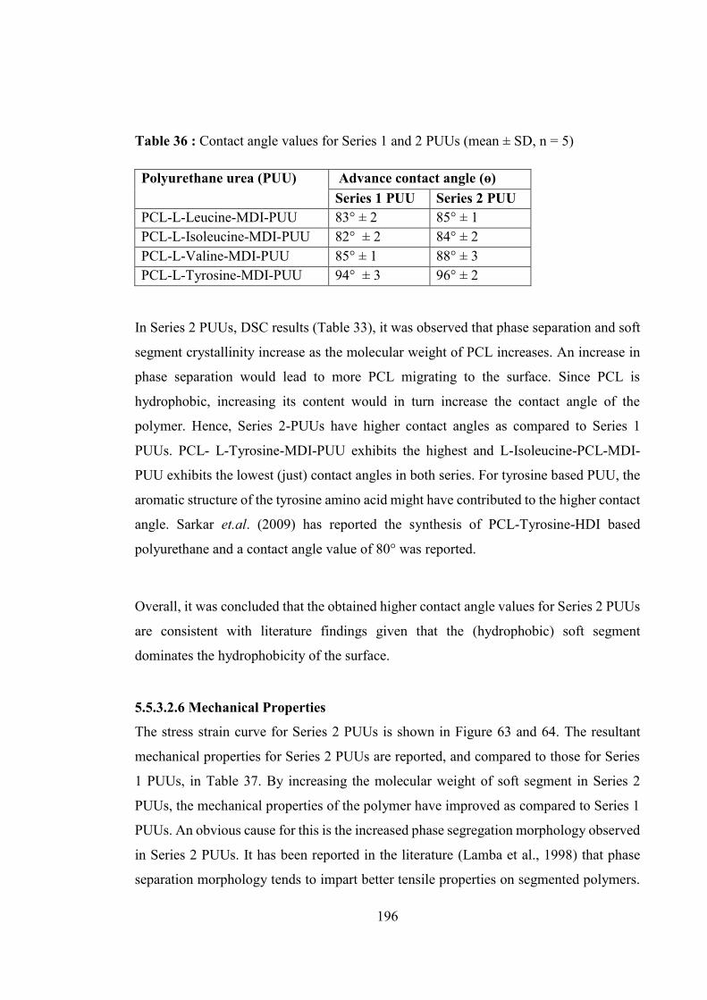
196
Table 36 : Contact angle values for Series 1 and 2 PUUs (mean ± SD, n = 5)
Polyurethane urea (PUU) Advance contact angle (ɵ) Series 1 PUU Series 2 PUU
PCL-L-Leucine-MDI-PUU 83° ± 2 85° ± 1 PCL-L-Isoleucine-MDI-PUU 82° ± 2 84° ± 2 PCL-L-Valine-MDI-PUU 85° ± 1 88° ± 3 PCL-L-Tyrosine-MDI-PUU 94° ± 3 96° ± 2
In Series 2 PUUs, DSC results (Table 33), it was observed that phase separation and soft
segment crystallinity increase as the molecular weight of PCL increases. An increase in
phase separation would lead to more PCL migrating to the surface. Since PCL is
hydrophobic, increasing its content would in turn increase the contact angle of the
polymer. Hence, Series 2-PUUs have higher contact angles as compared to Series 1
PUUs. PCL- L-Tyrosine-MDI-PUU exhibits the highest and L-Isoleucine-PCL-MDI-
PUU exhibits the lowest (just) contact angles in both series. For tyrosine based PUU, the
aromatic structure of the tyrosine amino acid might have contributed to the higher contact
angle. Sarkar et.al. (2009) has reported the synthesis of PCL-Tyrosine-HDI based
polyurethane and a contact angle value of 80° was reported.
Overall, it was concluded that the obtained higher contact angle values for Series 2 PUUs
are consistent with literature findings given that the (hydrophobic) soft segment
dominates the hydrophobicity of the surface.
5.5.3.2.6 Mechanical Properties
The stress strain curve for Series 2 PUUs is shown in Figure 63 and 64. The resultant
mechanical properties for Series 2 PUUs are reported, and compared to those for Series
1 PUUs, in Table 37. By increasing the molecular weight of soft segment in Series 2
PUUs, the mechanical properties of the polymer have improved as compared to Series 1
PUUs. An obvious cause for this is the increased phase segregation morphology observed
in Series 2 PUUs. It has been reported in the literature (Lamba et al., 1998) that phase
separation morphology tends to impart better tensile properties on segmented polymers.
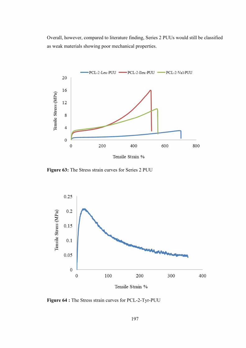
197
Overall, however, compared to literature finding, Series 2 PUUs would still be classified
as weak materials showing poor mechanical properties.
Figure 63: The Stress strain curves for Series 2 PUU
Figure 64 : The Stress strain curves for PCL-2-Tyr-PUU

198
Table 37 : Mechanical properties of Series 1 and 2 PUUs (mean ± SD, n = 6)
Series 1 and 2 PUU Modulus of Elasticity (MPa)
Tensile Strength (MPa)
Elongation at Break (%)
PCL-1-Leu-PUU* 4.3 ± 1 0.52 ± 0 982.2 ± 11 PCL-2-Leu-PUU 13.8 ± 2 2.1 ± 1 700 ± 66 PCL-1-Ileu-PUU 5.1 ± 1 1.1 ± 0 337.0 ± 46 PCL-2-Ileu-PUU 32.6 ± 3.2 12.0 ± 3.6 479.2± 55.7 PCL-1-Val-PUU 7.2 ± 2 2.0 ± 1 563.7 ± 22 PCL-2-Val-PUU 41.8 ± 11.9 5.4 ± 1.1 533.7 ± 46.9 PCL-1-Tyr-PUU 11.6 ± 8 7.3 ± 3 421.7 ± 58 PCL-2-Tyr-PUU 2.0 ± 1 0.03 ± 0 350.1 ± 1
*Sample did not break during tensile testing.
A very low value for the tensile properties of PCL-2-Tyr-PUU (Figure 64) was obtained,
consistent with the material being weak and tacky to handle. The reason for obtaining
such low tensile properties is not known, however the improvement in mechanical
properties (in most cases) from Series 1 to Series 2 suggests that optimising the soft
segment may be a useful future research direction. The low molecular weight and high
polydispersity (Table 32) in comparison to typical segmented PUUs is probably the main
reason for obtaining low tensile properties for PCL-2-Tyr-PUU. It has been reported in
the literature (Schollenberger and Dinbergs, 1973) that tensile properties increase as
molecular weight of the segmented PUs increases. For most polymers, properties such as
tensile strength, modulus and elongation are affected by molecular weight increases since
longer chains are more likely to act as connectors between the crystalline or glassy state,
allowing greater toughness along with soft segment crystallinity present in the polymer
(R.B. and Carraher, 1984, Skarja, 2001).
The relatively high values for the moduli of elasticity suggests that Series 2 PUUs are
stiffer than Series 1 PUUs and the tensile strengths for Series 2 PUUs are higher than
Series 1 PUUs (except for PCL-2-Tyr-PUU which was very low for Series 2 PUU).
Elongation properties were more robust for Series 2 PUUs as compared to Series 1 PUUs,
possibly due to the change from phase mixed to phase segregated morphology as the PCL
molecular weight increases. Again, the tensile properties for Series 2 PUUs appear
improved as compared to Series 1 PUUs, but remain on the poor side compared to

199
literature findings ((Azzam et al., 2007, Skarja, 2001). Another major factor which may
contribute in the attaining poor tensile properties for Series 2 PUUs is the use of non-
linear and bulky chain extenders as compared to conventional small, linear chain
extenders such as BDO (Caracciolo et al., 2008, Bagdi et al., 2009, Caracciolo et al.,
2009). The non-linear structure of the chain extender has resulted in a lack of hard
segment crystallization, which reduces the tendency of the hard segment to act as
reinforcing filler or physical cross link site. Hence the overall mechanical properties of
the polymer is reduced. It was also observed during DSC analysis (Section 5.5.3.2.2) that
the higher percent of soft segment crystallization in Series 2 PUUs (compared to Series 1
PUUs) improved the mechanical properties of Series 2 PUUs. Soft segment crystal
structure may act as physical cross links in a manner normally attributed to the hard
segments. Increasing phase segregation morphology is accompanied by increasing PCL
content which may yield superior mechanical properties for polyester based PUUs (Foks
et al., 1990).
Overall, it was observed that the mechanical properties of the Series 2 PUUs were much
improved as compared to Series 1 PUUs but still low as compared to the literature. The
main reason for the improved mechanical properties for Series 2 PUUs might be the
increase in the soft segment crystallinity and the existence of phase segregated
morphology for Series 2 PUUs. The importance of both soft segment crystallinity and
phase separation is highlighted by the low tensile properties of Series 1 PUUs. Overall, it
was observed that PUUs made from these bulky and non-linear chain extenders have
generally poor mechanical properties as compared to the literature findings and as
expected no hard segment ordering was observed to contribute toward obtaining good
mechanical properties.
5.6 Conclusion
A series of PUs and PUUs synthesised using 2,000 Dalton (Mw) PCL as soft segment
and using novel chain extenders exhibited a variable degree of phase behaviour depending
on the chemistry of the soft segment, structure and composition of the polymer. Detailed
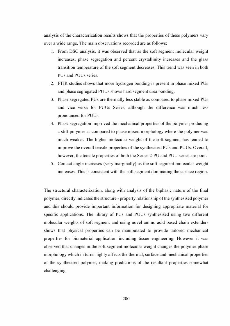
200
analysis of the characterization results shows that the properties of these polymers vary
over a wide range. The main observations recorded are as follows:
1. From DSC analysis, it was observed that as the soft segment molecular weight
increases, phase segregation and percent crystallinity increases and the glass
transition temperature of the soft segment decreases. This trend was seen in both
PUs and PUUs series.
2. FTIR studies shows that more hydrogen bonding is present in phase mixed PUs
and phase segregated PUUs shows hard segment urea bonding.
3. Phase segregated PUs are thermally less stable as compared to phase mixed PUs
and vice versa for PUUs Series, although the difference was much less
pronounced for PUUs.
4. Phase segregation improved the mechanical properties of the polymer producing
a stiff polymer as compared to phase mixed morphology where the polymer was
much weaker. The higher molecular weight of the soft segment has tended to
improve the overall tensile properties of the synthesised PUs and PUUs. Overall,
however, the tensile properties of both the Series 2-PU and PUU series are poor.
5. Contact angle increases (very marginally) as the soft segment molecular weight
increases. This is consistent with the soft segment dominating the surface region.
The structural characterization, along with analysis of the biphasic nature of the final
polymer, directly indicates the structure - property relationship of the synthesised polymer
and this should provide important information for designing appropriate material for
specific applications. The library of PUs and PUUs synthesised using two different
molecular weights of soft segment and using novel amino acid based chain extenders
shows that physical properties can be manipulated to provide tailored mechanical
properties for biomaterial application including tissue engineering. However it was
observed that changes in the soft segment molecular weight changes the polymer phase
morphology which in turns highly affects the thermal, surface and mechanical properties
of the synthesised polymer, making predictions of the resultant properties somewhat
challenging.

201
Chapter 6: Cells Cytotoxic studies on
polyurethane and polyurethane urea films

202
6.1 Introduction
This chapter describes cytotoxicity screening of the amino acid based Series 1 and 2
polyurethanes and polyurethane ureas. Mouse Fibroblast L929 (ATCC) cells were chosen
as the test cells for the study and the LIVE/DEAD® Viability/Cytotoxicity Assay Kit
(Molecular Probes L-3224) was used to investigate short term cytotoxicity of the
synthesised polymer films. Fibroblast cells have been used in literature (Rechichi et al.,
2008), (Henry et al., 2007), (Harris et al., 2006), (Li, 2009) to examine the
biocompatibility of the synthesised polyurethane. This assay examines basic cytotoxicity
and hence the actual cell type used is not crucial. It is possible that the polymer may
remain cytotoxic to specific cell type not tested here and hence further testing would be
required for specific applications. The test here, however, will determine whether or not
there are substantial generic cytotoxic concerns with the use of these polymers as
biomaterials.
It is reported in the literature that polyurethanes based on 4,4-methylenediphenyl
diisocyanate (MDI) produce the highly toxic aromatic amine, 4,4-methylenedianiline
(MDA) upon degradation , (Vermette, 2001). MDA is mutagenic and carcinogenic in
animals and is therefore not desirable for in vivo applications (Kavlock et al., 2007),
(Szycher and Siciliano, 1991). There is, however, some doubt as to whether the
concentration of these harmful degradation products reach any physiologically relevant
level upon in vivo applications (Blais, 1990), (Coury, 2004). The ill effects of MDI for
polyurethane synthesis can also be ameliorated by:
changing the concentration of MDI used in polymer synthesis,
choosing a final application (in vitro rather than in vivo) where toxicity is less
problematic,
changing the duration (short/long term) of the polymer, and/or its degradation
products, for in vivo applications
It is reported in literature (Guelcher, 2008) that the data currently available for toxicity of
MDI and its by-product (MDA) suggest that the physiological effects of aromatic

203
polyamines depend on the degradation rate of the material, as well as the clearance rate
of the degradation product from the tissue.
In the present study, MDI as diisocyanate was chosen for PU/PUU synthesis due to its
high reactivity and ability to impart superior physical properties to the polymers. There
will therefore be a balance between toxicity and polymer properties. Despite the
drawbacks, several researchers (de Groot et al., 1996) have pioneered the use of MDI in
polyurethane and polyurethane urea products. They cast doubt over magnitude of the
toxicity problem and argue that the risk benefit of improved polymer properties may out-
weigh those problems (which would hopefully be eventually ameliorated in any case).
For example, Jun et.al. (2004) and Taite et. al. (2008) have described the use and
modification of MDI based polyurethane to develop scaffolding for vascular graft
application. In this work, they have demonstrated the successful attachment and
proliferation of bovine aortic endothelial cells on MDI based polyurethanes. The use of
aromatic polyurethane plotting compounds for biomedical devices (Vermette, 2001) and
in biomedical research (Hsu and Lin, 2004), (Liljensten et al., 2002), (Gisselfält et al.,
2002) are further examples reported in the literature. In another report, aromatic
diisocyanate based polyurethanes have been used for the fabrication of extracorporeal
medical devices such as haemodialyzers, oxygenators, haemo-concentrators (Guelcher et
al., 2005), (Reed et al., 1994).
Nevertheless, based on the uncertainty of cytotoxicity of MDI based polyurethane and
polyurethane ureas, it was felt important to perform basic cell toxicity tests on the
polymers synthesised in this thesis, to determine if that is likely to be a significant
drawback to the novel polymers produced.
To perform the cell toxicity test, Mouse fibroblast cells were seeded onto the amino acid
based PUs and PUUs films and the cells viability was evaluated after short period of time
(2 days) through LIVE/DEAD® assay kit. Tissue culture polystyrene (TCPS) was used as
a positive control substrate. TCPS is the most commonly used in vitro cell culture surface
and has often been used as a primary reference material for the evaluation of new
biomaterials (Bonfield et al., 1989), (Yun et al., 1995), (Bélanger and Marois, 2001),

204
(Matheson et al., 2007), (Vashi et al., 2013). 1, 4-butanediol (BDO) based polyurethane
(chapter 4) was used as a secondary control due to its closer similarity in chemical
structure to the synthesised polymer samples. Several authors have reported that BDO
based polyurethanes supports cell growth (Liu et al., 2012a).
The mouse fibroblast L929 (ATCC) cell line was chosen as the model test cell, because
it is one of the most common cell lines used in cell culture and is known to be strongly
adhesive to TCPS surface (Vashi et al., 2013).
The LIVE/DEAD® Viability/Cytotoxicity assay Kit (L-3224) (Invitrogen, Carlsbad, CA)
was used to perform the test. It provides a two colour fluorescence viability assay that is
based on the simultaneous determination of live and dead cells. Calcein acetoxymethyl
(calcein AM) and ethidiumhomodimer (EthD-1) are the dyes used for this assay. Simply,
metabolically active cells permit calcein acetoxymethyl ester (calcein AM) to enter
through the intact plasma membrane of the cells, where the dye is cleaved by cytoplasmic
esterase yielding green fluorescence in live cells (ex/em ~ 495nm /~515nm). By contrast,
ethidium homodimer – 1 (EthD-1), which is membrane impermeable, binds to the DNA
of membrane of the compromised cells, yielding bright red fluorescence (ex/em ~ 495nm
/~635nm) in dead, or damaged (membrane compromised), cells.. Live/dead assay results
provide information regarding cell adhesion and viability on the various materials after a
specified time (in present case, two days) of cell culture. Note that strictly, the DEAD cell
determination is really a measure of compromised cells rather than simply dead cells,
however it is assumed that compromised cells will not be viable and it is common practise
to include them as “dead”.
The use of the LIVE/DEAD® assay to assess the cell viability and cytotoxicity of cells on
polyurethane surfaces has been previously reported in literature. For example, Parrag
(2010) used this method to visualise the live and dead cells present on peptide based
polyurethane materials. Other reports are available in literature which demonstrate the
use of this technique includes (Park et al., 2011), (Lee et al., 2009), (Blit et al., 2012),
(Fromstein et al., 2008),(Shah and Yun, 2011).

205
6.2 Materials
LIVE/DEAD® Viability/Cytotoxicity Kit (L-3224, Molecular Probes, MoBiTec GmbH,
Göttingen, Germany) was used to perform cell viability assays. Polymer films were
prepared by the method describe in section 4.4.7, to act as substrate for cell culture. PUs
and PUUs from Series 1 and 2 were chosen randomly for the cell cytotoxicity experiment.
Specifically, PCL-1-Z-Ser-PU, PCL-2-Z-Ser-PU, PCL-1-Z-Thr-PU, PCL-2-Z-Thr-PU,
PCL-1-Leu-PUU, PCL-2-Leu-PUU, PCL-1-Val-PUU and PCL-2-Val-PUU samples
were selected from both the series as exemplars for this experiment, tissue culture plates
(Nunc, Denmark), Mouse Fibroblast (L929, ATCC) cells, faetal calf serum (FCS),
phosphate buffer solution (PBS), Antibiotic- Antimycotic (Gibco-Invitrogen), minimum
essential medium (MEM) media.
6.3 Experimental Procedure
Polymer films were cut into small discs using an 8 mm diameter puncher. The discs were
sterilized by soaking in 0.1M phosphate buffer solution (PBS) containing 2X Antibiotic-
Antimycotic (Gibco-Invitrogen) for two days in the bottom of a 96-well tissue culture
plate (Nunc, Denmark) at 37 C in 5% CO2. Following this and prior to cell seeding,
polymer discs were soaked again in PBS culture media for 2 hours. Mouse Fibroblast
(L929, ATCC) cells were seeded on each polymer disk at the bottom of the 96 well tissue
culture plates at a density of 50,000 cells/cm2 in 10% faetal calf serum (FCS) containing
minimum essential medium (MEM) media. Seeded polymer films were incubated for two
days at 37°C, in a humidified atmosphere of 5% CO2 and 95% air. Cells were then washed
with PBS twice. The viability of attached and spread cells was assessed using the
LIVE/DEAD® Viability/Cytotoxicity Kit assay (Molecular Probes) at 48 hours according
to the manufacturer’s instructions, using a Nikon TE2000-U fluorescent microscope
equipped with digital camera. Live cells were stained green by Calcein AM while dead
cells stained red with EthD-1. Both Calcein AM and EthD-1 were excited at 654 nm and
530 nm, respectively. Images were viewed and photographed using a Colour View
Soft/Imaging system connected to a Nikon T2000 microscope. Viable and non-viable
cells were observed for each well at a magnification of X 200.

206
6.4 Result and Discussion
An ideal biomaterial should enhance cell growth, attachment, proliferation and other
cellular functions, all of which can be determined, to a first approximation by the
Live/Dead Cell assay. Figure 65 and 66 shows mouse fibroblast L929 (ATCC) cells
attachment onto the PUs and PUUs surface for Series 1 and 2 respectively. In both cases,
all images produced green fluorescence showing live cells with only a few spots of red
fluorescence indicating dead cells.
Figure 65 : Fluorescence images of mouse fibroblast cells on Series 1 polymers surfaces
(A) 1,4- Butanediol (Control), (B) & (C) Series 1 PU, (D) TCPS control, (E) and (F)
Series 1 PUU (magnification x 200). Circle showing dead cells with red fluorescence.
The results demonstrate that the cells seeded on Series 1 and 2 of amino acid based PUs
and PUUs films were substantially viable, with excellent cell attachment onto the
synthesised polymer films. Furthermore, the cell growth on amino acid based PUs/PUUs
samples were equally comparable to the cell growth on the positive test control (TCPS)
sample and the polymer control (BDO based PU) sample. This shows that the synthesised
polymers have retained a similar cell growth response to the commercially viable and
well known BDO based PU and TCPS.

207
Figure 66 : Fluorescence images of mouse fibroblast cells on Series 2 polymers surfaces
(A) PCL-1-BDO-PU (Control), (B) & (C) Series 2 PU, (D) TCPS control, (E) and (F)
Series 2 PUU (magnification x 200). Circle showing dead cells with red fluorescence.
The cells spreading pattern on synthesised PUs/PUUs resembles that of spindle
morphology. The attached cells show normal flattened appearance. Active adhesion is
shown by cells, as cells grow on the polymer surface and spread on these samples. The
cells consumed metabolic energy during this process, which is indicative of active
adhesion. These observations suggest that the chemical and/or physical structure of the
substrate controls the degree of cell adhesion and proliferation. This is further evidence
that the cell culture experiments shows good cell attachment in all the PUs and PUUs
samples used in this experiment, indicating that the scaffolds have a high level of
biocompatibility.
Closer evaluation of cell growth in Series 1 and 2 PUs/PUUs indicates that PCL-1-Z-Ser-
PU and PCL-2-Z-Ser-PU display a high, and even, level of cell attachment and viability,

208
i.e. fibroblast cells proliferated well and were evenly distributed on the serine based
polyurethane surface. This, however, was not the case for the threonine, leucine and
valine amino acid based PUs/PUUs where cell growth was patchy and cells were
unevenly distributed onto the polymer surface. The reason for this uneven distribution is
not clear, however it might be that the cells which grow on these polymer surfaces may
need little longer to spread evenly. It may also be a simple reflection of unusual growth
pattern on amino acid based polymer surface. Regardless of the reason for patchy growth,
all the Series 1 and 2 PUs/PUUs based materials supported cells growth (~90%) with a
little or few dead cells. It was observed that the cell growth was healthy and comparable
to the cell growth on the TCPS and BDO based PU controls. Overall, there was no
evidence of significant cytotoxic response observed by cultured cells on any polymer
surface. The cells appear well attached on all polymer substrates, with no apparent
orientation. In general, a fairly confluent layer of cells was identified for the culture
surfaces with cells in close contact or on top of each other in many spots. This
phenomenon likely reflective of high cell density, which in turn shows good cell
compatibility.
It is possible that dead cells may be washed off the polymer surface during medium
changes and washing carried out during the assay and this may result in an artificially
good result. All the tested polyurethanes and polyurethane ureas, however, supported
attachment, proliferation, and high viability of fibroblast cells in vitro. The major
observations obtained from the cell study experiment can be summarised as:
1. Cell viability test using LIVE/DEAD® assay kit provides no evidence of a
significant cytotoxic response by cultured fibroblast to any of the PUs and PUUs
synthesised in this thesis. The lack of cytotoxicity indicates that the polyurethanes
can be considered relatively cell compatible and have a cell adhesive surface. This
result may prove very useful to the intended end polymer application as an
implanted device that is designed to degrade.
2. While no cytotoxic response was detected, there was variation in cell viability
depending on the polyurethane chemistry. For example, cells were evenly
proliferated on PCL-Z-Ser-PU surface in both series 1 and 2, presumably due to
the hydrophilic nature of the serine amino acid supporting cell growth. By
contrast, the others synthesised polymer surfaces showed uneven distribution of
cell growth. It might be the case that the cells on these surfaces needs little longer
209
to fully cover the surface evenly, or even that the patchy areas could result from
tiny air bubbles present in the polymer film while preparing the polymer sample
for cell study. Surfaces which are less strongly adhesive would overcome and
force out such air bubbles slower and there may not have been enough time for
this to occur which the polymers investigated here. It should, however, be kept in
mind that the amount of dead cells detected was insignificant, strongly suggesting
a good biocompatible surface even if adhesion is slightly slower than on other
biocompatible surfaces.
3. Cells grown on the synthesised amino acid based PUs/PUUs surface did not differ
phenotypically from cells grown on the tissue culture polystyrene (TCPS) plates
or the control BDO based PU (as assessed by the cell growth, pattern and
proliferation).
4. PUs and PUUs, from both Series 1 and 2, used in this experiment, all supported
cell growth and proliferation, showing that biocompatibility is irrespective of the
molecular weight of the soft segment.
6.5 Conclusion
Polyurethanes are promising materials for soft tissue engineering applications. The
cytotoxicity assay employed here indicates that the amino acid based polymers
synthesised in this thesis do not show any significant cytotoxic response. Specifically, the
results from this study indicate that the polymer films can support a high density of viable
cells. Fibroblast cells growth, attachment and proliferation onto the polymer samples
were comparable to the positive TCPS and BDO based polymer controls. Seeded cells
appeared as a fairly confluent layer on all the substrate and were adherent and spread out
on the polymer surface, with no apparent orientation. The study is a suitable, but rapid
and basic experiment to test the cytocompatible behaviour of the synthesised polymers.
Further study, for example using other cell lines, are required before it can be said with
certainty that the polymers are fully biocompatible, however the results presented here
show that further development of the novel compounds developed in this thesis is worth
pursuing, i.e. continued improvement of physical polymer properties whilst minimising
any cytotoxic effect.

210
Chapter 7: Conclusion and future work

211
7.1 Conclusion
The focus of this dissertation was to design, synthesise and characterise segmented
polyurethanes and polyurethane ureas containing L-amino acids as chain extenders.
Novel L-amino acids based diamine/dihydroxyl groups containing organic compounds
were successfully synthesised and subsequently used as chain extenders to formulate
segmented polyurethanes and polyurethane urea. A detailed physical, mechanical and
chemical characterisation, including cell study, was performed on synthesised polymers
and analysis of the resultant properties showed that the L-amino acid containing
polyurethanes and polyurethane ureas have good potential to use as a biomaterial in
future. A study of structure property correlation was performed by developing a library
of polyurethanes and polyurethane ureas. The main outcome of this dissertation is briefly
summarized as follow:
Synthesis and characterisation of L-amino acid based chain extenders: Five different L-
amino acids such as L-isoleucine (301), L-leucine (302), L-valine (303), L-tyrosine (304)
and L-phenylalanine (305) (Figure 10, Chapter 3) were used to synthesise diamine ester
compounds such as 311, 312, 313, 314 and 315 (Figure 11, Chapter 3). The purpose to
synthesise these diamine diester compounds was to use them later as chain extender for
polyurethane urea synthesis. Out of the total five synthesised diamine ester compounds,
L-phenylalanine based compound 315 was not used as chain extender for polyurethane
urea synthesis in this thesis. Compound similar in structure to 315 is already reported in
literature and used for polyurethane urea synthesis (Skarja, 2001). Hence it was not worth
pursuing it further. Three amine group protected L-amino acids such as L-Z-serine (306),
L-Z-threonine (307) and L-Z-Tyrosine (308) (Figure 10, Chapter 3) were used to
synthesise dihydroxyl ester compounds such as 318, 319, 320 (Figure 20, Chapter 3) to
act as chain extender for polyurethane synthesis. PU synthesised based on compound 320
as chain extender was of very low molecular weight and was very tacky to handle. Hence
this PU was not used for further detail polymer characterisation. Rest of the L-amino acid
based synthesised compounds (311,312,313,314,318,319) were incorporated into
polyurethane and polyurethane urea as chain extender.
212
Fischer esterification reaction was employed to synthesise novel diamine diester
compounds (311, 312, 313, 314 and 315 – section 3.3, Figure 11. The reaction yield was
~ 80% in all cases. The final compounds were characterised, and confirmed, by NMR,
FTIR and MS spectroscopy.
Esterification reaction with caesium salt was employed to synthesise amine group
protected L-amino acid based dihydroxyl ester compounds. Reaction conditions were
optimised and a purification scheme was developed to remove impurities and to obtain a
pure product. The amine group of the L-amino acid used for this reaction was protected
by a benzyloxycarbonyl (Z) group. L-Z-amino acids were used to synthesise dihydroxy
ester compounds 318, 319 and 320. The percent yield of reaction in all cases was
approximately 85%. The final products were analysed, and confirmed, by NMR, FTIR
and MS spectroscopy.
A series of segmented polyurethanes (PUs) and polyurethane ureas (PUUs) were
synthesised using the L-amino acid based dihydroxyl and diamine chain extenders,
polycaprolactone (PCL, Mw=1000 Dalton) act as soft segment, 4,4-methylene diphenyl
diisocyanate (MDI) was used as diisocyanate. Synthesised PUs/PUUs series with
PCL1000 as soft segment were called Series-1-Chain Extender-PUs/PUUs. Their
structure property relationships were investigated, and compared with a control
polyurethane synthesised with 1,4-butanediol as the chain extender and polycaprolactone
(PCL, Mw=1000 Dalton) was used for soft segment and 4,4-methylene diphenyl
diisocyanate (MDI) was used as diisocyanate and abbreviated as PCL-1-BDO-PU. The
deprotection of the Z group of the chain extenders was optimised to obtain minimum
polymer backbone cleavage. The structure of synthesised polymers were confirmed by
NMR, GPC and FTIR technique. The thermal, surface and mechanical characterisation
of the synthesised PUs and PUUs were done by DSC, TGA, contact angle and tensile
testing. In general, compared to the control PCL-1-BDO-PU, the synthesised novel L-
amino acid based PCL-1- PUs/PUUs were:
Weak in mechanical strength. The L-amino acid based PUs/PUUs were soft
materials with high elongation at break values and with low values of tensile
strength and modulus of elasticity. In contrast, the control polyurethane behaved
as a strong and stiff polymer with a high value of modulus and tensile strength but
213
with low percent of elongation. The reason for obtaining poor mechanical
properties is due to the less ordered hard segment domain observed in L-amino
acid based PUs/PUUs.
More phase mixed with semicrystalline soft segment and amorphous hard segment
as shown by DSC. The reason for the absence of hard segment Tg and Tm for the
PUs/PUUs investigated here, is the unsymmetrical structure of the chain extender,
which prevents any ordering of the hard segments through hydrogen bonding.
Control polyurethane act as strong material with high modulus and tensile strength.
BDO based polyurethane showed phase segregated morphology with amorphous
soft segment and semi crystalline hard segment.
More thermally stable (PUs only) as compared to the control BDO based PU as
observed by TGA study. However, TGA result is reverse for Series-1-PUUs, which
is less thermally stable then control PU.
More hydrophobic in nature as shown by contact angle values. Series-1-PUs/PUUs
showed higher contact angle values as compared to control PU which implies to
the hydrophobic surface of the polymer.
Overall, it was observed that the properties shown by Series 1-PCL-L-amino acid
based-PUs/PUUs clearly demonstrate the effect of the unsymmetrical structure of
the chain extender on the physiochemical properties of the synthesised polymers.
Structure property relationship of the synthesised polymers (PUs/PUUs) was
studied by making another series with higher molecular weight of the soft segment
(PCL). The PCL of 2000 Dalton molecular weight was used and this series is called
Series 2-PU/PUU. L-amino acids based diamine and dihydroxy chain extenders
were used and MDI act as diisocyanate. FTIR, DSC and TGA techniques were
used for the primary characterisation. Series-2-PUs/PUUs were compared with
Series-1-PUs/PUUs to see the effect of increased molecular weight of the soft
segment (PCL) on physiochemical properties of the synthesised polymers. The
214
results shows that domain morphology of the polyurethanes changes considerably
with the change in the molecular weight of the soft segment. Depending on the
molecular weight of soft segment, polymers tends to show the phase mixed and
phase segregated morphology. For example, PUs formed with higher molecular
weight (PCL=2000 Mw) soft segments are more phase segregated and the soft
segment crystallinity increases as PCL molecular weight increases. However,
PUs/PUUs based on low molecular weight PCL showed phase mixed morphology.
For thermal stability, it was observed that as the molecular weight of the soft
segment increases, phase segregated morphology dominates and makes the
polymer less thermally stable presumably due to poor intramolecular interactions
such as hydrogen bonding. A different trend was also observed in mechanical
properties of both the series. On comparing both the Series 1 and 2 PUs/PUUs, it
was observed that Series 2 PUs/PUUs based polymers are hard and stiff with high
values of modulus of elasticity and low values of percent elongation and tensile
strength. Series 1 PUs behave as more flexible materials with higher values of
tensile strength and percent elongation and lower values of modulus of elasticity.
No evidence of cell cytotoxicity was observed when initial mouse fibroblast L929
(ATCC) cell line based cell compatibility studies were carried out on synthesised
Series 1 and 2 PUs/ PUUs films. Hence it can be assume that the incorporation of
L-amino acids into the PUs and PUUs has retained the similar cell response as it
is with commercial available and well known test control i.e. tissue culture
polystyrene (TCPS) and polymer control, PCL-1-BDO-PU. This observation
suggests that the synthesised PUs/PUUs may pave their way for possible use in
tissue engineering applications.
7.2 Key contributions of research
Design and synthesis of novel L-amino acid based diester compounds with
dihydroxy and diamine groups that were later used as chain extender for
polyurethane and polyurethane urea synthesis.

215
Development of a series of segmented polyurethane containing L-amino acid
based chain extenders and determination of the structure property relationship of
these newly synthesised materials.
Building a library of polyurethanes and polyurethane ureas with different
molecular weight of polyol used as soft segment and investigation of structure
property relationship of synthesised polymers in the design of appropriate
polyurethane.
Characterisation, and confirmation, of polymer structure using NMR, FTIR and
GPC technique.
Analysis of thermal, surface and mechanical properties of the synthesised polymer.
Determination of initial cell toxicity behaviour of novel synthesised polyurethanes
and polyurethane ureas.
Demonstration that the structure of the chain extender (linear/non-linear) has a
profound effect on overall property of synthesised polyurethane and polyurethane
ureas.
7.3 Future Work
Expanding the library of polyurethanes and polyurethane ureas by developing new
polyurethane with bio-friendly diisocyanate. Reported novel L-amino acid based
diester compounds in this thesis can act as chain extender.
Expanded cell compatibility studies to further investigate the suitability of these
polyurethanes and polyurethane ureas for in vivo applications.

216
An in-depth analysis of polyurethanes and polyurethane ureas degradation
(especially enzyme mediated degradation) aimed at identifying the degradation
product released as a result of different degradation method used would be very
useful to elucidate the site of cleavage during degradation process.
Investigation of alternative protection/deprotection chemistry to remove the Z-
group from the L-amino acid present in the polyurethanes chains. Use of other
protecting group and different protection chemistry may increase the deprotection
efficiency and reduce the risk of polymer backbone cleavage.
Developing blends of polyurethanes and polyurethane ureas of different
composition and structure such as a combination of different hard/soft segment
ratio can be examined to observe any change in physiochemical properties of the
polyurethanes and polyurethane ureas.
Development of new polyurethanes and polyurethane ureas through the
copolymerisation of soft segment. For example, blending of polycaprolactone and
polyethylene oxide based polyurethane to obtain an intermediate range of material
and degradation properties.
Fabrication and characterisation of three dimensional scaffold for tissue
engineering applications.

217
References
ABEDALWAFA, M., WANG, F., WANG, L. & LI, C. 2013. Biodegradable poly-
epsilon-caprolactone (PCL) for tissue engineering applications: A review. Review
on advance materials science, 34, 123 - 140.
ABOUZAHR, S., WILKES, G. L. & OPHIR, Z. 1982,. 'Structure-property behaviour of
segmented polyether-MDI-butanediol based urethanes: effect of composition
ratio'. Polymer, 23, 1077-1086.
ADHIKARI, R., GUNATILLAKE, P. A., MCCARTHY, S. J. & MEIJS, G. F. 2000.
Mixed macrodiol-based siloxane polyurethanes: Effect of the comacrodiol
structure on properties and morphology. Journal of Applied Polymer Science, 78,
1071-1082.
AHN, T. O., JUNG, S. U., JEONG, H. M. & LEE, S. W. 1994. The properties of
polyurethanes with mixed chain extenders and mixed soft segments. Journal of
Applied Polymer Science, 51, 43-49.
ALFANI, R., IANNACE, S. & NICOLAIS, L. 1998. Synthesis and characterization of
starch-based polyurethane foams. Journal of Applied Polymer Science, 68, 739-
745.
ANDERSON, G. W., BLODINGER, J. & WELCHER, A. D. 1952. Tetraethyl
Pyrophosphite as a Reagent for Peptide Syntheses1. Journal of the American
Chemical Society, 74, 5309-5312.
ASEFNEJAD, A., KHORASANI, M. T., BEHNAMGHADER, A., FARSADZADEH,
B. & BONAKDAR, S. 2011 Manufacturing of biodegradable polyurethane
scaffolds based on polycaprolactone using a phase separation method: physical
properties and in vitro assay International Journal of Nanomedicine 6, 2375-2384.

218
ATEF EL-SAYED, A., EL GABRY, L. & ALLAM, O. 2010. Application of prepared
waterborne polyurethane extended with chitosan to impart antibacterial properties
to acrylic fabrics. Journal of Materials Science: Materials in Medicine, 21, 507-
514.
AZZAM, R. A., MOHAMED, S. K., TOL, R., EVERAERT, V., REYNAERS, H. &
GODERIS, B. 2007. Synthesis and thermo-mechanical characterization of high
performance polyurethane elastomers based on heterocyclic and aromatic diamine
chain extenders. Polymer Degradation and Stability, 92, 1316-1325.
BAE, J. Y., CHUNG, D. J., AN, J. H. & SHIN, D. H. 1999. Effect of the structure of
chain extenders on the dynamic mechanical behaviour of polyurethane. Journal
of Materials Science, 34, 2523-2527.
BAGDI, K. 2010. Role of interactions on the structure and properties of segmented
polyurethane elastomers. PhD Thesis, Budapest University of Technology and
Economics.
BAGDI, K., MOLNÁR, K., PUKÁNSZKY, B. & PUKÁNSZKY, B. 2009. Thermal
analysis of the structure of segmented polyurethane elastomers. Journal of
Thermal Analysis and Calorimetry, 98, 825-832.
BAYER, O. 1937. "The diisocyanate polyaddition process (polyurethanes). Description
of a new principle for building up high molecular compounds". Angew. Chem,
A59:257.
BÉLANGER, M. C. & MAROIS, Y. 2001. Hemocompatibility, biocompatibility,
inflammatory and in vivo studies of primary reference materials low-density
polyethylene and polydimethylsiloxane: A review. Journal of Biomedical
Materials Research, 58, 467-477.
BEN-ISHAI, D. & BERGER, A. 1952. Cleavage of N-Carbobenzoxy Groups by dry
Hydrogen Bromide and Hydrogen Chloride. The Journal of Organic Chemistry,
17, 1564-1570.

219
BIEMOND, G. J. E., BRASSPENNING, K. & GAYMANS, R. J. 2011. Synthesis and
selected properties of polyurethanes with monodisperse hard segments based on
hexane diisocyanate and three types of chain extenders. Journal of Applied
Polymer Science, n/a-n/a.
BIESMAN, G. 2002. The global polyurethane market. In: Randell D, Lee S (eds) The
polyurethane book, Wiley, UK.
BIL, M., RYSZKOWSKA, J., WOZNIAK, P., KURZYDLOWSKI, K. J. &
LEWANDOWSKA-SZUMIEL, M. 2009. Optimization of the structure of
polyurethanes for bone tissue engineering applications. Acta Biomaterialia, 6,
2501-2510.
BLACKWELL, J. & GARDNER, K. H. 1979. Structure of the hard segments in
polyurethane elastomers. Polymer, 20, 13-17.
BLACKWELL, J. & NAGARAJAN, M. R. 1981. Conformational analysis of poly(MDI-
butandiol) hard segment in polyurethane elastomers. Polymer, 22, 202-208.
BLACKWELL, J., NAGARAJAN, M. R. & HOITINK, T. B. 1982. Structure of
polyurethane elastomers: effect of chain extender length on the structure of
MDI/diol hard segments. Polymer, 23, 950-956.
BLACKWELL, J., QUAY, J. R., NAGARAJAN, M. R., BORN, L. & HESPE, H. 1984.
Molecular parameters for the prediction of polyurethane structures. Journal of
Polymer Science: Polymer Physics Edition, 22, 1247-1259.
BLAIS, P. 1990. Letter to the editor. Journal of Applied Biomaterials, 1, 197-197.
BLIT, P. H., BATTISTON, K. G., YANG, M., PAUL, S. J. & WOODHOUSE, K. A.
2012. Electrospun elastin-like polypeptide enriched polyurethanes and their
interactions with vascular smooth muscle cells. Acta Biomaterialia, 8, 2493-2503.

220
BOGDANOV, B., TONCHEVA, V., SCHACHT, E., FINELLI, L., SARTI, B. &
SCANDOLA, M. 1999. Physical properties of poly(ester-urethanes) prepared
from different molar mass polycaprolactone-diols. Polymer, 40, 3171-3182.
BONART, R., MORBITZER, L. & HENTZE, G. 1969. X-ray investigations concerning
the physical structure of cross-linking in urethane elastomers. II. Butanediol as
chain extender. Journal of Macromolecular Science, Part B, 3, 337-356.
BONART, R., MORBITZER, L. & MÜLLER, E. H. 1974. X-ray investigations
concerning the physical structure of crosslinking in urethane elastomers. III.
Common structure principles for extensions with aliphatic diamines and diols.
Journal of Macromolecular Science, Part B, 9, 447-461.
BONFIELD, T. L., COLTON, E. & ANDERSON, J. M. 1989. Plasma protein adsorbed
biomedical polymers: Activation of human monocytes and induction of
interleukin 1. Journal of Biomedical Materials Research, 23, 535-548.
BORETOS, J. W. & PIERCE, W. S. 1967. Segmented Polyurethane: A New Elastomer
for Biomedical Applications. Science, 158, 1481-1482.
BORN, L. & HESPE, H. 1985. On the physical crosslinking of amine-extended
polyurethane urea elastomers: A crystallographic analysis of bis-urea from
diphenyl methane-4-isocyanate and 1,4-butane diamine. Colloid and Polymer
Science, 263, 335-341.
BOURKE, S. L. & KOHN, J. 2003. Polymers derived from the amino acid l-tyrosine:
polycarbonates, polyarylates and copolymers with poly(ethylene glycol).
Advanced Drug Delivery Reviews, 55, 447-466.
BRUIN, P., SMEDINGA, J., PENNINGS, A. J. & JONKMAN, M. F. 1990.
Biodegradable lysine diisocyanate-based poly(glycolide-co-ϵ-caprolactone)-
urethane network in artificial skin. Biomaterials, 11, 291-295.

221
BRUNETTE, C. M., HSU, S. L., ROSSMAN, M., MACKNIGHT, W. J. &
SCHNEIDER, N. S. 1981. Thermal and mechanical properties of linear
segmented polyurethanes with butadiene soft segments. Polymer Engineering &
Science, 21, 668-674.
BUCKLEY, C. P., PRISACARIU, C. & MARTIN, C. 2010. Elasticity and inelasticity of
thermoplastic polyurethane elastomers: Sensitivity to chemical and physical
structure. Polymer, 51, 3213-3224.
BUMMER, P. M. & KNUTSON, K. 1990. Infrared spectroscopic examination of the
surfaces of hydrated copoly(ether-urethane-ureas). Macromolecules, 23, 4357-
4362.
CAMBERLIN, Y. & PASCAULT, J. P. 1983. Quantitative DSC evaluation of phase
segregation rate in linear segmented polyurethanes and polyurethaneureas.
Journal of Polymer Science: Polymer Chemistry Edition, 21, 415-423.
CAMBERLIN, Y. & PASCAULT, J. P. 1984. Phase segregation kinetics in segmented
linear polyurethanes: Relations between equilibrium time and chain mobility and
between equilibrium degree of segregation and interaction parameter. Journal of
Polymer Science: Polymer Physics Edition, 22, 1835-1844.
CARACCIOLO, P. C., BUFFA, F. & ABRAHAM, G. A. 2009. Effect of the hard
segment chemistry and structure on the thermal and mechanical properties of
novel biomedical segmented poly(esterurethanes). Journal of Materials Science:
Materials in Medicine, 20, 145-155.
CARACCIOLO, P. C., DE QUEIROZ, A. A. A., HIGA, O. Z., BUFFA, F. &
ABRAHAM, G. A. 2008. Segmented poly(esterurethane urea)s from novel urea-
diol chain extenders: Synthesis, characterization and in vitro biological properties.
Acta Biomaterialia, 4, 976-988.
CHAN-CHAN, L., TKACZYK, C., VARGAS-CORONADO, R., CERVANTES-UC, J.,
TABRIZIAN, M. & CAUICH-RODRIGUEZ, J. 2013. Characterization and

222
biocompatibility studies of new degradable poly(urea)urethanes prepared with
arginine, glycine or aspartic acid as chain extenders. Journal of Materials Science:
Materials in Medicine, 24, 1733-1744.
CHANG, A. L. & THOMAS, E. L. 1979. Morphological studies of PCP\MDI\BDO based
segmented polyurethanes, Multiphase Polymers, Chapter 2, ACS, Washington.
CHANGHONG, Z. 2006. Elastic degradable polyurethanes for biomedical applications.
Master of Science, Clemson University, USA.
CHANGHONG, Z., XUEJUN, W., VYAVAHARE, N. R. & BOLAND, T. 2008.
Synthesis and characterization of biodegradable elastomeric polyurethane
scaffolds fabricated by the inkjet technique. Biomaterials, 29, 3781-3791.
CHAO, H. & TIAN, N. 2002. Property Difference of Polybutadiene-derived
Thermoplastic Polyurethanes Based on Preparative Methods [Online]. Cray
Valley USA, LLC Exton, Pennsylvania USA.
CHASIN, M. & LANGER, R. 1990. Biodegradable polymers as drug delivery systems,
New York, Marcel Dekker.
CHEN-TSAI, C. H. Y., THOMAS, E. L., MACKNIGHT, W. J. & SCHNEIDER, N. S.
1986. Structure and morphology of segmented polyurethanes: 3. Electron
microscopy and small angle X-ray scattering studies of amorphous random
segmented polyurethanes. Polymer, 27, 659-666.
CHEN, T. K., CHUI, J. Y. & SHIEH, T. S. 1997. Glass Transition Behaviors of a
Polyurethane Hard Segment based on 4,4‘-Diisocyanatodiphenylmethane and
1,4-Butanediol and the Calculation of Microdomain Composition.
Macromolecules, 30, 5068-5074.
CIARDELLI, G., RECHICHI, A., CERRAI, P., TRICOLI, M., BARBANI, N. &
GIUSTI, P. 2004. Segmented Polyurethanes for Medical Applications: Synthesis,

223
Characterization and in vitro Enzymatic Degradation Studies. Macromolecular
Symposia, 218, 261-272.
CLOUGH, S. B., SCHNEIDER, N. S. & KING, A. O. 1968. Small-angle X-Ray
scattering from polyurethane elastomers. Journal of Macromolecular Science,
Part B, 2, 641-648.
COLEMAN, M. M., LEE, K. H., SKROVANEK, D. J. & PAINTER, P. C. 1986.
Hydrogen bonding in polymers. 4. Infrared temperature studies of a simple
polyurethane. Macromolecules, 19, 2149-2157.
COLEMAN, M. M., SKROVANEK, D. J., HU, J. & PAINTER, P. C. 1988. Hydrogen
bonding in polymer blends. 1. FTIR studies of urethane-ether blends.
Macromolecules, 21, 59-65.
COLEMAN, M. M., SOBKOWIAK, MARIA, PEHLERT, GEORGE J., PAINTER,
PAUL C., IQBAL, TAHIR 1997. Infrared temperature studies of a simple
polyurea. Macromolecular Chemistry and Physics, 198, 117-136.
COOPER, S. L. & TOBOLSKY, A. V. 1966. Properties of linear elastomeric
polyurethanes. Journal of Applied Polymer Science, 10, 1837-1844.
COURY, A. 2004. Chemical and biochemical degradation of polymers. In: Ratner B,
Hoffman A, Schoen F, Lemons J, editors. Biomaterials Science: an introduction
to materials in medicine. Boston: Elsevier, Academic Press, 411-30.
DAVIS, F. J. & MITCHELL, G. R. 2008. Polyurethane based materials with applications
in medical devices. Bio-Material and Prototyping Applications in Medicine.
Springer.
DE GROOT, J. H., DE VRIJER, R., PENNINGS, A. J., KLOMPMAKER, J., VETH, R.
P. H. & JANSEN, H. W. B. 1996. Use of porous polyurethanes for meniscal
reconstruction and meniscal prostheses. Biomaterials, 17, 163-173.
224
DENG, C., CHEN, X., YU, H., SUN, J., LU, T. & JING, X. 2007. A biodegradable
triblock copolymer poly(ethylene glycol)-b-poly(l-lactide)-b-poly(l-lysine):
Synthesis, self-assembly, and RGD peptide modification. Polymer, 48, 139-149.
DIETERICH, D., GRIGAT, E., HAHN, W., HESPE, H. & SCHMELZER, H. G. 1994.
Principles of polyurethane chemisry and special applications, G.Oertel Edition,
Hanser Publishers, Munich.
DIJKSTRA, G., KRUIZINGA, W. H. & KELLOGG, R. M. 1987. An assessment of the
causes of the "cesium effect". The Journal of Organic Chemistry, 52, 4230-4234.
DOU, Q., WANG, C., CHENG, C., HAN, W., THÜNE, P. C. & MING, W. 2006. PDMS-
Modified Polyurethane Films with Low Water Contact Angle Hysteresis.
Macromolecular Chemistry and Physics, 207, 2170-2179.
DUMITRIU, S. 2002. Polymeric biomaterials : structure and function, Marcel Dekker :
New York.
ELLIOTT, S. L., FROMSTEIN, J. D., SANTERRE, P. J. & WOODHOUSE, K. A. 2002.
Identification of biodegradation products formed by L-phenylalanine based
segmented polyurethaneureas. Journal of Biomaterials Science, Polymer Edition,
13, 691-711.
ENGELBERG, I. & KOHN, J. 1991. Physico-mechanical properties of degradable
polymers used in medical applications: A comparative study. Biomaterials, 12,
292-304.
ENGELS, H. W., PIRKL, H. G., ALBERS, R., ALBACH, R. W., KRAUSE, J.,
HOFFMANN, A., CASSELMANN, H. & DORMISH , J. 2013. Polyurethanes:
Versatile Materials and Sustainable Problem Solvers for Today’s Challenges.
Angewandte Chemie International Edition, 52, 9422-9441.
225
ESTES, G. M., SEYMOUR, R. W. & COOPER, S. L. 1971. Infrared Studies of
Segmented Polyurethane Elastomers. II. Infrared Dichroism. Macromolecules, 4,
452-457.
FAÇANHA FILHO, P. F., FREIRE, P. T. C., LIMA, K. C. V., MENDES FILHO, J.,
MELO, F. E. A. & PIZANI, P. S. 2008. High temperature Raman spectra of L-
leucine crystals. Brazilian Journal of Physics, 38, 131-137.
FERGUSON, J. & PETROVIC, Z. 1976. Thermal stability of segmented polyurethanes.
European Polymer Journal, 12, 177-181.
FERNÁNDEZ D'ARLAS, B., RUEDA, L., DE LA CABA, K., MONDRAGON, I. &
ECEIZA, A. 2008. Microdomain composition and properties differences of
biodegradable polyurethanes based on MDI and HDI. Polymer Engineering &
Science, 48, 519-529.
FIÉTIER, I., BORGNE, A. & SPASSKY, N. 1990. Synthesis of functional polyesters
derived from serine. Polymer Bulletin, 24, 349-353.
FOKS, J., JANIK, H. & RUSSO, R. 1990. Morphology, thermal and mechanical
properties of solution-cast polyurethane films. European Polymer Journal, 26,
309-314.
FRISCH, K. C. & KORDOMENOS, P. 1985. Urethane Coatings. Applied Polymer
Science. American Chemical Society.
FROMSTEIN, J. D. & WOODHOUSE, K. A. 2002. Elastomeric biodegradable
polyurethane blends for soft tissue applications. Journal of Biomaterials Science,
Polymer Edition, 13, 391-406.
FROMSTEIN, J. D., ZANDSTRA, P. W., ALPERIN, C., ROCKWOOD, D., RABOLT,
J. F. & WOODHOUSE, K. A. 2008. Seeding bioreactor-produced embryonic
stem cell-derived cardiomyocytes on different porous, degradable, polyurethane
226
scaffolds reveals the effect of scaffold architecture on cell morphology. Tissue
Eng Part A, 14, 369-78.
FRONTINI, P. M., RINK, M. & PAVAN, A. 1993. Development of polyurethane
engineering thermoplastics. I. Preparation and structure. Journal of Applied
Polymer Science, 48, 2003-2022.
GARRETT, J. T., XU, R., CHO, J. & RUNT, J. 2003. Phase separation of diamine chain-
extended poly(urethane) copolymers: FTIR spectroscopy and phase transitions.
Polymer, 44, 2711-2719.
GISIN, B. F. 1973. The Preparation of Merrifield-Resins Through Total Esterification
With Cesium Salts. Helvetica Chimica Acta, 56, 1476-1482.
GISSELFÄLT, K., EDBERG, B. & FLODIN, P. 2002. Synthesis and Properties of
Degradable Poly(urethane urea)s To Be Used for Ligament Reconstructions.
Biomacromolecules, 3, 951-958.
GISSELFÄLT, K. & HELGEE, B. 2003. Effect of Soft Segment Length and Chain
Extender Structure on Phase Separation and Morphology in Poly(urethane urea)s.
Macromolecular Materials and Engineering, 288, 265-271.
GOGOLEWSKI, S. 1989. Selected topics in biomedical polyurethanes. A review. Colloid
and Polymer Science, 267, 757-785.
GOGOLEWSKI, S., GALLETTI, G. & USSIA, G. 1987. Polyurethane vascular
prostheses in pigs. Colloid & Polymer Science, 265, 774-778.
GOGOLEWSKI, S. & PENNINGS, A. J. 1983. An artificial skin based on biodegradable
mixtures of polylactides and polyurethanes for full-thickness skin wound
covering. Die Makromolekulare Chemie, Rapid Communications, 4, 675-680.
GORNA, K. & GOGOLEWSKI, S. 2002. In vitro degradation of novel medical
biodegradable aliphatic polyurethanes based on [epsilon]-caprolactone and

227
Pluronics® with various hydrophilicities. Polymer Degradation and Stability, 75,
113-122.
GORNA, K. & GOGOLEWSKI, S. 2006. Biodegradable porous polyurethane scaffolds
for tissue repair and regeneration. Journal of Biomedical Materials Research Part
A, 79A, 128-138.
GORNA, K., POLOWINSKI, S. & GOGOLEWSKI, S. 2002. Synthesis and
characterization of biodegradable poly(ϵ-caprolactone urethane)s. I. Effect of the
polyol molecular weight, catalyst, and chain extender on the molecular and
physical characteristics. Journal of Polymer Science Part A: Polymer Chemistry,
40, 156-170.
GROSAN, C. B., VARODI, C., VULCU, A., OLENIC, L., PRUNEANU, S. &
ALMASAN, V. 2012. Structural and electrochemical characterization of novel
leucine–gold nanoparticles modified electrode. Electrochimica Acta, 63, 146-152.
GU, Z., WANG, F., LU, H., WANG, X. & ZHENG, Z. 2015. Trypsin-inspired
poly(urethane-urea)s based on poly-lysine oligomer segment. Journal of
Biomaterials Science, Polymer Edition, 26, 311-321.
GUAN, J., FUJIMOTO, K. L., SACKS, M. S. & WAGNER, W. R. 2005. Preparation
and characterization of highly porous, biodegradable polyurethane scaffolds for
soft tissue applications. Biomaterials, 26, 3961-3971.
GUAN, J., SACKS, M. S., BECKMAN, E. J. & WAGNER, W. R. 2002. Synthesis,
characterization, and cytocompatibility of elastomeric, biodegradable poly(ester-
urethane)ureas based on poly(caprolactone) and putrescine. Journal of
Biomedical Materials Research, 61, 493-503.
GUAN, J., SACKS, M. S., BECKMAN, E. J. & WAGNER, W. R. 2004. Biodegradable
poly(ether ester urethane)urea elastomers based on poly(ether ester) triblock
copolymers and putrescine: synthesis, characterization and cytocompatibility.
Biomaterials, 25, 85-96.

228
GUAN, J. & WAGNER, W. R. 2005. Synthesis, Characterization and Cytocompatibility
of Polyurethaneurea Elastomers with Designed Elastase Sensitivity.
Biomacromolecules, 6, 2833-2842.
GUELCHER, S. A. 2008. Biodegradable Polyurethane: Synthesis and Applications in
Regenerative Medicine. Tissue Engineering Part B: Reviews, 14, 3-17.
GUELCHER, S. A., GALLAGHER, K. M., DIDIER, J. E., KLINEDINST, D. B.,
DOCTOR, J. S., GOLDSTEIN, A. S., WILKES, G. L., BECKMAN, E. J. &
HOLLINGER, J. O. 2005. Synthesis of biocompatible segmented polyurethanes
from aliphatic diisocyanates and diurea diol chain extenders. Acta Biomaterialia,
1, 471-484.
GUNATILLAKE, P. A. & ADHIKARI, R. 2003. Biodegradable synthetic polymers for
tissue engineering European Cella and Material, 5, 1-16.
GUNATILLAKE, P. A., MARTIN, D. J., MEIJS, G. F., MCCARTHY, S. J. &
ADHIKARI, R. 2003. Designing Biostable Polyurethane Elastomers for
Biomedical Implants. Australian Journal of Chemistry, 56, 545-557.
GUNATILLAKE, T. & ADHIKARI, R. 2011. Biodegradable Polyurethanes: Design,
Synthesis, Properties and Potential Applications. In: FELTON, G. P. (ed.)
Biodegradable Polymers: Processing, degradation and applications. Nova
Science Publishers, Hauppauge, NY.
GUNATILLAKE, T. & RAJU, A. 2011. Biodegradable Polyurethanes: Design,
Synthesis, Properties and Potential Applications. In: FELTON, G. P. (ed.)
Biodegradable Polymers: Processing, degradation and applications. Nova
Science Publishers, Hauppauge, NY.
GUO, B. & MA, P. 2014. Synthetic biodegradable functional polymers for tissue
engineering: a brief review. Science China Chemistry, 57, 490-500.

229
GUPTA, A. S. & LOPINA, S. T. 2004. Synthesis and characterization of l-tyrosine based
novel polyphosphates for potential biomaterial applications. Polymer, 45, 4653-
4662.
GUPTA, T. & ADHIKARI, B. 2003. Thermal degradation and stability of HTPB-based
polyurethane and polyurethaneureas. Thermochimica Acta, 402, 169-181.
HAN, J., CAO, R. W., CHEN, B., YE, L., ZHANG, A. Y., ZHANG, J. & FENG, Z. G.
2011. Electrospinning and biocompatibility evaluation of biodegradable
polyurethanes based on L-lysine diisocyanate and L-lysine chain extender.
Journal of Biomedical Materials Research Part A, 96A, 705-714.
HANADA, T., LI, Y. J. & NAKAYA, T. 2001. Synthesis and Hemocompatibilities of
Cellulose-Containing Segmented Polyurethanes. Macromolecular Chemistry and
Physics, 202, 97-104.
HARRIS, L. G., GORNA, K., GOGOLEWSKI, S. & RICHARDS, R. G. 2006.
Biodegradable polyurethane cytocompatibility to fibroblasts and staphylococci.
Journal of Biomedical Materials Research Part A, 77A, 304-312.
HARTHCOCK, M. A. 1989. Probing the complex hydrogen bonding structure of
urethane block copolymers and various acid containing copolymers using infra-
red spectroscopy. Polymer, 30, 1234-1242.
HASSAN, M. K., MAURITZ, K. A., STOREY, R. F. & WIGGINS, J. S. 2006.
Biodegradable aliphatic thermoplastic polyurethane based on poly(ε-
caprolactone) and L-lysine diisocyanate. Journal of Polymer Science Part A:
Polymer Chemistry, 44, 2990-3000.
HEIJKANTS, R. G. J. C., CALCK, R. V., TIENEN, T. G., GROOT, J. H., BUMA, P.,
PENNINGS, A. J., VETH, R. P. H. & SCHOUTEN, A. J. 2005. Uncatalyzed
synthesis, thermal and mechanical properties of polyurethanes based on
poly([epsilon]-caprolactone) and 1,4-butane diisocyanate with uniform hard
segment. Biomaterials, 26, 4219-4228.

230
HEIKENS, D., MEIJERS, A. & VON RETH, P. H. 1968. The difference in mechanical
properties of fibres of linear polyesterurethanes, prepared with different diamines.
Polymer, 9, 15-18.
HENRY, J. A., SIMONET, M., PANDIT, A. & NEUENSCHWANDER, P. 2007.
Characterization of a slowly degrading biodegradable polyesterurethane for tissue
engineering scaffolds. Journal of Biomedical Materials Research Part A, 82A,
669-679.
HEPBURN, C. 1982. Polyurethane Elastomers, Elsevier Applied Science: London.
HEPBURN, C. (ed.) 1991. Polyurethane Elastomers, London: Elsevier.
HERNÁNDEZ, J. R. & KLOK, H. A. 2003. Synthesis and ring-opening
(co)polymerization of L-lysine N-carboxyanhydrides containing labile side-chain
protective groups. Journal of Polymer Science Part A: Polymer Chemistry, 41,
1167-1187.
HERNANDEZ, R., WEKSLER, J., PADSALGIKAR, A., CHOI, T., ANGELO, E., LIN,
J. S., XU, L. C., SIEDLECKI, C. A. & RUNT, J. 2008. A Comparison of Phase
Organization of Model Segmented Polyurethanes with Different Intersegment
Compatibilities. Macromolecules, 41, 9767-9776.
HINRICHSEN, G. 1994. Polyurethane handbook (2nd ed.). Edited by G. Oertel, Hanser
Publishers, Munich 1993, 770 pp., DM 358, ISBN 3-446-17198-3. Acta
Polymerica, 45, 398-398.
HOLLINGER, J. O. 2012. An introduction to biomaterials, second edition, CRC Press.
HONG, J. H., JEON, H. J., YOO, J. H., YU, W. R. & YOUK, J. H. 2007. Synthesis and
characterization of biodegradable poly(ɛ-caprolactone-co-β-butyrolactone)-
based polyurethane. Polymer Degradation and Stability, 92, 1186-1192.

231
HONG, J. P., LILLYA, C. P. & CHIEN, J. C. W. 1992. Degree of phase separation in
polyether-polyurethane copolymers with different chemical structures of hard
segments. Polymer, 33, 4347-4351.
HOOD, M. A. 2012. Correlating the structure and property of highly plastic segmneted
polyurethane nanocomposites containing low silicon dioxide filler weight
fractions. Doctor of Pholosophy, Drexel University.
HSU, S. H. & LIN, Z. C. 2004. Biocompatibility and biostability of a series of
poly(carbonate)urethanes. Colloids and Surfaces B: Biointerfaces, 36, 1-12.
HU, C. B., WARD, R. S. & SCHNEIDER, N. S. 1982. A new criterion of phase
separation: The effect of diamine chain extenders on the properties of
polyurethaneureas. Journal of Applied Polymer Science, 27, 2167-2177.
HUANG, S. J., BANSLEBEN, D. A. & KNOX, J. R. 1979. Biodegradable polymers:
Chymotrypsin degradation of a low molecular weight poly(ester-urea) containing
phenylalanine. Journal of Applied Polymer Science, 23, 429-437.
IN'T VELD, P. J. A., DIJKSTRA, P. J. & FEIJEN, J. 1992. Synthesis of biodegradable
polyesteramides with pendant functional groups. Die Makromolekulare Chemie,
193, 2713-2730.
ISHIHARA, H., KIMURA, I., SAITO, K. & ONO, H. 1974. Infrared studies on
segmented polyurethane-urea elastomers. Journal of Macromolecular Science,
Part B, 10, 591-618.
JAGUR-GRODZINSKI, J. 2006. Polymers for tissue engineering, medical devices, and
regenerative medicine. Concise general review of recent studies. Polymers for
Advanced Technologies, 17, 395-418.
JAYAKUMAR, R., NANJUNDAN, S. & PRABAHARAN, M. 2006. Metal-containing
polyurethanes, poly(urethane–urea)s and poly(urethane–ether)s: A review.
Reactive and Functional Polymers, 66, 299-314.

232
JAYASHREE, V. 1999. Synthesis and characterisation of block copolymers from NR and
MDI- based polyurethanes. Doctor of Philosophy, Mahatma Gandhi University.
JHURRY, D. & DEFFIEUX, A. 1997. Sucrose-based polymers: Polyurethanes with
sucrose in the main chain. European Polymer Journal, 33, 1577-1582.
JINHUANG, W., YANHUI, S. & XINDE, F. 1989. Synthesis of poly (ether - urethane)
containing pendant amino group. Chinese Journal of Polymer Science, 7, 90 - 96.
JOSHI, V. P. 2009. Studies on synthesis and characterisation of thermoplastic
polyurethane urea copolymers. Doctor of Philosophy, PhD, University of Pune.
JUN, H. W. & WEST, J. 2004. Development of a YIGSR-peptide-modified
polyurethaneurea to enhance endothelialization. Journal of Biomaterials Science,
Polymer Edition, 15, 73-94.
KARTVELISHVILI, T., KVINTRADZE, A. & KATSARAVA, R. 1996. Amino acid
based bioanalogous polymers. Synthesis of novel poly(urethane amide)s based on
N,N′-(trimethylenedioxydicarbonyl)bis(phenylalanine). Macromolecular
Chemistry and Physics, 197, 249-257.
KARTVELISHVILI, T., TSITLANADZE, G., EDILASHVILI, L., JAPARIDZE, N. &
KATSARAVA, R. 1997. Amino acid based bioanalogous polymers. Novel
regular poly(ester urethane)s and poly(ester urea)s based on bis(L-phenylalanine)
α,ω-alkylene diesters. Macromolecular Chemistry and Physics, 198, 1921-1932.
KATCHALSKI, E., GROSSFELD, I. & FRANKEL, M. 1948. Poly-condensation of α-
Amino Acid Derivatives. III. Poly-lysine1. Journal of the American Chemical
Society, 70, 2094-2101.
KATSARAVA, R., BERIDZE, V., ARABULI, N., KHARADZE, D., CHU, C. C. &
WON, C. Y. 1999. Amino acid-based bioanalogous polymers. Synthesis, and
study of regular poly(ester amide)s based on bis(α-amino acid) α,ω-alkylene

233
diesters, and aliphatic dicarboxylic acids. Journal of Polymer Science Part A:
Polymer Chemistry, 37, 391-407.
KAVLOCK, K. D., PECHAR, T. W., HOLLINGER, J. O., GUELCHER, S. A. &
GOLDSTEIN, A. S. 2007. Synthesis and characterization of segmented
poly(esterurethane urea) elastomers for bone tissue engineering. Acta
Biomaterialia, 3, 475-484.
KAVLOCK, K. D., WHANG, K., GUELCHER, S. A. & GOLDSTEIN, A. S. 2013.
Degradable segmented polyurethane elastomers for bone tissue engineering:
effect of polycaprolactone content. J Biomater Sci Polym Ed, 24, 77-93.
KELLOGG, R. M. 2006 Science of synthesis. Georg Thieme Verlag KG.
KENDAGANNA SWAMY, B. K. & SIDDARAMAIAH 2003. Structure–property
relationship of starch-filled chain-extended polyurethanes. Journal of Applied
Polymer Science, 90, 2945-2954.
KIM, D. H., KWON, O. J., YANG, S. R. & PARK, J. S. 2007. Preparation of starch-
based polyurethane films and their mechanical properties. Fibers and Polymers,
8, 249-256.
KLOSS, J., MUNARO, M., DE SOUZA, G. P., GULMINE, J. V., WANG, S. H.,
ZAWADZKI, S. AND AKCELRUD, L. 2002. Poly(ester urethane)s with
polycaprolactone soft segments: A morphological study. J. Polym. Sci. A Polym.
Chem, 40, 4117-4130.
KOBERSTEIN, J. T., GANCARZ, I. & CLARKE, T. C. 1986. The effects of
morphological transitions on hydrogen bonding in polyurethanes: Preliminary
results of simultaneous DSC–FTIR experiments. Journal of Polymer Science Part
B: Polymer Physics, 24, 2487-2498.
KORLEY, L. S. T., JAMES, P., BRIAN, D., THOMAS, E. L. & HAMMOND, P. T.
2006. Effect of the degree of soft and hard segment ordering on the morphology
234
and mechanical behavior of semicrystalline segmented polyurethanes. Polymer,
47, 3073-3082.
KOUTSKY, J. A., HIEN, N. V. & COOPER, S. L. 1970. Some results on electron
microscope investigations of polyether-urethane and polyester-urethane block
copolymers. Journal of Polymer Science Part B: Polymer Letters, 8, 353-359.
KRISHNAN, G. R., MICHAEL, J. H. & DEBANJAN, S. 2014. Degradable Elastomers
for Tissue Regeneration. Tissue and Organ Regeneration. Pan Stanford
Publishing.
KRÓL, P. 2007. Synthesis methods, chemical structures and phase structures of linear
polyurethanes. Properties and applications of linear polyurethanes in polyurethane
elastomers, copolymers and ionomers. Progress in Materials Science, 52, 915-
1015.
KRUIZINGA, W. H. & KELLOGG, R. M. 1981. Preparation of macrocyclic lactones by
ring closure of cesium carboxylates. Journal of the American Chemical Society,
103, 5183-5189.
KRUIZINGA, W. H., STRIJTVEEN, B. & KELLOGG, R. M. 1981. Cesium
carboxylates in dimethyl formamide. Reagents for introduction of hydroxyl
groups by nucleophilic substitution and for inversion of configuration of
secondary alcohols. The Journal of Organic Chemistry, 46, 4321-4323.
KUMAR, N., RAVIKUMAR, M. N. V. & DOMB, A. J. 2001. Biodegradable block
copolymers. Advanced Drug Delivery Reviews, 53, 23-44.
KYLMÄ, J. & SEPPÄLÄ, J. V. 1997. Synthesis and Characterization of a Biodegradable
Thermoplastic Poly(ester−urethane) Elastomer. Macromolecules, 30, 2876-2882.
LAKSHMI, S. N. & CATO, T. L. 2006. Polymers as Biomaterials for Tissue Engineering
and Controlled Drug Delivery. Tissue Engineering I. Springer-Verlag Berlin
Heidelberg.

235
LAMBA, N. M. K., WOODHOUSE, K. A., COOPER, S. L. & LELAH, M. D. 1998.
Polyurethanes in Biomedical Applications.
LAMBA, N. M. K., WOODHOUSE, K.A., COOPER, S.L., 1998. Polyurethanes in
biomedical
applications., Boca Raton, USA: CRC Press, Washington DC.
LANZA, R. P., LANGER, R. & CHICK, W. L. 1997. Editors: Principles of tissue
engineering, California, Academic Press.
LEE, D. K. & TSAI, H. B. 2000. Properties of segmented polyurethanes derived from
different diisocyanates. Journal of Applied Polymer Science, 75, 167-174.
LEE, H. S., WANG, Y. K. & HSU, S. L. 1987. Spectroscopic analysis of phase separation
behavior of model polyurethanes. Macromolecules, 20, 2089-2095.
LEE, K. H., KWON, G. H., SHIN, S. J., BAEK, J. Y., HAN, D. K., PARK, Y. & LEE,
S. H. 2009. Hydrophilic electrospun polyurethane nanofiber matrices for hMSC
culture in a microfluidic cell chip. Journal of Biomedical Materials Research Part
A, 90A, 619-628.
LELAH, M. D. & COOPER, S. L. 1986. Polyurethanes in medicine, CRC press,
BocaRaton
LI, A. 2009. Evaluation of a novel biodegradable polymer for the generation of a dermal
matrix. Journal of Burn Care & Research, 30, 717-728.
LI, C., GOODMAN, S. L., ALBRECHT, R. M. & COOPER, S. L. 1988. Morphology of
segmented polybutadiene-polyurethane elastomers. Macromolecules, 21, 2367-
2375.
LI, W., RYAN, A. J. & MEIER, I. K. 2002. Effect of Chain Extenders on the Morphology
Development in Flexible Polyurethane Foam. Macromolecules, 35, 6306-6312.

236
LI, Y., KANG, W., STOFFER, J. O. & CHU, B. 1994. Effect of hard-segment flexibility
on phase separation of segmented polyurethanes. Macromolecules, 27, 612-614.
LI, Y., REN, Z., ZHAO, M., YANG, H. & CHU, B. 1993. Multiphase structure of
segmented polyurethanes: effects of hard-segment flexibility. Macromolecules,
26, 612-622.
LIAW, D. J. 1997. The relative physical and thermal properties of polyurethane
elastomers: Effect of chain extenders of bisphenols, diisocyanate, and polyol
structures. Journal of Applied Polymer Science, 66, 1251-1265.
LILJENSTEN, E., GISSELFÄLT, K., EDBERG, B., BERTILSSON, H., FLODIN, P.,
NILSSON, A., LINDAHL, A. & PETERSON, L. 2002. Studies of polyurethane
urea bands for ACL reconstruction. Journal of Materials Science: Materials in
Medicine, 13, 351-359.
LIPATOVA, T., PKHAHADZE, G., VASIL'CHENKO, D., VORONA, V. & SJILOV,
V. 1983. Structural pecularities of block copolyurethanes with peptide links as
rigid block extenders. Biomaterials, 4, 201- 204.
LIU, C., ABEDIAN, R., MEISTER, R., HAASPER, C., HURSCHLER, C., KRETTEK,
C., VON LEWINSKI, G. & JAGODZINSKI, M. 2012a. Influence of perfusion
and compression on the proliferation and differentiation of bone mesenchymal
stromal cells seeded on polyurethane scaffolds. Biomaterials, 33, 1052-1064.
LIU, C., QIAN, Z. Y., GU, Y. C., FAN, L. Y., LI, J., CHAO, G., T., , JIA, W. J. & TU,
M. J. 2006. Synthesis, characterization, and thermal properties of biodegradable
aliphatic copolyester based on ε-caprolactone, adipic acid, and 1,6-hexanediol.
Materials Letters, 60, 31-38.
LIU, Q., JIANG, L., SHI, R. & ZHANG, L. 2012b. Synthesis, preparation, in vitro
degradation, and application of novel degradable bioelastomers—A review.
Progress in Polymer Science, 37, 715-765.

237
LU, H., SUN, P., ZHENG, Z., YAO, X., WANG, X. & CHANG, F.-C. 2012. Reduction-
sensitive rapid degradable poly(urethane-urea)s based on cystine. Polymer
Degradation and Stability, 97, 661-669.
LUO, WANG & YING 1997. Hydrogen-Bonding Properties of Segmented Polyether
Poly(urethane urea) Copolymer. Macromolecules, 30, 4405-4409.
LUO, N., WANG, D. N. & YING, S. K. 1996. Hydrogen bonding between urethane and
urea: band assignment for the carbonyl region of FTi.r. spectrum. Polymer, 37,
3045-3047.
LUTZ, J. F. & BÖRNER, H. G. 2008. Modern trends in polymer bioconjugates design.
Progress in Polymer Science, 33, 1-39.
MARCOS-FERNÁNDEZ, A., ABRAHAM, G. A., VALENTÍN, J. L. & ROMÁN, J. S.
2006. Synthesis and characterization of biodegradable non-toxic poly(ester-
urethane-urea)s based on poly(ε-caprolactone) and amino acid derivatives.
Polymer, 47, 785-798.
MARTINA, M. & HUTMACHER, D. W. 2007. Biodegradable polymers applied in
tissue engineering research: a review. Polymer International, 56, 145-157.
MATHESON, L. A., MCBANE, J. E., MALOWANY, J. I., SANTERRE, J. P. &
LABOW, R. S. 2007. Is cell culture stressful? Effects of degradable and
nondegradable culture surfaces on U937 cell function. Bio Techniques, 42, 744-
750.
MATTIA, J. & PAINTER, P. 2007. A Comparison of Hydrogen Bonding and Order in a
Polyurethane and Poly(urethane−urea) and Their Blends with Poly(ethylene
glycol). Macromolecules, 40, 1546-1554.
MCBANE, J. E., SANTERRE, J. P. & LABOW, R. S. 2007. The interaction between
hydrolytic and oxidative pathways in macrophage-mediated polyurethane
degradation. Journal of Biomedical Materials Research Part A, 82A, 984-994.

238
MCCARTHY, S. J., MEIJS, G. F., MITCHELL, N., GUNATILLAKE, P. A., HEATH,
G., BRANDWOOD, A. & SCHINDHELM, K. 1997. In-vivo degradation of
polyurethanes: transmission-FTIR microscopic characterization of polyurethanes
sectioned by cryomicrotomy. Biomaterials, 18, 1387-1409.
MCKIERNAN, R. L., HEINTZ, A. M., HSU, S. L., ATKINS, E. D. T., PENELLE, J. &
GIDO, S. P. 2002. Influence of Hydrogen Bonding on the Crystallization
Behavior of Semicrystalline Polyurethanes. Macromolecules, 35, 6970-6974.
MEDINE, C. N., KHAN, F., PERNAGALLO, S., ZHANG, R., TURA, O., BRADLEY,
M. & HAY, D. C. 2012. Identification and Application of Polymers as
Biomaterials for Tissue Engineering and Regenerative Medicine. Biomaterials
and Stem Cells in Regenerative Medicine. CRC Press.
MILLER, C. E., EDELMAN, P. G., RATNER, B. D. & EICHINGER, B. E. 1990. Near-
Infrared Spectroscopic Analyses of Poly(ether urethane urea) Block Copolymers.
Part II: Phase Separation. society for applied spectroscopy, 44, 581-586.
MILLER, J. A., LIN, S. B., HWANG, K. K. S., WU, K. S., GIBSON, P. E. & COOPER,
S. L. 1985. Properties of polyether-polyurethane block copolymers: effects of
hard segment length distribution. Macromolecules, 18, 32-44.
MORBITZER, L. & HESPE, H. 1972. Correlations between chemical structure, stress-
induced crystallization, and deformation behavior of polyurethane elastomers.
Journal of Applied Polymer Science, 16, 2697-2708.
MORI, A., IMANISHI, Y., ITO, T. & SAKAOKU, K. 1985. Effect of thermal treatment
on the structure and the biomedical properties of polyetherurethane film
containing dipeptides of l-serine. Biomaterials, 6, 325-337.
NAIR, L. S. & LAURENCIN, C. T. 2007. Biodegradable polymers as biomaterials.
Progress in Polymer Science, 32, 762-798.

239
NAIR, M., SUKUMAR, P. & JAYASHREE, V. 2010. Mechanical properties and
fractography of block copolymers based on NR and MDI-based polyurethanes.
Polymer Bulletin, 65, 83-96.
NAKAYAMA, K., INO, T. & MATSUBARA, I. 1969. Infrared Spectra and Structure of
Polyurethane Elastomers from Polytetrahydrofuran, Diphenylmethane-4, 4′-
diisocyanate, and Ethylenediamine. Journal of Macromolecular Science: Part A
- Chemistry, 3, 1005-1020.
NOSHAY, A. & MCGRATH, J. E. 1977. Block Copolymers: Overview and critical
survey, New York, Academic Press.
OERTEL, G. 1994. Polyurethane Handbook, Berlin, Hanser Gardner Publication.
OKUTO, H. 1966. Studies on the structure of polyurethane elastomers. II. High resolution
NMR spectroscopic determination of allophanate and biuret linkages in the cured
polyurethane elastomer: Degradation by amine. Die Makromolekulare Chemie,
98, 148-163.
OLEDZKA, E., SOBCZAK, M. & KOLODZIEJSKI, W. L. 2007. Polymers in medicine
- Review of recent studies. Polimery, 52, 795-803.
OPREA, S. 2002. Effect of structure on the thermal stability of curable polyester urethane
urea acrylates. Polymer Degradation and Stability, 75, 9-15.
ORTEL, G. 1994. Polyurethane Handbook, Berlin, Berlin: Hanser Gardner.
OZDIL, D. & AYDIN, H. M. 2014. Polymers for medical and tissue engineering
applications. Journal of Chemical Technology & Biotechnology, 89, 1793-1810.
PAIK SUNG, C. S., HU, C. B. & WU, C. S. 1980a. Properties of Segmented
Poly(urethaneureas) Based on 2,4-Toluene Diisocyanate. 1. Thermal Transitions,
X-ray Studies, and Comparison with Segmented Poly(urethanes).
Macromolecules, 13, 111-116.

240
PAIK SUNG, C. S. & SCHNEIDER, N. S. 1978. Structure-property relationships of
polyurethanes based on toluene di-isocyanate. Journal of Materials Science, 13,
1689-1699.
PAIK SUNG, C. S., SMITH, T. W. & SUNG, N. H. 1980b. Properties of Segmented
Polyether Poly(urethaneureas) Based of 2,4-Toluene Diisocyanate. 2. Infrared and
Mechanical Studies. Macromolecules, 13, 117-121.
PAINTER, P. C., SHENOY, S. L., BHAGWAGAR, D. E., FISHBURN, J. &
COLEMAN, M. M. 1991. Effect of hydrogen bonding on the melting point
depression in polymer blends where one component crystallizes.
Macromolecules, 24, 5623-5629.
PANDYA, M. V., DESHPANDE, D. D. & HUNDIWALE, D. G. 1986. Effect of
diisocyanate structure on viscoelastic, thermal, mechanical and electrical
properties of cast polyurethanes. Journal of Applied Polymer Science, 32, 4959-
4969.
PANDYA, M. V., DESHPANDE, D. D. & HUNDIWALE, D. G. 1988. Thermal behavior
of cast polyurethane elastomers. Journal of Applied Polymer Science, 35, 1803-
1815.
PARK, D., WU, W. & WANG, Y. 2011. A functionalizable reverse thermal gel based on
a polyurethane/PEG block copolymer. Biomaterials, 32, 777-786.
PARRAG, I. C. 2010. The development of elastomeric biodegradable polyurethane
scaffolds for cardiac tissue engineering Ph.D., University of Toronto.
PARRAG, I. C. & WOODHOUSE, K. A. 2010. Development of biodegradable
polyurethane scaffolds using amino acid and dipeptide-based chain extenders for
soft tissue engineering. J Biomater Sci Polym Ed, 21, 843-62.
PATTERSON, C. W., HANSON, D., REDONDO, A., SCOTT, S. L. & HENSON, N.
1999. Conformational analysis of the crystal structure for MDI/BDO hard

241
segments of polyurethane elastomers. Journal of Polymer Science Part B:
Polymer Physics, 37, 2303-2313.
PAUL, C. J., NAIR, M. R. G., KOSHY, P. & IDAGE, B. B. 1999. Segmented block
copolymers of natural rubber and bisphenol A–toluene diisocyanate oligomers.
Journal of Applied Polymer Science, 74, 706-721.
PERGAL, M. V., ANTIC, V. V., OSTOJIC, S., MARINOVIC - CINCOVIC, M. &
DJONLAGIC, J. 2011. Influence of the content of hard segments on the properties
of novel urethane - siloxane copolymers based on a polycaprolactone-b-poly
dimethylsiloxane-b-polycaprolactone troblock copolymer. Journal of Serbial
Chemical Society, 76, 1703-1723.
PERRIN, D. E., ENGLISH, J. P., DOMB, A. J., KOST, J. & WISEMAN, D. M. 1997.
Handbook of biodegradable polymers, Harwood, Amsterdam.
PETROVIC, Z. S. & FERGUSON, J. 1991. Polyurethane elastomers. Progress in
Polymer Science, 16, 695-836.
PETROVIĆ, Z. S., JAVNI, I. & DIVJAKOVIĆ, V. 1998. Structure and physical
properties of segmented polyurethane elastomers containing chemical crosslinks
in the hard segment. Journal of Polymer Science Part B: Polymer Physics, 36,
221-235.
PINCHUK, L. 1995. A review of the biostability and carcinogenicity of polyurethanes in
medicine and the new generation of 'biostable' polyurethanes. Journal of
Biomaterials Science, Polymer Edition, 6, 225-267.
PITT, C. G., CHASIN, M. & LANGER, R. 1990. Biodegradable polymers and drug
delivery systems, New York: Marcel Dekker.
PRISACARIU, C. 2011. Polyurethane elastomers: From morphology to mechanical
aspects, Springer-Verlag/Wien 2011.

242
PRISACARIU, C. & SCORTANU, E. 2010. Effect of Increasing the Hard Segment
Percentage on the Mechanical Response of Selected Polyurethane Films. High
Performance Polymers, 22, 876-887.
PULAPURA, S. & KOHN, J. 1992. Trends in the Development of Bioresorbable
Polymers for Medical Applications. Journal of Biomaterials Applications, 6, 216-
250.
PUSKAS, J. E., ANTONY, P., EL FRAY, M. & ALTSTÄDT, V. 2003. The effect of
hard and soft segment composition and molecular architecture on the morphology
and mechanical properties of polystyrene–polyisobutylene thermoplastic
elastomeric block copolymers. European Polymer Journal, 39, 2041-2049.
QIZHI, C., SHULING, L. & GEORGE, T. A. 2013. Elastomeric biomaterials for tissue
engineering. Progress in Polymer Science, 38, 584-671.
R.B., S. & CARRAHER, C. E. 1984. Structure-property relationships in polymers, New
York, Plenum Press.
RAFIEMANZELAT, F. & ABDOLLAHI, E. 2010. Synthesis and characterization of
hydrolysable poly(ether-urethane-urea)s derived from l-leucine anhydride
cyclopeptide; a green synthetic method for monomer. Polymer Degradation and
Stability, 95, 901-911.
RANDALL, D. & LEE, S. 2002. The polyurethanes book, Distributed by J. Wiley.
RAO, C. N. R. 1963. Chemical applications of infrared spectroscopy, New York,
Academic Press.
RECHICHI, A., CIARDELLI, G., D'ACUNTO, M., VOZZI, G. & GIUSTI, P. 2008.
Degradable block polyurethanes from nontoxic building blocks as scaffold
materials to support cell growth and proliferation. Journal of Biomedical
Materials Research Part A, 84A, 847-855.

243
REED, A. M., POTTER, J. & SZYCHER, M. 1994. A Solution Grade Biostable
Polyurethane Elastomer: ChronoFlex® AR. Journal of Biomaterials
Applications, 8, 210-236.
ROCKWOOD, D. N., WOODHOUSE, K. A., FROMSTEIN, J. D., CHASE, D. B. &
RABOLT, J. F. 2007. Characterization of biodegradable polyurethane microfibers
for tissue engineering. J Biomater Sci Polym Ed, 18, 743-58.
SANTERRE, J. P., WOODHOUSE, K., LAROCHE, G. & LABOW, R. S. 2005.
Understanding the biodegradation of polyurethanes: From classical implants to
tissue engineering materials. Biomaterials, 26, 7457-7470.
SARKAR, D. 2007. Development and characterization of L-Tyrosine based
polyurethanes for tissue engineering applications. Ph.D. , The University of
Akron
SARKAR, D., YANG, J. C., GUPTA, A. S. & LOPINA, S. T. 2009. Synthesis and
characterization of L-tyrosine based polyurethanes for biomaterial applications.
Journal of Biomedical Materials Research Part A, 90A, 263-271.
SARKAR, D., YANG, J. C. & LOPINA, S. T. 2008. Structure-property relationship of
L-tyrosine-based polyurethanes for biomaterial applications. Journal of Applied
Polymer Science, 108, 2345-2355.
SAUNDERS, J. H. 1964. Polyurethanes chemistry and technology. Part 2: Technology,
New York: Interscience Pub.
SCHNEIDER, N. S., PAIK SUNG, C. S., MATTON, R. W. & ILLINGER, J. L. 1975.
Thermal Transition Behavior of Polyurethanes Based on Toluene Diisocyanate.
Macromolecules, 8, 62-67.
SCHOLLENBERGER, C. & DINBERGS, K. 1973. Thermoplastic urethane molecular
weight-property relations. J. Elastoplast, 5, 222-251.

244
SCHOLLENBERGER, C., SCOTT, H, MOORE, GR 1958. "Polyurethane VC. Virtually
crosslinked elastomer". Rubber World, 137, 549.
SEEFRIED, C. G., KOLESKE, J. V. & CRITCHFIELD, F. E. 1975. Thermoplastic
urethane elastomers. I. Effects of soft-segment variations. Journal of Applied
Polymer Science, 19, 2493-2502.
SENICH, G. A. & MACKNIGHT, W. J. 1980. Fourier Transform Infrared Thermal
Analysis of a Segmented Polyurethane. Macromolecules, 13, 106-110.
SERRANO, M., MACKNIGHT, W. J., THOMAS, E. L. & OTTINO, J. M. 1987.
Transport-morphology relationships in segmented polybutadiene polyurethanes:
1. Experimental results. Polymer, 28, 1667-1673.
SEYMOUR, R. W. & COOPER, S. L. 1973. Thermal Analysis of Polyurethane Block
Polymers. Macromolecules, 6, 48-53.
SHAH, P. N., MANTHE, R. L., LOPINA, S. T. & YUN, Y. H. 2009. Electrospinning of
l-tyrosine polyurethanes for potential biomedical applications. Polymer, 50, 2281-
2289.
SHAH, P. N. & YUN, Y. H. 2011. Cellular interactions with biodegradable polyurethanes
formulated from L-tyrosine. Journal of Biomaterials Applications.
SHARMA, S. & PASHA, S. 1997. Rapid anchoring of cesium salts of Boc-protected
amino acids on chloromethyl polystyrene resin using dibenzo-18-crown-6 as a
catalyst. Bioorganic & Medicinal Chemistry Letters, 7, 2077-2080.
SHELKE, N. B., NAGARALE, R. K. & KUMBAR, S. G. 2014. Chapter 7 -
Polyurethanes. In: DENG, S. G. K. T. L. (ed.) Natural and Synthetic Biomedical
Polymers. Oxford: Elsevier.
SHI, D. 2004. Biomaterials and Tissue Engineering, Springer Verlag - Berlin
Heidelberg.

245
SILVA, S. S., MENEZES, S. M. C. & GARCIA, R. B. 2003. Synthesis and
characterization of polyurethane-g-chitosan. European Polymer Journal, 39,
1515-1519.
SKARJA, G. A. 2001. The development and characterization of degradable, segmented
polyurethanes containing amino acid based chain extenders. Ph.D, Doctoral
Degree, Ph.D thesis, University of Toronto.
SKARJA, G. A. & WOODHOUSE, K. A. 1998. Synthesis and characterization of
degradable polyurethane elastomers containing an amino acid-based chain
extender. Journal of Biomaterials Science, Polymer Edition, 9, 271-295.
SKARJA, G. A. & WOODHOUSE, K. A. 2000. Structure-property relationships of
degradable polyurethane elastomers containing an amino acid-based chain
extender. Journal of Applied Polymer Science, 75, 1522-1534.
SKARJA, G. A. & WOODHOUSE, K. A. 2001. In vitro degradation and erosion of
degradable, segmented polyurethanes containing an amino acid-based chain
extender. Journal of Biomaterials Science -- Polymer Edition, 12, 851-873.
SKARJA, G. A. & WOODHOUSE, K. A. 2002. Elastomeric biodegradable polyurethane
blends for soft tissue applications. Journal of Biomaterials Science, Polymer
Edition, 13, 391-406.
SKARJA, G. A. A. K. A. W. 1998. Synthesis and characterization of degradable
polyurethane elastomers containing an amino acid-based chain extender. Journal
of Biomaterials Science, Polymer Edition, 9, 271-295.
SMITH, R., WILLIAMS, D. F. & OLIVER, C. 1987. The biodegradation of poly(ether
urethanes). Journal of Biomedical Materials Research, 21, 1149-1165.
SMITH, T. L. 1977. Strength of elastorners—a perspective. Polymer Engineering &
Science, 17, 129-143.

246
SOBCZAK, M. 2015. Biodegradable Polyurethane Elastomers for Biomedical
Applications – Synthesis Methods and Properties. Polymer-Plastics Technology
and Engineering, 54, 155-172.
SOLETTI, L., NIEPONICE, A., HONG, Y., YE, S. H., STANKUS, J. J., WAGNER, W.
R. & VORP, D. A. 2011. In vivo performance of a phospholipid-coated
bioerodable elastomeric graft for small-diameter vascular applications. Journal of
Biomedical Materials Research Part A, 96A, 436-448.
SONG, Y. M., CHEN, W. C., YU, T. L., LINLIU, K. & TSENG, Y. H. 1996. Effect of
isocyanates on the crystallinity and thermal stability of polyurethanes. Journal of
Applied Polymer Science, 62, 827-834.
SOSA, K., MORI, A., SISIDO, M. & IMANISHI, Y. 1985. Synthesis of
polyetherurethanes containing l-serine dipeptide and their use as materials for
biomedical films. Biomaterials, 6, 312-324.
SREENIVASAN, K. 1991. Effect of the type of polyol on the soft segment crystallinity
in polyurethane based on methylene bis(cyclohexyl diisocyanate). European
Polymer Journal, 27, 811-813.
SRICHATRAPIMUK, V. W. & COOPER, S. L. 1978. Infrared thermal analysis of
polyurethane block polymers. Journal of Macromolecular Science, Part B, 15,
267-311.
STOREY, R. F., WIGGINS, J. S. & PUCKETT, A. D. 1994. Hydrolyzable poly(ester-
urethane) networks from L-lysine diisocyanate and D,L-lactide/ϵ-caprolactone
homo- and copolyester triols. Journal of Polymer Science Part A: Polymer
Chemistry, 32, 2345-2363.
STRIJTVEEN, B. & KELLOGG, R. M. 1986. Synthesis of (racemization prone) optically
active thiols by SN2 substitution using cesium thiocarboxylates. The Journal of
Organic Chemistry, 51, 3664-3671.

247
SUNG, C. S. P. & HU, C. B. 1981. Orientation studies of segmented polyether
poly(urethaneurea) elastomers by infrared dichroism. Macromolecules, 14, 212-
215.
SUNG, C. S. P. & SCHNEIDER, N. S. 1975. Infrared Studies of Hydrogen Bonding in
Toluene Diisocyanate Based Polyurethanes. Macromolecules, 8, 68-73.
SUNG, C. S. P. & SCHNEIDER, N. S. 1977. Temperature Dependence of Hydrogen
Bonding in Toluene Diisocyanate Based Polyurethanes. Macromolecules, 10,
452-458.
SZYCHER, M. 1999. Handbook of polyurethanes, Boca Raton, FL, CRC Press.
SZYCHER, M. & SICILIANO, A. A. 1991. An Assessment of 2,4 TDA Formation from
Surgitek Polyurethane Foam under Simulated Physiological Conditions. Journal
of Biomaterials Applications, 5, 323-336.
TAITE, L. J., YANG, P., JUN, H. W. & WEST, J. L. 2008. Nitric oxide-releasing
polyurethane–PEG copolymer containing the YIGSR peptide promotes
endothelialization with decreased platelet adhesion. Journal of Biomedical
Materials Research Part B: Applied Biomaterials, 84B, 108-116.
TAKAHARA, A., TASHITA, J. I., KAJIYAMA, T., TAKAYANAGI, M. &
MACKNIGHT, W. J. 1985. Microphase separated structure and blood
compatibility of segmented poly(urethaneureas) with different diamines in the
hard segment. Polymer, 26, 978-986.
TANG, W., MACKNIGHT, W. J. & HSU, S. L. 1995. Segmented Polyurethane
Elastomers with Liquid Crystalline Hard Segments. 3. Infrared Spectroscopic
Study. Macromolecules, 28, 4284-4289.
TATAI, L., MOORE, T. G., ADHIKARI, R., MALHERBE, F., JAYASEKARA, R.,
GRIFFITHS, I. & GUNATILLAKE, P. A. 2007. Thermoplastic biodegradable

248
polyurethanes: The effect of chain extender structure on properties and in-vitro
degradation. Biomaterials, 28, 5407-5417.
TEO, L. S., CHEN, C. Y. & KUO, J. F. 1997. Fourier Transform Infrared Spectroscopy
Study on Effects of Temperature on Hydrogen Bonding in Amine-Containing
Polyurethanes and Poly(urethane−urea)s. Macromolecules, 30, 1793-1799.
THILAK GUNATILLAKE, R. A. 2011. Biodegradable Polyurethanes: Design,
Synthesis, Properties and Potential Applications. In: FELTON, G. P. (ed.)
Biodegradable Polymers: Processing, degradation and applications. Nova
Science Publishers, Hauppauge, NY.
THOTTAPPILLIL, N. & NAIR, P. D. 2015. Scaffolds in vascular regeneration: current
status. Vascular Health and Risk Management, 11, 79-91.
UMARE, S. S. & CHANDURE, A. S. 2008. Synthesis, characterization and
biodegradation studies of poly(ester urethane)s. Chemical Engineering Journal,
142, 65-77.
V.F. ANTIPOVA, A. I. M., N.P. APUKHTINA, L.V. MOZZHUKHINA AND V.I.
MELAMED 1970. Vysokomolek, Soedin, A(12), 2242.
VAN BOGART, J. W. C., BLUEMKE, D. A. & COOPER, S. L. 1981. Annealing-
induced morphological changes in segmented elastomers. Polymer, 22, 1428-
1438.
VAN BOGART, J. W. C., GIBSON, P. E. & COOPER, S. L. 1983. Structure-property
relationships in polycaprolactone-polyurethanes. Journal of Polymer Science:
Polymer Physics Edition, 21, 65-95.
VASHI, A. V., RAMSHAW, J. A. M., GLATTAUER, V., ELVIN, C. M., LYONS, R.
E. & WERKMEISTER, J. A. 2013. Controlled surface modification of tissue
culture polystyrene for selective cell binding using resilin-inspired polypeptides.
Biofabrication, 5.

249
VELANKAR, S. & COOPER, S. L. 1998. Microphase Separation and Rheological
Properties of Polyurethane Melts. 1. Effect of Block Length. Macromolecules, 31,
9181-9192.
VERMETTE, P. 2001. Biomedical applications of polyurethanes, Landes Bioscience,
Georgetown, TX; Austin TX (2001). .
VLAD, S. 2008. Influence of the chain extender length and diisocyanate amount on
thermal stability and mechanical properties of some polyurethane. Materiale
Plastice, 45, 394 - 397.
VLAD, S., CRISTEA, M., CIOBANU, C., MACOCINSCHI, D., FILIP, D., NISTOR, A.
& GRADINARU, L. M. 2010. Characterization of some soft polyesterurethane
films. Journal of Optoelectronics and Advanced Materials, 12, 2278-2287.
VLAJIC, M., ENES, T., ASA, S. & VAHID, S. 1990. Chemical structure and properties
of polyurethane elastomers II. The effect of the chain extenders and crosslinkers
on the properties of thermoplastic and thermosetting elastomers. Polimeri, 11, 15-
8.
VROMAN, I. & LAN, T. 2009. Biodegradable Polymers. Materials, 2, 307-344.
WADE, L. J. 1987. Jr. Organic chemistry, Prentice Hall,Inc., Englewood cliffs, New
Jersey.
WANG, C. B. & COOPER, S. L. 1983. Morphology and properties of segmented
polyether polyurethaneureas. Macromolecules, 16, 775-786.
WANG, F. 1998. Polydimethylsiloxane modification of segmented thermoplastic
polyurethanes and polyureas. Doctor of Philosophy, Virginia State University.
WANG, F., ZHENG, Z., WANG, W., GU, Z., WANG, J. & WANG, X. 2014. Trypsin-
inspired poly(urea-urethane)s containing phenylalanine-lysine ethyl ester-
phenylalanine units. Polymer Degradation and Stability, 100, 86-92.

250
WANG, G., FANG, B. & ZHANG, Z. 1994. Study of the domain structure and
mechanical properties of the ethylene oxide endcapped poly(propylene oxide)
polyol/4,4′-diphenylmethane diisocyanate/ethylenediol polyurethane system.
Polymer, 35, 3178-3183.
WANG, S. H., SILVA, L. F., KLOSS, J., MUNARO, M., PINTO DE SOUZA, G.,
WADA, M. A., GOMEZ, J. G. C., ZAWADZKI, S. & AKCELRUD, L. 2003.
Polycaprolactone based biodegradable polyurethanes. Macromolecular
Symposia, 197, 255-264.
WANG, S. S., GISIN, B. F., WINTER, D. P., MAKOFSKE, R., KULESHA, I. D.,
TZOUGRAKI, C. & MEIENHOFER, J. 1977. Facile synthesis of amino acid and
peptide esters under mild conditions via cesium salts. The Journal of Organic
Chemistry, 42, 1286-1290.
WANG, T. L. & HSIEH, T. H. 1997. Effect of polyol structure and molecular weight on
the thermal stability of segmented poly(urethaneureas). Polymer Degradation and
Stability, 55, 95-102.
WANG, W. & WANG, C. 2012. Polyurethane for biomedical applications: A review of
recent developments. In: DAVIM, J. P. (ed.) The Design and Manufacture of
Medical Devices. Woodhead Publishing.
WILKES, G. L., SAMUELS, S. L. & CRYSTAL, R. 1974. Scanning and transmission
electron microscopy studies on a model series of spherulitic segmented
polyurethanes. Journal of Macromolecular Science, Part B, 10, 203-229.
WILKES, G. L. & WILDNAUER, R. 1975. Kinetic behavior of the thermal and
mechanical properties of segmented urethanes. Journal of Applied Physics, 46,
4148-4152.
WILLIAMS, D. F. 2014. There is no such thing as a biocompatible material.
Biomaterials, 35, 10009-10014.
251
WIRPSZA, Z. 1993. Polyurethanes: Chemistry, Technology and Applications (1st Ed),
London, Ellis Horwood, London.
WON, C. Y., CHU, C. C. & LEE, J. D. 1998. Synthesis and characterization of
biodegradable poly(L-aspartic acid-co-PEG). Journal of Polymer Science Part A:
Polymer Chemistry, 36, 2949-2959.
WOO, G. L. Y., MITTELMAN, M. W. & SANTERRE, J. P. 2000. Synthesis and
characterization of a novel biodegradable antimicrobial polymer. Biomaterials,
21, 1235-1246.
WRIGHT, P. & CUMMING, A. P. C. 1969. Solid Polyurethane Elastomers, LONDON,
Maclaren & Sons, London.
XIE, Z., LU, C., CHEN, X., CHEN, L., HU, X., SHI, Q. & JING, X. 2007. A facile
approach to biodegradable poly(&z.epsiv;-caprolactone)-poly(ethylene glycol)-
based polyurethanes containing pendant amino groups. European Polymer
Journal, 43, 2080-2087.
XU, M., MACKNIGHT, W. J., CHEN-TSAI, C. H. Y. & THOMAS, E. L. 1987. Structure
and morphology of segmented polyurethanes: 4. Domain structures of different
scales and the composition heterogeneity of the polymers. Polymer, 28, 2183-
2189.
YEGANEH, H., LAKOURAJ, M. M. & JAMSHIDI, S. 2005. Synthesis and properties
of biodegradable elastomeric epoxy modified polyurethanes based on poly(ε-
caprolactone) and poly(ethylene glycol). European Polymer Journal, 41, 2370-
2379.
YEH, F., HSIAO, B. S., SAUER, B. B., MICHEL, S. & SIESLER, H. W. 2003. In-Situ
Studies of Structure Development during Deformation of a Segmented
Poly(urethane−urea) Elastomer. Macromolecules, 36, 1940-1954.

252
YEN, M. S., CHEN, P. Y. & TSAI, H. C. 2003. Synthesis, properties, and dyeing
application of nonionic waterborne polyurethanes with different chain length of
ethyldiamines as the chain extender. Journal of Applied Polymer Science, 90,
2824-2833.
YEN, M. S. & CHENG, K. L. 1994. The effects of soft segments on the physical
properties and water vapor permeability of H12MDI-PU cast films. Journal of
Applied Polymer Science, 52, 1707-1717.
YILGÖR, E., BURGAZ, E., YURTSEVER, E. & YILGÖR, İ. 2000. Comparison of
hydrogen bonding in polydimethylsiloxane and polyether based urethane and urea
copolymers. Polymer, 41, 849-857.
YILGÖR, I., RIFFLE, J. S., WILKES, G. L. & MCGRATH, J. E. 1982. Siloxane-urea
segmented copolymers. Polymer Bulletin, 8, 535-542.
YILGOR, I. & YILGOR, E. 2007. Structure‐Morphology‐Property Behavior of
Segmented Thermoplastic Polyurethanes and Polyureas Prepared without Chain
Extenders. Polymer Reviews, 47, 487-510.
YUN, J. K., DEFIFE, K., COLTON, E., STACK, S., AZEEZ, A., CAHALAN, L.,
VERHOEVEN, M., CAHALAN, P. & ANDERSON, J. M. 1995. Human
monocyte/macrophage adhesion and cytokine production on surface-modified
poly(tetrafluoroethylene/hexafluoropropylene) polymers with and without protein
preadsorption. Journal of Biomedical Materials Research, 29, 257-268.
YUN, J. K., YOO, H. J. & KIM, H. D. 2007. Preparation and properties of waterborne
polyurethane-urea/sodium alginate blends for high water vapor permeable coating
materials. Journal of Applied Polymer Science, 105, 1168-1176.
ZDRAHALA, R. J. & ZDRAHALA, I. J. 1999. Biomedical applications of
polyurethanes: a review of past promises, present realities, and a vibrant future. J
Biomater Appl, 14, 67-90.

253
ZETTERLUND, P., HIROSE, S., HATAKEYAMA, T., HATAKEYAMA, H. &
ALBERTSSON, A. C. 1997. Thermal and Mechanical Properties of
Polyurethanes Derived from Mono- and Disaccharides. Polymer International,
42, 1-8.
ZHANG, C., ZHANG, N. & WEN, X. 2006. Improving the elasticity and cytophilicity of
biodegradable polyurethane by changing chain extender. Journal of Biomedical
Materials Research Part B: Applied Biomaterials, 79B, 335-344.
ZHANG, J. Y., BECKMAN, E. J., PIESCO, N. P. & AGARWAL, S. 2000. A new
peptide-based urethane polymer: synthesis, biodegradation, and potential to
support cell growth in vitro. Biomaterials, 21, 1247-1258.
ZHOU, Q. X. & KOHN, J. 1990. Preparation of poly(L-serine ester): a structural analog
of conventional poly(L-serine). Macromolecules, 23, 3399-3406.
ZIA, K. M., ZUBER, M., BARIKANI, M., BHATTI, I. A. & SHEIKH, M. A. 2009.
Structural characteristics of UV-irradiated polyurethane elastomers extended with
α,ω-alkane diols. Journal of Applied Polymer Science, 113, 2843-2850.
ZOLTOWSKA, K., SOBCZAK, M. & OLEDZKA, E. 2014. Polyurethanes in pharmacy
- current state and perspectives of the development. Polimery, 59, 689-698.

254
Appendix A
Octane-1, 8-diyl bis (2-amino-4-methylpentanoate) (311)
The synthesis process for 311 is described in detail in chapter 3, section 3.3 and based on
amino acid 301. A light yellow color oil was obtained as final product (311). Yield 80%. 1H-NMR (400MHz, d6-DMSO): δ (ppm) = 0.9 (6H, dd, i, h); 1.4 – 1.7 (9H, m, b, c, d, f,
g); 3.3 (1H, m, C-H, e); 4.0 (2H, m, a). 13C-NMR (200MHz, d6-DMSO): δ (ppm) =
13.5(2C), 15.2, 21.4, 24.3, 24.6, 28.2, 55.8, 59.9, 171.4. MS (ESI)-Found m/z 373.4
[M+H] +. C20H40N2O4 requires 372.54. FTIR (neat, cm-1) 3376, 3305, 2953, 2930, 2857,
1730, 1728, 1380, 1168 cm-1.
Octane-1, 8-diyl bis (2-amino-3-methylpentanoate) (312)
302 based titled compound 312 was synthesised following the reaction conditions
described for 311 chapter 3, section 3.3. The crude product was purified by washing and
extracted to yield 312 as pure light yellow coloured oil. Yield 80%. 1H-NMR (400MHz,
d6-DMSO): δ (ppm) = 0.9 (6H, dd,), 1.2 (1H, m), 1.3 (4H, m), 1.4 (1H, m), 1.5 (3H, m),
3.1 (1H, d, N-H), 4.0 (2H, m). 13C-NMR (200MHz, d6-DMSO): δ (ppm) = 15.9, 25.5,
25.6, 28.3, 28.7(2C), 58.6, 64.0, 175.5. MS (ESI)- Found m/z 359.52 [M+H] +.
C19H38N2O4 requires 358.52. IR (neat, cm-1): 3384, 3315, 2960, 2931, 2875, 2858, 1728,
1389, 1174 cm-1.
Octane-1, 8-diyl bis (2-amino-3-methylbutanoate) (313)
Compound 313 was synthesized following the method outlined for 311 chapter 3, section
3.3. Compound 303 was used as starting material. The product as light yellow colored oil
was obtained after purification. Yield 80%. 1H-NMR (400MHz, d6-DMSO): δ (ppm) =
0.8 (6H, dd,); 1.2 (4H, m,); 1.5 (2H, m,); 1.8 (1H, m,); 3.1 (1H, N-H,); 4.0 (2H, m,). 13C-
NMR (200MHz, d6-DMSO): δ (ppm) = 17.8, 19.4, 25.8(2C), 28.6, 28.9, 59.7, 64.5, 171.4.
MS (ESI)- Found m/z 345.3 [M+H] +. C20H40N2O4 requires 344.4. FTIR (neat, cm-1) 3348,
3315, 2929, 2856, 1728, 1389, 1174 cm-1.
Octane-1, 8-diyl bis (2-amino-3-(4-hydroxyphenyl) propanoate (314)
The aromatic 304 based compound 314 was synthesized along the general procedure
outlined for compound 311 chapter 3, section 3.3. The crude white product isolated was
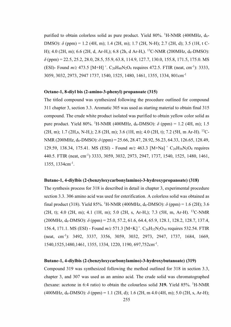
255
purified to obtain colorless solid as pure product. Yield 80%. 1H-NMR (400MHz, d6-
DMSO): δ (ppm) = 1.2 (4H, m); 1.4 (2H, m); 1.7 (2H, N-H); 2.7 (2H, d); 3.5 (1H, t C-
H); 4.0 (2H, m); 6.6 (2H, d, Ar-H,); 6.8 (2h, d Ar-H,). 13C-NMR (200MHz, d6-DMSO):
δ (ppm) = 22.5, 25.2, 28.0, 28.5, 55.9, 63.8, 114.9, 127.7, 130.0, 155.8, 171.5, 175.0. MS
(ESI)- Found m/z 473.5 [M+H] +. C20H40N2O4 requires 472.5. FTIR (neat, cm-1): 3333,
3059, 3032, 2973, 2947 1737, 1540, 1525, 1480, 1461, 1355, 1334, 801cm-1
Octane-1, 8-diyl bis (2-amino-3-phenyl) propanoate (315)
The titled compound was synthesized following the procedure outlined for compound
311 chapter 3, section 3.3. Aromatic 305 was used as starting material to obtain final 315
compound. The crude white product isolated was purified to obtain yellow color solid as
pure product. Yield 80%. 1H-NMR (400MHz, d6-DMSO): δ (ppm) = 1.2 (4H, m); 1.5
(2H, m); 1.7 (2H,s, N-H,); 2.8 (2H, m); 3.6 (1H, m); 4.0 (2H, t); 7.2 (5H, m Ar-H). 13C-
NMR (200MHz, d6-DMSO): δ (ppm) = 25.66, 28.47, 28.92, 56.23, 64.33, 126.65, 128.49,
129.59, 138.34, 175.41. MS (ESI) - Found m/z 463.3 [M+Na] + C26H36N2O4 requires
440.5. FTIR (neat, cm-1) 3333, 3059, 3032, 2973, 2947, 1737, 1540, 1525, 1480, 1461,
1355, 1334cm-1.
Butane-1, 4-diylbis (2-(benzyloxycarbonylamino)-3-hydroxypropanoate) (318)
The synthesis process for 318 is described in detail in chapter 3, experimental procedure
section 3.3. 306 amino acid was used for esterification. A colorless solid was obtained as
final product (318). Yield 85%. 1H-NMR (400MHz, d6-DMSO): δ (ppm) = 1.6 (2H); 3.6
(2H, t); 4.0 (2H, m); 4.1 (1H, m); 5.0 (2H, s, Ar-H,); 7.3 (5H, m, Ar-H). 13C-NMR
(200MHz, d6-DMSO): δ (ppm) = 25.0, 57.2, 61.6, 64.4, 65.9, 128.1, 128.2, 128.7, 137.4,
156.4, 171.1. MS (ESI) - Found m/z 571.3 [M+K] +. C26H32N2O10 requires 532.54. FTIR
(neat, cm-1): 3492, 3337, 3356, 3059, 3032, 2973, 2947, 1737, 1684, 1669,
1540,1525,1480,1461, 1355, 1334, 1220, 1190, 697,752cm-1.
Butane-1, 4-diylbis (2-(benzyloxycarbonylamino)-3-hydroxybutanoate) (319)
Compound 319 was synthesized following the method outlined for 318 in section 3.3,
chapter 3, and 307 was used as an amino acid. The crude solid was chromatographed
(hexane: acetone in 6:4 ratio) to obtain the colourless solid 319. Yield 85%. 1H-NMR
(400MHz, d6-DMSO): δ (ppm) = 1.1 (2H, d); 1.6 (2H, m 4.0 (4H, m); 5.0 (2H, s, Ar-H);

256
7.3 (5H, m, Ar-H). 13C-NMR (200MHz, d6-DMSO): δ (ppm) = 20.5, 25.0, 60.6, 64.3,
66.0, 66.8, 128.0, 128.2, 128.7, 137.3, 156.8, 171.2. MS (ESI)- Found m/z 583.3 [M+ Na]
+.C28H36N2O10 requires 560.59. FTIR (neat, cm-1): 3395, 3059, 3032, 2973, 2947, 1737,
1684, 1669, 1540, 1525, 1480, 1461, 1355, 1334, 1220, 1190, 697, 752cm-1.
Butane-1, 4-diylbis (2-(benzyloxycarbonylamino)-3(4-hydroxyphenyl) propanoate)
(320)
Compound 320 was synthesized following the method outlined for 318 in section 3.3,
chapter 3. This compound is based on 308 as starting material. The crude product was
purified by column chromatography (hexane: acetone in 6:4 ratio) and obtain a colourless
solid. Yield 85%. 1H-NMR (400MHz, d6-DMSO): δ (ppm) = 1.3 (2H, m); 2.8 (2H, dd,
Ar-H); 3.9 (2H, m); 4.1(IH, m N-H); 5.0 (2H, s Ar-H); 6.6 (2H, d); 7.0 (2H, d); 7.3 (5H,
m). 13C-NMR (200MHz, d6-DMSO): δ (ppm) = 24.9, 36.2, 56.4, 64.3, 65.8, 115.4, 127.7,
127.9, 128.1, 128.7, 130.4, 137.3, 156.3, 156.4, 172.4. MS (ESI)- Found m/z 708.3 [M+
Na] +.C38H40N2O10 requires 684.73. FTIR (neat, cm-1) 3333, 3059, 3032, 2973, 2947,
1737, 1684, 1669, 1540, 1525, 1480, 1461, 1355, 1334, 1220, 1190, 801cm-1.

257
Appendix B
Series 1 PU - 1H-NMR and FTIR Spectroscopy data
PCL-1-Z-Ser-PU: 1H- NMR (400MHz, DMSO-d6): δ(ppm) = 0.8 – 0.9 (-CH3- in
neopentyl glycol in PCL), 1.1 – 1.5 (-CH2- in PCL, serine chain extender), 2.24 (-CH2-
in PCL), 3.7-3.8 (-CH2- PCL, MDI), 4.0 - 4.2 (-CH2-C=O PCL, serine chain extender and
-CH- α proton of amino acid), 4.4 (-CH2- in side chain of serine chain extender), 5.02 (-
CH2- ϕ in Z- group of amino acid), 7.0 (-C6H4- MDI), 7.3 (-C6H4- MDI, Z-group), 7.8
(N-H- urethane link).
FTIR (neat, cm-1) = 3345, 3336, 2907, 2867, 2857, 2948, 1731, 1699, 1532, 1220, 1167,
1412, 1083, 1066, 697.
PCL-1-Z-Thr-PU: 1H-NMR (400MHz, DMSO-d6): δ(ppm) = δ 0.8 – 0.9 (-CH3- in
neopentyl glycol in PCL), 1.1 – 1.5 (-CH2- in PCL, threonine chain extender), 2.2 (-CH2-
in PCL), 3.7-3.8 (-CH2- PCL, MDI), 4.0 -4.2 (-CH2-C=O PCL, threonine chain extender
and -CH- α proton of amino acid), 4.5 (-CH2- in side chain of threonine based chain
extender), 5.03 (-CH2- in Z- group of amino acid), 7.2 (-C6H4- MDI), 7.4 (-C6H4- MDI,
Z-group), 8.0 (-N-H- urethane link).
FTIR (neat, cm-1) = 3345, 3336, 2907, 2867 2857, 2948, 1731 1699 1532, 1412, 1083,
1066, 697.
PCL-1-BDO-PU: 1H-NMR (400MHz, DMSO-d6): δ(ppm) = δ 0.8 – 0.9 (-CH3- in
neopentyl glycol in PCL, m-CH2- BDO), 1.1 – 1.5 (-CH2- in PCL), 2.2 (-CH2- in PCL),
3.7-3.8 (-CH2- PCL, MDI), 4.0-4.2 (-CH2-C=O PCL), 4.5 (-CH2-C=O PCL,-CH2-
terminal BDO), 7.2 (-C6H4- MDI), 7.4 (-C6H4- MDI), 8.0 (-N-H- urethane link).
FTIR (neat, cm-1) = 3339, 3330, 3191, 3125, 2924, 2854, 1735, 1597, 1530, 1448, 1365
1239, 1060 1067, 1311.

258
Series 1 PUU - 1H-NMR and FTIR Spectroscopy data
PCL-1-Leu-PUU: 1H- NMR (400MHz, DMSO-d6): δ(ppm) = δ 0.8 – 0.9 (-CH3- in
neopentyl glycol in PCL, d- CH3 of leucine chain extender), 1.1 – 1.5 (-CH2- in PCL, m,-
CH2, -CH- leucine chain extender), 2.2 (-CH2- in PCL), 3.5-3.6 (-CH2- PCL, MDI), 4.0 -
4.2 (-CH2-C=O PCL, leucine chain extender and -CH- α proton of amino acid), 6.2 (N-H
amino acid-urethane proton), 7.1 (-C6H4- MDI), 7.2 (-C6H4- MDI), 8.4 (-N-H- urethane
link).
FTIR (neat, cm-1) = 3339, 3330, 3191, 3125 2924, 2854 1735, 1645, 1597, 1530, 1448,
1365, 1239, 1060, 1067, 1311.
PCL-1-Ileu-PUU: 1H- NMR (400MHz, DMSO-d6): δ(ppm) = δ 0.8 – 0.9 (-CH3- in
neopentyl glycol in PCL, d- CH3 of isoleucine chain extender), 1.2 – 1.6 (-CH2- in PCL,
m,-CH2, -CH- isoleucine chain extender), 2.3 (-CH2- in PCL), 3.7-3.8 (-CH2- PCL, MDI),
4.0 -4.2 (-CH2-C=O PCL, leucine chain extender and -CH- α proton of amino acid), 6.4
(N-H- amino acid-urethane proton), 7.0 (-C6H4- MDI), 7.3 (-C6H4- MDI), 8.5 (-N-H-
urethane link).
FTIR (neat, cm-1) = 3339, 3330, 3191, 3125 2924, 2854 1735 1645, 1597, 1530, 1239,
1060.
PCL-1-Val-PUU : 1H-NMR (400MHz, DMSO-d6): δ(ppm) = δ 0.8 – 0.9 (-CH3- in
neopentyl glycol in PCL, d- CH3 of valine chain extender), 1.3 – 1.5 (-CH2- in PCL, m,-
CH2, -CH- valine chain extender), 2.2 (-CH2- in PCL), 3.7-3.8 (-CH2- PCL, MDI), 4.0 -
4.2 (-CH2-C=O PCL, valine chain extender and -CH- α proton of amino acid), 6.2 (N-H-
amino acid-urethane proton), 7.0 (-C6H4- MDI), 7.3 (-C6H4- MDI), 8.4 (-N-H- urethane
link).
FTIR (neat, cm-1) = 3339, 3330, 3191, 3125, 2924, 2854, 1735, 1645, 1597, 1530, 1448,
1365, 1239, 1060, 1067, 1311.
PCL-1-Tyr-PUU : 1H-NMR (400MHz, DMSO-d6): δ(ppm) = δ 1.1 – 1.3 (-CH3- in
neopentyl glycol in PCL, d- CH2 of tyrosine chain extender), 1.4 – 1.4 (-CH2- in PCL, m,-
CH2, tyrosine chain extender), 2.2 (-CH2- in PCL), 2.8 (-CH2- benzyl proton tyrosine),
259
3.7-3.8 (-CH2- PCL, MDI), 4.0 -4.2 (-CH2-C=O PCL, tyrosine chain extender and -CH-
α proton of amino acid), 6.2 (N-H- amino acid-urethane proton), 6.2-6.9 (-C6H4-
Tyrosine), 7.0 (-C6H4- MDI), 7.3 (-C6H4- MDI), 8.3 (-N-H- urethane link).
FTIR (neat, cm-1) = 3339, 3330, 3191, 3125, 2924, 2854, 1735, 1645, 1597, 1530, 1448,
1365, 1239, 1060 1067, 1311.

![Fisa de produs SX4 S-Cross 16 - Autonet Suzuki · 2019-09-18 · s ] µ v ] Z ] } ] v W ^^/KE z z z z z z z z z z z z z z z z z z z z z z z z z z z z z z z z z z z z z z z z z z z](https://img.pdfslide.us/doc/110x75/5e9311f76a68671aec7ec141/fisa-de-produs-sx4-s-cross-16-autonet-suzuki-2019-09-18-s-v-z-v.jpg)


![National Biodiesel Board [Read-Only] - Montana …leg.mt.gov/.../energy_group/staffmemos/biodiesel_board.pdfBiodiesel is already less than 15 ppm sulfur content ... France 1 Cognis/Diester](https://img.pdfslide.us/doc/110x75/5adb4d1e7f8b9add658dc05c/national-biodiesel-board-read-only-montana-legmtgovenergygroupstaffmemosbiodieselboardpdfbiodiesel.jpg)

![ADMISSION LETTER 2018...3DJH RI :HEVLWH ZZZ KHOE FR NH $'0,66,21 /(77(5 W } v o ] o E u W z z z z z z z z z z z z z z z z z z z z z z z z z z z z z z z z z z z z z z 3DJH RI ð î](https://img.pdfslide.us/doc/110x75/5ff2e8f32328856d162b0400/admission-letter-3djh-ri-hevlwh-zzz-khoe-fr-nh-06621-775-w-v-o-o.jpg)


![D ] v P µ o ] v P v ] v P ô ñ l ð u X ( o U ] l o ] Z Ç ( o · z z z z z z z z z z z z z z z z z z z z z z z z z z z z z z z z z z z z z z z z z z z z z z z z z z z z z z z z](https://img.pdfslide.us/doc/110x75/5f2b2b7f34c1dd164151f33c/d-v-p-o-v-p-v-v-p-l-u-x-o-u-l-o-z-o-z-z-z-z-z-z-z-z.jpg)
![INNOCrodaChromaveil Article a Diester to Protect Hair From Color Fade and Sun Damage May 2009[1]](https://img.pdfslide.us/doc/110x75/55cf94cd550346f57ba479ab/innocrodachromaveil-article-a-diester-to-protect-hair-from-color-fade-and-sun.jpg)

![6688==88..,, 66:::,,))777 - Autonet Suzuki · s ] µ v ] Z ] } ] v KK> z z z z z z z z z z z z z z z z z z z z z z z z z z z z z z z z z z z z z z z z z z z z z z z z z z z z z z](https://img.pdfslide.us/doc/110x75/5e9312c274650c20c60d46b4/668888-66777-autonet-suzuki-s-v-z-v-kk-z-z-z-z-z.jpg)