Embed Size (px)
Citation preview
Andrews University Andrews University
Digital Commons @ Andrews University Digital Commons @ Andrews University
Honors Theses Undergraduate Research
2012
Developing an Intracellular Neurobiology Lab Sequence for Use in Developing an Intracellular Neurobiology Lab Sequence for Use in
an Interdisciplinary Neurobiology Course an Interdisciplinary Neurobiology Course
Aleksandra Kozlova-Harris
Follow this and additional works at: https://digitalcommons.andrews.edu/honors
Recommended Citation Recommended Citation Kozlova-Harris, Aleksandra, "Developing an Intracellular Neurobiology Lab Sequence for Use in an Interdisciplinary Neurobiology Course" (2012). Honors Theses. 33. https://digitalcommons.andrews.edu/honors/33
This Honors Thesis is brought to you for free and open access by the Undergraduate Research at Digital Commons @ Andrews University. It has been accepted for inclusion in Honors Theses by an authorized administrator of Digital Commons @ Andrews University. For more information, please contact [email protected].

Thank you for your interest in the
Andrews University Digital Library
Please honor the copyright of this document by
not duplicating or distributing additional copies
in any form without the author’s express written
permission. Thanks for your cooperation.
Abstract:
The goal of my research was to evaluate procedures currently used in the Intracellular
Leech Lab in order to modify the Neurobiology ZOOL475 Lab to increase the rate of successful
intracellular recordings for future Neurobiology students. Two approaches were used: the first
was to evaluate the possibility of using a less expensive leech species in order to allow students
unlimited attempts at recording from ganglia without budget constraints. The second was to
evaluate various raising conditions (water, temperature, and age) to optimize the quality of the
leech ganglia. Both approaches provided useful information to improve lab success.
Introduction:
The concept for this project stemmed from an attempt to improve Andrews University’s
Neurobiology Lab. Historically, undergraduate level Neurobiology courses often did not offer
laboratory sections to accompany their classes. Due to a grant from the National Science
Foundation, Andrews University became one of the first universities to offer a laboratory course
at the undergraduate level.
In recent years, the demographics of the students taking Neurobiology at Andrews University
has changed - the enrolees were primarily upper division Biology majors, but currently there are
both upper and lower division students from the Biology and Psychology Departments. The class
size has likewise increased from 2 students in 2008 to 15 enrolled students in 2011. As the
demographics have altered, so has the lab. No manuals for an interdisciplinary Neurobiology Lab
exist, thus one has been composed by Dr. Atkins. At the end of the Neurobiology Lab Sequence,
there are a series of 3 labs entitled “Intracellular recordings from Leech neurons” in which the
students attempt to obtain and record and intracellular action potential recordings from a leech
ganglion.
The reason that the Hirudo medicinalis leech is the species of choice for these labs is in its
use in both research and medicine – doctors use leeches post-operatively for skin grafts,
replantation of the digits, periorbital hematomas, and in a variety of other surgeries to restore
blood flow and reduce inflammation through leeching (Mory et al. 2000). Researchers chose
leeches based on the rhythmic dorsoventral contraction that is involved in their swimming
pattern, which they used in order to create a neuronal model for behavior (Chen, et al. 2010). H.
medicinalis became a popular research animal because of the abundance and bilateral,
homologous, and linear positioning of the ganglia within each organism (Ort, et al. 1974).
Leeches are likewise popular specimens to use in Neurobiology, due to the large size and great
number of ganglia enclosed within each animal.
The nervous system of the leech is laid out in a series of connected ganglia. One ganglion
contains a bundle of neurons, from which one can measure action potentials. The leech is also
the species of choice for intracellular labs due to the opacity of their ganglia, which makes the
individual cells in the ganglion easy to identify under the dissection microscope (unlike the
cricket ganglia, which take much a higher resistance electrode to puncture the ganglion due to
the lesser opacity of their ganglia, which indicates a thicker ganglionic sheath). (Atkins &
Pollack, 1987). This is the only undergraduate lab at Andrews University in which the students
have the ability to record intracellular action potentials – all other recordings are extracellular.
The limitations of this particular lab at Andrews University have been the high cost of the
equipment and supplies. The cost of one medicinal leech, which previous years have used, is
$20.00 plus shipping, while a Common leech costs $4.00 plus shipping. Thus, if the ganglion
opacity and sheath thickness is similar for a Common Leech, subsequent Neurobiology classes
could use the cheaper species, which would allow more Common Leeches to be bought, in turn
giving students more ganglia to practice on for the same cost as having fewer Medicinal Leech
ganglia.
Yet another limitation in the Neurobiology Lab is the electrode puller. The settings for the
electrode puller (both Heat and Solenoid settings) need to be within a narrow, specified range in
order to pull intracellular electrodes under the desired resistance range of 30-50 MΩ (Atkins Lab
Manual). This desired range of resistance indicates an electrode tip width that is sharp enough to
puncture the ganglion sheath yet is narrow enough not to break the cells.
Leech care conditions have led to difficulties in obtaining “neuronally healthy” leeches that
have opaque ganglia that are not excessively rubbery. Qualitative observations (“hunches”) by
Dr. Atkins in previous years of leech care seem to indicate differences in ganglion sheath
thickness and opacity due to differences in: age, water, and temperature conditions.
Due to the difficulties encountered in recent years in obtaining data, I tested a series of
variables within the parameters of the laboratory setup in an attempt to increase the number of
successful intracellular leech recordings. My main goal in this lab was to ask: How can we
improve the success rate of data collection for the Neurobiology Intracellular Leech Lab? To
answer this question, I asked specific sub- questions: What are the optimal Pipette Puller settings
to pull electrodes with the necessary resistance range (30-50 MΩ)? Is there a difference in
ganglia consistency between Medicinal and Common Leech Species? What are the optimal
raising conditions to obtain neuronally “healthy” leech ganglia?
Once I began my experiment, I discovered that the electrode puller displayed great variability
between individual electrodes pulled under the same setting. Pulling multiple electrodes in series
changed the shape of the coil and the puller oftentimes stopped pulling for some amount of time.
Multiple pulls within a short interval changed the production of heat and pull by the same heat
and solenoid settings. One electrode coil completely burnt out during the pulling process, and the
settings had to be recalibrated to pull within the desired resistance range. There was very little
repeatability in the electrode pulls; the standard deviations were high, indicating that there was
not an optimal combination of heat and solenoid settings that would consistently pull at the
desired resistance range.
Another issue I ran into was the difficulty of obtaining intracellular recordings. The time it
took to obtain a single intracellular recording from a ganglion was 4 hours, disregarding
preparation and dissection time. In order to obtain an intracellular recording for 3 ganglia from
36 leeches, totaling 108 ganglia, I would have had to spend 432 hours at the lab. Thus, my
methodology was changed from obtaining intracellular recordings to an indirect measure of
ganglion sheath thickness through a comparison of opacity. Research shows that thicker, less
penetrable ganglion sheaths are associated with less opaque ganglia (Atkins, et al. 1987; Muller,
et al. 1981). Thus, by testing opacity (optical density) via a photographic method, one will be
able to infer penetrability (neuronal “healthiness”) of the leech ganglia.
The rationale for my new procedure involved taking 10 leech ganglia recordings from 1 leech
since it is not cost effective to test 1 ganglion from 10 separate leeches under each condition
(Approximate total cost: $3600 for Medicinalis and $720 for Common Leech = $4320 total). I
tested for opacity by keeping F-stop the same and recording shutter speed. As a backup method
of analysis I kept the F-stop and shutter speed the same and took photographs to compare
opacity.
Methodology
Initial Methodology
Electrode Condition:
First, I pulled electrodes under a series of heat settings and solenoid settings. My range of
heat settings varied from 18.8 to 19.1 and my solenoid settings ranged from 3.0-4.0, by
gradations of 0.5. Once the electrodes were pulled, I connected each electrode to the intracellular
recording apparatus. Then, I would conduct an Ohm’s test in order to measure the resistance of
the electrode to verify whether that particular electrode’s resistance was in the desired 30-50 MΩ
range. The electrode settings that were not in the desired range were deemed undesirable settings
for Neurobiology lab while the electrode settings that were within the desired range were deemed
to be the optimal settings for Neurobiology lab.
Leech Care Condition:
The initial methodology involved caring for leeches under a series of varying raising
conditions, for temperature, age, and water. The temperature conditions were: 4°C, 10°C, and
21°C. The age conditions were old (3-4 weeks) and young (1-2 weeks). The water conditions
were: lab-prepared solution (Basic stock solution per liter: 2.8 g NaCl, 0.05 g KCl, 0.08 g
Ca(NO3)2 • 4 H2O, 0.025 g MgSO4 • 7 H2O, 0.56 g Tris Buffer (pH to 7.4), the store-bought
freshwater solution, and the Carolina® Biological Supply Company water in which the leeches
were shipped (Muller, et al., 1981). One H. medicinalis leech and one Common leech would be
raised under each combination of the 3 conditions, resulting in 9 young H. medicinalis leeches, 9
old H. medicinalis leeches, 9 young Common leeches, and 9 old Common leeches. Next, I
dissected each of the leeches and removed 3 ganglia from each leech. Each ganglion was pinned
down, the electrophysiology equipment was set up, and an electrode with a proper range of
resistance was used to puncture through the sheath of the ganglion. Then, the electrode
placement was manipulated until an intracellular action potential was recorded on the LabChart®
Program. Finally, I would conduct statistical analysis to find which leech care conditions and
which species of leech obtained the most intracellular recordings, to determine which conditions
were optimal to suggest for use in subsequent Neurobiology Labs. Due to time constraints and
the difficulty of obtaining intracellular recordings, I was not able to continue this initial
methodology past the attempts at obtaining intracellular recordings. Thus, I conducted a different
methodology, to evaluate optimal leech species and care condition, using an indirect measure of
ganglion sheath permeability.
Alternate Methodology
Leech Dissection Condition:
Following the period of leech care under varying conditions (found in the initial methodology), I
dissected each of the 36 leeches, and extracted 10 ganglia from each leech. Then, I placed the
ganglia under the microscope lens, with a drop of saline, and viewed each of the 10 ganglia
under the light microscope. I took a photograph, using an underwater camera, by keeping the F-
stop the same (F-stop: 4.4), with the dependent variable being the shutter speed, which I then
recorded. Then, I converted the shutter speed fraction to a decimal, which indirectly indicated the
optical density. As a backup method of analysis, I took a second photograph of each ganglion,
this time keeping the F-stop and shutter speed constant (F-stop: 4.4, Shutter Speed: 1/125), in
order to compare the opacity of the photographs in the Lightroom® program. I analyzed the data
from the shutter speed fractions, conducting T-tests for age and species, one-way ANOVA’s for
average electrode resistances, the variability between 3 Leeches under the same condition, slide

preparation wait time, temperature conditions, and water conditions, and a Chi-square test for
Leech mortality under varying water conditions.
Results:
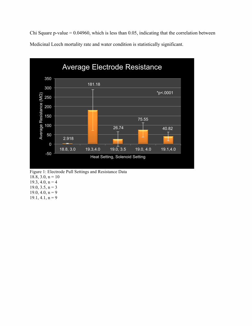
The average resistances for the combined heat and solenoid settings for the electrode
puller were (Fig. 1): Heat: 18.8, Solenoid: 3.0; 2.918 MΩ with a standard deviation of: 2.49.
Heat: 19.0, Solenoid: 3.5; 26.74 MΩ with a standard deviation of: 38.04. Heat: 19.0, Solenoid:
4.0; 75.55 MΩ with a standard deviation of: 38.06. Heat: 19.1, Solenoid: 4.0; 40.82 MΩ with a
standard deviation of: 22.87. Heat: 19.3, Solenoid: 4.0; 181.18 MΩ with a standard deviation of:
109.32. +Even though two of the electrode settings, of Heat 19.0 and Solenoid 4.0 and Heat 19.1
and Solenoid 4.0, had average resistances within the desired range, the standard deviations of the
averages were too high, indicating inconsistent electrode pulls.
Next, I tested the control of variability within 1 set of care conditions (Fig. 2):
Condition - Species: H. medicinalis Leech; Water: Carolina Shipment; Temperature: 10°C.
Mean: 0.00668 with a standard deviation of: 0.000406. The standard deviation range was small,
indicating little variability within one set of care conditions.
I conducted a comparison of Mean Shutter Speed Comparison Between H. medicinalis
and Common Leech Species (Fig. 3): the Common Leech mean shutter speed: 0.01761 with a
standard deviation of: 0.01021. The H. medicinalis mean shutter speed: 0.00807 with a standard
deviation of: 0.00186. I conducted a 2-tailed T-test p-value: 0.000488, which is less than p =
0.05, indicating a statistically significant difference between leech species.
I analyzed the Mean Shutter Speed Comparison of varying intervals of time between
leech dissection and slide preparation of H. medicinalis (Fig. 4): The mean shutter speed for 0
Hours: 0.007285 with a standard deviation of: 0.00124. The mean shutter speed for 2-4 Hours:
0.00887 with a standard deviation of: 0.00183. The mean shutter speed for 6-8 Hours: 0.00724
with a standard deviation of: 0.00170. The p-value for the ANOVA between the three time
intervals was: 0.33264, which is greater than p = 0.05. The difference between wait intervals was
not statistically significant.
The comparison of Mean Shutter Speeds of Varying Temperature Conditions for
Common Leech (Fig. 5): Mean shutter speed for 4°C: 0.01267 with a standard deviation of:
0.00673. The mean shutter speed for 10°C: 0.02009 with a standard deviation of: 0.01092. The
mean shutter speed for 21°C: 0.02127 with a standard deviation of: 0.01154. The p-value for the
ANOVA of 0.43451 is greater than 0.05, indicating no significant difference in shutter speed of
Common Leech ganglia under varying temperature conditions.
The comparison of Mean Shutter Speeds of Varying Temperature Conditions for
Medicinal Leech (Fig. 6): Mean shutter speed for 4°C: 0.00773 with a standard deviation of:
0.00249. The mean shutter speed for 10°C: 0.00806 with a standard deviation of: 0.00208. The
mean shutter speed for 21°C: 0.00832 with a standard deviation of: 0.00171. The p-value for the
ANOVA of 0.84517 is greater than 0.05, indicating no significant difference in shutter speed of
Medicinal Leech ganglia under varying temperature conditions.
The comparison of Mean Shutter Speeds of Varying Age Conditions for Common Leech
(Fig. 7): Mean shutter speed for old leeches: 0.02128 with a standard deviation of: 0.01317. The
mean shutter speed for young leeches: 0.01145 with a standard deviation of: 0.00425. The p-
value for the 2-tailed T-test was 0.15839, which is greater than 0.05, indicating no significant
difference in shutter speed of Common Leech ganglia under varying age conditions.
The comparison of Mean Shutter Speed Comparison of Varying Age Conditions for
Medicinal Leech (Fig. 8): Mean shutter speed for old leeches: 0.00804 with a standard deviation
of: 0.00179. The mean shutter speed for young leeches: 0.00794 with a standard deviation of:
0.00201. The p-value for the 2-tailed T-test was 0.91053, which is greater than 0.05, indicating
no significant difference in shutter speed of Medicinal Leech ganglia under varying age
conditions.
The Mean Shutter Speed Comparison of Varying Water Conditions for Common Leech
(Fig. 9): Mean shutter speed for Store Bought Solution: 0.01988 with a standard deviation of:
0.01696. The mean shutter speed for Lab Prepared Solution: 0.02024 with a standard deviation
of: 0.00358. The mean shutter speed for Carolina Supply Company Solution: 0.01610 with a
standard deviation of: 0.01141. The p-value for the ANOVA was 0.79064, which is greater than
0.05, indicating no significant difference in shutter speed of Common Leech ganglia under
varying water conditions.
The Mean Shutter Speed Comparison of Varying Water Conditions for Medicinal Leech
(Fig. 10): Mean shutter speed for Store Bought Solution: 0.00767 with a standard deviation of:
0.00097. The mean shutter speed for Lab Prepared Solution: 0.00797 with a standard deviation
of: 0.00177. The mean shutter speed for Carolina Supply Company Solution: 0.00810 with a
standard deviation of: 0.00216. The p-value for the ANOVA was 0.96089, which is greater than
0.05, indicating no significant difference in shutter speed of Medicinal Leech ganglia under
varying water conditions.
I conducted a comparison of Leech Mortality Rate vs. Water Condition for Common
Leech (Fig. 11): Store Bought: 5 dead; Lab Prepared: 1 dead; Carolina Supply Company: 0 dead.
Chi Square p-value = 0.03010, which is less than 0.05, indicating that the correlation between
Common Leech mortality rate and water condition is statistically significant.
I conducted a comparison of Leech Mortality Rate vs. Water Condition for Medicinal
Leech (Fig. 11): Store Bought: 3 dead; Lab Prepared: 0 dead; Carolina Supply Company: 0 dead.
Chi Square p-value = 0.04960, which is less than 0.05, indicating that the correlation between
Medicinal Leech mortality rate and water condition is statistically significant.
Figure 1: Electrode Pull Settings and Resistance Data 18.8, 3.0, n = 10 19.3, 4.0, n = 4 19.0, 3.5, n = 3 19.0, 4.0, n = 9 19.1, 4.1, n = 9
-50
0
50
100
150
200
250
300
350
18.8, 3.0 19.3,4.0 19.0, 3.5 19.0, 4.0 19.1,4.0
Aver
age
Res
ista
nce
(MΩ
)
Heat Setting, Solenoid Setting
Average Electrode Resistance
2.918
26.74
75.55
40.82
*p<.0001
181.18
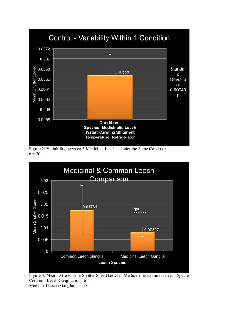
Figure 2: Variability between 3 Medicinal Leeches under the Same Condition n = 30
Figure 3: Mean Difference in Shutter Speed between Medicinal & Common Leech Species Common Leech Ganglia, n = 16 Medicinal Leech Ganglia, n = 18
0.00668
0.0058
0.006
0.0062
0.0064
0.0066
0.0068
0.007
0.0072
Mea
n S
hutte
r Spe
ed
Condition - Species: Medicinalis Leech Water: Carolina Shipment Temperature: Refrigerator
Control - Variability Within 1 Condition
Standard
Deviation:
0.000406
0.01761
0.00807
0
0.005
0.01
0.015
0.02
0.025
0.03
Common Leech Ganglia Medicinal Leech Ganglia
Mea
n S
hutte
r Spe
ed
Leech Species
Medicinal & Common Leech Comparison
*p=.000488
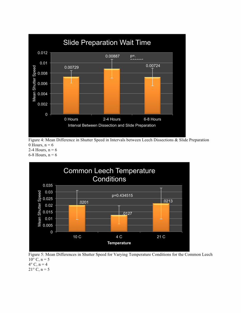
Figure 4: Mean Difference in Shutter Speed in Intervals between Leech Dissections & Slide Preparation 0 Hours, n = 6 2-4 Hours, n = 6 6-8 Hours, n = 6
Figure 5: Mean Differences in Shutter Speed for Varying Temperature Conditions for the Common Leech 10° C, n = 5 4° C, n = 4 21° C, n = 5
0.00729
0.00887
0.00724
0
0.002
0.004
0.006
0.008
0.01
0.012
0 Hours 2-4 Hours 6-8 Hours
Mea
n S
hutte
r Spe
ed
Interval Between Dissection and Slide Preparation
Slide Preparation Wait Time
p=.960885
0
0.005
0.01
0.015
0.02
0.025
0.03
0.035
10 C 4 C 21 C
Mea
n S
hutte
r Spe
ed
Temperature
Common Leech Temperature Conditions
.0127
.0201 .0213 p=0.434515
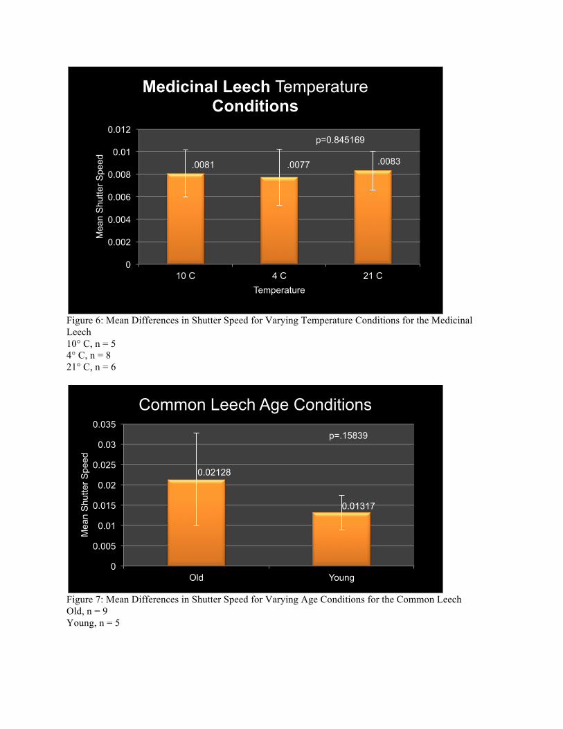
Figure 6: Mean Differences in Shutter Speed for Varying Temperature Conditions for the Medicinal Leech 10° C, n = 5 4° C, n = 8 21° C, n = 6
Figure 7: Mean Differences in Shutter Speed for Varying Age Conditions for the Common Leech Old, n = 9 Young, n = 5
0
0.002
0.004
0.006
0.008
0.01
0.012
10 C 4 C 21 C
Mea
n S
hutte
r Spe
ed
Temperature
Medicinal Leech Temperature Conditions
.0081 .0077 .0083
p=0.845169
0.02128
0.01317
0
0.005
0.01
0.015
0.02
0.025
0.03
0.035
Old Young
Mea
n S
hutte
r Spe
ed
Common Leech Age Conditions
p=.15839
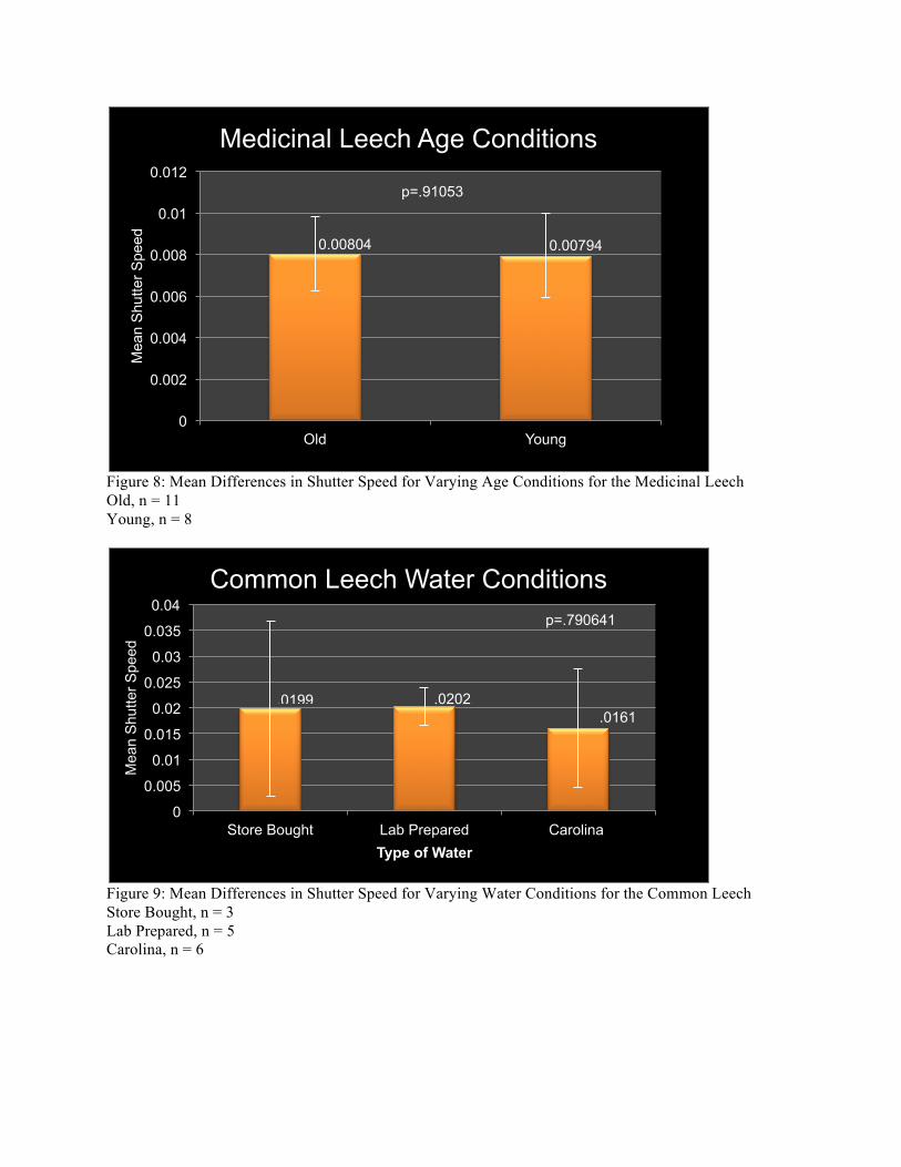
Figure 8: Mean Differences in Shutter Speed for Varying Age Conditions for the Medicinal Leech Old, n = 11 Young, n = 8
Figure 9: Mean Differences in Shutter Speed for Varying Water Conditions for the Common Leech Store Bought, n = 3 Lab Prepared, n = 5 Carolina, n = 6
0.00804 0.00794
0
0.002
0.004
0.006
0.008
0.01
0.012
Old Young
Mea
n S
hutte
r Spe
ed
Medicinal Leech Age Conditions
p=.91053
0
0.005
0.01
0.015
0.02
0.025
0.03
0.035
0.04
Store Bought Lab Prepared Carolina
Mea
n S
hutte
r Spe
ed
Type of Water
Common Leech Water Conditions
.0199 .0202
p=.790641
.0161
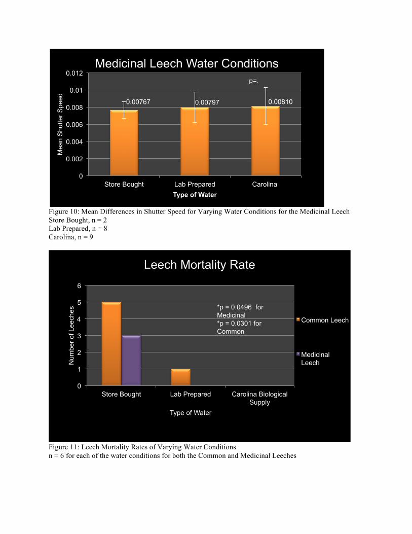
Figure 10: Mean Differences in Shutter Speed for Varying Water Conditions for the Medicinal Leech Store Bought, n = 2 Lab Prepared, n = 8 Carolina, n = 9
Figure 11: Leech Mortality Rates of Varying Water Conditions n = 6 for each of the water conditions for both the Common and Medicinal Leeches
0.00767 0.00797 0.00810
0
0.002
0.004
0.006
0.008
0.01
0.012
Store Bought Lab Prepared Carolina
Mea
n S
hutte
r Spe
ed
Type of Water
Medicinal Leech Water Conditions p=.960885
0
1
2
3
4
5
6
Store Bought Lab Prepared Carolina Biological Supply
Num
ber o
f Lee
ches
Type of Water
Leech Mortality Rate
Common Leech
Medicinal Leech
*p = 0.0496 for Medicinal *p = 0.0301 for Common
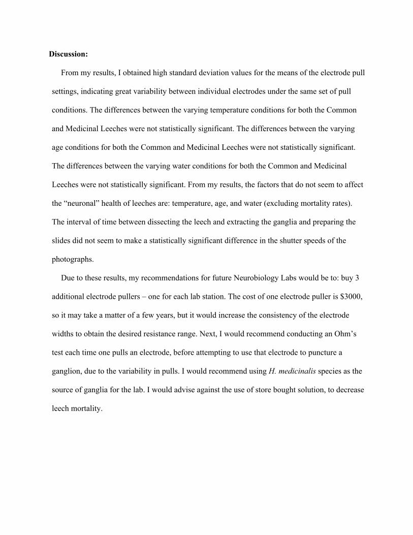
Discussion:
From my results, I obtained high standard deviation values for the means of the electrode pull
settings, indicating great variability between individual electrodes under the same set of pull
conditions. The differences between the varying temperature conditions for both the Common
and Medicinal Leeches were not statistically significant. The differences between the varying
age conditions for both the Common and Medicinal Leeches were not statistically significant.
The differences between the varying water conditions for both the Common and Medicinal
Leeches were not statistically significant. From my results, the factors that do not seem to affect
the “neuronal” health of leeches are: temperature, age, and water (excluding mortality rates).
The interval of time between dissecting the leech and extracting the ganglia and preparing the
slides did not seem to make a statistically significant difference in the shutter speeds of the
photographs.
Due to these results, my recommendations for future Neurobiology Labs would be to: buy 3
additional electrode pullers – one for each lab station. The cost of one electrode puller is $3000,
so it may take a matter of a few years, but it would increase the consistency of the electrode
widths to obtain the desired resistance range. Next, I would recommend conducting an Ohm’s
test each time one pulls an electrode, before attempting to use that electrode to puncture a
ganglion, due to the variability in pulls. I would recommend using H. medicinalis species as the
source of ganglia for the lab. I would advise against the use of store bought solution, to decrease
leech mortality.
References
ATKINS, Neurobiology Lab Syllabus and Intracellular Leech Lab Instruction Packets. ATKINS, G. & POLLACK, G.S. (1987). Response properties of prothoracic, interganglionic,
sound-activated interneurons in the cricket Teleogryllus oceanicus. Journal of Comparative Physiology A: Neuroethology, Sensory, Neural, and Behavioral Physiology. Vol. 161, No. 5, 681-693.
DEITMER, J. W. & SCHLUE, W. R. (1981). Measurements of the intracellular potassium
activity of Retzius cells in the leech central nervous system. Journal of Experimental Biology 91, 87-101.
MIYAZAKI, S. & NICHOLLS, J.G. (1976). The Properties and Connections of Nerve Cells in
Leech Ganglia Maintained in Culture. Proceedings of the Royal Society of London, Series b, Biological Sciences Vol. 194, No. 1116, 295-311.
MULLER, K.J., NICHOLLS, J.G., & STENT, G.S. (1981). Neurobiology of the Leech. Cold
Spring Harbor Laboratory,1-285. ROOSENBERG, J. & RIGGS, H. (1994). Staining & Identification of Selected Neurons of
Leech Segmental Ganglia. Neurobiology Student Lab Project.
J. N. Andrews Honors Program Andrews University
Honors Thesis
Developing an Intracellular Neurobiology Lab Sequence for Use In An Interdisciplinary Neurobiology Course
Aleksandra Kozlova-Harris
May 5, 2012
Advisor: Dr. Gordon Atkins
Primary Advisor Signature: _________________
Department: ____________________________


![Regulation of the intracellular Ca2+. Regulation of intracellular [H]:](https://img.pdfslide.us/doc/110x75/5a4d1b717f8b9ab0599b56a5/regulation-of-the-intracellular-ca2-regulation-of-intracellular-h.jpg)