Embed Size (px)
Citation preview

Ka et al. Cell Death Discovery (2020) 6:34
https://doi.org/10.1038/s41420-020-0268-1 Cell Death Discovery
ART ICLE Open Ac ce s s
Deubiquitinase USP47-stabilized splicing factor IKregulates the splicing of ATM pre-mRNAHye In Ka1, Sunyi Lee2, Sora Han3, Ae Lee Jeong4, Ji Young Park1, Hyun Jeong Joo1, Su Jung Soh1, Doyeon Park1 andYoung Yang1
AbstractIK depletion leads to an aberrant mitotic entry because of chromosomal misalignment through the enhancement ofAurora B activity at the interphase. Here, we demonstrate that IK, a spliceosomal component, plays a crucial role in theproper splicing of the ATM pre-mRNA among other genes related with the DNA Damage Response (DDR). Intron 1 inthe ATM pre-mRNA, having lengths <200 bp, was not spliced in the IK-depleted cells and led to a deficiency of theATM protein. Subsequently, the IK depletion-induced ATM protein deficiency impaired the ability to repair thedamaged DNA. Because the absence of SMU1 results in IK degradation, the mechanism underlying IK degradation wasexploited. IK was ubiquitinated in the absence of SMU1 and then subjected to proteolysis through the 26Sproteasome. To prevent the proteolytic degradation of IK, a deubiquitinating enzyme, USP47, directly interacted withIK and stabilized it through deubiquitination. Collectively, our results suggest that IK is required for proper splicing ofthe ATM pre-mRNA and USP47 contributes toward the stabilization of IK.
IntroductionSpliceosomes are large complex molecular machines
that remove introns from transcribed pre-mRNAs. Spli-ceosomes, which are associated with more than 150accessory proteins, are formed from five different smallnuclear ribonucleoproteins (snRNP)1–5. In the first step ofsplicing, the U1 snRNP recognizes the 5′-splice site (SS)region and U2 snRNP is recruited to the branch site (BS)region by binding the U2 auxiliary factor (U2AF). TheTri-U4/U6·U5 snRNP is recruited to the U1 and U2assembly and the helicase Prp28 helps the displacement ofthe U1 snRNP at the 5′-splice site into the U6 snRNA.Simultaneously, B-specific proteins, including IK andSMU1, are recruited to the B complex6,7. This B complexis transformed into an activated spliceosome (Bact) byunwinding of the U4/U6 RNA duplex triggered by the
helicase Brr2. The remaining U6 snRNP interacts with theU2 snRNA. During the course of B activation, B-specificproteins, including IK and SMU1, are dissociated. BecauseIK and SMU1 have no orthologs in yeast, these twoproteins are considered to act as regulatory proteins8. Inaddition to this splicing function of IK and SMU1, IKplays a role in the cell cycle regulation, as reported pre-viously9. IK localizes to the spindle pole and colocalizeswith MAD1 at the spindle poles. The depletion of IK leadsto a failure of the spindle pole localization of MAD110.The IK depletion is also able to disrupt the interactionbetween Aurora B kinase and protein phosphatase 2(PP2A)11, leading to aberrant mitotic entry with chro-mosomal misalignment and chromosomal instability12,13.Posttranslational modification of spliceosomal proteins
allows the exact splicing of pre-mRNAs through dynamicregulation of protein interactions14,15. Presently, it isknown that the U4/U6.U5 tri-snRNP complex of spli-ceosome is subjected to ubiquitination for dynamic acti-vation of the spliceosome. The splicing factor, Prp8, acomponent of the U5 snRNP, is ubiquitinated withintriple snRNPs yielding the stabilization of the U4/U6-U5
© The Author(s) 2020OpenAccessThis article is licensedunder aCreativeCommonsAttribution 4.0 International License,whichpermits use, sharing, adaptation, distribution and reproductionin any medium or format, as long as you give appropriate credit to the original author(s) and the source, provide a link to the Creative Commons license, and indicate if
changesweremade. The images or other third partymaterial in this article are included in the article’s Creative Commons license, unless indicated otherwise in a credit line to thematerial. Ifmaterial is not included in the article’s Creative Commons license and your intended use is not permitted by statutory regulation or exceeds the permitted use, you will need to obtainpermission directly from the copyright holder. To view a copy of this license, visit http://creativecommons.org/licenses/by/4.0/.
Correspondence: Young Yang ([email protected])1Department of Biological Sciences, Sookmyung Women’s University, Seoul04310, Korea2Drug Evaluation Group, R&D Center CJ HealthCare, Icheon 04551, KoreaFull list of author information is available at the end of the articleThese authors contributed equally: Hye In Ka, Sunyi LeeEdited by: M. V. Niklison Chirou
Official journal of the Cell Death Differentiation Association
1234
5678
90():,;
1234
5678
90():,;
1234567890():,;
1234
5678
90():,;

triple snRNP by repressing the U4/U6 unwinding16.PRP31, a component of the U4 snRNP, is modified withK63-linked ubiquitin chains by the PRP19 complex and isdeubiquitinated by the ternary complex of USP15,SART3, and USP4. Thus, the ubiquitination and deubi-quitination status of PRP31 regulates its interaction withthe U5 snRNP component, PRP8, by stabilizing the U4/U6.U5 tri-snRNP complex17. Ubiquitin has seven lysineresidues. K11- or K48-linked ubiquitinated proteins arethe targets of degradation by 26S proteasome, whereasK63-linked ubiquitinated proteins mediateprotein–protein interaction instead of proteolysis18–20.Until now, little is known about K11- or K48-linkedubiquitination of a spliceosomal component which mightbe relevant to regulation of splicing.Activation of ataxia-telangiectasia mutated (ATM)
kinase is an initial event in the DNA damage repair21–23.The activated ATM upon DNA damage leads to sub-sequent phosphorylation of downstream targets, includ-ing CHK1, CHK2, and p5324–26. These phosphorylationspromptly initiate the recruitment of damage repair factorsat DNA lesions to repair DNA double-strand breaks(DSBs). Previous studies showed that ATM and spliceo-some are reciprocally regulated27,28. When RNA poly-merase II (RNAPII) encounters DNA lesions, snRNPs toform spliceosome are displaced and followed by facilita-tion of R-loop formation, which results in non-canonicalactivation of ATM to repair damage. On the other hand,the activation of ATM also was known to regulate alter-native pre-mRNA splicing29. However, the splicingmechanism of ATM pre-mRNA by spliceosome is not yetclearly understood.In the present study, we observed that the depletion of
the splicing factor IK leads to intron 1 retention in ATM,but not in ATR, indicating that IK stabilization is veryimportant for the proper splicing of ATM. In addition, wedemonstrate that the stability of spliceosomal protein IKis regulated by ubiqutination-mediated proteolysis.USP47, which belongs to the ubiquitin-specific protease(USP) family of deubiquitinating enzymes (DUBs), pre-vents the proteolysis of IK through deubiquitination.
Materials and methodsCell cultureHeLa and HEK 293T cells obtained from the ATCC
were maintained in Dulbecco’s modified Eagle’s medium(DMEM, Hyclone) supplemented with 10% heat-inactivated fetal bovine serum at 37 °C in a humidified5% CO2 incubator, as described previously13. The cellswere treated with the following DNA-damaging reagents:thymidine (Thy), mitomycin C (MMC), neocarzinostatin(NCS), camptothecin (CPT), etoposide (ETP), andhydroxyurea (HU). The cells were also treated with theprotein synthesis inhibitor cycloheximide (CHX), the
lysosomes inhibitor bafilomycin (Baf), the autophagyinhibitor wortmannin (Wor), and the proteasome inhi-bitor bortezomib (BTZ) or MG132.
PlasmidsFull-length human IK and USP47 cDNAs were purchased
from OriGene (OriGene Technologies, Inc., Rockville, MD)and cloned into pcDNA 3.1, pcDNA 3.0, and pCMV-tag-2Bvectors. In the restoration assay, IK cloned into pCMV-tag-2B vector was used. The full-length mouse IK cDNA waspurchased from Origene and cloned into pcDNA 3.1 vector.Plasmid transfection was performed using a poly-ethylenimine (PEI) solution, X-tremeGENE HP DNATransfection Reagent, and jetPRIME (Polyplus) reagent,according to the manufacturer’s instructions.
AntibodiesPrimary antibodies used for immunoblotting and
immunofluorescence analyses were as follows: rabbitpolyclonal anti-IK (Santa Cruz, sc-1335485), rabbit poly-clonal anti-IK (Bethyl Laboratories, A301-708A), mousemonoclonal anti-USP47 (Santa Cruz, sc-100633), mousemonoclonal anti-β-actin (Santa Cruz, sc-47778), rabbitmonoclonal anti-pATM S1981 (Cell Signaling, #5883),rabbit monoclonal anti-ATM (Cell Signaling, #2873),rabbit polyclonal anti-pATR S428 (Cell Signaling, #2853),rabbit monoclonal ATR (Cell Signaling, #13934), rabbitmonoclonal anti-pCHK1 S345 (Cell Signaling, #2348),rabbit polyclonal anti-pCHK1 S317 (Cell Signaling,#2344), rabbit polyclonal anti-pCHK2 T68 (Cell Signaling,#2661), mouse monoclonal anti-SMU1 (Santa Cruz, sc-100896), mouse monoclonal anti-Ub (Santa Cruz, sc-8017), mouse monoclonal anti-cleaved PARP (Cell Sig-naling, #9546), rabbit polyclonal anti-Cleaved Caspase-3(Asp175) (Cell Signaling, #9661), rabbit monoclonal anti-Cleaved Caspase-9 (Asp315) (Cell Signaling, #20750),rabbit monoclonal anti-Mre11(Cell Signaling, #4847),rabbit polyclonal anti-pMre11(Ser676) (Cell Signaling,#4859), rabbit polyclonal anti-Rad50 (Cell Signaling,#3427), rabbit polyclonal anti-phospho p95 (Cell Signal-ing, #3001), rabbit monoclonal anti-p95 (Cell Signaling,#14956), rabbit monoclonal anti-phospho-Histone H2A.X(Ser139) (Cell Signaling, #9718), mouse monoclonal anti-HA (Santa Cruz, sc-7392), mouse monoclonal anti-FLAG(Sigma, F1804), mouse monoclonal anti-GFP (Santa Cruz,sc-9996), mouse monoclonal anti-SC-35 (Santa Cruz, sc-53518), and mouse monoclonal anti-BrdU (Cell Signaling,#5292) antibodies. The HRP-conjugated goat anti-mouseor anti-rabbit IgG (Fab) secondary antibodies were pur-chased from Enzo Life Sciences.
RNAiFor RNA interference assays, IK siRNA duplexes were
designed to repress IK (#1, 5′-CAAAGGUUGCAAGAU
Ka et al. Cell Death Discovery (2020) 6:34 Page 2 of 15
Official journal of the Cell Death Differentiation Association

GUUU-3′; #2, 5′-CUACCAAGGAGUUGAUCAA-3′; #3,5′-GCAUUCCAGUAUGGUAUCA-3′; #4, 5′-AGACCACACUGACCACAAA-3′; #5, 5′-AGCUGAGAUUGCCAGCAAA-3′) and were used at a concentration of 20 nM10.The SMU1 siRNA duplexes (5′-ACCACAGAAUGUUCAAAUA-3′) and USP47 siRNA duplexes (#1, 5′-GACUCUGAUAGUGUAGCAU-3′; #2, 5′-GCUCAGAUCCCUUUGGCUATT-3′; #3, 5′-GGCGUCAAGUCAACAUAUATT-3′) were also designed. The siRNAs were synthesizedby Bioneer. For DUB siRNA screening, the Bioneerscreening AccuTarget™ Human Ubiquitin siRNA set[SHS-0240] was used. The siRNAs were transfected intoHeLa cells using Lipofectamine RNAiMax TransfectionReagent (Invitrogen) according to the manufacturer’stransfection protocol. For IK restoration assays, the cellswere transfected with a human or mouse IK-expressingplasmid, and then with the IK siRNA 18 h after trans-fection with the IK plasmid. The cell lysates were pre-pared 48 h after transfection with the IK siRNA.
ImmunofluorescenceHeLa cells grown on coverslips were immediately per-
meabilized with 0.1% Triton X-100 in phosphate-bufferedsaline (PBS) for 3 min and were subsequently fixed with4% paraformaldehyde in PBS for 10min. The cells werethen washed twice with PBS, permeabilized with 0.5% NP-40 in PBS for 5 min, and blocked with PBS-BT (3% BSAand 0.1% Triton X-100 in PBS) for 30min at room tem-perature. The cells on the coverslips were subsequentlyincubated with primary and secondary antibodies dilutedin PBS-BT for 1 h at RT. The nuclei of the fixed cells werestained with Hoechst 33258 or DAPI mounting medium.Images were acquired on an LSM‐700 Confocal LaserScanning Microscope (Carl Zeiss) using a ×63 oilimmersion objective lens and ZEN software (Nikon).
Immunoblot analysisFor immunoblot analysis, cells were lysed in lysis buffer
[50 mM Tris-HCl (pH 8.0), 150mM NaCl, 1 mM EDTA,and 1% NP‐40, supplemented with a protease and phos-phatase inhibitor mixture (Roche)]. Cell lysates wereobtained by centrifugation for 15min at 4 °C at 20,000 × g,and concentrations of the supernatants were quantifiedusing the Pierce BCA Protein Assay Kit (Thermo Scien-tific). Total protein lysates were prepared using 5× SDSsample buffer and heating at 99 °C for 10min. Proteinswere separated electrophoretically on aSDS–polyacrylamide gel and transferred onto a 0.45‐μmpore size nitrocellulose membrane. The membrane wasincubated overnight with antibodies containing 3% BSAin TBS‐T (150mM NaCl, 20 mM Tris-HCl (pH 8.0), and0.05% Tween‐20) at 4 °C, followed by incubation withHRP‐conjugated goat anti‐mouse or anti‐rabbit IgG (Fab)(Enzo Life Sciences) in 5% skim milk in TBS‐T at room
temperature for 2 h. Proteins were visualized with ECLwestern blotting reagent and analyzed on a Fusion Solo‐Simage analyzer (Vilber).
Immunoprecipitation assayCells were lysed with lysis buffer [50 mM Tris-HCl (pH
8.0), 150mM NaCl, 1 mM EDTA, and 1% NP‐40, sup-plemented with a protease and phosphatase inhibitormixture (Roche)]. Cell lysates were obtained by cen-trifugation for 15 min at 4 °C at 20,000 × g. For endogen-ous protein immunoprecipitation, cell lysates wereincubated with 3 μg of antibody for overnight followed byincubation with protein G agarose beads (Amicogen,2010005) for 2 h at 4 °C. The immunocomplexes werethen washed with lysis buffer for four times, and theimmunocomplexes were separated bySDS–polyacrylamide gel and immunoblotting analysiswas performed as described above.
RNA immunoprecipitationFor RNA immunoprecipitation, HeLa cells were resus-
pended in nuclear isolation buffer (1.28M sucrose,40 mM Tris-HCl pH7.5, 20 mM MgCl2, 4% Triton X-100), and nuclear protein was extracted using the RIPbuffer. The anti-IK antibody was mixed with HeLanuclear extract in the RIP buffer, and then incubatedovernight at 4 °C. Next, 30 μL of protein G-agarose beadswas added to each binding reaction, and the samples werefurther incubated at RT for 3 h. The beads were subse-quently washed five times with the RIP buffer, and RNAwas extracted with TRIzol and reverse transcribed. Theresulting cDNA was used as a template for PCR. Theprimers used for detecting ATM were designed todemonstrate that the detected signals were due to theRNA specifically binding to IK.
Subcellular fractionationCell fractionation was performed using the PARIS kit
(Ambion), according to the manufacturer’s instructions.The total isolated RNA was reverse transcribed usingreverse transcriptase (Fermentas). The resulting cDNAwas used as a template for PCR with the indicated primersets. The values were normalized to those of GAPDH.
ATM mini-gene splicing assayThe 262-bp human ATM mini-gene construct was
cloned into the pEGFP-N2 vector. The ATM fragmentcontaining exon 1 with a 72-bp added start codon, 79-bpof intron 1 (containing the 5′-GU-A-AG-3′, splicingrecognition site), and 111-bp exon 2, flanked by theengineered EcoRI and BamHI restriction sites, wasamplified from the cDNA extracted from the IK-depletedcells. The mini-gene construct (0.5 μg) was transfectedwith 2 μL of HP DNA transfection reagent (Roche), 24 h
Ka et al. Cell Death Discovery (2020) 6:34 Page 3 of 15
Official journal of the Cell Death Differentiation Association

after siRNA transfection. After an additional 24 h incu-bation, protein was extracted using the lysis buffer andimmunoblotted with an anti-GFP antibody, and theexpression of green fluorescence protein (GFP) wasobserved by fluorescence microscopy.
Metaphase spreadsThe cells were arrested by treating with colcemid
(Sigma-Aldrich) at a concentration of 0.2 μg/mL for90min, then harvested by incubating for 15min at 37 °Cin 0.075M KCl, and subsequently fixed in a freshly pre-pared methanol:acetic acid (3:1 v/v) solution. Metaphasespreads were prepared by dropping the cell suspensiononto slides pre-wetted with ddH20. The slides were driedat 42 °C for 60min before staining with DAPI mountingmedium (Sigma-Aldrich) in Gurr Buffer for 3 min. Afterrinsing with fresh Gurr Buffer followed by rinsing withdistilled water, the slides were fully dried and then mon-itored under a Zeiss microscope30.
RT-PCRFor RNA extraction, 500 μL of the TRIzol reagent
was directly added to cell culture plates, and the sus-pension was harvested and mixed with 100 μL ofchloroform. After centrifugation, 200 μL of the clearsupernatant was mixed with 200 μL of 100% iso-propanol and centrifuged at 12,225 × g for 15 min at4 °C. The pellet was resuspended in 30 μL of DNase/RNase free DW (Invitrogen), and the concentration oftotal RNA was measured using an Epoch microplate.The RNA samples (3 μg) were reverse transcribed usingrandom hexamer primers. For quantification of bandintensities, images from different sets of experimentswere analyzed using the Image J software. The primersequences used for RT-PCR analysis are available inSupplementary Table S1.
Cell viability assayTo assess the effects of DNA damage inducers on the
viability of IK-depleted cells, HeLa cells were transfectedwith siIK #1 for 24 h, and the cells were plated in 48-wellplates. CPT and ETP (10 μM) were added to the cells 48 hafter transfection, and the kinetics of the cells was mon-itored using an IncuCyte instrument (ESSENBioSCIENCE).
Annexin V stainingTo assess the cell death, the transfected HeLa cells were
seeded onto 6-well plates, treated with trypsin-EDTA,centrifuged, and resuspended in PBS. Thereafter, the cellsuspensions were incubated at room temperature with PI/Annexin V for 15min and analyzed using BD FACSCanto II. All the antibodies were purchased fromeBioscience (eBioscience Inc. San Diego, CA).
Statistical analysisValues are presented as the means ± standard deviation
(SD). Multiple comparisons within groups were per-formed by one-way analysis of variance (ANOVA), anddifferences between the means of individual groups wereevaluated using Student’s t-test. A value of p < 0.05 wasconsidered as the threshold for significant differences (*p< 0.05, **p < 0.01, ***p < 0.001).
ResultsIK depletion results in abnormal fragmentation ofchromosomesWe have previously shown that IK depletion causes a
marked increase in the proportion of mitotic cells show-ing misaligned chromosomes because of the increase inphosphorylation of Aurora A kinase, Aurora B kinase, andPLK111,13. In the present study, we classified the variousaberrant chromosomal structures induced by the IKdepletion. To this end, five different siRNAs targetingvarious regions of IK, including the coding and 3′-untranslated regions (UTRs), were designed (Fig. 1a). Allthe five IK siRNAs successfully reduced the IK expressionin HeLa cells and caused significant nuclear abnormalities(Fig. 1b). Because IK siRNA #1 targeted the 3′-UTR, weused it for subsequent experiments including a rescueexperiment. To classify the aberrant chromosomalstructures, we examined the metaphase chromosomespreads after transfection with siIK #1 or siIK #3. The lossof IK resulted in various atypical chromosome shapes,including a parallel shape indicating cohesion defects anda fragmented shape indicating an impaired DNA repairsystem (Fig. 1c). Together, these results revealed that IKplays a crucial role in genome stability and stable cellviability in human cells.
IK depletion inactivates the DNA damage-induced ATMsignaling pathway via decrease in the ATM levelBecause IK depletion-induced chromosome fragmen-
tation, we inferred that IK might be associated with theDNA damage repair system. To determine whether IKaffects the DNA damage repair pathway under genotoxicstress, we treated the IK-depleted HeLa cells with neo-carzinostatin (NCS), which causes DNA DSBs. Inresponse to NCS treatment, IK-depleted cells showed aweak increase protein levels in ATM phosphorylation atS1981, CHK1 phosphorylation at S345, S317, and CHK2phosphorylation at T68, but the levels of ATR protein andphosphorylation were not affected (Fig. 2a). To furtherconfirm this result, the IK-depleted cells were also treatedwith other DNA damage inducers including thymidine(Thy), mitomycin C (MMC), camptothecin (CPT), eto-poside (ETP), and hydroxyurea (HU), and showed asimilar pattern with NCS treatment (Supplemental Fig.S1a, b). In addition, IK-depleted cells showed low percent
Ka et al. Cell Death Discovery (2020) 6:34 Page 4 of 15
Official journal of the Cell Death Differentiation Association

of co-localization between phosphorylated ATM andphosphorylation of the histone variant H2AX (γH2AX)foci, which is formed when double-strand breaks appearupon HU treatment (Supplemental Fig. S1c, d), which islikely to due to lack of ATM amount. Next, to completelyexclude the possibility that the decrease in ATM was dueto an off-target effect of the IK siRNA, we transfected full-length human or mouse IKs to IK-depleted cells, and theATM protein level was almost restored in the IK-
knockdown cells (Fig. 2b). Because IK depletion causes adecrease in the ATM protein levels, we hypothesized thatthe IK-depleted cells would exhibit an impairment of theATM-mediated DNA damage repair. To examine thishypothesis, BrdU incorporation assay was performed tomeasure the frequency of ssDNA breaks in the IK-depleted cells. The IK-depleted cells showed a dramaticincrease in the formation of BrdU foci (Fig. 2c). However,it is possible that the increase in BrdU incorporationwould be due to DNA damage as well as checkpointdefect. To further prove this possibility, the IK-depletedcells were treated with CPT and ETP and showed sig-nificantly reduction in cell number (Fig. 2d).The DNAdamage-inducing drugs treatment may affect the IK-depleted cells highly susceptible to decrease in cell pro-liferation. Collectively, these results indicate that IK isassociated with the DNA damage repair system throughthe regulation of endogenous ATM protein levels.
IK depletion decreases the spliceosomal excision of intron1 in the ATM pre-mRNABecause IK functions as a splicing factor, it was deter-
mined whether the decrease of ATM protein levels in theIK-depleted cells was related to the aberrant function inspliceosomes. Thus, first, the levels of mRNA wereexamined using exon–exon specific primers designedbased on different ATM exon regions and the level ofATMmRNA was found to be decreased in the IK-depletedcells (Fig. 3a). Because previous report showed IK wasrelated in splicing of short intron8, we also checked theintron sizes in ATM pre-mRNA using the human NCBItranscript reference sequences (Refseq: NM_000051) anddisplayed as a schematic view in ATM E1-E6 (Fig. 3b). Wedesigned only primer targeting exon1 and exon 2 regionwhich is only short length in ATM pre-mRNA and per-formed RT-PCR. The intron 1 retention between exon 1and exon 2 in the ATM pre-mRNA was markedlyincreased in the IK-depleted cells (Fig. 3c), showing a ratioof ~40% spliced intensity in ATM pre-mRNA (Fig. 3d).However, ATR splicing, which is responsible for the single-strand DDR, was processed normally. In addition,CDC25A, CDC25B, and CDC25C, which are inhibited bythe ATM-mediated pathway31, and the E2F1 transcriptionfactor, which increases the ATM pre-mRNA levels32, werealso normally processed without any intron retention.From these result, we confirmed that IK is a critical spli-cing factor involved in the processing of ATM pre-mRNA.In addition, the intron retention of ATM pre-mRNAsplicing was slightly rescued when IK was re-expressedusing a siIK-resistant expression plasmid (Fig. 3e). Therewas no significant effect in ATM splicing at exons 1–2upon IK overexpression (Fig. 3f). Together, these findingssuggest that IK depletion decreases the splicing of ATMpre-mRNA especially between exon1 and 2.
Fig. 1 IK depletion results in abnormal fragmentation ofchromosomes. a Schematic representation of the IK siRNA targetsites in IK. b Cells transfected with five different siIK constructs werestained with Hoechst 33258 after transfection for 48 h. Changes innuclear morphology were examined by confocal laser microscopy,and the level of IK was evaluated. Quantification was performed inquintuplicate (n > 100); the graph shows the percentage of abnormalnuclei in each case. c Cells transfected with siIK #1 or siIK #3 were usedto prepare the metaphase spreads. Representative images ofmetaphase spreads from the IK-depleted cells obtained by confocallaser microscopy were examined, and the results of quantification ofthe chromosomal shapes were graphed. ***p < 0.001.
Ka et al. Cell Death Discovery (2020) 6:34 Page 5 of 15
Official journal of the Cell Death Differentiation Association

IK depletion causes intron retention of ATM pre-mRNA andIK directly binds to the ATM pre-mRNANext, it was determined whether exon skipping is a
possible event in the IK-depleted cells and the regionbetween exons 1 and 2 was examined. In IK-depletedcells, intron 1 retention was observed without exonskipping (Fig. 4a). To further validate the intron 1retention, we constructed an ATM mini-gene containingexon 1–intron 1–exon 2 positioned upstream of the GFP-coding DNA (Fig. 4b). The IK-depleted cells failed toremove the intron 1. Thus, the level of GFP expressionwas markedly decreased, owing to a premature stopcodon arising from a reading frame shift resulting fromintron 1 retention in the absence of IK (Fig. 4b). Fur-thermore, we examined the RNA levels of ATM in thecytoplasmic and nuclear fractions of IK-depleted cells todetermine whether intron retention-ATM transcripts wasexported to the cytoplasm. Most of the intron 1-retainingforms of ATM transcript accumulated in the nuclearfraction, without being exported to the cytoplasm (Fig.4c). Next, to confirm whether the ATM transcript loca-lizes at the spliceosome containing IK, the co-presence ofATM transcript and IK protein in the nuclear extracts wasexamined. For confirmation of their co-presence, weprecipitated nuclear extracts of HeLa cells with an anti-IKantibody and used the precipitates to amplify the ATME1-E2 pre-mRNA primer. The amplified ATM bands wereobserved in the anti-IK precipitates but not in the controlIgG precipitates (Fig. 4d).
IK is degraded by the proteasome complex in the absenceof SMU1The interaction between SMU1 and IK is essential for
their spliceosomal functions and depletion of SMU1 or IKcauses the changes in the expression level and alternativesplicing profiles of several genes with intron specificeffect33. We confirmed an endogenous interaction of IKwith SMU1 using a pull-down assay (Fig. 5a). Next, wehave examined the localization of IK within nuclearspeckles where splicing factors are stored when not usedin splicing by co-staining with SC35. The IK was co-stained with SC35 (Supplementary Fig. S2) and dis-appeared during mitosis (Fig. 5b). When SMU1 wasdepleted, the level of the IK was reduced (Fig. 5c). Thus,SMU1 depletion resulted in a decrease in the ATM totaland phosphorylation protein level as well as in the IKexpression level (Fig. 5d). To investigate whether SMU1depletion decreases the efficiency of ATM pre-mRNAsplicing as observed in the case of IK depletion, the spli-cing of the ATM pre-mRNA was examined in the SMU1-depleted cells. The loss of SMU1 increased intron reten-tion between exon 1 and exon 2 similar to that observedwith IK depletion (Fig. 5e), suggesting that ATM pre-mRNA splicing is regulated by IK which is stabilized bySMU1 binding. Next, to determine how SMU1 affects thestability of the IK protein, the SMU1-depleted cells weretreated with bafilomycin (Baf) to inhibit the lysosomes(Fig. 5f), wortmannin (Wor) to inhibit the autophagy (Fig.5g), and bortezomib (BTZ) to inhibit the proteasome (Fig.
Fig. 2 IK depletion inactivates the DNA damage-induced ATM signaling pathway via decrease in the ATM level. a Cells transfected with siIK#1 for 47 h were treated with neocarzinostatin (NCS) at the indicated concentrations for an additional 1 h, and the levels of IK, pATM S1981, pATRS428, CHK1, CHK2, pCHK1 S345, pCHK1 S317, and pCHK2 T68 were examined. b Cells transfected with human IK or mouse IK for 18 h were transfectedwith siIK #1 for an additional 48 h, and the levels of IK and ATM were examined. c Cells transfected with siIK #1 for 24 h were treated with BrdU at100 μM for an additional 24 h and stained with an anti-BrdU antibody. Representative images obtained by confocal laser microscopy were examined.The percentages of cells exhibiting more than 15 BrdU foci per nucleus (n > 162) are graphed. ***p < 0.001. d Cells transfected with siIK #1 for 48 hwere treated with 10 μM camptothecin (CPT) or etoposide (ETP) for an additional 24 h. The cell growth was monitored for an additional 24 h using anIncuCyte live-cell imaging system. n= 3.
Ka et al. Cell Death Discovery (2020) 6:34 Page 6 of 15
Official journal of the Cell Death Differentiation Association

5h). When treated with BTZ, degradation of IK was onlyinhibited in SMU1-depleted cells. Moreover, we observedthat another representative proteasome inhibitor, MG132treatment also inhibited the IK degradation induced bySMU1 depletion (Fig. 5i). To further confirm this obser-vation, we examined the ubiquitination of IK in cellsoverexpressing SMU1. The overexpression of SMU1inhibited the ubiquitination of IK (Fig. 5j), indicating thatSMU1 is required for the stabilization of IK. Takentogether, these data shows IK is degraded by the protea-some complex in the absence of SMU1.
USP47 is a candidate deubiquitinase for IKBecause IK is regulated by a ubiquitin-dependent
mechanism, we further investigated as to which deubiquiti-nating enzymes (DUBs) are able to stabilize IK. To identifythe DUBs that could regulate the IK stability, the IK proteinlevels were determined after the treatment of siRNAs tar-geting each of the 76 DUBs (Fig. 6a; Supplementary Fig. S3).Among the 76 DUBs, 11 that are known to associate withDDR were separately examined because IK may play a role in
the DNA damage repair (Fig. 6b). The USP47 depletionconsistently decreased the endogenous levels of the IK pro-tein. Next, USP47 was depleted with three different siRNAstargeting different regions of the USP47 (Fig. 6c). Amongthree siRNAs, siRNA#1 targeting the 3′-UTR was mosteffective and was, therefore, used in subsequent experiments.The USP47-depleted cells showed a decrease in the levels ofboth IK and SMU1 (Fig. 6d) without any decrease in the IKmRNA levels (Fig. 6e). Moreover, the USP47 overexpressionrescued the levels of IK and SMU1 in the USP47-depletedcells (Fig. 6f). Next, to examine whether USP47 directly bindsto IK, cell lysates expressing HA-IK and Flag-USP47 wereused in a pull-down assay with anti-HA or -Flag antibodies.USP47 and IK were observed in both the immunoprecipi-tates (Fig. 6g, h). In addition, cell lysates obtained without theoverexpression of HA-IK and Flag-USP47 were immuno-precipitated with the anti-USP47 monoclonal antibody. Theendogenous IK was also included in the immunoprecipitates(Fig. 6i). Taken together, these results imply thatUSP47 should be a deubiquitinase responsible for the reg-ulation of the endogenous IK.
Fig. 3 IK depletion decreases the spliceosomal excision of intron 1 in the ATM pre-mRNA. a Cells were transfected with siIK #1 for 48 h, and thelevels of ATMmRNA were determined. The intensity of each band was graphed. n= 3. b A schematic view of the ATM pre-mRNA (E1 to E6). Scale bar= 250 bp. c Cells were transfected with siIK #1 for 48 h, and the levels of ATM, ATR, CDC25A/B/C, and E2F1 mRNA were determined. d A graph of thepercentage spliced in (PSI), which correspond to (Intensity of the spliced form PCR band)/(intensity of the unspliced+ intensity of the spliced formPCR bands) from ATM E1–E2 in c. e Cells overexpressed with FLAG-IK for 18 h were transfected with siIK #1 for an additional 48 h, and the relative ATMmRNA (E1–E2) were determined using qRT-PCR. f Cells were transfected with HA-IK for 24 h, and the levels of IK, ATM E1-E2, and ATR mRNA weredetermined.
Ka et al. Cell Death Discovery (2020) 6:34 Page 7 of 15
Official journal of the Cell Death Differentiation Association

USP47 stabilizes IK through deubiquitinationTo further confirm the role of USP47 as a deubiquiti-
nase of IK, the USP47-depleted cells were treated with aninhibitor of protein translation cycloheximide (CHX), toexamine the IK stability. The USP47-depleted cellsshowed markedly reduced half-life of the endogenous IKcompared to that in the non-depleted cells (Fig. 7a).Subsequently, the USP47-depleted cells and non-depletedcells were transfected with Flag-IK, and then treated withCHX. The USP47-depleted cells showed consistentlyreduced exogenous levels of IK (Fig. 7b). Next, to examinewhether the degradation of IK in the USP47-depleted cellswas mediated through the proteasomal pathway, theUSP47-depleted cells were treated with the proteasomeinhibitor, MG132. The IK degradation was impaired inthe USP47-depleted cells treated with MG132 (Fig. 7c). Inaddition, the overexpression of USP47 increased the half-life of IK (Fig. 7d). To provide more direct evidence forthe deubiquitination of IK by USP47, IK was immuno-precipitated from the lysates of USP47-depleted cells afterMG132 treatment and then the level of ubiquitination wasdetermined. The level of IK ubiquitination in the MG132-
treated cells was greatly increased compared to that in thenon-treated USP47-depleted cells (Fig. 7e). Moreover,forced expression of USP47 decreased the level ofubiquitinated-IK even after the MG132 treatment (Fig. 7f)and the expression of USP47 increased the level of IK in adose-dependent manner (Fig. 7g).
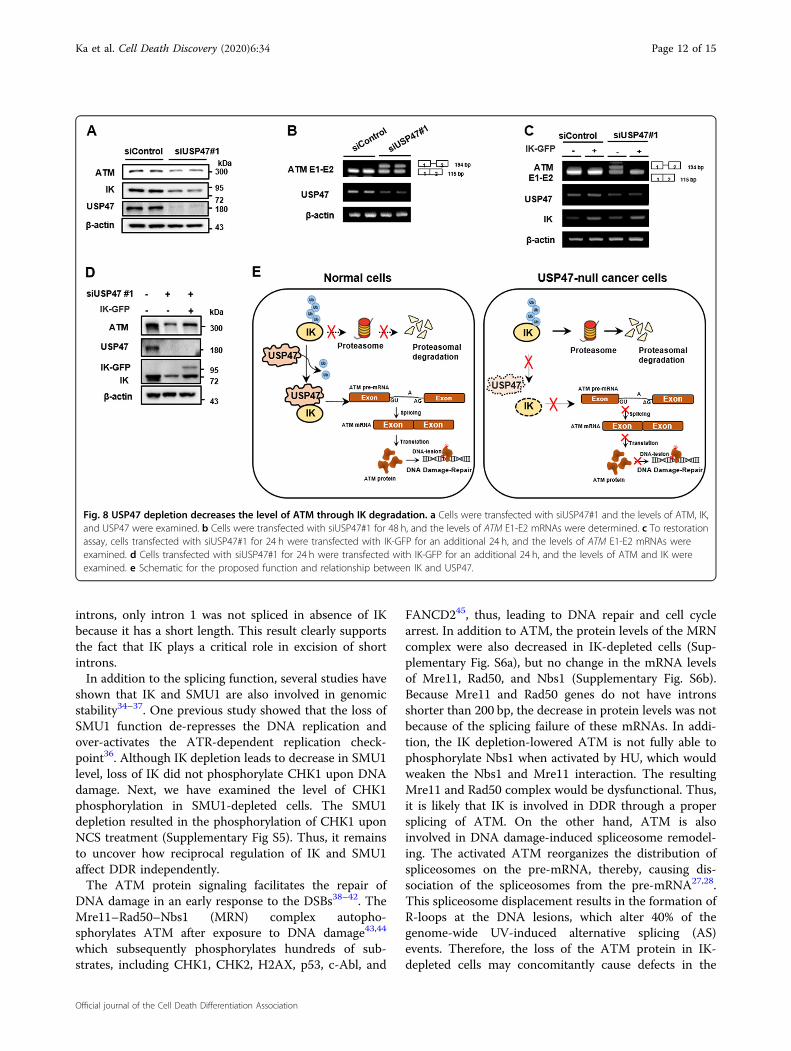
USP47 depletion decreases the level of ATM through IKdegradationBecause IK is associated with the proper splicing of
ATM pre-mRNA, the ATM levels were examined in theUSP47-depleted cells. Expectedly, the USP47-depletedcells which have low levels of IK showed lower levels ofATM compared to that in the non-depleted cells (Fig. 8a).Moreover, the ratio between intron retention/splicedisoforms of ATM transcript at the junction of exons 1–2was increased (Fig. 8b). To examine whether the aberrantsplicing of ATM transcript at exons 1–2 could be rescuedby restoration of IK, the USP47-depleted cells weretransfected with IK-GFP, and successful ATM pre-mRNAsplicing at exons 1–2 was observed (Fig. 8c). Moreover,the overexpression of IK partially rescued the low level of
Fig. 4 IK depletion causes intron retention of ATM pre-mRNA and IK directly binds to the ATM pre-mRNA. a Cells were transfected with siIK#1 or siIK #3 for 48 h, and the levels of ATM E1-EJ23 mRNA were determined. b Cells transfected with siIK #1 for 24 h were transfected using the GFP-ATM-mini-gene construct for an additional 24 h, and the levels of IK and GFP were examined. Representative images of green fluorescent protein-expressing cells obtained using fluorescence microscopy were examined, and the intensity of green fluorescence was quantified. n= 3. *p < 0.05. cCells were transfected with siIK #1 for 48 h, and cytoplasmic RNA and nuclear RNA were extracted separately. The levels of ATM mRNA wereexamined, and the intensity of each band was graphed. *p < 0.05. d HEK-293T cells were immunoprecipitated with anti-IK antibodies, and the levelsof ATM mRNA were determined.
Ka et al. Cell Death Discovery (2020) 6:34 Page 8 of 15
Official journal of the Cell Death Differentiation Association

Fig. 5 IK is degraded by the proteasome complex in the absence of SMU1. a Cells were immunoprecipitated with anti-IK and anti-SMU1 antibodies,and the levels of IK and SMU1 were examined. b Mitotic division of HeLa cells expressing GFP-tagged IK were recorded using time-lapse microscopy. Timefrom round-up is indicated. IK localized with nuclear speckle was marked with an arrow. c Cells transfected with siSMU1 for 48 h were stained with an anti-IKantibody. Representative images obtained using confocal laser microscopy were examined, and the intensity of IK was quantified using the ZEN softwarepackage and graphed with GraphPad Prism 5. ***p< 0.001. d Cells were transfected with siSMU1 for 48 h, and the levels of IK, pATM S1981, and ATM wereexamined. e Cells were transfected with siSMU1 for 48 h, and the levels of ATM mRNA were determined. f Cells were transfected with siSMU1 or siIK #1 for42 h and treated with Bafilomycin at 100 nM for indicated times. LC3 is used as positive control of Autophagy. g Cells were transfected with siSMU1 or siIK #1for 42 h and treated with Wortmannin at 5 µM for indicated times. LC3 is used as positive control of Autophagy. h Cells were transfected with siSMU1 or siIK#1 for 42 h and treated with Bortezomib at 200 nM for indicated times. i Cells were transfected with siSMU1 for 42 h and treated with 5 μM MG132, and thelevels of SMU1 and IK were examined. j HEK-293T cells transfected with HA-Ub and FLAG-SMU1 for 24 h were treated with MG132 at 5 μM for 5 h andsubjected to immunoprecipitation with an anti-IK antibody. The levels of ubiquitin, IK, and SMU1 were examined. *p< 0.05, **p< 0.01, ***p< 0.001.
Ka et al. Cell Death Discovery (2020) 6:34 Page 9 of 15
Official journal of the Cell Death Differentiation Association

ATM protein upon USP47 depletion (Fig. 8d). Besides, toexamine whether the USP47-depleted cells have animpairment of the ATM-mediated DDR, the USP47-depleted cells were treated with HU for 24 h (Supple-mentary Fig. S4a). Similar to the result observed in the IK-
depleted cells (Supplementary Fig. S1b), the USP47-depleted cells showed a slight increase in ATM phos-phorylation upon treatment of HU. It seems to be due tothe decrease in the ATM protein levels upon IK depletion.Because the failure of ATM-mediated DDR increases
Fig. 6 USP47 is a candidate deubiquitinase for IK. a HeLa cells were transfected for 48 h with individual siRNAs from a pool of 76 siRNA targetingdeubiquitinases (DUBs). Next, the level of endogenous IK was measured by western blotting and protein bands were quantified using the Image Jsoftware. b Cells were transfected with 11 DUB siRNAs involved in DNA Damage Response (DDR), and the level of IK was examined. c Cells weretransfected with three different siUSP47s for 48 h, and the levels of USP47 were examined. d Cells were transfected with siUSP47#1 for 48 h, and thelevels of IK, SMU1, and USP47 were examined. e Cells were transfected with siUSP47#1 for 48 h, and the levels of IK mRNA were determined. Therelative ratios of mRNA were normalized to those of β-actin and graphed. *p < 0.05. f Cells transfected with siUSP47 #1 for 24 h were transfected withFlag-USP47 for an additional 24 h, and the levels of IK and SMU1 were determined. g, h HEK-293T cells transfected with HA-IK or/and Flag-USP47were immunoprecipitated with an anti-HA or anti-Flag antibody, and the levels of HA and FLAG were examined. i HeLa cells wereimmunoprecipitated with anti-USP47, and the levels of IK and USP47 were examined.
Ka et al. Cell Death Discovery (2020) 6:34 Page 10 of 15
Official journal of the Cell Death Differentiation Association

apoptosis, the USP47-depleted cells treated with HU werecollected undergoing apoptosis in a living context. TheHU-treated USP47-depleted cells showed an increase incell death marker including cleaved PARP and cleavedcaspase9 (Supplementary Fig. S4b) followed by a hugeincrease in apoptosis compared to that in the control cells(Supplementary Fig. S4c). Taken together, these resultsimply that USP47-mediated IK stabilization contributes tothe ATM-mediated DDR through proper ATM pre-mRNA splicing (Fig. 8e).
DiscussionThe spliceosomal B-specific protein IK and SMU1,
which are known to be involved in B to Bact transition,interact not only with each other but also with theU2 snRNP protein, SF3B3, and the RNA helicase, Brr2, to
link the two proteins. Furthermore, knockdown of IK orSMU1 in human cells causes significant alternative spli-cing of many genes involved in cell death and survival33.During this work, Keiper et al.8 reported that IK andSMU1 play a critical role in the excision of short introns.In other words, introns retained after the knockdown ofIK or SMU1 were predominantly shorter than 200 nt,whereas the majority of introns found in human cells aremuch longer than 200 nt. In the case of introns having along distance between the 5-SS and BS, the U2 domain isflexible enough to move toward Brr2. However, in thecase of short introns (~56 nt or less), it is hard to movebecause of a structural constraint. To overcome thisproblem, IK and SMU1 extend the U2/Brr2 bridge. In thisstudy, we showed that depletion of IK impaired the propersplicing of ATM pre-mRNA. Although ATM has many
Fig. 7 USP47 stabilizes IK through deubiquitination. a Cells were transfected with USP47#1 and treated with 40 μg/mL of cycloheximide (CHX) forthe indicated times to determine the levels of IK. b Cells were transfected with siUSP47#1 for 24 h and incubated with Flag-IK for an additional 24 h,and the levels of IK were examined. Green-fluorescent protein (GFP) was used as a transfection control. c Cells were transfected with siUSP47#1 for48 h and treated with 10 μM MG132 for an additional 6 h, and the levels of IK were detected. d Cells transfected with Flag-USP47 for 24 h weretreated with 40 μg/mL of CHX for the indicated times and the levels of endogenous IK were examined. e Cells were transfected with siUSP47#1 for42 h and treated with 10 μM MG132 for 6 h. Cells were lysed and immunoprecipitated with an anti-IK antibody, followed by immunoblotting using ananti-Ub antibody. f Cells were transfected with Flag-USP47 and treated with 10 μM MG132 for 6 h. Cells were lysed and immunoprecipitated using ananti-IK antibody, followed by immunoblotting using an anti-Ub antibody. g Cells were transfected with a GFP-IK and increasing amounts of Flag-USP47, and the levels of GFP-IK and Flag-USP47 were examined.
Ka et al. Cell Death Discovery (2020) 6:34 Page 11 of 15
Official journal of the Cell Death Differentiation Association
introns, only intron 1 was not spliced in absence of IKbecause it has a short length. This result clearly supportsthe fact that IK plays a critical role in excision of shortintrons.In addition to the splicing function, several studies have
shown that IK and SMU1 are also involved in genomicstability34–37. One previous study showed that the loss ofSMU1 function de-represses the DNA replication andover-activates the ATR-dependent replication check-point36. Although IK depletion leads to decrease in SMU1level, loss of IK did not phosphorylate CHK1 upon DNAdamage. Next, we have examined the level of CHK1phosphorylation in SMU1-depleted cells. The SMU1depletion resulted in the phosphorylation of CHK1 uponNCS treatment (Supplementary Fig S5). Thus, it remainsto uncover how reciprocal regulation of IK and SMU1affect DDR independently.The ATM protein signaling facilitates the repair of
DNA damage in an early response to the DSBs38–42. TheMre11–Rad50–Nbs1 (MRN) complex autopho-sphorylates ATM after exposure to DNA damage43,44
which subsequently phosphorylates hundreds of sub-strates, including CHK1, CHK2, H2AX, p53, c-Abl, and
FANCD245, thus, leading to DNA repair and cell cyclearrest. In addition to ATM, the protein levels of the MRNcomplex were also decreased in IK-depleted cells (Sup-plementary Fig. S6a), but no change in the mRNA levelsof Mre11, Rad50, and Nbs1 (Supplementary Fig. S6b).Because Mre11 and Rad50 genes do not have intronsshorter than 200 bp, the decrease in protein levels was notbecause of the splicing failure of these mRNAs. In addi-tion, the IK depletion-lowered ATM is not fully able tophosphorylate Nbs1 when activated by HU, which wouldweaken the Nbs1 and Mre11 interaction. The resultingMre11 and Rad50 complex would be dysfunctional. Thus,it is likely that IK is involved in DDR through a propersplicing of ATM. On the other hand, ATM is alsoinvolved in DNA damage-induced spliceosome remodel-ing. The activated ATM reorganizes the distribution ofspliceosomes on the pre-mRNA, thereby, causing dis-sociation of the spliceosomes from the pre-mRNA27,28.This spliceosome displacement results in the formation ofR-loops at the DNA lesions, which alter 40% of thegenome-wide UV-induced alternative splicing (AS)events. Therefore, the loss of the ATM protein in IK-depleted cells may concomitantly cause defects in the
Fig. 8 USP47 depletion decreases the level of ATM through IK degradation. a Cells were transfected with siUSP47#1 and the levels of ATM, IK,and USP47 were examined. b Cells were transfected with siUSP47#1 for 48 h, and the levels of ATM E1-E2 mRNAs were determined. c To restorationassay, cells transfected with siUSP47#1 for 24 h were transfected with IK-GFP for an additional 24 h, and the levels of ATM E1-E2 mRNAs wereexamined. d Cells transfected with siUSP47#1 for 24 h were transfected with IK-GFP for an additional 24 h, and the levels of ATM and IK wereexamined. e Schematic for the proposed function and relationship between IK and USP47.
Ka et al. Cell Death Discovery (2020) 6:34 Page 12 of 15
Official journal of the Cell Death Differentiation Association

ATM-dependent spliceosome mobilization, thus, leadingto wide-ranging changes in AS events.IK/SMU1 is also involved in proper spindle attachment
through interaction with MAD1 and causes cell cyclearrest at the mitotic phase. Other studies have demon-strated the connections between pre-mRNA splicing andthe cell cycle46–49. In general, splicing is inhibited inextracts of mitotic cells, like the other steps in geneexpression. Shin and Manley50 showed that the SR (rich inserine and arginine) protein, SRp38, represses the splicingduring the mitotic phase through mitotic phase-specificdephosphorylation of SRp38. We also observed that IKlocalized at nuclear speckles and dissociated duringmitotic phase (Supplementary Figs. S2 and 5b). Thus,splicing would be inhibited during mitotic phase throughsome other mechanism and IK released from nuclearspeckle might play a role in the proper segregation ofchromosomes. Hence, there should be a mechanism tokeep IK from dissociation of nuclear speckle with inde-pendent functions and we show that USP47 may performthis role at the mitotic phase.As DUBs modulate the half-life of many cellular pro-
teins related to the signaling pathway, tumor suppression,tumorigenesis, immune response, and DDR, defects inDUBs induce various pathophysiological processes in thecell51,52. Among the DUBs, many are associated withDDR. USP14 regulates the IR-induced NHEJ DNArepair53. USP49 deubiqutinates p53 to prevent its degra-dation, and thus, USP49-depleted colon tumors have lowlevels of p53, which makes them more susceptible to DNAdamage-inducers54. USP1 removes the mono-ubiquitination of PCNA, and thus UV-induced USP1cleavage enables monoubiquitinated PCNA to accumulateand to activate translesion synthesis (TLS)55; as a result,USP1 knock-out mice are genetically unstable andhypersensitive to DNA damage. DNA damage-inducedphosphorylation of USP10 moves it into the nucleus,allowing it to stabilize p5356 and USP28 stabilizes Chk2and 53BP1 in response to DNA damage57. USP47 isidentified as a major deubiquitinase responsible for thestabilization of DNA polymerase β, which plays a criticalrole in DNA base excision repair58. In this study, we foundthat USP47 deubiquitinates the spliceosomal factor IKand associates with DDR through the complete splicing ofATM by IK stabilization.Moreover, recent studies have revealed that pre-mRNA
splicing depends on ubiquitination and deubiquitinationcycles of the spliceosomal component. For example, thetri-snRNP proteins, PRP3 and PRP31, are reported toregulate the spliceosome through ubiquitination anddeubiquitination17,59–61. First, the ubiquitin ligase, PRP19,containing the U-box spliceosomal protein, ubiquitinatesPRP3 and PRP31, and then the ubiquitinated PRP3 and
PRP31 complex binds PRP8 and stabilizes the tri-snRNPcomplex. The ubiquitinated PRP3 and PRP31 are deubi-quitinated by the ubiquitin-specific proteases, USP4 andUSP15, respectively. It facilitates the ejection of U4 pro-teins from the spliceosome during maturation of its activesite and progresses the splicing cycle. Ubiquitination playsa role as a mediator of protein–protein interaction in theabove example and also functions in protein destabiliza-tion as for IK. For the proper spatio-temporal recruitmentof IK into the spliceosomes, the stability of IK should becontinuously regulated by the USP47 deubiquitinase. Itneeds to be investigated as to which E3 ligase ubiquiti-nates IK under a specific condition. In addition, the pos-sibility that the ubiquitinated IK affects the modificationof other spliceosomal complexes like PRP3 and PRP31cannot be ruled out.In summary, we found that IK depletion impairs the
proper splicing of ATM pre-mRNA at exons 1–2, whichresults in ATM deficiency followed by chromosomefragmentation and that USP47 directly binds to IK anddeubiquitinates it leading to its stabilization. Thus, weprovide the evidence that USP47 is indirectly involved inDDR through the complete splicing of ATM pre-mRNAby stabilization of the spliceosomal protein IK.
AcknowledgementsFinancial support was given by the National Research Foundation of Koreagrant funded by the Korean government, MISP (Ministry of ScienceInformation and Communication Technology, and Future Planning; SRC(Science Research Center) program (Cellular Heterogeneity Research Center:2016R1A5A1011974).
Author details1Department of Biological Sciences, Sookmyung Women’s University, Seoul04310, Korea. 2Drug Evaluation Group, R&D Center CJ HealthCare, Icheon04551, Korea. 3Research Institute of Women’s Health, Sookmyung Women’sUniversity, Seoul 04310, Korea. 4New Drug Development Center, OsongMedical Innovation Foundation, Osong 28160, Korea
Conflict of interestThe authors declare that they have no conflict of interest.
Publisher’s noteSpringer Nature remains neutral with regard to jurisdictional claims inpublished maps and institutional affiliations.
The online version of this article (https://doi.org/10.1038/s41420-020-0268-1)contains supplementary material, which is available to authorized users.
Received: 16 February 2020 Revised: 24 March 2020 Accepted: 21 April2020
References1. Wahl, M. C., Will, C. L. & Luhrmann, R. The spliceosome: design principles of a
dynamic RNP machine. Cell 136, 701–718 (2009).2. Dominguez, D. & Burge, C. B. Interactome analysis brings splicing into focus.
Genome Biol. 16, 135 (2015).
Ka et al. Cell Death Discovery (2020) 6:34 Page 13 of 15
Official journal of the Cell Death Differentiation Association

3. Hegele, A. et al. Dynamic protein-protein interaction wiring of the humanspliceosome. Mol. Cell 45, 567–580 (2012).
4. Zhou, Z., Licklider, L. J., Gygi, S. P. & Reed, R. Comprehensive proteomic analysisof the human spliceosome. Nature 419, 182–185 (2002).
5. Lee, Y. & Rio, D. C. Mechanisms and regulation of alternative pre-mRNAsplicing. Annu. Rev. Biochem. 84, 291–323 (2015).
6. Bessonov, S., Anokhina, M., Will, C. L., Urlaub, H. & Luhrmann, R. Isolation of anactive step I spliceosome and composition of its RNP core. Nature 452,846–850 (2008).
7. Ulrich, A. K., Schulz, J. F., Kamprad, A., Schutze, T. & Wahl, M. C. Structural basisfor the functional coupling of the alternative splicing factors Smu1 and RED.Structure 24, 762–773 (2016).
8. Keiper, S. et al. Smu1 and RED are required for activation of spliceo-somal B complexes assembled on short introns. Nat. Commun. 10, 3639(2019).
9. Neumann, B. et al. Phenotypic profiling of the human genome by time-lapse microscopy reveals cell division genes. Nature 464, 721–727(2010).
10. Yeh, P. C., Yeh, C. C., Chen, Y. C. & Juang, Y. L. RED, a spindle pole-associatedprotein, is required for kinetochore localization of MAD1, mitotic progression,and activation of the spindle assembly checkpoint. J. Biol. Chem. 287,11704–11716 (2012).
11. Lee, S. et al. IK-guided PP2A suppresses Aurora B activity in the interphase oftumor cells. Cell. Mol. Life Sci. 73, 3375–3386 (2016).
12. Hu, L., Yang, F., Liu, X., Xu, D. & Dai, W. Nuclear protein IK undergoes dynamicsubcellular translocation and forms unique nuclear bodies during the cellcycle. Biomark. Res. 1, 11 (2013).
13. Lee, S., Han, S., Jeong, A. L., Park, J. S. & Yang, Y. Depletion of IK causes mitoticarrest through aberrant regulation of mitotic kinases and phosphatases. FEBSLett. 588, 2844–2850 (2014).
14. Will, C. L. & Luhrmann, R. Spliceosome structure and function. Cold Spring Harb.Perspect. Biol. 3, 3707 (2011).
15. Shi, Y. Mechanistic insights into precursor messenger RNA splicing by thespliceosome. Nat. Rev. Mol. Cell Biol. 18, 655–670 (2017).
16. Bellare, P. et al. A role for ubiquitin in the spliceosome assembly pathway. Nat.Struct. Mol. Biol. 15, 444–451 (2008).
17. Das, T. et al. USP15 regulates dynamic protein-protein interactions of thespliceosome through deubiquitination of PRP31. Nucleic Acids Res. 45,4866–4880 (2017).
18. Callis, J. The ubiquitination machinery of the ubiquitin system. ArabidopsisBook 12, e0174 (2014).
19. Glickman, M. H. & Ciechanover, A. The ubiquitin-proteasome proteolyticpathway: destruction for the sake of construction. Physiol. Rev. 82, 373–428(2002).
20. Lecker, S. H., Goldberg, A. L. & Mitch, W. E. Protein degradation by theubiquitin-proteasome pathway in normal and disease states. J. Am. Soc.Nephrol. 17, 1807–1819 (2006).
21. Guleria, A. & Chandna, S. ATM kinase: much more than a DNA damageresponsive protein. DNA Repair 39, 1–20 (2016).
22. Shiloh, Y. ATM and related protein kinases: safeguarding genome integrity.Nat. Rev. Cancer 3, 155–168 (2003).
23. Sancar, A., Lindsey-Boltz, L. A., Unsal-Kacmaz, K. & Linn, S. Molecularmechanisms of mammalian DNA repair and the DNA damage checkpoints.Annu. Rev. Biochem. 73, 39–85 (2004).
24. Abraham, R. T. Cell cycle checkpoint signaling through the ATM and ATRkinases. Genes Dev. 15, 2177–2196 (2001).
25. Kastan, M. B. & Lim, D. S. The many substrates and functions of ATM. Nat. Rev.Mol. Cell Biol. 1, 179–186 (2000).
26. Matsuoka, S. et al. ATM and ATR substrate analysis reveals extensiveprotein networks responsive to DNA damage. Science 316, 1160–1166(2007).
27. Tresini, M. et al. The core spliceosome as target and effector of non-canonicalATM signalling. Nature 523, 53–58 (2015).
28. Tresini, M., Marteijn, J. A. & Vermeulen, W. Bidirectional coupling of splicing andATM signaling in response to transcription-blocking DNA damage. RNA Biol.13, 272–278 (2016).
29. Katzenberger, R. J., Marengo, M. S. & Wassarman, D. A. ATM and ATR pathwayssignal alternative splicing of Drosophila TAF1 pre-mRNA in response to DNAdamage. Mol. Cell Biol. 26, 9256–9267 (2006).
30. Garner, E., Kim, Y., Lach, F. P., Kottemann, M. C. & Smogorzewska, A. HumanGEN1 and the SLX4-associated nucleases MUS81 and SLX1 are essential forthe resolution of replication-induced Holliday junctions. Cell Rep. 5, 207–215(2013).
31. Falck, J., Mailand, N., Syljuasen, R. G., Bartek, J. & Lukas, J. The ATM-Chk2-Cdc25Acheckpoint pathway guards against radioresistant DNA synthesis. Nature 410,842–847 (2001).
32. Hong, S., Paulson, Q. X. & Johnson, D. G. E2F1 and E2F3 activate ATM throughdistinct mechanisms to promote E1A-induced apoptosis. Cell Cycle 7, 391–400(2008).
33. Papasaikas, P., Tejedor, J. R., Vigevani, L. & Valcarcel, J. Functional splicingnetwork reveals extensive regulatory potential of the core spliceosomalmachinery. Mol. Cell 57, 7–22 (2015).
34. Sugaya, K., Hongo, E., Ishihara, Y. & Tsuji, H. The conserved role of Smu1 insplicing is characterized in its mammalian temperature-sensitive mutant. J. CellSci. 119, 4944–4951 (2006).
35. Paulsen, R. D. et al. A genome-wide siRNA screen reveals diverse cellularprocesses and pathways that mediate genome stability. Mol. Cell 35, 228–239(2009).
36. Ren, L. et al. Loss of Smu1 function de-represses DNA replication and over-activates ATR-dependent replication checkpoint. Biochem. Biophys. Res. Com-mun. 436, 192–198 (2013).
37. Sugaya, K., Hongo, E. & Tsuji, H. A temperature-sensitive mutation in the WDrepeat-containing protein Smu1 is related to maintenance of chromosomeintegrity. Exp. Cell Res. 306, 242–251 (2005).
38. Marechal, A. & Zou, L. DNA damage sensing by the ATM and ATR kinases. ColdSpring Harb. Perspect. Biol. 5, 12716 (2013).
39. Khanna, K. K., Lavin, M. F., Jackson, S. P. & Mulhern, T. D. ATM, a centralcontroller of cellular responses to DNA damage. Cell Death Differ. 8, 1052–1065(2001).
40. Rotman, G. & Shiloh, Y. ATM: a mediator of multiple responses to genotoxicstress. Oncogene 18, 6135–6144 (1999).
41. Lee, J. H. & Paull, T. T. Activation and regulation of ATM kinase activity inresponse to DNA double-strand breaks. Oncogene 26, 7741–7748 (2007).
42. Morrison, C. et al. The controlling role of ATM in homologous recombinationalrepair of DNA damage. EMBO J. 19, 463–471 (2000).
43. Lee, J. H. & Paull, T. T. ATM activation by DNA double-strand breaks throughthe Mre11-Rad50-Nbs1 complex. Science 308, 551–554 (2005).
44. Uziel, T. et al. Requirement of the MRN complex for ATM activation by DNAdamage. EMBO J. 22, 5612–5621 (2003).
45. Ahmed, K. M. & Li, J. J. ATM-NF-kappaB connection as a target for tumorradiosensitization. Curr. Cancer Drug Targets 7, 335–342 (2007).
46. Blencowe, B. J. Splicing regulation: the cell cycle connection. Curr. Biol. 13,R149–R151 (2003).
47. Lundgren, K. et al. A connection between pre-mRNA splicing and the cellcycle in fission yeast: cdc28+ is allelic with prp8+ and encodes an RNA-dependent ATPase/helicase. Mol. Biol. Cell 7, 1083–1094 (1996).
48. Shea, J. E., Toyn, J. H. & Johnston, L. H. The budding yeast U5 snRNP Prp8 is ahighly conserved protein which links RNA splicing with cell cycle progression.Nucleic Acids Res. 22, 5555–5564 (1994).
49. Karamysheva, Z., Diaz-Martinez, L. A., Warrington, R. & Yu, H. Graded require-ment for the spliceosome in cell cycle progression. Cell Cycle 14, 1873–1883(2015).
50. Shin, C. & Manley, J. L. The SR protein SRp38 represses splicing in M phasecells. Cell 111, 407–417 (2002).
51. Komander, D. Mechanism, specificity and structure of the deubiquitinases.Subcell. Biochem 54, 69–87 (2010).
52. Mevissen, T. E. T. & Komander, D. Mechanisms of deubiquitinase specificity andregulation. Annu. Rev. Biochem. 86, 159–192 (2017).
53. Sharma, A. et al. USP14 regulates DNA damage repair by targeting RNF168-dependent ubiquitination. Autophagy 14, 1976–1990 (2018).
54. Tu, R. et al. USP49 participates in the DNA damage response by forming apositive feedback loop with p53. Cell Death Dis. 9, 553 (2018).
55. Huang, T. T. et al. Regulation of monoubiquitinated PCNA by DUB auto-cleavage. Nat. Cell Biol. 8, 339–347 (2006).
56. Yuan, J., Luo, K., Zhang, L., Cheville, J. C. & Lou, Z. USP10 regulates p53localization and stability by deubiquitinating p53. Cell 140, 384–396(2010).
Ka et al. Cell Death Discovery (2020) 6:34 Page 14 of 15
Official journal of the Cell Death Differentiation Association

57. Zhang, D., Zaugg, K., Mak, T. W. & Elledge, S. J. A role for the deubiquitinatingenzyme USP28 in control of the DNA-damage response. Cell 126, 529–542(2006).
58. Parsons, J. L. et al. USP47 is a deubiquitylating enzyme that regulates baseexcision repair by controlling steady-state levels of DNA polymerase beta. Mol.Cell 41, 609–615 (2011).
59. Song, E. J. et al. The Prp19 complex and the Usp4Sart3 deubiquitinatingenzyme control reversible ubiquitination at the spliceosome. Genes Dev. 24,1434–1447 (2010).
60. Hogg, R., McGrail, J. C. & O’Keefe, R. T. The function of the NineTeen Complex(NTC) in regulating spliceosome conformations and fidelity during pre-mRNAsplicing. Biochem. Soc. Trans. 38, 1110–1115 (2010).
61. Makarova, O. V., Makarov, E. M., Liu, S., Vornlocher, H. P. & Luhrmann, R. Protein61K, encoded by a gene (PRPF31) linked to autosomal dominant retinitispigmentosa, is required for U4/U6*U5 tri-snRNP formation and pre-mRNAsplicing. EMBO J. 21, 1148–1157 (2002).
Ka et al. Cell Death Discovery (2020) 6:34 Page 15 of 15
Official journal of the Cell Death Differentiation Association