Embed Size (px)
Citation preview
Journal of Chromatography B, 875 (2008) 557–561
Contents lists available at ScienceDirect
Journal of Chromatography B
journa l homepage: www.e lsev ier .com/ locate /chromb
Short communication
Determination of prostaglandin E2 by on-line solid-phase extraction–liquidchromatography with ultraviolet detection for microsomal prostaglandin E2
synthase-1 inhibitor screening
Jörg FabianInstitute of Pharmaceutical and Medicinal Chemistry, University of Münster, Hittorfstrasse 58-62, D-48149 Münster, Germany
a r t i c l e i n f o
Article history:Received 29 April 2008Accepted 29 September 2008Available online 10 October 2008
Keywords:
a b s t r a c t
A rapid, robust and selective on-line solid-phase extraction–liquid chromatographic method with ultra-violet detection (on-line SPE-LC-UV) for microsomal prostaglandin E2 synthase-1 (mPGES-1) inhibitorscreening was developed and validated. Disrupted A549 cells were used as mPGES-1 source and theformation of prostaglandin E2 (PGE2) out of the substrate prostaglandin H2 (PGH2) was determined at195 nm. Direct on-line sample clean up was achieved by automated column switch (C18 trap column)prior isocratic separation using a C18 analytical column. The on-line SPE-LC-UV method was accurate,
mPGES-1On-line SPE-LC-UVP
precise and reproducible in the range of 71–1763 ng/ml for PGE2 and met the generally accepted criteriafor bioanalytical methods. The method was successfully applied to determine the IC value of the known
.
1
2afmtdP
arAeih
muabra
[nnpils
m(Tctamf[tSp
1d
GE2mPGES-1 inhibitor NS-398
. Introduction
Prostaglandin E2 (PGE2) produced via cyclooxgenase-2 (COX-) and microsomal prostaglandin E2 synthase-1 (mPGES-1) playsn important role in the pathophysiology of inflammation, pain,ever and vascular regulation. mPGES-1 is a member of the
embrane-associated proteins involved in eicosanoid and glu-athione metabolism (MAPEG) superfamily [1–3]. The glutathioneependent isomerase converts the intermediate PGH2 to PGE2.GE2 exerts its action via four EP-receptor subtypes [4].
COX-2 inhibitors as a therapeutic concept for inflammationre known to increase cardiovascular adverse effects by dis-uption of the balance between prostacyclin and thromboxane2. Intriguingly, deletion of mPGES-1 in mice did not affectither thrombogenesis or blood pressure but retained the anti-nflammatory effects [5,6]. Taken together inhibition of mPGES-1olds great potential in the treatment of inflammatory diseases.
As a detailed three-dimensional (3D) X-ray crystal structure ofPGES-1 is still missing, combined structure predicting, molec-
lar docking, site-directed mutagenesis and enzymatic activity
ssays were employed to develop a 3D model of the substrate-inding domain [7–9]. Hence, until now rational design did notesult in the discovery of novel mPGES-1 inhibitors. The sulfon-mide NS-398 [10], fatty acid mimetica like 15-deoxy-�12,14PGJ2E-mail address: [email protected].
ssn[Sf
b
570-0232/$ – see front matter © 2008 Elsevier B.V. All rights reserved.oi:10.1016/j.jchromb.2008.09.038
50
© 2008 Elsevier B.V. All rights reserved.
11] and compounds derived from MK-886 [12] are literatureoted inhibitors. However, these substances were found to haveon-sufficient pharmacokinetic properties. Only the orally activehenanthrene imidazoles [13] show less strong protein binding and
nhibition in cellular systems. Therefore the identification of newead inhibitors is a reasonable research field and rapid, robust butelective inhibitor screening assays are necessary.
The most widely utilized methods for the detection and deter-ination of PGE2 in biological samples are radioimmunoassays
RIA) [14] and enzyme immunoassays (EIA) [15,16], respectively.hese methods are sensitive and simple to do but show timeonsumption (EIA), cross-reactivity and variability in quantifica-ion on the other hand. HPLC methods offer greater flexibilitynd specificity but often need time consuming sample pretreat-ents of the biological matrices. Most of the published methods
or PGE2 determinations used off-line SPE combined with LC-UV10,17] and ESI-MS/MS [18], respectively, or liquid–liquid extrac-ion (LLE) with subsequent LC-MS/MS determination of PGE2 [19].urprisingly a direct LC-UV determination of PGE2 with no sampleretreatment is described [20]. On-line sample clean up is a timeaving and mostly error reducing procedure. Recently an on-lineample preparation coupled with capillary LC–MS/MS for determi-ation of prostaglandins in cell culture supernatants was published
21]. Additionally on-line-SPE-LC methods combined with UV-Flowcintillation Analyzers (FSA) [22,23] and off-line RIA determinationor prostanoids in biological matrices are known [24], respectively.The starting point of the present study was an A549 cell lineased enzyme assay combined with a non-validated PGE2 determi-
5 ogr. B
nacl7rdci
2
2
(o
b(cmmAppaflBN(
ap11l(Mp
2
TmhaCcdPtst
ifiFtaatb−
2
bppsfhma
2
1GIar(Tt(tcs
2
w1pftdKw(jdcodocpbHiAAf
fanss
58 J. Fabian / J. Chromat
ation method using off-line SPE with LC-UV [10]. Here we describerobust mPGES-1 inhibitor-screening assay with disrupted A549
ells combined with a rapid, selective and fully validated on-ine SPE-LC-UV method. PGE2 was determined in the range from1–1763 ng/ml. This method meets the generally accepted crite-ia for bioanalytical methods, is capable for higher throughput andoes not require any sample pretreatment. The method was suc-essfully applied to determine the IC50 value of the known mPGES-1nhibitor NS-398.
. Experimental
.1. Chemicals and reagents
Prostaglandin E1 (PGE1), PGE2, PGH2, NS-398 (N-[2-cyclohexyloxy)-4-nitrophenyl]methanesulfonamide) werebtained from Cayman Europe (Tallinn, Estonia).l-Glutathione, reduced (GSH), Trizma®, phosphate-
uffered saline (PBS) tablets for the preparation of PBS0.1 M, pH 7.4), lipopolysaccharides (LPS) from Escherichiaoli 0111:B4, Protease Inhibitor Cocktail for the use withammalian cell and tissue extracts and phorbol-12-yristate-13-acetate (TPA) were purchased from Sigma–ldrich (St. Louis, Missouri). Acetonitrile (HPLC gradient grade),otassium dihydrogen phosphate (p.A.), dipotassium hydrogenhosphate (p.A.) and dimethylsulfoxide (DMSO) (p.A.) werecquired from VWR International (Darmstadt, Germany). Tri-uoroacetic acid (TFA) was obtained from Acros Organics (Geel,elgium) and phosphoric acid (85%) (p.A.) from J.T. Baker (Deventer,etherlands). Fe(II)Cl2·4H2O (p.A.) was purchased from AppliChem
Darmstadt, Germany).DMEM high glucose (4.5 g/l) with l-glutamin was
cquired from PPA laboratories (Cölbe, Germany), GibcoTM
enicillin–streptomycin (10,000 U/ml penicillin G and0,000 �g/ml streptomycin sulfate), GibcoTM trypsin–EDTA0× (0.5% trypsin, 5.3 mM EDTA-Na4) was bought from invitrogenife technologies (Karlsruhe, Germany). Tumor cell line CCL185A549, human adenocarcinoma) was kindly provided by Prof. R.M.
esters (University of Münster, Münster, Germany). Water wasurified using a Bi 18 system from Heraeus (Hanau, Germany).
.2. Cell culture
A549 is a nonsmall lung cancer human adenocarcinoma cell line.he morphology is epithelial-like and the cells grow as an adherentonolayer. A549 cells were cultured in DMEM supplemented with
eat-inactivated foetal bovine serum (10%), penicillin (166.7 U/ml)nd streptomycin (166.7 �g/ml) at 37 ◦C in an atmosphere of 5%O2. Approximately 1.2 × 10⁶ cells (improved Neubauer countinghamber) in 18 ml media were seeded in 75 cm2 flasks. After 7ays, confluence was reached and cells were washed with 5 mlBS buffer twice, then detached using 3 ml 0.05% trypsin (GibcoTM
rypsin–EDTA 10×: 1:10 diluted with PBS) at 37 ◦C in an atmo-phere of 5% CO2 for 15 min. Thereafter, 10 ml medium was addedo quench the trypsin and 1 ml cell suspension was reseeded.
In order to stimulate the mPGES-1 expression the cells werencubated with LPS (100 ng/ml final concentration) or TPA (1 �Mnal concentration) at 37 ◦C in an atmosphere of 5% CO2 for 24 h.or harvest, the cells were washed with 5 ml PBS buffer twice,hen trypsinated using 3 ml 0.05% trypsin solution at 37 ◦C in an
tmosphere of 5% CO2 for 15 min. Subsequently, 10 ml medium wasdded and the cells were centrifuged at 500 × g at room tempera-ure for 10 min. The pellet was resuspended twice with 5 ml PBSuffer and centrifuged each time. The final cell pellet was stored at80 ◦C.svoT3
875 (2008) 557–561
.3. Preparation of A549 cell microsomes
The cell pellets were resuspended in 1.0 ml homogenizationuffer consisting of potassium phosphate buffer (100 mM, pH 7.4),rotease inhibitor cocktail and sucrose (0.25 M). The samples wereulse sonicated 3 × 20 s in a −15 ◦C isopropanol bath. The suspen-ion was centrifuged at 10,000 × g for 15 min and at 100,000 × gor 60 min. The microsomal fraction was resuspended in 200 �lomogenization buffer and the protein concentration was deter-ined by modified Bradford-Assay [25] applying bovine serum
lbumin (BSA) as standard.
.4. Incubation procedures
In precooled eppendorf caps 8 �l PGH2 (0.28 mM in aceton),86 �l Tris-buffer (50 mM, pH 7.4 at 0 ◦C) containing reducedSH (2.5 mM) and 2 �l DMSO were pipetted and stored at 0 ◦C.
n case of inhibitor screening 2 �l of inhibitor DMSO solutionst different concentrations were added instead. After 2 min theeaction was initiated by the addition of 4 �l enzyme solution3.5 mg/ml protein) and the mixture was processed at 0 ◦C for 5 min.he reaction was quenched by the addition of 100 �l stop solu-ion (60 mM Fe(II)Cl2 in 0.05 M HCl) and 100 �l internal standardISTD) solution (PGE1, 20 �M in MeCN with 0.1% TFA). Two hundredwenty five microliters of this solution was injected onto the HPLColumn. Reference incubations were performed without enzymeolution in the same way.
.5. On-line SPE-LC-UV-analysis
A schematic presentation of the on-line SPE-LC-UV system 1ith a 10-port 2-position valve from Rheodyne (Waters, EV700-
02-WA) is given in Fig. 1. A Knauer HPLC Pump 64 (Berlin, Germany,ump 1) was coupled to a Waters autosampler model 717 plus (Mil-ord, USA) and connected to a Kromasil 100-5-C18 (40 mm × 4 mm)rap column (CS-Chromatographie, Germany). A Waters 515 gra-ient pump system (Milford, USA, pump 2) was connected to aromasil 100-5-C18 (250 mm × 3 mm) analytical column, whichas protected by a C18 (4 mm × 3 mm) SecurityGuardTM Cartidge
Phenomenex, Germany) and temperature controlled at 30 ◦C by aetstream plus column oven (Waters, Milford). A Waters UV–vis-etector model 2487 was used for detection at 195 nm and systemontrol was performed by Millennium32 software. The temperaturef the autosampler was kept at 10 ◦C. The mobile phase for the firstimension was MeCN-Aqua bidest.-TFA (8:92:0.1, v/v/v) and 225 �lf each sample was stacked at a flow of 0.5 ml/min on the trapolumn. After 5 min the 10-port 2-position valve was switched toosition 2. An isocratic separation was conducted with MeCN-Aquaidest.-H3PO4 (35:65:0.025, v/v/v) (A) and MeCN-Aqua bidest.-3PO4 (90:10:0.025, v/v/v) (B) with a flow rate of 0.6 ml/min. The
nitial composition of 100% A was retained after injection for 19 min.linear washing gradient with 0% A was programmed over 3 min.fter valve switch to position 1 at 22 min reequilibration was per-
ormed at 100% A for 6 min.Alternatively, the SPE-LC-UV system 2 (Fig. 1) can be employed
or chromatographic determination of PGE2. As two trap columnsre used for sample clean-up, the analysis time can be reduced sig-ificantly. The solvents for system 2 and flow rates were kept theame. In detail, before injection the 10-port 2-position valve waswitched on position 1. 225 �l of an injected sample probe was
tacked on trap column 2 with MeCN-Aqua bidest.-TFA (8:92:0.1,/v/v). At the same time the isocratic separation of a probe stackedn trap column 1 was conducted with 100% of solvent A for 6 min.hen a linear washing gradient with 0% A was programmed overmin. After 13 min the 10-port 2-position valve was switched to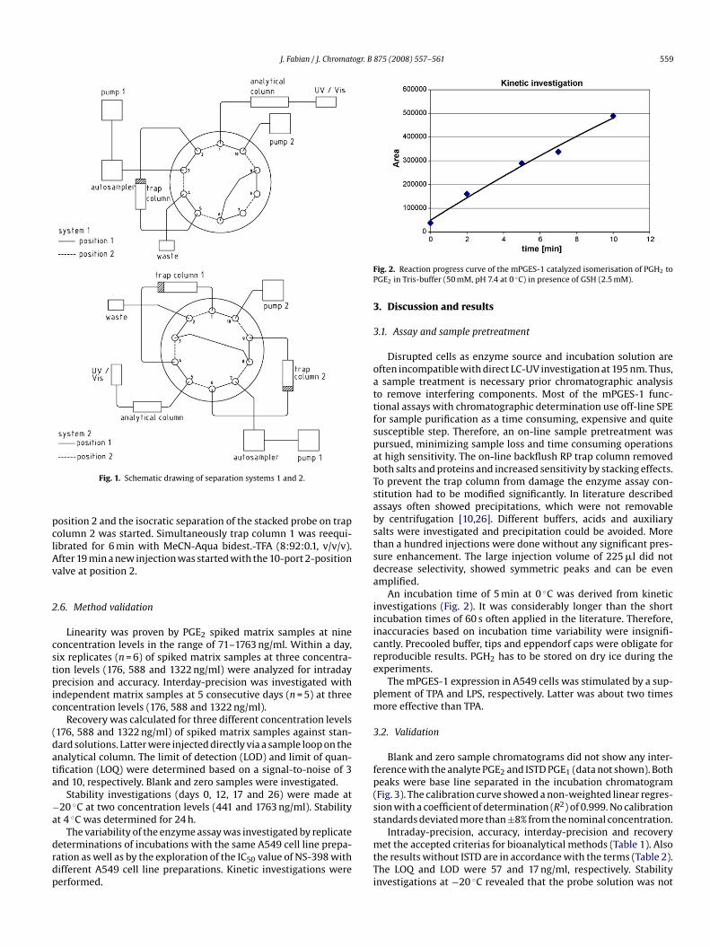
J. Fabian / J. Chromatogr. B 875 (2008) 557–561 559
pclAv
2
cstpic
(data
−a
drdp
FP
3
3
oattfspabTsabstsda
iiicre
pm
3
fp(s
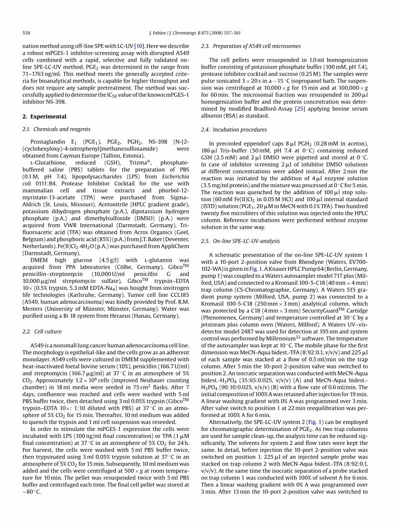
Fig. 1. Schematic drawing of separation systems 1 and 2.
osition 2 and the isocratic separation of the stacked probe on trapolumn 2 was started. Simultaneously trap column 1 was reequi-ibrated for 6 min with MeCN-Aqua bidest.-TFA (8:92:0.1, v/v/v).fter 19 min a new injection was started with the 10-port 2-positionalve at position 2.
.6. Method validation
Linearity was proven by PGE2 spiked matrix samples at nineoncentration levels in the range of 71–1763 ng/ml. Within a day,ix replicates (n = 6) of spiked matrix samples at three concentra-ion levels (176, 588 and 1322 ng/ml) were analyzed for intradayrecision and accuracy. Interday-precision was investigated with
ndependent matrix samples at 5 consecutive days (n = 5) at threeoncentration levels (176, 588 and 1322 ng/ml).
Recovery was calculated for three different concentration levels176, 588 and 1322 ng/ml) of spiked matrix samples against stan-ard solutions. Latter were injected directly via a sample loop on thenalytical column. The limit of detection (LOD) and limit of quan-ification (LOQ) were determined based on a signal-to-noise of 3nd 10, respectively. Blank and zero samples were investigated.
Stability investigations (days 0, 12, 17 and 26) were made at20 ◦C at two concentration levels (441 and 1763 ng/ml). Stability
t 4 ◦C was determined for 24 h.The variability of the enzyme assay was investigated by replicateeterminations of incubations with the same A549 cell line prepa-ation as well as by the exploration of the IC50 value of NS-398 withifferent A549 cell line preparations. Kinetic investigations wereerformed.
s
mtTi
ig. 2. Reaction progress curve of the mPGES-1 catalyzed isomerisation of PGH2 toGE2 in Tris-buffer (50 mM, pH 7.4 at 0 ◦C) in presence of GSH (2.5 mM).
. Discussion and results
.1. Assay and sample pretreatment
Disrupted cells as enzyme source and incubation solution areften incompatible with direct LC-UV investigation at 195 nm. Thus,sample treatment is necessary prior chromatographic analysis
o remove interfering components. Most of the mPGES-1 func-ional assays with chromatographic determination use off-line SPEor sample purification as a time consuming, expensive and quiteusceptible step. Therefore, an on-line sample pretreatment wasursued, minimizing sample loss and time consuming operationst high sensitivity. The on-line backflush RP trap column removedoth salts and proteins and increased sensitivity by stacking effects.o prevent the trap column from damage the enzyme assay con-titution had to be modified significantly. In literature describedssays often showed precipitations, which were not removabley centrifugation [10,26]. Different buffers, acids and auxiliaryalts were investigated and precipitation could be avoided. Morehan a hundred injections were done without any significant pres-ure enhancement. The large injection volume of 225 �l did notecrease selectivity, showed symmetric peaks and can be evenmplified.
An incubation time of 5 min at 0 ◦C was derived from kineticnvestigations (Fig. 2). It was considerably longer than the shortncubation times of 60 s often applied in the literature. Therefore,naccuracies based on incubation time variability were insignifi-antly. Precooled buffer, tips and eppendorf caps were obligate foreproducible results. PGH2 has to be stored on dry ice during thexperiments.
The mPGES-1 expression in A549 cells was stimulated by a sup-lement of TPA and LPS, respectively. Latter was about two timesore effective than TPA.
.2. Validation
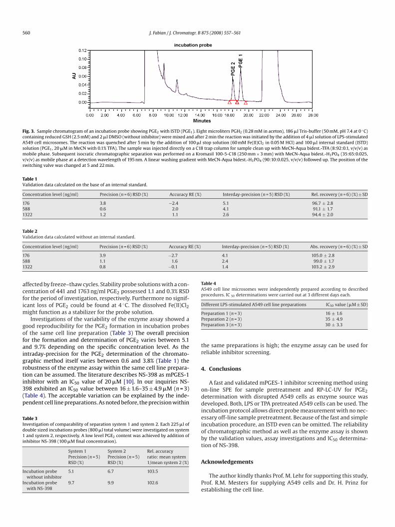
Blank and zero sample chromatograms did not show any inter-erence with the analyte PGE2 and ISTD PGE1 (data not shown). Botheaks were base line separated in the incubation chromatogramFig. 3). The calibration curve showed a non-weighted linear regres-ion with a coefficient of determination (R2) of 0.999. No calibrationtandards deviated more than ±8% from the nominal concentration.
Intraday-precision, accuracy, interday-precision and recoveryet the accepted criterias for bioanalytical methods (Table 1). Also
he results without ISTD are in accordance with the terms (Table 2).he LOQ and LOD were 57 and 17 ng/ml, respectively. Stabilitynvestigations at −20 ◦C revealed that the probe solution was not
560 J. Fabian / J. Chromatogr. B 875 (2008) 557–561
Fig. 3. Sample chromatogram of an incubation probe showing PGE2 with ISTD (PGE1). Eight microliters PGH2 (0.28 mM in aceton), 186 �l Tris-buffer (50 mM, pH 7.4 at 0 ◦C)containing reduced GSH (2.5 mM) and 2 �l DMSO (without inhibitor) were mixed and after 2 min the reaction was initiated by the addition of 4 �l solution of LPS-stimulatedA549 cell microsomes. The reaction was quenched after 5 min by the addition of 100 �l stop solution (60 mM Fe(II)Cl2 in 0.05 M HCl) and 100 �l internal standard (ISTD)solution (PGE1, 20 �M in MeCN with 0.1% TFA). The sample was injected directly on a C18 trap column for sample clean up with MeCN-Aqua bidest.-TFA (8:92:0.1, v/v/v) asmobile phase. Subsequent isocratic chromatographic separation was performed on a Kromasil 100-5-C18 (250 mm × 3 mm) with MeCN-Aqua bidest.-H3PO4 (35:65:0.025,v/v/v) as mobile phase at a detection wavelength of 195 nm. A linear washing gradient with MeCN-Aqua bidest.-H3PO4 (90:10:0.025, v/v/v) followed up. The position of theswitching valve was changed at 5 and 22 min.
Table 1Validation data calculated on the base of an internal standard.
Concentration level (ng/ml) Precision (n = 6) RSD (%) Accuracy RE (%) Interday-precision (n = 5) RSD (%) Rel. recovery (n = 6) (%) ± SD
176 3.8 −2.4 5.1 96.7 ± 2.8588 0.6 2.0 4.1 91.1 ± 1.71322 1.2 1.1 2.6 94.4 ± 2.0
Table 2Validation data calculated without an internal standard.
Concentration level (ng/ml) Precision (n = 6) RSD (%) Accuracy RE (%) Interday-precision (n = 5) RSD (%) Abs. recovery (n = 6) (%) ± SD
1 4.1 105.0 ± 2.85 2.4 99.0 ± 1.71 1.4 103.2 ± 2.9
acfim
gofaigrti3(p
TId1i
I
I
Table 4A549 cell line microsomes were independently prepared according to describedprocedures. IC 50 determinations were carried out at 3 different days each.
Different LPS-stimulated A549 cell line preparations IC50 value (�M ± SD)
PPP
tr
4
76 3.9 −2.788 1.1 1.6322 0.8 −0.1
ffected by freeze–thaw cycles. Stability probe solutions with a con-entration of 441 and 1763 ng/ml PGE2 possessed 1.1 and 0.3% RSDor the period of investigation, respectively. Furthermore no signif-cant loss of PGE2 could be found at 4 ◦C. The dissolved Fe(II)Cl2
ight function as a stabilizer for the probe solution.Investigations of the variability of the enzyme assay showed a
ood reproducibility for the PGE2 formation in incubation probesf the same cell line preparation (Table 3) The overall precisionor the formation and determination of PGE2 varies between 5.1nd 9.7% depending on the specific concentration level. As thentraday-precision for the PGE2 determination of the chromato-raphic method itself varies between 0.6 and 3.8% (Table 1) theobustness of the enzyme assay within the same cell line prepara-
ion can be assumed. The literature describes NS-398 as mPGES-1nhibitor with an IC50 value of 20 �M [10]. In our inquiries NS-98 exhibited an IC50 value between 16 ± 1.6–35 ± 4.9 �M (n = 3)Table 4). The acceptable variation can be explained by the inde-endent cell line preparations. As noted before, the precision withinable 3nvestigation of comparability of separation system 1 and system 2. Each 225 �l ofouble sized incubations probes (800 �l total volume) were investigated on systemand system 2, respectively. A low level PGE2 content was achieved by addition of
nhibitor NS-398 (100 �M final concentration).
System 1 System 2 Rel. accuracyPrecision (n = 5)RSD (%)
Precision (n = 5)RSD (%)
ratio: mean system1/mean system 2 (%)
ncubation probewithout inhibitor
5.1 6.7 103.5
ncubation probewith NS-398
9.7 9.9 102.6
oddieiobt
A
Pe
reparation 1 (n = 3) 16 ± 1.6reparation 2 (n = 3) 35 ± 4.9reparation 3 (n = 3) 30 ± 3.3
he same preparations is high; the enzyme assay can be used foreliable inhibitor screening.
. Conclusions
A fast and validated mPGES-1 inhibitor screening method usingn-line SPE for sample pretreatment and RP-LC-UV for PGE2etermination with disrupted A549 cells as enzyme source waseveloped. Both, LPS or TPA pretreated A549 cells can be used. The
ncubation protocol allows direct probe measurement with no nec-ssary off-line sample pretreatment. Because of the fast and simplencubation procedure, an ISTD even can be omitted. The reliabilityf chromatographic method as well as the enzyme assay is showny the validation values, assay investigations and IC50 determina-ion of NS-398.
cknowledgements
The author kindly thanks Prof. M. Lehr for supporting this study,rof. R.M. Mesters for supplying A549 cells and Dr. H. Prinz forstablishing the cell line.
ogr. B
R
[[[
[
[
[
[
[[[
[
[
[[
J. Fabian / J. Chromat
eferences
[1] B. Samuelsson, R. Morgenstern, P.J. Jakobsson, Pharmacol. Rev. 59 (2007) 207.[2] S. Thoren, R. Weinander, S. Saha, C. Jegerschold, P.L. Pettersson, B. Samuelsson,
H. Hebert, M. Hamberg, R. Morgenstern, P.J. Jakobsson, J. Biol. Chem. 278 (2003)22199.
[3] M. Ouellet, J.P. Falgueyret, P.H. Ear, A. Pen, J.A. Mancini, D. Riendeau, M.D. Per-cival, Protein Exp. Purif. 26 (2002) 489.
[4] S. Narumiya, Y. Sugimoto, F. Ushikubi, Physiol. Rev. 79 (1999) 1193.[5] T. Grosser, S. Fries, G.A. FitzGerald, J. Clin. Invest. 116 (2006) 4.[6] Y. Cheng, M. Wang, Y. Yu, J. Lawson, C.D. Funk, G.A. Fitzgerald, J. Clin. Invest. 116
(2006) 1391.[7] M.D. AbdulHameed, A. Hamza, J. Liu, X. Huang, C.G. Zhan, J. Chem. Inf. Model
48 (2008) 179.[8] X. Huang, W. Yan, D. Gao, M. Tong, H.H. Tai, C.G. Zhan, Bioorg. Med. Chem. 14
(2006) 3553.[9] A.A. San Juan, S.J. Cho, J. Mol. Model 13 (2007) 601.10] S. Thoren, P.J. Jakobsson, Eur. J. Biochem. 267 (2000) 6428.
11] O. Quraishi, J.A. Mancini, D. Riendeau, Biochem. Pharmacol. 63 (2002) 1183.12] D. Riendeau, R. Aspiotis, D. Ethier, Y. Gareau, E.L. Grimm, J. Guay, S. Guiral, H.Juteau, J.A. Mancini, N. Methot, J. Rubin, R.W. Friesen, Bioorg. Med. Chem. Lett.15 (2005) 3352.
13] B. Cote, L. Boulet, C. Brideau, D. Claveau, D. Ethier, R. Frenette, M. Gagnon, A.Giroux, J. Guay, S. Guiral, J. Mancini, E. Martins, F. Masse, N. Methot, D. Riendeau,
[
[[
875 (2008) 557–561 561
J. Rubin, D. Xu, H. Yu, Y. Ducharme, R.W. Friesen, Bioorg. Med. Chem. Lett. 17(2007) 6816.
14] M.C. Catley, J.E. Chivers, L.M. Cambridge, N. Holden, D.M. Slater, K.J. Staples,M.W. Bergmann, P. Loser, P.J. Barnes, R. Newton, FEBS Lett. 547 (2003) 75.
15] Y. Ikeda-Matsuo, Y. Ikegaya, N. Matsuki, S. Uematsu, S. Akira, Y. Sasaki, J. Neu-rochem. 94 (2005) 1546.
16] F. Masse, S. Guiral, L.J. Fortin, E. Cauchon, D. Ethier, J. Guay, C. Brideau, J. Biomol.Screen. 10 (2005) 599.
17] G. Dannhardt, M. Lehr, J. Pharm. Pharmacol. 44 (1992) 419.18] A. Margalit, K.L. Duffin, P.C. Isakson, Anal. Biochem. 235 (1996) 73.19] R. Schmidt, O. Coste, G. Geisslinger, J. Chromatogr. B Analyt. Technol. Biomed.
Life Sci. 826 (2005) 188.20] K. Olofsson, E. Suna, B. Pelcman, V. Ozola, M. Katkevics, I. Kalvins, WIPO (2005)
WO 2005/123673.21] S. Rinne, C. Ramstad Kleiveland, M. Kassem, T. Lea, E. Lundanes, T. Greibrokk, J.
Sep. Sci. 30 (2007) 1860.22] W.S. Powell, Anal. Biochem. 164 (1987) 117.23] W. Schafer, C. Strandberg, T. Gaillard, H.P. Zahradnik, Eicosanoids 1 (1988)
101.24] G. Hotter, I. Ramis, G. Bioque, C. Sarmiento, J.M. Fernandez, J. Rosello-Catafau,
E. Gelpi, Chromatographia 36 (1993) 33.25] C.M. Stoscheck, Anal. Biochem. 184 (1990) 111.26] F.J. Sweeney, T.S. Wachtmann, J.D. Eskra, K.A. Verdries, R.H. Lambalot, T.J. Carty,
J.R. Perez, L.P. Audoly, Mol. Cell. Endocrinol. 205 (2003) 151.