Embed Size (px)
Citation preview
D E T E R M I N I N G T H E O R G A N I S M S
A N D PA T H W A Y S O F I N F E C T I O N
L E A D I N G T O M A S T I T I S I N E W E S
S e l i n C o o p e r
U N I V E R S I T Y O F W A R W I C K
F E B R U A R Y 2 0 1 2
1 y e a r r e p o r t
S t a r t d a t e : 1 / 3 / 2 0 1 1
S u p e r v i s o r s : P r o f e s s o r L a u r a G r e e n
D r . K e v i n P u r d y
INTRODUCTION
Mastitis: an overview
Mastitis is an inflammation of the mammary gland primarily caused by bacterial infection
that results in inflammation of the mammary tissue (Khan & Khan 2006). Mastitis can
also be caused by viral infections, trauma, allergies, and physiological and metabolic
changes (Bergonier et al. 2003). Mastitis can be classified as clinical, where an animal
has overt clinical signs, or subclinical, where clinical signs are absent. Ewes that have
developed mastitis may experience discomfort and pain in the udder, or be unwell overall
or even die.
However, these are fairly rigid classifications of disease, and it can be argued that there is
a single udder disease in sheep, caused by a variety of bacteria, with differences in
clinical manifestation. It is thus important to consider mastitis with some flexibility of
thought; it is likely that there is a continual shift in condition of the udder from health to
disease and vice versa. There is no absolute point at which a mammary gland is diseased
but rather, after colonisation of bacteria, a change in the ewe results in the development
of disease. To date, much of the research on mastitis has tended to focus on causative
species determined by culture and clinical signs, due to technical limitations. This has not
allowed us to understand the dynamics of the disease in terms of when and how ewes’
udders become infected with potentially pathogenic bacterial strains.
Endemic diseases such as mastitis result in a direct and indirect economic loss for the
industry. In both ewes kept to produce milk (hereafter dairy ewes) and in ewes kept to
produce lambs for human consumption (hereafter suckler ewes), costs of diagnosis,
treatments, preventive measures, labour, carcass disposal, and ewe replacements result in
a substantial economic loss (Hogeveen et al. 2011; Pinzón-Sánchez et al. 2011). In dairy
ewes, decreased milk yield (by up to 55%)(Saratsis et al. 1999), downgrading of milk due
to high somatic cell counts (SCC) and higher bacterial counts would contribute to the
economic loss attributed to mastitis (Fthenakis & Jones 1990). Milk bacterial counts are
monitored due to the human health hazards posed. Milk is heated in order to minimise
this risk, however in cases of cheese made with raw milk, and the possibility of
thermostable toxins produced by mastitis causing pathogens surviving in pasteurised
milk, controlling mastitis becomes a priority (Contreras et al. 2007). In meat producing
sheep, costs occur through decreased live-weight gain of lambs, and loss of lambs that
would have been reared by the affected ewe. In addition, lamb performance is decreased
in lambs whose mothers have subclinical mastitis, due to decreased milk production
(Fthenakis & Jones 1990; Keisler et al. 1992), and changes in suckling behaviour
(Gougoulis et al. 2008), which is particularly important in suckler flocks. Improvement
in management of mastitis will benefit the health and welfare of sheep and lambs and
might help to reduce economic losses.
Aetiology
Over 130 different organisms have been associated with infection of the bovine
mammary gland (Jeffrey L 1988), it is probable that a similar number would be found in
the ovine mammary gland. The complexity of the infection is further highlighted when
we consider not only species, but strain types.
In suckler ewes, bacteria commonly isolated from subclinical mastitis include coagulase-
negative staphylococci (CNS), such as Staphylococcus epidermidis, Staphylococcus
simulans, Staphylococcus chromogenes, Staphylococcus xylosus (Fthenakis 1994) and
coagulase-positive staphylococci (CPS) such as Staphylococcus aureus (Bergonier et al.
2003; Kiossis et al. 2007; Kirk et al. 1996; Mork et al. 2007; Winter & Colditz 2002). In
dairy ewes, CNS are a common cause of subclinical (undetected) mastitis (Kirk et al.
1996; Mork et al. 2007; Pengov 2001). Bacteria isolated from clinical mastitis include
Staphylococcus aureus, Mannheimia haemolytica, Escherichia coli, Mycoplasma spp
(Bergonier & Berthelot 2003) and Streptococcus spp. (Fragkou et al. 2011; Fragkou et al.
2007; Fragkou et al. 2007; Las Heras et al. 2002) including Streptococcus uberis
(Marogna et al. 2010) and Streptococcus agalactiae (Lafi et al. 1998). Despite its
renowned link with subclinical mastitis, CNS have also been shown to play a role in
clinical mastitis in dairy ewes (Lafi et al. 1998).
The defensive role of the udder
There are three sources from which bacteria is likely to invade the mammary gland and
cause infection: the lamb’s mouth, the ewe’s udder skin or the environment, including
fields and bedding in housed sheep (Gougoulis et al. 2008; Piccinini et al. 2009).
Pathogenic bacteria are likely to enter the udder half through the teat orifice, colonising
the teat canal and cistern.
The mammary gland has a defensive role against pathogenic bacteria entering the teat
duct. Sphincter muscles keep the teat canal tightly closed to prevent the entry of
pathogens. The teat canal is lined with keratinocytes, considered to be the first line of
physical defense for the udder (Forbes 1970). It hinders the movement of bacteria up the
teat canal and also contains antimicrobial agents (Sordillo & Streicher 2002). In an
experimental study in dairy cows, when the keratin was partially removal from the teat
canal, the ability of the teat canal to act as a protective anatomical feature against
bacterial pathogens from the external environment was compromised (Capuco et al.
1992). In addition, the presence of induced subepithelial lymphoid tissue between the teat
duct and teat cistern appear to have a role in the protection of the mammary gland against
the early stages of bacterial infection (Fragkou et al. 2010; Fragkou et al. 2007).
Healthy mammary glands have bacteria colonised on the udder and teat skin (Fragkou et
al. 2007), which may be a source of bacteria that would be transferred into the teat duct
(Fragkou et al. 2011; Scott & Jones 1998). Interactions between the host’s mammary
defense system and the virulence of the invading pathogenic bacteria determines the
severity and extent of infection and disease. In order to understand the pathogenesis of a
multifactorial microbial disease such as mastitis, it is important to consider the flora of
the udder, as well as the bacterial content in the milk and in particular temporal
interactions between them.
Entrance of a pathogen into the teat does not necessarily result in infection (Mavrogianni
et al. 2006). For example, some bacteria may enter the teat canal but subsequently be
withdrawn during suckling (Gougoulis et al. 2008). In an experimental study,
Mannheimia haemolytica inoculated into the teat duct did not always cause clinical
infection suggesting a protective role for the teat (Mavrogianni et al. 2005). In the sequel
to that work, the same experimental design with ewes with natural or experimentally
generated lesions on the surface of the teat had pathogenic bacteria deposited into the teat
duct, resulting in the development of mastitis (Mavrogianni et al. 2006). It was postulated
that Mannheimia haemolytica may form part of the teat duct flora without resulting in
disease, suggesting that this bacteria does not necessarily always cause mastitis and for
mastitis to occur, it requires either excessive accumulation of the bacteria (Fragkou et al.
2007) or injury to the teat (Mavrogianni et al. 2006). Indeed it appears that there is an
increased risk in bacterial colonisation of the teat duct compared to the mammary gland
of the same ewe (Mavrogianni et al. 2007).
The protective role of udder skin microflora
In addition, bacteria present on the udder skin may act as an inhibitor of major mastitis
pathogens. For example, Staphylococcus chromogenes has been shown to protect udder
quarters against elevated somatic cell counts post-partum. Further experimentation in
vitro has revealed the in vitro inhibitory capability of Staphylococcus chromogenes
(isolated from teat apices of heifers) against Staphylococcus aureus, Streptococcus
dysgalactiae and Streptococcus uberis using the cross-streaking method (De Vliegher et
al. 2004). This also supports the earlier discussion of the inhibitory effect of CNS in the
milk from protecting against major mastitis pathogens from causing severe infection
(Woodward et al. 1987).
Persistence of infections in the mammary gland
Whilst a great deal of research has identified sources of infection, little research has been
done on the persistence of particular strains of bacteria in milk throughout lactation in
cows once bacteria have penetrated the mammary gland. Much of the treatment advice
for persistent infections in sheep still leverages on bovine mastitis research. There is no
research on the persistence of infections in sheep during the lactating or dry period.
Different strains within a bacterial species can differ in pathogenicity and transmission
routes, classification of isolates at the species level can incorrectly oversimplify control
measures recommended, hence the need for longitudinal strain typing methods.
Infection persistence during lactation
Recent studies have used strain typing techniques such as pulse field gel electrophoresis
to test whether the same strain of bacteria from the same mammary quarter is present
over time indicating persistence with CNS based on a study of 12,412 milk samples from
3 dairy research herds (Gillespie et al. 2009). In fact, CNS persisted for up to 10 months
(Gillespie et al. 2009). Escherichia coli has also been shown to persist in the bovine
mammary gland, in a study of 300 dairy cows, with an estimated occurrence of between
4.8% and 9.1% of the herd population (Döpfer et al. 1999; Lam et al. 1996). However
this was thought to be an underestimation. In addition the occurrence of recurrent
episodes of the same strain in more than one quarter in a cow was high (Döpfer et al.
1999), suggesting transmission between quarters, which could increase persistence of the
strain. Lipman et al., (1995) also found persistence of the same Escherichia coli
serotypes in the bovine mammary gland, although different methods (serotyping and
DNA polymorphism patterns) were used. In addition, infection with Eschierichia coli
more than once in a lactation was infrequent (Lipman et al. 1995). Conversely, in a study
of 503 cows from 5 herds, quarters were often infected with multiple Streptococcus
uberis strain types, despite the ability of Streptococcus uberis to persist in the udder
(McDougall et al. 2004). This suggests that some pathogens have a superior mechanism
of bacterial persistence than others. In fact, some pathogens subtypes, such as Listeria
moncytogenes, are able to persist in the milking parlour which could be a source of
reinfection (Ho et al. 2007).
The role of the dry period
The bovine mammary gland is thought to be particularly susceptible to new
environmental coliform and streptococcal infections during the dry period (Larry Smith
et al. 1985; Oliver & Mitchell 1983; Todhunter et al. 1995), including Streptococcus
uberis (Todhunter et al. 1995) and Escherichia coli (Bradley & Green 2001; Döpfer et al.
1999; Lipman et al. 1995). Indeed, experimental studies have shown the ability of
pathogens to remain within the udder, causing clinical disease after the onset of lactation
(A.J 2002; McDonald & Anderson 1981).
Two studies in particular highlight the significance of the dry period in the persistence of
mastitis causing pathogens.
In a study of 629 cows from 6 commercial herds, samples were collected during the dry
period and from clinical quarters of these cows during the subsequent lactation allowing
comparisons to be made between these time periods. DNA fingerprinting showed the
persistence of enterobacterial organisms acquired during the dry period, causing disease
after the onset of lactation (Bradley & Green 2000). One quarter remained persistently
infected for >200 days before resulting in severe clinical mastitis (A.J 2002). Of all the
enterobacterial mastitis occurring in the first 100 days of lactation, 52.6% arose in
quarters previously colonized with the same strain of bacteria during the dry period
(Bradley & Green 2000).
Bradley and Green (2001), observed 6 commercial herds over a period of 12 months in
order to identify changes in the behavior of Escherichia coli as a mastitis
pathogen. DNA
fingerprinting allowed the identification of the genotypes of strains involved in recurrent
cases of clinical E.coli mastitis. In the majority of cases, the same genotype was
implicated as the cause of disease in recurrent cases and often the same genotype was
identified in different quarters of the same cow suggesting that the same genotype may
persist in the mammary environment (for more than 100 days), causing recurring
infections and that bacteria may be spread between quarters (Bradley & Green 2001).
PCR based DNA fingerprinting identified the same Streptococcus uberis and
Streptococcus dysgalactiae subtypes from some infected mammary glands from one
lactation to the next, highlighting the persistence of these organisms through the dry
period and during lactation (Oliver et al. 1998).
Statistical modelling has been used to identify the relationship between intramammary
infection during the dry period, and clinical mastitis in the next lactation. The probability
of an udder quarter developing clinical mastitis increased when Streptococcus
dysgalactiae, Streptococcus faecalis, Escherichia coli, or Enterobacter spp. were
cultured at drying off. In addition, the risk of clinical mastitis for specific pathogens
increased if they were cultured in 2 or more late dry and post calving samples.
Interestingly, the time that an isolate was identified was important in whether it increased
or decreased the risk of clinical mastitis development; Corynebacterium spp., when
isolated at drying off were associated with an increase, but when isolated in the late dry
or post calving samples was associated with a reduction in the risk of clinical mastitis
(Green et al. 2002). Although this study did not use strain typing to prove the persistence
of certain strains in the mammary gland, it provides evidence for the significance of the
dry period on clinical mastitis development.
AIMS, OBJECTIVES AND HYPOTHESES
The overall aim of this PhD is to contribute to the understanding of the pathways of
infection in the udder. Ultimately this will assist in identifying when and how ewes’
udders become infected and whether certain strains of bacteria are responsible for
infection.
Specific aims:
• Investigate the development of intramammary infection, from colonisation with
an infecting strain to development of disease
• Determine links between strains of bacteria found on udder skin and the
intramammary infections they cause
• Identify management and environmental factors that are associated with a ewe’s
risk of developing intramammary infection
Objectives:
Stored samples of milk and udder swabs will be used, and new data will be collected
during field work on sheep farms in England. Analysis will integrate results from culture,
molecular analysis and statistical modelling. All samples will be analysed using MALDI-
TOF MS. A subset of samples will be validated using other techniques such as PFGE and
MLST alongside traditional culturing methods.
Hypotheses:
1. Development of intramammary infection is as a result of colonisation of the udder with
certain strains of bacteria.
2. Ewes with intramammary infection have certain bacterial strains in their milk in the
weeks after the same bacterial strains have been isolated from the environment (lambs
mouths, bedding, teat skin/teat lesions).
ORIGINAL RESEARCH PLAN YEAR 1
The general research plan up to 2 years is shown in appendix 1.
Year 1
The model on risk factors for intramammary infection from the cross-sectional
study is ongoing.
Texel flock samples used as a pilot to test and parameterise all laboratory
techniques, as well as producing results on this sample flock.
Year 2
All of the samples (both milk and swabs) from the longitudinal studies LS_2010
and LS_2011 are analysed and bacterial strains identified. Statistical comparisons
made between the milk and swabs for each ewe and other data could also be
combined to produce a multilevel model.
Field-work studies have been designed in order to collect the remaining data
required.
The model on risk factors for intramammary infection from the cross-sectional
study finished.
Year 3
Field-work on farms in England.
Lab analysis continues on samples on days that there are no farm visits allowing
the studies to progress quickly.
Year 4
Statistical analysis and modelling in order to explore patterns within the datasets
thus contributing to the understanding of the pathways of infection in the udder.
By the end of year 4, all samples would be analysed and statistical analysis would
be completed. Thesis submission.

RESEARCH PLAN YEAR 2 (DETAILED)
Texel flock
By the beginning of my second year, I hope to have begun PFGE on selected Texel
isolates. Ideally, this would only continue a few months into my second year, and so
would give me the opportunity to write this chapter up in the early stages of year 2.
Cross sectional study
The majority of the statistical analyses for this study will be done in year 2. I intend on
using R for this study and thus extra time must be allowed for this (10 months in total). I
may have an undergraduate or MSc student working on this part of the project with me. I
hope to complete this as a chapter in my second year.
LS_2010 and LS_2011
These two studies will begin in my second year. They will require a large amount of time
in the lab and I will also need to work for a few months in Somerset in order to use the
MALDI-TOF if we do not have this here. I have given myself a year for both studies
running alongside one another. Although I anticipate that the chapters will not be
completed until my third year.
New field work study set-up
I will be able to assess my progress towards the end of my second year and decide
whether I need more samples, and if I do then I can design a study that will directly
address my remaining aims.
Year 3
Set up new study to address remaining aims
o Field work, laboratory analyses and statistical analyses
Begin statistical analysis (cross-classified mixed effect model)
Decide on remaining studies
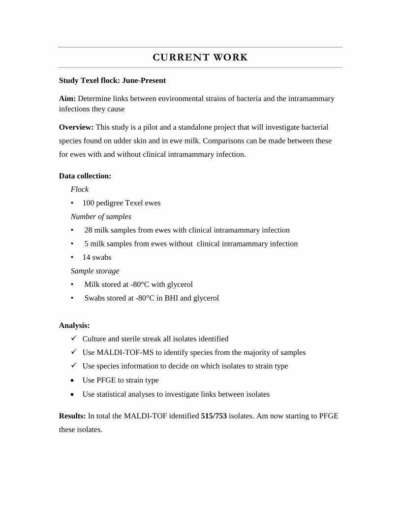
CURRENT WORK
Study Texel flock: June-Present
Aim: Determine links between environmental strains of bacteria and the intramammary
infections they cause
Overview: This study is a pilot and a standalone project that will investigate bacterial
species found on udder skin and in ewe milk. Comparisons can be made between these
for ewes with and without clinical intramammary infection.
Data collection:
Flock
• 100 pedigree Texel ewes
Number of samples
• 28 milk samples from ewes with clinical intramammary infection
• 5 milk samples from ewes without clinical intramammary infection
• 14 swabs
Sample storage
• Milk stored at -80°C with glycerol
• Swabs stored at -80°C in BHI and glycerol
Analysis:
Culture and sterile streak all isolates identified
Use MALDI-TOF-MS to identify species from the majority of samples
Use species information to decide on which isolates to strain type
Use PFGE to strain type
Use statistical analyses to investigate links between isolates
Results: In total the MALDI-TOF identified 515/753 isolates. Am now starting to PFGE
these isolates.
CURRENT WORK: CONTINUED
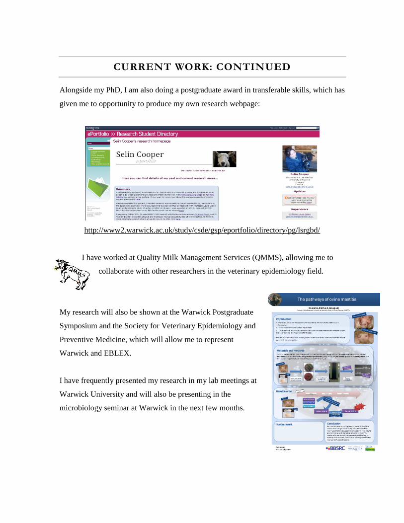
Alongside my PhD, I am also doing a postgraduate award in transferable skills, which has
given me to opportunity to produce my own research webpage:
http://www2.warwick.ac.uk/study/csde/gsp/eportfolio/directory/pg/lsrgbd/
I have worked at Quality Milk Management Services (QMMS), allowing me to
collaborate with other researchers in the veterinary epidemiology field.
My research will also be shown at the Warwick Postgraduate
Symposium and the Society for Veterinary Epidemiology and
Preventive Medicine, which will allow me to represent
Warwick and EBLEX.
I have frequently presented my research in my lab meetings at
Warwick University and will also be presenting in the
microbiology seminar at Warwick in the next few months.
3
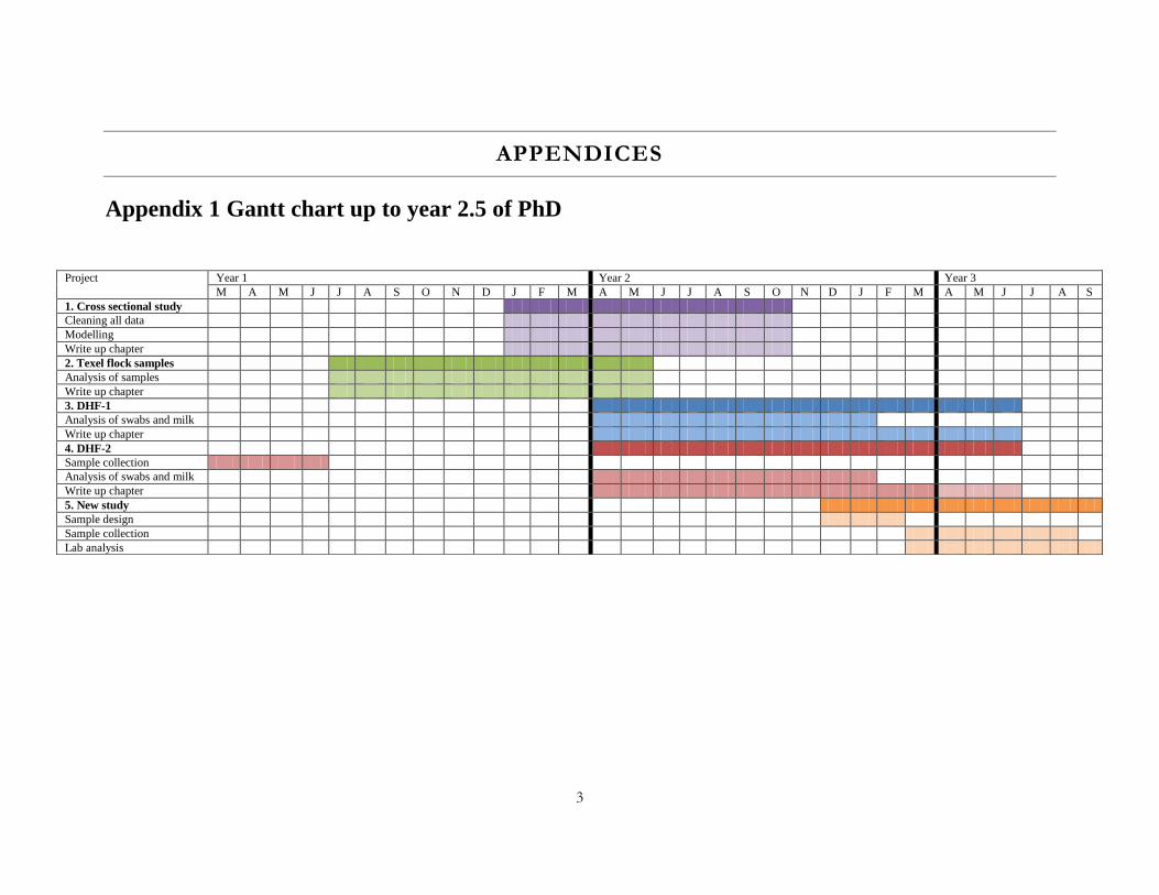
APPENDICES
Appendix 1 Gantt chart up to year 2.5 of PhD
Project Year 1 Year 2 Year 3
M A M J J A S O N D J F M A M J J A S O N D J F M A M J J A S
1. Cross sectional study
Cleaning all data
Modelling
Write up chapter
2. Texel flock samples
Analysis of samples
Write up chapter
3. DHF-1
Analysis of swabs and milk
Write up chapter
4. DHF-2
Sample collection
Analysis of swabs and milk
Write up chapter
5. New study
Sample design
Sample collection
Lab analysis
4
REFERENCES
A.J, B. (2002) Bovine Mastitis: An Evolving Disease, The Veterinary Journal 164, 116-128. Batavani, R. A., Mortaz, E., Falahian, K., and Dawoodi, M. A. (2003) Study on frequency,
etiology and some enzymatic activities of subclinical ovine mastitis in Urmia, Iran, Small Ruminant Research 50, 45-50.
Beheshti, R., J., S., Eshratkhah , B., Ghalehkandi, J. G., and Maheri-Sis, N. (2010) Prevalence
and etiology of subclinical mastitis in ewes of the Tabriz region, Iran, Global Veterinaria 4, 299-302.
Bergonier, D., and Berthelot, X. (2003) New advances in epizootiology and control of ewe
mastitis, Livestock Production Science 79, 1-16. Bergonier, D., de Cremoux, R., Rupp, R., Lagriffoul, G., and Berthelot, X. (2003) Mastitis of
dairy small ruminants, Veterinary Research 34, 689 - 716. Berriatua, E., Ziluaga, I., Miguel-Virto, C., Uribarren, P., Juste, R., Laevens, S., Vandamme,
P., and Govan, J. R. W. (2001) Outbreak of Subclinical Mastitis in a Flock of Dairy Sheep Associated with Burkholderia cepacia Complex Infection, Journal of Clinical Microbiology 39, 990-994.
Bradley, A. J., and Green, M. J. (2000) A Study of the Incidence and Significance of
Intramammary Enterobacterial Infections Acquired During the Dry Period, Journal of Dairy Science 83, 1957-1965.
Bradley, A. J., and Green, M. J. (2001) Adaptation of Escherichia coli to the Bovine
Mammary Gland, J. Clin. Microbiol. 39, 1845-1849. Capuco, A. V., Bright, S. A., Pankey, J. W., Wood, D. L., Miller, R. H., and Bitman, J. (1992)
Increased Susceptibility to lntramammary Infection Following Removal of Teat Canal Keratin, Journal of Dairy Science 75, 2126-2130.
De Vliegher, S., Opsomer, G., Vanrolleghem, A., Devriese, L. A., Sampimon, O. C., Sol, J.,
Barkema, H. W., Haesebrouck, F., and de Kruif, A. (2004) In vitro growth inhibition of major mastitis pathogens by Staphylococcus chromogenes originating from teat apices of dairy heifers, Veterinary Microbiology 101, 215-221.
Deinhofer, M., and Pernthaner, A. (1995) Staphylococcus spp. as mastitis-related pathogens
in goat milk, Veterinary Microbiology 43, 161-166. Desmasures, N., Opportune, W., and Guéguen, M. (1997) Lactococcus spp., yeasts and
Pseudomonas spp. on teats and udders of milking cows as potential sources of milk contamination, International Dairy Journal 7, 643-646.
Döpfer, D., Barkema, H. W., Lam, T. J. G. M., Schukken, Y. H., and Gaastra, W. (1999)
Recurrent Clinical Mastitis Caused by Escherichia coli in Dairy Cows, Journal of Dairy Science 82, 80-85.
Ericsson Unnerstad, H., Lindberg, A., Persson Waller, K., Ekman, T., Artursson, K., Nilsson-Öst, M., and Bengtsson, B. (2009) Microbial aetiology of acute clinical mastitis and agent-specific risk factors, Veterinary Microbiology 137, 90-97.
Forbes, D. (1970) The survival of micrococcaceae in bovine teat canal keratin, The British
Veterinary Journal 126, 268 - 274. Fragkou, I. A., Dagleish, M. P., Papaioannou, N., Cripps, P. J., Boscos, C. M., Ververidis, H.
N., Orfanou, D. C., Solomakos, N., Finlayson, J., Govaris, A., Kyriazakis, I., and Fthenakis, G. C. (2010) The induction of lymphoid follicle-like structures in the ovine teat duct following experimental infection with Mannheimia haemolytica, The Veterinary Journal 184, 194-200.
Fragkou, I. A., Gougoulis, D. A., Billinis, C., Mavrogianni, V. S., Bushnell, M. J., Cripps, P.
J., Tzora, A., and Fthenakis, G. C. (2011) Transmission of Mannheimia haemolytica from the tonsils of lambs to the teat of ewes during sucking, Veterinary Microbiology 148, 66-74.
Fragkou, I. A., Mavrogianni, V. S., Cripps, P. J., Gougoulis, D. A., and Fthenakis, G. C.
(2007) The bacterial flora in the teat duct of ewes can protect against and can cause mastitis, Veterinary Research 38, 525-545.
Fragkou, I. A., Mavrogianni, V. S., Papaioannou, N., Boscos, C., Cripps, P. J., Skoufos, J.,
and Fthenakis, G. C. (2007) Presence of sub-epithelial lymphoid tissues in the teat of ewe-lambs and adult ewes, Small Ruminant Research 70, 286-291.
Fragkou, I. A., Papaioannou, N., Cripps, P. J., Boscos, C. M., and Fthenakis, G. C. (2007)
Teat lesions predispose to invasion of the ovine mammary gland by Mannheimia haemolytica, Journal of Comparative Pathology 137, 239-244.
Fragkou, I. A., Skoufos, J., Cripps, P. J., Kyriazakis, I., Papaioannou, N., Boscos, C. M.,
Tzora, A., and Fthenakis, G. C. (2007) Differences in susceptibility to Mannheimia haemolytica-associated mastitis between two breeds of dairy sheep, Journal of Dairy Research 74, 349-355.
Fthenakis, G. C. (1994) Prevalence and aetiology of subclinical mastitis in ewes of Southern
Greece, Small Ruminant Research 13, 293-300. Fthenakis, G. C., and Jones, J. E. T. (1990) Incidence and aetiology of clinical ovine mastitis
in flocks in central Macedonia (Greece), Delt. Ellen. Kten. Etair 41, 133 – 141. Fthenakis, G. C., Leontides, L., Skoufos, J., Taitzoglou, I. A., and Tzora, A. (2004) Case
report: high prevalence rate of ovine mastitis, caused by coagulase-negative staphylococci and predisposed by increased gossypol consumption, Small Ruminant Research 52, 185-189.
Fthenakis, G. C., Saratsis, P., Tzora, A., and Linde, K. (1998) Naturally occurring subclinical
ovine mastitis associated with Listeria monocytogenes, Small Ruminant Research 31, 23-27. Gillespie, B. E., Headrick, S. I., Boonyayatra, S., and Oliver, S. P. (2009) Prevalence and
persistence of coagulase-negative Staphylococcus species in three dairy research herds, Veterinary Microbiology 134, 65-72.
Gougoulis, D. A., Kyriazakis, I., Tzora, A., Taitzoglou, I. A., Skoufos, J., and Fthenakis, G. C. (2008) Effects of Lamb Sucking on the Bacterial Flora of Teat Duct and Mammary Gland of Ewes, Reproduction in Domestic Animals 43, 22-26.
Green, M. J., Green, L. E., Medley, G. F., Schukken, Y. H., and Bradley, A. J. (2002)
Influence of Dry Period Bacterial Intramammary Infection on Clinical Mastitis in Dairy Cows, Journal of dairy science 85, 2589-2599.
Ho, A. J., Lappi, V. R., and Wiedmann, M. (2007) Longitudinal Monitoring of Listeria
monocytogenes Contamination Patterns in a Farmstead Dairy Processing Facility, Journal of Dairy Science 90, 2517-2524.
Jeffrey L, W. (1988) Etiological agents of bovine mastitis, Veterinary Microbiology 16, 41-66. Khan, M. Z., and Khan, A. (2006) Basic facts of mastitis in dairy animals, Pakistan Veterinary
Journal 26, 204 - 208. Kiossis, E., Brozos, C. N., Petridou, E., and Boscos, C. (2007) Program for the control of
subclinical mastitis in dairy Chios breed ewes during lactation, 73, 194-199. Kirk, J. H., Glenn, J. S., and Maas, J. P. (1996) Mastitis in a flock of milking sheep, Small
Ruminant Research 22, 187-191. Lafi, S. Q., Al-Majali, A. M., Rousan, M. D., and Alawneh, J. M. (1998) Epidemiological
studies of clinical and subclinical ovine mastitis in Awassi sheep in northern Jordan, Preventive Veterinary Medicine 33, 171-181.
Lam, T. J., Lipman, L. J., Schukken, Y. H., Gaastra, W., and Brand, A. (1996)
Epidemiological characteristics of bovine clinical mastitis caused by Staphylococcus aureus and Escherichia coli studied by DNA fingerprinting., Am J Vet Res 57, 39 - 42.
Larry Smith, K., Todhunter, D. A., and Schoenberger, P. S. (1985) Environmental Pathogens
and Intramammary Infection During the Dry Period1,2, Journal of Dairy Science 68, 402-417. Las Heras, A., Vela, A. I., Fernandez, E., Legaz, E., Dominguez, L., and Fernandez-
Garayzabal, J. F. (2002) Unusual Outbreak of Clinical Mastitis in Dairy Sheep Caused by Streptococcus equi subsp. zooepidemicus, J. Clin. Microbiol. 40, 1106-1108.
Lipman, L. J. A., de Nijs, A., Lam, T. J. G. M., and Gaastra, W. (1995) Identification of
Escherichia coli strains from cows with clinical mastitis by serotyping and DNA polymorphism patterns with REP and ERIC primers, Veterinary Microbiology 43, 13-19.
Marogna, G., Rolesu, S., Lollai, S., Tola, S., and Leori, G. (2010) Clinical findings in sheep
farms affected by recurrent bacterial mastitis, Small Ruminant Research 88, 119-125. Mavrogenis, A. P., Koumas, A., Kakoyiannis, C. K., and Taliotis, C. H. (1995) Use of
somatic cell counts for the detection of subclinical mastitis in sheep, Small Ruminant Research 17, 79-84.
Mavrogianni, V., S. , Cripps, P., J. , Papaioannou, N., Taitzoglou, I., and Fthenakis, G. C.
(2006) Teat disorders predispose ewes to clinical mastitis after challenge with Mannheimia haemolytica, Veterinary Research 37, 89-105.
Mavrogianni, V. S., Cripps, P. J., and Fthenakis, G. C. (2006) Description and validation of a
novel technique to study the bacterial flora of the teat duct of ewes, Small Ruminant Research 66, 258-264.
Mavrogianni, V. S., Cripps, P. J., and Fthenakis, G. C. (2007) Bacterial flora and risk of
infection of the ovine teat duct and mammary gland throughout lactation, Preventive Veterinary Medicine 79, 163-173.
Mavrogianni, V. S., Cripps, P. J., Papaioannou, N., Taitzoglou, I., and Fthenakis, G. C.
(2006) Teat disorders predispose ewes to clinical mastitis after challenge with Mannheimia haemolytica, Vet. Res. 37, 89-105.
Mavrogianni, V. S., Cripps, P. J., Tzora, A., Skoufos, I., and Fthenakis, G. C. (2006) Effects
of hand milking on the bacterial flora of mammary gland and teat duct of ewes, Journal of Dairy Research 73, 353-356.
Mavrogianni, V. S., Fthenakis, G. C., Brooks, H., Papaioannou, N., Cripps, P. J., Taitzoglou,
I., Brellou, G., and Saratsis, P. (2005) The effects of inoculation of Mannheimia haemolytica into the teat of lactating ewes, Veterinary Research 36, 13-25.
McDonald, J. S., and Anderson, A. J. (1981) Experimental intramammary infection of the
dairy cow with Escherichia coli during the nonlactating period., Am J Vet Res 42, 229 - 231. McDougall, S., Parkinson, T. J., Leyland, M., Anniss, F. M., and Fenwick, S. G. (2004)
Duration of Infection and Strain Variation in Streptococcus uberis Isolated from Cows’ Milk, Journal of Dairy Science 87, 2062-2072.
Mork, T., Waage, S., Tollersrud, T., Kvitle, B., and Sviland, S. (2007) Clinical mastitis in
ewes; bacteriology, epidemiology and clinical features, Acta Veterinaria Scandinavica 49, 1-8. Oliver, S. P., Gillespie, B. E., and Jayarao, B. M. (1998) Detection of new and persistent
Streptococcus uberis and Streptococcus dysgalactiae intramammary infections by polymerase chain reaction-based DNA fingerprinting, FEMS Microbiology Letters 160, 69-73.
Oliver, S. P., and Mitchell, B. A. (1983) Susceptibility of Bovine Mammary Gland to
Infections During the Dry Period1, Journal of Dairy Science 66, 1162-1166. Osman, K. M., El-Enbaawy, M. I., Ezzeldeen, N. A., and Hussein, H. M. G. (2009) Mastitis
in dairy buffalo and cattle in Egypt due to Clostridium perfringens: prevalence, incidence, risk factors and costs, Revue scientifique et technique International Office of Epizootics 28, 975 - 986.
Pengov, A. (2001) The Role of coagulase-negative Staphylococcus spp. and associated somatic
cell counts in the ovine mammary gland, Journal of Dairy Science 84, 572-574. Piccinini, R., Cesaris, L., Daprà, V., Borromeo, V., Picozzi, C., Secchi, C., and Zecconi, A.
(2009) The role of teat skin contamination in the epidemiology of Staphylococcus aureus intramammary infections, Journal of Dairy Research 76, 36-41.
Rendos, J. J., Eberhart, R. J., and Kesler, E. M. (1975) Microbial Populations of Teat Ends
of Dairy Cows, and Bedding Materials1, Journal of Dairy Science 58, 1492-1500.
Saratsis, P., Leontides, L., Tzora, A., Alexopoulos, C., and Fthenakis, G. C. (1998) Incidence risk and aetiology of mammary abnormalities in dry ewes in 10 flocks in Southern Greece, Preventive Veterinary Medicine 37, 173-183.
Scott, M. J., and Jones, J. E. T. (1998) The carriage of Pasteurella haemolytica in sheep and its
transfer between ewes and lambs in relation to mastitis, Journal of Comparative Pathology 118, 359-363.
Sieber, R. L., and Farnsworth, R. J. (1984) Differential diagnosis of bovine teat lesions., Vet
Clin North Am Large Anim Pract. 6, 313 - 321. Sordillo, L. M., and Streicher, K. L. (2002) Mammary Gland Immunity and Mastitis
Susceptibility, Journal of Mammary Gland Biology and Neoplasia 7, 135-146. Supre, K., De Vliegher, S., Cleenwerck, I., Engelbeen, K., Van Trappen, S., Piepers, S.,
Sampimon, O. C., Zadoks, R. N., De Vos, P., and Haesebrouck, F. (2010) Staphylococcus devriesei sp. nov., isolated from teat apices and milk of dairy cows, Int J Syst Evol Microbiol 60, 2739-2744.
Todhunter, D. A., Smith, K. L., and Hogan, J. S. (1995) Environmental Streptococcal
Intramammary Infections of the Bovine Mammary Gland1, Journal of Dairy Science 78, 2366-2374.
Trinidad, P., Nickerson, S. C., and Alley, T. K. (1990) Prevalence of Intramammary Infection
and Teat Canal Colonization In Unbred and Primigravid Dairy Heifers1, Journal of Dairy Science 73, 107-114.
Tzora, A., and Fthenakis, G. C. (1998) Mastitis in dairy ewes associated with Serratia
macrescens, Small Ruminant Research 29, 125-126. Vissers, M. M. M., Driehuis, F., Te Giffel, M. C., De Jong, P., and Lankveld, J. M. G. (2007)
Short Communication: Quantification of the Transmission of Microorganisms to Milk via Dirt Attached to the Exterior of Teats, Journal of Dairy Science 90, 3579-3582.
Winter, P., and Colditz, I. G. (2002) Immunological responses of the lactating ovine udder
following experimental challenge with Staphylococcus epidermidis, Veterinary Immunology and Immunopathology 89, 57-65.
Woodward, W. D., Besser, T. E., Ward, A. C., and Corbeil, L. B. (1987) In vitro growth
inhibition of mastitis pathogens by bovine teat skin normal flora, Canadian journal of veterinary research = Revue canadienne de recherche veterinaire 51, 27-31.
Zdanowicz, M., Shelford, J. A., Tucker, C. B., Weary, D. M., and von Keyserlingk, M. A. G.
(2004) Bacterial Populations on Teat Ends of Dairy Cows Housed in Free Stalls and Bedded with Either Sand or Sawdust, Journal of Dairy Science 87, 1694-1701.


![[Ahmed Riahi-Belkaoui] Earnings Measurement, Deter](https://img.pdfslide.us/doc/110x75/577cd32a1a28ab9e7896d9d4/ahmed-riahi-belkaoui-earnings-measurement-deter.jpg)


![[Ths]2012 deter-indeter](https://img.pdfslide.us/doc/110x75/54122a068d7f72dd728b4719/ths2012-deter-indeter.jpg)