Embed Size (px)
Citation preview
Detection of VulnerableDetection of Vulnerable Plaque by Coronary CT:Plaque by Coronary CT:
Evolution and more?Evolution and more?
James K. Min, MD FACCPresident Society of Cardiovascular Computed TomographyPresident, Society of Cardiovascular Computed TomographyAssociate Professor of Medicine, UCLA School of Medicine
Associate Professor of Medicine and Imaging Cedars-Sinai Medical CenterAssociate Professor of Medicine and Imaging, Cedars Sinai Medical CenterCo-Director, Cardiac Imaging, Cedars-Sinai Heart Institute
Director, Cardiac Imaging Research, Cedars-Sinai Medical Center
Disclosures: Research support (NHLBI; Qatar National Research Fund; GE Healthcare; Philips Medical, Vital Images, Infinitt/Xelis); Medical Advisory Board (GE Healthcare); Medical Consultant (Edwards Life Sciences); Equity Interest (TC3 Cardiovascular Core Laboratories; Cedars-Sinai Medical Center)
Case: 46 y/o Caucasian Man• Chest pain: Atypical chest pain prompted CT
angiogram at OSHangiogram at OSH• Reported to have left main dissection• High grade stenosis in LAD• High-grade stenosis in LAD• Other coronaries reported as “moderate”• Now CP free• Now CP-free
• Self refers to 2 cardiologists for 2nd and 3rd• Self-refers to 2 cardiologists for 2nd and 3rd
opinion• Coronary CT angiogram re reviewed• Coronary CT angiogram re-reviewed
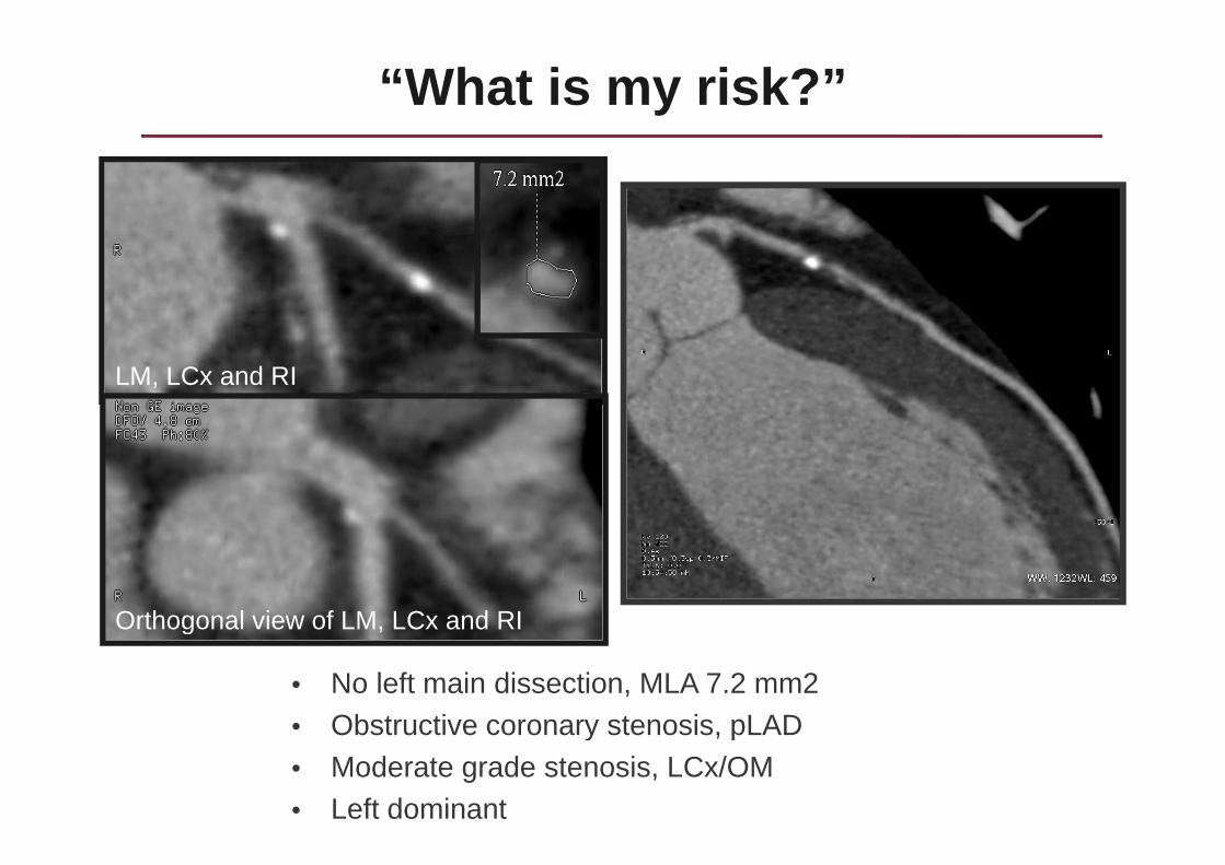
“What is my risk?”
LM, LCx and RI
Orthogonal view of LM, LCx and RI
• No left main dissection, MLA 7.2 mm2• Obstructive coronary stenosis pLAD
Orthogonal view of LM, LCx and RI
• Obstructive coronary stenosis, pLAD• Moderate grade stenosis, LCx/OM• Left dominant
Case: 46 y/o Caucasian Man• Patient recommended to undergo LHC
LM IVUS (7 0 mm2)• LM IVUS (7.0 mm2)• Severe CAD in LAD• Recommended to undergo CABG• Recommended to undergo CABG
• Patient flies to 2 additional hospitals for 4th and 5th• Patient flies to 2 additional hospitals for 4th and 5th
opinion, and then returns to PCP for 6th opinion• Based upon COURAGE recommended to• Based upon COURAGE, recommended to
undergo optimal medical therapy alone
• Patient presents 2 weeks later with CP and large lateral STEMI from proximal LCx lesionlateral STEMI from proximal LCx lesion
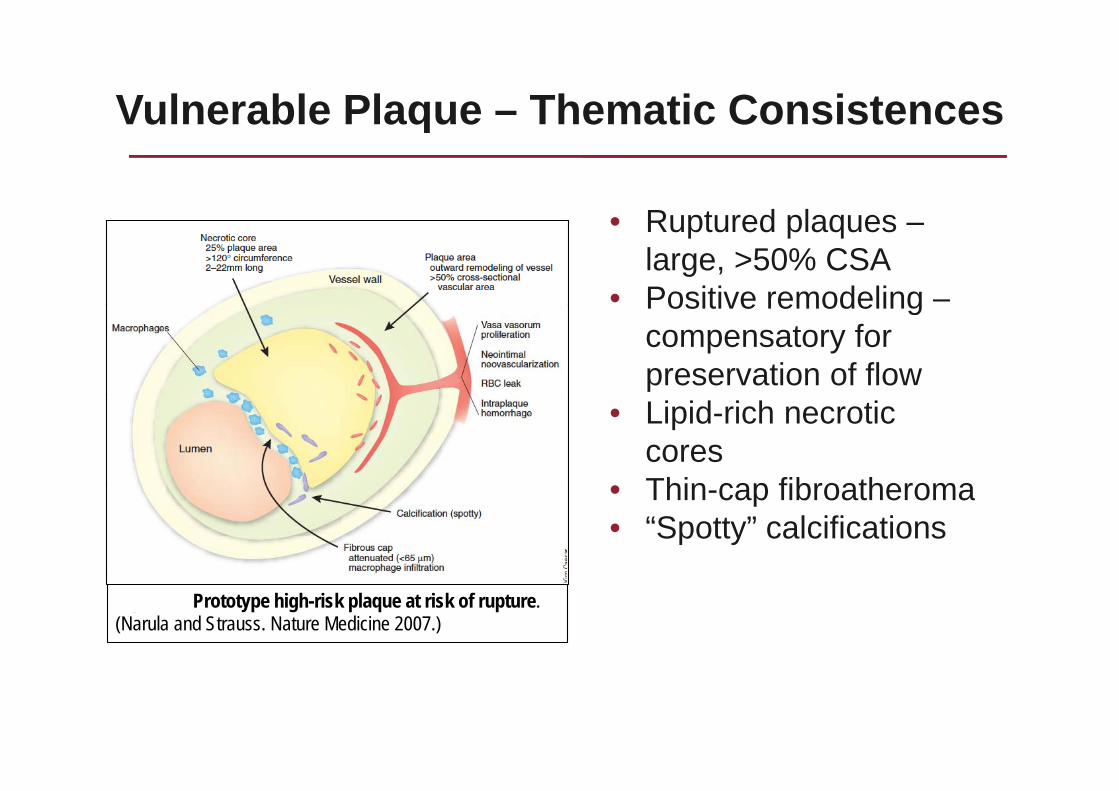
Vulnerable Plaque – Thematic Consistencesq
Ruptured plaques• Ruptured plaques –large, >50% CSA
• Positive remodeling –Positive remodeling compensatory for preservation of flow
• Lipid-rich necrotic cores
• Thin cap fibroatheroma• Thin-cap fibroatheroma• “Spotty” calcifications
Figure 1. Prototype high-risk plaque at risk of rupture. (Narula and Strauss. Nature Medicine 2007.)
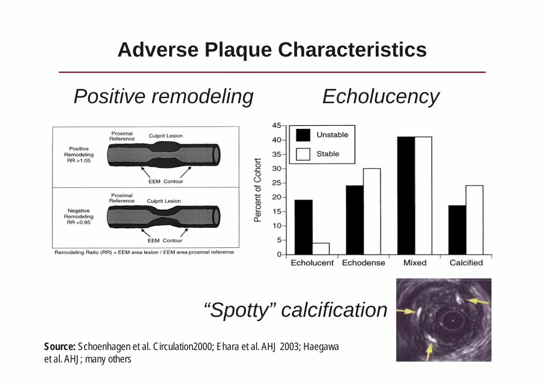
Adverse Plaque Characteristics
Positive remodeling Echolucencyg y
“Spotty” calcificationSource: Schoenhagen et al. Circulation2000; Ehara et al. AHJ 2003; Haegawaet al. AHJ; many others
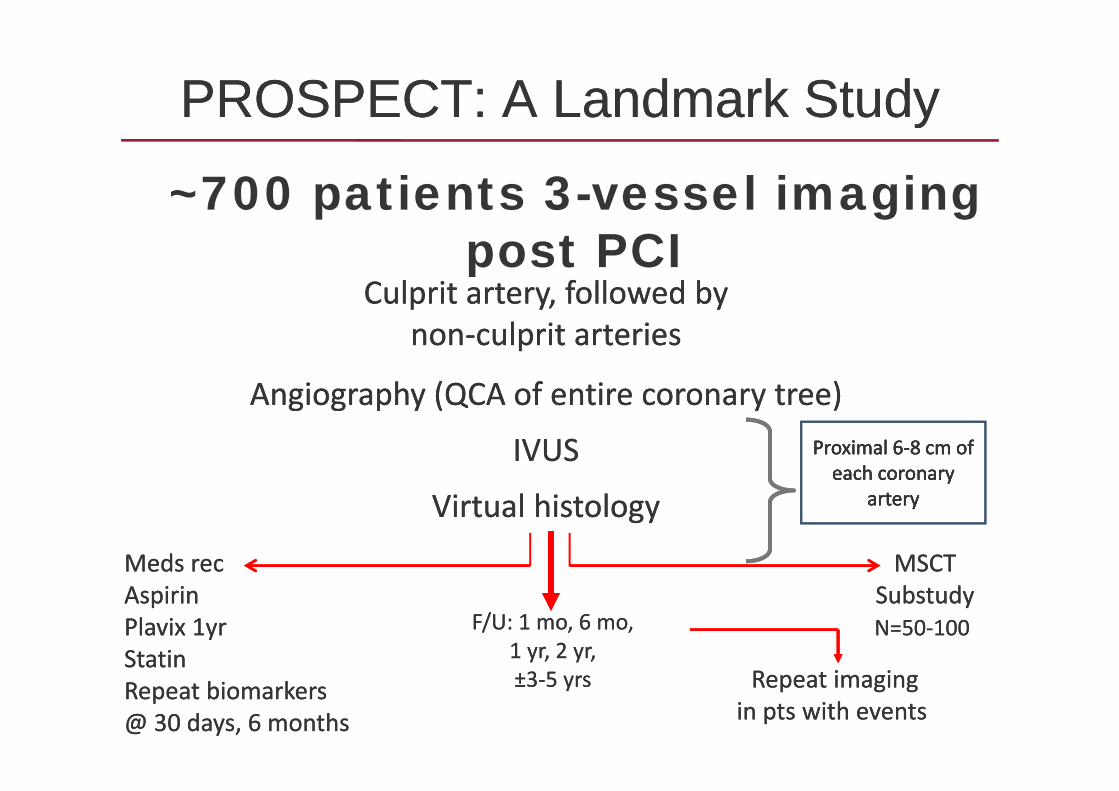
PROSPECT:PROSPECT: A Landmark StudyA Landmark Study
~700 patients 3-vessel imaging
yy
p g gpost PCI
Culprit artery, followed byCulprit artery, followed byp y, yp y, ynonnon‐‐culprit arteriesculprit arteries
Angiography (QCA of entire coronary tree)Angiography (QCA of entire coronary tree)Angiography (QCA of entire coronary tree)Angiography (QCA of entire coronary tree)
IVUSIVUS Proximal 6Proximal 6‐‐8 cm of 8 cm of each coronary each coronary
Proximal 6Proximal 6‐‐8 cm of 8 cm of each coronary each coronary
Virtual Virtual histologyhistology
Meds Meds recrec
arteryarteryarteryartery
MSCTMSCTAspirinAspirinPlavixPlavix 1yr1yrStatinStatin
SubstudySubstudyN=50N=50‐‐100100F/U: 1 mo, 6 mo,F/U: 1 mo, 6 mo,
1 yr, 2 yr,1 yr, 2 yr,Repeat imagingRepeat imaging
in pts with events in pts with events
StatinStatinRepeat biomarkersRepeat biomarkers@ 30 days, 6 months @ 30 days, 6 months
±±33‐‐5 yrs5 yrs
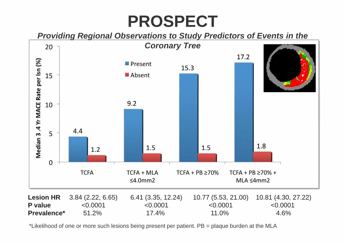
PROSPECTProviding Regional Observations to Study Predictors of Events in the g g y
Coronary Tree
Lesion HR 3.84 (2.22, 6.65) 6.41 (3.35, 12.24) 10.77 (5.53, 21.00) 10.81 (4.30, 27.22)P value <0 0001 <0 0001 <0 0001 <0 0001P value <0.0001 <0.0001 <0.0001 <0.0001Prevalence* 51.2% 17.4% 11.0% 4.6%
*Likelihood of one or more such lesions being present per patient. PB = plaque burden at the MLA
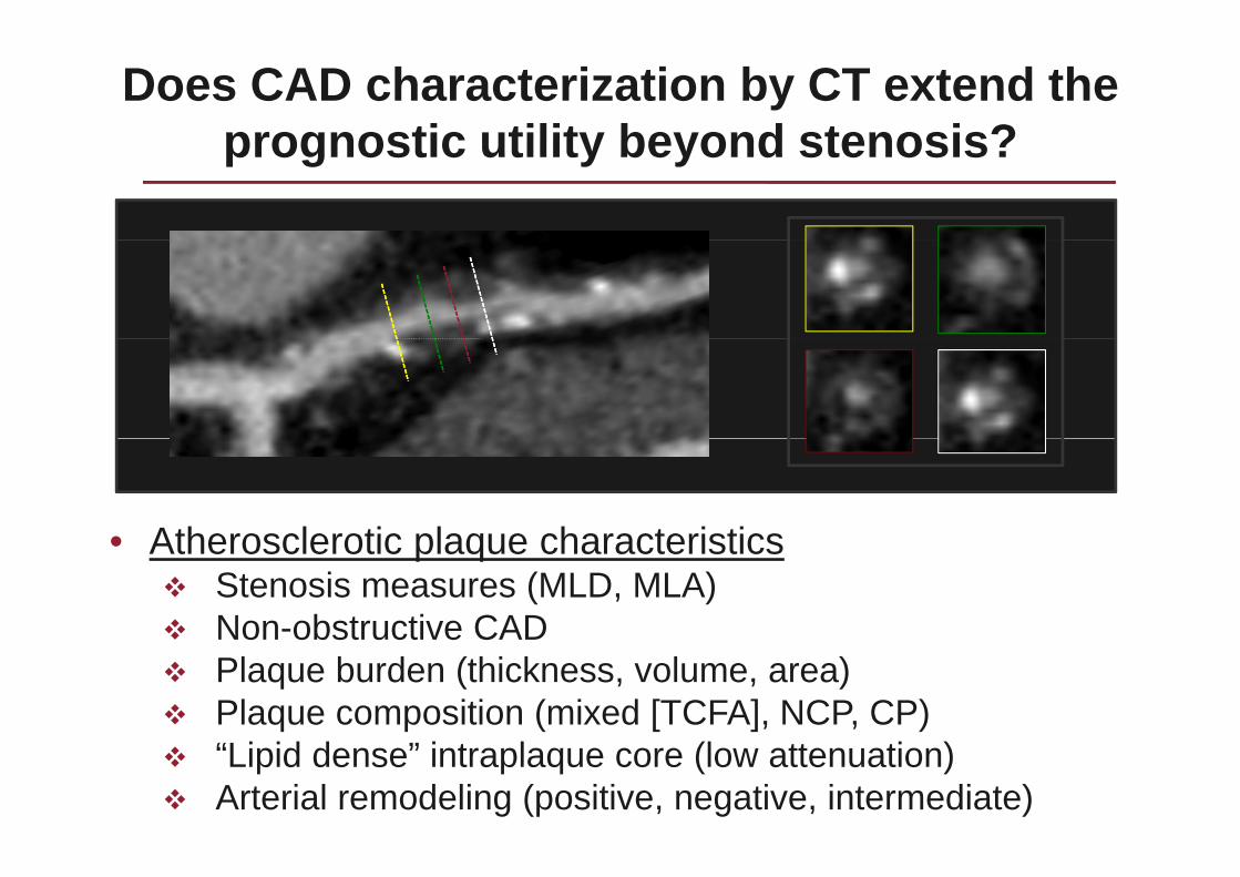
Does CAD characterization by CT extend the prognostic utility beyond stenosis?prognostic utility beyond stenosis?
• Atherosclerotic plaque characteristics• Atherosclerotic plaque characteristicsStenosis measures (MLD, MLA)Non-obstructive CADNon obstructive CADPlaque burden (thickness, volume, area)Plaque composition (mixed [TCFA], NCP, CP)“Lipid dense” intraplaque core (low attenuation)Arterial remodeling (positive, negative, intermediate)
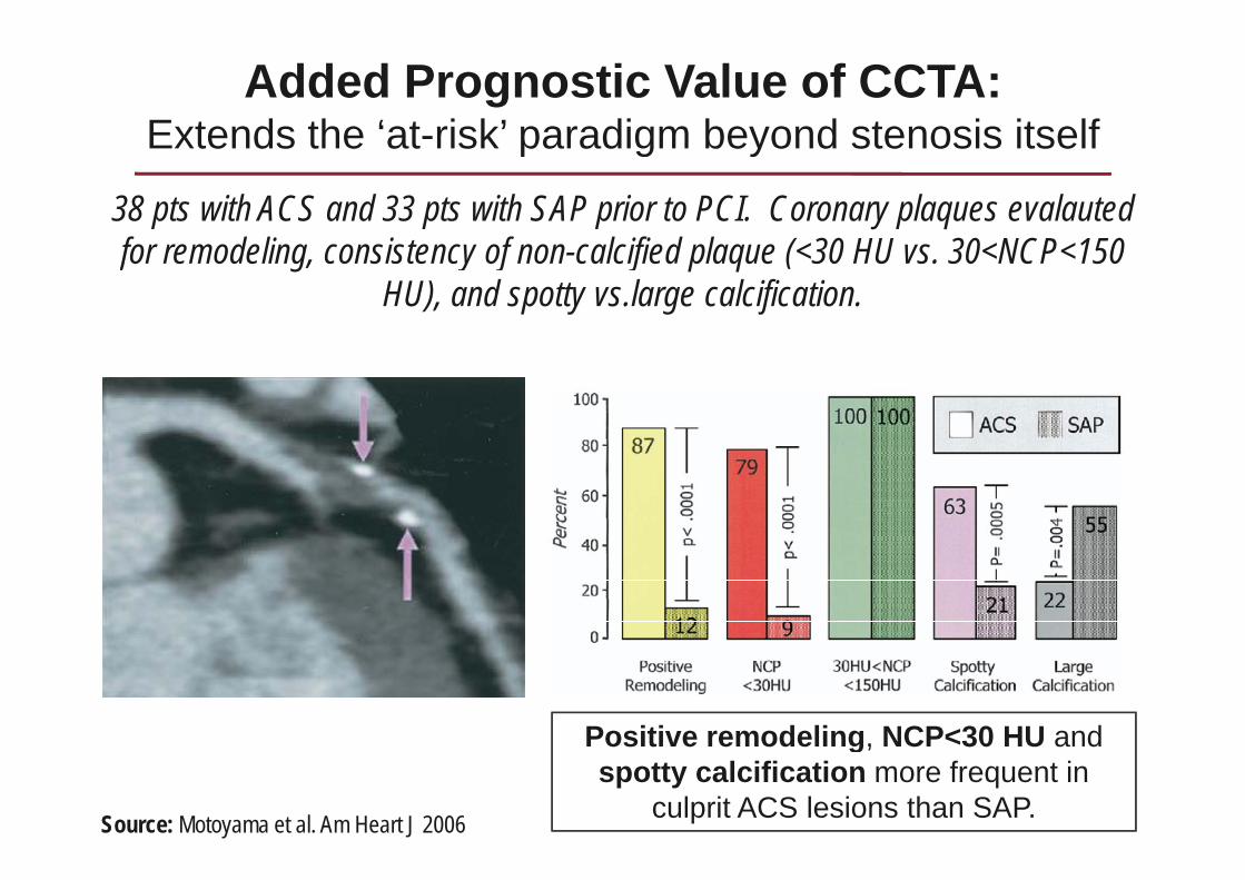
Added Prognostic Value of CCTA:Extends the ‘at-risk’ paradigm beyond stenosis itselfExtends the at risk paradigm beyond stenosis itself
38 pts with ACS and 33 pts with SAP prior to PCI. Coronary plaques evalauted f d li i t f l ifi d l ( 30 HU 30 NCP 150 for remodeling, consistency of non-calcified plaque (<30 HU vs. 30<NCP<150
HU), and spotty vs.large calcification.
Positive remodeling NCP<30 HU and
Source: Motoyama et al. Am Heart J 2006
Positive remodeling, NCP<30 HU and spotty calcification more frequent in
culprit ACS lesions than SAP.
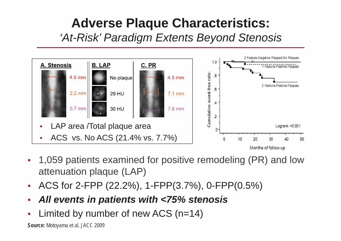
Adverse Plaque Characteristics: ‘At-Risk’ Paradigm Extents Beyond StenosisAt-Risk Paradigm Extents Beyond Stenosis
• LAP area /Total plaque area• ACS vs. No ACS (21.4% vs. 7.7%)
• 1,059 patients examined for positive remodeling (PR) and low attenuation plaque (LAP)p q ( )
• ACS for 2-FPP (22.2%), 1-FPP(3.7%), 0-FPP(0.5%)• All events in patients with <75% stenosis
Source: Motoyama et al. JACC 2009
All events in patients with 75% stenosis• Limited by number of new ACS (n=14)
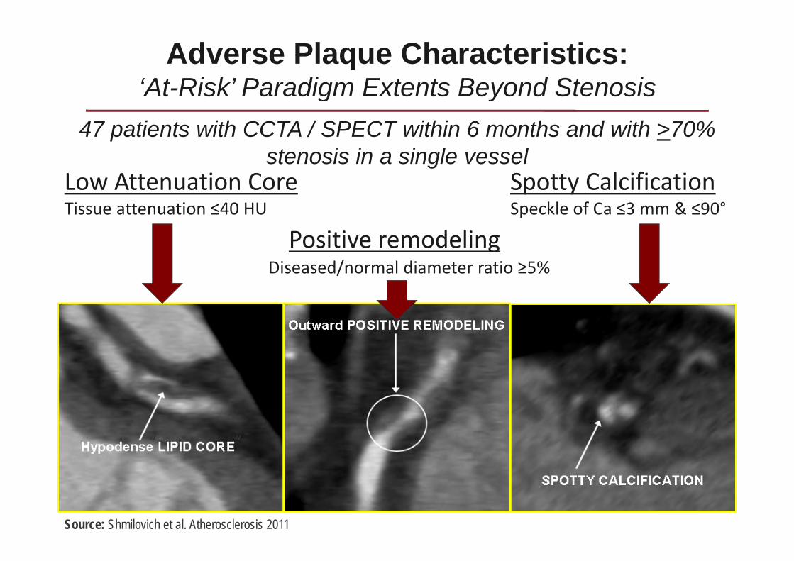
Adverse Plaque Characteristics: ‘At-Risk’ Paradigm Extents Beyond Stenosis
47 patients with CCTA / SPECT within 6 months and with >70% stenosis in a single vessel
At-Risk Paradigm Extents Beyond Stenosis
Low Attenuation Core Spotty CalcificationTissue attenuation ≤40 HU Speckle of Ca ≤3 mm & ≤90°
stenosis in a single vessel
p
Positive remodelingDiseased/normal diameter ratio ≥5%
““
Source: Shmilovich et al. Atherosclerosis 2011
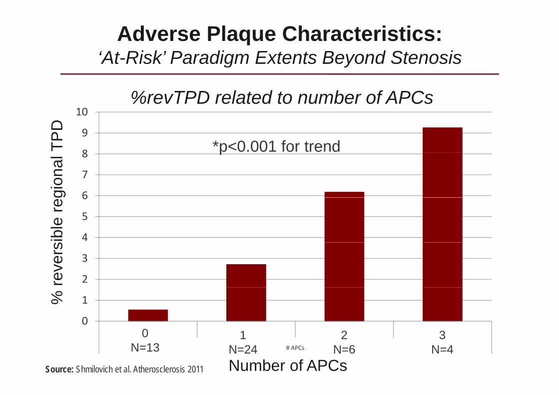
Adverse Plaque Characteristics: ‘At-Risk’ Paradigm Extents Beyond Stenosis
10%revTPD related to number of APCs
At Risk Paradigm Extents Beyond Stenosis
8
9
10
TPD
*p<0.001 for trend
6
7
8
gion
al p
4
5
6
ible
reg
2
3
reve
rs
0
1
0 1 2 30 31 2
%
0 1 2 3
# APCs
0N=13
3N=4
1N=24
2N=6
Number of APCsSource: Shmilovich et al. Atherosclerosis 2011
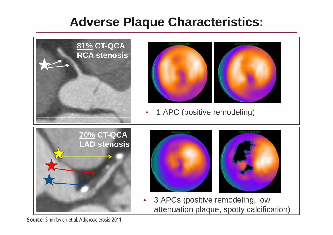
Adverse Plaque Characteristics:
81% CT-QCA RCA stenosis
• 1 APC (positive remodeling)
70% CT-QCA LAD stenosisLAD stenosis
3 APC ( iti d li l• 3 APCs (positive remodeling, low attenuation plaque, spotty calcification)
Source: Shmilovich et al. Atherosclerosis 2011
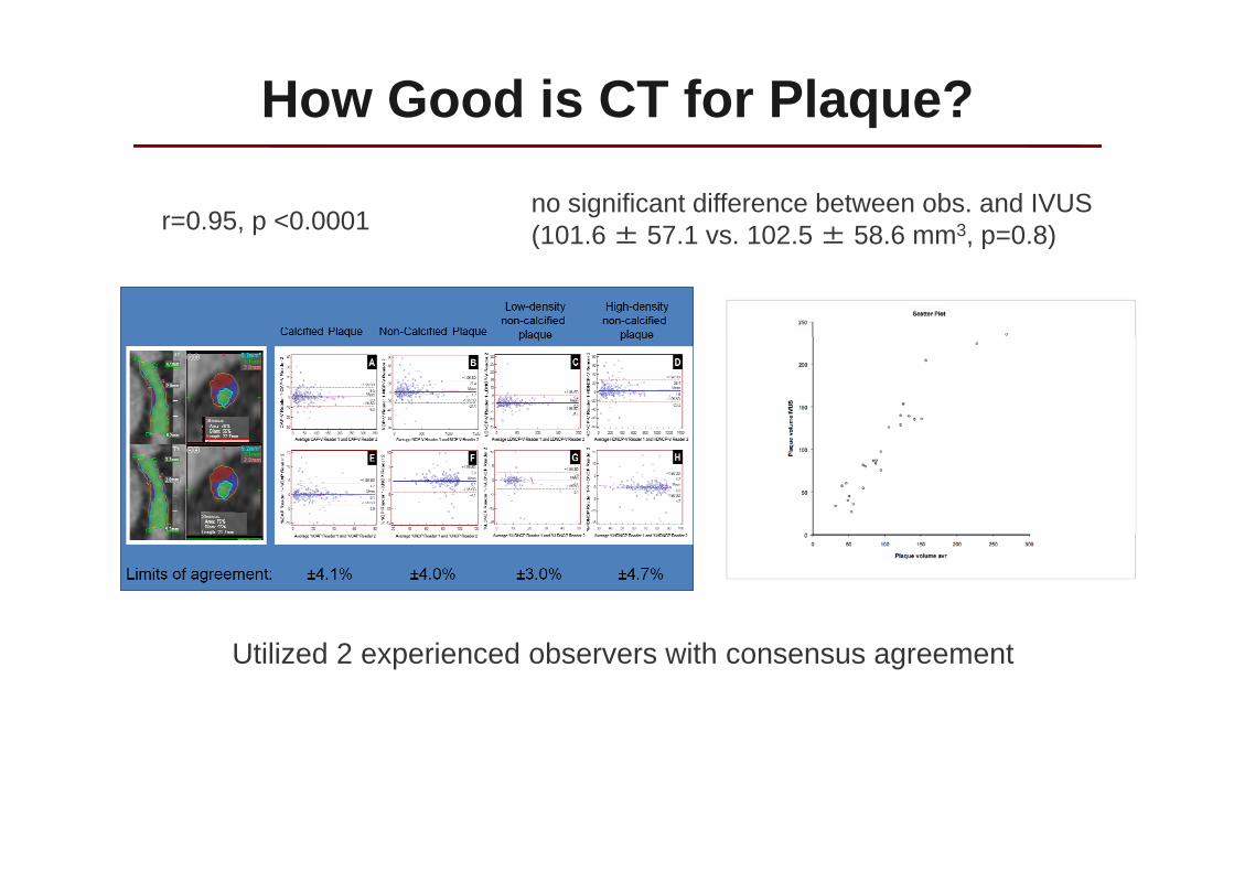
How Good is CT for Plaque?
r=0.95, p <0.0001no significant difference between obs. and IVUS(101 6 ± 57 1 vs 102 5 ± 58 6 mm3 p=0 8)(101.6 ± 57.1 vs. 102.5 ± 58.6 mm , p=0.8)
Utilized 2 experienced observers with consensus agreement
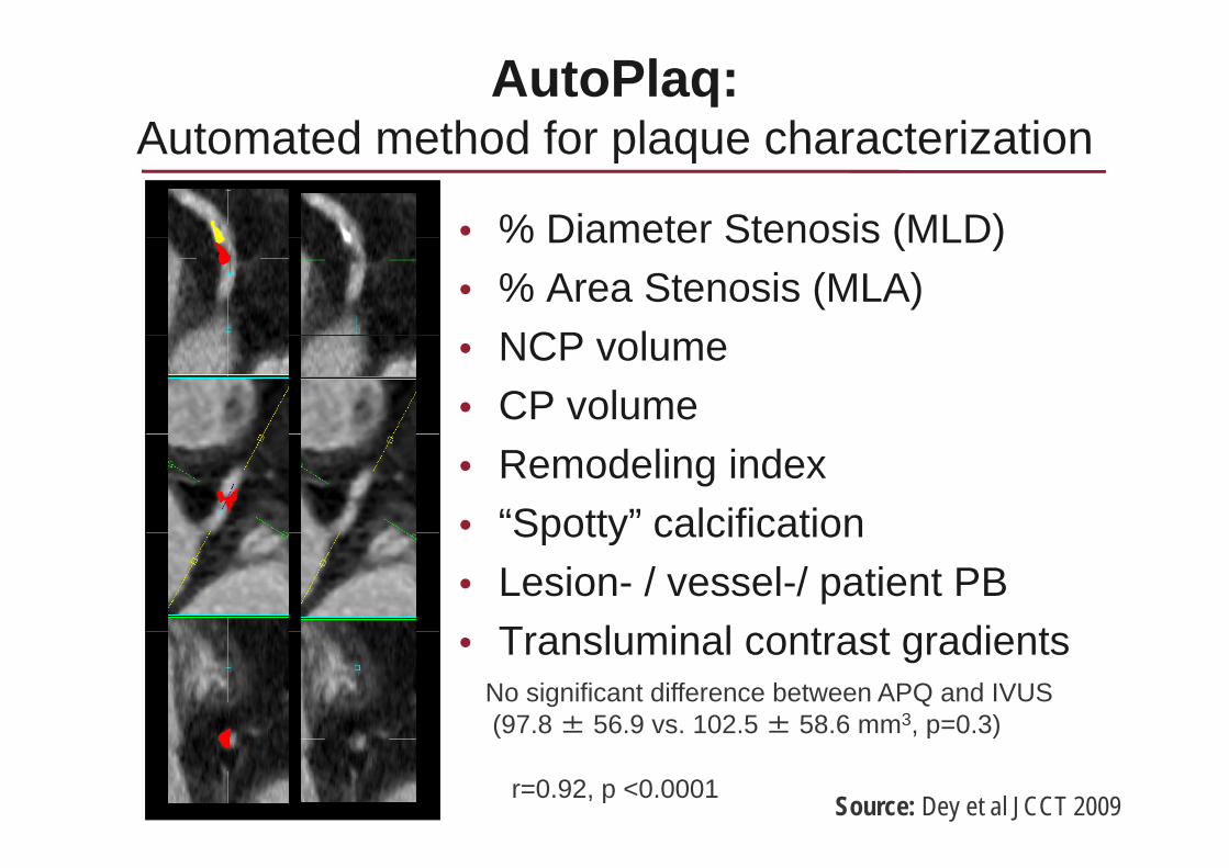
AutoPlaq: Automated method for plaque characterizationAutomated method for plaque characterization
• % Diameter Stenosis (MLD)% Diameter Stenosis (MLD)• % Area Stenosis (MLA)
NCP l• NCP volume• CP volume• Remodeling index• “Spotty” calcificationSpotty calcification• Lesion- / vessel-/ patient PB
T l i l t t di t• Transluminal contrast gradientsNo significant difference between APQ and IVUS(97 8 ± 56 9 vs 102 5 ± 58 6 mm3 p=0 3)
Source: Dey et al JCCT 2009r=0.92, p <0.0001
(97.8 ± 56.9 vs. 102.5 ± 58.6 mm , p 0.3)
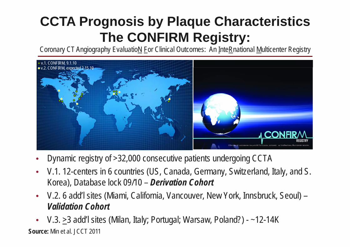
CCTA Prognosis by Plaque CharacteristicsThe CONFIRM Registry:The CONFIRM Registry:
Coronary CT Angiography EvaluatioN For Clinical Outcomes: An InteRnational Multicenter Registryv.1. CONFIRM, 9.1.10 v 2 CONFIRM expected 2 15 10 v.2. CONFIRM, expected 2.15.10
• Dynamic registry of >32,000 consecutive patients undergoing CCTA• V.1. 12-centers in 6 countries (US, Canada, Germany, Switzerland, Italy, and S.
K ) D t b l k 09/10 D i ti C h tKorea), Database lock 09/10 – Derivation Cohort• V.2. 6 add’l sites (Miami, California, Vancouver, New York, Innsbruck, Seoul) –
Validation CohortValidation Cohort• V.3. >3 add’l sites (Milan, Italy; Portugal; Warsaw, Poland?) - ~12-14K
Source: Min et al. JCCT 2011
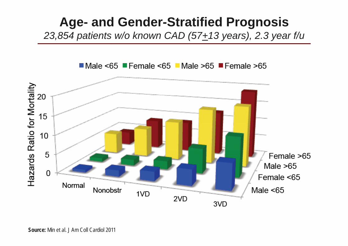
Age- and Gender-Stratified Prognosis23,854 patients w/o known CAD (57+13 years), 2.3 year f/u, p ( y ), y
Source: Min et al. J Am Coll Cardiol 2011
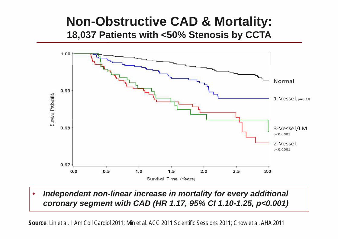
Non-Obstructive CAD & Mortality: 18,037 Patients with <50% Stenosis by CCTA, % y
• Independent non-linear increase in mortality for every additional t ith CAD (HR 1 17 95% CI 1 10 1 25 0 001)
Source: Lin et al. J Am Coll Cardiol 2011; Min et al. ACC 2011 Scientific Sessions 2011; Chow et al. AHA 2011
coronary segment with CAD (HR 1.17, 95% CI 1.10-1.25, p<0.001)
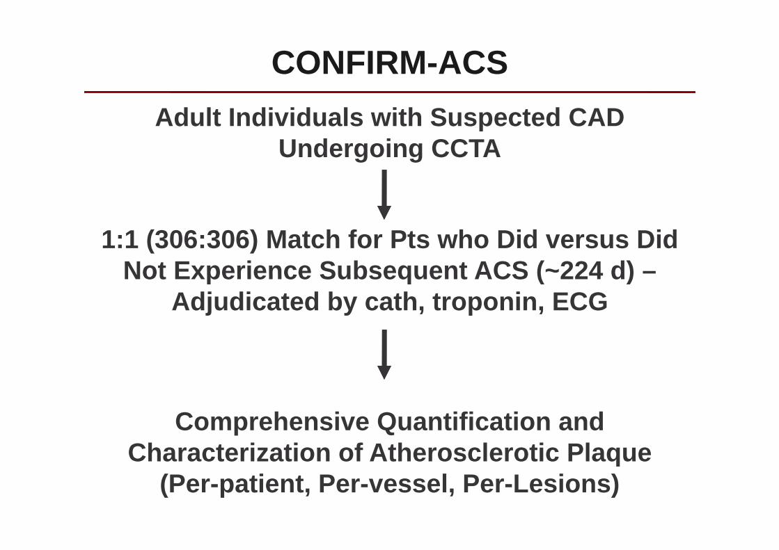
CONFIRM-ACSAdult Individuals with Suspected CAD
Undergoing CCTAUndergoing CCTA
1:1 (306:306) Match for Pts who Did versus Did Not Experience Subsequent ACS (~224 d) –Not Experience Subsequent ACS ( 224 d)
Adjudicated by cath, troponin, ECG
Comprehensive Quantification and Characterization of Atherosclerotic Plaqueq
(Per-patient, Per-vessel, Per-Lesions)
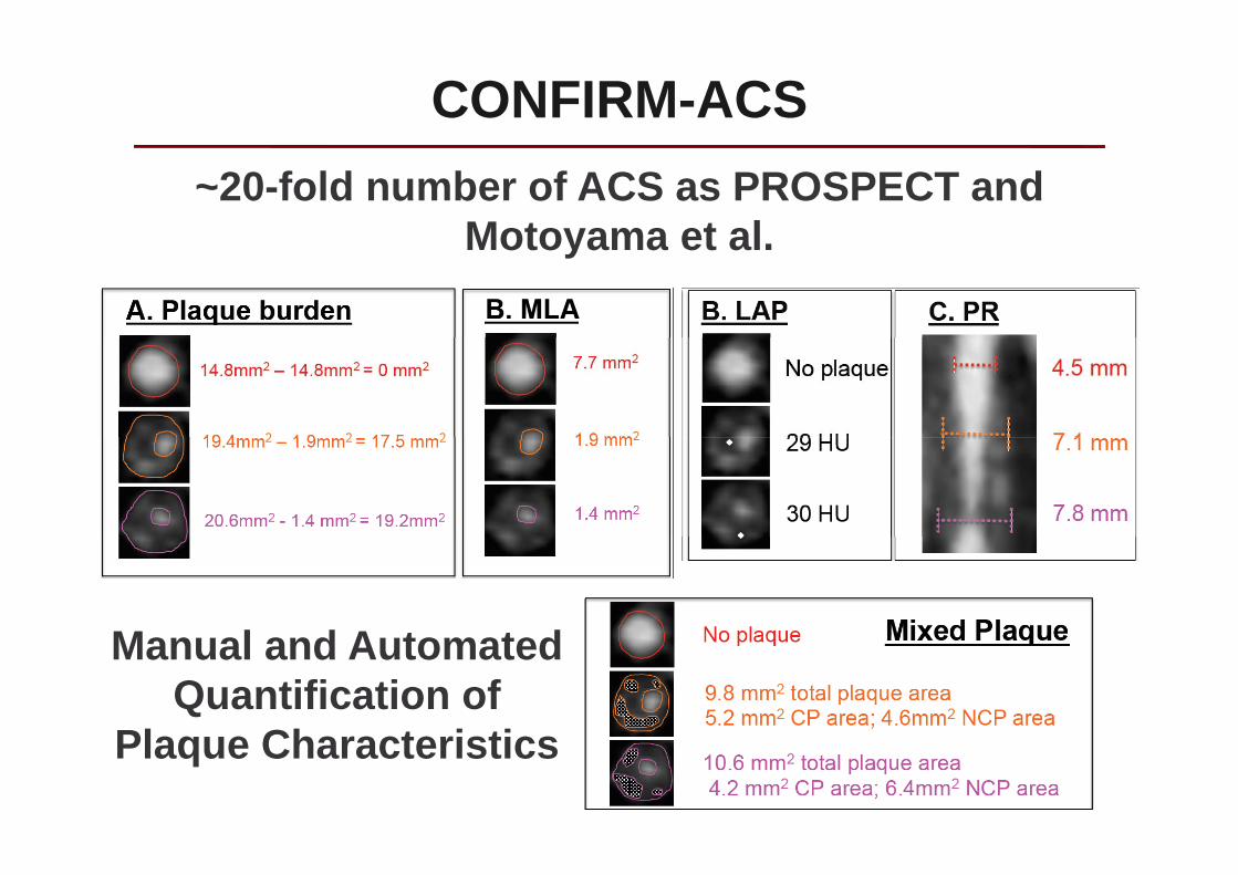
CONFIRM-ACS~20-fold number of ACS as PROSPECT and
Motoyama et alMotoyama et al.
M l d A t t dManual and Automated Quantification of
Pl Ch t i tiPlaque Characteristics
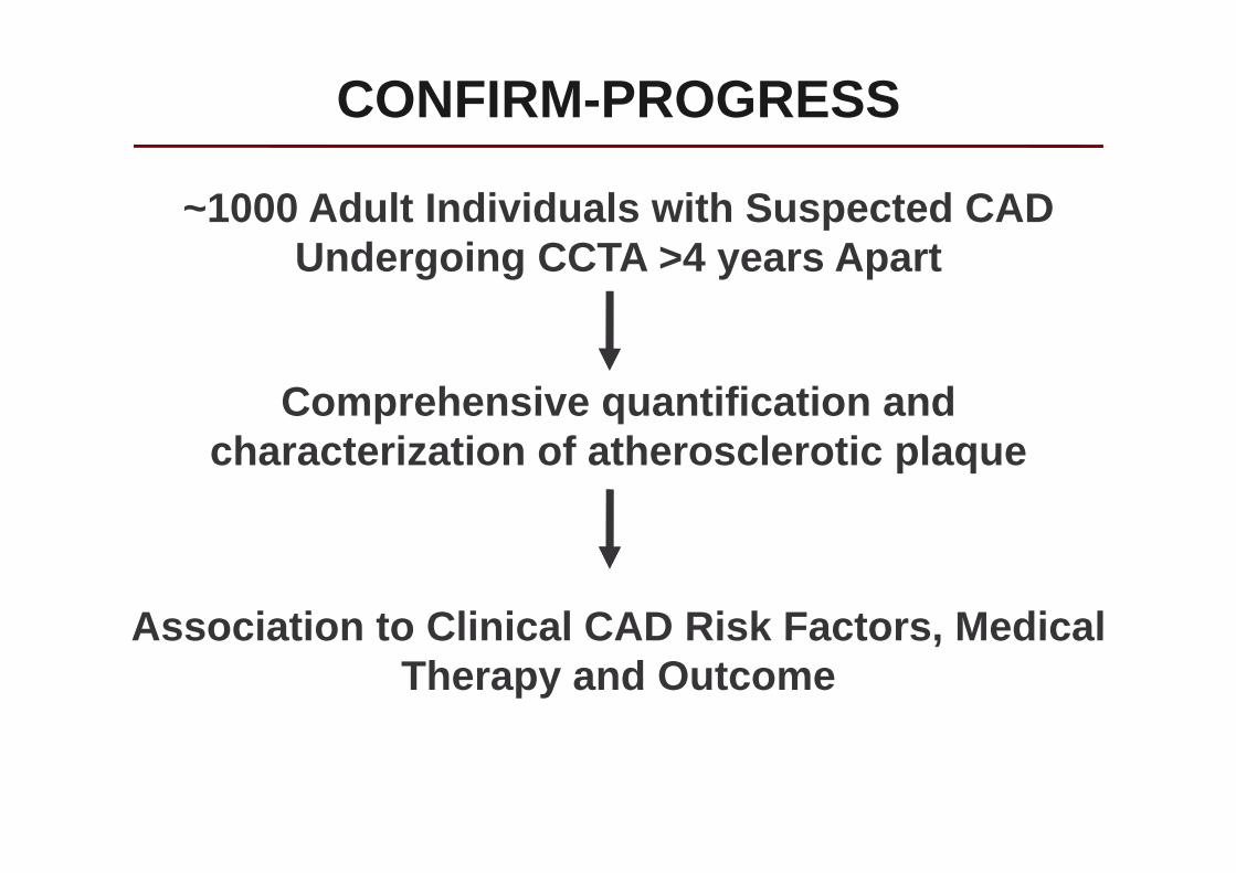
CONFIRM-PROGRESS
~1000 Adult Individuals with Suspected CAD Undergoing CCTA >4 years Apart
Comprehensive quantification and h t i ti f th l ti lcharacterization of atherosclerotic plaque
Association to Clinical CAD Risk Factors, MedicalAssociation to Clinical CAD Risk Factors, Medical Therapy and Outcome
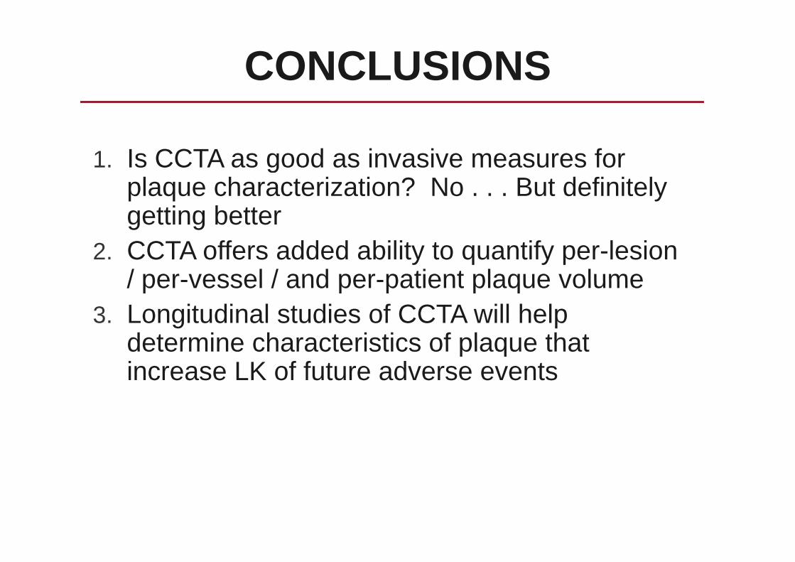
CONCLUSIONS
1 Is CCTA as good as in asi e meas res for1. Is CCTA as good as invasive measures for plaque characterization? No . . . But definitely getting bettergetting better
2. CCTA offers added ability to quantify per-lesion / per-vessel / and per-patient plaque volume/ per-vessel / and per-patient plaque volume
3. Longitudinal studies of CCTA will help determine characteristics of plaque thatdetermine characteristics of plaque that increase LK of future adverse events