1
1 Ra Labo In signifi “preco In this gold c M isoflur scanne were m maps 40s). T FA = 3, 4 ho source metho 90 mi the bl input f Re LnCap tumor hours POST perme effects Figure accum effects monit Refere al., Ma Funded CytImm Dete Isabelle I adiology, Univers oratory, Minneapo ntroduction. Re icantly improve onditioning”, ha s work, we prop construct (CYT-6 Materials & Me rane in O 2 /N 2 O ed using an RF v monitored throu prior to contrast The same basic 30º, total acquis ours after an intr e DICOM viewi ods using the pre nutes of the exp ood to ve) and k function (AIF) w esults. In one ca p tumors after in r region is show after injection o T, while kep dro eability and rete s were observed e 1c. Along with mulation of thera s of CYT in viv or novel precond ences. 1. Goel, R., agn Reson Med, 20 d by NIH P41 R mune Sciences, In ction of the ef ltis 1 , Jeunghwan C ity of Minnesota - olis, MN, United St States, 4 ecently, it has be d by using TNF s been studied u ose to use Dyna 6091) [1] in mic ethods. Four to (50/50). A cathe volume coil (Mi ughout the exper t injection (FOV acquisition was sition time = 1h ravenous injectio ng software (Os e-contrast T 1 ma periment. The pa kep (rate constan was determined f ase, the data we ntraperitoneal inj wn (B, average o of CYT. The par ops considerably ention (EPR) [6] d in the muscle, h improving cry apeutic agents p vo with non-des ditioning of ther , et al., Mol Cance 005. 5. Ocak, I., e RR008079, The W nc. for providing C ffect of nanopa Choi 2 , Emily Colon CMRR, Minneapo tates, 3 Biomedical 4 Department of Ur een shown that th F- based nanopa using invasive or amic Contrast En ce bearing LnCap o 5 weeks after eter was placed i illipede, Agilent riment. A multis V = 35 mm, matr s used to acquire 29min. Animals on of CYT (POS siriX) [2]. Param aps [3, 4]. The a arameters ve (ex nt between the v from a posterior re discarded due jection of Gd (A of 10 tumors). In rametric maps o y after CYT, sug ]. The results of the greatest chan yosurgical proced preferentially wi tructive method rapeutic interven er Ther, 2007. 2. R t al., AJR Am J R WM KECK Found CYT-6091. article precon nna 1 , Manda Vollm olis, MN, United S Engineering, Uni rologic Surgery, U he outcome of cr articles before th r post-mortem m nhanced Imaging p tumors, before implantation on in the intraperito t, Palo Alto) at a lice gradient ech rix size = 128 x e DCE-MRI dat s were scanned o ST). Enhanceme metric maps were area under the ga xtravascular and ve and plasma) vessel within th e to movements A). Enhancement n both the tumo of a typical mous ggesting slightly f this study dem nges in contrast dures, the retenti ithin a tumor, th ds is important a ntions will be adv Rosset, A., et al., J Roentgenol, 2007. dation and AHC nditioning in a mers 1 , Mithun She States, 2 Mechanica versity of Minneso University of Minne ryosurgery, a pro he procedure is methods in anima g (DCE-MRI) as e and after inject n both hindlimb oneal cavity for a 9.4T magnet eq ho sequence was x 128, 1 mm thic a to follow cont on day 1 withou ent curves were o e obtained by fir adolinium conce extracellular vo were calculated he imaging volum during the acqu t curves of the si r and the muscl se are also show y slower “washi monstrate that en retention are fo ion of contrast in hus increasing e as such compoun vantageous in fu J Digit Imaging, 2 189(4): p. 849. 6. Translational Re a mouse model enoi 3 , Joel Slaton 4 , al Engineering, Un ota - Bioheat and M esota, Minneapolis ocedure gaining performed [1]. als, but has neve s a method to m tion of CYT-609 bs of LnCap tum Gadolinium-DT quipped with a V s acquired at mu ckness, TE = 1.4 trast kinetics wi ut injection of C obtained in regio rst converting th entration curve ( olume per unit vo by applying the me to mitigate su uisition. Figure 1 ignal relative to le (not shown), wn (C). In the tu in” and a substa tumor, respec slightly increa between PRE increased (5-fo the AUGC m 250% in the tu AUGC maps processing da modeling is d small size of th Discussion the effect of construct can using DCE-M Preconditionin physiological nhanced EPR is ound in the tumo n these studies m efficacy while re nds progress tow uture translationa 004. 3. Daldrup, H Shenoi, M.M., et esearch Grant Pro Figure 1. injection ( signal enh dynamic c vivo in tu Data are m The solid l of prostate c , John Bischof 2 , an niversity of Minnes Mass Transfer Lab s, MN, United Stat g acceptance for The effect of th er been character monitor in vivo th 91 (CYT). mors, 6 male nu TPA (Gd) injecti Varian console. ultiple flip angle 43 ms, TR = 65 th the following YT (PRE), reco on of interests (t he signal changes (AUGC) was de olume of tissue) e two-compartm ubject and exper 1 shows an axial the maximum s we observed th umor region, Ktr antially slower ctively. In the ase post-CYT. V E and POST co old) in the area o maps show that umor, and by on are independen ata. This is sign difficult to ident he vessels in the n and Conclus “preconditionin be detected non MRI in a mou ng has been event involving, likely the resul or as demonstrat may indicate the educing toxicity wards clinical tr al studies. H.E., et al., Magn al., Nanomedicine ogram 2009 09-0 Axial view of a (A). Regions of in hancement at each curves of relative umors before and mean ± SEM. In C white line delineaancer by MRI nd Gregory J Metz sota - Bioheat and boratory, Minneap tes prostate cancer t his nanoparticle rized in vivo in t he effect of a we ude mice were a on (0.6 mmol/kg Temperature an es (from 5 to 40 ms, 4 averages, g parameter chan overed, and scann tumor and muscl s to concentratio etermined by int ), KTrans (transf ment Kety model rimental variatio view of a typic signal observed a at contrast uptak rans is slightly lo washout” of th muscle, these s Ve is comparab onditions, while of the tumor PO Gd concentrati nly 18% in the m nt of the kinetic nificant as the A tify reliably in e imaging volum sion. In this wo ng” of the tum n-invasively in t use model of shown to b , among other ch lt of CYT admi ted in the pharm e potential of CY y. The ability to rials. Non-invasi Reson Med, 1998 e (Lond), 2011. Ac 08. We also grate mouse after CYT nterest for the tumh time point is oe signal enhancem after CYT treatC), the parametric tes the tumor regio I zger 1 Mass Transfer polis, MN, United treatment, can b on the tumor, o the intact anima ell-defined TNF- anesthetized wit g). Animals wer nd respiration rat 0º) to calculate T , acquisition tim nges: 8 averages ned again on da le) using an ope on using standar egrating over th fer constant from l [5]. The arteria ons. al mouse bearin after CYT for th ke is enhanced ower PRE versu he contrast in th same parameter ble in the muscl it is drasticall OST. Remarkably ion increases b muscle. Note tha c model used fo AIF used for th mice due to th me rk, we show tha mor by a TNF- the intact anima prostate cancer be a comple hanges, enhance inistration. Whil macokinetic maps YT to enhance th o characterize th ive techniques t 8. 4. Deoni, S.C., e cknowledgements efully acknowledg and gado-linium or are drawn and btained (B). The ment obtained in ment are shown. maps are shown. on. e or l. - h re te T 1 me s, y n d e m al g he 4 us he rs e y y, y at or e he at - al r. x d e s, e he o et s. ge 3020 Proc. Intl. Soc. Mag. Reson. Med. 20 (2012)

Detection of the effect of nanoparticle preconditioning in ... filebtained in regio st converting th ntration curve (olume per unit vo by applying the e to mitigate su uisition. Figure

Embed Size (px)

Citation preview

1RaLabo

Insignifi“precoIn thisgold c

Misoflurscannewere mmaps 40s). TFA = 3, 4 hosourcemetho90 mithe blinput f

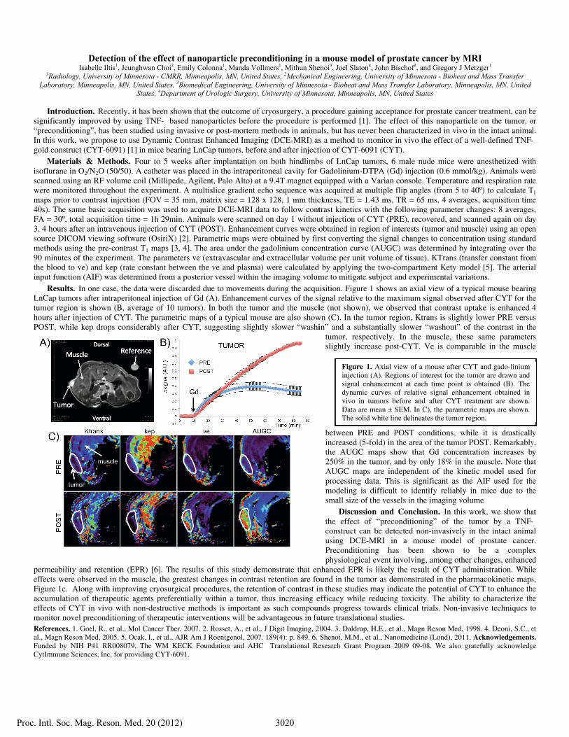
ReLnCaptumorhours POST

permeeffectsFigureaccumeffectsmonitRefereal., MaFundedCytImm

DeteIsabelle I

adiology, Universoratory, Minneapo

ntroduction. Reicantly improveonditioning”, has work, we propconstruct (CYT-6

Materials & Merane in O2/N2O ed using an RF vmonitored throuprior to contrastThe same basic 30º, total acquisours after an intre DICOM viewiods using the prenutes of the expood to ve) and kfunction (AIF) wesults. In one cap tumors after inr region is showafter injection o

T, while kep dro

eability and retes were observede 1c. Along with

mulation of theras of CYT in vivor novel precond

ences. 1. Goel, R.,agn Reson Med, 20d by NIH P41 Rmune Sciences, In

ction of the efltis1, Jeunghwan City of Minnesota -

olis, MN, United StStates, 4

ecently, it has bed by using TNFs been studied uose to use Dyna6091) [1] in micethods. Four to(50/50). A cathevolume coil (Mi

ughout the expert injection (FOVacquisition was

sition time = 1h ravenous injectiong software (Ose-contrast T1 maperiment. The pakep (rate constanwas determined fase, the data wentraperitoneal inj

wn (B, average oof CYT. The parops considerably

ention (EPR) [6]d in the muscle, h improving cryapeutic agents pvo with non-desditioning of ther, et al., Mol Cance005. 5. Ocak, I., e

RR008079, The Wnc. for providing C

ffect of nanopaChoi2, Emily Colon

CMRR, Minneapotates, 3Biomedical 4Department of Ur

een shown that thF-� based nanopausing invasive oramic Contrast Ence bearing LnCapo 5 weeks after eter was placed iillipede, Agilentriment. A multisV = 35 mm, matrs used to acquire29min. Animalson of CYT (POSsiriX) [2]. Paramaps [3, 4]. The aarameters ve (exnt between the vfrom a posterior re discarded duejection of Gd (A

of 10 tumors). Inrametric maps oy after CYT, sug

]. The results ofthe greatest chan

yosurgical procedpreferentially witructive method

rapeutic intervener Ther, 2007. 2. Rt al., AJR Am J R

WM KECK FoundCYT-6091.

article preconnna1, Manda Vollmolis, MN, United SEngineering, Uni

rologic Surgery, U

he outcome of crarticles before thr post-mortem mnhanced Imagingp tumors, beforeimplantation onin the intraperitot, Palo Alto) at alice gradient echrix size = 128 xe DCE-MRI dats were scanned oST). Enhanceme

metric maps werearea under the gaxtravascular and ve and plasma) vessel within th

e to movements A). Enhancementn both the tumoof a typical mousggesting slightly

f this study demnges in contrast dures, the retentiithin a tumor, th

ds is important antions will be advRosset, A., et al., J

Roentgenol, 2007. dation and AHC

nditioning in amers1, Mithun SheStates, 2Mechanicaversity of Minneso

University of Minne

ryosurgery, a prohe procedure is

methods in animag (DCE-MRI) ase and after injectn both hindlimboneal cavity for a 9.4T magnet eqho sequence was

x 128, 1 mm thica to follow conton day 1 withou

ent curves were oe obtained by firadolinium conceextracellular vowere calculated

he imaging volumduring the acqut curves of the sir and the musclse are also showy slower “washi

monstrate that enretention are fo

ion of contrast inhus increasing eas such compounvantageous in fuJ Digit Imaging, 2189(4): p. 849. 6.

Translational Re

a mouse modelenoi3, Joel Slaton4,al Engineering, Unota - Bioheat and Mesota, Minneapolis

ocedure gainingperformed [1].

als, but has neves a method to mtion of CYT-609bs of LnCap tumGadolinium-DTquipped with a Vs acquired at muckness, TE = 1.4trast kinetics wiut injection of Cobtained in regiorst converting thentration curve (olume per unit vo

by applying theme to mitigate su

uisition. Figure 1ignal relative to le (not shown),

wn (C). In the tuin” and a substa

tumor, respecslightly increa

between PREincreased (5-fothe AUGC m250% in the tuAUGC maps processing damodeling is dsmall size of th

Discussionthe effect of construct can using DCE-MPreconditioninphysiological

nhanced EPR is ound in the tumon these studies mefficacy while rends progress towuture translationa004. 3. Daldrup, HShenoi, M.M., et esearch Grant Pro

Figure 1.injection (signal enhdynamic cvivo in tuData are mThe solid w

l of prostate c, John Bischof2, anniversity of MinnesMass Transfer Labs, MN, United Stat

g acceptance for The effect of th

er been charactermonitor in vivo th91 (CYT). mors, 6 male nuTPA (Gd) injectiVarian console. ultiple flip angle43 ms, TR = 65 th the followingYT (PRE), recoon of interests (t

he signal changes(AUGC) was deolume of tissue)e two-compartmubject and exper

1 shows an axialthe maximum swe observed th

umor region, Ktrantially slower “ctively. In the ase post-CYT. V

E and POST coold) in the area o

maps show that umor, and by onare independen

ata. This is signdifficult to identhe vessels in then and Conclus

“preconditioninbe detected non

MRI in a moung has been event involving,likely the resul

or as demonstratmay indicate theeducing toxicitywards clinical tral studies. H.E., et al., Magn al., Nanomedicineogram 2009 09-0

Axial view of a m(A). Regions of inhancement at eachcurves of relativeumors before and mean ± SEM. In Cwhite line delineat

ancer by MRInd Gregory J Metzsota - Bioheat and boratory, Minneaptes

prostate cancer this nanoparticlerized in vivo in the effect of a we

ude mice were aon (0.6 mmol/kgTemperature an

es (from 5 to 40ms, 4 averages,

g parameter chanovered, and scanntumor and muscls to concentratio

etermined by int), KTrans (transf

ment Kety modelrimental variatio view of a typic

signal observed aat contrast uptak

rans is slightly lo“washout” of thmuscle, these sVe is comparab

onditions, while of the tumor POGd concentrati

nly 18% in the mnt of the kineticnificant as the Atify reliably in

e imaging volumsion. In this wong” of the tumn-invasively in tuse model of

shown to b, among other chlt of CYT admited in the pharme potential of CYy. The ability torials. Non-invasi

Reson Med, 1998e (Lond), 2011. Ac

08. We also grate

mouse after CYT nterest for the tumoh time point is obe signal enhancem

after CYT treatmC), the parametric tes the tumor regio

I zger1 d Mass Transfer polis, MN, United

treatment, can bon the tumor, o

the intact animaell-defined TNF-

anesthetized witg). Animals wer

nd respiration rat0º) to calculate T, acquisition timnges: 8 averagesned again on dale) using an opeon using standaregrating over thfer constant froml [5]. The arteriaons. al mouse bearinafter CYT for thke is enhanced ower PRE versuhe contrast in thsame parameter

ble in the muscl

it is drasticallOST. Remarkably

ion increases bmuscle. Note thac model used foAIF used for thmice due to th

me rk, we show tha

mor by a TNF-the intact animaprostate cancerbe a complehanges, enhanceinistration. Whil

macokinetic mapsYT to enhance tho characterize thive techniques t

8. 4. Deoni, S.C., ecknowledgements

efully acknowledg

and gado-linium or are drawn and btained (B). The ment obtained in ment are shown. maps are shown.

on.

e or l. -�

h re te T1

me s, y n d e

m al

g he 4

us he rs e

y y, y at or e

he

at -� al r. x d e s, e

he o

et s.

ge

3020Proc. Intl. Soc. Mag. Reson. Med. 20 (2012)