Embed Size (px)
Citation preview

Sensing prokaryotic mRNA signifies microbial viability andpromotes immunity
Leif E. Sander, Michael J. Davis‡,1, Mark V. Boekschoten§,1, Derk Amsen†, Christopher C.Dascher, Bernard Ryffel*, Joel A. Swanson‡, Michael Müller§, and J. Magarian Blander
Immunology Institute, Department of Medicine, Mount Sinai School of Medicine, 1425 MadisonAvenue, New York, New York 10029 ‡Department of Microbiology and Immunology, University ofMichigan, Ann Arbor, Michigan, USA §Nutrition, Metabolism and Genomics Group, Division ofHuman Nutrition, Wageningen University, Wageningen, The Netherlands *Laboratory ofMolecular Immunology and Embryology, University of Orleans and Centre National de laRecherche Scientifique, Orleans, France †Department of Cell Biology and Histology, AcademicMedical Center, University of Amsterdam, Amsterdam, The Netherlands
AbstractLive vaccines have long been known to trigger far more vigorous immune responses than theirkilled counterparts1–6. This has been attributed to the ability of live microorganisms to replicateand express specialized virulence factors that facilitate invasion and infection of their hosts7.However, protective immunization can often be achieved with a single injection of live, but notdead, attenuated microorganisms stripped of their virulence factors. Pathogen associated molecularpatterns (PAMPs), which serve to alert the immune system8,9, are present in both live and killedvaccines, suggesting that certain poorly characterized aspects of live microorganisms, notincorporated in dead vaccines, are particularly effective at inducing protective immunity. Here weshow that the innate immune system can directly sense microbial viability through detection of aspecial class of viability-associated PAMPs (vita-PAMPs). We identify prokaryotic messengerRNA (mRNA) as a vita-PAMP present only in viable bacteria, recognition of which elicits aunique innate response and a robust adaptive antibody response. Notably, the innate responseevoked by viability and prokaryotic mRNA was thus far considered to be reserved for pathogenicbacteria, but we show that even nonpathogenic bacteria in sterile tissues can trigger similarresponses, provided they are alive. Thus, the immune system actively gauges the infectious risk byscouring PAMPs for signatures of microbial life and thus infectivity. Detection of vita-PAMPstriggers an alert mode not warranted for dead bacteria. Vaccine formulations that incorporate vita-PAMPs could thus combine the superior protection of live vaccines with the safety of deadvaccines.
Correspondence and requests for materials should be addressed to J.M.B. ([email protected]).1these authors contributed equally to this work.The authors declare no competing financial interests.Author Contributions. L.E.S. and J.M.B. designed experiments and directed the study. L.E.S. performed all experiments. M.J.D. andL.E.S. performed experiments measuring lysosomal leakage. J.A.S. helped with the design and analysis of the lysosomal leakageexperiments. M.V.B. performed gene microarray analysis. M.V.B. and M.M. analyzed the gene microarray data and helped with datainterpretation. D.A. and J.M.B. performed experiments during development phase of the project, and C.C.D. helped with design ofRNA related experiments. B.R. provided bone marrow progenitor cells from Nlrp3−/−, Asc−/−, and Casp1−/− mice. L.E.S., D.A. andJ.M.B. wrote the manuscript. J.M.B. conceived of the study.Author information. Affymetrix Microarray data have been deposited with the NCBI Gene Expression Omnibus(http://www.ncbi.nlm.nih.gov/geo/) under accession no. GSE27960.
NIH Public AccessAuthor ManuscriptNature. Author manuscript; available in PMC 2012 February 29.
Published in final edited form as:Nature. ; 474(7351): 385–389. doi:10.1038/nature10072.
NIH
-PA Author Manuscript
NIH
-PA Author Manuscript
NIH
-PA Author Manuscript

We hypothesized that the innate immune system may sense the most fundamentalcharacteristic of microbial infectivity, microbial viability itself, and activate a robustimmune response regardless of the presence of more specialized factors that regulatemicrobial virulence7. To study sensing of bacterial viability without compounding effects ofreplication or virulence factors, we used thymidine auxotrophs of nonpathogenicEscherichia coli K12, strain DH5α (ECThyA—). Viable and heat-killed (HK) ECThyA—
similarly activated nuclear factor-κB (NF-κB) and mitogen-activated protein kinase p38(supplementary Fig. 1) in bone marrow-derived macrophages and elicited production ofsimilar amounts of interleukin-6 (IL-6) and tumor necrosis factor alpha (TNF-α) (Fig. 1a). Instark contrast, viable ECThyA— induced higher levels of IFN-β than HK ECThyA— orlipopolysaccharide (LPS) (Fig. 1b), and only viable ECThyA— induced IL-1β secretion (Fig.1c; supplementary Fig. 2). Pro-IL-1β transcription was equally induced by both viable andHK ECThyA— (Fig. 1c), suggesting that viable bacteria specifically elicit cleavage of pro-IL-1β. This process is catalyzed by caspase-1 in Nod-like receptor (NLR)-containinginflammasome complexes, assembly of which can be triggered by the activity of bacterialvirulence factors10,11. Notably, avirulent viable but not HK ECThyA— inducedinflammasome activation and pro-caspase-1 cleavage (Fig. 1d). Finally, viable but not HKECThyA— induced caspase-1-dependent inflammatory cell death, termed pyroptosis10,11,resulting in release of lactate dehydrogenase (LDH) (Fig. 1e) and appearance of 7-amino-actinomycin D (7AAD)+Annexin-V−/low cells (Fig. 1f). Similar responses were observed inperitoneal macrophages and both splenic and bone marrow-derived dendritic cells(supplementary Fig. 2b). Killing ECThyA— by UV irradiation, antibiotics, or ethanol, alsoselectively abrogated IL-1β secretion and pyroptosis without affecting IL-6 production (Fig.1g; supplementary Fig. 3), suggesting that a general determinant associated with bacterialviability is detected.
To determine whether pathogenic bacteria can also activate the inflammasome in theabsence of virulence factors, we studied attenuated strains of selected pathogens: Shigellaflexneri virulence plasmid-cured strain BS10312, Salmonella typhimurium,SL1344ΔSpi1ΔSpi2, lacking the Salmonella pathogenicity islands SPI-1 and SPI-210, andListeria monocytogenes ΔHlyΔfliC, lacking listeriolysin O and flagellin10. These mutantsinduced IL-1β production at levels comparable to those induced by ECThyA— (Fig. 1h), butlower and with slower kinetics than their pathogenic counterparts (supplementary Fig. 4).IL-1β production was abolished when these bacteria were killed, whereas IL-6 productionwas similar (Fig. 1h). Thus, immune cells detect universal characteristics of viabilitydifferent from virulence factors.
Caspase-1 activation, pyroptosis and IL-1β production in response to ECThyA— wereabrogated in macrophages deficient for NLRP3 or for the inflammasome adaptor ApoptosisSpeck protein with Caspase recruitment (ASC or PYCARD)11 (Figs. 1i,j), while NLRC4was dispensable (supplementary Fig. 5). Pyroptosis and IL-1β production induced by viableECThyA— were abrogated in Casp1−/− macrophages (Fig. 1j) and suppressed by inhibitorsfor caspase-1, but not caspase-8 (supplementary Fig. 6).
Induction of IFN-β mRNA and protein by viable ECThyA— required the Toll-like receptor(TLR) adaptor TRIF9 (Figs. 2a,b) and downstream interferon regulatory factor-3 (IRF3)9
(supplementary Fig. 7), but not MyD88, the main TLR adaptor9 (Figs. 2a,b). In contrast,transcription of pro-IL-1β was largely dependent on MyD88. Consequently, Myd88−/− cellssecreted no IL-1β (Figs. 2c,d), whereas pyroptosis and caspase-1 cleavage were intact (Fig.2e,f). Notably, while TRIF was dispensable for pro-IL-1β transcription (Fig. 2c), Trif−/−
cells failed to secrete IL-1β (Fig. 2d), were protected from pyroptosis (Fig. 2e), and did notactivate caspase-1 (Fig. 2f). These findings revealed an unexpected role for TRIF in NLRP3inflammasome activation in response to viable ECThyA—. In contrast, pyroptosis induced by
Sander et al. Page 2
Nature. Author manuscript; available in PMC 2012 February 29.
NIH
-PA Author Manuscript
NIH
-PA Author Manuscript
NIH
-PA Author Manuscript

pathogenic S. typhimurium10, proceeded independently of TRIF (supplementary Fig. 8).Differential involvement of TRIF, together with differences in magnitude and kinetics of theresponse (Fig. 1h; supplementary Fig. 4), indicated that inflammasome activation inresponse to virulence factors occurs in a manner distinct from that to viability.
Genome-wide transcriptional analysis of wild type and Trif−/− macrophages before and afterphagocytosis of viable ECThyA— showed differential regulation of several clusters of genes(supplementary Fig. 9) including IFN-regulated genes, as expected9 (Figs. 2a,b;supplementary Fig. 10a), while most of Rel/NF-κB target genes were comparable(supplementary Fig. 10b). Nlrp3 expression was induced independently of TRIF (Figs. 2g,h)and negative regulators of inflammasome activity such as those encoded by Mediterraneanfever (Mefv), Nlrp10 and Casp12 genes were also unchanged or expressed at higher levels inwild type macrophages (Fig. 2g), possibly due to negative feedback. Thus, the role of TRIFin inflammasome activation upon phagocytosis of viable ECThyA— is not explained bytranscriptional control of inflammasome components (so called priming11). Furthermore,ATP and reactive oxygen species (ROS)11,13, known activators of the NLRP3inflammasome, were not involved, as deficiency for P2X7R, required for ATP-mediatedNLRP3 activation, did not affect pyroptosis or IL-1β production (supplementary Figs.11a,b), and ROS accumulated equally in response to viable and HK ECThyA—
independently of TRIF (supplementary Fig. 11c).
Injection of viable and HK ECThyA— into mice induced similarly high serum levels of IL-6(Fig. 2i). In contrast, circulating IL-1β was detected only in mice infected with viablebacteria (Fig. 2i), while IFN-β levels were undetectable in all groups (not shown).Confirming our results in vitro, production of IL-1β (but not IL-6) in vivo also requiredTRIF, ASC, and NLRP3 (Fig. 2i). Injection of non-pathogenic S. typhimurium inducedserum IL-1β levels comparable to those elicited by ECThyA—, which similarly depended onTRIF (supplementary Fig. 12). Although pathogenic S. typhimurium elicited higher levels ofserum IL-1β than non-pathogenic Salmonella, this response was also severely reduced inTrif−/− mice, suggesting a previously unappreciated role for TRIF in Salmonella infection(supplementary Fig. 12). Importantly, deficiency in TRIF, ASC and NLRP3 impairedbacterial clearance during systemic infection with replication sufficient non-pathogenic E.coli (Fig. 2j). This failure was more dramatic in Trif−/− than in Asc−/− or Nlrp3−/− mice,possibly due to the central upstream role of TRIF in inflammasome activation and IFN-βproduction.
The ability to sense microbial viability through pathways downstream of pattern recognitionreceptors suggests the existence of vita-PAMPs; PAMPs associated with viable but not deadbacteria. In contrast to LPS and genomic DNA that remained constant after killingECThyA— with heat, total bacterial RNA was rapidly lost (Figs. 3a,b; supplementary Fig.13). Total RNA content was also lost with antibiotic treatment, and little ribosomal RNA(rRNA) remained after killing with UV and ethanol (supplementary Fig. 14). Only fixationwith paraformaldehyde (PFA) efficiently killed the bacteria (not shown), but preserved totalRNA content (supplementary Fig. 15a). Remarkably, unlike bacteria killed by other means,PFA-killed bacteria induced pyroptosis and IL-1β to levels similar to those induced byviable bacteria (supplementary Fig. 15b). Thus, the presence or absence of RNA correlatedwith the ability to activate pathways involved in sensing viability.
These results suggest that prokaryotic RNA represents a labile PAMP closely associatedwith bacterial viability, which might signify microbial life to the immune system. Indeed,addition of purified total bacterial RNA fully restored the ability of HK ECThyA— to inducepyroptosis, IL-1β and IFN-β production (Fig. 3c). These responses were dependent on TRIF,NLRP3 and caspase-1, just as those elicited by viable bacteria (Fig. 3d compared to Figs. 1j
Sander et al. Page 3
Nature. Author manuscript; available in PMC 2012 February 29.
NIH
-PA Author Manuscript
NIH
-PA Author Manuscript
NIH
-PA Author Manuscript

and 2a–f). The NLRP3 inflammasome mediates recognition of viral RNA during InfluenzaA infection14. Together with our results and others’15, this suggests a more general role forNLRP3 in responses to RNAs of microbial origin. RNA can activate the NLRP3inflammasome when delivered into the cytosol (where NLRP3 resides) with transfectionreagents15. In contrast, inflammasome activation by the combination of total bacterial RNAand dead ECThyA— did not require RNA transfection (Figs. 3c,d). Administration of total E.coli RNA alone or in combination with LPS (to mimic an E. coli-derived PAMP+RNA) hadlittle effect on NLRP3 inflammasome activation unless the RNA was delivered to thecytosol using Lipofectamine (supplementary Fig. 16) or in combination with ATP, asreported previously15. Thus, phagocytosis of viable bacteria is a natural context of bacterialRNA-mediated NLRP3 inflammasome activation.
These findings raised the question as to how vita-PAMPs in phagolysosomes gain access tocytosolic receptors such as NLRP3 in the absence of invasion, auxiliary secretion systems orpore forming toxins. To address this question, we exploited the pH sensitive excitationspectrum of fluorescein: the acidic pH in phagolysosomes quenches fluorescence whilerelease into the pH neutral cytosol allows a regain in fluorescence16. Phagocytosis ofavirulent ECThyA— in the presence of fluorescein-conjugated dextran (Fdx), consistentlyinduced low level release of Fdx into the cytosol of macrophages (Figs. 3e,f; supplementaryFig. 17). This indicates that phagosomes carrying E. coli exhibit intrinsic leakiness, aproperty previously described for particles such as beads and crystals that inducephagolysosomal destabilization16,17. Interestingly, killed E. coli also induced Fdx release,although to a slightly lower extent than viable E. coli (Figs. 3e,f) demonstrating thatphagosomal leakage occurs independently of bacterial viability. Therefore, RNA fromviable bacteria could gain access to cytosolic receptors via intrinsic phagosomal leakage.These results may also explain the reported ability of phagosome-degraded mutants ofListeria monocytogenes or Staphylococcus aureus to induce a transcriptional responsedependent on cytosolic NLRs18,19.
Digestion of total RNA from E. coli with exonuclease RNase I and double-stranded RNA(dsRNA)-specific endonuclease RNase III abrogated LDH and IL-1β release, while DNasetreatment had no effect (Fig. 4a). Of E. coli RNA species, mRNA most potently inducedpyroptosis as well as production of IL-1β and IFN-β. Small RNA (sRNA) or the mostabundant RNA, ribosomal RNA (rRNA), had little or no detectable effects (Fig. 4b;supplementary Fig. 18). E. coli rRNA undergoes extensive modifications not found inmRNA20, which may underlie the differential activity of these RNA species. The relativeamount of mRNA was <1% of the total RNA and accordingly, mRNA was approximately100-fold more effective than total RNA (Figs. 3c, 4a,b; supplementary Fig. 18).
In vitro transcribed single-stranded mRNA of the E. coli Gro-operon (supplementary Figs.19a,b), which is strongly expressed upon phagocytosis of bacteria21, induced caspase-1cleavage and subsequent pyroptosis and IL-1β production when phagocytosed together withHK ECThyA— (Fig. 4c,e; supplementary Figs. 19c–e). The single-stranded Gro-mRNAsequence had a predicted secondary structure with regions of high probability for basepairing (Fig. 4d), consistent with susceptibility of the stimulatory activity to RNase IIItreatment (Fig. 4a). Indeed, fully dsGro-mRNA (supplementary Fig. 19b) induced similarresponses as single-stranded Gro-mRNA of the appropriate length (Fig. 4e; supplementaryFig. 19d). Other transcripts also induced such responses, showing that theimmunostimulatory property is independent of RNA sequence (Fig. 4f).
Strikingly, eukaryotic RNA was unable to elicit the responses induced by E. coli mRNA(Fig. 4b). Unlike eukaryotic mRNA, tri-phosphate moieties at the 5’end of bacterial mRNAsare not capped with 7-methyl-guanosine (7m7G)22, and might betray the prokaryotic origin
Sander et al. Page 4
Nature. Author manuscript; available in PMC 2012 February 29.
NIH
-PA Author Manuscript
NIH
-PA Author Manuscript
NIH
-PA Author Manuscript
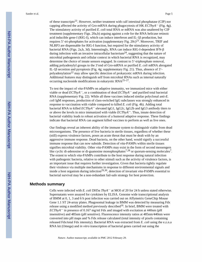
of these transcripts23. However, neither treatment with calf intestinal phosphatase (CIP) norcapping affected the activity of Gro-mRNA during phagocytosis of HK ECThyA— (Fig. 4g).The stimulatory activity of purified E. coli total RNA or mRNA was also unaltered by CIPtreatment (supplementary Figs. 20a,b) arguing against a role for the RNA helicase retinoicacid inducible gene-I (RIG-I), which can induce interferon and IL-1β production, butrequires 5’-tri-phosphates for activation (supplementary Fig. 20c)23. Moreover, TRIF andNLRP3 are dispensable for RIG-I function, but required for the stimulatory activity ofbacterial RNA (Figs. 2a,b, 3d). Interestingly, RNA can induce RIG-I-dependent IFN-βduring infection with an invasive intracellular bacterium24, suggesting that the nature ofmicrobial pathogenesis and cellular context in which bacterial RNA is recognized, maydetermine the choice of innate sensors engaged. In contrast to 5’-triphosphate removal,adding polyadenylyl groups to the 3’end of Gro-mRNA or purified E. coli mRNA abrogatedIL-1β secretion and pyroptosis (Fig. 4g; supplementary Fig. 21). Thus, absence of 3’-polyadenylation22 may allow specific detection of prokaryotic mRNA during infection.Additional features may distinguish self from microbial RNAs such as internal naturallyoccurring nucleoside modifications in eukaryotic RNA25–27.
To test the impact of vita-PAMPs on adaptive immunity, we immunized mice with eitherviable or dead ECThyA—, or a combination of dead ECThyA— and purified total bacterialRNA (supplementary Fig. 22). While all three vaccines induced similar polyclonal anti-E.coli IgM responses, production of class-switched IgG subclasses was strongly enhanced inresponse to vaccination with viable compared to killed E. coli (Fig. 4h). Adding totalbacterial RNA to killed ECThyA— elevated IgG1, IgG2c, IgG2b and IgG3 antibody titers toor above the levels in mice immunized with viable ECThyA—. Thus, innate detection ofbacterial viability leads to robust activation of a humoral adaptive response. These findingsindicate that bacterial RNA can augment killed vaccines to perform as well as live ones.
Our findings reveal an inherent ability of the immune system to distinguish viable from deadmicroorganisms. The presence of live bacteria in sterile tissues, regardless of whether these(still) express virulence factors, poses an acute threat that must be dealt with by anaggressive immune response. Dead bacteria, on the other hand, would signify a successfulimmune response that can now subside. Detection of vita-PAMPs within sterile tissuessignifies microbial viability. Other vita-PAMPs may exist in the form of second messengerslike cyclic di-adenosine or di-guanosine monophosphates7,28 or quorum-sensing molecules7.The extent to which vita-PAMPs contribute to the host response during natural infectionwith pathogenic bacteria, relative to other stimuli such as the activity of virulence factors, isan important issue that requires further investigation. Given that bacteria tightly regulatetheir virulence via multiple mechanisms in response to different environmental signals andinside a host organism during infection29,30, detection of invariant vita-PAMPs essential tobacterial survival may be a non-redundant fail-safe strategy for host protection.
Methods summaryCells were infected with E. coli DH5α ThyA— at MOI of 20 for 24 h unless stated otherwise.Supernatants were assayed for cytokines by ELISA. Genome wide transcriptional analysisof BMM at 0, 1, 3 and 6 h post infection was carried out on Affymetrix GeneChip MouseGene 1.1 ST 24-array plates. Phagosomal leakage in BMM was detected by measuring Fdxrelease using a modified method previously described16. In brief, BMM were treated withECThyA— in presence of 0.167 mg/ml Fdx and imaged with excitation at 440nm (pHinsensitive) and 485nm (pH sensitive). Fluorescence intensity ratios at 485nm/440nm wereconverted into pH maps and % Fdx release calculated (total intensity of pixels containingreleased Fdx/total Fdx intensity). Bacterial RNA was extracted from E. coli using the e.z.n.aRNA kit (Omega) and in vitro transcription of bacterial genes carried out using the
Sander et al. Page 5
Nature. Author manuscript; available in PMC 2012 February 29.
NIH
-PA Author Manuscript
NIH
-PA Author Manuscript
NIH
-PA Author Manuscript

MEGAscript kit (Ambion) followed by DNase digestion and RNA purification usingMEGAclear kit (Ambion). RNA polyadenylation was performed with the poly-A-tailing kit(Ambion). Vaccinations were performed as a prime-boost regimen (see full Methods).C57BL/6J and P2rx7−/− mice were purchased from the Jackson Laboratory. MyD88−/−,Trif−/− mice were provided by S. Akira, Myd88−/x−Trif−/− by R. Medzhitov, Nlrp3−/−,Asc−/−, Nlrc4−/− by Millenium, and Caspase-1−/− by R. Flavell. Animal care andexperimentation were performed in accordance with approved MSSM Institutional AnimalCare and Use Committee protocols.
MethodsCells
Bone marrow (BM)-derived dendritic cell (DC) cultures were grown as previouslydescribed31 in RPMI 1640 supplemented with GM-CSF and 5% fetal bovine serum (FBS),plus 100 µg/ml penicillin, 100µg/ml streptomycin, 2 mM L-glutamine, 10 mM HEPES, 1nM sodium pyruvate, 1% MEM non-essential amino acids, and 2.5 µM β-mercaptoethanol(all SIGMA). Semi-adherent cells were harvested on ice on day 5 and re-plated immediatelyin fresh RPMI 1640 medium containing 10% FBS at 1×10⁶ cells/well in 24-well tissueculture-treated plates. Stimuli were added immediately after re-plating in the same mediumand the cells were centrifuged for 2’ at 2000 rpm. Murine macrophages were derived fromthe BM (BMM) of either C57BL/6J, Myd88−/−, Trif−/−, Trif−/−xMyd88−/−, Nlrp3−/−, Asc−/−
or Casp1−/− mice, as described previously32, in RPMI 1640 supplemented with M-CSF and10% FBS, plus 100µg/ml penicillin, 100µg/ml streptomycin, 10mM HEPES and 1nMsodium pyruvate (all SIGMA). For some experiments macrophages were derived from BMof Irf3−/− or P2rx7−/− mice. Peritoneal macrophages were harvested 72h after intra-peritoneal injection of 1ml thioglycollate (BD Bioscience), grown overnight in RPMI 1640medium supplemented with 10% FBS and 100µg/ml penicillin and 100µg/ml streptomycin,hereafter referred to as ‘complete medium’. Mouse embryonic fibroblasts (MEFs) deficientfor RIG-I (RIG-I−/−) were kindly provided by A. Ting with permission from S. Akira, andgrown in DMEM medium containing 10% FBS and 100 µg/ml penicillin, 100µg/mlstreptomycin.
MiceC57BL/6J and P2rx7−/− mice were purchased from Jackson Laboratories. Myd88−/− andTrif−/− mice were originally provided by S. Akira, Myd88−/− and Trif−/− mice wereinterbred to homozygosity to generate TrifxMyd88−/− mice, and were provided by R.Medzhitov. Nlrp3−/−, Asc−/− or Casp1−/− bone marrow was provided by B. Ryffel and micefor in vivo studies were acquired from R. Flavell (through Millenium) and have beendescribed previously33,34. Irf3−/− mice were provided by C.B. Lopez and were previouslydescribed35. We used 8–10 week old animals for all experiments. All experiments wereapproved by the institutional ethics committee and carried out in agreement with the ‘Guidefor the Care and Use of Laboratory Animals’ (NIH publication 86-23, revised 1985).
BacteriaE. coli K12, strain DH5α were purchased from Invitrogen. Naturally occurring Thymidineauxotrophs (ThyA—) were selected on Luria-Bertani (LB) agar plates containing 50µg/mltrimethoprim and 500µg/ml thymidine (both SIGMA). Auxotrophy was confirmed byinoculation and overnight culture of single colonies in LB medium. ThyA— E. coli grew onlyin the presence of thymidine and were resistant to trimethoprim. For phagocytosisexperiments, ThyA— E. coli were grown to mid log phase, washed 3 times in phosphatebuffered saline (PBS) to remove thymidine and LB salts prior to addition to cells. For heatkilling, ThyA— E. coli were grown to log phase, washed and resuspended in PBS at an
Sander et al. Page 6
Nature. Author manuscript; available in PMC 2012 February 29.
NIH
-PA Author Manuscript
NIH
-PA Author Manuscript
NIH
-PA Author Manuscript

OD600 of 0.6, and subsequently incubated at 60°C for 60 minutes. ThyA— heat killed (HK)E. coli were stored up to 18 hours at 4°C or used immediately after cooling. Efficient killingwas confirmed by overnight plating on thymidine/trimethoprim supplemented LB-agarplates. For gentamicin killing, ThyA— E. coli were grown to mid log phase, washed andresuspended in LB medium containing thymidine, trimethoprim and 50µg/ml gentamicinsulfate and incubated in a shaking incubator at 37°C overnight. Ethanol killing was carriedout by resuspending log phase ThyA— E. coli in 70% Ethanol for 10 minutes, followed byextensive washing in PBS. For UV killing, log phase ThyA— E. coli were resuspended inPBS at an OD600 of 0.6, UV-irradiated with 1000mJ/cm2 in a Petri dish followed bywashing with PBS. Paraformaldehyde (PFA) fixation was performed by resuspending logphase ThyA— E. coli in 4% PFA in PBS for 10 minutes followed by extensive washing andresuspension in PBS. Shigella flexneri virulence plasmid-cured strain BS103 was kindlyprovided by M.B. Goldberg12,36. ThyA— S. flexneri were selected similarly to E. coliThyA—. D.M. Monack kindly provided Salmonella typhimurium, strain SL1344 ΔSpi1ΔSpi2,lacking the Salmonella pathogenicity island SPI-1 and SPI-2 Type-III secretion systems37.SL1344 ΔSpi1ΔSpi2 was grown in LB medium containing 25µg/ml kanamycin and 12µg/mltetracycline. Listeria monocytogenes ΔHlyΔfliC lacking listeriolysin O (LLO) and flagellinexpression were kindly provided by D. Portnoy38.
Treatment of macrophages and dendritic cells with viable and killed bacteriaMacrophages were detached and re-plated 4 hours prior to the experiment. BMDC were re-plated immediately before addition of bacteria or soluble ligands. Unless stated otherwise,bacteria were used at a multiplicity of infection (MOI) of 20. All experiments were carriedout in antibiotic-free ‘complete medium’. One hour after addition of bacteria, penicillin(100µg/ml) and streptomycin (100µg/ml) were added to the medium in order to kill anyremaining extracellular bacteria. Alternatively, gentamicin sulfate (50µg/ml) was used. Wealso compared this approach to washing the cells and replacing the antibiotic-free mediumwith penicillin/streptomycin containing medium after one hour and found no differenceswith regards to the cellular responses measured. Supernatants were collected 24 hours afteraddition of the bacteria unless stated otherwise in the figure legends.
Cytokine Enzyme-linked immunoabsorbent assays (ELISA)Supernatants from cultured BMM or BMDC were collected at 24 hours after stimulation orat the times indicated. ELISA antibody (Ab) pairs used for IL-6, IL-1β and TNF-α were aslisted below. All ELISA Abs were used at 2µg/ml capture and 0.5 µg/ml detection, with theexception of IL-6 capture, which was used at 1µg/ml. Detection antibodies were biotinylatedand labeled by streptavidin-conjugated horseradish peroxidase (HRP), and visualized byaddition of o-phenylenediamine dihydrochloride (SIGMA) (from tablets) or 3,3', 5,5’-tetramethylbenzidine solution (TMB, KPL). Color development was stopped with 3MH2SO4 or TMB-Stop Solution (KPL), respectively. Recombinant cytokines served asstandards and were purchased from Peprotech. Absorbances at 492 or 450nm weremeasured, respectively, on a tunable microplate reader (VersaMax, Molecular Devices).Cytokine supernatant concentrations were calculated by extrapolating absorbance valuesfrom standard curves where known concentrations were plotted against absorbance usingSoftMax Pro 5 software. Capture/detection Ab pairs were as follows. IL-6: MP5-20F3/MP5-32C11 (BDPharmingen), IL-1β: B12/rabbit polyclonal Ab (eBioscience), TNF-α:TN3-19/rabbit polyclonal Ab (eBioscience). IFN-β production was measured fromsupernatants using the VeriKine™ Mouse IFN-Beta ELISA Kit (PBL Interferon source)following manufacturer’s instructions.
Sander et al. Page 7
Nature. Author manuscript; available in PMC 2012 February 29.
NIH
-PA Author Manuscript
NIH
-PA Author Manuscript
NIH
-PA Author Manuscript

Anti-E. coli antibody ELISA96-well microtiter plates were coated overnight with E. coli lysates (3µg/ml) that wegenerated from log-phase cultures of ThyA— E. coli. Serum samples from immunized micewere serially diluted (12 dilutions) and incubated in the pre-coated plates for 12 hours at 4°Cfollowed by washing and incubation with rabbit anti-mouse isotype specific Ig-HRP(Southern Biotech) for 1 hour. Bound rabbit anti-mouse Ig-HRP was visualized by additionof o-phenylenediamine dihydrochloride (SIGMA) from tablets, and the anti-E. coli antibodytiters for each mouse were determined by absorbance readings at 490nm.
Measurement of inflammatory cell deathCell death of macrophages or BMDC was measured using the Cytotox96 cytotoxicity assay(Promega) following manufacturer’s instructions. The assay measures release of lactatedehydrogenase (LDH) into the supernatant calculated as percent of total LDH content,measured from cellular lysates (100%). LDH released by unstimulated cells was used forbackground correction.
Flow cytometric assessment of cell deathCells were stimulated overnight, stained for Annexin V / 7AAD using the Annexin V-PE /7AAD Apoptosis Detection kit (BD Pharmingen), and analyzed by flow cytometry(FACSCalibur, BD).
Flow cytometric measurement of reactive oxygen species (ROS)-productionBMM were loaded with the ROS indicator dye H2DCFDA (Molecular Probes/Invitrogen,10mM in PBS) for 30 minutes followed by a recovery time of 30 min in fresh pre-warmed‘complete medium’. BMM were then stimulated with viable or HK E. coli for 60 minutes,washed and analyzed by flow cytometry (FACSCalibur, BD).
Western BlotsFor detection of Caspase-1, protein extracts were separated on 4–12% SDS-gradient gels(Invitrogen). For detection of all other proteins, samples were run on 10% SDS-polyacrylamide gels. Proteins were transferred to PVDF membranes (Millipore).Membranes were blocked with 5% milk in PBS and probed with the following Abs:Caspase-1 p10 (M-20) / rabbit polyclonal Ab, IκB-α (C-21) / rabbit polyclonal Ab (bothfrom Santa Cruz Biotechnologies), Phospho-IRF3 (Ser396) / rabbit polyclonal Ab, IRF3 /rabbit polyclonal Ab, Phospho-p38 MAPK (Thr180/Tyr182) / rabbit polyclonal Ab, p38MAPK / rabbit polyclonal Ab (all from Cell Signalling Technology), α-tubulin (DM1A) /rabbit monoclonal Ab (Novus Biologicals).
Real time PCRTotal RNA was isolated from macrophages using the RNeasy kit (QIAGEN). Contaminatinggenomic DNA was removed by DNase digestion (DNase I, Promega). Reverse transcriptionwas performed using Superscript III (Invitrogen) and cDNA was used for subsequent realtime PCR reactions. Quantitative real-time RT-PCR was conducted on an ABI Prism 7900instrument using the Maxima™ SYBR green qPCR Master Mix (Fermentas) with thefollowing primer pairs. β-Actin: FW 5’-GAAGTCCCTCACCCTCCCAA-3’, RV 5’-GGCATGGACGCGACCA-3’; Il1b: FW 5’ AAAGACGGCACACCCACCCTGC-3’, RV 5’TGTCCTGACCACTGTTGTTTCCCAG-3’; Ifnb: FW 5’ GCACTGGGTGGAAT 3’, RV 5’TTCTGAGGCATCAA 3’; Nlrp3: FW 5’ CGAGACCTCTGGGAAAAAGCT 3’, RV 5’GCATACCATAGAGGAATGTGATGTACA 3’. All reactions were performed induplicates and the samples were normalized to β-actin. “Fold inductions” were calculatedusing the ΔΔCt method relative to unstimulated BMM.
Sander et al. Page 8
Nature. Author manuscript; available in PMC 2012 February 29.
NIH
-PA Author Manuscript
NIH
-PA Author Manuscript
NIH
-PA Author Manuscript

Transcriptome analysisBMM derived from wild type (wt) or Trif−/− mice were stimulated with viable E. coli for 0,1, 3 or 6 hours and total RNA was extracted using the RNeasy kit (QIAGEN). RNA fromthree independent experiments was used for transcriptional analysis. RNA integrity waschecked on an Agilent 2100 Bioanalyzer (Agilent Technologies, Amsterdam, TheNetherlands) with 6000 Nano Chips. RNA was judged as suitable only if samples showedintact bands of 18S and 28S ribosomal RNA subunits, displayed no chromosomal peaks orRNA degradation products, and had a RNA integrity number (RIN) above 8.0.
One hundred nanograms of RNA were used for whole transcript cDNA synthesis with theAmbion WT expression kit (Applied Biosystems, Nieuwekerk a/d IJssel, The Netherlands).Hybridization, washing and scanning of an Affymetrix GeneChip Mouse Gene 1.1 ST 24-array plate was carried out according to standard Affymetrix protocols on a GeneTitaninstrument (Affymetrix, Santa Clara, CA).
Packages from the Bioconductor project, integrated in an in-house developed managementand analysis database for microarray experiments, were used for analysis of the scannedarrays39. Arrays were normalized using the Robust Multi-array Average method40,41. Probesets were defined according to Dai et al.42. With this method probes are assigned to uniquegene identifiers, in this case Entrez IDs. The probes on the Gene 1.1 ST arrays represent19,807 genes that have at least 10 probes per identifier. For the analysis, only genes that hadan intensity value of > 20 on at least two arrays were taken into account. In addition, theinterquartile range of log2 intensities had to be at least 0.25. These criteria were met by9,921 genes. Changes in gene expression are represented as signal log ratios betweentreatment and control. Multiple Experiment Viewer software (MeV 4.6.1) was used to createheatmaps43,44. Genes were clustered by average linkage hierarchical clustering usingPearson correlation. Significantly regulated genes were identified by Intensity-basedmoderated t-statistics45. Obtained p-values were corrected for multiple testing by a falsediscovery rate method46.
IFN-regulated genes were identified using the Interferome database(www.interferome.org)47 and grouped in a heat map. Rel/NF-κB target genes wereidentified using another online database (http://bioinfo.lifl.fr/NF-KB/) which compiles Rel/NF-κB target genes identified by various groups48,(http://people.bu.edu/gilmore/nfkb/index.html). Inflammasome-related genes were compiledbased on the current literature11,49.
Measuring release from bacterial phagosomesMeasurement of fluorescein-dextran (Fdx) release from macrophage phagosomes wasperformed using a modified method previously described16. BMM were plated onto Mat-tekcoverslip dishes (MatTek Corp. Ashland, MA, USA) and incubated overnight. BMM werestimulated with viable or gentamicin-killed red fluorescent protein (RFP)-expressing ThyA—
E. coli in the presence of 0.167 mg/mL Fdx in 200 µL of medium. After 120 minutes of co-culture, additional Fdx and gentamicin containing medium was added to the coverslip dishesto prevent drying and to prevent bacterial overgrowth. Cells were imaged after 2, 4 and 8hours to measure release of Fdx. Microscopic imaging was performed on an IX70 invertedmicroscope (Olympus, Center Valley, PA, USA) equipped with an X-cite 120 metal halidelight source (EXFO, Mississauga, ON, Canada) and excitation and emission filter wheels.Phase contrast and two fluorescent images were acquired for each field of cells. Thefluorescent images used the same emission settings, but used different excitation band-passfilters. Fdx fluorescence intensity using an excitation filter centred at 440 nm is relativelyinsensitive to pH, while fluorescence intensity using an excitation filter centered at 485 is
Sander et al. Page 9
Nature. Author manuscript; available in PMC 2012 February 29.
NIH
-PA Author Manuscript
NIH
-PA Author Manuscript
NIH
-PA Author Manuscript

very sensitive to pH. The ratio of fluorescence intensity at 485 nm divided by 440 nm wasconverted to into pH maps using calibration curves generated by imaging BMM with Fdxcontaining compartments at a series of fixed pH conditions. As described previously16,pixels with pH above 5.5 were designated as representing Fdx which has been released fromendo-lysosomal compartments. Percent of Fdx release was calculated by dividing the totalintensity of pixels containing released Fdx by the total Fdx intensity for each cell.
Infections and vaccinationsFor measurements of systemic cytokine levels, C57BL/6J wt, Trif−/−, Asc−/− or Nlrp3−/−
mice were injected with 1×10⁹ viable or 5×10⁹ HK ThyA— E. coli, respectively. Bloodsamples were drawn 6 hours post infection, and cytokine concentrations were measured byELISA. For determination of bacterial clearance, we infected mice with 5×10⁷ viablereplication-sufficient E. coli by intraperitoneal injection. Mice were monitored daily andmoribund animals were sacrificed according to humane criteria established and approved byour institutional IACUC committee. After 60 hours, animals were euthanized and thespleens were explanted, homogenized, serially diluted and plated on LB-agar platesovernight followed by colony forming units (cfu) counting.
For vaccinations, we followed a prime-boost regimen as shown in the schematic in Figure4h that was adopted from a previous study50. In brief, mice received an initial vaccinationintraperitoneally with 5×10⁷ cfu of viable or HK ThyA— E. coli or a combination of 5×10⁷
cfu HK ThyA— E. coli and 30µg of purified E. coli total RNA, followed by two boosts(5×10⁶ cfu) after 10 and 20 days. Polyclonal class-specific anti-E. coli antibody productionwas measured in the serum after 25 days by ELISA.
Bacterial RNATotal bacterial RNA was isolated from ThyA— E. coli using the e.z.n.a. Bacterial RNA Kit(Omega Bio-Tek), following the manufacturer’s instructions. Contaminating DNA wasremoved by DNase digestion (TURBO DNase, Ambion/Applied Biosystems). Alternatively,total purified E. coli (DH5α) RNA was purchased from Ambion/Applied Biosystems, andsimilar results were obtained. Fractionation of bacterial RNA species was performed asfollows. First, ribosomal 16S and 23S RNA (rRNA) was removed by a magnetic bead basedcapture hybridization approach using the MICROBExpress™ kit (Ambion/AppliedBiosystems). The enriched RNA was then separated into messenger RNA (mRNA) andsmall RNA (sRNA, including 5S rRNA) using the MEGAClear™ kit (Ambion/AppliedBiosystems). All separated RNA fractions were precipitated with ammonium acetate andresuspended in nuclease free water. RNA concentration and purity were determined bymeasuring the absorbance at 260/280 and 260/230 nm. RNA preparations were furthervisualized by 1% agarose gel electrophoresis.
In vitro RNA transcriptionThe E. coli Gro-operon encoding the bacterial chaperonins GroEL and GroES, the GTPaseEra-operon or the DNA-polymerase-III-operon were PCR amplified from genomic DNAisolated from ThyA— E. coli using primer pairs containing a T7 promotor sequence (T7) ineither the FW or both FW and RV primer. Gro-FWT7 5’-TAATACGACTCACTATAGGGCACCAGCCGGGAAACCACG-3’; Gro-RVT7 5’-TAATACGACTCACTATAGGAAAAGAAAAACCCCCAGACAT-3’; Gro-RV 5’-AGATGACCAAAAGAAAAACCCCCAGACATT-3’; Era-FWT7 5’-TAATACGACTCACTATAGGGCATATGAGCATCGATAAAAGTTAC-3’; Era-RV 5’-TTTAAAGATCGTCAACGTAACCGAG-3’; DNApol-FWT7 5’-TAATACGACTCACTATAGGGATGTCTGAACCACGTTTCGT-3’; DNApol-RV 5’-AGTCAAACTCCAGTTCCACCTGCTCCGAA-3’.
Sander et al. Page 10
Nature. Author manuscript; available in PMC 2012 February 29.
NIH
-PA Author Manuscript
NIH
-PA Author Manuscript
NIH
-PA Author Manuscript

PCR fragments were purified using Nucleospin Extract II PCR purification kit (Macherey-Nagel), and used as DNA templates for in vitro transcription. In vitro transcription wasperformed using MEGAscript kit T7 (Ambion/Applied Biosystems) following themanufacturer’s instructions. DNA templates generated with Gro-FWT7 and Gro-RV primersonly contained a T7 promotor site in the sense strand and yielded single-stranded RNA,whereas PCR templates generated with Gro-FWT7 and Gro-RVT7 primers contained T7promotor sequences in both strands, allowing transcription of two complementary strands,yielding double-stranded RNA. For generation of 5’-capped RNA, m7G(5’)ppp(5’)G capanalog (Ambion/Applied Biosystems) was included in the transcription reaction at aGTP:cap ratio of 1:4.
RNA digestion, dephosphorylation and polyadenylationIn vitro transcribed Gro RNA, total E. coli RNA or E. coli mRNA were digested usingRNase I (Promega) and RNase III (Ambion/Applied Biosystems). To remove 5’-triphosphates, RNA dephosphorylation was performed by incubating 10µg in vitrotranscribed RNA or total E. coli RNA or 1mg of E. coli mRNA with 30U of calf intestinalalkaline phosphatase (CIP, New England Biolabs) for 2 hours at 37°C, as describedpreviously51. Polyadenylation of in vitro transcribed and purified bacterial mRNA wasperformed using the poly(A) Tailing kit (Ambion) following the manufacturer’s instructions.
Transfection of macrophages and MEFsFor direct cytosolic delivery of total purified E. coli RNA or in vitro transcribed Gro RNA,5×105 BMM or 2×10⁵ MEFs were transfected with 1mg of RNA using 2µl of Lipofectamine2000 (Invitrogen) in 24- or 12-well plates, respectively.
Soluble ligands, inhibitors and other reagentsLipopolysaccharide (LPS) was purchased from SIGMA (E. coli 055:B5, phenol extracted).Caspase inhibitors z-YVAD, z-IEDT, Q-VD-OPH (all SM Biochemicals) were used at50µM, and added 30’ prior to stimulation of cells.
Statistical AnalysisStatistical significances were tested by ANOVA Kruskall-Wallis test and Bonferroni-Dunnpost hoc correction. Significances are represented in the figures as follows; *; p≤0.05, **;p≤0.01, ***; p≤0.001. n.s., not statistically significant, #, ‘Not detected’.
Supplementary MaterialRefer to Web version on PubMed Central for supplementary material.
AcknowledgmentsWe are grateful to R. Medzhitov and J. C. Kagan for critical reading of the manuscript, C.B. Lopez for Irf3−/−mice, D.M. Monack for Salmonella ΔSpi1ΔSpi2, M.B. Goldberg for Shigella BS103, and D.A. Portnoy for ListeriaΔHlyΔFliC. We thank M. Rivieccio, I. Brodsky, M. Blander, S.J. Blander, J. Sander and Blander lab members forinsightful discussions, help and support. L.E.S. was supported by Deutsche Forschungsgemeinschaft grantSA-1940/1-1, D.A. by fellowships from the Academic Medical Center and the Landsteiner Foundation for BloodResearch, M.V.B. and M.M. by the Netherlands Nutrigenomics Centre. This work was supported by NIH grantAI080959A, and the Kinship Foundation Searle Scholar award to J.M.B.
Sander et al. Page 11
Nature. Author manuscript; available in PMC 2012 February 29.
NIH
-PA Author Manuscript
NIH
-PA Author Manuscript
NIH
-PA Author Manuscript

References1. Brockstedt DG, et al. Killed but metabolically active microbes: a new vaccine paradigm for eliciting
effector T-cell responses and protective immunity. Nat Med. 2005; 11:853–860. [PubMed:16041382]
2. Cheers C, Zhan Y. How do macrophages distinguish the living from the dead? Trends Microbiol.1996; 4:453–455. [PubMed: 8950815]
3. Detmer A, Glenting J. Live bacterial vaccines--a review and identification of potential hazards.Microb Cell Fact. 2006; 5:23. [PubMed: 16796731]
4. Kawamura I, et al. Antigen provoking gamma interferon production in response to Mycobacteriumbovis BCG and functional difference in T-cell responses to this antigen between viable and killedBCG-immunized mice. Infect Immun. 1994; 62:4396–4403. [PubMed: 7927701]
5. Lauvau G, et al. Priming of memory but not effector CD8 T cells by a killed bacterial vaccine.Science. 2001; 294:1735–1739. [PubMed: 11721060]
6. von Koenig CH, Finger H, Hof H. Failure of killed Listeria monocytogenes vaccine to produceprotective immunity. Nature. 1982; 297:233–234. [PubMed: 6176874]
7. Vance RE, Isberg RR, Portnoy DA. Patterns of pathogenesis: discrimination of pathogenic andnonpathogenic microbes by the innate immune system. Cell Host Microbe. 2009; 6:10–21.[PubMed: 19616762]
8. Medzhitov R. Approaching the asymptote: 20 years later. Immunity. 2009; 30:766–775. [PubMed:19538928]
9. Takeuchi O, Akira S. Pattern recognition receptors and inflammation. Cell. 2010; 140:805–820.[PubMed: 20303872]
10. Mariathasan S, Monack DM. Inflammasome adaptors and sensors: intracellular regulators ofinfection and inflammation. Nat Rev Immunol. 2007; 7:31–40. [PubMed: 17186029]
11. Schroder K, Tschopp J. The inflammasomes. Cell. 2010; 140:821–832. [PubMed: 20303873]12. Wing HJ, Yan AW, Goldman SR, Goldberg MB. Regulation of IcsP, the outer membrane protease
of the Shigella actin tail assembly protein IcsA, by virulence plasmid regulators VirF and VirB. JBacteriol. 2004; 186:699–705. [PubMed: 14729695]
13. Zhou R, Yazdi AS, Menu P, Tschopp J. A role for mitochondria in NLRP3 inflammasomeactivation. Nature. 2011; 469:221–225. [PubMed: 21124315]
14. Pang IK, Iwasaki A. Inflammasomes as mediators of immunity against influenza virus. TrendsImmunol. 2011; 32:34–41. [PubMed: 21147034]
15. Kanneganti TD, et al. Bacterial RNA and small antiviral compounds activate caspase-1 throughcryopyrin/Nalp3. Nature. 2006; 440:233–236. [PubMed: 16407888]
16. Davis MJ, Swanson JA. Technical advance: Caspase-1 activation and IL-1beta release correlatewith the degree of lysosome damage, as illustrated by a novel imaging method to quantifyphagolysosome damage. J Leukoc Biol. 2010; 88:813–822. [PubMed: 20587739]
17. Hornung V, et al. Silica crystals and aluminum salts activate the NALP3 inflammasome throughphagosomal destabilization. Nat Immunol. 2008; 9:847–856. [PubMed: 18604214]
18. Herskovits AA, Auerbuch V, Portnoy DA. Bacterial ligands generated in a phagosome are targetsof the cytosolic innate immune system. PLoS Pathog. 2007; 3:e51. [PubMed: 17397264]
19. Shimada T, et al. Staphylococcus aureus evades lysozyme-based peptidoglycan digestion that linksphagocytosis, inflammasome activation, and IL-1beta secretion. Cell Host Microbe. 2010; 7:38–49. [PubMed: 20114027]
20. Piekna-Przybylska D, Decatur WA, Fournier MJ. The 3D rRNA modification maps database: withinteractive tools for ribosome analysis. Nucleic Acids Res. 2008; 36:D178–D183. [PubMed:17947322]
21. Buchmeier NA, Heffron F. Induction of Salmonella stress proteins upon infection of macrophages.Science. 1990; 248:730–732. [PubMed: 1970672]
22. Belasco JG. All things must pass: contrasts and commonalities in eukaryotic and bacterial mRNAdecay. Nat Rev Mol Cell Biol. 2010; 11:467–478. [PubMed: 20520623]
Sander et al. Page 12
Nature. Author manuscript; available in PMC 2012 February 29.
NIH
-PA Author Manuscript
NIH
-PA Author Manuscript
NIH
-PA Author Manuscript

23. Rehwinkel J, Reis e Sousa C. RIGorous detection: exposing virus through RNA sensing. Science.2010; 327:284–286. [PubMed: 20075242]
24. Monroe KM, McWhirter SM, Vance RE. Identification of host cytosolic sensors and bacterialfactors regulating the type I interferon response to Legionella pneumophila. PLoS Pathog. 2009; 5e1000665.
25. Nallagatla SR, Toroney R, Bevilacqua PC. A brilliant disguise for self RNA: 5'-end and internalmodifications of primary transcripts suppress elements of innate immunity. RNA Biol. 2008;5:140–144. [PubMed: 18769134]
26. Anderson BR, et al. Incorporation of pseudouridine into mRNA enhances translation bydiminishing PKR activation. Nucleic Acids Res. 2010; 38:5884–5892. [PubMed: 20457754]
27. Kariko K, Buckstein M, Ni H, Weissman D. Suppression of RNA recognition by Toll-likereceptors: the impact of nucleoside modification and the evolutionary origin of RNA. Immunity.2005; 23:165–175. [PubMed: 16111635]
28. Woodward JJ, Iavarone AT, Portnoy DA. c-di-AMP secreted by intracellular Listeriamonocytogenes activates a host type I interferon response. Science. 2010; 328:1703–1705.[PubMed: 20508090]
29. Gripenland J, et al. RNAs: regulators of bacterial virulence. Nat Rev Microbiol. 2010; 8:857–866.[PubMed: 21079634]
30. Raskin DM, Seshadri R, Pukatzki SU, Mekalanos JJ. Bacterial genomics and pathogen evolution.Cell. 2006; 124:703–714. [PubMed: 16497582]
31. Torchinsky MB, Garaude J, Martin AP, Blander JM. Innate immune recognition of infectedapoptotic cells directs T(H)17 cell differentiation. Nature. 2009; 458:78–82. [PubMed: 19262671]
32. Blander JM, Medzhitov R. Regulation of phagosome maturation by signals from toll-likereceptors. Science. 2004; 304:1014–1018. [PubMed: 15143282]
33. Sutterwala FS, et al. Critical role for NALP3/CIAS1/Cryopyrin in innate and adaptive immunitythrough its regulation of caspase-1. Immunity. 2006; 24:317–327. [PubMed: 16546100]
34. Kuida K, et al. Altered cytokine export and apoptosis in mice deficient in interleukin-1 betaconverting enzyme. Science. 1995; 267:2000–2003. [PubMed: 7535475]
35. Sato M, et al. Distinct and essential roles of transcription factors IRF-3 and IRF-7 in response toviruses for IFN-alpha/beta gene induction. Immunity. 2000; 13:539–548. [PubMed: 11070172]
36. Maurelli AT, Baudry B, d'Hauteville H, Hale TL, Sansonetti PJ. Cloning of plasmid DNAsequences involved in invasion of HeLa cells by Shigella flexneri. Infect Immun. 1985; 49:164–171. [PubMed: 2989179]
37. Haraga A, Ohlson MB, Miller SI. Salmonellae interplay with host cells. Nat Rev Microbiol. 2008;6:53–66. [PubMed: 18026123]
38. Schnupf P, Portnoy DA. Listeriolysin O: a phagosome-specific lysin. Microbes Infect. 2007;9:1176–1187. [PubMed: 17720603]
39. Gentleman RC, et al. Bioconductor: open software development for computational biology andbioinformatics. Genome Biol. 2004; 5:R80. [PubMed: 15461798]
40. Bolstad BM, Irizarry RA, Astrand M, Speed TP. A comparison of normalization methods for highdensity oligonucleotide array data based on variance and bias. Bioinformatics. 2003; 19:185–193.[PubMed: 12538238]
41. Irizarry RA, et al. Summaries of Affymetrix GeneChip probe level data. Nucleic acids research.2003; 31:e15. [PubMed: 12582260]
42. Dai M, et al. Evolving gene/transcript definitions significantly alter the interpretation of GeneChipdata. Nucleic acids research. 2005; 33:e175. [PubMed: 16284200]
43. Saeed AI, et al. TM4: a free, open-source system for microarray data management and analysis.Biotechniques. 2003; 34:374–378. [PubMed: 12613259]
44. Saeed AI, et al. TM4 microarray software suite. Methods Enzymol. 2006; 411:134–193. [PubMed:16939790]
45. Sartor MA, et al. Intensity-based hierarchical Bayes method improves testing for differentiallyexpressed genes in microarray experiments. BMC Bioinformatics. 2006; 7:538. [PubMed:17177995]
Sander et al. Page 13
Nature. Author manuscript; available in PMC 2012 February 29.
NIH
-PA Author Manuscript
NIH
-PA Author Manuscript
NIH
-PA Author Manuscript

46. Storey JD, Tibshirani R. Statistical significance for genomewide studies. Proc Natl Acad Sci U SA. 2003; 100:9440–9445. [PubMed: 12883005]
47. Samarajiwa SA, Forster S, Auchettl K, Hertzog PJ. INTERFEROME: the database of interferonregulated genes. Nucleic acids research. 2009; 37:D852–D857. [PubMed: 18996892]
48. Pahl HL. Activators and target genes of Rel/NF-kappaB transcription factors. Oncogene. 1999;18:6853–6866. [PubMed: 10602461]
49. Coll RC, O'Neill LA. New insights into the regulation of signalling by toll-like receptors and nod-like receptors. J Innate Immun. 2010; 2:406–421. [PubMed: 20505309]
50. Lim SY, Bauermeister A, Kjonaas RA, Ghosh SK. Phytol-based novel adjuvants in vaccineformulation: 2. Assessment of efficacy in the induction of protective immune responses to lethalbacterial infections in mice. Journal of immune based therapies and vaccines. 2006; 4:5. [PubMed:17059608]
51. Hornung V, et al. 5'-Triphosphate RNA is the ligand for RIG-I. Science. 2006; 314:994–997.[PubMed: 17038590]
Sander et al. Page 14
Nature. Author manuscript; available in PMC 2012 February 29.
NIH
-PA Author Manuscript
NIH
-PA Author Manuscript
NIH
-PA Author Manuscript

Figure 1. Sensing bacterial viability induces IFN-β and activates the NLRP3 inflammasome inthe absence of virulence factorsa. IL-6, TNF-α, b. IFN-β protein, mRNA (at 2h) in BMM stimulated with medium (contr),Lipopolysaccharide (LPS), ThyA— E. coli (EC), and heat-killed EC (HKEC), MOI=20. c.IL-1β (top), and Il1b mRNA (left y-axis), secreted IL-1β (right y-axis) at indicated times(bottom). d, i. Caspase-1 immunoblots at 18h. Pyroptosis by LDH release (e), and FACS (f)at 18h. IL-6 and IL-1β in response to EC, viable or killed by different means (g, BMDC), orviable or HK: EC, attenuated Shigella, Salmonella, and Listeria, or virulent SalmonellaSL1344 (h). j. LDH, IL-1β, and IL-6. All responses by BMM and measured at 24h unlessindicated otherwise. #, ‘Not detected’. Data represent ≥5 experiments. All bars representmean ± s.e.m.
Sander et al. Page 15
Nature. Author manuscript; available in PMC 2012 February 29.
NIH
-PA Author Manuscript
NIH
-PA Author Manuscript
NIH
-PA Author Manuscript
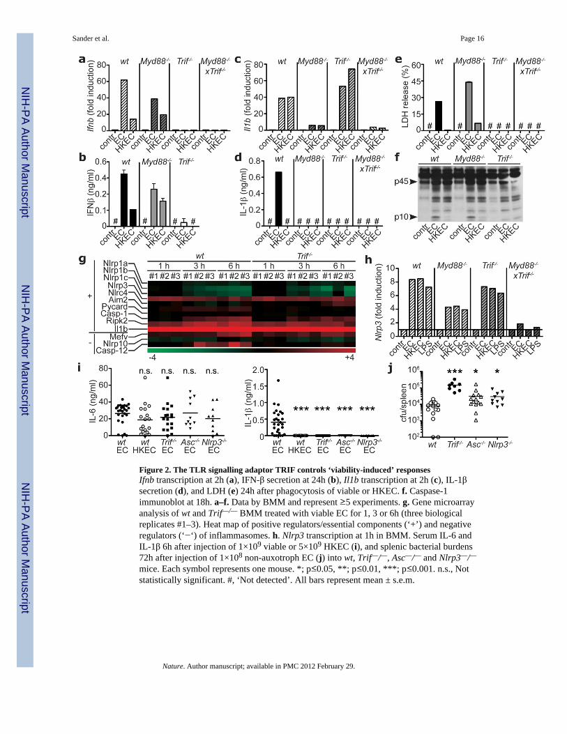
Figure 2. The TLR signalling adaptor TRIF controls ‘viability-induced’ responsesIfnb transcription at 2h (a), IFN-β secretion at 24h (b), Il1b transcription at 2h (c), IL-1βsecretion (d), and LDH (e) 24h after phagocytosis of viable or HKEC. f. Caspase-1immunoblot at 18h. a–f. Data by BMM and represent ≥5 experiments. g. Gene microarrayanalysis of wt and Trif—/— BMM treated with viable EC for 1, 3 or 6h (three biologicalreplicates #1–3). Heat map of positive regulators/essential components (‘+’) and negativeregulators (‘−‘) of inflammasomes. h. Nlrp3 transcription at 1h in BMM. Serum IL-6 andIL-1β 6h after injection of 1×10⁹ viable or 5×10⁹ HKEC (i), and splenic bacterial burdens72h after injection of 1×10⁸ non-auxotroph EC (j) into wt, Trif—/—, Asc—/— and Nlrp3—/—mice. Each symbol represents one mouse. *; p≤0.05, **; p≤0.01, ***; p≤0.001. n.s., Notstatistically significant. #, ‘Not detected’. All bars represent mean ± s.e.m.
Sander et al. Page 16
Nature. Author manuscript; available in PMC 2012 February 29.
NIH
-PA Author Manuscript
NIH
-PA Author Manuscript
NIH
-PA Author Manuscript

Figure 3. Bacterial RNA is a vita-PAMP that accesses cytosolic receptors during phagocytosis,and in the absence of virulence factorsa. LPS/Endotoxin, genomic DNA and total RNA in EC before and at indicated times afterheat killing. b. Agarose gel electrophoresis of EC total RNA before and after heat killing at60°C for 60min followed by 4°C incubation for indicated times. c,d. LDH, IL-1β, IFN-β andIL-6 at 24h in response to viable and HKEC, or HKEC with 10µg/ml total EC RNA (HKEC+RNA). # in c,d, ‘Not detected’. a–d, Data by BMM represent ≥5 experiments. e.Representative ratiometric epifluorescence imaging of BMM at 8h with Fdx alone (ctr 8h),Fdx and viable EC (EC 8h) or gentamicin-killed EC (Gent EC) (colour code indicates pHscale). Positive control: Ground silica (silica 1h). f. Quantification of cytosolic Fdx (% oftotal Fdx/cell). Each dot represents % released Fdx/individual cell. Grey bars represent meanFdx release. *; p≤0.05, **; p≤0.01, ***; p≤0.001. All bars represent mean ± s.e.m.
Sander et al. Page 17
Nature. Author manuscript; available in PMC 2012 February 29.
NIH
-PA Author Manuscript
NIH
-PA Author Manuscript
NIH
-PA Author Manuscript

Figure 4. Bacterial messenger RNA constitutes an active vita-PAMPa–c and e–g. LDH, IL-1β and IL-6 at 24h. a. Total EC RNA treated with RNAse I andRNAse III, RNAse III alone, or DNAse prior to stimulation of BMDC. b. BMM treated withviable or HKEC, or HKEC with 0.1µg/ml of different bacterial RNA; ribosomal (rRNA),messenger (mRNA), small (sRNA) or eukaryotic RNA (eukRNA). c. BMDC responses.Gro-RNA; in vitro transcribed EC Gro-operon RNA. d. Predicted secondary structure ofGro-RNA. Colour code indicates base pairing probability. e. BMM treated with in vitrotranscribed Gro-RNA or Gro dsRNA alone or with HKEC. f. BMDC responses. Era-RNAand DNApol-RNA; in vitro transcribed EC Era-GTPase and DNA-polymerase-III RNA,respectively. g. BMM treated with different doses of unmodified (control), or modified Gro-
Sander et al. Page 18
Nature. Author manuscript; available in PMC 2012 February 29.
NIH
-PA Author Manuscript
NIH
-PA Author Manuscript
NIH
-PA Author Manuscript

RNA with HKEC (5’cap, 5’ m7G capping; CIP, calf intestinal phosphatase; 3’poly(A), 3’-polyadenylation). a–g. #, ‘not detected’, all RNA at 10µg/ml except as noted, data represent≥5 experiments. h. Mice vaccinated and boosted twice with viable EC, HKEC or HKECwith 30µg total purified bacterial RNA (HKEC+RNA) (vaccination regimen in suppl. Fig.22). Class-specific anti-E. coli antibody serum titers at 25 days. *; p≤0.05, **; p≤0.01, ***;p≤0.001. All bars represent mean ± s.e.m.
Sander et al. Page 19
Nature. Author manuscript; available in PMC 2012 February 29.
NIH
-PA Author Manuscript
NIH
-PA Author Manuscript
NIH
-PA Author Manuscript


![Reviews of Physiology, Biochemistry and Pharmacology 155 · [eIFs] in eukaryotes and prokaryotic translation initiation factors [IFs] in prokaryotes) bind the mRNA, deliver the initiator](https://img.pdfslide.us/doc/110x75/5f09fea37e708231d4298040/reviews-of-physiology-biochemistry-and-pharmacology-155-eifs-in-eukaryotes-and.jpg)