Embed Size (px)
Citation preview

PAPER
CRIMINALISTICS
Andrew Farrar,1 B.Sc. (Hons); Glenn Porter,1 M.App.Sc.; and Adrian Renshaw,1 Ph.D.
Detection of Latent Bloodstains Beneath Painted Surfaces UsingReflected Infrared Photography
ABSTRACT: Bloodstain evidence is a highly valued form of physical evidence commonly found at scenes involving violent crimes.However, painting over bloodstains will often conceal this type of evidence. There is limited research in the scientific literature that describesmethods of detecting painted-over bloodstains. This project employed a modified digital single-lens reflex camera to investigate the effective-ness of infrared (IR) photography in detecting latent bloodstain evidence beneath a layer or multiple layers of paint. A qualitative evaluationwas completed by comparing images taken of a series of samples using both IR and standard (visible light) photography. Further quantitativeimage analysis was used to verify the findings. Results from this project indicate that bloodstain evidence can be detected beneath up to sixlayers of paint using reflected IR; however, the results vary depending on the characteristics of the paint. This technique provides crime scenespecialists with a new field method to assist in locating, visualizing, and documenting painted-over bloodstain evidence.
KEYWORDS: forensic science, infrared photography, bloodstains, forensic photography, invisible radiation, latent evidence
Blood evidence recovered at crime scenes is generallyconsidered as having a high degree of forensic value. Severalcontemporary techniques may be used to detect the presence ofblood at scenes where violent crimes were committed (1–3).However, when blood evidence has been tampered with, hidden,or covered up, it becomes increasingly difficult to detect.A common question facing forensic practitioners when examin-ing blood is whether a red-brown stain is actually blood. Thismay lead to further questions such as whether or not the bloodis of human origin, and if so, whether the bloodstains can belinked to a particular individual using DNA. Furthermore, blood-stain pattern analysis (BPA) may also be used to assist in recon-structing the event. Because bloodstain evidence has such anintrinsic forensic value, perpetrators frequently attempt to con-ceal or remove this type of evidence from the scene (4). Onemethod of concealing blood spatter is to paint over the wall orsurface where the stains are located.Painting over bloodstains on walls will conceal blood evi-
dence beneath the paint layers. This makes detection of blood-stains more difficult during crime scene examinations. There islimited research described in the forensic science literature thatinvestigates the visualization of bloodstains located beneathpainted surfaces. Some research suggests a nondestructive tech-nique using monochromatic light sources such as a PolilightTM
(5); however, some work also suggested that the blood reagentluminol could still produce a chemiluminescence reaction evenwhen the blood is located beneath a layer of paint (6,7). Previ-ous studies have also demonstrated that paint does not appear to
cause false positive results with luminol (8). While there are alimited number of published studies involving painted-overbloodstains in the context of crime scene investigation, extensiveresearch has been completed in the field of art conservation inregards to visualizing artifacts beneath paint layers. The applica-tion of infrared (IR) photography has been used in conservationpractices to evaluate the authenticity of paintings by lookingbeneath its uppermost paint surface (9).IR radiation is capable of penetrating visually opaque material
to reveal object states that the naked eye cannot see. It is alsocapable of discerning between different material that may appearoptically similar under visible light but produce distinct differ-ences when irradiated by IR. These variations occur becauseobjects can absorb, reflect, or transmit IR by varying degrees(10). The two main uses for using IR within art conservationpractices are (i) to detect any alterations or restoration work thathas been made to the painting and (ii) to visualize any prelimin-ary drawings or markings beneath the painted surface that wereused to form the composition of the artwork. These originalmarkings also referred to as “underdrawings” may act as a signa-ture of the painting and are usually not present in forgeries. IRhas proven to be a successful technique in this context and canrecord artifacts that are beneath the paint surface (11).Previous research into the interaction between IR radiation
and paint determined that a layer of white paint could increasethe degree of transmission when illuminated by IR radiation.Mairinger (12) suggested the following factors that increase IRpenetration through paint:
i) the greater the wavelength of the incident radiation, ii)the smaller the thickness of the paint layer, iii) the smallerthe number of particles in the layer (pigment/volume con-centration) and iv) the lesser the difference of refractiveindices of pigment and medium (12, p. 42).
1School of Science & Health, University of Western Sydney, Locked Bag1797, NSW 2751, Australia.
Received 10 Oct. 2010; and in revised form 15 Feb. 2011; accepted 16April 2011.
© 2012 American Academy of Forensic Sciences 1
J Forensic Sci, 2012doi: 10.1111/j.1556-4029.2012.02231.x
Available online at: onlinelibrary.wiley.com

A practical study into the transmission of IR radiation throughpaint pigments determined that certain combinations of opticalfilters are able to penetrate some pigments, whereas other paintswere completely opaque and that multiple pigmented paintsimpair IR penetrability (13).IR photography has already found numerous applications
within forensic science practices. Early research identified IR asbeing capable of visualizing bite marks on human skin (14) aswell as the assessment of inks during document examination(15). Recent studies have found IR useful in the visualization ofgun shot residue patterns on dark and multicolored clothing (16)as well as detecting and visualizing the presence of a tattoo thathad been removed by laser surgery (17). IR is applied within theBPA discipline with the most common use assisting the visuali-zation of latent bloodstains on dark surfaces (18). Other recentstudies using IR involve the detection of blood on different typesof clothing, fabrics, and patterns (19) and identifying bloodstainson black fabric using various blood dilutions (20). The mostnoteworthy conclusion to draw from the scientific literature isthat blood is capable of absorbing IR radiation. Chun-Yen Linet al.’s (20) research indicated that blood continued to absorb IRradiation up to 1/8th of its original concentration.Previous studies have indicated two key points relating to this
work: (i) that paint layers, depending on pigment and thickness,are capable of allowing IR transmission and (ii) that bloodabsorbs IR radiation. Combining these two concepts produce alikely hypothesis suggesting it is possible for IR to visualize alatent bloodstain located beneath a layer of paint. Theoretically,IR radiation will transmit through certain paint pigments, thusincreasing the transparency of paint layers, be absorbed byblood, while the background reflects IR. These differences inspectral response (transmission, absorption, and reflection) arerecorded by IR and latent bloodstains could be detected.This project employed a modified digital single-lens reflex
(SLR) camera, converted to exclusively record IR wavelengths,to investigate the effectiveness of IR in detecting latent bloodbeneath layers of paint. A qualitative evaluation was completedby comparing images taken from a series of samples using IRand standard visible light photography. Quantitative image anal-ysis was also conducted to verify the photographic results.Throughout this study, several objectives were investigated
and included (i) standardizing or calibrating exposure values(EVs) between the specially modified IR camera and a standardcamera, to optimize image quality and provide an accurate com-parative image analysis; (ii) developing a method to quantify theeffectiveness of IR in detecting and visualizing blood beneathlayers of paint; and (iii) investigate the effects of variables suchas different types of paints, different colors, and interferences.
Materials and Methods
Spectral Sensitivity of the Digital Camera
This project utilized a specially modified Canon EOS 10DTM
digital SLR camera (Canon U.S.A., Inc., Lake Success, NY) thatwas converted to record exclusively within the IR spectrum. Thecamera modification was conducted by a commercial photo-graphic engineering company. Digital cameras are naturallysensitive to the IR region because of the spectral sensitivity of thesemiconductors used by digital cameras. However, IR blockingfilters are installed over these sensors to improve optical perfor-mance and assist chromatic aberration correction by attenuatingthe IR wavelengths recorded by the sensor. Modification
involved replacing the IR blocking filter with an IR transmissionfilter directly onto the sensor. The replacement filter is a longpass optical filter equivalent to a Wratten 87C (IR transmis-sion > 820 nm). For the remainder of this article, the modifiedCanon EOS 10DTM SLR camera that photographs exclusivelywithin the IR spectrum will be referred to as the IR camera andthe standard unmodified camera used to photograph controlimages will be referred to as the VIS camera.
Calibrating Exposure Values and Camera Settings
To enable accurate comparative evaluation between each typeof recorded image (IR and visible light), each photograph wastaken at a consistent EV. Owing to each recording techniqueusing different components of the electromagnetic spectrum andusing different optical filters on each camera sensor, calibrationbetween exposures is necessary. A critical component of thisexperimental design was to determine “equal” exposures for bothstandard white light and IR techniques. An experiment wasundertaken to ensure that equivalent exposures could provide ameaningful comparison between each recording method.Testing EVs consisted of taking a series of images using a
KodakTM Q13 (Rochester, NY) test target standard. Cameraswere attached to a copy stand and evenly lit using two tungstenhalogen lights. Several test images were taken of the Q13 testtarget with a range of altering exposures. The images wereanalyzed using Adobe PhotoshopTM (Adobe Systems Inc., SanJose, CA) by first removing all color information and thenmeasuring the segment defined as “M” on the Q13 target. Thisvalue is considered as having an 18% reflectance and is a stan-dard reflectance value in photographic science. The eyedroppertool was used to measure the pixel brightness, which has anumerical range for an 8-bit image between 0 and 255 (256different brightness values). By analyzing the “M” segment,EVs for each photographic method were chosen where the “M”
was closest to match the 18% gray value, which consisted of abrightness value of 128 (or 256/2). For the IR camera, thisexposure was calculated to be f/2.8 at 1/250th sec (EV 11) andf/3.5 at 1/320th sec (EV 11.9) for the VIS camera at 100ISO.The camera settings used throughout the experiment are detailedin Table 1.
Sample Preparation
Sheets of 10 mm plasterboard were sourced from a localhardware store. A white acrylic primer was applied to the plas-terboard to seal the surface and provide an undercoat similar toa wall surface. A single sheet of plasterboard was also used as acontrol surface and was not primed. The primer was left to dryfor a minimum of 12 h before any further sample preparationwas completed. After the plasterboard was sealed, it was cut intoa series of 200 9 200 mm sized pieces.
TABLE 1––Camera settings for each camera used in the experiment.
IR Camera VIS Camera
Lens 50 mm macro 50 mm macrof/stop 2.8 3.5Shutter speed 250 320ISO 100 100“u” distance 0.8 m 0.8 mEV 11 11.9
2 JOURNAL OF FORENSIC SCIENCES

Stencil Construction
A perspex stencil was constructed to assist in placing thebloodstains on the sample surface. The stencil positioned thebloodstains in the same arrangement on each sample to aid inthe quantitative image analysis component of the research. Theperspex stencil included a series of small holes aligned in a3 9 3 grid. The holes aligned to a grid with total dimensions of100 9 100 mm, and each hole was c. 50 mm horizontally orvertically away from one another. The stencil also had foursmaller holes located peripherally to this grid, large enough for apin to be inserted. Once the template had been drawn onto theperspex surface, 10-mm holes were drilled for each blood holeand 2 mm for the peripheral holes. A small red triangle adhesivewas attached to the front of the stencil to ensure it consistentlyfaced the same direction while applying blood to the samplesurface. Four large magnetic weights were also attached to thebottom of the stencil to create a gap between the stencil and theplasterboard while pipetting blood to prevent pooling. A sche-matic of the stencil design is shown in Fig. 1.
Detection of Blood Beneath Paint
Three test conditions were investigated in this study andinclude types of white paint, colored paints, and interferences.The three types of white paint selected were acrylic, oil-based,and white spray paint; the acrylic colored paints selected wereblack, red, yellow, blue, green, orange, and purple; and the inter-ferences tested included using undiluted blood, diluted blood to1/100th and 1/1000th, cleaning up the blood after dropping it onthe plasterboard, using water in lieu of blood, having no under-coat present, and then using white acrylic paint on a blackundercoat and black acrylic paint on a black undercoat. A blanksample where no blood was applied to the sample surface wasalso used as a control. The basic procedure for preparing allsamples, painting, and photography is outlined below.The perspex stencil was placed over a prepared piece of
plasterboard and secured. Four pins were inserted through theperipheral holes in the stencil to create an indentation in theplasterboard. The stencil was then removed and four black mappins were placed securely into the indentations. A control photo-graph (C0) of the sample was then taken using both the IR andthe VIS cameras. The cameras were attached to a copy stand,and the samples were evenly lit with two tungsten halogenlamps that were set up permanently for the study. All samples ateach stage of the project were photographed on a piece of black
cardboard with a linear scale, a Q13 target, and the respectivesample number. The layout of how samples were photographedis shown in Fig. 2.The stencil was then placed back onto the plasterboard using
the map pins to guide it to its original position. Two hundredmicroliters of defibrinated horse blood (sourced from commercialbiological supplier) that had been diluted to 1/10th of its originalconcentration was dropped directly through each of the nineholes in the stencil using a micropipette. The stencil wasremoved and the blood was left to dry for a minimum of 48 h.Once the blood had dried, the layer zero photograph (L0) wastaken using both IR and VIS cameras. After photographing theL0 layer, the four map pins were removed from the plasterboardand a single layer of selected paint was applied to the sampleusing small disposable paint rollers. To maintain a level ofconsistency, the painting technique was standardized. Thirty mil-liliters of paint was considered a “single paint layer” and coveredthe entire surface of the plasterboard. The pins were then placedback into their original positions, and the paint was left to dryfor a minimum of 24 h. Once the paint had dried, the layer onephotograph (L1) was taken in the same manner as the L0 photo-graph. This process was repeated when applying layers twothrough to six (L2–L6). After the photography had been com-pleted, visual examination and image analysis were conducted.A slightly altered method was required when applying the white
spray paint. Once both the C0 and L0 photographs were taken, thecan of paint was shaken for c. 2 min before spraying and for a fur-ther 10 sec every few minutes during application. The can washeld c. 150–200 mm from the surface and the paint was appliedby moving back and forth in even strokes. This procedure wasthen repeated to apply layers two through to six (L2–L6).A slightly altered method was also needed when introducing
interferences to the samples as changes were made to the way inwhich the paint or blood was applied to the sample surfaces. Allinterferences used white acrylic paint unless otherwise specified;one sample used undiluted blood instead of 1/10th diluted blood;two samples used 1/100th and 1/1000th diluted blood,respectively; one sample involved allowing the blood to drybefore wiping it off with a cleaning product before applying thefirst layer (L1) of paint; one sample substituted water for blood;one sample used a plain piece of plasterboard without anyprimer applied; one sample involved the plasterboard having ablack undercoat before applying layer one through to six (L1–L6) of white acrylic paint; and one sample involved the plaster-board having a black undercoat before applying layer onethrough to six (L1–L6) of black acrylic paint.
FIG. 1––Schematic of the stencil design.
FARRAR ET AL. . DETECTION OF LATENT BLOODSTAINS BENEATH PAINTED SURFACES 3
Visual Examination of Photographs and Image Analysis
The qualitative analysis component of this study involved avisual comparison of the resultant photographs of all samples ateach stage of the painting process. By visually examining theimages on a calibrated computer monitor, the paint layer atwhich the bloodstain became latent was recorded, and the suc-cess of the technique was rated.Image analysis used a combination of Adobe PhotoshopTM
and ImageJTM (National Institutes of Health, Bethesda, MD) toquantify the effectiveness of this technique. Adobe PhotoshopTM
was used to prepare the photographs for analysis, and ImageJwas used to record pixel brightness values from the image.Image analysis was completed for the C0, L0, L1, L2, and L3photographs of each sample. Each photograph to be analyzedwas cropped into a 1000 9 1000 pixel image by using the pinson each sample as a guide to form a square. The image wasthen transformed into an 8-bits/channel image by selectingImage > Mode and by checking both “Grayscale” and “8-bits/channel” so that all color information was discarded and tomaintain consistency during analysis. The image was thenopened in ImageJ and the linear tool was used to take sixindividual readings at the 150, 500, and 850 pixel mark, bothhorizontally and vertically, and ensuring it crossed through threebloodstains each time. By selecting Analyze > Plot Profile onImageJ, a profile of the line was graphed and the brightnessvalues were saved into Excel spreadsheets. Each pixel isassigned a brightness value (a numerical value from 0 to 255)that represents its brightness on a gray scale. A value of 0(zero) refers to a pixel being completely black and a value of255 refers to a pixel being completely white. By generating a
plot profile, graphic information can be turned into empiricaldata, and visual changes across the image, such as brightness,can be recorded.
Results
The results from this study are expressed using two methods(i) a qualitative evaluation using a visual comparison betweenimages taken using IR and standard visible light photographyand (ii) a quantitative method using image analysis (mean pixelbrightness values).Comparison of photographs using IR and white light photog-
raphy demonstrated that IR could detect latent bloodstainsbeneath up to six layers of paint. Certain paints, such as blackand purple acrylic, allowed greater IR transmission than otherpaints used in this study. Bloodstains located beneath whiteacrylic could not successfully be photographed using IR after afew layers had been applied to the sample. In cases where theblood remained visible beneath multiple layers when photo-graphed using both standard and IR, such as with red acrylicpaint, IR did improve the visualization of the bloodstains. Fig-ure 3 summarizes the information obtained from the qualitativeanalysis of all test conditions investigated and reports the layerwhere the bloodstain was last visible. Figure 4 features a seriesof photographs that were taken during the study to demonstratethe comparison of results between reflected IR and visible lightphotography.
Types of White Paint
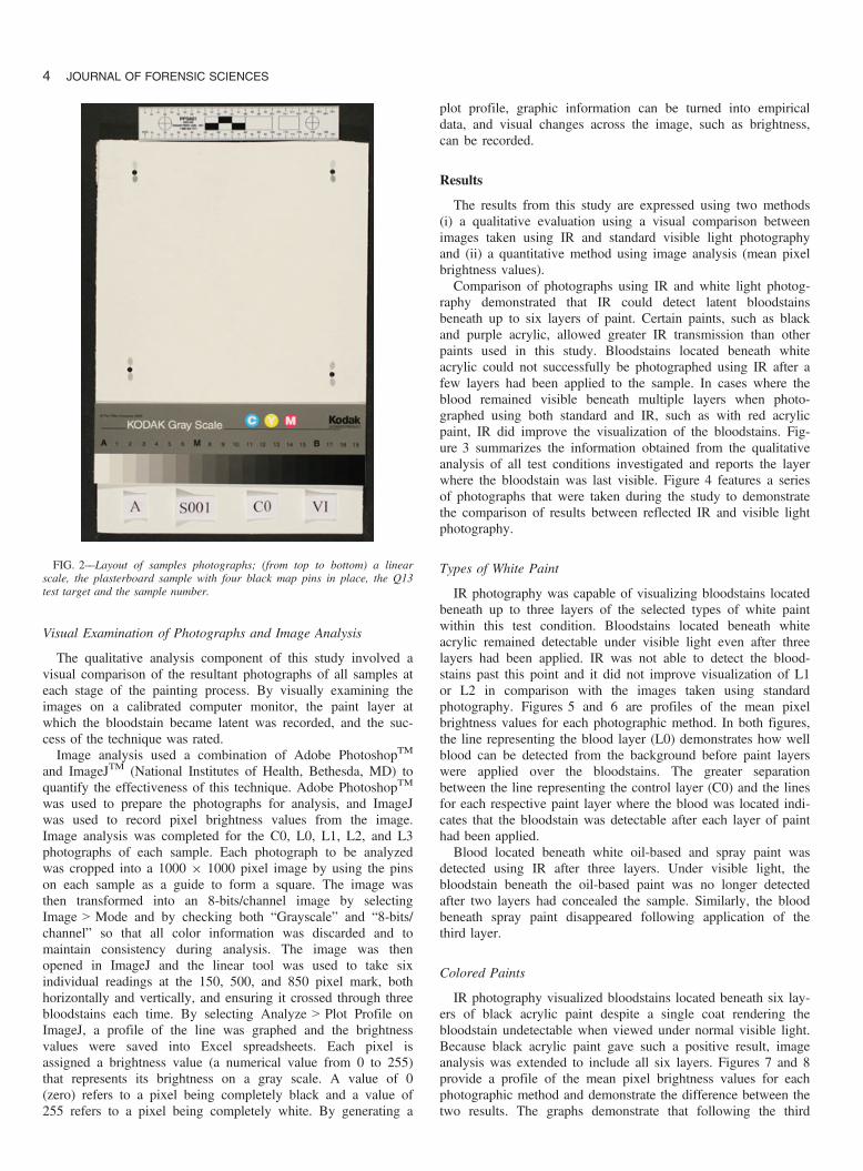
IR photography was capable of visualizing bloodstains locatedbeneath up to three layers of the selected types of white paintwithin this test condition. Bloodstains located beneath whiteacrylic remained detectable under visible light even after threelayers had been applied. IR was not able to detect the blood-stains past this point and it did not improve visualization of L1or L2 in comparison with the images taken using standardphotography. Figures 5 and 6 are profiles of the mean pixelbrightness values for each photographic method. In both figures,the line representing the blood layer (L0) demonstrates how wellblood can be detected from the background before paint layerswere applied over the bloodstains. The greater separationbetween the line representing the control layer (C0) and the linesfor each respective paint layer where the blood was located indi-cates that the bloodstain was detectable after each layer of painthad been applied.Blood located beneath white oil-based and spray paint was
detected using IR after three layers. Under visible light, thebloodstain beneath the oil-based paint was no longer detectedafter two layers had concealed the sample. Similarly, the bloodbeneath spray paint disappeared following application of thethird layer.
Colored Paints
IR photography visualized bloodstains located beneath six lay-ers of black acrylic paint despite a single coat rendering thebloodstain undetectable when viewed under normal visible light.Because black acrylic paint gave such a positive result, imageanalysis was extended to include all six layers. Figures 7 and 8provide a profile of the mean pixel brightness values for eachphotographic method and demonstrate the difference between thetwo results. The graphs demonstrate that following the third
FIG. 2––Layout of samples photographs; (from top to bottom) a linearscale, the plasterboard sample with four black map pins in place, the Q13test target and the sample number.
4 JOURNAL OF FORENSIC SCIENCES

layer of paint, the application of additional layers caused detec-tion of the bloodstains using IR to reach a limit where it couldno longer quantitatively be distinguished from the surrounding
background. However, close visual examination of the IRphotographs confirmed minor traces of the bloodstain even afterthe sixth layer had been applied.
FIG. 4––Photographs of layers L0, L1, L2 and L3 for (a) white acrylic paint, (b) black acrylic paint, (c) red acrylic paint, (d) blue acrylic paint, (e) greenacrylic paint, and (f) purple acrylic paint using infrared (IR) photography and standard visible light photography (VIS).
FIG. 3––Visual examination results when recorded using visible light photography (VIS) and infrared (IR) photography; the paint layer value refers to thelayer where the bloodstain was last visible in the photographs.
FARRAR ET AL. . DETECTION OF LATENT BLOODSTAINS BENEATH PAINTED SURFACES 5
Red acrylic paint was incapable of effectively concealing thebloodstains even after six coats had been applied. However, IRimproved the contrast between the blood and the backgroundand improved visualization. Yellow, blue, and orange acrylicpaint all allowed bloodstains to be detected after six layers hadbeen applied. Green acrylic completely concealed the bloodstainsafter three layers but it remained visible in the IR photographsuntil the fourth layer. IR could visualize bloodstains locatedbeneath six layers of purple acrylic compared to three layers toconceal the bloodstains using standard photography.
Interferences
On samples where no bloodstain was present, including theblank and the sample that used water in lieu of blood, nothingwas detected at any stage of photography using either method.Furthermore, when a bloodstain had dried and was cleaned off,no residual bloodstain or artifacts were detected visually orquantitatively using image analysis after any paint was applied.Diluted blood decreased the ability of IR to detect the latent
bloodstains. Figure 9 visually compares the IR L0 photograph
FIG. 5––Image analysis results (mean pixel brightness value) of paint layers C0, L0, L1, L2 and L3 for white acrylic paint using standard visible lightphotography (VIS). Bloodstains are located on the 150, 500, and 850 pixel distances and results either side of these markers are the background.
FIG. 6––Image analysis results (mean pixel brightness value) of paint layers C0, L0, L1, L2, and L3 for white acrylic paint using infrared (IR) photography.
6 JOURNAL OF FORENSIC SCIENCES

taken of each of the dilutions. As demonstrated, diluting theblood to 1/100th and 1/1000th greatly impaired visualizationwhen using IR. After a single coat of paint, both bloodstainswere concealed. In cases where blood was diluted greater than1/10th of its original concentration, the bloodstain was more vis-ible when photographed using standard photography in compari-son with images taken using reflected IR.The absence of an undercoat decreased the success of the tech-
nique. Two coats of paint completely concealed the bloodstainand no difference in visualization was detected between images
using IR and visible light photography. A black undercoat didnot affect results when white paint was used as the overcoat.However, the black undercoat with a black overcoat decreasedthe number of layers that IR could visualize the bloodstains.
Discussion
The aim of this study was to investigate whether bloodstainscan be detected or visualized beneath layers of paint whenphotographed using reflected IR. As stated, within a forensic
FIG. 7––Image analysis results (mean pixel brightness value) of paint layers C0, L0, L1, L2, L3, L4, L5, and L6 for black acrylic paint using standard visiblelight photography (VIS).
FIG. 8––Image analysis results (mean pixel brightness value) of paint layers C0, L0, L1, L2, L3, L4, L5, and L6 for black acrylic paint using infrared (IR)photography.
FARRAR ET AL. . DETECTION OF LATENT BLOODSTAINS BENEATH PAINTED SURFACES 7

context, IR has been used for other types of bloodstain analysisin cases where the blood was difficult to detect because of thelack of contrast between the bloodstain and the surface where itwas deposited (18–20). The study illustrated in this paper issomewhat different to previous research. The bloodstains are notsitting on the surface of the substrate but beneath layers of paint.This means that the bloodstain is concealed when viewed undervisible light or when photographed using standard photography.The various optical enhancement parameters that apply to IRused in this study are• The spectral transmission of the paint: Because the blood-
stains are located beneath layers of paint, the paint layersmust be capable of transmitting IR radiation and result in thepaint becoming transparent to IR wavelengths. Variations inthe chemical composition of the paint, paint color, and thick-ness of the layers will also alter the spectral transmissionqualities of the overlaying paint.
• The spectral absorption of blood and reflectance of the back-ground: To record a distinction between the bloodstains andthe background, the blood must absorb IR while the back-ground must do the opposite and reflect IR. This differencebetween the spectral response of the bloodstains and thebackground is critical. The concentration of a bloodstain alsoalters the IR absorption properties of blood.
• The spectral sensitivity of the camera: The camera must be sen-sitive to IR radiation. It was necessary to modify a standarddigital SLR camera to achieve spectral sensitivity in the IRspectrum. Most digital SLR cameras are not sensitive to thisregion because an IR blocking filter is installed over the sensor.
• The spectral transmission of the camera filter: An IR trans-mission filter is required to eliminate all visible light andexclusively record wavelengths featuring components of theIR spectrum. An IR transmission filter was installed directlyonto the camera’s sensor during its modification to allow theexclusive recording of IR radiation.
• The spectral distribution of the light source: The light sourceused for IR must contain components of IR. Tungsten halo-gen lamps were used throughout this project to illuminate thespecimens.
The paints selected for this study represent a small sample ofconditions that painted-over bloodstains could be created or con-cealed beneath. These included using three different types ofwhite paint, colored paints, and introducing variables such asdiluting the blood to determine how this would affect the effi-ciency of IR. The paints selected were chosen because they wereamong some of the most commonly sold paint and available atlocal hardware stores. It was not considered viable during thisstudy to test all available paints and colors due to the immensenumber available.Of the three types of white paints selected, it was found that
white oil-based paint was the most effective in concealing blood-
stains when using standard photography, however, it was foundthat each of the white paints appeared to be very effective inconcealing blood, even when photographed using IR. The varia-tion of results achieved from standard visible light photographymay be attributed to the chemical make-up of the individualtypes of paint. The chemical differences between the three painttypes may inhibit visible light and IR from penetrating theuppermost layers, thus concealing bloodstains more effectively.Colored paints were more successful in allowing the detection
of bloodstains using IR. Black acrylic paint exhibited the great-est difference when comparing both photographic methods. Asingle layer of black paint was enough to conceal bloodstainsunder standard photography, however, even after six layers IRwas still able to detect blood.Purple acrylic paint also produced a strong result, whereas
green acrylic inhibited IR penetration. Vandenberg and Oorschot(5) also found green paint to effectively conceal blood, evenwhen viewed using monochromatic light with a central band-width of 415 nm. The variation in results among the coloredpaints indicate that pigment may also play an important role inthe effectiveness of whether or not IR can detect a painted-overbloodstain. This further supports the conclusions proposed byWise (13) who stated that certain pigments allowed greater IRpenetration than others did when using IR.Diluted blood decreased the efficiency of IR to detect a
painted-over bloodstain. Undiluted blood was visible after sixlayers of paint had been applied using both photographicmethods but this was attributable to the topographical nature ofthe sample. By diluting the blood to 1/10th of its original con-centration as well as 1/100th and 1/1000th respectively, the levelof IR absorption was noticeably weaker than undiluted blood.Diluted blood caused the blood drops to appear lighter. This wasmost likely due to lower concentrations of hemoglobin mole-cules being present in the diluted blood and therefore less IRabsorption occurred. The results from the diluted samples mayrepresent scenarios where attempts to remove bloodstains havebeen made and leaving only residual or diluted traces of blood.Water drops were not detectable using standard or IR before orafter any stage of the painting process.Results indicate that the technique worked more effectively on
samples where an undercoat was present beneath the bloodstain.The sample without an undercoat caused the blood to appearwith less clarity in comparison with samples where an undercoatwas applied. An undercoat may provide a more reflective surfaceand increasing the effectiveness of IR and enhancing the contrastbetween the bloodstain (IR absorption) and the background (IRreflectance).In photographs taken using IR where the paint still effectively
concealed the bloodstain, it was possible that (i) the IR radiationemitted from the tungsten halogen lamps was not strong enoughto penetrate particular types of paints, (ii) the layer or layerswere too thick to allow IR radiation to successfully transmitthrough the paint, (iii) a particular chemical constituent in thepaint such as the pigment or binder was inhibiting IR penetra-tion, or (iv) a combination of all three reasons. This supports theproposed conditions provided by Mairinger (12) under which thetransparency of a paint layer depends on the interaction betweenIR radiation and the physical dimensions of the layers of paint.By utilizing both visual comparative examination and image
analysis results could be verified. By completing a quantitativeanalysis, controls were used to ensure that all images werephotographed under the same conditions. Controls also ensuredsamples were exposed to the same amount of light consistently
FIG. 9––Visual comparison of (a) undiluted blood, (b) diluted blood (1/10th),(c) diluted blood (1/100th), and (d) diluted blood (1/1000th). Images takenusing infrared (IR) photography.
8 JOURNAL OF FORENSIC SCIENCES
at every stage of the photographic process. However, imageanalysis was only completed for the control layer, blood layer,and first three layers of paint (C0, L0, L1, L2, and L3). Thephotographs taken for L4 through to L6 were not processedthrough ImageJ and were deemed unnecessary. Image analysisplots that contained the first three layers of paint were consid-ered sufficient to demonstrate the success of this technique;however, all photographs were still visually examined and wereincluded in the overall results presented in Fig. 2. The onlyexception was for black acrylic paint where photographs for L4,L5, and L6 were also analyzed.
Conclusion
Results from this study indicate that bloodstains are capableof being detected beneath layers of paint using reflected IR. Byemploying qualitative analysis that involved visually examiningphotographs using both IR and visible light photography, theresult indicates that IR could detect bloodstains beneath up tosix layers of paint. These results were further supported by aquantitative method that used image analysis to determine themean pixel brightness of the images and demonstrate the effectsmultiple layers of paint had on the clarity of bloodstains. Col-ored paints gave the most successful results in allowing IR todetect painted-over bloodstains. Black paint exhibited the great-est difference between the two recording techniques, and blood-stains were successfully detected after six layers of paint hadbeen applied.In instances where the blood remained visible following the
application of multiple layers of paint, IR increased the contrastbetween the background and the blood with improved visualiza-tion. The application of multiple layers of paint graduallydecreased the clarity at which bloodstains could be detectedusing IR. Furthermore, diluting the blood was found to greatlydecrease the level at which IR could detect bloodstains.This study demonstrates that IR was successful in detecting
bloodstains located beneath a number of layers of paint depend-ing on various optical and physical parameters. These parame-ters include (i) the spectral transmission of the paint, (ii) thephysical dimensions of the paint and the particles containedwithin the paint layers, (iii) variations in the spectral absorptionof blood and the spectral reflectance of the background, and (iv)the IR spectral properties of the photographic recording system.
References
1. Barni F, Lewis S, Berti A, Miskelly G, Lago G. Forensic application ofthe luminol reaction as a presumptive test for latent blood detection.Talanta 2007;72(3):896–913.
2. Miskelly GM, Wagner JH. Using spectral information in forensic imag-ing. Forensic Sci Int 2005;115(2–3):112–8.
3. Thorogate R, Moreira J, Jickells S, Miele M, Daniel B. A novel fluores-cence based method in forensic science for the detection of blood in situ.Forensic Sci Int Genet 2008;2(4):363–71.
4. Bevel T, Gardner RM. Bloodstain pattern analysis with an introductionto crime scene reconstruction, 3rd edn. Boca Raton, FL: CRC Press,2008.
5. Vandenberg N, Oorschot RA. The use of Polilight® in the detection ofseminal fluid, saliva and bloodstains and comparison with conventionalchemical-based screening tests. J Forensic Sci 2006;51(2):361–70.
6. Robinson E. Crime scene photography. Burlington, MA: AcademicPress, 2007.
7. Bily C, Maldonado H. The application of luminol to bloodstains con-cealed by multiple layers of paint. J Forensic Identi 2006;56(6):896–905.
8. Quickenden TI, Creamer JI. A study of common interferences withthe forensic luminol test for blood. J Biolumin Chemilumin 2001;16(4):295–8.
9. Seracini M. Diagnostic investigations on the adoration of the magi byLeonardo Da Vinci. In: Gauluzzi P, editor. Mind of Leonardo—the uni-versal genius at work. Florence, Italy: Giunti, 2006;94–101.
10. Porter G. Photography: marks impressions and documents. In: JamiesonA, Moenssens A, editors. Wiley encyclopedia of forensic science. NewYork, NY: Wiley & Sons, 2009;2036–57.
11. Verhoeven G. Imaging the invisible using modified digital still camerasfor straightforward and low-cost archaeological near-infrared photogra-phy. J Archaeol Sci 2008;35(12):3087–100.
12. Mairinger F. The infrared examination of paintings. In: Creagh DC,Bradley DA, editors. Radiation in art and archeometry. New York, NY:Elsevier Science, 2000;40–55.
13. Wise D. Through a glass, darkly: practical applications of infraredphotography. Melb J Tech Stud Art 2005;2(6):85–94.
14. Wright FD. Photography in bite mark and patterned injury document.J Forensic Sci 1998;43(4):877–80.
15. Ellen DM, Creer KE. Infrared luminescence in the examination of docu-ments. J Forensic Sci 1970;10(3):159–64.
16. Bailey J. Digital infrared photography to develop GSR patterns. Aust JForensic Sci 2007;39(1):33–40.
17. McKechnie ML, Porter G, Langlois N. The detection of latent residuetattoo ink pigments in skin using invisible radiation photography. Aust JForensic Sci 2008;40(1):65–72.
18. Raymond MA, Hall RL. An interesting application of infra-red reflectionphotography to blood splash pattern interpretation. Forensic Sci Int1986;31(3):189–94.
19. Perkins M. The application of infrared photography in bloodstain patterndocumentation of clothing. J Forensic Identi 2005;55(1):1–9.
20. Chun-Yen Lin A, Hsieh H, Tsai L, Linacre A, Lee JC. Forensic applica-tions of infrared imaging for the detection and recording of latent evi-dence. J Forensic Sci 2007;52(5):1148–50.
Additional information and reprint requests:Glenn Porter, M.App.Sc.School of Science & HealthUniversity of Western SydneyLocked Bag 1797NSW 2751AustraliaE-mail: [email protected]
FARRAR ET AL. . DETECTION OF LATENT BLOODSTAINS BENEATH PAINTED SURFACES 9