Embed Size (px)
Citation preview

ARTHRITIS & RHEUMATISM Volume 36 Number 10, October 1993, pp 1353-1363 0 1993, American CoHege of Rheumatology 1353
DETECTION OF INTRAARTICULAR ABNORMALITIES IN OSTEOARTHRITIS OF THE KNEE
A Pilot Study Comparing Needle Arthroscopy with Standard Arthroscopy
ROBERT W. IKE and KENNETH S. O’ROURKE
Objective. To determine whether intraarticular abnormalities in osteoarthritis (OA) of the knee can be detected as well by needle arthroscopy as by standard arthroscopy.
Methods. Needle arthroscopy followed by stan- dard arthroscopy was performed on 10 patients with knee OA (diagnosed according to American College of Rheumatology criteria) whose symptoms were not en- tirely attributable to the OA and were therefore an indication for further evaluation. Each knee was as- sessed for abnormalities of the menisci, articular carti- lage (6 sites), and synovium (6 sites).
Results. Evaluation of the 18 menisci visualized with both techniques yielded the same results: 6 abnor- mal and 12 normal. Among the 54 articular cartilage sites evaluable with both procedures, 16 were judged normal by both needle arthroscopy and standard ar- throscopy. Of the 38 cartilage sites judged abnormal by standard arthroscopy, 34 (89%) were abnormal by needle arthroscopy. Both techniques indicated cartilage changes were the same at 42 (78%) of the 54 sites;
Presented in part at the 55th Annual Scientific Meeting of the American College of Rheumatology, Boston, MA, November 1991.
From the Division of Rheumatology, Rackham Arthritis Research Unit and Michigan Multipurpose Arthritis Center, Depart- ment of Internal Medicine, University of Michigan Medical Center, Ann Arbor.
Supported in part by NIH/NIAMS grant 5P60-AR-20557. Robert W. Ike, MD: Assistant Professor; Kenneth S.
O’Rourke, MD: Lecturer (current address: Assistant Professor, Section on Rheumatology, Bowman Gray School of Medicine, Winston-Salem, NC).
Address reprint requests to Robert W. Ike, MD, Division of Rheumatology, 3918 Taubman Center, 1500 East Medical Center Drive, Ann Arbor, MI 48109-0358.
Submitted for publication December 1, 1992; accepted in revised form April 1, 1993.
changes at the other 12 sites were 1 grade higher by standard arthroscopy than by needle arthroscopy. Both needle arthroscopy and standard arthroscopy revealed 51 evaluable sites in the synovium. Of 34 areas judged abnormal by standard arthroscopy, 24 (71%) were also judged abnormal by needle arthroscopy; 17 areas were judged normal by both techniques. The 2 techniques assigned the same macroscopic score in 27 (53%) of 51 areas of the synovium, with a higher grade by standard arthroscopy in all but 1 of the other 16 areas.
Conclusion. These pilot data suggest that in knee OA, needle arthroscopy can 1) accurately detect menis- cal abnormalities, 2) detect cartilage abnormalities, but may underestimate the severity, and 3) detect most synovial abnormalities, but often underestimates the severity. Needle arthroscopy is a potentially valuable rheumatologic tool for the assessment of OA of the knee.
The arthroscope provides a direct, magnified view of the intraarticular anatomy. Rheumatologists employing arthroscopy during the early days of its development recognized the potential utility of the technique as a diagnostic aid and as a research tool (1). In the 20 years since its reintroduction to North America, the technique has been expanded primarily in the therapeutic arena, as orthopedic surgeons have adopted arthroscopy for many of the resective proce- dures that previously required open arthrotomy. As a consequence, current practice standards dictate that arthroscopy be performed in an operating room (OR) environment, under sterile conditions strict enough to assure the safety of any open procedure that might be performed concurrently. These standards, while pru- dent for procedures in which the joint is invaded repeatedly through multiple stab incisions with instru-

1354 IKE AND O’ROURKE
ments directed at tissue resection, may be excessive for arthroscopic procedures in which inspection and directed biopsy of the joint contents are the sole objectives.The potential for transforming arthroscopy for diagnostic and research usage from an OR proce- dure to an office procedure has been greatly enhanced by the recent development of a small-diameter (1.8- mm, - 16-gauge) fiberoptic arthroscope (also called a “needle scope”) (Optical Catheter System; Medical Dynamics, Englewood, CO) that can be inserted into the joint through needle puncture rather than stab incision. The image “seen” by the needle scope is transmitted to a videocamera via an 11,000-pixel fi- beroptic cable and, as a result, is of lower quality than if it were transmitted through the multiple glass lens system of a standard 4.0-mm arthroscope. Neverthe- less, a preliminary study by Halbrecht and Jackson (2) has indicated that in patients with standard “orthope- dic” conditions of the knee, these images are of sufficient quality to permit clinical decision-making.
Reports of the use of arthroscopy in osteoar- thritis (OA) of the knee have largely been limited to discussions of surgical therapy (3). The complexity of the pathologic anatomy of symptomatic knee OA is only hinted at in these discussions. Other than Lind- blad and Hedfors’ careful documentation of the syno- vial abnormalities in knee OA (4), the involvement of rheumatologists in analyses of the information ob- tained at arthroscopy for knee arthritis has been limited to collaborative work in which the clinical aspects-including the performance of the arthro- scopic procedure-have been under the control of an orthopedic surgeon. The availability of arthroscopy as a tool to be used directly by the rheumatologist for the evaluation of patients with knee arthritis could have tremendous implications for research and clinical care. The needle scope could provide the means by which this arthroscopic capability might be acquired.
Two factors account for the differences in im- aging capability between the needle scope and the standard arthroscope. Besides the aforementioned dif- ference in scope-to-camera transmission that produces a somewhat dimmer and “grainier” image from the needle scope, the 2 scopes view differently. The standard scope has an angled lens that provides a wider field of view from a single point within the knee as the scope is rotated about that point, a feature that cannot be duplicated by the fore-viewing needle scope. Whether these differences translate into an appreciable discrepancy between the capabilities of
the 2 arthroscopes to reveal important features of pathologic anatomy is Q critical concern.
This pilot study was undertaken to determine whether the intraarticular abnormalities of knee OA can be identified by the needle scope as well as they can by the standard arthroscope. In this study, needle arthroscopy was performed on 10 patients prior to their undergoing standard arthroscopy. The results indicate that abnormalities of menisci, articular carti- lage, and synovium in knee OA are revealed to a similar degree by the needle scope as by the standard arthroscope, and that assessment of the degree of severity of synovial abnormalities and cartilage dam- age can be accomplished nearly as well with the needle scope as with the standard arthroscope. Needle arthros- copy appears to be a technique worthy of further devel- opment by rheumatologists as a tool for the assessment of OA and other disorders involving the knee.
PATIENTS AND METHODS Patient selection. The study subjects consisted of 10
patients attending the Rheumatology Clinics at the Univer- sity of Michigan who underwent arthroscopy for the evalu- ation of knee symptoms that were not completely explained by clinical data and had not responded to medical therapy. Medical therapy had consisted of oral analgesics and anti- inflammatory agents in all 10 patients, intraarticular injec- tions of corticosteroids in 9 patients, and physical therapy in 8 patients. The patient who did not receive a corticosteroid injection was young (age 23) and wished first to undergo arthroscopy to assess a focal problem (a large osteophyte) that potentially could be remedied under arthroscopic guidance.
Data collected on all subjects were sufficient to judge whether American College of Rheumatology (ACR) classifi- cation criteria for OA of the knee (5) were met by each subject. These data were the results of physical assessment, laboratory studies of blood and synovial fluid, and radio- graphs of the knees with weight bearing. All subjects satis- fied at least 1 of the 6 possible sets of ACR criteria for classification of knee OA (see Figures 1-3 and Table 8 in ref. 5). Only 1 patient had an additional rheumatic disease diagnosis (a 63-year-old woman with rheumatoid arthritis [RA]) and was included in the study because she had “typical” secondary OA and satisfied 5 of the 6 possible sets of ACR criteria. Another patient, a 49-year-old man, had an inflammatory monarthritis of 1 2 months’ duration that was superimposed on radiographic OA; his condition satisfied 3 of the 6 possible sets of ACR criteria.
Arthroscopic techniques. In all but 1 patient, needle arthroscopy was performed in the OR immediately before standard arthroscopy. The anesthesia the patients had cho- sen for standard arthroscopy (epidural in 5, spinal in 2 , and general in 2) was administered prior to the needle arthros- copy. One patient underwent needle arthroscopy under local
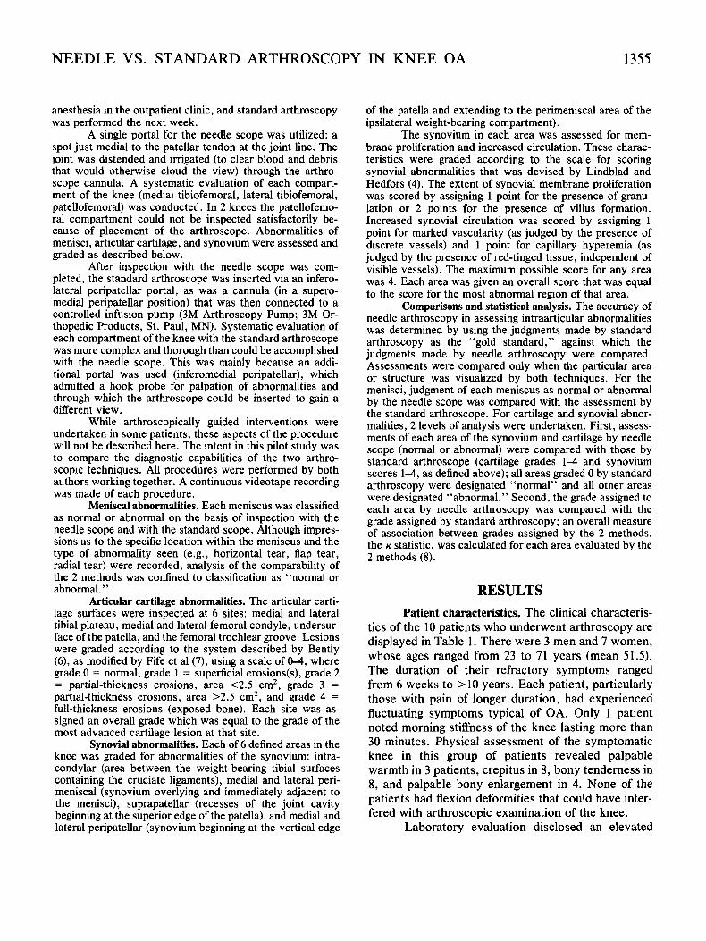
NEEDLE VS. STANDARD ARTHROSCOPY IN KNEE OA 1355
anesthesia in the outpatient clinic, and standard arthroscopy was performed the next week.
A single portal for the needle scope was utilized: a spot just medial to the patellar tendon at the joint line. The joint was distended and irrigated (to clear blood and debris that would otherwise cloud the view) through the arthro- scope cannula. A systematic evaluation of each compart- ment of the knee (medial tibiofemoral, lateral tibiofemoral, patellofemoral) was conducted. In 2 knees the patellofemo- ral compartment could not be inspected satisfactorily be- cause of placement of the arthroscope. Abnormalities of menisci, articular cartilage, and synovium were assessed and graded as described below.
After inspection with the needle scope was com- pleted, the standard arthroscope was inserted via an infero- lateral peripatellar portal, as was a cannula (in a supero- medial peripatellar position) that was then connected to a controlled infusion pump (3M Arthroscopy Pump; 3M Or- thopedic Products, St. Paul, MN). Systematic evaluation of each compartment of the knee with the standard arthroscope was more complex and thorough than could be accomplished with the needle scope. This was mainly because an addi- tional portal was used (inferomedial peripatellar), which admitted a hook probe for palpation of abnormalities and through which the arthroscope could be inserted to gain a different view.
While arthroscopically guided interventions were undertaken in some patients, these aspects of the procedure will not be described here. The intent in this pilot study was to compare the diagnostic capabilities of the two arthro- scopic techniques. All procedures were performed by both authors working together. A continuous videotape recording was made of each procedure.
Meniscal abnormalities. Each meniscus was classified as normal or abnormal on the basis of inspection with the needle scope and with the standard scope. Although impres- sions as to the specific location within the meniscus and the type of abnormality seen (e.g., horizontal tear, flap tear, radial tear) were recorded, analysis of the comparability of the 2 methods was confined to classification as “normal or abnormal. ’ ’
Articular cartilage abnormalities. The articular carti- lage surfaces were inspected at 6 sites: medial and lateral tibial plateau, medial and lateral femoral condyle, undersur- face of the patella, and the femoral trochlear groove. Lesions were graded according to the system described by Bently (6), as modified by Fife et a1 (7), using a scale of 0-4, where grade 0 = normal, grade 1 = superficial erosions(s), grade 2 = partial-thickness erosions, area <2.5 cm2, grade 3 = partial-thickness erosions, area >2.5 cm2, and grade 4 = full-thickness erosions (exposed bone). Each site was as- signed an overall grade which was equal to the grade of the most advanced cartilage lesion at that site.
Synovial abnormalities. Each of 6 defined areas in the knee was graded for abnormalities of the synovium: intra- condylar (area between the weight-bearing tibial surfaces containing the cruciate ligaments), medial and lateral peri- meniscal (synovium overlying and immediately adjacent to the menisci), suprapatellar (recesses of the joint cavity beginning at the superior edge of the patella), and medial and lateral peripatellar (synovium beginning at the vertical edge
of the patella and extending to the perimeniscal area of the ipsilateral weight-bearing compartment).
The synovium in each area was assessed for mem- brane proliferation and increased circulation. These charac- teristics were graded according to the scale for scoring synovial abnormalities that was devised by Lindblad and Hedfors (4). The extent of synovial membrane proliferation was scored by assigning 1 point for the presence of granu- lation or 2 points for the presence of villus formation. Increased synovial circulation was scored by assigning 1 point for marked vascularity (as judged by the presence of discrete vessels) and 1 point for capillary hyperemia (as judged by the presence of red-tinged tissue, independent of visible vessels). The maximum possible score for any area was 4. Each area was given an overall score that was equal to the score for the most abnormal region of that area.
Comparisons and statistical analysis. The accuracy of needle arthroscopy in assessing intraarticular abnormalities was determined by using the judgments made by standard arthroscopy as the “gold standard,” against which the judgments made by needle arthroscopy were compared. Assessments were compared only when the particular area or structure was visualized by both techniques. For the menisci, judgment of each meniscus as normal or abnormal by the needle scope was compared with the assessment by the standard arthroscope. For cartilage and synovial abnor- malities, 2 levels of analysis were undertaken. First, assess- ments of each area of the synovium and cartilage by needle scope (normal or abnormal) were compared with those by standard arthroscope (cartilage grades 1-4 and synovium scores 1-4, as defined above); all areas graded 0 by standard arthroscopy were designated “normal” and all other areas were designated “abnormal.” Second, the grade assigned to each area by needle arthroscopy was compared with the grade assigned by standard arthroscopy; an overall measure of association between grades assigned by the 2 methods, the K statistic, was calculated for each area evaluated by the 2 methods (8).
RESULTS Patient characteristics. The clinical characteris-
tics of the 10 patients who underwent arthroscopy are displayed in Table 1. There were 3 men and 7 women, whose ages ranged from 23 to 71 years (mean 51.5). The duration of their refractory symptoms ranged from 6 weeks to > 10 years. Each patient, particularly those with pain of longer duration, had experienced fluctuating symptoms typical of OA. Only 1 patient noted morning stiffness of the knee lasting more than 30 minutes. Physical assessment of the symptomatic knee in this group of patients revealed palpable warmth in 3 patients, crepitus in 8, bony tenderness in 8, and palpable bony enlargement in 4. None of the patients had flexion deformities that could have inter- fered with arthroscopic examination of the knee.
Laboratory evaluation disclosed an elevated

1356 IKE AND O’ROURKE
Table 1. Clinical, laboratory, radiographic, and arthroscopic characteristics and the ACR classification criteria in 10 patients who underwent needle arthroscotw and standard arthroscorw of the knee*
Patient number
Characteristic ~~~~ ~
1 2 3 4 5 6 7 8 9 10
Agelsex Duration of pain Morning stiffness (>30 minutes) Joint warmth Joint crepitus Bony tenderness Bony enlargement ESR (mm/hour) Rheumatoid factor Synovial fluid analysis
Clear Viscous Total WBC/ml
Osteophytes Joint space narrowing Chondrocalcinosis Kellgren-Lawrence class
ACR criteria fulfilled Traditional method
Clinical ( 2 3 of 6) Clinical and laboratory (25 of 9) Clinical and radiographic ( I of 3)
Clinical Clinical and laboratory Clinical, laboratory, and
Radiographic findings
Classification tree
radiographic Arthroscopic abnormalities
Menisci Articular cartilage Synovium
48lF 44lF 6 wks. 2 yrs.
+ - + + + + + - 10 21
ND ND
Yes Yes Yes Yes 440 18
+ - + -
3 0
- -
- -
5 4 8 6
Yes No
Yes Yes Yes Yes Yes Yes
- - + + + +
54lF 1 yr. - - + - -
ND ND
Yes Yes ND
+ + 2 -
4 5
Yes
Yes Yes Yes
- + +
49lM 1 yr. - - - - - 57
<1:80
No No
13,000
+ - - 1
1 2
Yes
No No Yes
- + +
40lM 3 yrs.
+ + +
ND ND
Yes Yes 237
+ + 3
-
-
-
4 5
Yes
Yes No Yes
+ + +
23lM 2 yrs. - - - + +
ND ND
Yes Yes ND
+ - - 1
3 3
No
Yes No Yes
- - +
62lF 2 mos.
- - + + 17
<1:80
Yes No 256
-
- - - 0
5 8
No
Yes Yes Yes
+ + +
71lF >10 yrs.
- - + + + 15
<1:80
Yes Yes ND
+ + 2 -
5 8
Yes
Yes Yes Yes
+ + +
63lF 61F 4mos. 9mos.
+ + + + + + + - 76 10
1:2.560 <1:80
- -
No Yes No No
13,800 245
+ + + + 3 3 - -
5 4 5 7
Yes Yes
Yes Yes No Yes Yes Yes
+ + + + + +
* American College of Rheumatology (ACR) classification criteria for osteoarthritis of the knee were applied to the findings (see ref. 5). ESR = erythrocyte sedimentation rate; ND = not done; WBC = white blood cells.
erythrocyte sedimentation rate (ESR) in 2 of the 7 patients tested. Of the 5 patients in whom rheumatoid factor was determined, only 1 (a patient who had clinical RA) had a positive test result. Synovial fluid was obtained from the affected knee of all 10 patients. Eight samples were noninflammatory (according to appearance in 3 and by appearance and cell count in 5 ) and 2 samples (from the 2 patients with elevated ESR values) were inflammatory. Crystals were not detected in any of the 9 synovial fluid samples that were examined under the polarized light microscope.
Radiographs of the affected knee, with weight bearing, disclosed features consistent with OA in 8 patients, all of whom had osteophytes. Joint space narrowing was seen in 5 of these 8 patients. Classifi- cation of the abnormalities according to the Kellgren- Lawrence system (9) revealed grade 1 changes (osteo- phytes only) in 2, grade 2 changes (osteophytes plus
minimal to mild joint space narrowing) in 2, and grade 3 changes (osteophytes plus moderate to marked joint space narrowing) in 4.
With these clinical data, we were able to deter- mine whether each patient. met the ACR criteria for knee OA ( 5 ) . Four patients were classified as having OA by all 6 schema employed, 4 patients satisfied 5 of the 6, 1 satisfied 3 of the 6, and 1 patient met 2 of the 6 schema.
Meniscal abnormalities. All menisci were visu- alized by at least one of the techniques. In 1 patient, it was impossible to visualize the lateral tibiofemoral compartment (and the normal meniscus contained therein) with the needle scope, but with the standard scope, this area was well visualized. In another pa- tient, the standard scope did not visualize the posterior horn of the medial meniscus, but with the needle scope, this area was visualized (and was found to be

NEEDLE VS. STANDARD ARTHROSCOPY IN KNEE OA 1357
Figure 1. Knee menisci as visualized by needle arthroscopy (left) and standard arthroscopy (right), with a schematic of the visualized area (center): f = femoral condyle surface; m = meniscus; t = tibia1 plateau surface; h = posterior horn of the medial meniscus. Top panel, Normal medial meniscus in the right knee of patient 2. Bottom panel, Medial meniscus with horizontal tear in the left knee of patient 5 . The metallic object in the periphery of the standard arthroscopy image (at 1 o’clock position) is the tip of the hook probe used to inspect the tear.
torn). Both menisci were normal in 5 patients (Figure 1, top panel). In the other 5 patients, 7 of the 10 menisci were abnormal (Figure 1, bottom panel). Five of the 7 abnormal menisci were torn (the medial meniscus in 3 knees and the lateral meniscus in 2), and the other 2 were diffusely frayed along their central margins.
Eighteen menisci were visualized with both the needle scope and the standard scope. All menisci judged normal on evaluation by standard scope were also judged normal by needle scope, and all menisci judged abnormal by standard scope were judged ab- normal by needle scope. Thus, the “accuracy” of the needle scope for the assessment of meniscal abnormal- ities in these 10 patients was 100%.
Articular cartilage abnormalities. All but 1 of the knees arthroscoped contained abnormal articular cartilage (Figure 2). Among the 60 sites to be assessed (6 sites per knee, 10 knees), only 54 sites were visual- ized by both techniques. Four sites could not be examined with the needle scope (both surfaces in 1 patellofemoral compartment and both surfaces in 1 lateral tibiofemoral compartment). The patellofemoral compartment was not inspected with the standard arthroscope in 1 patient because of time constraints (a resection was performed after the diagnostic phase); thus, 2 cartilage surface sites were excluded from comparison. In 1 case in which the entire course of the medial meniscus could not be visualized with the standard arthroscope (although it had been seen well

1358
4 -
3 -
2 -
1 -
0 -
IKE AND O’ROURKE
Figure 2. Articular cartilage surfaces as visualized by needle arthroscopy (left) and standard arthroscopy (right), with a schematic of the visualized area (center): f = femoral condyle surface; t = tibial plateau surface). The surface of the femoral condyle in the medial tibiofemoral compartment of left knee of patient 10 is ulcerated to the bone, appearing as circumscribed, darker area surrounded by lighter-appearing cartilage. The difference in apparent space between the surfaces of the femoral condyle and the tibial plateau is because of differences in the positioning of the knee (flexed for viewing by needle scope; extended, with slight applied valgus stress, for inspection by standard arthroscope).
with the needle scope), it nevertheless was possible to examine and grade the femoral and tibial articular cartilage surfaces in that compartment.
Of the 54 sites graded, 38 sites were judged abnormal by standard arthroscopy; 34 of these 38 sites were judged abnormal by needle scope. Thus, the sensitivity of needle arthroscopy, as compared with
standard arthroscopy, for articular cartilage abnormal- ities in these 54 sites was 89%. The remaining 16 sites were judged normal by both techniques. The compar- ative specificity of needle arthroscopy for articular cartilage abnormalities in these 10 subjects, therefore, was 100%.
The grades assessed for each of the 54 sites
not 4 seen ....
= equivalent grades for assessed sites
kappa statistic (for sites seen by both techniques) = 0.71
significance P c 0.001
I I I I I I I
0 1 2 3 4 not seen
cartilage grade by standard arthroscopy
Figure 3. Comparison of grades for cartilage abnormalities as determined by needle arthroscopy and standard arthroscopy.

NEEDLE VS. STANDARD ARTHROSCOPY IN KNEE OA 1359
Figure 4. Synovium as visualized by needle arthroscopy (left) and standard arthroscopy (right), with a schematic of the visualized area (center): pat = patellar undersurface; s = synovium, focal accumulation; f = femoral trochlear surface. There is focal accumulation of synovium adjacent to the medial aspect of the patella in the left knee of patient 1. The difference in orientation of the synovium and femoral trochlear surface is because of the closer view and the slightly flexed position of the knee for needle arthroscopy. Standard arthroscopy also shows a portion of the cartilage ulceration to bare bone on the undersurface of the patella.
inspected with the 2 arthroscopic techniques are dis- played in Figure 3. The apparent high concordance of assigned grades is borne out by the K statistic gener- ated (for sites evaluated by both techniques K = 0.71, P < 0.001). For each of the 12 sites assessed as less severe by needle scope, the assigned grade was just 1 step lower than that assigned using the standard ar- throscope.
Synovial abnormalities. The 6 sites of synovial accumulation scored for abnormalities lay in areas that did not directly correspond to those that had been inspected for meniscal or articular cartilage abnormal- ities. For example, perimeniscal synovium accumu- lates on the outer rim of the meniscus and extends away from the weight-bearing portion of the tibiofem- oral compartment that contains the meniscus; thus, it is possible to assess abnormalities of perimeniscal synovium without fully inspecting either the meniscus or the tibiofemoral articular cartilage. It was therefore possible to use the needle scope to assess the peri- meniscal synovium in all 10 knees, despite its inade- quacy for completely visualizing 1 lateral tibiofemoral compartment for assessment of the meniscus and articular cartilage and 1 medial tibiofemoral compart- ment for assessment of the meniscus therein.
Adequate scoring of 3 synovial sites in each knee requires thorough assessment of the patellofem- oral and suprapatellar spaces. These sites could not be completely assessed by needle scope in 2 knees, and in another knee, it was not possible to enter the patello-
femoral compartment with the standard scope; these 3 sites were therefore excluded. As a result, 51 of 60 possible areas of the synovium were graded using both arthroscopic techniques.
All 10 knees contained at least 1 area of abnormal-appearing synovium (Figure 4). Thirty-four of the 51 areas visualized were scored as abnormal by standard arthroscopy, and 24 of these 34 areas were scored as abnormal by needle arthroscopy. Thus, the comparative sensitivity of needle arthroscopy for sy- novial abnormalities in these 10 subjects was 71%. The remaining 17 areas were scored as normal by both techniques; thus, the comparative specificity of needle arthroscopy for synovial abnormalities in these 10 subjects was 100%.
The scores for each of the 60 synovial areas inspected with at least 1 the 2 arthroscopic techniques are displayed in Figure 5. The degree of concordance was not as great as that for cartilage abnormalities, but was nevertheless substantial (for sites evaluated by both techniques K = 0.36, P < 0.01). Twenty-three areas were assessed as less severe by needle scope, and 12 areas were assigned scores 2 or more points lower than those assigned using the standard arthro- scope. While areas of abnormal synovium were often scored lower by needle arthroscopy than by standard arthroscopy, synovial abnormalities were seldom missed entirely. All 6 areas assigned the highest pos- sible score by standard arthroscopy were judged to be abnormal by needle arthroscopy, and 7 of 9 areas with

1360
4 -
3 -
il e
f 2 - aY p B 1 - e!
.- a B 0 -
2
H
IKE AND O’ROURKE
not 4 Seen
... ...
= equivalent scores for assessed areas
kappa statistic (for areas seen by both techniques) = 0.36
significance P< 0.01
I I I I I I I
not wen 0 1 2 3 4
synovial score by standard arthroscopy
Figure 5. Comparison of scores for synovial abnormalities as determined by needle arthroscopy and standard arthroscopy.
the next highest score by standard arthroscopy were judged abnormal by needle technique. In 2 knees judged to be devoid of abnormal synovium by needle scope, areas of slightly abnormal synovium (maximum score 2) were detected by the standard technique.
DISCUSSION In these 10 patients with knee OA who under-
went arthroscopy because of symptoms deemed dis- proportionate to clinical assessments, it was possible to detect and assess the intraarticular abnormalities of menisci, cartilage, and synovium nearly as well with a small-bore, fore-viewing, fiberoptic arthroscope as with a standard, angled-viewing, glass lens arthro- scope. Abnormal menisci were accurately detected, and the needle scope could occasionally access some places, such as the posterior horn of the medial meniscus, where the larger arthroscope could not go. Cartilage abnormalities were accurately detected, al- though some subtle abnormalities were missed and some sites of pathology were judged as less severe by needle arthroscopy than by standard arthroscopy. Areas of synovium revealed as abnormal by standard arthroscopy were usually detected by the needle scope, but some areas judged to be normal by needle scope were considered abnormal with the standard scope. Areas of abnormal synovium were often scored
as less severe by needle scope than by standard arthroscopy.
The purpose of this, pilot study was to deter- mine whether a smalI-bore arthroscope adaptable to office use (“needle scope”) could deliver images of sufficient quality to permit qualitative and semi- quantitative assessments of intraarticular pathologic anatomy for clinical and research applications. The method of comparing the 2 arthroscopic techniques- performing a needle arthroscopic inspection of the knee in the OR after induction of anesthesia for the standard arthroscopy that was to follow-does not duplicate the conditions under which it is likely that the needle scope would be used in an “office proce- dure.” Only 1 patient was evaluated under those conditions: in the outpatient clinic special procedure room, subcutaneous and intraarticular anesthestic was administered, and the needle arthroscopic procedure was accomplished satisfactorily. For the other 9 pa- tients, however, it is possible that the degree of muscle relaxation achieved by general or regional anesthesia facilitated visualization by the needle scope that might not have been achievable under local anesthesia in the office setting.
Differences in the technical aspects of the 2 arthroscopic methods we compared put the needle scope at a considerable disadvantage in this contest. The video image generated by the needle scope was

NEEDLE VS. STANDARD ARTHROSCOPY IN KNEE OA 1361
never as sharp and clear as that transmitted by the standard arthroscope. While this was due mostly to differences between the light and image transmission systems of the 2 techniques, at least some of the difference in clarity could also be ascribed to the superior joint irrigation (clearing the joint of blood and debris) achieved with the larger-bore, higher-flow, standard arthroscopic system. In this study, we used a single portal of entry with the needle scope, whereas as many as 5 portals were used with the standard arthroscope to assure a thorough inspection of the joint by providing views from several perspectives. This restriction to a single portal for the needle scope was imposed in order to limit the amount of time that would be spent inspecting the joint and to follow the technique described by Johnson (lo), who used an earlier version of the needle arthroscope for inspection of the knee under local anesthesia through a single inferomedial peripatellar portal. This is an artificial restriction, however, and anesthesia of other peri- articular areas for use as portals for the needle scope could easily be included in the protocol for the office- based procedure, yielding the refinement in assessment provided by multiple views of the same structure.
Our experience with standard arthroscopy is considerably greater than with needle arthroscopy. Thus, a number of factors contributing to discrepancy between observations made by the two techniques should improve with experience with the needle tech- nique. Whether there will be technological advances in the equipment itself remains to be seen, but enhance- ment of image quality via transmission of a larger number of pixels through the fiberoptic cable and development of some degree of angled viewing capa- bility would help to bring the needle arthroscopic image in line with that of the standard arthroscope.
It is likely that clinicians would not have deemed all of our patients’ cases as “typical” knee OA. Two patients had normal findings on radiographs, 2 patients had “inflammatory” synovial fluid (one with unexplained monarthritis and preexisting mild OA; the other with RA and moderately severe secondary OA), and 1 patient, who had a large osteophyte with abnor- mal synovium overlying the bony outgrowth, had normal articular cartilage at arthroscopy. Yet, arthros- copy disclosed abnormal articular cartilage, the cur- rent sine qua non for diagnosis of OA (1 l), in 4 of these 5 patients. Thus, the spectrum of OA is broadened by the findings in these study patients. As rigorous dis- ease classification criteria and arthroscopic examina- tion are used together, we anticipate an expansion of
the disease definition to include “early” stages of OA (which might be missed by conventional diagnostic measures) as well as expansion of the indications to include patients in whom the defined disorder under evaluation may be only part of the overall clinical problem.
It was not intended that the description of the intraarticular abnormalities seen should serve as a representative catalog of the pathologic anatomy en- countered in OA of the knee. While a formal arthro- scopic survey of intraarticular abnormalities in knee OA has not appeared in the rheumatology literature, several orthopedic series describing arthroscopic de- bridement as a therapeutic intervention for knee OA also mention the abnormalities encountered during the procedure (12-16). Abnormalities of the menisci were the most common, being found in 3&100% of OA knees arthroscoped. Other abnormalities-often not appreciated from the findings of the clinical examina- tion and imaging studies-included loose bodies (14- 21%), ligament disruption (6-13%), and focal accumu- lation of synovium permitting repeated impingement.
Advanced lesions of articular cartilage (grade 4, exposed bone) can be present regardless of radio- graphic stage, as has been noted by Fife et a1 (7) and validated by findings of magnetic resonance imaging studies (17,18). The contribution of these intraarticular abnormalities to the genesis of symptoms in OA re- mains an unsettled issue, as does the role of specific resection of these abnormal structures and the proper selection of patients with symptomatic knee OA who will undergo arthroscopic intervention. The needle scope could play a valuable role in prospective studies designed to answer these questions.
The criteria by which arthroscopic abnormali- ties are classified have several drawbacks. The system used for articular cartilage abnormalities fails to take into account the size of the lesion, beyond the distinc- tion between partial-thickness defects that are larger or smaller than 2.5 cm2 (about one-third the surface of the medial tibia1 plateau, for example). Thus, the same grade will be assigned for a cartilage site with a tiny area of exposed bone as for an entire surface that has been denuded of cartilage. While this is the system most widely used in arthroscopic studies, new scales that take into account the size as well as the depth of cartilage lesions, such as the system proposed by Noyes and Stabler (19), should be developed for use in future arthroscopic studies in rheumatology .
Systems for scoring synovial abnormalities are less well developed and are far less extensively em-

1362 IKE AND O’ROURKE
ployed in arthroscopic studies than are cartilage grad- ing systems. The system used in the present study, assigning weight to such “early” signs of synovial inflammation as hyperemia, vascularity, and villus formation (4), is adequate for scoring mildly abnormal synovial changes but does not adequately grade more florid or more extensive synovitis. As a consequence, an area containing an isolated spot of highly vascular, reddened synovial villi would be scored higher than an area containing diffuse villus synovitis that was not highly vascular. Further, by assigning weight to fea- tures that may be discernible only with a very clear arthroscopic image, such as vascularity and granula- tion, this system puts the needle scope at a distinct disadvantage to the standard arthroscope. The ten- dency to “under-score” synovial abnormalities when using the needle scope may be due in large part to reliance on this scoring system.
The methods by which these data were ana- lyzed were admittedly crude. The nonblinded collec- tion and review of the arthroscopic data provide 2 major sources of potential bias. A bias favoring needle arthroscopy could arise from an unstated desire to “make the new technique look good.” A more pro- found source of bias-and one that heavily favors standard arthroscopy over needle arthroscopy-is the “focusing and amplification” phenomenon that occurs when the second imaging technique is used to examine an area deemed abnormal by the first imaging tech- nique. For example, an area of synovium that looked slightly hyperemic when examined by needle scope would be examined more closely and intently with the standard arthroscope, and the finer details of the abnormality merely hinted at by the first method would be established by the second. This could ac- count for at least some of the tendency to under-score that occurred with the needle scope. The converse of this phenomenon can also occur. Thus, an area judged normal by needle scope might be given less intense scrutiny by standard arthroscope than it would have received if only the latter procedure were being per- formed. As a consequence, subtle abnormalities might have been missed, which, if recorded, would have reduced the apparent concordance of the 2 techniques. However, it seems unlikely that this particular phe- nomenon played an important role in our study, since 4 cartilage sites and 10 synovial areas judged normal by needle scope were later judged abnormal by stan- dard arthroscope.
Finally, the sequential performance of 2 arthro- scopic procedures raises the possibility that some of
the lesions seen by the second scope were induced by the first procedure and were not part of the preexisting pathology. Detection by standard arthroscopy of these induced lesions, such as increased synovial hyperemia and scuffing of articular cartilage, would further con- tribute to the apparent lower “sensitivity” of needle arthroscopy. Since the findings of needle arthroscopy nearly kept pace with the “gold standard” of standard arthroscopy under these conditions of potential bias, further development of the technique as a means of assessing chronic knee conditions presents itself as a worthwhile endeavor for rheumatologists and others interested in joint disorders who seek an alternative to conventional OR-based arthroscopy.
Careful assessment of new technology applica- ble to rheumatic disorders is a necessary effort that should precede dissemination of the technology into practice and training settings (20). This pilot study has taken a step along that path for the promising new technology of needle arthroscopy. While this tech- nique seems capable of assessing the OA knee nearly as well as OR-based conventional arthroscopy, many questions regarding its research and clinical capabili- ties remain unanswered. With the coordinated efforts of others interested in the role of arthroscopy in OA and other rheumatic disorders, answering these ques- tions should prove to be an achievable goal.
ACKNOWLEDGMENTS The authors wish to extend thanks to Anthony
Schork, PhD, for assistance with the biostatistical analysis and to William Schnarr of Medical Dynamics for provid- i ng -on loan-the needle arthroscopy equipment.
REFERENCES I. Jayson MIV, Dixon AJ: Arthroscopy of the knee in rheumatic
diseases. Ann Rheum Dis 27:503-511, 1968 2. Halbrecht JL, Jackson DW: Office arthroscopy: a diagnostic
alternative. Arthroscopy 8:32&326, 1992 3. Burks RT: Arthroscopy and degenerative arthritis of the knee: a
review of the literature. Arthroscopy 6:43-47, 1990 4. Lindblad S, Hedfors E: Arthroscopic and immunohistologic
characterization of knee joint synovitis in osteoarthritis. Arthri- tis Rheum 30:1081-1088, 1987
5 . Altman R, Asch E, Bloch D, Bole G, Borenstein D, Brandt K, Christy W, Cooke TD, Greenwald R, Hochberg M, Howell D, Kaplan D, Koopman W, Longley S 111, Mankin H, McShane DJ, Medsger T Jr, Meenan R, Mikkelsen W, Moskowitz R, Murphy W, Rothschild B, Segal M, Sokoloff L, Wolfe F: Development of criteria for the: classification of osteoarthritis of the knee: classification of osteoarthritis of the knee. Arthritis Rheum 29:103!3-1049, 1986
6. Bently G: Chondromalacia patellae. J Bone Joint Surg [Am] 52~221-232, 1970
NEEDLE VS. STANDARD ARTHROSCOPY IN KNEE OA
7. Fife RS, Brandt KD, Braunstein EM, Katz BP, Shelbourne KD, Kalasinski LA, Ryan S: Relationship between arthroscopic evidence of cartilage damage and radiographic evidence of joint space narrowing in early osteoarthritis of the knee. Arthritis Rheum 34:377-382, 1991
8. Fleiss JL: Statistical Methods for Rates and Proportions. Sec- ond edition. New York, Wiley, 1981
9. Kellgren JH, Lawrence JS: Radiological assessment of osteo- arthritis. Ann Rheum Dis 16:494-501, 1957
10. Johnson LL: Comprehensive Arthroscopic Examination of the Knee. St. Louis, CV Mosby, 1977
1 1. McAlindon T, Dieppe PA: Osteoarthritis: definitions and crite- ria. Ann Rheum Dis 48531-532, 1989
12. Sprague NF: Arthroscopic debridement for degenerative knee joint disease. Clin Orthop 160:118-123, 1981
13. Baumgaertner MR, Cannon WD, Vittori JM, Schmidt ES, Maurer RC: Arthroscopic debridement of the arthritic knee. Clin Orthop 253: 197-202, 1990
14. Timoney JM, Kneisl JS, Barrack RL, Alexander AH: Arthros-
1363
copy in the osteoarthritic knee: long-term follow-up. Orthop Rev 19:371-379, 1990
5. Ogilvie-Harris DJ, Fitsialos DP: Arthroscopic management of the degenerative knee. Arthroscopy 7: 151-157, 1991
6. Rand JA: Role of arthroscopy in osteoarthritis of the knee. Arthroscopy 7:358-363, 1991
17. McAlindon TE, Watt I, McCrae F, Goddard P, Dieppe PA: Magnetic resonance imaging in osteoarthritis of the knee: cor- relation with radiographic and scintigraphic findings. Ann Rheum Dis 50: 14-19, 1991
18. Chan WP, Lang P, Stevens MP, Sack K, Majumdar S, Stoller DW, Basch C, Genant HK: Osteoarthritis of the knee: compar- ison of radiography, CT, and MR imaging to assess extent and severity. AJR Am J Roentgen01 157:799-806, 1991
19. Noyes FR, Stabler CL: A system for grading articular cartilage lesions at arthroscopy. Am J Sports Med 1750.5-513, 1989
20. Meenan RF: Looking back and looking ahead: five years of the American College of Rheumatology . Arthritis Rheum 35:249- 254, 1992