Embed Size (px)
Citation preview

Tissue Antigens ISSN 0001-2815
Detection of complement-fixing and non-fixing antibodiesspecific for endothelial precursor cells and lymphocytesusing flow cytometryA. AlMahri1,2,3, J. Holgersson2 & M. Alheim1
1 Division of Clinical Immunology and Transfusion Medicine, Karolinska Institute, Karolinska University Hospital, Stockholm, Sweden2 Department of Clinical Chemistry and Transfusion Medicine, Sahlgrenska Academy, University of Gothenburg, Gothenburg, Sweden3 Sheikh Khalifa Medical City (SKMC), Abu Dhabi, United Arab Emirates
Key words
complement factor; endothelial precursorcell crossmatch; human leukocyte antigenantibodies; lymphocyte crossmatch;non-human leukocyte antigen antibodies
Correspondence
Mats Alheim, PhDDivision of Clinical Immunology andTransfusion MedicineKarolinska University HospitalHuddingeF79, SE-141 86 StockholmSwedenTel: +46 8 585 850 59Fax: +46-585 813 90e-mail: [email protected]
Received 8 February 2012; revised 21 June2012; accepted 30 July 2012
doi: 10.1111/j.1399-0039.2012.01954.x
Abstract
Donor human leukocyte antigen (HLA)-specific antibodies (Abs) with the abilityto activate complement are associated with an increased risk of early Ab-mediatedrejection (AMR) of kidney allografts. In recent years, also non-HLA Abs-bindingendothelial cells have been shown to elicit early AMR. Donor-specific anti-endothelialcell Abs escape detection in the pre-transplant evaluation if only lymphocytes are usedas target cells in crossmatch tests. We addressed whether endothelial precursor cells(EPCs) could be used for detection of complement-fixing as well as non-fixing Absand if complement factor and immunoglobulin G (IgG) deposition on co-purified Tand B cells correlated to the outcome of the T- and B-cell complement-dependentcytotoxicity assay. Deposition of complement factors C3c and C3d, but not C1q norC4d, were detected on EPCs and lymphocytes upon incubation with HLA Ab-positivesera. There was a correlation between the amount of C3c deposition and IgG bindingon EPCs (R2 = 0.71, P = 0.0012) and T cells (R2 = 0.74, P = 0.0006) but not forB cells (R2 = 0.34, P = 0.059). The specificity and sensitivity for C3d depositionon endothelial precursor cell crossmatch (EPCXM) T cells vs the T complement-dependent cytotoxicity (CDC) assay were 69% and 72%, respectively. The EPCXMB-cell C3d assay had considerably lower sensitivity (39%) than the B CDC assay.Altogether, this novel assay based on the detection of complements factors on EPCsand lymphocytes by flow cytometry may widen the diagnostic repertoire and therebyimprove the clinical management of patients undergoing kidney transplantation.
Introduction
Preformed complement-fixing donor human leukocyte antigen(HLA)-specific antibodies (Abs) (HLA DSA) induce kidneyallograft rejections (1). Therefore it is of outmost importanceto perform pre-transplant testing for the presence of potentiallyharmful HLA Abs (2). The method of choice for the last40 years is the complement-dependent cytotoxicity (CDC)assay (3, 4). The CDC has become the ‘golden standard’in crossmatch testing and has clearly served its purpose.However, the technique has limited sensitivity, a subjectiveread-out and a high frequency of false-positive tests dueto non-HLA Abs, auto-Abs and/or non-deleterious IgM Abs(reviewed in (4)). Several modifications of the assay havebeen introduced over the years in order to avoid some of theseproblems (5–8). In addition the relevance of using cytolysisas read-out for complement activation has been questioned
(9). It has recently been shown that detection of complementfactors deposited on lymphocytes could be a more accurateway of determining HLA Ab-triggered complement activation(10, 11). One of the major drawbacks of the CDC assay, as it isdesigned today, is that it only detects Ab reactivity to antigensexpressed on T and B lymphocytes. It is known from severalstudies that also non-HLA Abs (e.g. anti-endothelial cell Ab;AECA), not detectable in regular lymphocyte crossmatchtests, play a role in graft rejection (12–16). Three casesof kidney graft rejection caused by non-HLA-specific Absat our Tx center led to the development of a novel cell-based crossmatch assay for the determination of donor-specificAECA (17–19). Since 2007, we routinely use this endothelialprecursor cell crossmatch (EPCXM) assay in the selectionof living kidney donors ((20), M.Alheim, manuscript inpreparation). Recently, we showed that the EPCXM can be
404 © 2012 John Wiley & Sons A/STissue Antigens, 2012, 80, 404–415
A. AlMahri et al. Detection of complement-fixing and non-fixing antibodies
used for simultaneous detection of Abs against donor EPCsand lymphocytes (21). Along this path of method developmentwe here addressed whether the EPCXM could be used forthe detection of complement-fixing Abs. A multicolor flowcytometric crossmatch assay was evaluated using EPCs andlymphocytes isolated on magnetic nanoparticles carrying anti-Tie-2 Abs as target cells. Deposition of complement factorswas used as the determinant of Ab-induced complementactivation.
Materials and methods
Cells and human sera
Cells were isolated from peripheral blood of healthy individ-uals. HLA Ab-negative and -positive sera were from patientson the wait list for kidney transplantation. The HLA Abimmunoglobulin G (IgG) reactivity of the sera was deter-mined by FlowPRA
®, LABScreen
®PRA or single antigen
assay (One Lambda Inc., Canoga Park, CA). A panel of 42different sera were grouped into four separate categories basedon their panel-reactive Ab (PRA) reactivity; HLA I−/II−(0% PRA; n = 13), HLA I−/II+ (9–77% PRA; n = 6), HLAI+/II− (13–87% PRA; n = 4), HLA I+/II+ (6–100% PRA;n = 19). AB serum from male blood donors were used asnegative control serum (NS). Pooled serum from highly immu-nized patients >70% PRA was used as positive control serum(PS). Human sera used as source of complement were fromhealthy individuals. The presence of HLA Abs of IgM classwas determined by the LABScreen
®Mixed assay using phyco-
erythrin (PE)-conjugated secondary donkey anti-human IgMAb (Jackson ImmunoResearch Europe Ltd, Suffolk, UK).
Isolation of EPCs and peripheral blood mononuclear
cells
Tie-2+ EPCs were isolated with the commercially availablekit, XM-ONE
®(AbSorber AB, Stockholm, Sweden), as pre-
viously described (20). T and B cells co-purified with EPCsare hereafter referred to as EPCXM T cells and EPCXM Bcells, respectively (21). Peripheral blood mononuclear cells(PBMC) were isolated by density gradient centrifugation(Lymphoprep™; Axis-Shield PoC AS, Oslo, Norway).
Measurement of complement deposition by flow
cytometry
PBMCs and EPCs were incubated with 50 μl of HLA Ab-NS, HLA Ab-PS or patient sera (HLA Ab − or +) for30 min in room temperature. Cells were washed twice withPBS + 0.1% bovine serum albumin (BSA). Fifty microliters ofcomplement-active normal human serum or heat-inactivatedserum (56◦C, 30 min) with reduced complement activity wereadded as source of complement to target cells and incu-bated at 37◦C for 20 min. Thereafter the cells were washed
three times with PBS + 0.1% BSA and stained with one ofthe following FITC-conjugated complement-specific Abs; C1q(F0254; Dako, Glostrup, Denmark), C3c (F0201; Dako), C3d(F0323; Dako), C4d (12–500; American Research Products,Waltham, MA) or Alexa Fluor
®488-conjugated C3d (A207;
Quidel Corporation, San Diego, CA). The latter Ab was con-jugated with Alexa Fluor
®488 using a commercial labeling kit
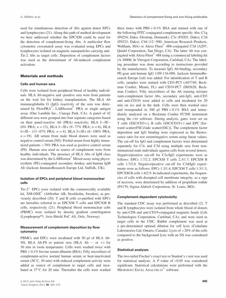
(A-30006; In Vitrogen Corporation, Carlsbad, CA). The label-ing procedure was done according to instructions providedby the manufacturer. To measure IgG Ab-binding, secondaryPE-goat anti-human IgG (109-116-098; Jackson ImmunoRe-search Europe Ltd) was added. For identification of T and Bcells, samples were stained with CD3-PC5 (A07749; Beck-man Coulter, Miami, FL) and CD19-PC7 (IM3628; Beck-man Coulter). Fifty microiliters of the Ab staining mixture(anti-complement factor Abs, secondary anti-IgG, anti-CD3and anti-CD19) were added to cells and incubated for 20min on ice and in the dark. Cells were then washed onceand resuspended in 300 μl PBS + 0.1% BSA and imme-diately analyzed on a Beckman Coulter FC500 instrumentusing the cxp software. During analysis, gates were set onT cells (SSC/CD3+), B cells (SSC/CD19+) and EPC [for-ward scatter(FSC)/side scatter(SSC)]. The complement factordeposition and IgG binding were expressed as the fluores-cence ratio for test serum/negative serum using linear values.The cut-off for IgG and complement factors were determinedseparately for C3c and C3d using multiple sera from non-immunized male individuals against cells from several donors.Negative/positive cut-off for C3c/IgG experiments were asfollows: EPCs 1.7/2.2; EPCXM T cells 2.3/1.7; EPCXM Bcells 1.7/2.0. Negative/positive cut-off for C3d/IgG experi-ments were as follows: EPCs 1.3/1.4; EPCXM T cells 1.3/1.3;EPCXM B cells 1.6/2.9. In indicated experiments, the frequen-cies of cells with disrupted cell membrane integrity, as a signof necrosis, were determined by addition of propidium iodide(P4170; Sigma-Aldrich Corporation, St. Louis, MO).
Complement-dependent cytotoxicity
The standard CDC assay was performed as described (2). Tand B lymphocytes were isolated from whole blood of donorsby anti-CD8 and anti-CD19-conjugated magnetic beads (LifeTechnologies Corporation, Carlsbad, CA), and were used astarget cells in the CDC. Rabbit complement was used ata pre-determined optimal dilution for cell lysis (CedarlaneLaboratories Ltd, Ontario, Canada). Lysis of >20% of the cellscompared to the background lysis with an NS was consideredas positive.
Statistical analyses
The two-tailed Fischer’s exact test or Student’s t-test was usedfor statistical analysis. A P -value of <0.05 was consideredsignificant. Statistical calculations were performed with theMicrosoft Excel Analyse-it
®software.
© 2012 John Wiley & Sons A/S 405Tissue Antigens, 2012, 80, 404–415

Detection of complement-fixing and non-fixing antibodies A. AlMahri et al.
(D)(A)
(E)(B)
(F)(C)
CD3+CD19+
C3c FITC C3c FITC
CD3+ CD19+CD3+ CD19+
IgG PE IgG PE
Figure 1 Flow cytometric detection of complement factor C3c and immunoglobulin G (IgG) deposition on peripheral blood mononuclear cells (PBMC)gated on CD3+ T cells (A–C) and CD19+ B cells (D–F). PBMC were incubated with human leukocyte antigen (HLA) antibody-negative control serum(NS; solid line) and HLA antibody-positive control serum (PS; dotted line).
Results
Complement factor deposition on EPCs
With a previously described flow cytometric assay we initiallyverified that human complement factors are deposited on thesurface of peripheral blood T and B cells upon binding ofcomplement-fixing HLA Abs (Figure 1; (10, 11)). Severaldifferent Abs specific for human complement were tested(see Materials and methods). Generally the C3c and C3d Absresulted in the strongest fluorescence signal. The Abs directedagainst C1q and C4d resulted in a modest signal with
fluorescence ratios barely above 1 (data not shown).Apart from detection of complement factors, the assaysupports simultaneous detection of IgG as depicted inFigure 1C, F. Next, we set-out to determine whether this typeof complement-binding assay also could be applied on EPCisolated with the XM-ONE
®kit. A set of pilot experiments
(n = 10) with Tie-2+ cells isolated from peripheral bloodshowed that complement factors C3c and C3d are depositedon EPCs (Figure 2A, B) and co-purified lymphocytes(Figure 2A, C). C1q and C4d (data not shown) were notdetected at any substantial levels. Use of the viable/dead cell
406 © 2012 John Wiley & Sons A/STissue Antigens, 2012, 80, 404–415

A. AlMahri et al. Detection of complement-fixing and non-fixing antibodies
(A)
EPC
(B)
NSNS
NSPS
PSPS
EPC
CTIF d3CCTIF q1C C3c FITC
(C)NS
PS
CTIF d3CCTIF c3CCTIF q1C
SNPSNS
PSLY
(D)
EPC PI+1.0%
LY PI+36.8%
Figure 2 Flow cytometric detection of complement factors on endothelial precursor cells with low induction of cytolysis. Endothelial precursor cells(EPCs) and co-purified lymphocytes (LY) isolated with anti-Tie-2 antibody (Ab)-conjugated beads were incubated with human leukocyte antigen (HLA)Ab-negative control serum (NS) and HLA Ab-positive control serum (PS) and stained with C1q, C3c and C3d Abs. The EPCs (B) and lymphocytes (C)were FSC/SSC gated and the deposition of the complement factors C1q, C3c and C3d was determined. The frequencies of non-viable [propidiumiodide (PI)+] EPCs and lymphocytes after incubation with HLA Ab-positive control serum are shown in (D). One representative experiment is shown.
© 2012 John Wiley & Sons A/S 407Tissue Antigens, 2012, 80, 404–415
Detection of complement-fixing and non-fixing antibodies A. AlMahri et al.
exclusion dye, propidium iodide (PI), showed that humanserum or rabbit serum used as source of complement inducedonly modest necrosis of endothelial cells (Figure 2D and datanot shown). In contrast, for the lymphocytes (co-purified withEPC) cytolysis were induced resulting in 30%–50% deadcells upon exposure to serum with complement-fixing HLAAbs (Figure 2D). Higher frequencies of dead lymphocytes(50%–80%) were observed when rabbit serum was used ascomplement source (data not shown).
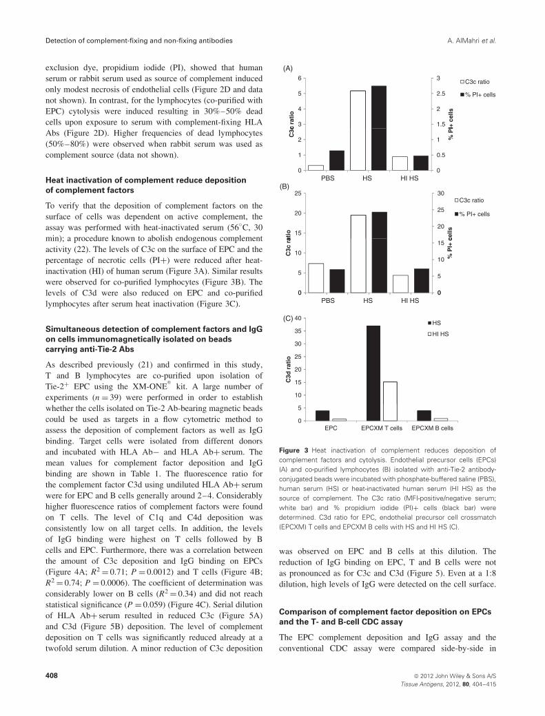
Heat inactivation of complement reduce deposition
of complement factors
To verify that the deposition of complement factors on thesurface of cells was dependent on active complement, theassay was performed with heat-inactivated serum (56◦C, 30min); a procedure known to abolish endogenous complementactivity (22). The levels of C3c on the surface of EPC and thepercentage of necrotic cells (PI+) were reduced after heat-inactivation (HI) of human serum (Figure 3A). Similar resultswere observed for co-purified lymphocytes (Figure 3B). Thelevels of C3d were also reduced on EPC and co-purifiedlymphocytes after serum heat inactivation (Figure 3C).
Simultaneous detection of complement factors and IgG
on cells immunomagnetically isolated on beads
carrying anti-Tie-2 Abs
As described previously (21) and confirmed in this study,T and B lymphocytes are co-purified upon isolation ofTie-2+ EPC using the XM-ONE
®kit. A large number of
experiments (n = 39) were performed in order to establishwhether the cells isolated on Tie-2 Ab-bearing magnetic beadscould be used as targets in a flow cytometric method toassess the deposition of complement factors as well as IgGbinding. Target cells were isolated from different donorsand incubated with HLA Ab− and HLA Ab+ serum. Themean values for complement factor deposition and IgGbinding are shown in Table 1. The fluorescence ratio forthe complement factor C3d using undiluted HLA Ab+ serumwere for EPC and B cells generally around 2–4. Considerablyhigher fluorescence ratios of complement factors were foundon T cells. The level of C1q and C4d deposition wasconsistently low on all target cells. In addition, the levelsof IgG binding were highest on T cells followed by Bcells and EPC. Furthermore, there was a correlation betweenthe amount of C3c deposition and IgG binding on EPCs(Figure 4A; R2 = 0.71; P = 0.0012) and T cells (Figure 4B;R2 = 0.74; P = 0.0006). The coefficient of determination wasconsiderably lower on B cells (R2 = 0.34) and did not reachstatistical significance (P = 0.059) (Figure 4C). Serial dilutionof HLA Ab+ serum resulted in reduced C3c (Figure 5A)and C3d (Figure 5B) deposition. The level of complementdeposition on T cells was significantly reduced already at atwofold serum dilution. A minor reduction of C3c deposition
(A)
1.5
2
2.5
3
3
4
5
6
I+ c
ells
c ra
tio
C3c ratio
% PI+ cells
0
0.5
1
0
1
2 % P
I
C3c
HS HI HSPBS(B)
20
25
30
15
20
25
cells
atio
C3c ratio
% PI+ cells
0
5
10
15
0
5
10
% P
I+ c
C3c
ra
00HS HI HSPBS
(C)
30
35
40HS
HI HS
10
15
20
25
C3d
rat
io
0
5
EPC EPCXM T cells EPCXM B cells
Figure 3 Heat inactivation of complement reduces deposition ofcomplement factors and cytolysis. Endothelial precursor cells (EPCs)(A) and co-purified lymphocytes (B) isolated with anti-Tie-2 antibody-conjugated beads were incubated with phosphate-buffered saline (PBS),human serum (HS) or heat-inactivated human serum (HI HS) as thesource of complement. The C3c ratio (MFI-positive/negative serum;white bar) and % propidium iodide (PI)+ cells (black bar) weredetermined. C3d ratio for EPC, endothelial precursor cell crossmatch(EPCXM) T cells and EPCXM B cells with HS and HI HS (C).
was observed on EPC and B cells at this dilution. Thereduction of IgG binding on EPC, T and B cells were notas pronounced as for C3c and C3d (Figure 5). Even at a 1:8dilution, high levels of IgG were detected on the cell surface.
Comparison of complement factor deposition on EPCs
and the T- and B-cell CDC assay
The EPC complement deposition and IgG assay and theconventional CDC assay were compared side-by-side in
408 © 2012 John Wiley & Sons A/STissue Antigens, 2012, 80, 404–415

A. AlMahri et al. Detection of complement-fixing and non-fixing antibodies
Table 1 Simultaneous detection of complement factors and IgG onEPCs, EPCXM T and EPCXM B cellsa
C3c C3d C1q C4d IgG
EPC 2.1 ± 0.5 3.4 ± 1.4 0.6 ± 0.4 0.7 ± 0.5 5.0 ± 2.2EPCXM T cells 43.9 ± 25.1 46.7 ± 23.3 0.9 ± 0.4 1.6 ± 0.9 28.3 ± 20EPCXM B cells 1.7 ± 0.5 4.2 ± 1.3 1.4 ± 0.8 1.8 ± 0.6 8.7 ± 3.9
EPC, endothelial precursor cell; EPCXM, endothelial precursor cellcrossmatch; IgG, immunoglobulin G.aResults are expressed as a fluorescence ratio (mean ± SD) of HLAAbs-positive serum over negative serum. C3c (n = 11), C3d (n = 4),C1q (n = 3) and C4d (n = 3). IgG represents mean ratio ± SD from allexperiments (n = 21). Target cells were from different donors.
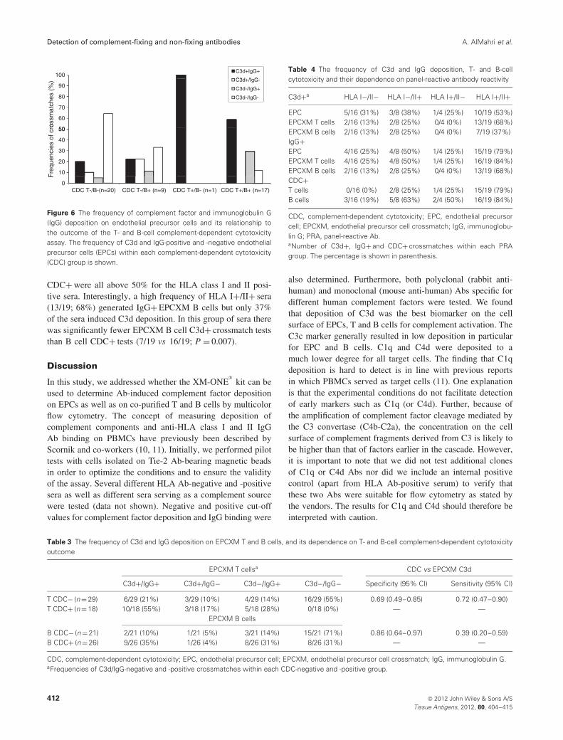
10 separate experiments. Target cells from different donors(n = 9) were crossmatch tested against HLA class I and IIAb-negative and -positive sera from patients on the kidneytransplant waiting list. In total 47 different target cell/serumcombinations were tested (Table 2). The outcome of theEPCXM following detection of C3d/IgG deposition wascompared with the outcome of the CDC assay (Figure 6).The CDC crossmatch outcome was grouped as: CDC T−/B−(n = 20), CDC T−/B+ (n = 9), CDC T+/B− (n = 1), CDCT+/B+ (n = 17). The majority (14/20, 70%) of the CDCT−/B− crossmatch tests were EPC C3d−. Thirty per cent(6/20) of the target/serum combinations that were CDCT−/B− resulted in C3d deposition on EPC. Four of the 6(67%) sera resulted in IgG binding. However, none of thesesera were found to have HLA class I or II Abs as determinedby solid-phase Ab screening (data not shown). Furthermore,tests with CDC T−/B+, CDC T+/B− and CDC T+/B+ seraresulted in 44%, 100% and 59% EPCs with C3d deposition,respectively. In the latter T+/B+ CDC group, 88% (15/17)of the target/serum combinations resulted in IgG bindingon EPCs.
Comparison of complement factor deposition on T and
B cells co-purified with EPCs and the T- and B-cell CDC
assay
EPCXM T and B cells co-purified with EPCs can be usedfor simultaneous detection of C3d and IgG on all threecell subsets. Therefore, we addressed whether there was anycorrelation between the T and B cell CDC assay and the C3dassay when gating on EPCXM T and B cells. Seventy-two percent (13/18) of the T CDC+ tests were T cell C3d+. Ten of the13 (77%) were IgG+. Notably, all three T C3d+/IgG− withinthe T CDC− group had a C3d ratio just above cut-off (datanot shown). Twenty-nine of 47 target cell/serum combinationsresulted in a negative T cell CDC (T CDC−; Table 3). Sixty-nine per cent (20/29) of these were negative with regard toC3d deposition on T cells co-purified with EPCs. Of theC3d+ T-cell crossmatch tests that were negative in the T CDC,67% were IgG positive. The specificity and sensitivity for the
y = 4,1223x - 3,2445R² = 0,7052
4
5
6
7
8
9
10EPCs
(A)
0
1
2
3
4
0 1 2 3 4 5
Flu
ores
cenc
e ra
tio Ig
G
Fluorescence ratio C3c
y = 0,7852x + 3,4699100
(B)y 0,7852x 3,4699
R² = 0,7429
20
30
40
50
60
70
80
90
100EPCXMT cells
0
10
20
0 10 20 30 40 50 60 70 80 90 100
Flu
ores
cenc
e ra
tio Ig
G
Fluorescence ratio C3c
y = 5 1373x - 0 064220
(C)
y = 5,1373x - 0,0642R² = 0,3413
4
6
8
10
12
14
16
18
20EPCXM B cells
0
2
4
0 1 2 3 4
Flu
ores
cenc
e ra
tio Ig
G
Fluorescence ratio C3c
Figure 4 Relationship between the amount of complement factordeposition (C3c) and immunoglobulin G (IgG) binding on endothelialprecursor cells (EPCs), endothelial precursor cell crossmatch (EPCXM) Tcells and EPCXM B cells. Linear regression analysis of C3c deposition vsIgG binding on EPCs (A; n = 11), EPCXM T cells (B; n = 11) and EPCXMB cells (C; n = 11). Each data point represents a separate experimentwith a given donor/serum combination. Different donors were used ineach experiment. The same negative and positive control serum wasused throughout the experiments.
flow cytometric assessment of C3d deposition on EPCXM Tcells vs the T CDC assay were 69% and 72%, respectively.
Eighty-six per cent (18/21) of the B CDC− were C3d−on EPCXM B cells. Thirty-eight per cent (10/26) of the BCDC+ were C3d+ on EPCXM B cells. Nine of the 10 (90%)were also IgG+. In tests that were B CDC+/EPCXM B cellC3d− the percentage of cell lysis in the B CDC ranged
© 2012 John Wiley & Sons A/S 409Tissue Antigens, 2012, 80, 404–415

Detection of complement-fixing and non-fixing antibodies A. AlMahri et al.
355
15
20
25
30
35
3
4
5
(A) (B)
EPCs C3c
IgG
0
5
10
15
0
1
2
PS neat 1:2 1:4 1:8
Flu
ore
scen
ce r
atio
IgG
Flu
ore
scen
ce r
atio
C3c
25
30
35
15
20 EPCXM T cells C3c
IgG
5
10
15
20
5
10
00PS neat 1:2 1:4 1:8
30
355 EPCXM B cells C3c
IgG
10
15
20
25
30
1
2
3
4g
0
5
0
1
PS neat 1:2 1:4 1:8
145 EPCs C3d
6
8
10
12
2
3
4IgG
0
2
4
0
1
PS neat 1:2 1:4 1:8 1:16
Flu
ore
scen
ce r
atio
IgG
Flu
ore
scen
ce r
atio
C3d
Flu
ore
scen
ce r
atio
IgG
Flu
ore
scen
ce r
atio
C3c
Flu
ore
scen
ce r
atio
IgG
Flu
ore
scen
ce r
atio
C3d
Flu
ore
scen
ce r
atio
IgG
Flu
ore
scen
ce r
atio
C3c
Flu
ore
scen
ce r
atio
IgG
Flu
ore
scen
ce r
atio
C3d
15
20EPCXM T cells C3d
IgG
0
5
10
10
12
14
16
0
2
4
6
8
00PS neat 1:2 1:4 1:8 1:16
25
30
35
4
5 EPCXM B cells C3dIgG
10
15
20
25
1
2
3
0
5
0
1
PS neat 1:2 1:4 1:8 1:16
Figure 5 Relationship between deposition of C3c (A), C3d (B) and immunoglobulin G (IgG) binding on endothelial precursor cells (EPCs), endothelialprecursor cell crossmatch (EPCXM) T and EPCXM B cells at different serum dilutions. Results are expressed as a fluorescence ratio of humanleukocyte antigen antibodies-positive serum over negative serum. Data are from representative experiments.
between 25% and 100%. In the majority (11/16; 69%) of tests,at least 50% lysis was observed (data not shown). Notably, in5 of those 11 tests (45%) no IgG deposition on B cells wasobserved. The majority of sera (6/8) that were B CDC+ andEPCXM B cell C3d−/IgG− were also negative for HLA Absof IgM class (data not shown). The flow cytometric EPCXMB cell C3d assay had high specificity (86%), but considerablylower sensitivity (39%) than the B CDC assay.
The width of the panel-reactive Ab repertoire and its
impact on C3d/IgG deposition and CDC
The panel of sera (n = 42) were grouped into four separate cat-egories based on the PRA reactivity; HLA I−/II− (0% PRA),HLA I−/II+ (9%–77% PRA), HLA I+/II− (13%–87%PRA), HLA I+/II+ (6%–100% PRA). The HLA I−/II−group of sera resulted in 31% EPC C3d+, 13% EPCXM Tcell C3d+ and 13% EPCXM B cell C3d+ crossmatch tests
410 © 2012 John Wiley & Sons A/STissue Antigens, 2012, 80, 404–415

A. AlMahri et al. Detection of complement-fixing and non-fixing antibodies
Table 2 Complement factor deposition and IgG binding on T and B cells co-purified with endothelial precursor cells and the T- and B-cellcomplement-dependent cytotoxicity assaya
EPCXM T cells EPCXM B cells EPC
Crossmatch no.Serum PRAclass I/II (%) C3d IgG
T CDC(% lysis) C3d IgG
B CDC(% lysis) C3d IgG
1 0/0 2.0 3.1 10 1.3 2.0 5 1.8 5.72 0/0 1.7 2.9 10 1.2 1.9 5 1.5 4.63 0/0 0.9 1.3 10 0.4 0.8 10 0.6 1.14 0/0 0.9 1.2 10 0.3 0.7 10 0.8 0.65 0/0 1.1 1.0 5 0.9 0.9 15 0.8 1.16 0/0 1.2 1.0 15 1.4 1.2 15 1.3 1.17 0/0 0.9 1.7 10 2.5 7.2 20 3.9 7.88 0/0 1.0 0.9 10 1.1 0.3 20 0.8 1.49 0/0 0.5 1.6 20 2.0 8.8 20 1.7 8.010 0/0 1.2 0.9 20 1.3 1.4 20 0.9 1.011 0/0 1.1 1.0 5 0.9 1.2 25 0.8 1.112 0/0 0.9 0.8 5 1.0 0.6 40 0.9 0.713 0/0 1.0 1.0 10 1.0 1.3 80 1.3 1.114 0/0 0.9 0.8 10 1.1 0.8 10 1.0 1.315 0/0 0.9 0.9 15 1.1 1.2 5 1.0 0.816 0/0 0.8 0.7 10 0.8 0.4 10 0.8 0.917 0/57 1.4 2.2 10 0.9 5.4 10 0.9 1.518 0/43 1.1 0.9 5 1.0 1.4 20 1.0 1.119 0/71 2.2 4.5 5 1.3 2.7 25 1.3 1.220 0/40 0.4 1.7 20 1.8 9.4 30 1.8 8.121 0/40 0.9 0.8 20 0.7 0.7 75 0.8 0.922 0/77 1.2 1.1 60 1.2 1.7 60 0.9 1.823 0/77 0.5 1.7 90 2.0 17.9 80 2.0 8.424 0/9 0.9 0.8 10 1.3 0.7 15 0.9 1.225 49/0 1.0 0.8 10 1.1 1.3 35 2.1 0.926 13/0 1.0 0.9 20 0.7 0.7 10 1.0 1.127 16/0 0.7 0.8 5 0.6 0.3 10 0.9 0.928 87/0 0.9 6.2 25 1.1 2.1 50 0.8 1.829 73/83 1.2 1.2 15 0.5 0.9 15 1.7 1.230 75/83 1.4 3.2 15 1.2 2.5 20 0.8 0.731 67/89 1.1 7.8 40 0.9 6.4 30 1.1 2.332 65/66 0.5 4.8 30 2.0 17.2 50 4.0 8.433 99/76 1.1 10.1 50 0.8 2.1 60 1.0 1.034 84/94 1.8 0.9 60 0.9 1.4 90 0.9 1.035 98/94 1.9 28.8 40 1.0 20.6 100 0.9 3.036 85/100 54.4 35.2 80 3.4 9.7 100 4.0 5.537 98/94 96.6 12.0 90 10.8 5.9 100 11.7 5.138 65/63 1.8 7.7 90 0.6 2.9 100 0.5 1.539 93/97 40.6 36.5 100 12.8 40.6 100 19.4 78.940 96/100 34.1 11.4 100 3.5 13.3 100 2.6 6.341 89/83 1.8 25.6 70 0.8 20.0 70 0.9 8.942 75/89 18.6 0.8 100 2.8 0.5 100 1.7 4.543 6/61 1.0 4.1 10 1.1 4.4 60 1.1 1.544 85/100 3.2 2.9 80 7.4 8.5 100 8.4 35.545 67/86 1.7 6.4 50 1.1 9.0 20 1.9 7.346 84/26 23.0 26.4 100 1.0 15.5 100 1.1 3.947 84/86 1.2 4.3 20 1.5 3.1 30 1.7 4.5
CDC, complement-dependent cytotoxicity; EPC, endothelial precursor cell; EPCXM, endothelial precursor cell crossmatch; IgG, immunoglobulin G;PRA, panel-reactive Ab.aData from all crossmatches (n = 47). Serum PRA class I and II (%) for each crossmatch are shown together with C3d deposition and IgG binding.CDC lysis >20% were considered as positive.
(Table 4). Nineteen per cent (3/16) of the tests were B-cellCDC+. None (0/16; 0%) of the tests were CDC T+. TheHLA I−/II+ group of sera resulted in similar frequenciesof EPC C3d+, EPCXM T cell C3d+ and EPCXM B cellC3d+ crossmatch tests. A high frequency of tests (5/8; 63%)
was B CDC+ in the HLA I−/II+ group of sera. Furthermore,as expected, the highest number of positive crossmatch testswas observed with HLA class I and II positive sera. Apartfrom the EPCXM B cell C3d assay with only 37% pos-itive crossmatch tests, the frequencies of C3d+, IgG+ and
© 2012 John Wiley & Sons A/S 411Tissue Antigens, 2012, 80, 404–415
Detection of complement-fixing and non-fixing antibodies A. AlMahri et al.
100C3d+IgG+
C3d+/I G
50
60
70
80
90
ossm
atch
es (
%) C3d+/IgG-
C3d-/IgG+
C3d-/IgG-
10
20
30
40
50
requ
enci
es o
f cro
0CDC T-/B-(n=20) CDC T-/B+ (n=9) CDC T+/B- (n=1) CDC T+/B+ (n=17)
F
Figure 6 The frequency of complement factor and immunoglobulin G(IgG) deposition on endothelial precursor cells and its relationship tothe outcome of the T- and B-cell complement-dependent cytotoxicityassay. The frequency of C3d and IgG-positive and -negative endothelialprecursor cells (EPCs) within each complement-dependent cytotoxicity(CDC) group is shown.
CDC+ were all above 50% for the HLA class I and II posi-tive sera. Interestingly, a high frequency of HLA I+/II+ sera(13/19; 68%) generated IgG+ EPCXM B cells but only 37%of the sera induced C3d deposition. In this group of sera therewas significantly fewer EPCXM B cell C3d+ crossmatch teststhan B cell CDC+ tests (7/19 vs 16/19; P = 0.007).
Discussion
In this study, we addressed whether the XM-ONE®
kit can beused to determine Ab-induced complement factor depositionon EPCs as well as on co-purified T and B cells by multicolorflow cytometry. The concept of measuring deposition ofcomplement components and anti-HLA class I and II IgGAb binding on PBMCs have previously been described byScornik and co-workers (10, 11). Initially, we performed pilottests with cells isolated on Tie-2 Ab-bearing magnetic beadsin order to optimize the conditions and to ensure the validityof the assay. Several different HLA Ab-negative and -positivesera as well as different sera serving as a complement sourcewere tested (data not shown). Negative and positive cut-offvalues for complement factor deposition and IgG binding were
Table 4 The frequency of C3d and IgG deposition, T- and B-cellcytotoxicity and their dependence on panel-reactive antibody reactivity
C3d+a HLA I−/II− HLA I−/II+ HLA I+/II− HLA I+/II+
EPC 5/16 (31%) 3/8 (38%) 1/4 (25%) 10/19 (53%)EPCXM T cells 2/16 (13%) 2/8 (25%) 0/4 (0%) 13/19 (68%)EPCXM B cells 2/16 (13%) 2/8 (25%) 0/4 (0%) 7/19 (37%)IgG+EPC 4/16 (25%) 4/8 (50%) 1/4 (25%) 15/19 (79%)EPCXM T cells 4/16 (25%) 4/8 (50%) 1/4 (25%) 16/19 (84%)EPCXM B cells 2/16 (13%) 2/8 (25%) 0/4 (0%) 13/19 (68%)CDC+T cells 0/16 (0%) 2/8 (25%) 1/4 (25%) 15/19 (79%)B cells 3/16 (19%) 5/8 (63%) 2/4 (50%) 16/19 (84%)
CDC, complement-dependent cytotoxicity; EPC, endothelial precursorcell; EPCXM, endothelial precursor cell crossmatch; IgG, immunoglobu-lin G; PRA, panel-reactive Ab.aNumber of C3d+, IgG+ and CDC+ crossmatches within each PRAgroup. The percentage is shown in parenthesis.
also determined. Furthermore, both polyclonal (rabbit anti-human) and monoclonal (mouse anti-human) Abs specific fordifferent human complement factors were tested. We foundthat deposition of C3d was the best biomarker on the cellsurface of EPCs, T and B cells for complement activation. TheC3c marker generally resulted in low deposition in particularfor EPC and B cells. C1q and C4d were deposited to amuch lower degree for all target cells. The finding that C1qdeposition is hard to detect is in line with previous reportsin which PBMCs served as target cells (11). One explanationis that the experimental conditions do not facilitate detectionof early markers such as C1q (or C4d). Further, because ofthe amplification of complement factor cleavage mediated bythe C3 convertase (C4b-C2a), the concentration on the cellsurface of complement fragments derived from C3 is likely tobe higher than that of factors earlier in the cascade. However,it is important to note that we did not test additional clonesof C1q or C4d Abs nor did we include an internal positivecontrol (apart from HLA Ab-positive serum) to verify thatthese two Abs were suitable for flow cytometry as stated bythe vendors. The results for C1q and C4d should therefore beinterpreted with caution.
Table 3 The frequency of C3d and IgG deposition on EPCXM T and B cells, and its dependence on T- and B-cell complement-dependent cytotoxicityoutcome
EPCXM T cellsa CDC vs EPCXM C3d
C3d+/IgG+ C3d+/IgG− C3d−/IgG+ C3d−/IgG− Specificity (95% CI) Sensitivity (95% CI)
T CDC− (n = 29) 6/29 (21%) 3/29 (10%) 4/29 (14%) 16/29 (55%) 0.69 (0.49–0.85) 0.72 (0.47–0.90)T CDC+ (n = 18) 10/18 (55%) 3/18 (17%) 5/18 (28%) 0/18 (0%) — —
EPCXM B cells
B CDC− (n = 21) 2/21 (10%) 1/21 (5%) 3/21 (14%) 15/21 (71%) 0.86 (0.64–0.97) 0.39 (0.20–0.59)B CDC+ (n = 26) 9/26 (35%) 1/26 (4%) 8/26 (31%) 8/26 (31%) — —
CDC, complement-dependent cytotoxicity; EPC, endothelial precursor cell; EPCXM, endothelial precursor cell crossmatch; IgG, immunoglobulin G.aFrequencies of C3d/IgG-negative and -positive crossmatches within each CDC-negative and -positive group.
412 © 2012 John Wiley & Sons A/STissue Antigens, 2012, 80, 404–415

A. AlMahri et al. Detection of complement-fixing and non-fixing antibodies
In the early phase of this study during the optimizing ofthe assay conditions, purified rabbit complement was testedas complement source. Interestingly, no lysis of EPCs wasdetected in any of the experiments irrespective of the lengthof incubation time (up to 1 h; data not shown). In contrast,a large fraction (50%–80%) of the co-purified lymphocyteswas lysed. Furthermore, only very weak lysis of EPCs wasobserved with human serum as complement source. Thesedata suggest that the frequency of necrotic cells as read-out will not be suitable to asses Ab-induced complementactivation on isolated EPCs. This observation is in line withthe notion that endothelial cells lining the graft are relativelymore resistant to Ab-induced complement-mediated lysis thanother cells of the graft; cells not normally exposed to blood(23). Endothelial cells are known to express a plethora ofcomplement-regulatory proteins (CRP) such as CD59, CD46and CD55, which can protect against complement-inducedlysis (24–26). The expression level of complement-inhibitorymolecules on EPCs is yet to be determined. However, wehypothesize that CRPs are expressed on Tie-2+ EPCs andthat they may influence complement factor deposition andsusceptibility to lysis. The level of C3c/d deposition on EPCsand EPCXM B cells was in general significantly lower thanon EPCXM T cells. Whether the relatively low C3c/d ratioon EPCs and EPCXM B cells is due to higher backgroundstaining generating a lower signal-to-noise ratio compared toT cells and/or due to other factors such as the repertoire ordensity of CRPs expressed is presently unknown. It is well-known that B cells have an intrinsically higher backgroundstaining in part because of Fc-receptor expression (27). Fc-receptors are expressed on Tie-2+ EPCs (Maria Sundback,personal communication, 18 January 2012).
We found a correlation between the level of IgG and C3cdeposition with a given negative/positive serum combinationagainst target cells isolated from different donors. The positiveserum predominantly contained IgG Abs specific for HLAclass I and II. Only low or modest levels of HLA IgM Abswere detected (data not shown). Interestingly, C3c depositionsharply dropped upon serum dilution. In contrast, the IgGbinding was not affected to the same extent and was stillabove cut-off after several dilutions. These findings are inline with previous data by Scornik et al. (11). An appealingidea, presented by this group, is to use C3 deposition asa tool to monitor the efficacy of patient desensitization(e.g. following immunoadsorption and plasma exchange). Ourexperience is that determination of IgG levels by solid-phase techniques after pre-treatment strategies rarely give anyconclusive results. In most cases IgG is readily detected evenafter several rounds of Ab removal.
In our side-by-side comparison of the EPCXM C3d deposi-tion assay and the CDC assays, we found that a considerablenumber (11/27; 41%) of T and/or B CDC+ tests did not showup as positive for C3d. In particular, the comparison betweenB CDC and the C3d deposition on B cells co-purified with
the EPCs showed a low degree of concordance (see Table 3).This could either be explained by a high proportion of false-positive CDC tests or a poor sensitivity of the flow cytometricC3d assay. As shown in Table 4, the HLA I+/II− group didnot result in any EPCXM T or B cell C3d+ crossmatch. Onlyone serum supported IgG deposition on EPCs and T cells.The HLA class I specificity determination of those four serashowed that three of them had rather low levels of HLA IgGAbs with mean fluorescence intensity (MFI) values below3000 in the LabScreen
®single antigen assay. Two of the sera
resulted in positive CDC B-cell crossmatch tests. However,none of them induced IgG deposition on EPCXM B cells.Notably, one of the sera had high levels of HLA-C Abs (MFI10,000–12,000) and induced considerable IgG deposition onT cells (ratio: 6; data not shown). The IgG ratio for EPCXMB cells with that particular serum was below cut-off. Thismay indicate that the HLA-C expression is lower on B cellsthan on T cells (28).
Unexpectedly, 50% of the sera from the HLA I−/II+ groupresulted in EPCXM T cells IgG binding (Table 4). The IgGratio for two of those sera (crossmatch no. 20 and 23, Table 1)were close to cut-off value which may call for re-evaluation ofthe IgG cut-off level. Furthermore, the patient whose sera wereused in crossmatch no. 19 (IgG: 4.5, Table 1) were 3 monthslater found to have HLA class I Abs (highest MFI ∼4000)as determined by the more sensitive Labscreen single antigenassay. This may explain the crossmatch IgG results.
Another important observation was that 30% of the target/serum combinations that were CDC T−/B− resulted in C3ddeposition on EPC. However, none of these sera were foundto have HLA class I or II Abs as determined by solid-phaseAb screening. Interestingly, this data are thus in line with ourprevious observation that approximately 25% of all LXM−(CDC and FCXM) are EPCXM+ (manuscript submitted forpublication). Importantly, this shows that EPCXM C3d assaymay permit detection of complement-fixing non-HLA Absmissed by conventional LXM.
It is important to acknowledge though that the assay proce-dures for CDC and complement factor deposition are differentwith regard to target cell isolation, kinetics, read-out mode andserum source. In particular the latter will greatly influencethe XM outcome. Purified rabbit complement induces lym-phocytotoxicity more efficiently than human serum (data notshown; (2)). The CDC assay has been the method of choicesince many years for the detection of complement-fixing anti-HLA class I and II Abs. Nevertheless, we argue that a positiveCDC (in particular a positive B cell CDC) XM does not nec-essarily indicate a donor-specific Ab response. In this studywe were not able to HLA-type the donors and thus it wasnot possible to check for a possible correlation between DSAand crossmatch outcome. It is generally believed that auto-Abs(both IgM and IgG) may induce false-positive B-cell XM. Thishighlights the importance of routinely performing autologouscrossmatch tests to rule out this possibility. In future studies it
© 2012 John Wiley & Sons A/S 413Tissue Antigens, 2012, 80, 404–415
Detection of complement-fixing and non-fixing antibodies A. AlMahri et al.
will be important to investigate whether there is a differencebetween the CDC and the complement factor deposition XMwith regard to their capability of distinguishing between auto-and allo-Abs.
In summary, we show that the EPC crossmatch kit, XM-ONE
®, can be applied for simultaneous detection of HLA
class I and II IgG Ab binding and induced complement acti-vation (C3c/d) on the surface of EPCs, T and B lymphocytes.The combined flow cytometric technique could potentiallyminimize the number of assays needed in the work-up priorto kidney transplantation. A potentially important advantagewith the C3 assay is that the read-out, in contrast to whenCDC is used, is objective and semi-quantitative. Important forthe future will be to perform this flow cytometric complementactivation assay with sera containing HLA and non-HLAAbs of known specificity and target cells from well-typeddonors in order to fully evaluate its usefulness in a routinelaboratory setting.
Acknowledgments
This study was financially supported by the Swedish ResearchCouncil/Medicine (JH), the Stockholm and Vastra GotalandCounty Councils (ALF; JH) and the Marianne and MarcusWallenberg Foundation (JH). AA is fully sponsored by SheikhKhalifa Medical City (SKMC) in Abu Dhabi, United ArabEmirates. The authors wish to thank Guido Moll for providingreagents and excellent technical assistance.
Conflict of interest
JH is a part time Medical Director and board member ofAbSorber AB, the company manufacturing the XM-ONE
®
crossmatch test. He is also a shareholder in Allenex AB,the venture capital company that is the majority owner ofAbSorber AB, and his wife receives a royalty from XM-ONE
®
sales.
References
1. Terasaki PI. Humoral theory of transplantation. Am JTransplant 2003: 3: 665–73.
2. Terasaki PI, McClelland JD. Microdroplet assay of humanserum cytotoxins. Nature 1964: 204: 998–1000.
3. Patel R, Terasaki PI. Significance of the positive crossmatchtest in kidney transplantation. N Engl J Med 1969: 280: 735–9.
4. Gebel HM, Bray RA, Nickerson P. Pre-transplant assessment ofdonor-reactive HLA-specific antibodies in renal transplantation:contraindications vs. risk. Am J Transplant 2003: 3: 1488–500.
5. Amos DB, Bashir H, Bogle W et al. A simplemicrocytotoxicity test. Transplantation 1970: 7: 220–3.
6. Johnson AH, Rossen RD, Butler WT. Detection ofalloantibodies using a sensitive antiglobulin microcytotoxicitytest: identification of low levels of preformed antibodies inaccelerated allograft rejection. Tissue Antigens 1972: 2:215–26.
7. Thorne N, Klingman LL, Teresi GA et al. Effects of heatinactivation and DTT treatment of serum on immunoglobulinbinding. Hum Immunol 1993: 37 (Suppl. 1): 123.
8. Okuno T, Kondelis N. Evaluation of dithiothreitol (DTT) forinactivation of IgM antibodies. J Clin Pathol 1978: 31: 1152–5.
9. Spiller OB. Measurement of C3 fragment deposition on cells.In: Methods in Molecular Biology , Vol. 150 . Totowa, NJ :Humana Press Inc, 2000 , 131 –7 .
10. Kushihata F, Watanabe J, Mulder A, Claas F, Scornik JC.Human leukocyte antigen antibodies and human complementactivation: role of IgG subclass, specificity, and cytotoxicpotential. Transplantation 2004: 78: 995–1001.
11. Watanabe J, Scornik JC. Measuring human complementactivation by HLA antibodies. Arch Pathol Lab Med 2006:130: 368–73.
12. Ahern AT, Artruc SB, DellaPelle P et al. Hyperacute rejectionof HLA-AB-identical renal allografts associated with Blymphocyte and endothelial reactive antibodies. Transplantation1982: 33: 103–6.
13. Cerilli J, Bay W, Brasile L. The significance of the monocytecrossmatch in recipients of living-related HLA identical kidneygrafts. Hum Immunol 1983: 7: 45–50.
14. Kalil J, Guilherme L, Neumann J et al. Humoral rejection intwo HLA identical living related donor kidney transplants.Transplant Proc 1989: 21: 711–3.
15. Amico P, Honger G, Bielmann D et al. Incidence andprediction of early antibody-mediated rejection due tonon-human leukocyte antigen-antibodies. Transplantation 2008:85: 1557–63.
16. Sumitran-Holgersson S, Holgersson J. Clinical importance ofnon-HLA antibodies in solid organ transplantation. Curr OpinOrgan Transplant 2006: 11: 425–32.
17. Sumitran-Karuppan S, Tyden G, Reinholt F, Berg U, Moller E.Hyperacute rejections of two consecutive renal allografts andearly loss of the third transplant caused by non-HLA antibodiesspecific for endothelial cells. Transpl Immunol 1997: 5: 321–7.
18. Vermehren D, Sumitran-Holgersson S. Isolation of precursorendothelial cells from peripheral blood for donor-specificcrossmatching before organ transplantation. Transplantation2002: 74: 1479–86.
19. Nowak G, Karrar A, Holmen C et al. Expression of vascularendothelial growth factor receptor-2 or Tie-2 on peripheralblood cells defines functionally competent cell populationscapable of reendothelialization. Circulation 2004: 110:3699–707.
20. Breimer ME, Rydberg L, Jackson AM et al. Multicenterevaluation of a novel endothelial cell crossmatch test in kidneytransplantation. Transplantation 2009: 87: 549–56.
21. Alheim M, Johansson SM, Hauzenberger D, Grufman P,Holgersson J. A flow cytometric crossmatch test forsimultaneous detection of antibodies against donor lymphocytesand endothelial precursor cells. Tissue Antigens 2010: 75:269–77.
22. Pillemer L, Seifter S, Ecker EE. The role of the components ofcomplement in specific immune fixation. J Exp Med 1942: 75:421–35.
23. Bach FH, Hancock WW, Ferran C. Protective genes expressedin endothelial cells: a regulatory response to injury. ImmunolToday 1997: 18: 483–6.
414 © 2012 John Wiley & Sons A/STissue Antigens, 2012, 80, 404–415

A. AlMahri et al. Detection of complement-fixing and non-fixing antibodies
24. Brooimans RA, Van der Ark AA, Tomita M, Van Es LA, DahaMR. CD59 expressed by human endothelial cells functions as aprotective molecule against complement-mediated lysis. Eur JImmunol 1992: 22: 3135–40.
25. Asch AS, Kinoshita T, Jaffe EA, Nussenzweig V.Decay-accelerating factor is present on cultured humanumbilical vein endothelial cells. J Exp Med 1986: 163: 221–6.
26. McNearney T, Ballard L, Seya T, Atkinson JP. Membranecofactor protein of complement is present on human fibroblast,
epithelial, and endothelial cells. J Clin Invest 1989: 84:538–45.
27. Lobo PI, Winfield JB, Craig A, Westervelt FB Jr. Utility ofprotease-digested human peripheral blood lymphocytes for thedetection of lymphocyte-reactive alloantibodies by indirectimmunofluorescence. Transplantation 1977: 23: 16–21.
28. Johnson DR. Differential expression of human majorhistocompatibility class I loci: HLA-A, -B, and -C. HumImmunol 2000: 61: 389–96.
© 2012 John Wiley & Sons A/S 415Tissue Antigens, 2012, 80, 404–415


![Enzyme-LinkedImmunosorbent Assay Detection Antibodies ... filecardiolipin, lecithin, andcholesterol(VDRL[VenerealDiseaseResearchLaboratory]ELISA)is described. The ... nontreponemal](https://img.pdfslide.us/doc/110x75/5ccac51588c993cc428c6a4f/enzyme-linkedimmunosorbent-assay-detection-antibodies-lecithin-andcholesterolvdrlvenerealdiseaseresearchlaboratoryelisais.jpg)
![Detection of antibodies to Oropouche virus in non-human ... · Detection of antibodies to Oropouche virus in non-human primates in Goiânia City, Goiás Marize Moreira Gibrail[1]](https://img.pdfslide.us/doc/110x75/5c6816f609d3f226188cbafa/detection-of-antibodies-to-oropouche-virus-in-non-human-detection-of-antibodies.jpg)