Embed Size (px)
Citation preview

/. Sci. Food Agric. 1981, 32, 539-546
Detection and Estimation of Collagen^
David J. Etherington and Trevor J. Sims
ARC Meat Research Institute, Longford, Bristol BS18 7D Y
(Manuscript received 27 June 1980)
A. R. C.STITUTE
Collagen exists in at least five genetically distinct forms and the relative abundance ofthese is determined by the type and age of the tissue. Methods are presented for thedetection and estimation of these collagens based on appropriate chemical, physical,histochemical and immunological techniques. The specific difficulties in providingaccurate quantitative data for collagen in meat and meat products are discussed.
1. Introduction
Collagen is the most abundant protein in the body and is present mainly in bone, skin and tendon.It is found in muscle as 1-9% of the dry, fat-free mass1 where it exists as networks of fibres. Inthe living muscle these fibres resist over-extension which may cause damage to the tissue. The forceof contraction is transmitted to the tendons through sheets of intramuscular connective tissuewhich enclose the individual muscle fibres. Three collagenous structures can be distinguishedmorphologically—endomysium, enclosing each fibre, perimysium, surrounding bundles of thesefibres, and epimysium, surrounding the whole muscle (Figure 1). The subunit of the collagen fibre
Figure 1. Connective tissues ofmuscle. Each muscle fibre is en-closed in a delicate collagenousmeshwork, the endomysium. Bund-les of these muscle fibres, fasciculi,are surrounded by a thicker sheathof connective tissue, the peri-mysium. The strong outer layer ofconnective tissue, the epimysium,is continuous with the peritendi-neum. The collagen fibres in thetendon appear to connect withboth the perimysium and the endo-mysium.
Epimysium
Tendon fibres
Epitendineum
Endomysium
Muscle fibre
Perimysium
is tropocollagen, or the collagen monomer, which is a long rod-like molecule, 1.5 .̂m in diameterand 300 /Ltm long. Tropocollagen has a molecular weight of about 300 000 daltons and is comprisedof three polypeptide chains wound into a helix. These have a high content of glycine, proline andhydroxyproline located in regular repeating sequences of Gly, Pro, Y and Gly, X, Hyp.2 In thecollagen fibril the tropocollagen molecules are aligned in parallel and held together by covalentintermolecular cross-links. The structural features of collagen are shown in Figure 2.
The development of these cross-links is an age-related process and it is the increase in the numberof the heat-stable covalent attachments that makes meat from older animals much tougher oncooking.3 Therefore, in assessing the quality of meat it is necessary to consider both the quantity
" Paper read at the Society of Chemical Industry Food Group-Meat Panel Meeting Utilisation and identificationof meat analogues and extenders in meat products held at Sutton Bonington, Leics., on 20-21 March 1980.
0022-5142/81/0600-0539 $02.00 © 1981 Society of Chemical Industry
539

D. J. Etherington and T. J. Sims
1. PRIMARY SEQUENCE
G L Y - P R O - Y - G L Y - X - Y - G L Y - X - H Y P - G L Y -
2. TRIPLE HELIX
U. COLLAGEN F I B R E
Figure 2. Structure of collagen.(1) In the primary sequence of thea-chain glycine is found predomi-nantly at every third residue. Inthese triplets with glycine at thefirst position, proline is locatedgenerally at the second and hydro-xyproline at the third position.(T) The a-chain takes up a tighthelical conformation and the threea-chains of tropocollagen arewound together in a super triplehelix. (3) The collagen fibril iscomposed of many rod-like tro-pocollagen molecules aligned inparallel but showing a regularquarter-stagger end overlap withthe repeat distance (D) being 64nm. The fibril is stabilised bycovalent cross-links. These cross-links (not shown) are generated atthe non-helical ends of the mole-cule and connect with the helicalregion of adjacent molecules. (4)It is this precise arrangement of thetropocollagen molecules in thefibre that produces the regularcross-striations typically revealedin the electron microscope.
and the maturity of the connective tissue component. Meat that is less desirable for direct sale tothe consumer may be utilised in processed foods, where the high cooking temperature reducesmuch of the toughness caused by highly cross-linked collagen. Furthermore, tissues rich in collagen,such as tendon (90 %) and skin (40 %), can be included in such products as sausages where mechanicalcomminution of the ingredients overcomes any toughness and effectively disperses the collagenoustissue.
The detection and accurate determination of collagen is an essential requirement for the meatscientist who is attempting to resolve the contribution of the connective tissues to the structureand quality of meat. The food technologist also requires reliable analytical techniques to help main-tain a uniform product that will also conform to any regulations governing composition. There areseveral experimental approaches to the detection and estimation of collagen, depending upon thenature of the investigation. These various methods will be considered separately.
2. Chemical methods
Proline is incorporated into collagen to > 20 % of the total amino acids and nearly half is hydroxy-lated to 4-hydroxyproline immediately following synthesis of the polypeptide chain. The largeamount of hydroxyproline in collagen is a unique feature of this protein and is generally found tobe about 14% of the dry weight of collagen. Therefore, chemical methods for the detection ofcollagen are conveniently based on the determination of protein-bound hydroxyproline in thetissues. It is because of this unusual composition that most interest has been given to the develop-ment of accurate methods for measuring this imino acid. In recent years different phenotypes ofcollagen have been identified and purified and the amino acid composition for each collagen typeis now known.4'5 The hydroxyproline content is lowest in type I collagen, found mainly in bones

Detection and estimation of collagen 541
and tendons, and highest in the type III collagen which is abundant in arterial walls and embryonictissues. The recent data on composition show that hydroxyprolyl residues are present in the majorphenotype of collagen, type I, to the extent of approximately 11.3 % by weight. The weights of thehydroxyprolyl residue in type II cartilage collagen and the type IV collagen of basement membranesare found to be 12.9 and 14.3%, respectively,4 and about 15.0% in type III collagen.4'5 However,all chemical determinations are made on the free imino acid after hydrolysis of the protein. Whena correction is made for the addition of water, the hydroxyproline value of type I collagen is raisedfrom 11.3 to 13.1 %. The corrected values for the free imino acid of the other collagens are: type11—15.0%, type III—17.4% and type IV—16.6%. Clearly then, the actual hydroxyproline valuefor a mixed connective tissue will vary according to the ratio of these different types. Overall,the traditionally applied value of 14 % is probably a fair approximation for muscle connective tissue.
Hydroxyproline is also found in small amounts in other proteins such as the collagen-like sequen-ces found in acetylcholinesterase and the subcomponent of serum complement, Clq.6 Elastin (1.5%hydroxyproline) is found mainly in ligaments, arterial walls and, to a lesser extent, in lung andskin.6 Unless this is physically separated out it can interfere in the collagen hydroxyproline deter-mination. The amount of elastin in whole muscle is generally low but where significant amountsare believed to be present, as in processed meats, the elastin hydroxyproline is best determinedseparately. Elastin is insoluble in hot alkali7 and this extractant can be used to remove all of thecollagen as soluble gelatins. The residue of insoluble elastin is then analysed for hydroxyprolineand the amount deducted from the value determined for the whole tissue.
For the determination of muscle connective tissue some laboratories prefer to extract as muchof the myofibrillar protein as possible before analysing for hydroxyproline. Extraction is performedwith a strong potassium iodide solution and this may be followed by chloroform-methanol whichis used to remove any lipids.8 In this laboratory the preferred method is to extract initially withHasselbach-Schneider pyrophosphate solution9 which dissociates the actomyosin complex andfacilitates treatment with KI. The main disadvantage of this extraction procedure is that somepoorly cross-linked collagen, particularly in the muscles of young animals, may be lost.
Tissues to be analysed are first acid hydrolysed to release the hydroxyproline from peptidelinkage. This is generally achieved using 6M HC1 at 110°C for 18-24 h, either in sealed tubes orunder reflux with an air condenser. For rapid hydrolysis it was found that 3 h at 130°C is adequate.10
Other workers have employed 72% perchloric acid at 100°C for 2-4 h.11'12 The free imino acid ismost conveniently determined colorimetrically after oxidation to pyrrole (Figure 3) which is thenreacted specifically with /7-dimethyl-amino-benzaldehyde (Ehrlich's reagent) to yield an intensered-brown colour.
H
(VI)
(+0)
COOH
(-C02)
COOH
COOH
COOH
COOH
(IV)
Figure 3. Postulated mechanism for the oxidation of hydroxyproline to pyrrole. Hydroxyproline (I) is oxidisedfirst to the linear a-keto-y-hydroxy-S-aminovaleric acid (II), which is in equilibrium with the cyclic structure A'-pyrroline-4-hydroxy-2-carboxylic acid (III). Loss of water gives an unstable structure (IV) which spontaneouslyrearranges to pyrrole-2-carboxylic acid (V). The final decarboxylation step to pyrrole (VI) occurs during heatingafter the addition of the chromogenic reagent for pyrrole, /?-dimethylaminobenzaldehyde.

542 D. J. Etherington and T. J. Sims
In 1970 Mitchell and Taylor13 critically examined the available methods and concluded that nosingle method could combine accuracy with freedom from errors caused by interferring substances.Consequently, several different experimental approaches can be found in the literature, dependingupon the nature of the test material.
Chloramine T is now generally preferred for use as an oxidant in the formation of pyrrole. Forcollagen-rich materials three methods10'14> 15 are in common use and that described by Woessner10
is probably the most popular. The International Standards Organisation method16 has beendeveloped from these specifically for the determination of hydroxyproline in meat or meat products.Here, SnCla is incorporated into the acid used for hydrolysis and reduces the losses caused by sidereactions. The colour yield from hydroxyproline can be depressed by such substances as otheramino acids and salts. However, in tissues low in collagen, such as muscle, there is usually anoverestimate of the amount of hydroxyproline, and in this laboratory the colour value for musclehydroxyproline is found to be increased by as much as 15-20%. The reasons for this are unclearbut may be caused by the weak chromogenic reaction of certain amino acids, e.g. tyrosine. Woes-sner10 claimed that for hydrolysates of tissues in which hydroxyproline is < 2 % of the total aminoacid content the colour value could not be accepted as accurate. Woessner's method II10 eliminatesthis excess colour by using benzene as the solvent extractant—this removes any contaminants andleaves only the chromogen derived from hydroxyproline in the aqueous layer. Bergman and Lox-ley17 overcame much of the problems arising from contaminants by modifying the conditions forchromogen formation. They reacted the oxidised hydroxyproline with p-dimethylamino-benzaldehydereagent overnight at room temperature, instead of at 60°C for 20 min as in Woessner's procedure.
Several accurate methods exist to correct for the depression of colour yield by such interferringsubstances as usually occur in urine.18"20 In these methods colour development is made in thepresence of added alanine and saturated KC1 and the hydroxyproline chromogen is separatedfrom other pigments by extraction with toluene. Prockop and Udenfriend18 also included a char-coal-resin step to adsorb the urinary pigments although in more recent methods19'20 this is notnecessary. These elaborate methods are less suited for routine assay, but where accuracy is requiredthey do provide a reliable means for the determination of hydroxyproline in whole tissues or pro-cessed foods.
Where the need is for routine analysis without correction, then autoanalyser methods offer aconvenient means for handling large numbers of samples. The method described by Grant,21
derived from Woessner's manual method I, is used in this laboratory and is accurate in the range1-8 ,u.g ml"1 hydroxyproline for neutralised hydrolysates of collagenous tissues. The methoddescribed by Bannister and Burns22 is based on Bergman and Loxley's manual method,17 but thesensitivity is reduced as the reaction times are shorter.
Several laboratories have provided methods for micro-scale analysis where there are only verysmall tissue samples (< 1 mg) available.23"25 In the ultramicro-method described by Airhartet «/.,26 a known quantity of (3H)-hydroxyproline is mixed with the sample hydrolysate and thenreacted with (14C)-dansyl chloride. The DNS amino acid mixture is resolved using thin layerchromatography and the DNS-hydroxyproline spot eluted. The ratio of (3H): (14C) in the recoveredfraction is then determined and from this value the amount of unlabelled hydroxyproline in theoriginal sample calculated using appropriate standards. The method is sensitive down to as littleas 2 pmol of amino acid.
The amino acid analyser is very reliable for the determination of hydroxyproline in the /^Mrange and will also resolve 3-hydroxyproline of basement membrane collagens from the moreabundant 4-hydroxyproline.27'28 Its sensitivity can be improved by fluorimetric monitoring ofthe eluate after reaction with fluorescamine29 or o-phthalaldehyde.30 In a simplified method, ashort acidic ion exchange column is used to adsorb most of the amino acids from a tissue hydro-lysate and the unadsorbed hydroxyproline then determined using a standard colorimetricmethod.31 Several published methods exist for the sensitive determination of both 3-hydroxy-proline and 4-hydroxyproline by gas-liquid chromatography32"35 using volatile derivatives, suchas the trifluoracetyl butyl esters32'33'35 or W-heptafluorobutyryl isobutyl esters,34 which can beprepared quantitatively after acid hydrolysis of the collagen.
i

Detection and estimation of collagen 543
3. Physical methods
Recently a nuclear magnetic resonance technique was investigated as a means for determininghydroxyproline in meat and meat products.36 The method examines the peak heights of the 13C-Fourier transform spectrum in which the peak for the carbon-4 in hydroxyproline is comparedwith the /3-carbon of the internal standard (/3-hydroxy-a-methyl phenethyl ammonium chloride).The method is expensive to operate and is not immediately applicable to routine analysis butwith further development it could be applied directly to unhydrolysed materials. High levels ofcarbohydrate were found to obscure the reference signals in the spectra which limited the potentialusefulness of this method for certain processed foods.
4. Histochemical methods
The structural organisation of the connective tissues can be examined by histochemical techniques.Van Gieson's stain37 was introduced during the last century for the specific detection of collagenfibres. Under the microscope collagen present in connective tissues appears bright red. Morerecently, Sirius supra red F3BA was introduced as an alternative to Van Gieson's stain.38 A methodis now available for the spectrophotometric determination of soluble collagen as a conjugate withSirius supra red F3BA.39 Lillie et al.40 have examined the specificity of Victoria Blue R stain. Incombination wi th phosphomolybdic acid or phosphotungstic acid, two histochemically distinctforms of collagen ha \ been revealed. They claim that dense fibrous tissue (dark blue) can bedistinguished from the fine fibres of fatty and areolar tissues (light green). Basement membranesassume a deep purple tint when the PAS reaction is subsequently performed on stained sections.
At least five genetically distinct forms of collagen are now known to exist and the locationsof the different collagen types have been examined by immunohistochemical techniques. Baileyet al. have prepared specific antibodies for the five collagen types from several different species.The indirect immunofluorescent or sandwich method was used to investigate the distribution of thecollagen types in muscle. Type I collagen was located in the epimysium and perimysium. Thefine lace-like fibres of type III collagen were located only in the perimysium. The reticulin is nowbelieved to be type III collagen. Type V collagen is associated with basement membranes and inmuscle this collagen type was found specifically in the endomysium which encloses each muscle.41
These authors also found that the tougher muscles in the carcass possessed an increased amountof perimysial type III collagen. The relationship, if any, of this increased content of type III colla-gen to toughness is not known. Immunohistochemical techniques are proving increasingly usefulfor qualitative assessment of the distribution and organisation of the different collagen phenotypes.
5. Immunochemical methods
The immunohistochemical techniques for the examination of connective tissue structure are nowallowing the quantitative determination of collagen by immunoassay. For radioimmunoassay itis essential to have the pure antigen radiolabelled to a high specific activity. Collagen has veryfew side chains available for direct iodination with 125I but recently the Bolton-Hunter reagent hasbeen used to introduce this label by modifying the side chains of the lysyl residues.42 The methodwas applied to both types I and III collagens which were shown to retain their immuno-reactivityafter labelling. Each collagen type in solution was determined by mixing with a known amount of125I-labelled antigen and then precipitating with the specific antibody. The amount of 125I in theimmune complex was found to be inversely related to the amount of the unlabelled sample collagenpresent.
The enzyme-linked immunosorbent assay (ELISA) pioneered by Engvall and Perlamann43 may,with suitable modification, eventually supersede radioimmunoassay for the determination ofantigenic components.44
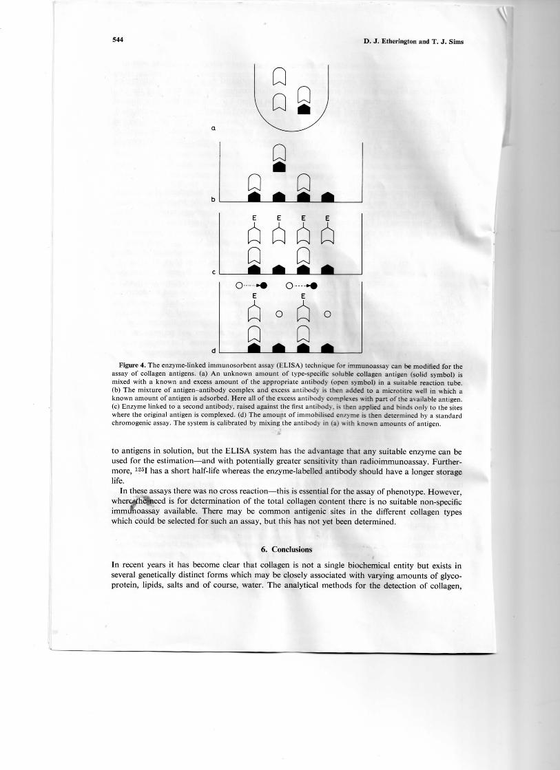
Recently, an ELISA technique was described for the specific assay of various connective tissuecomponents including collagen types I, II, III and IV45 (Figure 4). The method can be applied only
544 D. J. Etherington and T. J. Sims
Figure 4. The enzyme-linked immunosorbent assay (ELISA) technique for immunoassay can be modified for theassay of collagen antigens, (a) An unknown amount of type-specific soluble collagen antigen (solid symbol) ismixed with a known and excess amount of the appropriate antibody (open symbol) in a suitable reaction tube.(b) The mixture of antigen-antibody complex and excess antibody is then added to a microtitre well in which aknown amount of antigen is adsorbed. Here all of the excess antibody complexes with part of the a%ailable antigen.(c) Enzyme linked to a second antibody, raised against the first antibody, is then applied and binds only to the siteswhere the original antigen is complexed. (d) The amount of immobilised enzyme is then determined by a standardchromogenic assay. The system is calibrated by mixing the antibody in (a) with known amounts of antigen.
to antigens in solution, but the ELISA system has the advantage that any suitable enzyme can beused for the estimation—and with potentially greater sensitivity than radioimmunoassay. Further-more, 125I has a short half-life whereas the enzyme-labelled antibody should have a longer storagelife.
In these assays there was no cross reaction—this is essential for the assay of phenotype. However,wher^fheii*ieed is for determination of the total collagen content there is no suitable non-specificimmunoassay available. There may be common antigenic sites in the different collagen typeswhich could be selected for such an assay, but this has not yet been determined.
6. Conclusions
In recent years it has become clear that collagen is not a single biochemical entity but exists inseveral genetically distinct forms which may be closely associated with varying amounts of glyco-protein, lipids, salts and of course, water. The analytical methods for the detection of collagen,

Detection and estimation of collagen 545
therefore, must be selected according to the type of tissue. Accurate determination of collagen isnot always possible using the simpler colorimetric methods because the level of interferring sub-stances may be quite high and for some materials, e.g. processed foods, the nature of these sub-stances is not necessarily known or whether they have any predictable effect on chromogen forma-tion. The use of internal standards is advisable where a high degree of accuracy is required. Alterna-tively, a chromatographic method may be more suitable. If new and more stringent legislation isintroduced, such that the collagen content of meat products must be known with certainty, thenthis will increase the need for routine methods of analysis which are simple, reliable and sufficientlyrapid to avoid undue delays during processing.
References*
1. Lawrie, R. A. Chemical and biochemical constitution of muscle. Meat Science Pergamon Press, Oxford, 1979,3rdedn, pp. 75-131.
2. Piez, K. A. Primary structure. Biochemistry of Collagen (Ramachandran, G. N.; Reddi, A. H., Eds.), PlenumPress, New York, 1976, pp. 1-44.
3. Bailey, A. J. The basis of meat texture. /. Sci. Food Agric. 1972, 23, 995-1007.4. Gallop, P. M.; Paz, M. A. Post-translational protein modifications, with special attention to collagen and
elastin. Physiol. Rev. 1975, 55, 418-487.5. Bailey, A. J.; Sims, T. J. Chemistry of the collagen cross-links. Nature of the cross-links in the polymorphic
forms of dermal collagen during development. Biochem. J. 1976, 153, 211-215.6. Bailey, A. J.; Etherington, D. J. Metabolism of collagen and eastin. Comprehensive Biochemistry Vol. 19B
(Florkin, M.; Stotz, E., Eds), Elsevier, Amsterdam, 1980, Part I, pp. 299-460.7. Sykes, B. C.; Partridge, S. M. Salt-soluble elastin from lathyritic chicks. Biochem. J. 1974, 141, 567-572.8. Goll, D. E.; Bray, R. W.; Hoekstra, W. G. Age-associated changes in muscle composition. The isolation
and properties of a collagenous residue from bovine muscle. /. Fd Sci. 1963, 28, 503-509.9. Hasselbach, W.; Schneider, G. L-Myosin and actin contents of rabbit muscle. Biochem. Z. 1951, 321, 462-475.
10. Woessner, Jr J. F. The determination of hydroxyproline in tissue and protein samples containing small pro-portions of this imino acid. Arch. Biochem. Biophys. 1961, 93, 440-447.
11. Galasinski, W.; Gadek, A.; Ratkiewiez, A.; Rzeczycki, W. A convenient modification of the method forhydroxyproline determination in proteins. Anal. Biochem. 1978, 85, 550-555.
12. Baily, P.; Kilroe-Smith T. A.; Rollin, H. B.; Goldstein, B. The rapid determination of microgram amountsof total collagen in pathological lesions. Microchem. J. 1979, 24, 192-198.
13. Mitchell, A. D.; Taylor, I. E. P. The spectrophototometric determination of hydroxyproline: an analyticalinvestigation. Analyst 1970, 95, 1003-1011.
14. Bergman, I . ; Loxley, R. Two improved and simplified methods for the spectrophotometric determinationof hydroxyproline. Analyt. Chem. 1963, 35, 1961-1965.
15. Stegemann, H. B.; Stalder, K. Determination of hydroxyproline. Clin. Chim. Acta 1967, 18, 267-273.16. International Standards Organisation Methods of test for meat and meat products. Part II. Determination
of L(-)hydroxyproline content. 1978.17. Bergman, I.; Loxley, R. Lung tissue hydrolysates: studies of the optimum conditions for the spectrophoto-
metric determination of hydroxyproline. Analyst 1969, 94, 575-584.18. Prockop, D. J.; Udenfriend, S. A specific method for the analysis of hydroxyproline in tissues and urine.
Anal. Biochem. 1960, 1, 228-239.19. Kiviriklco, K. I.; Laitinen, O.; Prockop, D. J. Modifications of a specific assay for hydroxyproline in urine.
Anal. Biochem. 1967, 19, 249-255.20. Blumenkrantz, N.; Absoe-Hansen, G. An assay for hydroxyproline and proline on one sample and a simplified
method for hydroxyproline. Anal. Biochem. 1975, 63, 331-340.21. Grant. R. A. Estimation of hydroxyproline by the Auto Analyzer. /. Clin. Pathol. 1964,17, 685-686.22. Bannister, D. W.; Burns, A. B. Adaptation of the Bergman and Loxley technique for hydroxyproline deter-
mination to the Auto Analyzer and its use in determining plasma hydroxyproline in the domestic fowl. Analyst1970, 95, 596-600.
23. Switzer, B. R.; Summer, G. K. Improved method for hydroxyproline analysis in tissue hydrolysates. Anal.Biochem. 1971, 39, 487-491.
24. Bondjers, G.; Bjorkerud, S. Spectrophotometric determination of hydroxyproline in connective tissue on thenanogram level. Anal. Biochem. 1973, 52, 496-504.
25. Morales, T. I.; Woessner, Jr J. F.; Howell, D. S.; Marsh, J. M.; LeMaire, W. J. A microassay for the directdemonstration of collagenolytic activity in Graafian follicles of the rat. Biochem. Biophys. Acta 1978, 524,428-434.
26. Airhart, J.; Kelley, J.; Brayden, J. E.; Low, R. B.; Stirewalt, W. S. An ultramicro method of amino acidanalyses: application to studies of protein metabolism in cultured cells. Anal. Biochem. 1979, 96, 45-55.

546 D. J. Etherington and T. J. Sims
27. Miller, E. J. Structural studies on cartilage collagen employing limited cleavage and solubilization with pepsin.Biochemistry 1972, 11, 4903-4909. ,/
28. Callahan, P. X.; Shepard, J. A.; Ellis, S. Accelerated chromatographic method for determination of hydroxy-proline. Anal. Biochem. 1972, 49, 155-163. /
29. Felix, A. M.; Terkelsen, G. Determinatiorywf hydrozyproline in fluorometric amino acid analysis with fluores-camine. Anal. Biochem. 1973, 56, 610-61/
30. Bohlen, P.; Mellet, M. Automated fluorometric amino acid analysis: the determination of proline and hydro-xyproline. Anal. Biochem. 1979, 94, 313-3^
31. Partridge, S. M.; Elsden, D. F. Rapid methods for the determination of glucosamine, galactosamine andhydroxyproline. Biochem. J. 1961, 80, 34.
32. Mussini, E.; Marcucci, F. Separation of prolines and hydroxyprolines by gas chromatography. J. Chromatog.1965, 20,266-269.
33. l^ee, J. M. L. Specific assay of hydroxyproline by gas chromatography. /. Chromatog. 1973, 87, 155-161.34. MacKenzie, S. L.; Tenaschuk, D. Analysis of hydroxyproline and hydroxylysine: improved gas chromato-
graphic method. /. Chromatog. 1975, 104, 176-177.35. Pefier, C.; Ronziere, M. C.; Rattner, A.; Frey, J. Employment of gas liquid chromatography for the analysis
of collagen amino acids in biopsy tissue. /. Chromatog. 1980, 182, 155-162.36. O'Neill, I. K.; Trimble, M. L.; Casey, J. C. Carbon-13 pulsed Fourier transform nuclear magnetic resonance
spectroscopic determination of 4-hydroxy-L-proline in meat—comparison with the colorimetric method.Meat Sci. 1979, 3, 223-232.
37. Disbrey, B. D.; Rack, J. H. Connective tissues, Histological Laboratory Methods Livingstone, Edinburgh,1970, pp. 108-124.
38. Sweat, F.; Puchtler, H.; Rosenthal, S. T. Sirius red F313A as a strain for connective tissue. Arch. Pathol.1964, 78,69-72.
39. Junqueira, L. C. U.; Bignolas, G.; Brentani, R. R. A simple and sensitive method for the quantitative estima-tion of collagen. Anal. Biochem. 1979, 94, 96-99.
40. Lillie, R. D.; Reynolds, C.; Pizzolato, P. Phosphomolybdic and phosphototungstic acid-Victoria blue Rstrains two histochemically distinct collagens; dense dark blue and loose areolar pale green. /. Histochem.Cytochem. 1979 27, 1092-1094.
41. Bailey, A. J.; Restall, D. J.; Sims, T. J.; Duance, V. C. Meat tenderness: immunofluorescent localisation ofthe isomorphic forms of collagen in bovine muscles of varying texture. /. Sci. Food Agric. 1979, 30, 203-210.
42. Roll, F. J.; Madri, J. A.; Furthmayr, H. A new method of iodinating collagens for use in radioimmunoassay.Anal. Biochem. 1979, 96, 489-499.
43. Engvall, E.; Perlmann, P. Enzyme-linked immunosorbent assay, ELISA 111 Quantitation of specific antibodiesby enzyme-labelled anti immunoglobulin in antigen-coated tubes. /. Immitnol. 1972, 109, 129-135.
44. ELISA: a replacement for radioimmuno assay? Lancet 1976, ii, 406-407.45. Rennard, S. I.; Borg, R.; Martin, G. R.; Foidart, J. M.; Gehron Robey, P. Enzyme-linked immunoassay
(ELISA) for connective tissue components. Anal. Biochem. 1980, 104, 205-214.