Embed Size (px)
Citation preview
Detecting the Inferior Thoracic Aperture using Statistical Shape
Models
Pahal DalalDepartment of Computer Science &
Engineering,University of South Carolina
Outline
• Introduction– What is the Inferior Thoracic Aperture (ITA)?– Why segment the ITA?– Why is segmenting the ITA difficult?
• Construction of Shape Model
• Detecting the ITA
• Conclusion
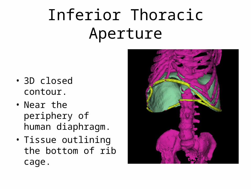
Inferior Thoracic Aperture
• 3D closed contour.• Near the periphery of
human diaphragm.• Tissue outlining the
bottom of rib cage.
Relation to Diaphragm
• Diaphragm hangs off the ITA.
• Diaphragm related to normal pulmonary function.
• Extracting ITA can help extract diaphragm.

Difficulty
• Diaphragm hangs off the ITA.
• Difference between ITA and boundary of diaphragm difficult to find.
• CT images very fuzzy in certain parts.
Outline
• Introduction– What is the Inferior Thoracic Aperture (ITA)?– Why segment the ITA?– Why is segmenting the ITA difficult?
• Construction of Shape Model
• Detecting the ITA
• Conclusion
Partitioning ITA shape
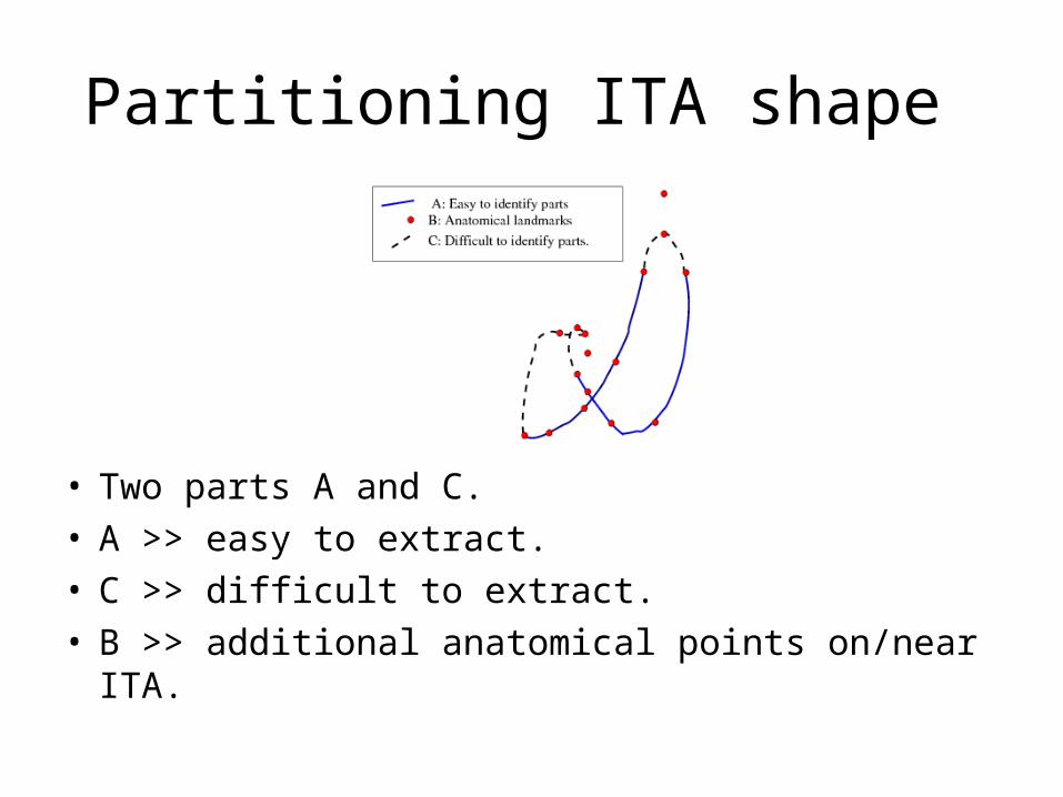
• Two parts A and C.• A >> easy to extract.• C >> difficult to extract.• B >> additional anatomical points on/near ITA.
Additional points considered
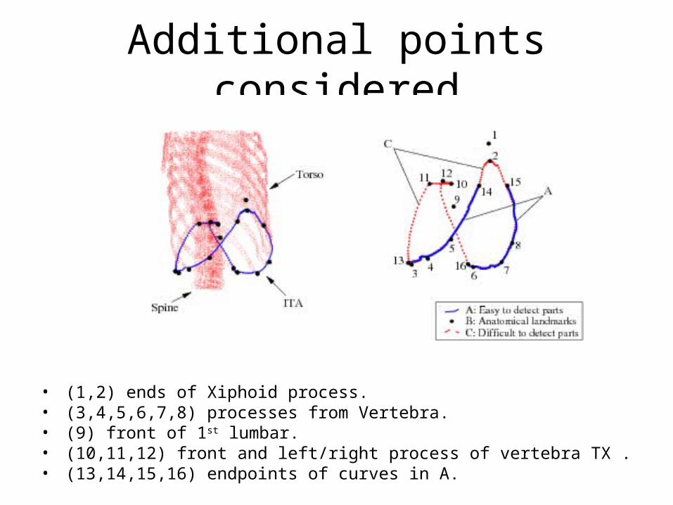
• (1,2) ends of Xiphoid process. • (3,4,5,6,7,8) processes from Vertebra.• (9) front of 1st lumbar.• (10,11,12) front and left/right process of vertebra TX .• (13,14,15,16) endpoints of curves in A.
Identifying Landmarks
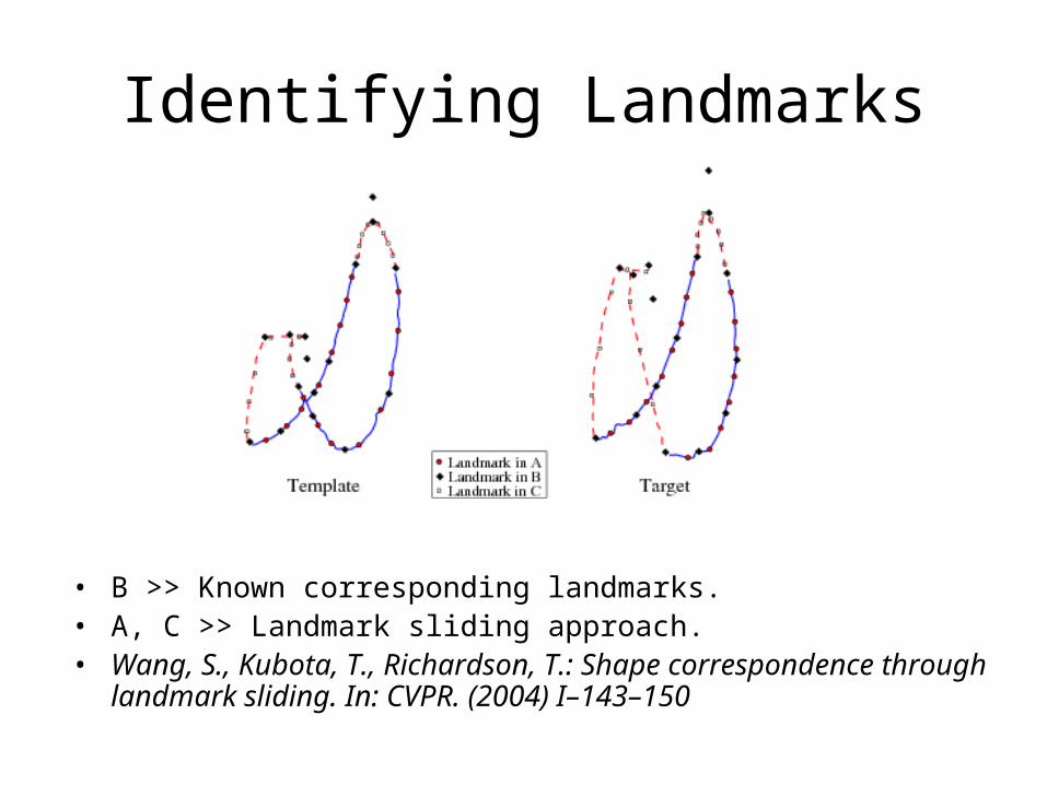
• B >> Known corresponding landmarks.• A, C >> Landmark sliding approach.• Wang, S., Kubota, T., Richardson, T.: Shape correspondence
through landmark sliding. In: CVPR. (2004) I–143–150
Identifying Landmarks
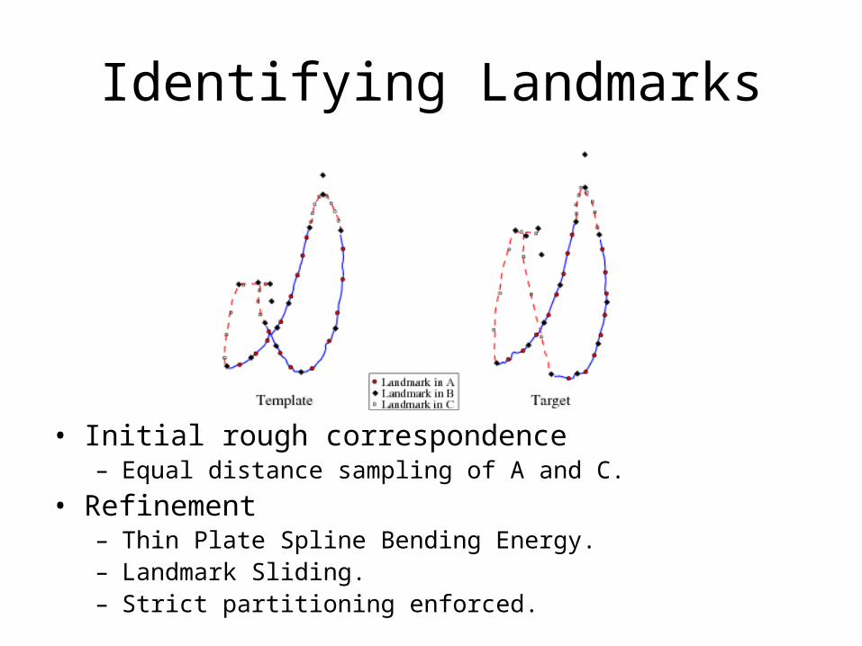
• Initial rough correspondence– Equal distance sampling of A and C.
• Refinement– Thin Plate Spline Bending Energy.– Landmark Sliding.– Strict partitioning enforced.

Constructing PDM
• Set of training shapes.
• Find corresponding landmarks on each.
• Mean shape >> m.
• Co-variance >> S.
• Shape model >> (m, S).

Outline
• Introduction– What is the Inferior Thoracic Aperture (ITA)?– Why segment the ITA?– Why is segmenting the ITA difficult?
• Construction of Shape Model
• Detecting the ITA
• Conclusion

Detecting the ITA
• v is the set of landmarks along shape to be found.
• m = [ mP mQ ]
• v = [ vP vQ ]
• P >> landmarks along A and B.
• Q >> landmarks along C.
Detecting landmarks along A, B
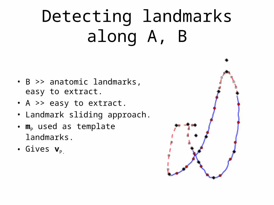
• B >> anatomic landmarks, easy to extract.
• A >> easy to extract.• Landmark sliding approach.
• mP used as template landmarks.
• Gives vP.
Mahalanobis distance
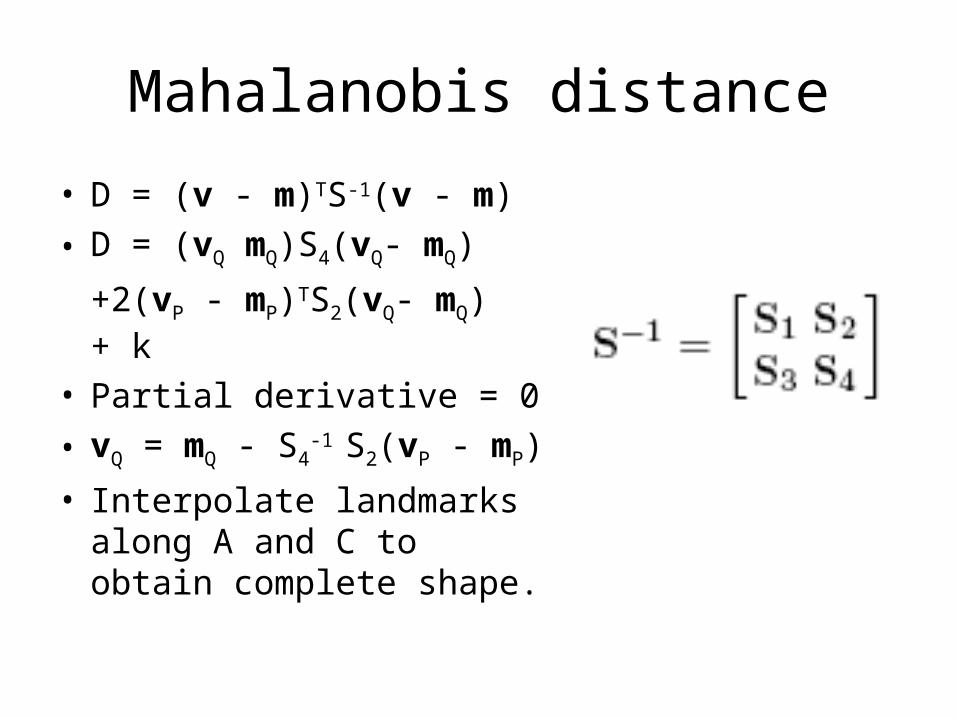
• D = (v - m)TS-1(v - m)
• D = (vQ mQ)S4(vQ- mQ)
+2(vP - mP)TS2(vQ- mQ) + k
• Partial derivative = 0
• vQ = mQ - S4-1 S2(vP - mP)
• Interpolate landmarks along A and C to obtain complete shape.

Experiments
• 14 shapes.
• Leave one out.
• e1 = average distance between predicted shape and truth.
• e2 = average distance between predicted landmark and truth landmark.
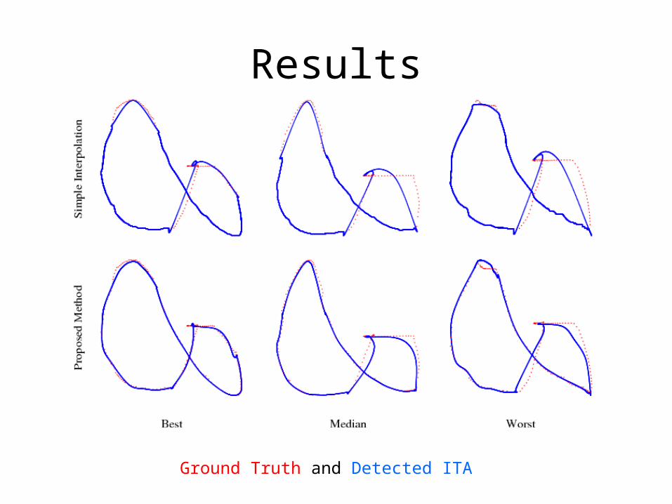
Results
Ground Truth and Detected ITA
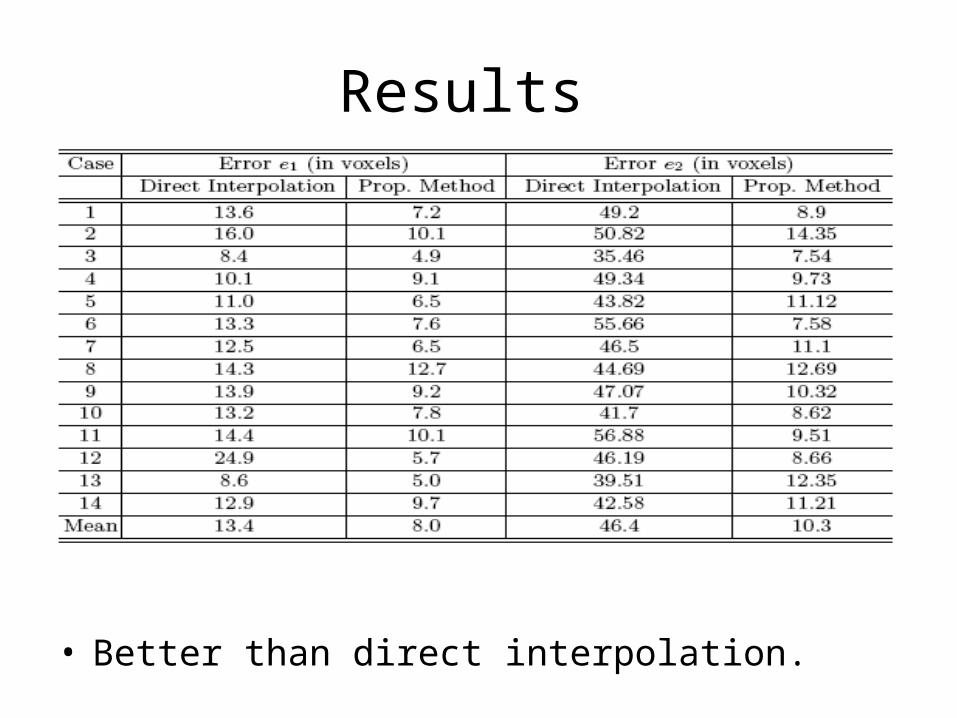
Results
• Better than direct interpolation.
Conclusion
• A new method to detect the Inferior Thoracic Aperture.
• Better performance than direct interpolation.

Questions?