Embed Size (px)
Citation preview
The Journal of Molecular Diagnostics, Vol. 13, No. 6, November 2011
Copyright © 2011 American Society for Investigative Pathology
and the Association for Molecular Pathology.
Published by Elsevier Inc. All rights reserved.
DOI: 10.1016/j.jmoldx.2011.06.003
Detailed Characterization of Alterations ofChromosomes 7, 9, and 10 in Glioblastomas as
Assessed by Single-Nucleotide Polymorphism ArraysInês Crespo,*† Ana Luísa Vital,*†
Ana Belen Nieto,‡ Olinda Rebelo,§ Hermínio Tão,¶
Maria Celeste Lopes,*† Catarina Resende Oliveira,*�
Pim J. French** Alberto Orfao,‡ andMaría Dolores Tabernero‡††‡‡
From the Center for Neuroscience and Cell Biology,* and the
Faculties of Pharmacy,† and Medicine,� University of Coimbra,
Coimbra, Portugal; the Center for Cancer Research,‡ Department
of Medicine, University of Salamanca, Salamanca, Spain; the
Neurosurgery Service,§ and the Neuropathology Laboratory,
Neurology Service,¶ University Hospital of Coimbra, Coimbra,
Portugal; the Josephine Nefkens Institute,�� Department of
Neurology, Erasmus Medical Center Rotterdam, Rotterdam, The
Netherlands; the Research Unit,†† University Hospital of
Salamanca, Salamanca, Spain; and the Instituto de Estudios de
Ciencias de la Salud de Castilla y León,‡‡ Soria, Spain
Glioblastomas are cytogenetically heterogeneous tu-mors that frequently display alterations of chromo-somes 7, 9p, and 10q. We used high-density (500K)single-nucleotide polymorphism arrays to investigategenome-wide copy number alterations and loss ofheterozygosity in 35 primary glioblastomas. We fo-cused on the identification and detailed characteriza-tion of alterations involving the most frequently al-tered chromosomes (chromosomes 7, 9, and 10), theidentification of distinct prognostic subgroups of glio-blastomas based on the cytogenetic patterns of alter-ation for these chromosomes, and validation of theirprognostic impact in a larger series of tumors frompublic databases. Gains of chromosome 7 (97%), withor without epidermal growth factor receptor (EGFR)amplification, and losses of chromosomes 9p (83%)and 10 (91%) were the most frequent alterations. Suchalterations defined five different cytogenetic groupswith a significant effect on patient survival; notably,EGFR amplification (29%) was associated with a bet-ter survival among older patients, as confirmed bymultivariate analysis of a larger series of glioblasto-mas from the literature. In addition, our results pro-vide further evidence about the relevance of othergenes (eg, EGFR, CDKN2A/B, MTAP) in the pathogen-esis of glioblastomas. Altogether, our results confirmthe cytogenetic heterogeneity of glioblastomas and
suggest that their stratification based on combined634
assessment of cytogenetic alterations involving chro-mosomes 7, 9, and 10 may contribute to the prognos-tic evaluation of glioblastomas. (J Mol Diagn 2011, 13:634–647; DOI: 10.1016/j.jmoldx.2011.06.003)
Gliomas are a heterogeneous group of malignant tu-mors that show variable localization, histopathologicfeatures, and genetic profiles, together with a hetero-geneous response to therapy but a uniformly fatal out-come.1–11 Although no common genetic signature hasbeen detected in all gliomas, multiple chromosomalchanges have been described so far, which frequentlyinclude gains of chromosome 7 and deletions of chro-mosomes 9 and 10 and to a less extent also of chro-mosomes 1 and 19.12–14 These genetic changes areassociated with amplification of oncogenes [eg, epi-dermal growth factor receptor (EGFR)] together withdeletion and/or mutation of tumor suppressor genes[eg, tumor protein p53 (TP53), phosphatase and tensinhomolog (PTEN), and cyclin-dependent kinase inhibi-tor 2A (p16/CDKN2A)].15
Altogether, these results point out the potential in-volvement of different signaling pathways in gliomas,with alterations of chromosome 7, 9, and 10 participat-ing in the most frequent tumor subtypes (eg, glioblas-toma). In line with this hypothesis, we have recentlyshown the existence of distinct cytogenetic pathwaysin gliomas, by using interphase fluorescence in situhybridization (iFISH) analysis of intratumoral patternsof chromosomal alterations, at the single-cell level.16
Notably, specific genomic aberrations and cytogeneticprofiles are associated with particular tumor histo-
Supported by the Portuguese Foundation for Science and Technology(FCT) grant PIC/IC/83108/2007, FCT PhD fellowships SFRH/BD/23086/2005 and SFRH/BD/11820/2003, and the Spanish Network of CancerResearch Centers (Red Temática de Investigación Cooperativa enCáncer) grant RD06/0020/0035 from the Instituto de Salud Carlos III,Ministry of Science and Innovation, Madrid, Spain.
Accepted for publication June 10, 2011.
Supplemental material for this article can be found at http://jmd.amjpathol.org or at doi: 10.1016/j.jmoldx.2011.06.003.
The authors did not disclose any relevant financial relationships.
Address reprint requests to María Dolores Tabernero, M.D., Ph.D.,Research Unit of the University Hospital of Salamanca, Paseo San Vicente
58-182, Salamanca, Spain. E-mail: [email protected].
SNP Array Profiles in Glioblastomas 635JMD November 2011, Vol. 13, No. 6
pathologic features.17–19 Accordingly, amplification (orrearrangement) of EGFR is almost restricted to a frac-tion of all malignant gliomas, particularly glioblasto-mas. Among these cases, overexpression of the EGFRvariant 3 mutant is most frequently detected.20,21 Al-though this mutant protein is unable to bind to its ligands,it constitutively signals, conferring proliferation and sur-vival advantages to tumor cells.20–22 In turn, genomicdeletions of chromosomes 9 and 10 at regions that harbortumor suppressor genes are also typically found in glioblas-tomas, where they have been associated with the develop-ment of the tumor, its progression, and a poor progno-sis.23–26 Interestingly, monosomy 10 is associated with gainor amplification of the EGFR gene on chromosome 7p11.2,supporting the role of both alterations in gliomagenesis.27,28
Other genetic abnormalities that can be frequently foundin low-grade gliomas29,30 [eg, combined del(1p)/del(19q) and TP53 mutation] are less frequently de-tected in glioblastomas.31–35
In the past, most studies devoted to the identificationand characterization of genetic/chromosomal alterationsin glioblastomas have used conventional cytogenetic andmolecular techniques associated with relatively low-res-olution (eg, iFISH and comparative genomic hybridiza-tion). Recently, high-density single-nucleotide polymor-phism (SNP) arrays have been used to characterize themost frequent genetic alterations of glioblastomas.36–51
Newly available high-density SNP arrays allow the studyof copy number (CN) changes and loss of heterozygosity(LOH) at both coding and noncoding DNA regions of thewhole tumor cell genome, with high resolution; this pro-vides a more precise map of the genetic alterations as-sociated with CN changes in glioblastomas. Thus, SNParray studies performed in large series of patients with orwithout gene expression profiling have provided new in-sights into the potential role of new candidate genes (eg,ERBB2, NF1, and TP53), molecular changes (eg, PIK3R1and PDGFRA/IDH1 mutations), and signaling pathwaysinto the pathogenesis of glioblastomas.40 In turn, basedon gene expression profiles, a molecular classification ofglioblastomas has been proposed that reflects the in-volvement of different neural lineages.42 To the best ofour knowledge, however, no classification based on thegenetic changes involving the most frequently alteredchromosomes (eg, 7, 9, and 10) has been proposed sofar for glioblastomas.
We used high-density (500K) SNP array to investigategenome-wide CN alterations and LOH in a group of 35primary glioblastoma patients; we focused on the identifi-cation and detailed characterization of the genetic altera-tions of those chromosomes more frequently altered inthese tumors and the identification of groups of glioblasto-mas with distinct cytogenetic patterns of alteration for these3 chromosomes, which are potentially associated with thebehavior of the disease. Finally, the prognostic value of thepresence of amplification of the EGFR gene was confirmedin a larger number of cases from four different independentseries of glioblastoma patients, which have been previously
reported in the literature.41,42,45,50Materials and Methods
Patients and Samples
A total of 70 paired tumor (n � 35) and peripheral blood(PB; n � 35) samples from 35 patients (15 men and 20women) diagnosed as having glioblastomas (mean � SDage, 60 � 14 years; age range, 30 to 84 years) who wereadmitted to the Neurosurgery Service of the UniversityHospital of Coimbra (Coimbra, Portugal) were included inthis study. Before entering the study, each patient gavewritten informed consent to participate, and the studywas approved by the Hospital’s Ethics Committee. Of the35 patients, 5 underwent complete resection of the tumor;either partial resection or just a diagnostic biopsy wasperformed in the other 30 cases (Table 1). Distributionaccording to tumor localization was as follows: 16 tumorswere localized in the frontal lobe, 12 were temporal, 3were parietal, 2 were occipital, 1 was insular, and 1 hada deep localization. Tumors were diagnosed and classi-fied by an experienced neuropathologist according to theWorld Health Organization criteria.3 At the time of closingthe study, all patients had died, with a median overallsurvival of 11 months (range, 1 week to 67 months).
Representative parts of fresh tumor tissues left afterroutine diagnostic histopathologic procedures had beenperformed were immediately snap-frozen in liquid nitro-gen and stored at �80°C, until used for iFISH and DNAextraction for SNP array studies. In each case, a sectioncut from the tissue block used for this purpose was his-tologically assessed to estimate tumor cell contents.Specimens with 75% or more tumor cells in the absenceof contamination by normal brain parenchyma and tumornecrosis were systematically selected for further DNAextraction and SNP array studies.
DNA Extraction and SNP Array Hybridization
DNA from both frozen tumor tissues and their paired PBleukocyte samples was purified using the QIAamp DNAMini Kit (Qiagen, Valencia, CA) according to the manu-facturer’s instructions. DNA yield and purity were deter-mined with a NanoDrop-1000 spectrophotometer (Nano-Drop Technologies Inc., Wilmington, DE). DNA integritywas evaluated by conventional electrophoretic proce-dures in 1% agarose gel.
DNA samples were processed according to the Map-ping 500K Array Set (Affymetrix Inc., Santa Clara, CA)protocol with two arrays, each containing 250,000 SNPs,with a mean intermarker distance of 5.8 kb (250K Nspand Sty arrays). Briefly, total DNA (250 ng per array) frompaired tumor and PB samples was separately digestedwith the NspI and StyI restriction enzyme and ligated tothe corresponding adaptors that recognize overhangsgenerated by the restriction enzymes. All digested DNAfragments were then used as substrates for adaptor liga-tion, regardless of their size. A generic primer that rec-ognizes the adaptor sequence was used in triplicate toamplify adaptor-ligated DNA fragments through PCR.The amplified DNA was then fragmented, labeled, and
hybridized to the GeneChip Human Mapping 250K Nsp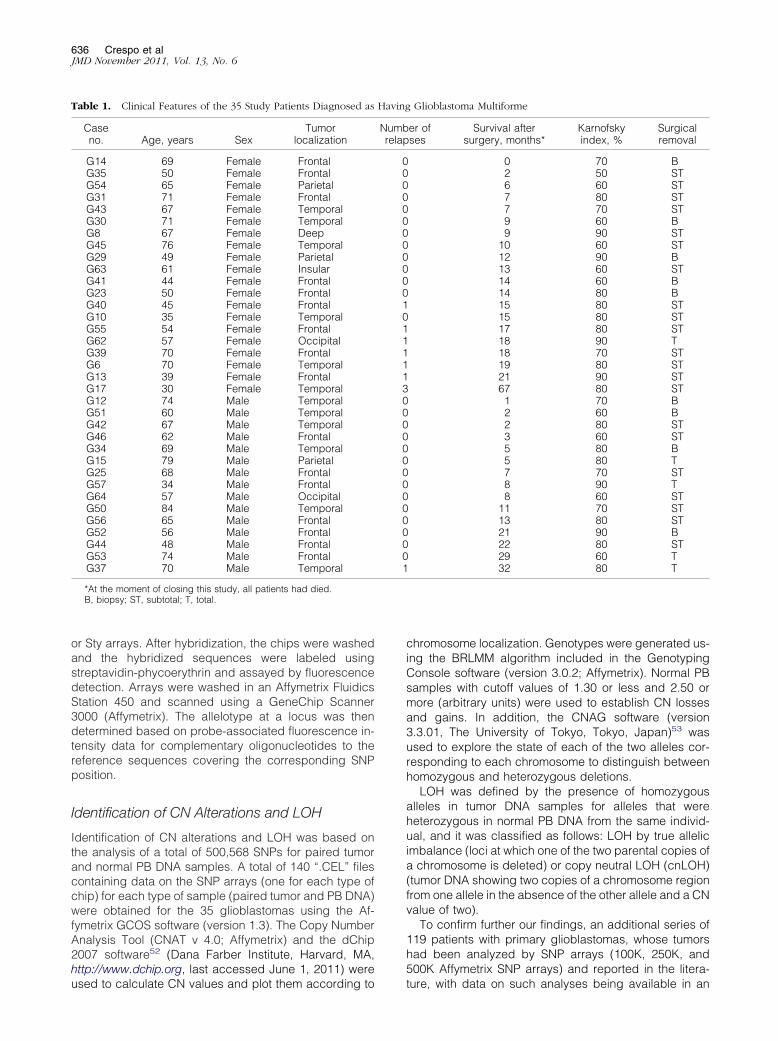
636 Crespo et alJMD November 2011, Vol. 13, No. 6
or Sty arrays. After hybridization, the chips were washedand the hybridized sequences were labeled usingstreptavidin-phycoerythrin and assayed by fluorescencedetection. Arrays were washed in an Affymetrix FluidicsStation 450 and scanned using a GeneChip Scanner3000 (Affymetrix). The allelotype at a locus was thendetermined based on probe-associated fluorescence in-tensity data for complementary oligonucleotides to thereference sequences covering the corresponding SNPposition.
Identification of CN Alterations and LOH
Identification of CN alterations and LOH was based onthe analysis of a total of 500,568 SNPs for paired tumorand normal PB DNA samples. A total of 140 “.CEL” filescontaining data on the SNP arrays (one for each type ofchip) for each type of sample (paired tumor and PB DNA)were obtained for the 35 glioblastomas using the Af-fymetrix GCOS software (version 1.3). The Copy NumberAnalysis Tool (CNAT v 4.0; Affymetrix) and the dChip2007 software52 (Dana Farber Institute, Harvard, MA,http://www.dchip.org, last accessed June 1, 2011) were
Table 1. Clinical Features of the 35 Study Patients Diagnosed as
Caseno. Age, years Sex
Tumorlocalization
G14 69 Female FrontalG35 50 Female FrontalG54 65 Female ParietalG31 71 Female FrontalG43 67 Female TemporalG30 71 Female TemporalG8 67 Female DeepG45 76 Female TemporalG29 49 Female ParietalG63 61 Female InsularG41 44 Female FrontalG23 50 Female FrontalG40 45 Female FrontalG10 35 Female TemporalG55 54 Female FrontalG62 57 Female OccipitalG39 70 Female FrontalG6 70 Female TemporalG13 39 Female FrontalG17 30 Female TemporalG12 74 Male TemporalG51 60 Male TemporalG42 67 Male TemporalG46 62 Male FrontalG34 69 Male TemporalG15 79 Male ParietalG25 68 Male FrontalG57 34 Male FrontalG64 57 Male OccipitalG50 84 Male TemporalG56 65 Male FrontalG52 56 Male FrontalG44 48 Male FrontalG53 74 Male FrontalG37 70 Male Temporal
*At the moment of closing this study, all patients had died.B, biopsy; ST, subtotal; T, total.
used to calculate CN values and plot them according to
chromosome localization. Genotypes were generated us-ing the BRLMM algorithm included in the GenotypingConsole software (version 3.0.2; Affymetrix). Normal PBsamples with cutoff values of 1.30 or less and 2.50 ormore (arbitrary units) were used to establish CN lossesand gains. In addition, the CNAG software (version3.3.01, The University of Tokyo, Tokyo, Japan)53 wasused to explore the state of each of the two alleles cor-responding to each chromosome to distinguish betweenhomozygous and heterozygous deletions.
LOH was defined by the presence of homozygousalleles in tumor DNA samples for alleles that wereheterozygous in normal PB DNA from the same individ-ual, and it was classified as follows: LOH by true allelicimbalance (loci at which one of the two parental copies ofa chromosome is deleted) or copy neutral LOH (cnLOH)(tumor DNA showing two copies of a chromosome regionfrom one allele in the absence of the other allele and a CNvalue of two).
To confirm further our findings, an additional series of119 patients with primary glioblastomas, whose tumorshad been analyzed by SNP arrays (100K, 250K, and500K Affymetrix SNP arrays) and reported in the litera-
Glioblastoma Multiforme
er ofses
Survival aftersurgery, months*
Karnofskyindex, %
Surgicalremoval
0 70 B2 50 ST6 60 ST7 80 ST7 70 ST9 60 B9 90 ST
10 60 ST12 90 B13 60 ST14 60 B14 80 B15 80 ST15 80 ST17 80 ST18 90 T18 70 ST19 80 ST21 90 ST67 80 ST
1 70 B2 60 B2 80 ST3 60 ST5 80 B5 80 T7 70 ST8 90 T8 60 ST
11 70 ST13 80 ST21 90 B22 80 ST29 60 T32 80 T
Having
Numbrelap
00000000000010111113000000000000001
ture, with data on such analyses being available in an
SNP Array Profiles in Glioblastomas 637JMD November 2011, Vol. 13, No. 6
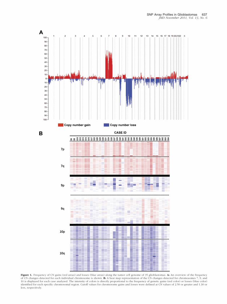
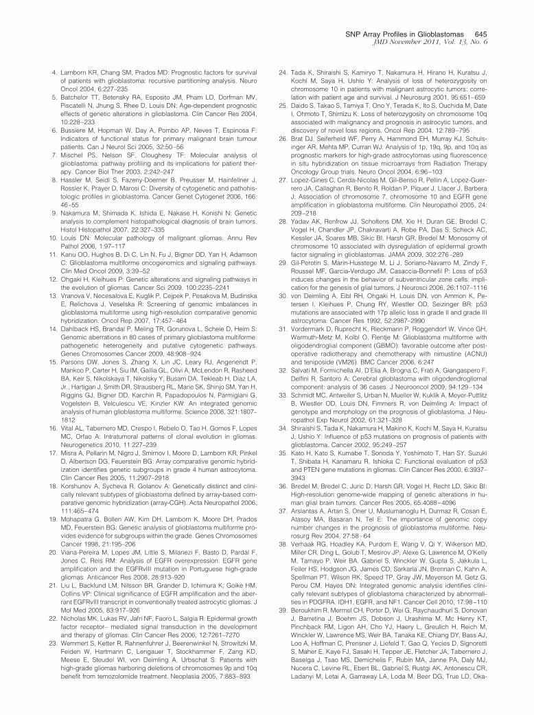
Figure 1. Frequency of CN gains (red areas) and losses (blue areas) along the tumor cell genome of 35 glioblastomas. A: An overview of the frequencyof CN changes detected for each individual chromosome is shown. B: A heat map representation of the CN changes detected for chromosomes 7, 9, and10 is displayed for each case analyzed. The intensity of colors is directly proportional to the frequency of genetic gains (red color) or losses (blue color)
identified for each specific chromosomal region. Cutoff values for chromosome gains and losses were defined at CN values of 2.50 or greater and 1.30 orless, respectively.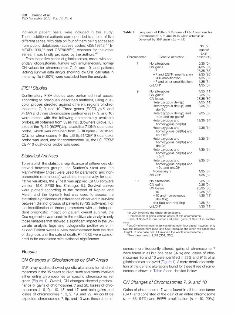
638 Crespo et alJMD November 2011, Vol. 13, No. 6
individual patient basis, were included in this study.These additional patients corresponded to a total of fivedifferent series, with data on four of them being accessedfrom public databases (access codes: GSE19612,42 E-MEXD-1330,45 and GSE963550), whereas for the otherseries, it was kindly provided by the authors.41
From these five series of glioblastomas, cases with sec-ondary glioblastomas, tumors with simultaneously normalCN values for chromosomes 7, 9, and 10, and patientslacking survival data and/or showing low SNP call rates inthe array file (�90%) were excluded from the analysis.
iFISH Studies
Confirmatory iFISH studies were performed in all cases,according to previously described methods, using dual-color probes directed against different regions of chro-mosomes 7, 9, and 10. Three genes (EGFR, p16, andPTEN) and three chromosome centromeres (7, 9, and 10)were tested with the following commercially availableprobes, all obtained from Vysis Inc. (Downers Grove, IL),except the 7p12 (EGFR)/alphasatellite 7 DNA dual-colorprobe, which was obtained from Q-BIOgene (Carlsbad,CA); for chromosome 9, the LSI 9p21/CEP-9 dual-colorprobe was used, and for chromosome 10, the LSI PTEN/CEP-10 dual-color probe was used.
Statistical Analyses
To establish the statistical significance of differences ob-served between groups, the Student’s t-test and theMann-Whitney U-test were used for parametric and non-parametric (continuous) variables, respectively; for qual-itative variables, the �2 test was applied (SPSS softwareversion 15.0, SPSS Inc, Chicago, IL). Survival curveswere plotted according to the method of Kaplan andMeier, and the log-rank test was used to assess thestatistical significance of differences observed in survivalbetween distinct groups of patients (SPSS software). Forthe identification of those parameters with an indepen-dent prognostic impact on patient overall survival, theCox regression was used; in the multivariate analysis onlythose variables that showed a significant impact in the uni-variate analysis (age and cytogenetic profile) were in-cluded. Patient overall survival was measured from the dateof diagnosis until the date of death. P � 0.05 were consid-ered to be associated with statistical significance.
Results
CN Changes in Glioblastomas by SNP Arrays
SNP array studies showed genetic alterations for all chro-mosomes in the 35 cases studied; such alterations involvedeither entire chromosomes or specific chromosomal re-gions (Figure 1). Overall, CN changes showed predomi-nance of gains of chromosomes 7 and 20, losses of chro-mosomes 4, 6, 9p, 10, 15, and 17, and both gains andlosses of chromosomes 1, 3, 9, 19, and 22. As could be
expected, chromosomes 7, 9p, and 10 were those chromo-somes more frequently altered: gains of chromosome 7were found in all but one case (97%) and losses of chro-mosomes 9p and 10 were identified in 83% and 91% of allglioblastomas analyzed (Figure 1). A more detailed descrip-tion of the genetic alterations found for these three chromo-somes is shown in Table 2 and detailed below.
CN Changes of Chromosomes 7, 9, and 10
Gains of chromosome 7 were found in all but one tumor(G41) and consisted of the gain of an entire chromosome
Table 2. Frequency of Different Patterns of CN Alterations forChromosomes 7, 9, and 10 in Glioblastomas asDetected by SNP Arrays (n � 35)
Chromosome Genetic alteration
No. ofcases/total
cases (%)
7 No alterations 0/35 (0)CN gains 34/35 (97)
�7 23/35 (65)�7 and EGFR amplification 9/35 (26)EGFR amplification 1/35 (3)�7 and other amplifications 1/35 (3)
cnLOH* 1/35 (3)
9 No alterations 4/35 (11)CN gains† 2/35 (6)CN losses 28/35 (80)
Heterozygous del(9p) 4/35 (11)Heterozygous del(9p) and
del(9q)2/35 (6)
Heterozygous del(9p) and�9q and 9p gains‡
2/35 (6)
Heterozygous andhomozygous del(9p)
12/35 (34)
Heterozygous andhomozygous del(9p) andcnLOH§
2/35 (6)
Heterozygous andhomozygous del(9p) anddel(9q)
2/35 (6)
Heterozygous andhomozygous del(9p) and�9q¶
1/35 (3)
Heterozygous andhomozygous del(9p) and�9q and cnLOH
2/35 (6)
Monosomy 9 1/35 (3)cnLOH* 1/35 (3)
10 No alterations 3/35 (9)CN gains 0/35 (0)CN losses 28/35 (80)
�10 22/35 (63)�10 and homozygous
del(10q)4/35 (11)
del(10p) and del(10q) 2/35 (6)cnLOH* 4/35 (11)
*cnLOH involving the whole chromosome.†Chromosome 9 gains without losses of this chromosome.‡Gain of 9p24.3 in one tumor and other gains of 9p21.1 in another
case.§cnLOH of chromosome 9p was detected in four cases; however, only
two are included here (G54 and G55) because the other two cases had�9q21. In one case cnLOH involved the whole chromosome 9.
¶Two case have cnLOH (G54, G55).
(n � 33; 94%) and EGFR amplification (n � 10; 29%).
62,691-bll excep
SNP Array Profiles in Glioblastomas 639JMD November 2011, Vol. 13, No. 6
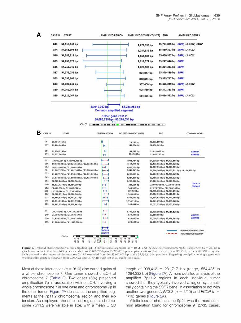
Most of these later cases (n � 9/10) also carried gains ofa whole chromosome 7. One tumor showed cnLOH ofchromosome 7 (Table 2). Two cases (6%) presentedamplification 7p in association with cnLOH, involving awhole chromosome 7 in one case and chromosome 7p inthe other tumor. Figure 2A delineates the amplified seg-ments at the 7p11.2 chromosomal region and their ex-tension. As displayed, the amplified regions at chromo-
Figure 2. Detailed characterization of the amplified 7p11.2 chromosomal seglioblastomas. Note that the EGFR gene extends from 55,086,725 bp to 55,27SNPs assayed in this region of chromosome 7p11.2 extended from the 55,0systematically deleted; however, both CDKN2A and CDKN2B were lost in a
some 7p11.2 were variable in size, with a mean � SD
length of 908,412 � 281,717 bp (range, 554,485 to1284,332 bp) (Figure 2A). A more detailed analysis of theamplified 7p11.2 regions in each individual tumorshowed that they typically involved a region systemati-cally containing the EGFR gene, in association or not withanother two genes: LANCL2 (n � 5/10) and ECOP (n �1/10) genes (Figure 2A).
Allelic loss of chromosome 9p21 was the most com-
(n � 10, A) and the deleted chromosome 9p21.3 sequences (n � 22, B) infrom pter positions (Entrez Gene, GeneID1956); in the 500K SNP array, thep to the 55,236,410-bp positions. Regarding del(9p21) no single gene wast one case.
gments5,031 bp
mon alteration found for chromosome 9 (27/35 cases;
s deleticases).
640 Crespo et alJMD November 2011, Vol. 13, No. 6
77%); in addition, monosomy 9 in association with ho-mozygous del(9p21) was detected in one case (caseG17; 3%), and cnLOH of an entire chromosome 9 wasfound in another case (case G57, 3%) (Table 2; see alsoSupplemental Table S1 at http://jmd.amjpathol.org).Seven tumors showed gains of chromosome 9q, consist-ing of partial gains (n � 5; cases G6, G14, G54, G55,G56, and G63) or gain of an entire chromosome 9 (n � 1;case G51); some of these cases (n � 5/7) showed addi-tional coexisting losses of chromosome 9p (cases G6,G14, G54, G55, and G56). A more detailed analysis ofchromosome 9 sequences in cases with del(9p) revealeda wide spectrum of allelic losses regarding the size of thedeleted regions, ranging from 44,787 to 5518,896 bp.Overall, deletions within the short arm occurred muchmore frequently than in the long arm of chromosome 9(n � 27 versus 4 cases), with several different patterns: i)heterozygous del(9p) (n � 8) (cases G41, G39, G6, G14,G29, G34, G64, and G35); ii) combined heterozygousand homozygous del(9p) (n � 15) (cases G23, G53, G44,G56, G30, G37, G13, G52, G62, G12, G45, G8, G10,G43, and G50); and iii) cnLOH combined with heterozy-gous and homozygous del(9p) (n � 4) (cases G55, G40,G31, and G54). From those cases showing cnLOH with orwithout del(9p) (n � 5), complete loss of chromosome 9pwas found in 3 cases (9%); the other two glioblastomashad cnLOH involving the whole chromosome 9, in asso-ciation with heterozygous and homozygous del(9p21) inone tumor (case G40) (Table 2; see also SupplementalTable S1 at http://jmd.amjpathol.org). Despite all thesepatterns, cases with del(9p21.3) (n � 22) or monosomy 9
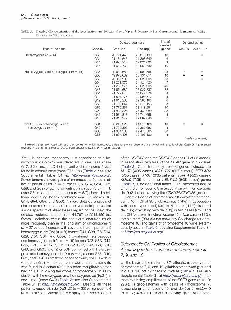
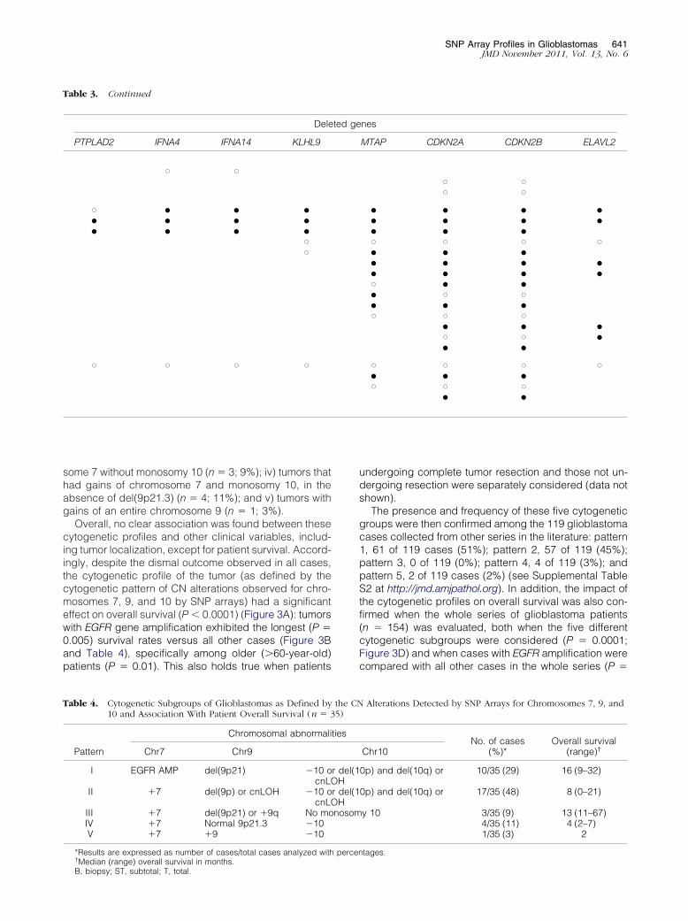
Table 3. Detailed Characterization of the Localization and DeletiDetected in Glioblastomas
Type of deletion Case ID
Heterozygous (n � 4) G6G34G14G44
Heterozygous and homozygous (n � 14) G37G56G52G8G53G43G54G10G12G50G62G23G45G13
cnLOH plus heterozygous andhomozygous (n � 4)
G31G40G30G55
Deleted genes are noted with a circle; genes for which homozygoumonosomy 9 and homozygous losses from 9p22.1 to p21.3. (n � 22/35
(n � 1) almost systematically displayed in common loss
of the CDKN2B and the CDKN2A genes (21 of 22 cases),in association with loss of the MTAP gene in 15 cases(Table 3). Other frequently deleted genes included theMLLT3 (4/35 cases), KIAA1797 (6/35 tumors), PTPLAD2(5/35 cases), IFNA4 (6/35 patients), IFNA14 (6/35 cases),KLHL9 (7/35 tumors), and ELAVL2 (9/35 cases) genes(Table 3). One additional tumor (G17) presented loss ofan entire chromosome 9 in association with homozygousdel(9p21) also involving the CDKN2A/CDKN2B genes.
Genetic losses of chromosome 10 consisted of mono-somy 10 in 26 of 35 glioblastomas (74%) in associationwith homozygous del(10q) in 4 cases (11%), isolateddel(10p) coexisting with del(10q) in two cases (6%), andcnLOH for the entire chromosome 10 in four cases (11%);three tumors (9%) did not show any CN change for chro-mosome 10, and gains of chromosome 10 were system-atically absent (Table 2; see also Supplemental Table S1at http://jmd.amjpathol.org).
Cytogenetic CN Profiles of GlioblastomasAccording to the Alterations of Chromosomes7, 9, and 10
On the basis of the pattern of CN alterations observed forchromosomes 7, 9, and 10, glioblastomas were groupedinto five distinct cytogenetic profiles (Table 4; see alsoSupplemental Table S1 at http://jmd.amjpathol.org): i) tu-mors exhibiting amplification of the EGFR gene (n � 10;29%); ii) glioblastomas with gains of chromosome 7,losses along chromosome 10, and del(9p) or cnLOH 9
of 9p and Commonly Lost Chromosomal Segments at 9p21.3
eleted segment No. ofdeletedgenes
Deleted genes
bp) End (bp) MLLT3 KIAA1797
,446 20,873,199 10 X,643 21,306,649 6,218 22,021,005 3,762 22,062,730 16
,652 24,901,868 128 X X,632 26,131,011 10 ● ●,906 22,021,005 53 X,575 24,124,420 7,575 22,021,005 146,689 26,027,837 32,848 24,047,376 4,777 22,093,813 3,200 22,586,163 6,644 22,273,153 3,251 23,118,281 10,326 25,441,989 33,818 26,741,666 5,279 22,062,040 2
,922 24,518,128 10 X ●,396 22,389,693 3,535 22,476,565 30,495 22,108,102 3
(table continues)
ons were observed are noted with a solid circle. Case G17 presented
on Size
D
Start (
20,79421,16421,97621,657
19,64919,97020,95121,28221,28221,67421,77721,80721,61621,72321,77021,88021,93421,913
20,24521,75021,85421,884
(n � 17; 48%); iii) tumors displaying gains of chromo-
SNP Array Profiles in Glioblastomas 641JMD November 2011, Vol. 13, No. 6
some 7 without monosomy 10 (n � 3; 9%); iv) tumors thathad gains of chromosome 7 and monosomy 10, in theabsence of del(9p21.3) (n � 4; 11%); and v) tumors withgains of an entire chromosome 9 (n � 1; 3%).
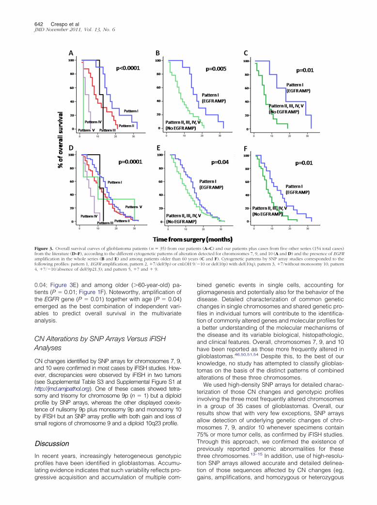
Overall, no clear association was found between thesecytogenetic profiles and other clinical variables, includ-ing tumor localization, except for patient survival. Accord-ingly, despite the dismal outcome observed in all cases,the cytogenetic profile of the tumor (as defined by thecytogenetic pattern of CN alterations observed for chro-mosomes 7, 9, and 10 by SNP arrays) had a significanteffect on overall survival (P � 0.0001) (Figure 3A): tumorswith EGFR gene amplification exhibited the longest (P �0.005) survival rates versus all other cases (Figure 3Band Table 4), specifically among older (�60-year-old)patients (P � 0.01). This also holds true when patients
Table 3. Continued
Dele
PTPLAD2 IFNA4 IFNA14 KLHL9
X X
X ● ● ●● ● ● ●● ● ● ●
XX
X X X X
Table 4. Cytogenetic Subgroups of Glioblastomas as Defined by10 and Association With Patient Overall Survival (n � 3
Pattern
Chromosomal abnormali
Chr7 Chr9
I EGFR AMP del(9p21) �10 ocnL
II �7 del(9p) or cnLOH �10 ocnL
III �7 del(9p21) or �9q No moIV �7 Normal 9p21.3 �10V �7 �9 �10
*Results are expressed as number of cases/total cases analyzed with†
Median (range) overall survival in months.B, biopsy; ST, subtotal; T, total.undergoing complete tumor resection and those not un-dergoing resection were separately considered (data notshown).
The presence and frequency of these five cytogeneticgroups were then confirmed among the 119 glioblastomacases collected from other series in the literature: pattern1, 61 of 119 cases (51%); pattern 2, 57 of 119 (45%);pattern 3, 0 of 119 (0%); pattern 4, 4 of 119 (3%); andpattern 5, 2 of 119 cases (2%) (see Supplemental TableS2 at http://jmd.amjpathol.org). In addition, the impact ofthe cytogenetic profiles on overall survival was also con-firmed when the whole series of glioblastoma patients(n � 154) was evaluated, both when the five differentcytogenetic subgroups were considered (P � 0.0001;Figure 3D) and when cases with EGFR amplification werecompared with all other cases in the whole series (P �
nes
MTAP CDKN2A CDKN2B ELAVL2
X XX X
● ● ● ●● ● ● ●● ● ●X X X X● ● ●● ● ● ●● ● ● ●X ● ●● X X● ● ●X X X
● ● ●X X ●● ●
X X X X● ● ●X X X
● ●
Alterations Detected by SNP Arrays for Chromosomes 7, 9, and
No. of cases(%)*
Overall survival(range)†Chr10
0p) and del(10q) or 10/35 (29) 16 (9–32)
0p) and del(10q) or 17/35 (48) 8 (0–21)
y 10 3/35 (9) 13 (11–67)4/35 (11) 4 (2–7)1/35 (3) 2
tages.
ted ge
the CN5)
ties
r del(1OHr del(1OHnosom
percen
LOH 9/�
642 Crespo et alJMD November 2011, Vol. 13, No. 6
0.04; Figure 3E) and among older (�60-year-old) pa-tients (P � 0.01; Figure 1F). Noteworthy, amplification ofthe EGFR gene (P � 0.01) together with age (P � 0.04)emerged as the best combination of independent vari-ables to predict overall survival in the multivariateanalysis.
CN Alterations by SNP Arrays Versus iFISHAnalyses
CN changes identified by SNP arrays for chromosomes 7, 9,and 10 were confirmed in most cases by iFISH studies. How-ever, discrepancies were observed by iFISH in two tumors(see Supplemental Table S3 and Supplemental Figure S1 athttp://jmd.amjpathol.org). One of these cases showed tetra-somy and trisomy for chromosome 9p (n � 1) but a diploidprofile by SNP arrays, whereas the other displayed coexis-tence of nulisomy 9p plus monosomy 9p and monosomy 10by iFISH but an SNP array profile with both gain and loss ofsmall regions of chromosome 9 and a diploid 10q23 profile.
Discussion
In recent years, increasingly heterogeneous genotypicprofiles have been identified in glioblastomas. Accumu-lating evidence indicates that such variability reflects pro-
Figure 3. Overall survival curves of glioblastoma patients (n � 35) from oufrom the literature (D–F), according to the different cytogenetic patterns of alamplification in the whole series (B and E) and among patients older thanfollowing profiles: pattern 1, EGFR amplification; pattern 2, �7/del(9p) or cn4, �7/�10/absence of del(9p21.3); and pattern 5, �7 and � 9.
gressive acquisition and accumulation of multiple com-
bined genetic events in single cells, accounting forgliomagenesis and potentially also for the behavior of thedisease. Detailed characterization of common geneticchanges in single chromosomes and shared genetic pro-files in individual tumors will contribute to the identifica-tion of commonly altered genes and molecular profiles fora better understanding of the molecular mechanisms ofthe disease and its variable biological, histopathologic,and clinical features. Overall, chromosomes 7, 9, and 10have been reported as those more frequently altered inglioblastomas.46,50,51,54 Despite this, to the best of ourknowledge, no study has attempted to classify glioblas-tomas on the basis of the distinct patterns of combinedalterations of these three chromosomes.
We used high-density SNP arrays for detailed charac-terization of those CN changes and genotypic profilesinvolving the three most frequently altered chromosomesin a group of 35 cases of glioblastomas. Overall, ourresults show that with very few exceptions, SNP arraysallow detection of underlying genetic changes of chro-mosomes 7, 9, and/or 10 whenever specimens contain75% or more tumor cells, as confirmed by iFISH studies.Through this approach, we confirmed the existence ofpreviously reported genomic abnormalities for thesethree chromosomes.13–15 In addition, use of high-resolu-tion SNP arrays allowed accurate and detailed delinea-tion of those sequences affected by CN changes (eg,
ts (A–C) and our patients plus cases from five other series (154 total cases)detected for chromosomes 7, 9, and 10 (A and D) and the presence of EGFR(C and F). Cytogenetic patterns by SNP array studies corresponded to the10 or del(10p) with del(10q); pattern 3, �7/without monosomy 10; pattern
r patienteration60 years
gains, amplifications, and homozygous or heterozygous
SNP Array Profiles in Glioblastomas 643JMD November 2011, Vol. 13, No. 6
deletions) and allelic imbalances (eg, LOH and cnLOH)and identification of the specific genes involved; overall,five different patterns of combined alterations for thesethree chromosomes were observed.
Noteworthy, gains of chromosome 7 were identified invirtually every case. This highlights the relevance of com-plete gains of this chromosome in the development ofglioblastomas and the potential pathogenic contributionof multiple oncogenes coded in it. In addition, we alsoconfirm and extend on previous observations that havesuggested that EGFR is the most frequently amplifiedoncogene in glioblastomas5,11,55,56 because EGFR wasthe only oncogene found to be amplified in common incases with multiple copies of the 7p11.2 chromosomalregion. Interestingly, precise localization of the EGFR am-plicons revealed amplification of other adjacent genes inmany cases, particularly the LANCL2 gene. Noteworthy,from those genes involved in 7p11.2 amplification, onlyEGFR showed increased expression in gene expressionprofiling,7,57 further supporting the unique and relevantrole of this oncogene in glioblastomas versus the othergenes (eg, LANCL2).
Regarding chromosome 9, more heterogeneous pat-terns of CN changes were observed, from whichheterozygous and/or homozygous del(9p) was the mostcommon alteration. Interestingly, although homozygousdeletions were restricted to relatively small sequences ofchromosome 9p21, heterozygous del(9p) extended tolarger chromosomal regions. Notably, common deletedsegments at 9p21 almost systematically involved theCDKN2B/p15 tumor suppressor gene in association withCDKN2A/p16 and the MTAP housekeeping genes.Del(9p21) is known to play an important role in the de-velopment and progression of many different types ofcancer through deregulation of cell cycle and/or apopto-sis.58 The CDKN2A locus has been claimed to play acrucial role in this regard. CDKN2A codes for two geneproducts, p16 and p14, that control both the Rb and thep53 pathways; p16 binds to CDK4 and CDK6 and inhibitsthe catalytic activity of CDK/cyclin D complexes to acti-vate cell cycle through RB phosphorylation. In turn, p14blocks MDM2 inhibition of p53 activity, thereby leading tostabilization of p53.11 Because deletion of the CDKN2A/Blocus causes deregulation of two crucial pathways in-volved in many types of cancer, loss of the MTAP geneactivity could be viewed as potentially irrelevant. How-ever, deficiency of the MTAP protein (an enzyme involvedin the metabolism of methionine and purines) has alsobeen detected in multiple types of malignant neoplasmsin association with deletion of the CDKN2A and CDKN2Bloci,59 as also found in our glioblastoma cases. Mostinterestingly, it has been shown that MTAP can be lostindependently of CDKN2A/p16, which suggests that lossof MTAP may indeed play a role in tumor biology.60–62
Taken together, these results raise the question aboutwhich of these three genes is/are critical target genes inglioblastomas. On the basis of our results, CDKN2A/p16and CDKN2B/p15 are the most frequently altered incases with heterozygous and homozygous deletions inline with previous large-scale multidimensional analyses
performed by The Cancer Genome Atlas Research Net-work.40 The CDKN2B gene (p15; INK4b) is located adja-cent to p16 (INK4a) on 9p21 and is co-deleted in a highproportion of human cancers. p15 (INK4b) is a memberof the family of cyclin-dependent protein kinases thatinhibits CDK4B. Because expression of CDKN2B is in-duced by transforming growth factor �, p15 may act asan effector of the transforming growth factor �–mediatedcell cycle arrest pathway. In line with our results, datafrom both mutational and functional studies indicate thatCDKN2B/p15 deletion could likely be the target ofdel(9p21).63
Regarding the specific mechanism by which thesegenes are inhibited, LOH at 9p21 was a relatively rareevent, whereas combined homozygous and heterozy-gous deletions (associated or not with cnLOH events)were relatively common in our and other studies64; thisfinding suggests that all three genes (CDKN2A/p16,CDKN2B/p15, and MTAP) may be inactivated in glioblas-tomas by a large deletion event. In line with this hypoth-esis, a large mapping study of 545 primary tumors65
showed that tumors containing homozygous del(9p21)minimally have a 170-kb region deleted that includesboth the MTAP and p16 loci, as also found here. How-ever, homozygous deletion does not seem to be the onlymechanism leading to inactivation of these tumor sup-pressor genes in glioblastomas because cases withheterozygous deletions were also found at higher fre-quency in our study. In another study on 85 brain tumorsamples of different histologic features and grade,CDKN2B/p15 and CDKN2A/p16 genes were found to bemethylated in only 4% and 7% of the cases, respectively;interestingly, CDKN2A was methylated only in glioblas-toma samples (6% of the cases), and none of the sam-ples showed simultaneous methylation of both the p15and p16 genes66; this finding suggests that methylationof these genes does not play a major role in the devel-opment of glioblastomas. Interestingly, however, geneexpression profiling of glioblastomas shows a significantimpact on the expression of CDKN2A in cases with notonly homozygous but also heterozygous del(9p21),whereas this does not affect the expression of the othertwo genes (data not shown). In any case, point mutationsof these genes should be investigated in parallel in thesecases. Because emerging CN analyses of glioblastomasamples confirmed the CDKN2A/CDKN2B locus to be themost common homozygous deletion at 9p21, detailedcharacterization of the deletion at chromosome 9p21 andthe lost genes becomes particularly relevant. In thisstudy, detailed mapping of the 9p21.3 region shows dis-tinct patterns and extents of del(9p21) among the tumorsanalyzed. In addition, our results also show that the de-leted locus encompassed not only genes with well-estab-lished tumor suppressor functions in glioblastomas butalso multiple other less known genes (eg, the ELAVL2,MLLT3, KIAA1797, PTPLAD2, and KLHL9 genes). Thesefindings strengthen the hypothesis that suggests thepresence of additional candidate tumor suppressorgenes mapped to this region.67
Overall, approximately three-quarters of all glioblasto-mas analyzed showed chromosome 10 losses, which
most frequently consisted of monosomy 10 and cnLOH of
644 Crespo et alJMD November 2011, Vol. 13, No. 6
an entire chromosome 10. These findings point to the lossof more than one tumor suppressor gene, localized bothin the short and the long arms of this chromosome. In thisregard, extensive losses of chromosome 10 sequenceshave been associated with progression of astrocytoma,68
and several regions along this chromosome (eg, 10q23,10q24, 10q25-26, 10p13, and 10p14-p15) have beenconsistently proposed to harbor tumor suppressor genes(eg, the PTEN/MMAC1, DMBT1, and LGI1 genes).69 Al-though it has been previously suggested that the PTENgene could be a preferential target of del(10q),70 in ourseries, losses of chromosome 10 mainly involved theentire chromosome. Despite this, our results highlight thefact that other regions at 10q11.21, 10q21.3, and10q.23.33 (with loss of the HNRPF, PAKDB, and CUL2genes, the CXXCC, CCPRL1, STOX1, and DDX50 genes;and the IRE gene, respectively) were more frequently lostand could act as potential preferential targets of deletionin glioblastomas. Likewise, those genes encompassedwithin these deleted loci could also represent novel can-didate tumor suppressor genes involved in glioblastomatumorigenesis, in addition to PTEN.
In this study, as in other larger series of glioblasto-mas,14,18,19,27,71 gains of chromosome 7 and losses ofchromosomes 9 and 10 frequently coexisted in the sametumor, but different patterns were observed for theseabnormalities. Accordingly, glioblastomas that exhibitedEGFR gene amplification also showed extensive losses ofchromosome 10, del(9p21), and trisomy 7 in all but onecase. Conversely, in more than half of the cases, mono-somy 10 coexisted with trisomy 7 in the absence of EGFRgene amplification with or without del(9p21). Altogether,these findings suggest that these alterations may occurindependently from each other, with EGFR amplificationappearing to be a later event in the development ofglioblastomas versus trisomy 7 and monosomy 10. Nev-ertheless, their combination could be crucial in the ma-lignant transformation process for which the underlyingmechanism is still poorly understood. In this regard, sev-eral candidate genes in chromosome 10 with putativereciprocal relationship to EGFR have been identified, withgreat emphasis on the PTEN gene. Complementary de-regulation of the EGFR and PTEN pathways often resultsin constitutional signaling through PI3-kinase and Akt,leading to altered cell proliferation and survival.72 A re-cent study by Yadav et al28 also suggests a tumorigenicsynergism between loss of the annexin A7 (ANXA7) geneat 10q21.1-q21.2 and EGFR amplification, with ANXA7haploinsufficiency acting as a positive regulator of EGFRsignaling in glioblastomas. This study also demonstratesa cross-talk among the ANXA7, PTEN, and EGFR genes,which leads to constitutive activation of the PI3K-AKTsignaling pathway and, ultimately, to malignant transfor-mation. Taken together, these findings suggest that cy-togenetic profiles, more than isolated chromosomal alter-ations, should be considered in evaluating the impact ofCN alterations in disease behavior.
On the basis of CN alterations of chromosomes 7, 9,and 10, five different genetic profiles were identified inour series and confirmed to be present in other series
from the literature41,42,45,50 from which cases with ampli-fication of the EGFR gene, in association with monosomy10 and del(9p21), clearly showed a better outcome in our35 cases and when data on 119 additional glioblastomapatients from four previously reported series41,42,45,50
were considered. Controversial results have been re-ported about the prognostic value of EGFR amplification/overexpression in glioblastomas. Although some authorsclaim there is no association with survival,73,74 othersstate that this aberration is a negative prognostic fac-tor.75,76 In turn, an association between EGFR overex-pression and a better prognosis in older glioblastomapatients has also been reported,25,26,33,77 in line with ourobservations. Noteworthy, we did not find an associationbetween tumor cytogenetics and other disease charac-teristics, such as patient age76 and tumor localization,among other features.55,78
Simmons et al78 and Batchelor et al5 have previouslyfound that EGFR overexpression is associated with atrend toward a worse prognosis in young patients and abetter outcome in older cases; likewise, in a series of 220primary glioblastomas Houillier et al55 also documentedan association between EGFR amplification and in-creased survival in older patients, which could be asso-ciated with the existence of additional as-yet-unidentifiedspecific molecular alterations in older patients. In thepresent study, we confirm the prognostic value of EGFRamplification in patients older than 60 years in oursmall patient series and in a larger series of patientsfrom four independent studies previously reported inthe literature.5,55,78,79
In summary, our high-density analysis of the CN alter-ations of chromosomes 7, 9, and 10 disclosed five sub-groups of patients defined by unique cytogenetic pro-files, which are associated with patient outcome, withtumors with EGFR amplification showing a longer overallsurvival among older patients. In addition, our resultsprovide further evidence about the relevance of theEGFR, CDKN2A/B, and MTAP genes, together with othergenes coded in chromosome 10, in the malignant trans-formation of glioblastomas. Further studies in larger se-ries of glioblastoma patients are necessary to investigatethe functional interaction between these genes and moreprecisely delineate their pathogenetic role and clinicalimpact in glioblastomas.
Acknowledgments
We thank Dr. Pim J. French (Josephine Nefkens Institute,Department of Neurology, Erasmus Medical Center, Rot-terdam, The Netherlands) for his valuable collaborationwith 21.CEL files and patient survival data.41
References
1. DeAngelis LM: Brain tumors. N Engl J Med 2001, 344:114–1232. Malmer B, Iselius L, Holmberg E, Collins A, Henriksson R, Gronberg
H: Genetic epidemiology of glioma. Br J Cancer 2001, 84:429–4343. Louis DN, Ohgaki H, Wiestler OD, Cavenee WK, Burger PC, Jouvet A,
Scheithauer BW, Kleihues P: The 2007 WHO classification of tumoursof the central nervous system. Acta Neuropathol 2007, 114:97–109
SNP Array Profiles in Glioblastomas 645JMD November 2011, Vol. 13, No. 6
4. Lamborn KR, Chang SM, Prados MD: Prognostic factors for survivalof patients with glioblastoma: recursive partitioning analysis. NeuroOncol 2004, 6:227–235
5. Batchelor TT, Betensky RA, Esposito JM, Pham LD, Dorfman MV,Piscatelli N, Jhung S, Rhee D, Louis DN: Age-dependent prognosticeffects of genetic alterations in glioblastoma. Clin Cancer Res 2004,10:228–233
6. Bussiere M, Hopman W, Day A, Pombo AP, Neves T, Espinosa F:Indicators of functional status for primary malignant brain tumourpatients. Can J Neurol Sci 2005, 32:50–56
7. Mischel PS, Nelson SF, Cloughesy TF: Molecular analysis ofglioblastoma: pathway profiling and its implications for patient ther-apy. Cancer Biol Ther 2003, 2:242–247
8. Hassler M, Seidl S, Fazeny-Doerner B, Preusser M, Hainfellner J,Rossler K, Prayer D, Marosi C: Diversity of cytogenetic and pathohis-tologic profiles in glioblastoma. Cancer Genet Cytogenet 2006, 166:46–55
9. Nakamura M, Shimada K, Ishida E, Nakase H, Konishi N: Geneticanalysis to complement histopathological diagnosis of brain tumors.Histol Histopathol 2007, 22:327–335
10. Louis DN: Molecular pathology of malignant gliomas. Annu RevPathol 2006, 1:97–117
11. Kanu OO, Hughes B, Di C, Lin N, Fu J, Bigner DD, Yan H, AdamsonC: Glioblastoma multiforme oncogenomics and signaling pathways.Clin Med Oncol 2009, 3:39–52
12. Ohgaki H, Kleihues P: Genetic alterations and signaling pathways inthe evolution of gliomas. Cancer Sci 2009, 100:2235–2241
13. Vranova V, Necesalova E, Kuglik P, Cejpek P, Pesakova M, BudinskaE, Relichova J, Veselska R: Screening of genomic imbalances inglioblastoma multiforme using high-resolution comparative genomichybridization. Oncol Rep 2007, 17:457–464
14. Dahlback HS, Brandal P, Meling TR, Gorunova L, Scheie D, Heim S:Genomic aberrations in 80 cases of primary glioblastoma multiforme:pathogenetic heterogeneity and putative cytogenetic pathways.Genes Chromosomes Cancer 2009, 48:908–924
15. Parsons DW, Jones S, Zhang X, Lin JC, Leary RJ, Angenendt P,Mankoo P, Carter H, Siu IM, Gallia GL, Olivi A, McLendon R, RasheedBA, Keir S, Nikolskaya T, Nikolsky Y, Busam DA, Tekleab H, Diaz LA,Jr., Hartigan J, Smith DR, Strausberg RL, Marie SK, Shinjo SM, Yan H,Riggins GJ, Bigner DD, Karchin R, Papadopoulos N, Parmigiani G,Vogelstein B, Velculescu VE, Kinzler KW: An integrated genomicanalysis of human glioblastoma multiforme. Science 2008, 321:1807–1812
16. Vital AL, Tabernero MD, Crespo I, Rebelo O, Tao H, Gomes F, LopesMC, Orfao A: Intratumoral patterns of clonal evolution in gliomas.Neurogenetics 2010, 11:227–239
17. Misra A, Pellarin M, Nigro J, Smirnov I, Moore D, Lamborn KR, PinkelD, Albertson DG, Feuerstein BG: Array comparative genomic hybrid-ization identifies genetic subgroups in grade 4 human astrocytoma.Clin Cancer Res 2005, 11:2907–2918
18. Korshunov A, Sycheva R, Golanov A: Genetically distinct and clini-cally relevant subtypes of glioblastoma defined by array-based com-parative genomic hybridization (array-CGH). Acta Neuropathol 2006,111:465–474
19. Mohapatra G, Bollen AW, Kim DH, Lamborn K, Moore DH, PradosMD, Feuerstein BG: Genetic analysis of glioblastoma multiforme pro-vides evidence for subgroups within the grade. Genes ChromosomesCancer 1998, 21:195–206
20. Viana-Pereira M, Lopes JM, Little S, Milanezi F, Basto D, Pardal F,Jones C, Reis RM: Analysis of EGFR overexpression. EGFR geneamplification and the EGFRvIII mutation in Portuguese high-gradegliomas. Anticancer Res 2008, 28:913–920
21. Liu L, Backlund LM, Nilsson BR, Grander D, Ichimura K, Goike HM,Collins VP: Clinical significance of EGFR amplification and the aber-rant EGFRvIII transcript in conventionally treated astrocytic gliomas. JMol Med 2005, 83:917–926
22. Nicholas MK, Lukas RV, Jafri NF, Faoro L, Salgia R: Epidermal growthfactor receptor– mediated signal transduction in the developmentand therapy of gliomas. Clin Cancer Res 2006, 12:7261–7270
23. Wemmert S, Ketter R, Rahnenfuhrer J, Beerenwinkel N, Strowitzki M,Feiden W, Hartmann C, Lengauer T, Stockhammer F, Zang KD,Meese E, Steudel WI, von Deimling A, Urbschat S: Patients with
high-grade gliomas harboring deletions of chromosomes 9p and 10qbenefit from temozolomide treatment. Neoplasia 2005, 7:883–89324. Tada K, Shiraishi S, Kamiryo T, Nakamura H, Hirano H, Kuratsu J,Kochi M, Saya H, Ushio Y: Analysis of loss of heterozygosity onchromosome 10 in patients with malignant astrocytic tumors: corre-lation with patient age and survival. J Neurosurg 2001, 95:651–659
25. Daido S, Takao S, Tamiya T, Ono Y, Terada K, Ito S, Ouchida M, DateI, Ohmoto T, Shimizu K: Loss of heterozygosity on chromosome 10qassociated with malignancy and prognosis in astrocytic tumors, anddiscovery of novel loss regions. Oncol Rep 2004, 12:789–795
26. Brat DJ, Seiferheld WF, Perry A, Hammond EH, Murray KJ, Schuls-inger AR, Mehta MP, Curran WJ: Analysis of 1p, 19q, 9p, and 10q asprognostic markers for high-grade astrocytomas using fluorescencein situ hybridization on tissue microarrays from Radiation TherapyOncology Group trials. Neuro Oncol 2004, 6:96–103
27. Lopez-Gines C, Cerda-Nicolas M, Gil-Benso R, Pellin A, Lopez-Guer-rero JA, Callaghan R, Benito R, Roldan P, Piquer J, Llacer J, BarberaJ: Association of chromosome 7, chromosome 10 and EGFR geneamplification in glioblastoma multiforme. Clin Neuropathol 2005, 24:209–218
28. Yadav AK, Renfrow JJ, Scholtens DM, Xie H, Duran GE, Bredel C,Vogel H, Chandler JP, Chakravarti A, Robe PA, Das S, Scheck AC,Kessler JA, Soares MB, Sikic BI, Harsh GR, Bredel M: Monosomy ofchromosome 10 associated with dysregulation of epidermal growthfactor signaling in glioblastomas. JAMA 2009, 302:276–289
29. Gil-Perotin S, Marin-Husstege M, Li J, Soriano-Navarro M, Zindy F,Roussel MF, Garcia-Verdugo JM, Casaccia-Bonnefil P: Loss of p53induces changes in the behavior of subventricular zone cells: impli-cation for the genesis of glial tumors. J Neurosci 2006, 26:1107–1116
30. von Deimling A, Eibl RH, Ohgaki H, Louis DN, von Ammon K, Pe-tersen I, Kleihues P, Chung RY, Wiestler OD, Seizinger BR: p53mutations are associated with 17p allelic loss in grade II and grade IIIastrocytoma. Cancer Res 1992, 52:2987–2990
31. Vordermark D, Ruprecht K, Rieckmann P, Roggendorf W, Vince GH,Warmuth-Metz M, Kolbl O, Flentje M: Glioblastoma multiforme witholigodendroglial component (GBMO): favorable outcome after post-operative radiotherapy and chemotherapy with nimustine (ACNU)and teniposide (VM26). BMC Cancer 2006, 6:247
32. Salvati M, Formichella AI, D’Elia A, Brogna C, Frati A, Giangaspero F,Delfini R, Santoro A: Cerebral glioblastoma with oligodendrogliomalcomponent: analysis of 36 cases. J Neurooncol 2009, 94:129–134
33. Schmidt MC, Antweiler S, Urban N, Mueller W, Kuklik A, Meyer-PuttlitzB, Wiestler OD, Louis DN, Fimmers R, von Deimling A: Impact ofgenotype and morphology on the prognosis of glioblastoma. J Neu-ropathol Exp Neurol 2002, 61:321–328
34. Shiraishi S, Tada K, Nakamura H, Makino K, Kochi M, Saya H, KuratsuJ, Ushio Y: Influence of p53 mutations on prognosis of patients withglioblastoma. Cancer 2002, 95:249–257
35. Kato H, Kato S, Kumabe T, Sonoda Y, Yoshimoto T, Han SY, SuzukiT, Shibata H, Kanamaru R, Ishioka C: Functional evaluation of p53and PTEN gene mutations in gliomas. Clin Cancer Res 2000, 6:3937–3943
36. Bredel M, Bredel C, Juric D, Harsh GR, Vogel H, Recht LD, Sikic BI:High-resolution genome-wide mapping of genetic alterations in hu-man glial brain tumors. Cancer Res 2005, 65:4088–4096
37. Arslantas A, Artan S, Oner U, Muslumanoglu H, Durmaz R, Cosan E,Atasoy MA, Basaran N, Tel E: The importance of genomic copynumber changes in the prognosis of glioblastoma multiforme. Neu-rosurg Rev 2004, 27:58–64
38. Verhaak RG, Hoadley KA, Purdom E, Wang V, Qi Y, Wilkerson MD,Miller CR, Ding L, Golub T, Mesirov JP, Alexe G, Lawrence M, O’KellyM, Tamayo P, Weir BA, Gabriel S, Winckler W, Gupta S, Jakkula L,Feiler HS, Hodgson JG, James CD, Sarkaria JN, Brennan C, Kahn A,Spellman PT, Wilson RK, Speed TP, Gray JW, Meyerson M, Getz G,Perou CM, Hayes DN: Integrated genomic analysis identifies clini-cally relevant subtypes of glioblastoma characterized by abnormali-ties in PDGFRA. IDH1, EGFR, and NF1. Cancer Cell 2010, 17:98–110
39. Beroukhim R, Mermel CH, Porter D, Wei G, Raychaudhuri S, DonovanJ, Barretina J, Boehm JS, Dobson J, Urashima M, Mc Henry KT,Pinchback RM, Ligon AH, Cho YJ, Haery L, Greulich H, Reich M,Winckler W, Lawrence MS, Weir BA, Tanaka KE, Chiang DY, Bass AJ,Loo A, Hoffman C, Prensner J, Liefeld T, Gao Q, Yecies D, SignorettiS, Maher E, Kaye FJ, Sasaki H, Tepper JE, Fletcher JA, Tabernero J,Baselga J, Tsao MS, Demichelis F, Rubin MA, Janne PA, Daly MJ,
Nucera C, Levine RL, Ebert BL, Gabriel S, Rustgi AK, Antonescu CR,Ladanyi M, Letai A, Garraway LA, Loda M, Beer DG, True LD, Oka-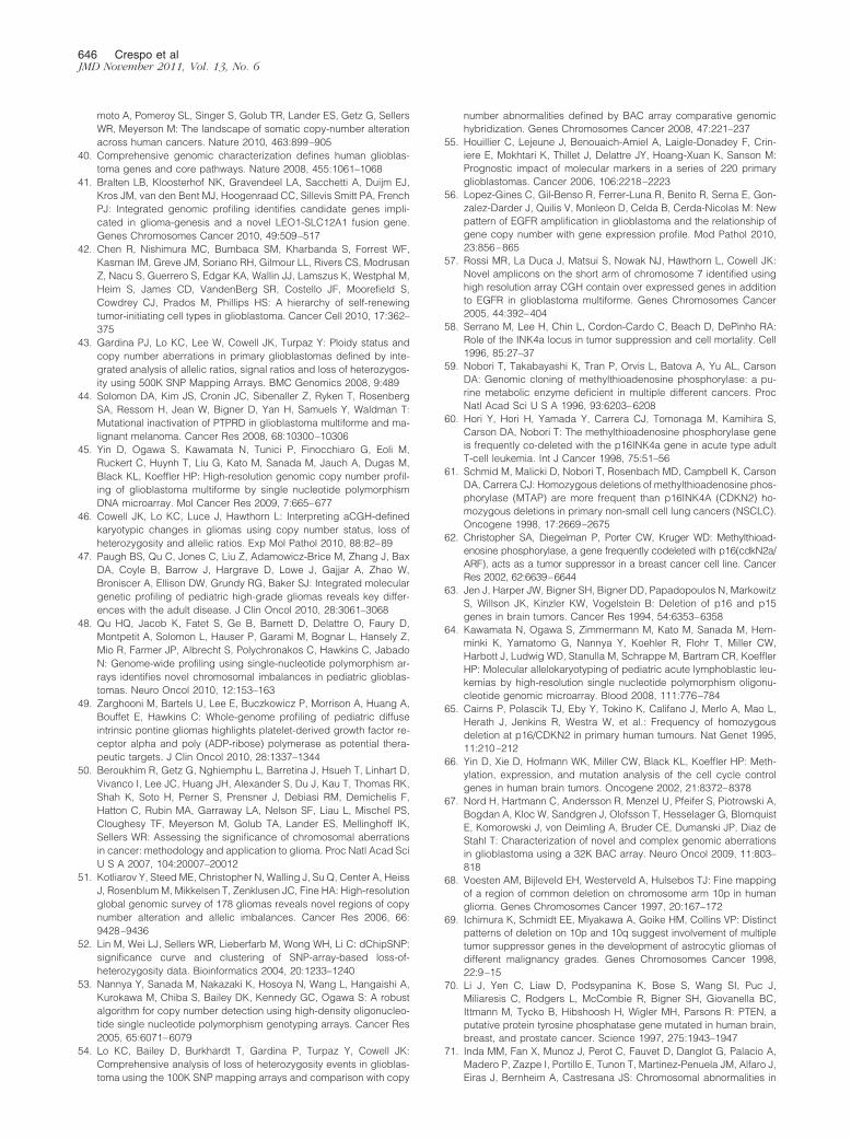
646 Crespo et alJMD November 2011, Vol. 13, No. 6
moto A, Pomeroy SL, Singer S, Golub TR, Lander ES, Getz G, SellersWR, Meyerson M: The landscape of somatic copy-number alterationacross human cancers. Nature 2010, 463:899–905
40. Comprehensive genomic characterization defines human glioblas-toma genes and core pathways. Nature 2008, 455:1061–1068
41. Bralten LB, Kloosterhof NK, Gravendeel LA, Sacchetti A, Duijm EJ,Kros JM, van den Bent MJ, Hoogenraad CC, Sillevis Smitt PA, FrenchPJ: Integrated genomic profiling identifies candidate genes impli-cated in glioma-genesis and a novel LEO1-SLC12A1 fusion gene.Genes Chromosomes Cancer 2010, 49:509–517
42. Chen R, Nishimura MC, Bumbaca SM, Kharbanda S, Forrest WF,Kasman IM, Greve JM, Soriano RH, Gilmour LL, Rivers CS, ModrusanZ, Nacu S, Guerrero S, Edgar KA, Wallin JJ, Lamszus K, Westphal M,Heim S, James CD, VandenBerg SR, Costello JF, Moorefield S,Cowdrey CJ, Prados M, Phillips HS: A hierarchy of self-renewingtumor-initiating cell types in glioblastoma. Cancer Cell 2010, 17:362–375
43. Gardina PJ, Lo KC, Lee W, Cowell JK, Turpaz Y: Ploidy status andcopy number aberrations in primary glioblastomas defined by inte-grated analysis of allelic ratios, signal ratios and loss of heterozygos-ity using 500K SNP Mapping Arrays. BMC Genomics 2008, 9:489
44. Solomon DA, Kim JS, Cronin JC, Sibenaller Z, Ryken T, RosenbergSA, Ressom H, Jean W, Bigner D, Yan H, Samuels Y, Waldman T:Mutational inactivation of PTPRD in glioblastoma multiforme and ma-lignant melanoma. Cancer Res 2008, 68:10300–10306
45. Yin D, Ogawa S, Kawamata N, Tunici P, Finocchiaro G, Eoli M,Ruckert C, Huynh T, Liu G, Kato M, Sanada M, Jauch A, Dugas M,Black KL, Koeffler HP: High-resolution genomic copy number profil-ing of glioblastoma multiforme by single nucleotide polymorphismDNA microarray. Mol Cancer Res 2009, 7:665–677
46. Cowell JK, Lo KC, Luce J, Hawthorn L: Interpreting aCGH-definedkaryotypic changes in gliomas using copy number status, loss ofheterozygosity and allelic ratios. Exp Mol Pathol 2010, 88:82–89
47. Paugh BS, Qu C, Jones C, Liu Z, Adamowicz-Brice M, Zhang J, BaxDA, Coyle B, Barrow J, Hargrave D, Lowe J, Gajjar A, Zhao W,Broniscer A, Ellison DW, Grundy RG, Baker SJ: Integrated moleculargenetic profiling of pediatric high-grade gliomas reveals key differ-ences with the adult disease. J Clin Oncol 2010, 28:3061–3068
48. Qu HQ, Jacob K, Fatet S, Ge B, Barnett D, Delattre O, Faury D,Montpetit A, Solomon L, Hauser P, Garami M, Bognar L, Hansely Z,Mio R, Farmer JP, Albrecht S, Polychronakos C, Hawkins C, JabadoN: Genome-wide profiling using single-nucleotide polymorphism ar-rays identifies novel chromosomal imbalances in pediatric glioblas-tomas. Neuro Oncol 2010, 12:153–163
49. Zarghooni M, Bartels U, Lee E, Buczkowicz P, Morrison A, Huang A,Bouffet E, Hawkins C: Whole-genome profiling of pediatric diffuseintrinsic pontine gliomas highlights platelet-derived growth factor re-ceptor alpha and poly (ADP-ribose) polymerase as potential thera-peutic targets. J Clin Oncol 2010, 28:1337–1344
50. Beroukhim R, Getz G, Nghiemphu L, Barretina J, Hsueh T, Linhart D,Vivanco I, Lee JC, Huang JH, Alexander S, Du J, Kau T, Thomas RK,Shah K, Soto H, Perner S, Prensner J, Debiasi RM, Demichelis F,Hatton C, Rubin MA, Garraway LA, Nelson SF, Liau L, Mischel PS,Cloughesy TF, Meyerson M, Golub TA, Lander ES, Mellinghoff IK,Sellers WR: Assessing the significance of chromosomal aberrationsin cancer: methodology and application to glioma. Proc Natl Acad SciU S A 2007, 104:20007–20012
51. Kotliarov Y, Steed ME, Christopher N, Walling J, Su Q, Center A, HeissJ, Rosenblum M, Mikkelsen T, Zenklusen JC, Fine HA: High-resolutionglobal genomic survey of 178 gliomas reveals novel regions of copynumber alteration and allelic imbalances. Cancer Res 2006, 66:9428–9436
52. Lin M, Wei LJ, Sellers WR, Lieberfarb M, Wong WH, Li C: dChipSNP:significance curve and clustering of SNP-array-based loss-of-heterozygosity data. Bioinformatics 2004, 20:1233–1240
53. Nannya Y, Sanada M, Nakazaki K, Hosoya N, Wang L, Hangaishi A,Kurokawa M, Chiba S, Bailey DK, Kennedy GC, Ogawa S: A robustalgorithm for copy number detection using high-density oligonucleo-tide single nucleotide polymorphism genotyping arrays. Cancer Res2005, 65:6071–6079
54. Lo KC, Bailey D, Burkhardt T, Gardina P, Turpaz Y, Cowell JK:Comprehensive analysis of loss of heterozygosity events in glioblas-
toma using the 100K SNP mapping arrays and comparison with copynumber abnormalities defined by BAC array comparative genomichybridization. Genes Chromosomes Cancer 2008, 47:221–237
55. Houillier C, Lejeune J, Benouaich-Amiel A, Laigle-Donadey F, Crin-iere E, Mokhtari K, Thillet J, Delattre JY, Hoang-Xuan K, Sanson M:Prognostic impact of molecular markers in a series of 220 primaryglioblastomas. Cancer 2006, 106:2218–2223
56. Lopez-Gines C, Gil-Benso R, Ferrer-Luna R, Benito R, Serna E, Gon-zalez-Darder J, Quilis V, Monleon D, Celda B, Cerda-Nicolas M: Newpattern of EGFR amplification in glioblastoma and the relationship ofgene copy number with gene expression profile. Mod Pathol 2010,23:856–865
57. Rossi MR, La Duca J, Matsui S, Nowak NJ, Hawthorn L, Cowell JK:Novel amplicons on the short arm of chromosome 7 identified usinghigh resolution array CGH contain over expressed genes in additionto EGFR in glioblastoma multiforme. Genes Chromosomes Cancer2005, 44:392–404
58. Serrano M, Lee H, Chin L, Cordon-Cardo C, Beach D, DePinho RA:Role of the INK4a locus in tumor suppression and cell mortality. Cell1996, 85:27–37
59. Nobori T, Takabayashi K, Tran P, Orvis L, Batova A, Yu AL, CarsonDA: Genomic cloning of methylthioadenosine phosphorylase: a pu-rine metabolic enzyme deficient in multiple different cancers. ProcNatl Acad Sci U S A 1996, 93:6203–6208
60. Hori Y, Hori H, Yamada Y, Carrera CJ, Tomonaga M, Kamihira S,Carson DA, Nobori T: The methylthioadenosine phosphorylase geneis frequently co-deleted with the p16INK4a gene in acute type adultT-cell leukemia. Int J Cancer 1998, 75:51–56
61. Schmid M, Malicki D, Nobori T, Rosenbach MD, Campbell K, CarsonDA, Carrera CJ: Homozygous deletions of methylthioadenosine phos-phorylase (MTAP) are more frequent than p16INK4A (CDKN2) ho-mozygous deletions in primary non-small cell lung cancers (NSCLC).Oncogene 1998, 17:2669–2675
62. Christopher SA, Diegelman P, Porter CW, Kruger WD: Methylthioad-enosine phosphorylase, a gene frequently codeleted with p16(cdkN2a/ARF), acts as a tumor suppressor in a breast cancer cell line. CancerRes 2002, 62:6639–6644
63. Jen J, Harper JW, Bigner SH, Bigner DD, Papadopoulos N, MarkowitzS, Willson JK, Kinzler KW, Vogelstein B: Deletion of p16 and p15genes in brain tumors. Cancer Res 1994, 54:6353–6358
64. Kawamata N, Ogawa S, Zimmermann M, Kato M, Sanada M, Hem-minki K, Yamatomo G, Nannya Y, Koehler R, Flohr T, Miller CW,Harbott J, Ludwig WD, Stanulla M, Schrappe M, Bartram CR, KoefflerHP: Molecular allelokaryotyping of pediatric acute lymphoblastic leu-kemias by high-resolution single nucleotide polymorphism oligonu-cleotide genomic microarray. Blood 2008, 111:776–784
65. Cairns P, Polascik TJ, Eby Y, Tokino K, Califano J, Merlo A, Mao L,Herath J, Jenkins R, Westra W, et al.: Frequency of homozygousdeletion at p16/CDKN2 in primary human tumours. Nat Genet 1995,11:210–212
66. Yin D, Xie D, Hofmann WK, Miller CW, Black KL, Koeffler HP: Meth-ylation, expression, and mutation analysis of the cell cycle controlgenes in human brain tumors. Oncogene 2002, 21:8372–8378
67. Nord H, Hartmann C, Andersson R, Menzel U, Pfeifer S, Piotrowski A,Bogdan A, Kloc W, Sandgren J, Olofsson T, Hesselager G, BlomquistE, Komorowski J, von Deimling A, Bruder CE, Dumanski JP, Diaz deStahl T: Characterization of novel and complex genomic aberrationsin glioblastoma using a 32K BAC array. Neuro Oncol 2009, 11:803–818
68. Voesten AM, Bijleveld EH, Westerveld A, Hulsebos TJ: Fine mappingof a region of common deletion on chromosome arm 10p in humanglioma. Genes Chromosomes Cancer 1997, 20:167–172
69. Ichimura K, Schmidt EE, Miyakawa A, Goike HM, Collins VP: Distinctpatterns of deletion on 10p and 10q suggest involvement of multipletumor suppressor genes in the development of astrocytic gliomas ofdifferent malignancy grades. Genes Chromosomes Cancer 1998,22:9–15
70. Li J, Yen C, Liaw D, Podsypanina K, Bose S, Wang SI, Puc J,Miliaresis C, Rodgers L, McCombie R, Bigner SH, Giovanella BC,Ittmann M, Tycko B, Hibshoosh H, Wigler MH, Parsons R: PTEN, aputative protein tyrosine phosphatase gene mutated in human brain,breast, and prostate cancer. Science 1997, 275:1943–1947
71. Inda MM, Fan X, Munoz J, Perot C, Fauvet D, Danglot G, Palacio A,
Madero P, Zazpe I, Portillo E, Tunon T, Martinez-Penuela JM, Alfaro J,Eiras J, Bernheim A, Castresana JS: Chromosomal abnormalities in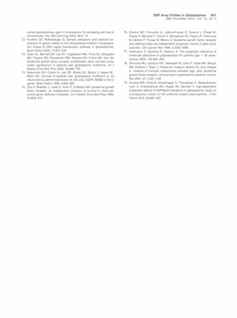
SNP Array Profiles in Glioblastomas 647JMD November 2011, Vol. 13, No. 6
human glioblastomas: gain in chromosome 7p correlating with loss inchromosome 10q. Mol Carcinog 2003, 36:6–14
72. Knobbe CB, Reifenberger G: Genetic alterations and aberrant ex-pression of genes related to the phosphatidyl-inositol-3=-kinase/pro-tein kinase B (Akt) signal transduction pathway in glioblastomas.Brain Pathol 2003, 13:507–518
73. Quan AL, Barnett GH, Lee SY, Vogelbaum MA, Toms SA, StaugaitisSM, Prayson RA, Peereboom DM, Stevens GH, Cohen BH, Suh JH:Epidermal growth factor receptor amplification does not have prog-nostic significance in patients with glioblastoma multiforme. Int JRadiat Oncol Biol Phys 2005, 63:695–703
74. Newcomb EW, Cohen H, Lee SR, Bhalla SK, Bloom J, Hayes RL,Miller DC: Survival of patients with glioblastoma multiforme is notinfluenced by altered expression of p16, p53. EGFR, MDM2 or Bcl-2genes. Brain Pathol 1998, 8:655–667
75. Zhu A, Shaeffer J, Leslie S, Kolm P, El-Mahdi AM: Epidermal growthfactor receptor: an independent predictor of survival in astrocytic
tumors given definitive irradiation. Int J Radiat Oncol Biol Phys 1996,34:809–81576. Etienne MC, Formento JL, Lebrun-Frenay C, Gioanni J, Chatel M,Paquis P, Bernard C, Courdi A, Bensadoun RJ, Pignol JP, FrancoualM, Grellier P, Frenay M, Milano G: Epidermal growth factor receptorand labeling index are independent prognostic factors in glial tumoroutcome. Clin Cancer Res 1998, 4:2383–2390
77. Korshunov A, Sycheva R, Golanov A: The prognostic relevance ofmolecular alterations in glioblastomas for patients age � 50 years.Cancer 2005, 104:825–832
78. Simmons ML, Lamborn KR, Takahashi M, Chen P, Israel MA, BergerMS, Godfrey T, Nigro J, Prados M, Chang S, Barker FG, 2nd, AldapeK: Analysis of complex relationships between age, p53, epidermalgrowth factor receptor, and survival in glioblastoma patients. CancerRes 2001, 61:1122–1128
79. Srividya MR, Thota B, Arivazhagan A, Thennarasu K, Balasubrama-niam A, Chandramouli BA, Hegde AS, Santosh V: Age-dependentprognostic effects of EGFR/p53 alterations in glioblastoma: study ona prospective cohort of 140 uniformly treated adult patients. J Clin
Pathol 2010, 63:687–691