Embed Size (px)
Citation preview

International Journal of
Molecular Sciences
Article
Design, Synthesis, and Cytotoxic Analysis of NovelHederagenin–Pyrazine Derivatives Based on PartialLeast Squares Discriminant Analysis
Kang Fang, Xiao-Hua Zhang, Yao-Tian Han, Gao-Rong Wu, De-Sheng Cai, Nan-Nan Xue,Wen-Bo Guo, Yu-Qin Yang, Meng Chen, Xin-Yu Zhang, Hui Wang, Tao Ma, Peng-Long Wang *and Hai-Min Lei *
School of Chinese Pharmacy, Beijing University of Chinese Medicine, Beijing 100102, China;[email protected] (K.F.); [email protected] (X.-H.Z.); [email protected] (Y.-T.H.);[email protected] (G.-R.W.); [email protected] (D.-S.C.); [email protected] (N.-N.X.);[email protected] (W.-B.G.); [email protected] (Y.-Q.Y.); [email protected] (M.C.);[email protected] (X.-Y.Z.); [email protected] (H.W.); [email protected] (T.M.)* Correspondence: [email protected] (P.-L.W.); [email protected] (H.-M.L.)
Received: 16 August 2018; Accepted: 25 September 2018; Published: 30 September 2018�����������������
Abstract: Hederagenin (He) is a novel triterpene template for the development of new antitumorcompounds. In this study, 26 new He–pyrazine derivatives were synthetized in an attempt todevelop potent antitumor agents; they were screened for in vitro cytotoxicity against tumor andnon-tumor cell lines. The majority of these derivatives showed much stronger cytotoxic activity thanHe. Remarkably, the most potent was compound 9 (half maximal inhibitory concentration (IC50) was3.45 ± 0.59 µM), which exhibited similar antitumor activities against A549 (human non-small-celllung cancer) as the positive drug cisplatin (DDP; IC50 was 3.85 ± 0.63 µM), while it showed lowercytotoxicity on H9c2 (murine heart myoblast; IC50 was 16.69 ± 0.12 µM) cell lines. Compound 9could induce the early apoptosis and evoke cell-cycle arrest at the synthesis (S) phase of A549 cells.Impressively, we innovatively introduced the method of cluster analysis modeled as partial leastsquares discriminant analysis (PLS-DA) into the structure–activity relationship (SAR) evaluation,and SAR confirmed that pyrazine had a profound effect on the antitumor activity of He. The presentstudies highlight the importance of pyrazine derivatives of He in the discovery and development ofnovel antitumor agents.
Keywords: hederagenin; PLS-DA; antitumor activity; pyrazine; apoptosis
1. Introduction
Natural molecules are an indispensable source of lead compounds for drug discovery [1–3].Pentacyclic triterpenes are widely present in medicinal plants, and they show a unique range ofpharmacological activities, including great antitumor, anti-inflammatory, and antiviral activities [4–6].They attracted the attention of scholars due to their enormous chemopreventive and antineoplasticpotential [7,8]. In view of this, the structural modification of pentacyclic triterpenoids is alwaysa focus in drug research [9–11]. Hederagenin (He, 3b,23-dihydroxyurs-12-ene-28-oic acid; Figure 1) isa member of the pentacyclic triterpenoid family, which is extracted mainly from the medicinal plantivy (Hedera helix L.) and Dipsacus asper [12,13]. A variety of pharmacological activities were ascribedto He, such as anticancer [14], anti-inflammatory [15], neuroprotective [16], and antiatherosclerosiseffects [17]. Notably, He exhibits potential antitumor activity in several cancer cell lines. He couldinduce apoptotic cell death in LoVo and breast cancer cells through regulation of the mitochondrialapoptosis pathway [18,19], as well as evoke apoptosis in cisplatin-resistant head and neck cancer
Int. J. Mol. Sci. 2018, 19, 2994; doi:10.3390/ijms19102994 www.mdpi.com/journal/ijms

Int. J. Mol. Sci. 2018, 19, 2994 2 of 23
cells by inhibiting the nuclear factor erythroid-2-related factor 2/antioxidant responsive element(Nrf2–ARE) pathway [15].
Numerous derivatization studies performed on He exhibited excellent anticancer activities.A previous study demonstrated that He derivatives carrying a heterocyclic group at C-28 had strongcell-growth-inhibitory activity against several cancer cell lines (half maximal inhibitory concentration(IC50) ranging from 1.1 to 6.5 µM) [20]. Furthermore, He derivatives with aryl-1H-1,2,3-triazol-4-yl-methyl esters at C-28 have stronger cell-growth-inhibitory activity than the amides against several cancercell lines [21]. The above literature studies suggested that He derivatives with heterocyclic group estersat C-28 might exhibit excellent antitumor activities. However, the number of He derivatives that wassynthesized and investigated with respect to antitumor activity is still quite limited. Therefore, there isa great interest in the synthesis and evaluation of novel He derivatives in order to develop more potentialantitumor drugs. Pyrazine is an N-heteroaromatic ring, and the incorporation of N-heteroaromatic ringsinto organic molecules could improve their antitumor activity and aqueous solubility [10,22,23]. Ourresearch group proved that the introduction of ligustrazine to betulinic acid, ursolic acid, glycyrrhetinicacid, and oleanolic acid could significantly improve their antitumor effects in vitro [24,25]. Amongthem (Figure 1), compound 3β-hydroxyolea-12-en-28-oic acid-3,5,6-trimethylpyrazin-2-methyl ester(I), 3β-hydroxyurs-12-en-28-oic acid-3,5,6-trimethylpyrazin-2-methyl ester (II), and 3β-hydroxy-lup-20(29)-ene-28-oic acid-3,5,6-trimethylpyrazin-2-methyl ester (III) exhibited significant cytotoxicityagainst a variety of tumor cell lines. As a consequence of the desirable properties of N-heteroaromaticrings, N-heteroaromatic rings became very important in the design and development of new drugs.However, to the best of our knowledge, there is no systematic literature concerning pyrazines withdifferent methyl numbers and positions in the aforementioned field. In this paper, this issue wasdiscussed preliminarily. In addition, the length of the carbon chain also had a great influence onbiological activity [26,27]. It could enrich compound diversity and provide more possibilities for thediscovery of lead drugs by introducing carbon chains.
The structure–activity relationship (SAR) evaluation of novel compounds plays a vital role indiscovering new drugs. Thus, finding and applying a rapid and trustworthy method is needed.Partial least squares discriminant analysis (PLS-DA) is a linear classification method that combinesthe discrimination power of a classification technique with the properties of partial least squaresregression. PLS-DA models could classify samples on the basis of calculating class thresholds on thebasis of the Bayes theorem (samples can be unclassified) or class probabilities (samples are assignedto a class), and it was widely used to judge how to classify research objects [28,29]. Compared withdirect observation, the introduction of PLS-DA into the structure–activity relationship evaluationcould reflect the structure–activity law more intuitively. In this paper, we provided a new reference forstructure–activity relationship analysis by applying cluster analysis to structure–activity analysis.
In line with these fascinating features, we reported 26 He–pyrazine derivatives. Their chemicalstructures were ingeniously designed, and they exhibited certain regularity. Based on the SAR,we preliminarily clarified the law of structure–activity on the antitumor effect of He in vitro, includingpyrazine with different numbers and positions of methyl groups, with a variable length of carbonchain as a linker, as well as a modification site. All new derivatives were screened for in vitrocytotoxicity against tumor and non-tumor cell lines including HepG2, A549, MCF-7, H9c2, and MDCKcells. Moreover, SAR was analyzed by PLS-DA. The result of SAR provided a reference for themodification of He for subsequent research. Furthermore, fluorescence staining observations and flowcytometric analyses were performed to investigate the potential mechanism of action of the most activecompound 9 in A549 cells.

Int. J. Mol. Sci. 2018, 19, 2994 3 of 23
2. Result and Discussion
2.1. Chemistry
Based on the structure of He and some reports [30,31], we chose available sites for chemicalmodification to improve antitumor activity at C-28 and C-23, and we designed 26 compounds bycombining a pyrazine ring (Figure 1). Firstly, hederagenin (He) was isolated from the himalayan teaselroot as detailed in the experimental section.
Int. J. Mol. Sci. 2018, 19, x FOR PEER REVIEW 3 of 23
combining a pyrazine ring (Figure 1). Firstly, hederagenin (He) was isolated from the himalayan
teasel root as detailed in the experimental section.
Figure 1. Structural modification of hederagenin (He).
As shown in Schemes 1–4, all the designed derivatives were synthesized [20,24]. In Scheme 1,
six kinds of chloromethyl pyrazines were produced by N-chlorosuccinimide (NCS) and benzoyl
peroxide (BPO) in carbon tetrachloride (CCl4) under light conditions with nitrogen protection, and
the yield ranged from 40–60%. Then, compounds 1–6 were obtained through a combination of He
and chloromethyl pyrazines in N,N-dimethylformamide (DMF) containing K2CO3 at 85 °C, and the
yield was 35–90%. As seen in Scheme 2, compound 7 was obtained from He with 2-bromoethanol in
DMF containing K2CO3 at 85 °C with 95% yield. Then, compounds 8–13 were produced with 20–90%
yield through a combination of He and pyrazinic acid with methyl groups using 1-ethyl-3-(3-
dimethylaminopropyl) carbodiimide hydrochloride (EDCI) and 4-dimethylaminopyridine (DMAP)
in dry dichloromethane (DCM). In addition, pyrazinic acid with methyl groups was obtained from
pyrazine with a methyl group in H2O with KMnO4, and the yield was 70–90%. Interestingly,
compounds 20–26 were obtained with 30–90% yield using a similar method to that shown in Scheme
2. In this reaction, the hydroxyl group of 4-chlorobutanol reacted with the carboxyl group of He
instead of methyl chloride. As described in Scheme 3, the intermediate was obtained by combining
pyrazinic acid with methyl groups with 4-chlorobutanol in EDCI and DMAP in DCM, and the yield
was 80–95%; then, compounds 14–19 were produced with 30–90% yield through a combination of He
and the intermediate under K2CO3 in DMF at 85 °C. Interestingly, according to the original routine,
we planned on gaining compounds 14–19 with the synthesis conditions of Scheme 4; however,
compounds 21–26 were obtained unexpectedly. Based on the data, we redesigned the experimental
steps as Scheme 3, and obtained compounds 14–19 successfully. All reactions were carried out as
detailed in the experimental section, which also describes the spectra for all compounds (spectra data
can be found in Supplementary Materials) [20,32–34].
Figure 1. Structural modification of hederagenin (He).
As shown in Schemes 1–4, all the designed derivatives were synthesized [20,24]. In Scheme 1,six kinds of chloromethyl pyrazines were produced by N-chlorosuccinimide (NCS) and benzoylperoxide (BPO) in carbon tetrachloride (CCl4) under light conditions with nitrogen protection, andthe yield ranged from 40–60%. Then, compounds 1–6 were obtained through a combination of Heand chloromethyl pyrazines in N,N-dimethylformamide (DMF) containing K2CO3 at 85 ◦C, and theyield was 35–90%. As seen in Scheme 2, compound 7 was obtained from He with 2-bromoethanolin DMF containing K2CO3 at 85 ◦C with 95% yield. Then, compounds 8–13 were produced with20–90% yield through a combination of He and pyrazinic acid with methyl groups using 1-ethyl-3-(3-dimethylaminopropyl) carbodiimide hydrochloride (EDCI) and 4-dimethylaminopyridine (DMAP)in dry dichloromethane (DCM). In addition, pyrazinic acid with methyl groups was obtained frompyrazine with a methyl group in H2O with KMnO4, and the yield was 70–90%. Interestingly, compounds20–26 were obtained with 30–90% yield using a similar method to that shown in Scheme 2. In thisreaction, the hydroxyl group of 4-chlorobutanol reacted with the carboxyl group of He instead of methylchloride. As described in Scheme 3, the intermediate was obtained by combining pyrazinic acid withmethyl groups with 4-chlorobutanol in EDCI and DMAP in DCM, and the yield was 80–95%; then,compounds 14–19 were produced with 30–90% yield through a combination of He and the intermediateunder K2CO3 in DMF at 85 ◦C. Interestingly, according to the original routine, we planned on gainingcompounds 14–19 with the synthesis conditions of Scheme 4; however, compounds 21–26 were obtainedunexpectedly. Based on the data, we redesigned the experimental steps as Scheme 3, and obtainedcompounds 14–19 successfully. All reactions were carried out as detailed in the experimental section,which also describes the spectra for all compounds (spectra data can be found in SupplementaryMaterials) [20,32–34].

Int. J. Mol. Sci. 2018, 19, 2994 4 of 23Int. J. Mol. Sci. 2018, 19, x FOR PEER REVIEW 4 of 23
Scheme 1. Synthesis of hederagenin (He) derivatives 1–6. Reagents and conditions: (i) N-
chlorosuccinimide (NCS), benzoyl peroxide (BPO), carbon tetrachloride (CCl4), N2, 60 °C, 8 h, with
60–80% yield; (ii) N,N-dimethylformamide (DMF), K2CO3, 85 °C, 4 h, with 35–90% yield.
Scheme 2. Synthesis of He derivatives 7–13. Reagents and conditions: (i) 2-bromoethanol, DMF,
K2CO3, 85 °C, 4 h, with 95% yield; (ii) KMnO4, H2O, 75 °C, 2 h, with 60–85% yield; (iii) CH2Cl2, 1-
ethyl-3-(3-dimethylaminopropyl) carbodiimide hydrochloride (EDCI), 4-dimethylaminopyridine
(DMAP), room temperature (rt), 12 h, with 20–90% yield.
Scheme 1. Synthesis of hederagenin (He) derivatives 1–6. Reagents and conditions: (i) N-chlorosuccinimide(NCS), benzoyl peroxide (BPO), carbon tetrachloride (CCl4), N2, 60 ◦C, 8 h, with 60–80% yield;(ii) N,N-dimethylformamide (DMF), K2CO3, 85 ◦C, 4 h, with 35–90% yield.
Int. J. Mol. Sci. 2018, 19, x FOR PEER REVIEW 4 of 23
Scheme 1. Synthesis of hederagenin (He) derivatives 1–6. Reagents and conditions: (i) N-
chlorosuccinimide (NCS), benzoyl peroxide (BPO), carbon tetrachloride (CCl4), N2, 60 °C, 8 h, with
60–80% yield; (ii) N,N-dimethylformamide (DMF), K2CO3, 85 °C, 4 h, with 35–90% yield.
Scheme 2. Synthesis of He derivatives 7–13. Reagents and conditions: (i) 2-bromoethanol, DMF,
K2CO3, 85 °C, 4 h, with 95% yield; (ii) KMnO4, H2O, 75 °C, 2 h, with 60–85% yield; (iii) CH2Cl2, 1-
ethyl-3-(3-dimethylaminopropyl) carbodiimide hydrochloride (EDCI), 4-dimethylaminopyridine
(DMAP), room temperature (rt), 12 h, with 20–90% yield.
Scheme 2. Synthesis of He derivatives 7–13. Reagents and conditions: (i) 2-bromoethanol, DMF,K2CO3, 85 ◦C, 4 h, with 95% yield; (ii) KMnO4, H2O, 75 ◦C, 2 h, with 60–85% yield; (iii) CH2Cl2,1-ethyl-3-(3-dimethylaminopropyl) carbodiimide hydrochloride (EDCI), 4-dimethylaminopyridine(DMAP), room temperature (rt), 12 h, with 20–90% yield.
Int. J. Mol. Sci. 2018, 19, 2994 5 of 23Int. J. Mol. Sci. 2018, 19, x FOR PEER REVIEW 5 of 23
Scheme 3. Synthesis of He derivatives 14–19. Reagents and conditions: (i) KMnO4, H2O, 75 °C, 2 h,
with 60–85% yield; (ii) CH2Cl2, 4-chlorobutanol, EDCI, DMAP, rt, 12 h, with 75–95% yield; (iii) DMF
K2CO3, 75 °C, 4 h, with 30–90% yield.
Scheme 4. Synthesis of He derivatives 20–26. Reagents and conditions: (i) DMF, K2CO3, 4-
chlorobutanol, 75 °C, 4 h, with 54% yield; (ii) CH2Cl2, EDCI, DMAP, rt, 12 h, with 30–90% yield.
2.2. Biological Screening
2.2.1. Cytotoxicity Assay
The cytotoxicity of hederagenin derivatives in vitro was evaluated on three tumor cell lines
(A549: human non-small-cell lung cancer; MCF-7: human mammary cancer; HepG2: human
hepatocellular carcinoma) using the 3-(4,5-dimethylthiazol-2-yl)-2,5-diphenyltetrazolium bromide
(MTT) assay. In addition, their toxicity evaluations were tested on H9c2 (murine heart myoblast) and
MDCK (Madin–Darby canine kidney) cells. The IC50 values of these compounds are summarized in
Table 1.
Scheme 3. Synthesis of He derivatives 14–19. Reagents and conditions: (i) KMnO4, H2O, 75 ◦C, 2 h,with 60–85% yield; (ii) CH2Cl2, 4-chlorobutanol, EDCI, DMAP, rt, 12 h, with 75–95% yield; (iii) DMFK2CO3, 75 ◦C, 4 h, with 30–90% yield.
Int. J. Mol. Sci. 2018, 19, x FOR PEER REVIEW 5 of 23
Scheme 3. Synthesis of He derivatives 14–19. Reagents and conditions: (i) KMnO4, H2O, 75 °C, 2 h,
with 60–85% yield; (ii) CH2Cl2, 4-chlorobutanol, EDCI, DMAP, rt, 12 h, with 75–95% yield; (iii) DMF
K2CO3, 75 °C, 4 h, with 30–90% yield.
Scheme 4. Synthesis of He derivatives 20–26. Reagents and conditions: (i) DMF, K2CO3, 4-
chlorobutanol, 75 °C, 4 h, with 54% yield; (ii) CH2Cl2, EDCI, DMAP, rt, 12 h, with 30–90% yield.
2.2. Biological Screening
2.2.1. Cytotoxicity Assay
The cytotoxicity of hederagenin derivatives in vitro was evaluated on three tumor cell lines
(A549: human non-small-cell lung cancer; MCF-7: human mammary cancer; HepG2: human
hepatocellular carcinoma) using the 3-(4,5-dimethylthiazol-2-yl)-2,5-diphenyltetrazolium bromide
(MTT) assay. In addition, their toxicity evaluations were tested on H9c2 (murine heart myoblast) and
MDCK (Madin–Darby canine kidney) cells. The IC50 values of these compounds are summarized in
Table 1.
Scheme 4. Synthesis of He derivatives 20–26. Reagents and conditions: (i) DMF, K2CO3, 4-chlorobutanol,75 ◦C, 4 h, with 54% yield; (ii) CH2Cl2, EDCI, DMAP, rt, 12 h, with 30–90% yield.
2.2. Biological Screening
2.2.1. Cytotoxicity Assay
The cytotoxicity of hederagenin derivatives in vitro was evaluated on three tumor cell lines (A549:human non-small-cell lung cancer; MCF-7: human mammary cancer; HepG2: human hepatocellularcarcinoma) using the 3-(4,5-dimethylthiazol-2-yl)-2,5-diphenyltetrazolium bromide (MTT) assay.In addition, their toxicity evaluations were tested on H9c2 (murine heart myoblast) and MDCK(Madin–Darby canine kidney) cells. The IC50 values of these compounds are summarized in Table 1.
Int. J. Mol. Sci. 2018, 19, 2994 6 of 23
Table 1. The in vitro cytotoxicity of compounds against various cell lines. A549—human non-small-cell lung cancer; MCF-7—human mammary cancer; HepG2—human hepatocellular carcinoma;MDCK—Madin–Darby canine kidney; H9c2—murine heart myoblast; He—hederagenin; DDP—cisplatin.
Half Maximal Inhibitory Concentration (IC50) Values (µM)
Compound A549 MCF-7 HepG2 MDCK H9c2
1 17.62 ± 0.37 16.05 ± 0.01 18.79 ± 0.04 18.06 ± 0.25 14.73 ± 0.512 20.55 ± 0.29 15.78 ± 0.22 27.23 ± 1.82 26.71 ± 1.64 25.09 ± 0.433 15.03 ± 0.35 16.83 ± 9.25 18.93 ± 0.35 18.42 ± 3.76 >304 17.07 ± 1.06 17.31 ± 0.48 25.53 ± 1.40 26.12 ± 1.02 20.42 ± 2.245 24.37 ± 0.37 >30 24.94 ± 0.06 23.53 ± 0.67 28.46 ± 0.296 25.52 ± 0.26 >30 >30 24.08 ± 0.76 >307 >30 >30 >30 >30 >308 23.92 ± 0.31 25.35 ± 0.09 17.41 ± 0.75 20.13 ± 0.30 21.66 ± 0.869 3.45 ± 0.59 8.73 ± 1.49 8.71 ± 0.38 14.11 ± 0.04 16.69 ± 0.12
10 20.93 ± 0.68 27.24 ± 2.09 28.17 ± 0.21 25.42 ± 0.28 19.60 ± 3.2911 14.12 ± 0.01 27.30 ± 0.57 14.84 ± 0.03 18.64 ± 0.18 14.72 ± 0.1612 10.05 ± 2.47 17.28 ± 0.58 14.42 ± 0.03 18.64 ± 0.71 13.16 ± 0.2013 8.15 ± 0.17 18.03 ± 0.18 13.24 ± 0.29 23.22 ± 0.01 10.51 ± 0.6114 17.60 ± 2.33 31.23 ± 0.15 7.05 ± 0.17 18.10 ± 0.12 15.90 ± 0.2215 17.98 ± 0.04 19.77 ± 1.27 6.71 ± 0.20 17.12 ± 1.01 17.56 ± 1.8516 24.78 ± 0.66 28.49 ± 0.28 6.99 ± 0.63 20.12 ± 0.06 14.99 ± 0.1217 19.57 ± 0.22 22.88 ± 0.66 8.15 ± 0.22 20.93 ± 3.03 22.31 ± 0.3018 16.17 ± 0.15 28.29 ± 0.41 11.91 ± 0.99 16.55 ± 0.22 >3019 12.23 ± 0.4 27.57 ± 0.52 9.06 ± 0.32 16.43 ± 0.04 16.50 ± 0.2220 >30 >30 >30 >30 >3021 9.29 ± 0.99 24.98 ± 0.18 7.99 ± 0.12 14.85 ± 0.06 16.68 ± 0.1022 10.40 ± 0.43 20.53 ± 3.63 7.15 ± 0.43 16.34 ± 1.57 16.26 ± 0.0723 18.67 ± 0.32 21.26 ± 1.28 7.17 ± 1.09 19.49 ± 2.47 23.35 ± 0.5524 21.97 ± 0.87 17.95 ± 2.80 6.89 ± 0.21 19.27 ± 1.47 27.87 ± 0.6225 >30 15.63 ± 1.41 6.42 ± 0.24 >30 >3026 >30 18.30 ± 1.16 7.15 ± 0.85 >30 >30He >50 >50 >50 >50 >50
DDP 3.85 ± 0.63 5.17 ± 0.28 3.42 ± 0.68 9.97 ± 1.12 5.31 ± 0.26
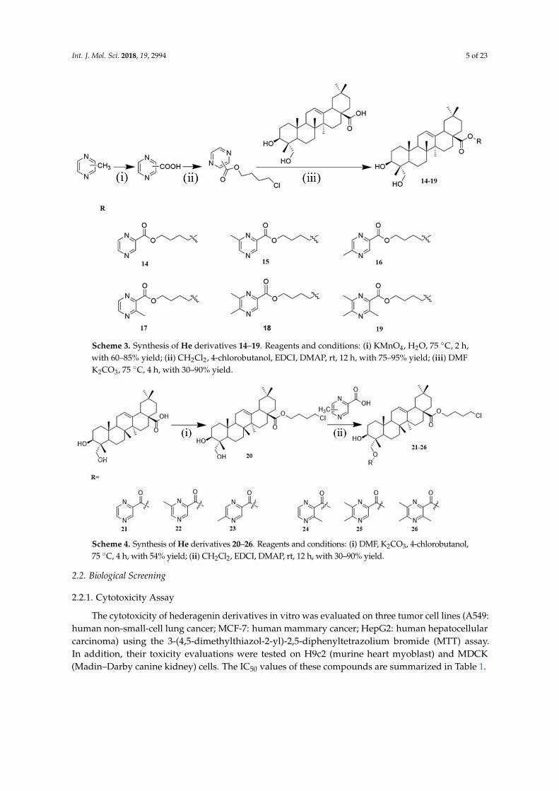
Many of the derivatives displayed more significant growth inhibition effects than He. Remarkably,as shown in Figure 2, the most promising was compound 9, which exhibited similar antitumor activitiesagainst A549 as the positive drug cisplatin (DDP), while it showed lower cytotoxicity than DDP onMDCK and H9c2 cell lines.
Int. J. Mol. Sci. 2018, 19, x FOR PEER REVIEW 6 of 23
Table 1. The in vitro cytotoxicity of compounds against various cell lines. A549—human non-small-
cell lung cancer; MCF-7—human mammary cancer; HepG2—human hepatocellular carcinoma;
MDCK—Madin–Darby canine kidney; H9c2—murine heart myoblast; He—hederagenin; DDP—
cisplatin.
Half Maximal Inhibitory Concentration (IC50) Values (μM)
Compound A549 MCF-7 HepG2 MDCK H9c2
1 17.62 ± 0.37 16.05 ± 0.01 18.79 ± 0.04 18.06 ± 0.25 14.73 ± 0.51
2 20.55 ± 0.29 15.78 ± 0.22 27.23 ± 1.82 26.71 ± 1.64 25.09 ± 0.43
3 15.03 ± 0.35 16.83 ± 9.25 18.93 ± 0.35 18.42 ± 3.76 >30
4 17.07 ± 1.06 17.31 ± 0.48 25.53 ± 1.40 26.12 ± 1.02 20.42 ± 2.24
5 24.37 ± 0.37 >30 24.94 ± 0.06 23.53 ± 0.67 28.46 ± 0.29
6 25.52 ± 0.26 >30 >30 24.08 ± 0.76 >30
7 >30 >30 >30 >30 >30
8 23.92 ± 0.31 25.35 ± 0.09 17.41 ± 0.75 20.13 ± 0.30 21.66 ± 0.86
9 3.45 ± 0.59 8.73 ± 1.49 8.71 ± 0.38 14.11 ± 0.04 16.69 ± 0.12
10 20.93 ± 0.68 27.24 ± 2.09 28.17 ± 0.21 25.42 ± 0.28 19.60 ± 3.29
11 14.12 ± 0.01 27.30 ± 0.57 14.84 ± 0.03 18.64 ± 0.18 14.72 ± 0.16
12 10.05 ± 2.47 17.28 ± 0.58 14.42 ± 0.03 18.64 ± 0.71 13.16 ± 0.20
13 8.15 ± 0.17 18.03 ± 0.18 13.24 ± 0.29 23.22 ± 0.01 10.51 ± 0.61
14 17.60 ± 2.33 31.23 ± 0.15 7.05 ± 0.17 18.10 ± 0.12 15.90 ± 0.22
15 17.98 ± 0.04 19.77 ± 1.27 6.71 ± 0.20 17.12 ± 1.01 17.56 ± 1.85
16 24.78 ± 0.66 28.49 ± 0.28 6.99 ± 0.63 20.12 ± 0.06 14.99 ± 0.12
17 19.57 ± 0.22 22.88 ± 0.66 8.15 ± 0.22 20.93 ± 3.03 22.31 ± 0.30
18 16.17 ± 0.15 28.29 ± 0.41 11.91 ± 0.99 16.55 ± 0.22 >30
19 12.23 ± 0.4 27.57 ± 0.52 9.06 ± 0.32 16.43 ± 0.04 16.50 ± 0.22
20 >30 >30 >30 >30 >30
21 9.29 ± 0.99 24.98 ± 0.18 7.99 ± 0.12 14.85 ± 0.06 16.68 ± 0.10
22 10.40 ± 0.43 20.53 ± 3.63 7.15 ± 0.43 16.34 ± 1.57 16.26 ± 0.07
23 18.67 ± 0.32 21.26 ± 1.28 7.17 ± 1.09 19.49 ± 2.47 23.35 ± 0.55
24 21.97 ± 0.87 17.95 ± 2.80 6.89 ± 0.21 19.27 ± 1.47 27.87 ± 0.62
25 >30 15.63 ± 1.41 6.42 ± 0.24 >30 >30
26 >30 18.30 ± 1.16 7.15 ± 0.85 >30 >30
He >50 >50 >50 >50 >50
DDP 3.85 ± 0.63 5.17 ± 0.28 3.42 ± 0.68 9.97 ± 1.12 5.31 ± 0.26
Many of the derivatives displayed more significant growth inhibition effects than He.
Remarkably, as shown in Figure 2, the most promising was compound 9, which exhibited similar
antitumor activities against A549 as the positive drug cisplatin (DDP), while it showed lower
cytotoxicity than DDP on MDCK and H9c2 cell lines.
Figure 2. Comparison of cytotoxicity between compound 9 and cisplatin (DDP). Figure 2. Comparison of cytotoxicity between compound 9 and cisplatin (DDP).

Int. J. Mol. Sci. 2018, 19, 2994 7 of 23
2.2.2. Cluster Analysis
Principal Component Analysis (PCA)
A PCA study was designed to verify the adaptability of experimental data to the model. For theinitial overview of the dataset and the detection of trends and outliers, PCA was carried out. Analysisshowed no samples being outside the Hotelling T2 95% confidence ellipse that could influence theanalyses, and high values of explained variation and predictive ability were obtained (Table 2). All datawere analyzed using SIMACA 13.0.
Table 2. The evaluation of explained variation (R2X) and predictive ability (Q2(cum)).
Cell Types R2X Q2(cum)
A549 0.761 0.159HepG2 0.872 0.697MCF-7 0.748 0.148
Partial Least Squares Discriminant Analysis (PLS-DA)
To further explore the structure–activity relationship, PLS-DA was performed, comparing alldesigned pairs of groups of He derivatives. Analyses revealed an antitumor activity discriminationbetween the different types of He derivatives compared in pairs. As Figures 3 and 4 depict, Hederivatives combined with the same pyrazine exhibited similar in vitro antitumor activities, and thelength of the carbon chain at the attachment site caused a difference in activity. In different tumorcell models, He derivatives linked with one carbon, two carbons, and four carbons were divided intothree groups according to the difference in in vitro antitumor activity (Figure 3). As for the effect of thestructure modification site of He, He derivatives modified at C-28 and C-23 were divided into twogroups according to the difference in in vitro antitumor activity (Figure 4). It is worth noticing thatthis put forward a reference point for the selection of structural modification sites for He. Throughdata analysis, we found that the number of methyl groups on the pyrazines also showed a certaindegree of regularity in their effect on activity. There was no law between the position of methyl groupson pyrazines and antitumor activity. Pyrazine derivatives with different numbers of methyl groupswere divided into six groups according to the difference in in vitro antitumor activity. Incredibly, theanalysis results of cluster analysis were consistent with the results of direct observation. For example,compounds 21–26 (similar activities on HepG2, IC50 ranging from 6.42 to 7.99 µM) could be separatedfrom the others (IC50 > 8 µM on HepG2) by direct observation. Similarly, the results of cluster analysisof PLS-DA supported this result (Figure 4). The cluster analysis of PLS-DA might provide us withfurther directions for further analysis of He derivatives.
Int. J. Mol. Sci. 2018, 19, x FOR PEER REVIEW 7 of 23
2.2.2. Cluster Analysis
Principal Component Analysis (PCA)
A PCA study was designed to verify the adaptability of experimental data to the model. For the
initial overview of the dataset and the detection of trends and outliers, PCA was carried out. Analysis
showed no samples being outside the Hotelling T2 95% confidence ellipse that could influence the
analyses, and high values of explained variation and predictive ability were obtained (Table 2). All
data were analyzed using SIMACA 13.0.
Table 2. The evaluation of explained variation (R2X) and predictive ability (Q2 (cum)).
Cell Types R2X Q2 (cum)
A549 0.761 0.159
HepG2 0.872 0.697
MCF-7 0.748 0.148
Partial Least Squares Discriminant Analysis (PLS-DA)
To further explore the structure–activity relationship, PLS-DA was performed, comparing all
designed pairs of groups of He derivatives. Analyses revealed an antitumor activity discrimination
between the different types of He derivatives compared in pairs. As Figures 3 and 4 depict, He
derivatives combined with the same pyrazine exhibited similar in vitro antitumor activities, and the
length of the carbon chain at the attachment site caused a difference in activity. In different tumor
cell models, He derivatives linked with one carbon, two carbons, and four carbons were divided into
three groups according to the difference in in vitro antitumor activity (Figure 3). As for the effect of
the structure modification site of He, He derivatives modified at C-28 and C-23 were divided into
two groups according to the difference in in vitro antitumor activity (Figure 4). It is worth noticing
that this put forward a reference point for the selection of structural modification sites for He.
Through data analysis, we found that the number of methyl groups on the pyrazines also showed a
certain degree of regularity in their effect on activity. There was no law between the position of
methyl groups on pyrazines and antitumor activity. Pyrazine derivatives with different numbers of
methyl groups were divided into six groups according to the difference in in vitro antitumor activity.
Incredibly, the analysis results of cluster analysis were consistent with the results of direct
observation. For example, compounds 21–26 (similar activities on HepG2, IC50 ranging from 6.42 to
7.99 µM) could be separated from the others (IC50 > 8 µM on HepG2) by direct observation. Similarly,
the results of cluster analysis of PLS-DA supported this result (Figure 4). The cluster analysis of PLS-
DA might provide us with further directions for further analysis of He derivatives.
Figure 3. Partial least squares discriminant analysis (PLS-DA) of carbon chain length on HepG2
(human hepatocellular carcinoma). Four carbon I—a carbon chain of four carbon atoms and pyrazine
were combined at the C-28 position; Four carbon II—a carbon chain of four carbon atoms was added
at the C-28 position, but pyrazine was added at the C-23 position.
Figure 3. Partial least squares discriminant analysis (PLS-DA) of carbon chain length on HepG2 (humanhepatocellular carcinoma). Four carbon I—a carbon chain of four carbon atoms and pyrazine werecombined at the C-28 position; Four carbon II—a carbon chain of four carbon atoms was added at theC-28 position, but pyrazine was added at the C-23 position.

Int. J. Mol. Sci. 2018, 19, 2994 8 of 23
Int. J. Mol. Sci. 2018, 19, x FOR PEER REVIEW 8 of 23
Figure 4. PLS-DA of combining position on HepG2. C-28—pyrazine was added at the C-28 position;
C-23—pyrazine was added at the C-23 position.
Structure–Activity Relationship Analysis
Combining the data analysis from Section 2.2.1 and Section Partial Least Squares Discriminant
Analysis (PLS-DA), we could easily find that the structural modification site of He, the length of the
carbon chain, and the type of pyrazine had an effect on the in vitro antitumor activity of the He
derivatives. In general, as observed for compounds 14–19 and 21–26, structural modification at
positions C-28 and C-23 could improve antitumor biological activity in vitro, while structural
transformation of C-23 might have more potential to enhance cytotoxicity on the same series of tumor
cells; it was confirmed that the IC50 values of compounds 21–26 were usually lower than those of
compounds 14–19. In general, compounds with chains containing two and four carbons were
superior to those with one carbon for improving the antitumor activity of the compounds. For
example, compounds 14–19 with a chain containing four carbons generally exhibited superior
antitumor activity to others, especially on HepG2. He derivatives 2, 9 and 15 with 2,6-
dimethylpiperazine showed better antitumor activity in vitro than others. In addition, comparing
compounds 7 and 20 with others, we knew that the introduction of the pyrazine structure was
essential for improving the in vitro antitumor activity of He. Notably, compound 9 (He combined
with 2,6-dimethylpiperazine via a chain containing two carbon atoms), the drug with the most
potential, was also in line with this law. It is worth mentioning that compounds 14–19 and 21–26
exhibited good antitumor activity and some cytotoxic selectivity toward HepG2 cells in vitro (the
selective inhibition (SI = IC50 MCF-7/IC50 HepG2) value of compound 14 was 4.43). What these compounds
had in common was that they were modified by four-carbon chains. This meant that changes in the
length of the carbon chain at C-28 and C-23 might alter the selectivity of compounds for different cell
lines; we speculated that this result might be caused by the combination of the compound and the
tumor cell target.
2.2.3. Analyses of Apoptosis
Morphological Detection of Apoptosis Using Giemsa Staining
To characterize the morphological detection of apoptosis induced by compound 9 on A549, the
nuclear and cytoplasmic morphological changes in compound-9-treated A549 were observed with
Giemsa staining. As shown in Figure 5, the number of A549 cells in the control group was higher
than other groups, and the cells appeared polygonal with a normal cell shape. With an increase in
dose, the phenomenon of cell shrinkage and cell disruption became more obvious. At the dose of 2
µM, the cells had a lot of puffing and fragmentation. When the concentration was 6 µM, there was
almost no normal cell morphology.
Figure 4. PLS-DA of combining position on HepG2. C-28—pyrazine was added at the C-28 position;C-23—pyrazine was added at the C-23 position.
Structure–Activity Relationship Analysis
Combining the data analysis from Section 2.2.1 and Section Partial Least Squares DiscriminantAnalysis (PLS-DA), we could easily find that the structural modification site of He, the length ofthe carbon chain, and the type of pyrazine had an effect on the in vitro antitumor activity of theHe derivatives. In general, as observed for compounds 14–19 and 21–26, structural modificationat positions C-28 and C-23 could improve antitumor biological activity in vitro, while structuraltransformation of C-23 might have more potential to enhance cytotoxicity on the same series of tumorcells; it was confirmed that the IC50 values of compounds 21–26 were usually lower than those ofcompounds 14–19. In general, compounds with chains containing two and four carbons were superiorto those with one carbon for improving the antitumor activity of the compounds. For example,compounds 14–19 with a chain containing four carbons generally exhibited superior antitumor activityto others, especially on HepG2. He derivatives 2, 9 and 15 with 2,6-dimethylpiperazine showed betterantitumor activity in vitro than others. In addition, comparing compounds 7 and 20 with others,we knew that the introduction of the pyrazine structure was essential for improving the in vitroantitumor activity of He. Notably, compound 9 (He combined with 2,6-dimethylpiperazine via a chaincontaining two carbon atoms), the drug with the most potential, was also in line with this law. It isworth mentioning that compounds 14–19 and 21–26 exhibited good antitumor activity and somecytotoxic selectivity toward HepG2 cells in vitro (the selective inhibition (SI = IC50
MCF-7/IC50HepG2)
value of compound 14 was 4.43). What these compounds had in common was that they were modifiedby four-carbon chains. This meant that changes in the length of the carbon chain at C-28 and C-23might alter the selectivity of compounds for different cell lines; we speculated that this result might becaused by the combination of the compound and the tumor cell target.
2.2.3. Analyses of Apoptosis
Morphological Detection of Apoptosis Using Giemsa Staining
To characterize the morphological detection of apoptosis induced by compound 9 on A549,the nuclear and cytoplasmic morphological changes in compound-9-treated A549 were observed withGiemsa staining. As shown in Figure 5, the number of A549 cells in the control group was higher thanother groups, and the cells appeared polygonal with a normal cell shape. With an increase in dose,the phenomenon of cell shrinkage and cell disruption became more obvious. At the dose of 2 µM,the cells had a lot of puffing and fragmentation. When the concentration was 6 µM, there was almostno normal cell morphology.

Int. J. Mol. Sci. 2018, 19, 2994 9 of 23Int. J. Mol. Sci. 2018, 19, x FOR PEER REVIEW 9 of 23
Figure 5. Morphological detection of apoptosis using Giemsa staining (200×) on A549 (human non-
small-cell lung cancer) cells treated with compound 9: (A) control group; (B) 2 µM; (C) 4 µM; (D) 6 µM.
4′,6-Diamidino-2-phenylindole (DAPI) Staining
To characterize the effects of apoptosis inducted by compound 9 on A549, the nuclear
morphological changes in compound-9-treated A549 were observed with DAPI staining. After
treating with compound 9 for 72 h, A549 cells showed nuclear morphological changes typical of
apoptosis in a dose-dependent manner. As pictured in Figure 6, in the control group, the nucleus was
intact and evenly colored. The light-blue fluorescence was diffuse and the cells did not show the
characteristics of apoptosis. When the concentration of the drug increased, nuclear condensation,
nuclear fragmentation, and the formation of apoptotic bodies appeared. When treated at 6 µM, the
number of cells decreased drastically, the shape of the cells became irregular, and nuclear
fragmentation occurred.
Figure 5. Morphological detection of apoptosis using Giemsa staining (200×) on A549 (humannon-small-cell lung cancer) cells treated with compound 9: (A) control group; (B) 2 µM; (C) 4 µM;(D) 6 µM.
4′,6-Diamidino-2-phenylindole (DAPI) Staining
To characterize the effects of apoptosis inducted by compound 9 on A549, the nuclearmorphological changes in compound-9-treated A549 were observed with DAPI staining. After treatingwith compound 9 for 72 h, A549 cells showed nuclear morphological changes typical of apoptosis ina dose-dependent manner. As pictured in Figure 6, in the control group, the nucleus was intact andevenly colored. The light-blue fluorescence was diffuse and the cells did not show the characteristics ofapoptosis. When the concentration of the drug increased, nuclear condensation, nuclear fragmentation,and the formation of apoptotic bodies appeared. When treated at 6 µM, the number of cells decreaseddrastically, the shape of the cells became irregular, and nuclear fragmentation occurred.
Detection of Apoptosis Using Annexin V Fluorescein Isothiocyanate (FITC)/Propidium Iodide(PI) Staining
To further depict the apoptosis induced by compound 9, apoptotic rates were analyzed by flowcytometry using Annexin V-FITC/PI staining. As depicted in Figure 7, with different concentrations (0,2, 5, and 10 µM) of compound 9 treatment, the percentage of apoptotic cells (including early and lateapoptosis ratios) increased from 3.7% of the control to 17.4%, 72.0%, and 91.5%, respectively. The resultindicates that compound 9 had the potential to induce the apoptosis of A549 cells.

Int. J. Mol. Sci. 2018, 19, 2994 10 of 23Int. J. Mol. Sci. 2018, 19, x FOR PEER REVIEW 10 of 23
Figure 6. Morphological detection of apoptosis using 4′,6-diamidino-2-phenylindole (DAPI) staining
(100×) on A549 cells treated with compound 9: (A) control group; (B) 2 µM; (C) 4 µM; (D) 6 µM.
Detection of Apoptosis Using Annexin V Fluorescein Isothiocyanate (FITC)/Propidium Iodide (PI)
Staining
To further depict the apoptosis induced by compound 9, apoptotic rates were analyzed by flow
cytometry using Annexin V-FITC/PI staining. As depicted in Figure 7, with different concentrations
(0, 2, 5, and 10 µM) of compound 9 treatment, the percentage of apoptotic cells (including early and
late apoptosis ratios) increased from 3.7% of the control to 17.4%, 72.0%, and 91.5%, respectively. The
result indicates that compound 9 had the potential to induce the apoptosis of A549 cells.
Figure 7. Detection of apoptosis using Annexin V fluorescein isothiocyanate (FITC)/propidium iodide
(PI) staining on A549 cells treated with compound 9: (A) control group; (B) 2 µM; (C) 5 µM; (D) 10 µM.
Figure 6. Morphological detection of apoptosis using 4′,6-diamidino-2-phenylindole (DAPI) staining(100×) on A549 cells treated with compound 9: (A) control group; (B) 2 µM; (C) 4 µM; (D) 6 µM.
Int. J. Mol. Sci. 2018, 19, x FOR PEER REVIEW 10 of 23
Figure 6. Morphological detection of apoptosis using 4′,6-diamidino-2-phenylindole (DAPI) staining
(100×) on A549 cells treated with compound 9: (A) control group; (B) 2 µM; (C) 4 µM; (D) 6 µM.
Detection of Apoptosis Using Annexin V Fluorescein Isothiocyanate (FITC)/Propidium Iodide (PI)
Staining
To further depict the apoptosis induced by compound 9, apoptotic rates were analyzed by flow
cytometry using Annexin V-FITC/PI staining. As depicted in Figure 7, with different concentrations
(0, 2, 5, and 10 µM) of compound 9 treatment, the percentage of apoptotic cells (including early and
late apoptosis ratios) increased from 3.7% of the control to 17.4%, 72.0%, and 91.5%, respectively. The
result indicates that compound 9 had the potential to induce the apoptosis of A549 cells.
Figure 7. Detection of apoptosis using Annexin V fluorescein isothiocyanate (FITC)/propidium iodide
(PI) staining on A549 cells treated with compound 9: (A) control group; (B) 2 µM; (C) 5 µM; (D) 10 µM.
Figure 7. Detection of apoptosis using Annexin V fluorescein isothiocyanate (FITC)/propidium iodide(PI) staining on A549 cells treated with compound 9: (A) control group; (B) 2 µM; (C) 5 µM; (D) 10 µM.
2.2.4. Detection of the Effect of Compound 9 on Cell-Cycle Progression
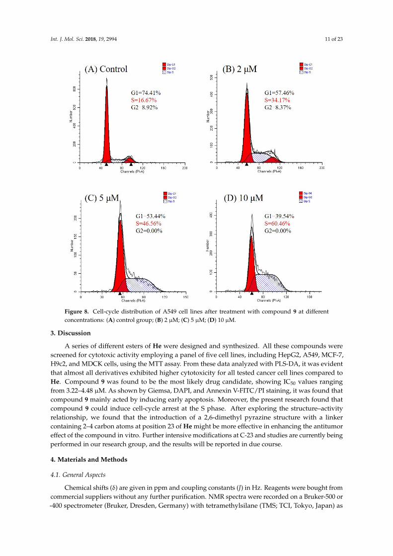
The remarkable activity of compound 9 against the A549 cell line prompted us to investigateits effect on the cell cycle of A549 by flow cytometry. As shown in Figure 8, when the concentrationincreased (0, 2, 5, and 10 µM), the percentage of A549 cells in the synthesis (S) phase overtly increased(from 16.67% to 60.46%) which clearly indicated that compound 9 arrested the cells in the S phase ina concentration-dependent manner.
Int. J. Mol. Sci. 2018, 19, 2994 11 of 23
Int. J. Mol. Sci. 2018, 19, x FOR PEER REVIEW 11 of 23
2.2.4. Detection of the Effect of Compound 9 on Cell-Cycle Progression
The remarkable activity of compound 9 against the A549 cell line prompted us to investigate its
effect on the cell cycle of A549 by flow cytometry. As shown in Figure 8, when the concentration
increased (0, 2, 5, and 10 µM), the percentage of A549 cells in the synthesis (S) phase overtly increased
(from 16.67% to 60.46%) which clearly indicated that compound 9 arrested the cells in the S phase in
a concentration-dependent manner.
Figure 8. Cell-cycle distribution of A549 cell lines after treatment with compound 9 at different
concentrations: (A) control group; (B) 2 µM; (C) 5 µM; (D) 10 µM.
3. Discussion
A series of different esters of He were designed and synthesized. All these compounds were
screened for cytotoxic activity employing a panel of five cell lines, including HepG2, A549, MCF-7,
H9c2, and MDCK cells, using the MTT assay. From these data analyzed with PLS-DA, it was evident
that almost all derivatives exhibited higher cytotoxicity for all tested cancer cell lines compared to
He. Compound 9 was found to be the most likely drug candidate, showing IC50 values ranging from
3.22–4.48 µM. As shown by Giemsa, DAPI, and Annexin V-FITC/PI staining, it was found that
compound 9 mainly acted by inducing early apoptosis. Moreover, the present research found that
compound 9 could induce cell-cycle arrest at the S phase. After exploring the structure–activity
relationship, we found that the introduction of a 2,6-dimethyl pyrazine structure with a linker
containing 2–4 carbon atoms at position 23 of He might be more effective in enhancing the antitumor
Figure 8. Cell-cycle distribution of A549 cell lines after treatment with compound 9 at differentconcentrations: (A) control group; (B) 2 µM; (C) 5 µM; (D) 10 µM.
3. Discussion
A series of different esters of He were designed and synthesized. All these compounds werescreened for cytotoxic activity employing a panel of five cell lines, including HepG2, A549, MCF-7,H9c2, and MDCK cells, using the MTT assay. From these data analyzed with PLS-DA, it was evidentthat almost all derivatives exhibited higher cytotoxicity for all tested cancer cell lines compared toHe. Compound 9 was found to be the most likely drug candidate, showing IC50 values rangingfrom 3.22–4.48 µM. As shown by Giemsa, DAPI, and Annexin V-FITC/PI staining, it was found thatcompound 9 mainly acted by inducing early apoptosis. Moreover, the present research found thatcompound 9 could induce cell-cycle arrest at the S phase. After exploring the structure–activityrelationship, we found that the introduction of a 2,6-dimethyl pyrazine structure with a linkercontaining 2–4 carbon atoms at position 23 of He might be more effective in enhancing the antitumoreffect of the compound in vitro. Further intensive modifications at C-23 and studies are currently beingperformed in our research group, and the results will be reported in due course.
4. Materials and Methods
4.1. General Aspects
Chemical shifts (δ) are given in ppm and coupling constants (J) in Hz. Reagents were bought fromcommercial suppliers without any further purification. NMR spectra were recorded on a Bruker-500 or-400 spectrometer (Bruker, Dresden, Germany) with tetramethylsilane (TMS; TCI, Tokyo, Japan) as

Int. J. Mol. Sci. 2018, 19, 2994 12 of 23
an internal standard; high-resolution mass spectra (HRMS) were acquired using a Thermo SientificTMLTQ Orbitrap XL hybrid FTMS instrument (Thermo Technologies, New York, NY, USA), and an X-5micro melting point apparatus (Beijing, China).
4.1.1. Isolation of Hederagenin (He)
Dried Dipsacus asper Wall.ex Henry was extracted with 70% ethanol; three extractions at refluxedtemperature were carried out, and the solvent was kept in contact with the sample for 3–9 h ata concentration ratio of 10 to 20 times the amount of ethanol than medicinal herbs. The combinedethanol extracts were concentrated under reduced pressure in a rotary evaporator. Then, 7%hydrochloric acid solution in water (20 times the residue) was added to this residue, and the mixturewas heated under reflux temperature for 4–8 h. After this hydrolysis, the residue was filtered off, andthe precipitate was obtained, washed with water to neutrality, and dried at 65 ◦C to obtain a crudeproduct of He. The crude product was purified by column chromatography on a silica gel with solvent(DCM:MeOH = 70:1), and the mixture was concentrated and evaporated to dryness. After standingovernight under 25–50 mL of anhydrous ethanol, the residue was filtered off, and He was obtained.Hederagenin (He): yield <10%; white powder, melting point (m.p.) >300 ◦C; HRMS (electrosprayionization (ESI)) m/z: [M + Cl]− 507.3274, calculated for C30H48ClO4 507.3247. All spectroscopic data(NMR and MS) were in agreement with the literature.
4.1.2. Preparation of Chloromethylpyrazine
Firstly, 2-methylpyrazine (10.63 mmol), NCS (1.42 g, 10.63 mmol), and BPO (0.26 g, 1.06 mmol)were added to a solution of CCl4 (40 mL), and the mixture was stirred for 2 h at room temperatureunder light conditions with nitrogen protection. The mixture was heated at reflux temperature for8 h, cooled down, and then stirred in an ice bath for 1 h, before being filtered by suction, washedwith 10 mL of carbon tetrachloride, and distilled under reduced pressure to give an irritating yellowoily liquid. The product was left untreated. Other methyl pyrazines included 2,3-dimethylpyrazine,2,5-dimethylpyrazine, 2,6-dimethylpyrazine, 2,3,5-trimethylpyrazine, and 2,3,5,6-tetramethyl pyrazine,carried out as 2-methylpyrazine [24,25].
4.1.3. Preparation of Pyrazinic Acid
Powdered potassium permanganate (3.3 g, 20.90 mmol) was dissolved in water (50 mL), thendropped into the aqueous solution of methyl pyrazine within 20 min. The mixture was stirred for 2 h at75 ◦C. The reaction was monitored with thin-layer chromatography (TLC) until the end of the reaction.The mixture was cooled down, filtered, and washed with 50 mL of water; then, the filtrate was adjustedto pH 1.5 with nitric acid, slowly warmed to 50 ◦C, and maintained for 10 min, before being cooleddown, extracted with 3× 20 mL of ethyl acetate, and dried over anhydrous sodium sulfate. The productwas left untreated. Other methyl pyrazines included 2,3-dimethylpyrazine, 2,6-dimethylpyrazine,2,3,5-trimethylpyrazine and 2,3,5,6-tetramethyl pyrazine, carried out as 2,6-dimethylpyrazine [35,36].
4.1.4. Preparation of Compound 7
He (1.246 g, 2.64 mmol) and K2CO3 (728 mg, 5.28 mmol) were added to a solution of2-bromoethanol (1.649 g, 13.20 mmol) in DMF (20 mL), and the mixture was stirred for 4 h at 85 ◦C.The reaction mixture was then diluted with ethyl acetate (30 mL), washed with water and brinesuccessively, dried over anhydrous sodium sulfate, and purified by column chromatography to yieldthe white powder.
2-Hydroxyethyl (4aS,6aS,6bR,9R,10S,12aR)-10-hydroxy-9-(hydroxymethyl)-2,2,6a,6b,9,12a-hexamethyl-1,3,4,5,6,6a,6b,7,8,8a,9,10,11,12,12a,12b,13,14b-octadecahydropicene-4a(2H)-carboxylate(compound 7): yield 95% (after chromatograph with DCM/MeOH 1–2%) as a white powder; m.p.246.6 ◦C. 1H NMR (400 MHz, CDCl3) δ 5.28 (s, 1H, H-12), 4.05 (t, J = 5.9 Hz, 2H, H-31), 3.72 (d,J = 10.2 Hz, 1H, H-23a), 3.63 (t, J = 7.7 Hz, 1H, H-3), 3.56 (t, J = 6.3 Hz, 2H, H-32), 3.43 (d, J = 9.9 Hz, 1H,
Int. J. Mol. Sci. 2018, 19, 2994 13 of 23
H-23a), 2.90–2.82 (m, 1H, H-18), 1.12 (s, 3H, –CH3), 0.96–0.88 (m, 12H, 4 × –CH3), 0.73 (s, 3H, –CH3).13C NMR (100 MHz, CDCl3) δ 178.2, 144.1, 122.3, 76.8, 72.1, 66.1, 61.4, 49.8, 47.6, 46.9, 45.8, 41.8, 41.8,41.5, 39.3, 38.1, 36.9, 33.8, 33.1, 32.5, 32.5, 30.7, 27.6, 26.7, 25.9, 23.6, 23.4, 23.0, 18.5, 17.1, 15.7, 11.4.HRMS (ESI) m/z: [M−H2O]+ 499.3790, calculated for C32H50O4 499.3782.
4.1.5. General Procedure for the Synthesis of Compounds 1–6
He (282.2 mg, 0.6 mmol), chloromethyl pyrazine (153.6 mg, about 0.6 mmol, prepared as detailedin Section 4.1.2), and K2CO3 (248.4 mg, 1.8 mmol) were added to DMF (20 mL), and the mixture wasstirred for 4 h at 85 ◦C. The reaction mixture was then diluted with ethyl acetate (20 mL), washed withwater and brine, dried over anhydrous sodium sulfate, and purified by column chromatography toyield the white powders.
Synthesis of Compound 1
Pyrazin-2-ylmethyl(4aS,6aS,6bR,9R,10S,12aR)-10-hydroxy-9-(hydroxymethyl)-2,2,6a,6b,9,12a-hexamethyl-1,3,4,5,6,6a,6b,7,8,8a,9,10,11,12,12a,12b,13,14b-octadecahydropicene-4a(2H)-carboxylate(compound 1): yield 40% (after chromatograph with DCM/MeOH, 1–1.5%) as a white powder; m.p.146.8 ◦C. 1H NMR (400 MHz, CDCl3) δ 8.68 (s, 1H, –N=C–H), 8.53 (d, J = 9.0 Hz, 2H, 2 × –N=C–H),5.31 (s, 1H, H-12), 5.22 (q, J = 13.9 Hz, 2H, H-31), 3.71 (d, J = 10.3 Hz, 1H, H-23a), 3.65–3.59 (m, 1H,H-3), 3.41 (d, J = 10.3 Hz, 1H, H-23b), 2.91 (m, 1H, H-18), 1.12 (s, 3H, –CH3), 0.93–0.89 (m, 9H, 3 ×–CH3), 0.87 (s, 3H, –CH3), 0.59 (s, 3H, –CH3). 13C NMR (100 MHz, CDCl3) δ 177.3, 152.2, 144.2, 144.0,143.9, 143.6, 122.8, 77.0, 72.2, 64.8, 49.9, 47.7, 47.1, 45.9, 41.9, 41.9, 41.5, 39.4, 38.2, 37.0, 33.9, 33.2, 32.6,32.6, 30.8, 27.8, 26.9, 26.0, 23.8, 23.5, 23.2, 18.6, 17.0, 15.8, 11.5. HRMS (ESI) m/z: [M + H]+ 565.4003,calculated for C35H53N2O4 565.4000.
Synthesis of Compound 2
(6-Methylpyrazin-2-yl)methyl(4aS,6aS,6bR,9R,10S,12aR)-10-hydroxy-9-(hydroxymethyl)-2,2,6a,6b,9,12a-hexamethyl-1,3,4,5,6,6a,6b,7,8,8a,9,10,11,12,12a,12b,13,14b-octadecahydropicene-4a(2H)-carboxylate (compound 2): yield 35% (after chromatograph with DCM/MeOH, 1–1.5%) asa white powder; m.p. 107.0 ◦C. 1H NMR (400 MHz, CDCl3) δ 8.47 (s, 1H, –N=C–H), 8.39 (s, 1H,–N=C–H), 5.30 (s, 1H, H-12), 5.18 (q, J = 13.8 Hz, 2H, H-31), 3.71 (d, J = 10.3 Hz, 1H, H-23a), 3.62 (t,J = 7.9 Hz, 1H, H-3), 3.41 (d, J = 10.3 Hz, 1H, H-23b), 2.91 (m, 1H, H-18), 2.56 (s, 3H, –CH3), 1.12 (s,3H, –CH3), 0.93–0.90 (m, 9H, 3 × –CH3), 0.88 (s, 3H, –CH3), 0.58 (s, 3H, –CH3). 13C NMR (100 MHz,CDCl3) δ 177.3, 153.4, 150.9, 143.7, 143.6, 140.4, 122.7, 77.0, 72.3, 64.9, 49.9, 47.7, 47.0, 46.0, 41.9, 41.8,41.5, 39.4, 38.2, 37.0, 33.9, 33.2, 32.6, 32.6, 30.8, 27.8, 27.1, 26.9, 26.1, 23.8, 23.5, 23.2, 21.6, 18.6, 17.0, 15.8,11.5. HRMS (ESI) m/z: [M + H]+ 579.4142, calculated for C36H55N2O4 579.4156.
Synthesis of Compound 3
(5-Methylpyrazin-2-yl)methyl(4aS,6aS,6bR,9R,10S,12aR)-10-hydroxy-9-(hydroxymethyl)-2,2,6a,6b,9,12a-hexamethyl-1,3,4,5,6,6a,6b,7,8,8a,9,10,11,12,12a,12b,13,14b-octadecahydropicene-4a(2H)-carboxylate (compound 3): yield 60% (after chromatograph with DCM/MeOH, 1–1.5%) asa white powder; m.p. 170.3 ◦C. 1H NMR (400 MHz, CDCl3) δ 8.45 (m, 2H, 2 × –N=C–H), 5.30 (d,J = 2.7 Hz, 1H, H-12), 3.71 (d, J = 10.3 Hz, 1H, H-23a), 3.41 (d, J = 10.3 Hz, 1H, H-23b), 2.90 (m, 1H,H-18), 2.57 (d, J = 6.7 Hz, 3H, –CH3), 1.12 (s, 3H, –CH3), 0.91 (d, J = 6.6 Hz, 9H, 3 × –CH3), 0.88 (s, 3H,–CH3), 0.57 (s, 3H, –CH3). 13C NMR (100 MHz, CDCl3) δ 177.4, 153.1, 148.8, 143.9, 143.7, 142.7, 122.7,77.0, 72.3, 64.7, 49.9, 47.7, 47.0, 45.9, 41.9, 41.8, 41.5, 39.4, 38.2, 37.0, 33.9, 33.2, 32.6, 32.6, 30.8, 27.8, 26.9,26.1, 23.8, 23.5, 23.2, 21.4, 18.6, 17.0, 15.8, 11.5. HRMS (ESI) m/z: [M + H]+ 579.4142, calculated forC36H55N2O4 579.4156.

Int. J. Mol. Sci. 2018, 19, 2994 14 of 23
Synthesis of Compound 4
(3-Methylpyrazin-2-yl)methyl(4aS,6aS,6bR,9R,10S,12aR)-10-hydroxy-9-(hydroxymethyl)-2,2,6a,6b,9,12a-hexamethyl-1,3,4,5,6,6a,6b,7,8,8a,9,10,11,12,12a,12b,13,14b-octadecahydropicene-4a(2H)-carboxylate (compound 4): yield 36% (after chromatograph with DCM/MeOH, 1–1.5%) asa white powder; m.p. 170.4 ◦C. 1H NMR (400 MHz, CDCl3) δ 8.41 (s, 1H, –N=C–H), 8.38 (s, 1H,–N=C–H), 5.22 (m, 3H, H-12, H-31a, H-32b), 3.71 (d, J = 10.3 Hz, 1H, H-23a), 3.64–3.59 (m, 1H, H-3),3.41 (d, J = 10.3 Hz, 1H, H-23b), 2.85 (m, 1H, H-18), 2.63 (s, 3H, –CH3), 1.10 (s, 3H, –CH3), 0.93–0.88 (m,12H, 4 × –CH3), 0.57 (s, 3H, –CH3). 13C NMR (100 MHz, CDCl3) δ 177.4, 153.1, 148.9, 143.9, 143.7,142.7, 122.7, 77.0, 72.3, 64.7, 49.9, 47.7, 47.0, 45.93, 41.9, 41.8, 41.5, 39.4, 38.2, 37.0, 33.9, 33.2, 32.6, 32.6,30.8, 27.8, 26.9, 26.1, 23.8, 23.5, 23.2, 21.4, 18.6, 17.0, 15.8, 11.5. HRMS (ESI) m/z: [M + H]+ 579.4145,calculated for C36H55N2O4 579.4156.
Synthesis of Compound 5
(5,6-Dimethylpyrazin-2-yl)methyl(4aS,6aS,6bR,9R,10S,12aR)-10-hydroxy-9-(hydroxymethyl)-2,2,6a,6b,9,12a-hexamethyl-1,3,4,5,6,6a,6b,7,8,8a,9,10,11,12,12a,12b,13,14b-octadecahydropicene-4a(2H)-carboxylate (compound 5): yield approximately 40% (after chromatograph with DCM/MeOH,1–1.5%) as a white solid; m.p. > 200 ◦C. 1H NMR (400 MHz, CDCl3) δ 8.24 (d, J = 13.0 Hz, 1H,–N=C–H), 5.22–5.09 (m, 3H, H-12, H-31), 3.67 (d, J = 10.1 Hz, 1H, H-23a), 3.62–3.57 (m, 1H, H-3),3.37 (d, J = 10.1 Hz, 1H, H-23b), 2.83 (m, 1H, H-18), 2.59–2.49 (m, 6H, 2 × –CH3), 1.07 (s, 3H, –CH3),0.90–0.85 (m, 12H, 4 × –CH3), 0.52 (d, J = 5.4 Hz, 3H, –CH3). 13C NMR (100 MHz, CDCl3) δ 177.3,152.4, 149.9, 146.3, 143.5, 141.2, 122.6, 76.8, 72.0, 65.0, 49.9, 47.6, 47.0, 45.9, 41.8, 41.7, 41.4, 39.3, 38.2, 37.0,33.9, 33.1, 32.6, 32.5, 30.8, 27.7, 26.6, 26.0, 23.7, 23.4, 23.1, 21.4, 21.2, 18.5, 16.9, 15.7, 11.6. HRMS (ESI)m/z: [M + H]+ 593.4295, calculated for C37H57N2O4 593.4313.
Synthesis of Compound 6
(3,5,6-Trimethylpyrazin-2-yl)methyl(4aS,6aS,6bR,9R,10S,12aR)-10-hydroxy-9-(hydroxymethyl)-2,2,6a,6b,9,12a-hexamethyl-1,3,4,5,6,6a,6b,7,8,8a,9,10,11,12,12a,12b,13,14b-octadecahydropicene-4a(2H)-carboxylate (compound 6): yield 90% (after chromatograph with DCM/MeOH, 1–1.5%) asa white powder; m.p. 127.8 ◦C. 1H NMR (400 MHz, CDCl3) δ 5.24–5.08 (m, 3H, H-12, H-31a, H-31b),3.72 (d, J = 10.2 Hz, 1H, H-23a), 3.62 (t, J = 7.8 Hz, 1H, H-3), 3.41 (d, J = 10.3 Hz, 1H, H-23b), 2.85 (m,1H, H-18), 2.55–2.48 (m, 9H, 3 × –CH3), 1.09 (s, 3H, –CH3), 0.93–0.88 (m, 12H, 4 × –CH3), 0.53 (s, 3H,–CH3). 13C NMR (100 MHz, CDCl3) δ 177.3, 151.1, 149.3, 148.9, 145.5, 143.7, 122.5, 77.0, 72.3, 65.0, 50.0,47.7, 47.0, 46.0, 41.9, 41.8, 41.4, 39.4, 38.2, 37.0, 34.0, 33.2, 32.6, 32.5, 30.8, 27.7, 26.9, 26.0, 23.8, 23.5, 23.2,21.8, 21.5, 20.7, 18.6, 17.0, 15.8, 11.6. HRMS (ESI) m/z: [M + H]+ 607.4459, calculated for C38H59N2O4
607.4469.
4.1.6. General Procedure for the Synthesis of Compounds 8–13
Pyrazinic acid (0.27 mmol, prepared as detailed in Section 4.1.3), EDCI (0.35 mmol), and DMAP(0.027 mmol) were added to dried dichloromethane (20 mL). Then, compound 7 (140 mg, 0.27 mmol)dissolved in dichloromethane (20 mL) was dropped into the aqueous solution of methyl pyrazinewithin 20 min, and the mixture was stirred for 12 h at room temperature. The reaction mixture wasthen diluted with dichloromethane (20 mL), washed with water and brine, dried over anhydroussodium sulfate, concentrated, and purified by column chromatography to yield the white powders.
Synthesis of Compound 8
2-(((4aS,6aS,6bR,9R,10S,12aR)-10-Hydroxy-9-(hydroxymethyl)-2,2,6a,6b,9,12a-hexamethyl-1,2,3,4,4a,5,6,6a,6b,7,8,8a,9,10,11,12,12a,12b,13,14b-icosahydropicene-4a-carbonyl)oxy)ethylpyrazine-2-carboxylate (compound 8): yield 20% (after chromatograph with DCM/MeOH, 1–2%) as a whitepowder; m.p. 154.7 ◦C. 1H NMR (400 MHz, CDCl3) δ 9.29 (d, J = 9.6 Hz, 1H, –N=C–H), 8.80–8.70

Int. J. Mol. Sci. 2018, 19, 2994 15 of 23
(m, 2H, 2 × –N=C–H), 5.30 (s, 1H, H-12), 3.62 (t, J = 7.5 Hz, 1H, H-3), 2.88 (m, 1H, H-18), 0.98 (s, 3H,–CH3), 0.91 (d, J = 10.4 Hz, 9H, 3 × –CH3), 0.77 (s, 3H, –CH3). 13C NMR (100 MHz, CDCl3) δ 178.3,163.9, 148.0, 146.2, 144.7, 144.3, 143.4, 122.4, 77.4, 71.9, 66.2, 61.6, 49.6, 48.0, 47.1, 45.9, 42.3, 41.9, 41.7,39.5, 38.3, 37.0, 34.0, 33.2, 32.6, 30.8, 27.7, 26.4, 26.1, 25.8, 23.7, 23.5, 23.1, 18.8, 17.2, 15.7, 12.3. HRMS(ESI) m/z: [M + H]+ 623.4045, calculated for C37H55N2O6 623.4055.
Synthesis of Compound 9
2-(((4aS,6aS,6bR,9R,10S,12aR)-10-Hydroxy-9-(hydroxymethyl)-2,2,6a,6b,9,12a-hexamethyl-1,2,3,4,4a,5,6,6a,6b,7,8,8a,9,10,11,12,12a,12b,13,14b-icosahydropicene-4a-carbonyl)oxy)ethyl-6-methylpyrazine-2-carboxylate (compound 9): yield 35% (after chromatograph with DCM/MeOH,1–2%) as a white powder; m.p. 122.9 ◦C. 1H NMR (400 MHz, CDCl3) δ 9.06 (d, J = 21.2 Hz, 1H,–N=C–H), 8.63 (d, J = 5.4 Hz, 1H, –N=C–H), 5.31 (s, 1H, H-12), 4.25–3.99 (m, 2H), 2.87 (m, 1H, H-18),2.67 (d, J = 9.0 Hz, 3H, –CH3), 1.11 (s, 3H, –CH3), 0.92 (m, 12H, 4× –CH3), 0.77 (s, 3H, –CH3). 13C NMR(100 MHz, CDCl3) δ 178.3, 164.2, 154.4, 148.1 144.2, 143.0, 142.2, 122.4, 74.1, 72.0, 66.2, 61.6, 49.7, 48.0,47.1, 45.9, 42.3, 41.9, 41.7, 39.4, 38.3, 37.0, 33.9, 33.2, 32.6, 32.7, 30.8, 27.7, 26.3, 26.1, 25.9, 23.5, 23.1, 21.9,18.8, 17.2, 15.7, 12.3. HRMS (ESI) m/z: [M +H]+ 637.4219, calculated for C38H57N2O6 637.4211.
Synthesis of Compound 10
2-(((4aS,6aS,6bR,9R,10S,12aR)-10-Hydroxy-9-(hydroxymethyl)-2,2,6a,6b,9,12a-hexamethyl-1,2,3,4,4a,5,6,6a,6b,7,8,8a,9,10,11,12,12a,12b,13,14b-icosahydropicene-4a-carbonyl)oxy)ethyl-5-methylpyrazine-2-carboxylate (compound 10): yield 55% (after chromatograph with DCM/MeOH,1–2%) as a white powder; m.p. 192.6 ◦C. 1H NMR (400 MHz, CDCl3) δ 9.17 (s, 1H, –N=C–H), 8.59 (s,1H, –N=C–H), 5.22 (s, 1H, H-12), 4.68–4.59 (m, 2H, H-31), 4.46–4.31 (m, 2H, H-32), 3.70 (d, J = 10.3 Hz,1H, H-23a), 3.61 (t, J = 7.8 Hz, 1H, H-3), 3.40 (d, J = 10.3 Hz, 1H, H-23b), 2.84 (m, 1H, H-18), 2.67 (s,3H, –CH3), 1.08 (s, 3H), 0.87 (s, 12H, 4 × –CH3), 0.65 (s, 3H, –CH3). 13C NMR (100 MHz, CDCl3) δ177.6, 163.9, 158.1, 145.5, 144.6, 143.6, 140.5, 122.6, 77.0, 72.2, 63.6, 62.0, 49.9, 47.7, 46.9, 45.9, 41.9, 41.8,41.3, 39.4, 38.2, 37.0, 33.9, 33.2, 32.5, 32.5, 30.8, 27.7, 26.9, 26.0, 23.7, 23.4, 23.0, 22.1, 18.6, 17.0, 15.7, 11.5.HRMS (ESI) m/z: [M + H]+ 637.4208, calculated for C38H57N2O6 637.4211.
Synthesis of Compound 11
2-(((4aS,6aS,6bR,9R,10S,12aR)-10-Hydroxy-9-(hydroxymethyl)-2,2,6a,6b,9,12a-hexamethyl-1,2,3,4,4a,5,6,6a,6b,7,8,8a,9,10,11,12,12a,12b,13,14b-icosahydropicene-4a-carbonyl)oxy)ethyl-3-methylpyrazine-2-carboxylate (compound 11): yield 35% (after chromatograph with DCM/MeOH,1–2%) as a white powder; m.p. 146.8 ◦C. 1H NMR (400 MHz, CDCl3) δ 8.62 (s, 1H, –N=C–H), 8.48(s, 1H, –N=C–H), 5.31 (s, 1H, H-12), 4.59 (d, J = 10.8 Hz, 1H, H-23a), 4.04 (d, J = 10.8 Hz, 1H, H-23b),3.67–3.58 (m, 1H, H-3), 2.87 (s, 4H, H-18, –CH3), 1.13 (s, 3H, –CH3), 0.94 (m, 12H, 4 × –CH3), 0.77 (s,3H, –CH3). 13C NMR (100 MHz, CDCl3) δ 178.3, 165.0, 155.7, 146.5, 144.2, 142.5, 141.7, 122.4, 74.8,73.7, 66.2, 61.6, 50.0, 47.9, 47.1, 45.9, 42.1, 42.0, 41.7, 39.4, 38.2, 37.0, 34.0, 33.2, 32.6, 32.6, 30.8, 27.8, 26.3,25.9, 23.7, 23.5, 23.3, 23.2, 19.0, 17.2, 15.6, 12.36. HRMS (ESI) m/z: [M + H]+ 637.4222, calculated forC38H57N2O6 637.4211.
Synthesis of Compound 12
2-(((4aS,6aS,6bR,9R,10S,12aR)-10-Hydroxy-9-(hydroxymethyl)-2,2,6a,6b,9,12a-hexamethyl-1,2,3,4,4a,5,6,6a,6b,7,8,8a,9,10,11,12,12a,12b,13,14b-icosahydropicene-4a-carbonyl)oxy)ethyl-5,6-dimethylpyrazine-2-carboxylate (compound 12): yield 25% (after chromatograph with DCM/MeOH,1–2%) as a white powder; m.p. 146.2 ◦C. 1H NMR (400 MHz, CDCl3) δ 8.41 (d, J = 60.1 Hz, 1H,–N=C–H), 5.31 (s, 1H, H-12), 4.59 (d, J = 10.5 Hz, 1H, H-23a), 4.00 (d, J = 10.7 Hz, 1H, H-23b), 2.83 (m,4H, H-18, –CH3), 2.58 (d, J = 6.8 Hz, 3H, –CH3), 1.13 (s, 3H, –CH3), 0.97–0.90 (m, 12H, 4 × –CH3), 0.76(s, 3H, –CH3). 13C NMR (100 MHz, CDCl3) δ 178.3, 165.2, 154.8, 151.0, 146.5, 144.2, 141.5, 122.4, 75.0,73.8, 66.2, 61.6, 50.2, 47.8, 47.1, 45.9, 42.1, 42.0, 41.6, 39.4, 38.2, 37.0, 33.9, 33.2, 32.6, 30.8,27.8, 26.2, 25.9,

Int. J. Mol. Sci. 2018, 19, 2994 16 of 23
23.7, 23.5, 23.2, 22.7, 22.0, 21.2, 19.0, 17.2, 15.6, 12.4. HRMS (ESI) m/z: [M + H]+ 651.4375, calculated forC39H59N2O6 651.4368.
Synthesis of Compound 13
2-(((4aS,6aS,6bR,9R,10S,12aR)-10-Hydroxy-9-(hydroxymethyl)-2,2,6a,6b,9,12a-hexamethyl-1,2,3,4,4a,5,6,6a,6b,7,8,8a,9,10,11,12,12a,12b,13,14b-icosahydropicene-4a-carbonyl)oxy)ethyl-3,5,6-trimethylpyrazine-2-carboxylate (compound 13): yield 90% (after chromatograph with DCM/MeOH,1–2%) as a white powder; m.p. 150.6 ◦C. 1H NMR (400 MHz, CDCl3) δ 5.31 (s, 1H, H-12), 4.60 (d,J = 10.7 Hz, 1H, H-23a), 3.94 (d, J = 10.7 Hz, 1H, H-23b), 3.66–3.59 (m, 1H, H-3), 2.87 (m, 1H, H-18), 2.79(s, 3H, –CH3), 2.57 (m, 9H, 3 × –CH3), 1.13 (s, 3H, –CH3), 0.99–0.89 (m, 15H, 5 × –CH3), 0.77 (s, 3H,–CH3). 13C NMR (100 MHz, CDCl3) δ 178.3, 165.3, 155.4, 152.2, 149.6, 144.2, 138.1, 122.4, 75.4, 74.6,66.2, 61.6, 50.7, 47.8, 47.1, 45.9, 42.0, 42.0, 41.6, 39.4, 38.2, 36.98, 34.0, 33.20, 32.60, 30.8, 29.8, 27.8, 26.2,26.0, 23.8, 23.5, 23.2, 22.7, 22.4, 22.3, 21.8, 19.1, 17.2, 15.5, 12.5. HRMS (ESI) m/z: [M + H]+ 665.4531,calculated for C40H61N2O6 665.4524.
4.1.7. General Procedure for the Synthesis of Compounds 14–19
Pyrazinic acid (248 mg, about 2 mmol, prepared as detailed in Section 4.1.3), 4-chloro-1-butanol(2.4 mmol), EDCI (2.4 mmol), and DMAP (0.2 mmol) were added into dried dichloromethane (20 mL).The mixture was stirred for 12 h at room temperature, then diluted with dichloromethane (20 mL),washed with water and brine, dried over anhydrous sodium sulfate, concentrated and purified bycolumn chromatography to yield the oily liquid. Next, He (236 mg, 0.5 mmol), K2CO3 (207 mg,1.5 mmol), and the oily liquid were added to DMF (20 mL), and the mixture was stirred for 4 h at 85 ◦C.The reaction mixture was then diluted with ethyl acetate (20 mL), washed with water and brine, driedover anhydrous sodium sulfate, concentrated and purified by column chromatography to yield thewhite powders.
Synthesis of Compound 14
4-(((4aS,6aS,6bR,9R,10S,12aR)-10-Gydroxy-9-(hydroxymethyl)-2,2,6a,6b,9,12a-hexamethyl-1,2,3,4,4a,5,6,6a,6b,7,8,8a,9,10,11,12,12a,12b,13,14b-icosahydropicene-4a-carbonyl)oxy)butyl-pyrazine-2-carboxylate (compound 14): yield 30% (after chromatograph with DCM/MeOH, 1–2%) as a whitepowder; m.p. 127 ◦C. 1H NMR (400 MHz, CDCl3) δ 9.31 (s, 1H, –N=C–H), 8.77 (s, 1H, –N=C–H), 8.74(s, 1H, –N=C–H), 5.27 (s, 1H, H-12), 4.48 (t, J = 6.3 Hz, 2H, H-34), 4.08 (t, J = 5.7 Hz, 2H, H-31), 3.72 (d,J = 10.3 Hz, 1H, H-23a), 3.62 (t, J = 7.5 Hz, 1H, H-3), 3.42 (d, J = 10.1 Hz, 1H, H-23b), 2.86 (m, 1H, H-18),1.12 (s, 3H, –CH3), 0.93–0.88 (m, 12H, 4 × –CH3), 0.71 (s, 3H, –CH3). 13C NMR (100 MHz, CDCl3) δ177.8, 164.0, 147.8, 146.4, 144.6, 143.9, 143.6, 122.5, 77.0, 72.3, 66.0, 63.7, 49.9, 47.7, 46.9, 46.0, 41.9, 41.9,41.5, 39.5, 38.2, 37.0, 34.0, 33.2, 32.7, 32.6, 30.8, 27.8, 26.9, 26.0, 25.7, 25.4, 23.8, 23, 23.1, 18.6, 17.2, 15.8,11.5. HRMS (ESI) m/z: [M + H]+ 651.4375, calculated for C39H59N2O6 651.4368.
Synthesis of Compound 15
4-(((4aS,6aS,6bR,9R,10S,12aR)-10-Hydroxy-9-(hydroxymethyl)-2,2,6a,6b,9,12a-hexamethyl-1,2,3,4,4a,5,6,6a,6b,7,8,8a,9,10,11,12,12a,12b,13,14b-icosahydropicene-4a-carbonyl)oxy)butyl-6-methylpyrazine-2-carboxylate (compound 15): yield 35% (after chromatograph with DCM/MeOH,1–2%) as a white powder; m.p. 128 ◦C. 1H NMR (400 MHz, CDCl3) δ 9.09 (s, 1H, –N=C–H), 8.63 (s, 1H,–N=C–H), 5.27 (s, 1H, H-12), 4.47 (t, J = 6.3 Hz, 2H, H-34), 4.08 (t, J = 5.8 Hz, 2H, H-31), 3.71 (d, J = 10.2Hz, 1H, H-23a), 3.62 (t, J = 7.5 Hz, 1H, H-3), 3.42 (d, J = 10.2 Hz, 1H, H-23b), 2.86 (m, 1H, H-18), 2.68 (s,3H, –CH3), 1.11 (s, 3H, –CH3), 0.92–0.87 (m, 12H, 4 × –CH3), 0.71 (s, 3H, –CH3). 13C NMR (100 MHz,CDCl3) δ 177.8, 164.3, 154.4, 147.8, 143.9, 143.2, 142.6, 122.5, 77.0, 72.3, 65.8, 63.7, 49.9, 47.7, 46.9, 46.0,41.9, 41.9, 41.5, 39.5, 38.2, 37.0, 34.0, 33.2, 32.7, 32.6, 30.8, 27.8, 26.9, 26.0, 25.7, 25.4, 23.8, 23.5, 23.1, 21.9,18.6, 17.2, 15.8, 11.5. HRMS (ESI) m/z: [M + H]+ 665.4527, calculated for C40H61N2O6 665.4524.

Int. J. Mol. Sci. 2018, 19, 2994 17 of 23
Synthesis of Compound 16
4-(((4aS,6aS,6bR,9R,10S,12aR)-10-Hydroxy-9-(hydroxymethyl)-2,2,6a,6b,9,12a-hexamethyl-1,2,3,4,4a,5,6,6a,6b,7,8,8a,9,10,11,12,12a,12b,13,14b-icosahydropicene-4a-carbonyl)oxy)butyl-5-methylpyrazine-2-carboxylate (compound 16): yield 26% (after chromatograph with DCM/MeOH,1–2%) as a white powder; m.p. 166.1 ◦C. 1H NMR (400 MHz, CDCl3) δ 9.17 (s, 1H, –N=C–H), 8.58 (s,1H, –N=C–H), 5.27 (s, 1H, H-12), 4.46 (t, J = 6.4 Hz, 2H, H-34), 4.08 (t, J = 5.9 Hz, 2H, H-31), 3.71 (d,J = 10.2 Hz, 1H, H-23a), 3.62 (t, J = 7.5 Hz, 1H, H-3), 3.42 (d, J = 10.2 Hz, 1H, H-23b), 2.85 (m, 1H, H-18),2.67 (s, 3H, –CH3), 1.11 (s, 3H, –CH3), 0.92–0.88 (m, 12H, 4 × –CH3), 0.71 (s, 3H, –CH3). 13C NMR(100 MHz, CDCl3) δ 177.8, 164.3, 158.0, 145.5, 144.5, 143.9, 140.8, 122.5, 77.0, 72.3, 65.7, 63.7, 49.9, 47.7,46.9, 46.0, 41.9, 41.9, 41.5, 39.5, 38.2, 37.0, 34.0, 33.2, 32.7, 32.6, 30.8, 27.8, 26.9, 26.0, 25.7, 25.4, 23.8,23.5, 23.1, 22.1, 18.6, 17.2, 15.8, 11.5. HRMS (ESI) m/z: [M + H]+ 665.4538, calculated for C40H61N2O6
665.4524.
Synthesis of Compound 17
4-(((4aS,6aS,6bR,9R,10S,12aR)-10-Hydroxy-9-(hydroxymethyl)-2,2,6a,6b,9,12a-hexamethyl-1,2,3,4,4a,5,6,6a,6b,7,8,8a,9,10,11,12,12a,12b,13,14b-icosahydropicene-4a-carbonyl)oxy)butyl-3-methylpyrazine-2-carboxylate (compound 17): yield 31% (after chromatograph with DCM/MeOH,1–2%) as a white powder; m.p. 145.2 ◦C. 1H NMR (400 MHz, CDCl3) δ 8.61 (s, 1H, –N=C–H), 8.52 (s,1H, –N=C–H), 5.29 (s, 1H, H-12), 4.44 (t, J = 6.5 Hz, 2H, H-34), 4.07 (t, J = 6.0 Hz, 2H, H-31), 3.71 (d,J = 10.1 Hz, 1H, H-23a), 3.62 (t, J = 7.6 Hz, 1H, H-3), 3.42 (d, J = 10.2 Hz, 1H, H-23b), 2.84 (s, 3H, –CH3),1.11 (s, 3H, –CH3), 0.92–0.87 (m, 12H, 4 × –CH3), 0.71 (s, 3H, –CH3). 13C NMR (100 MHz, CDCl3) δ177.8, 165.4, 155.3, 146.0, 143.9, 143.3, 141.6, 122.5, 77.0, 72.3, 65.8, 63.7, 49.9, 47.71, 46.9, 46.0, 41.9, 41.9,41.5, 39.5, 38.2, 37.0, 34.0, 33.2, 32.7, 32.6, 30.8, 27.8, 26.9, 26.0, 25.7, 25.4, 23.8, 23.5, 23.4, 23.1, 18.6, 17.2,15.8, 11.5. HRMS (ESI) m/z: [M + H]+ 665.4542, calculated for C40H61N2O6 665.4524.
Synthesis of Compound 18
4-(((4aS,6aS,6bR,9R,10S,12aR)-10-Hydroxy-9-(hydroxymethyl)-2,2,6a,6b,9,12a-hexamethyl-1,2,3,4,4a,5,6,6a,6b,7,8,8a,9,10,11,12,12a,12b,13,14b-icosahydropicene-4a-carbonyl)oxy)butyl-5,6-dimethylpyrazine-2-carboxylate (compound 18): yield 27% (after chromatograph with DCM/MeOH,1–2%) as a white powder; m.p. 134.7 ◦C. 1H NMR (400 MHz, CDCl3) δ 8.43 (d, J = 32.0 Hz, 1H,–N=C–H), 5.27 (s, 1H, H-12), 4.43 (d, J = 6.2 Hz, 2H, H-34), 4.07 (d, J = 3.0 Hz, 2H, H-34), 3.72 (d,J = 10.1 Hz, 1H, H-23a), 3.62 (t, J = 7.6 Hz, 1H, H-3), 3.42 (d, J = 10.2 Hz, 1H, H-23b), 2.88–2.59 (m,7H, H-18, 2 × –CH3), 1.11 (s, 3H, –CH3), 0.93–0.87 (m, 12H, 4 × –CH3), 0.71 (s, 3H, –CH3). 13C NMR(100 MHz, CDCl3) δ 177.8, 155.9, 154.5, 145.8, 143.9, 142.8, 141.4, 122.5, 77.0, 72.3, 65.6, 63.8, 49.9, 47.7,46.9, 46.0, 41.9, 41.9, 41.5, 39.5, 38.2, 37.0, 34.0, 33.2, 32.6, 30.8, 27.8, 26.9, 26.0, 25.7, 25.4, 23.8, 23.5, 23.1,22.6, 21.9, 21.3, 18.6, 17.2, 15.8, 11.5. HRMS (ESI) m/z: [M + H]+ 679.4682, calculated for C41H63N2O6
679.4681.
Synthesis of Compound 19
4-(((4aS,6aS,6bR,9R,10S,12aR)-10-Hydroxy-9-(hydroxymethyl)-2,2,6a,6b,9,12a-hexamethyl-1,2,3,4,4a,5,6,6a,6b,7,8,8a,9,10,11,12,12a,12b,13,14b-icosahydropicene-4a-carbonyl)oxy)butyl-3,5,6-trimethylpyrazine-2-carboxylate (compound 19): yield 90.4% (after chromatograph with DCM/MeOH,1–2%) as a white powder; m.p. 110.1 ◦C. 1H NMR (400 MHz, CDCl3) δ 5.27 (s, 1H, H-12), 4.42 (t, J = 6.3Hz, 2H, H-34), 4.08 (t, J = 5.7 Hz, 2H, H-31), 3.72 (d, J = 10.3 Hz, 1H, H-23a), 3.62 (t, J = 7.5 Hz, 1H,H-3), 3.42 (d, J = 10.2 Hz, 1H, H-23b), 2.86 (m, 1H, H-18), 2.74 (s, 3H, –CH3), 2.57 (s, 6H, 2 × –CH3),1.12 (s, 3H, –CH3), 0.92–0.87 (m, 12H, 4 × –CH3), 0.71 (s, 3H, –CH3). 13C NMR (100 MHz, CDCl3) δ177.8, 166.1, 154.5, 151.1, 149.5, 143.9, 139.8, 122.5, 77.0, 72.3, 65.5, 63.8, 49.9, 47.70, 46.8, 46.0, 41.9, 41.8,41.5, 39.4, 38.2, 37.0, 34.0, 33.2, 32.6, 32.6, 30.8, 27.8, 26.9, 26.0, 25.7, 25.5, 23.7, 23.5, 23.1, 22.7, 22.3, 21.7,18.6, 17.2, 15.8, 11.5. HRMS (ESI) m/z: [M + H]+ 693.4849, calculated for C42H65N2O6 693.4837.

Int. J. Mol. Sci. 2018, 19, 2994 18 of 23
4.1.8. Procedure for the Synthesis of Compound 20
He (1.236 g, 2.64 mmol) and K2CO3 (730 mg, 5.28 mmol) were added to a solution of4-chlorobutanol (1.43 g, 13.20 mmol) in DMF (20 mL), and the mixture was stirred for 4h at85 ◦C. The reaction mixture was then diluted with ethyl acetate (30 mL), washed with water andbrine successively, dried over anhydrous sodium sulfate, concentrated, and purified by columnchromatography to yield the white powder. Yield 54% (after chromatograph with DCM/MeOH, 1–2%)as a white powder, m.p. 170.2 ◦C. 1H NMR (400 MHz, CDCl3) δ 5.28 (s, 1H, H-12), 3.72 (d, J = 10.2 Hz,1H, H-23a), 3.63 (t, J = 7.6 Hz, 1H, H-3), 3.42 (d, J = 10.2 Hz, 1H, H-23b), 2.89–2.80 (m, 1H, H-18), 1.12 (s,3H, –CH3), 0.95 (s, 3H, –CH3), 0.93–0.88 (m, 9H, 3 × –CH3), 0.73 (d, J = 8.5 Hz, 3H, –CH3). 13C NMR(100 MHz, CDCl3) δ 177.7, 143.8, 122.4, 76.9, 72.2, 63.3, 49.8, 47.6, 46.7, 45.9, 44.5, 41.8, 41.7, 41.3, 39.3,38.1, 36.9, 33.9, 33.1, 32.5, 32.5, 30.7, 29.4, 27.6, 26.8, 26.1, 25.9, 23.6, 23.4, 23.0, 18.5, 17.1, 15.7, 11.4.HRMS (ESI) m/z: [M–H2O + H]+ 545.3752, calculated for C34H54ClO3 545.3756.
4.1.9. General Procedure for the Synthesis of Compounds 21–26
Pyrazinic acid (about 0.27 mmol, prepared as detailed in Section 4.1.3), EDCI (0.35 mmol), andDMAP (0.027 mmol) were added to dried dichloromethane (20 mL); then, compound 20 (152 mg,0.27 mmol) dissolved in dichloromethane (20 mL) was dropped into the aqueous solution of methylpyrazine within 20 min, and the mixture was stirred for 12 h at room temperature. The reaction mixturewas then diluted with dichloromethane (20 mL), washed with water and brine, dried over anhydroussodium sulfate, concentrated, and purified by column chromatography to yield the white powders.
Synthesis of Compound 21
((3S,4R,6aR,6bS,8aS,14bR)-8a-((4-Chlorobutoxy)carbonyl)-3-hydroxy-4,6a,6b,11,11,14b-hexamethyl-1,2,3,4,4a,5,6,6a,6b,7,8,8a,9,10,11,12,12a,14,14a,14b-icosahydropicen-4-yl)methyl-pyrazine-2-carboxylate (compound 21): yield 31% (after chromatograph with DCM/MeOH, 1–2%) as a whitepowder; m.p. 117.1 ◦C. 1H NMR (400 MHz, CDCl3) δ 9.28 (d, J = 9.3 Hz, 1H, –N=C–H), 8.85–8.59 (m,2H, 2 × –N=C–H), 5.28 (s, 1H, H-12), 2.86 (m, 1H, H-18), 1.09 (s, 3H, –CH3), 1.02–0.83 (m, 12H, 4 ×–CH3), 0.75 (s, 3H, –CH3). 13C NMR (100 MHz, CDCl3) δ 177.8, 163.9, 148.0, 146.4, 146.2, 144.7, 143.9,122.4, 74.0, 71.9, 63.5, 49.6, 48.0, 46.9, 45.9, 44.6, 42.3, 41.8, 41.5, 39.5, 38.3, 37.0, 34.0, 33.2, 32.6, 30.8, 29.5,27.7, 26.4, 26.2, 26.1, 25.8, 23.7, 23.5, 23.1, 18.8, 17.2, 15.7, 12.3. HRMS (ESI) m/z: [M + H]+ 669.4045,calculated for C39H58ClN2O5 669.4029.
Synthesis of Compound 22
((3S,4R,6aR,6bS,8aS,14bR)-8a-((4-Chlorobutoxy)carbonyl)-3-hydroxy-4,6a,6b,11,11,14b-hexamethyl-1,2,3,4,4a,5,6,6a,6b,7,8,8a,9,10,11,12,12a,14,14a,14b-icosahydropicen-4-yl)methyl6-methylpyrazine-2-carboxylate (compound 22): yield 35% (after chromatograph with DCM/MeOH,1–2%) as a white powder; m.p. 126.9 ◦C. 1H NMR (400 MHz, CDCl3) δ 9.07 (s, 1H, –N=C–H), 8.63 (s,1H, –N=C–H), 5.27 (s, 1H, H-12), 2.84 (m, 1H, H-18), 2.64 (s, 3H, –CH3), 1.09 (s, 3H, –CH3), 0.96 (s, 3H,–CH3), 0.89 (m, 9H, 3 × –CH3), 0.74 (s, 3H, –CH3). 13C NMR (100 MHz, CDCl3) δ 177.7, 164.6, 154.4,148.0, 143.8, 142.9, 142.2, 122.4, 74.0, 71.8, 63.4, 49.6, 47.9, 46.8, 45.9, 44.6, 42.2, 41.8, 41.5, 39.4, 38.3, 36.9,34.0, 33.2, 32.7, 32.6, 30.8, 29.5, 27.7, 26.3, 26.2, 25.8, 23.7, 23.5, 23.1, 21.8, 18.8, 17.2, 15.6, 12.3. HRMS(ESI) m/z: [M + H]+ 683.4185, calculated for C40H60ClN2O5 683.4185.
Synthesis of Compound 23
((3S,4R,6aR,6bS,8aS,14bR)-8a-((4-Chlorobutoxy)carbonyl)-3-hydroxy-4,6a,6b,11,11,14b-hexamethyl-1,2,3,4,4a,5,6,6a,6b,7,8,8a,9,10,11,12,12a,14,14a,14b-icosahydropicen-4-yl)methyl5-methylpyrazine-2-carboxylate (compound 23): yield 40% (after chromatograph with DCM/MeOH,1–2%) as a white powder; m.p. 175.8 ◦C. 1H NMR (400 MHz, CDCl3) δ 9.17 (s, 1H, –N=C–H), 8.56 (s,1H, –N=C–H), 5.29 (s, 1H, H-12), 2.87 (m, 1H, H-18), 2.66 (d, J = 5.3 Hz, 3H, –CH3), 1.10 (s, 3H, –CH3),

Int. J. Mol. Sci. 2018, 19, 2994 19 of 23
0.98 (s, 3H, –CH3), 0.91 (m, 9H, 3 × –CH3), 0.75 (s, 3H, –CH3). 13C NMR (100 MHz, CDCl3) δ 177.8,164.3, 158.2, 145.2, 144.6, 143.9, 140.5, 122.5, 74.1, 71.8, 63.5, 49.7, 48.0, 46.9, 46.0, 44.6, 42.3, 41.8, 41.5,39.5, 38.3, 37.0, 34.0, 33.3, 32.7, 32.6, 30.8, 29.5, 27.7, 27.1, 26.3, 25.9, 23.7, 23.5, 23.1, 22.1, 18.8, 17.2, 15.7,12.3. HRMS (ESI) m/z: [M + H]+ 683.4183, calculated for C40H60ClN2O5 683.4185.
Synthesis of Compound 24
((3S,4R,6aR,6bS,8aS,14bR)-8a-((4-Chlorobutoxy)carbonyl)-3-hydroxy-4,6a,6b,11,11,14b-hexamethyl-1,2,3,4,4a,5,6,6a,6b,7,8,8a,9,10,11,12,12a,14,14a,14b-icosahydropicen-4-yl)methyl3-methylpyrazine-2-carboxylate (compound 24): yield 36% (after chromatograph with DCM/MeOH,1–2%) as a white powder; m.p. 106.1 ◦C. 1H NMR (400 MHz, CDCl3) δ 8.62 (s, 1H, –N=C–H), 8.49 (s,1H, –N=C–H), 5.29 (s, 1H, H-12), 3.65–3.59 (m, 1H, H-3), 2.86 (m, 4H, H-18, –CH3), 1.12 (s, 3H, –CH3),0.93 (m, 12H, 3 × –CH3), 0.75 (s, 3H, –CH3). 13C NMR (100 MHz, CDCl3) δ 177.8, 165.0, 155.7, 146.4,143.9, 142.5, 141.7, 122.5, 74.9, 73.7, 63.5, 50.1, 47.9, 46.9, 46.0, 44.6, 42.1, 41.9, 41.5, 39.5, 38.2, 37.0, 34.0,33.3, 32.6, 32.6, 30.8, 29.5, 27.8, 26.3, 26.3, 25.9, 23.7, 23.5, 23.3, 23.1, 19.0, 17.2, 15.6, 12.4. HRMS (ESI)m/z: [M + H]+ 683.4175, calculated for C40H60ClN2O5 683.4185.
Synthesis of Compound 25
((3S,4R,6aR,6bS,8aS,14bR)-8a-((4-Chlorobutoxy)carbonyl)-3-hydroxy-4,6a,6b,11,11,14b-hexamethyl-1,2,3,4,4a,5,6,6a,6b,7,8,8a,9,10,11,12,12a,14,14a,14b-icosahydropicen-4-yl)methyl5,6-dimethylpyrazine-2-carboxylate (compound 25): yield 36% (after chromatograph withDCM/MeOH, 1–2%) as a white powder; m.p. 123.6 ◦C. 1H NMR (400 MHz, CDCl3) δ 8.42 (d,J = 59.1 Hz, 1H, –N=C–H), 5.29 (s, 1H, H-12), 2.83 (d, J = 8.1 Hz, 3H, –CH3), 2.59 (d, J = 6.8 Hz, 3H,–CH3), 1.12 (s, 3H, –CH3), 0.99–0.89 (m, 12H, 4 × –CH3), 0.75 (s, 3H, –CH3). 13C NMR (100 MHz,CDCl3) δ 177.8, 165.2, 156.4, 154.8, 146.4, 143.9, 141.5, 122.5, 75.0, 73.9, 63.5, 50.3, 47.9, 46.9, 46.0, 44.6,42.1, 41.9, 41.5, 39.4, 38.2, 37.0, 34.0, 33.2, 32.6, 30.8, 29.5, 27.8, 26.3, 25.9, 23.7, 23.5, 23.3, 23.1, 22.0, 21.2,19.0, 17.3, 15.6, 12.4, 12.4. HRMS (ESI) m/z: [M + H]+ 697.4343, calculated for C41H62ClN2O5 697.4342.
Synthesis of Compound 26
((3S,4R,6aR,6bS,8aS,14bR)-8a-((4-Chlorobutoxy)carbonyl)-3-hydroxy-4,6a,6b,11,11,14b-hexamethyl-1,2,3,4,4a,5,6,6a,6b,7,8,8a,9,10,11,12,12a,14,14a,14b-icosahydropicen-4-yl)methyl 3,5,6-trimethylpyrazine-2-carboxylate (compound 26): yield 90% (after chromatograph with DCM/MeOH,1–2%) as a white powder; m.p. 168.1 ◦C. 1H NMR (400 MHz, CDCl3) δ 5.28 (s, 1H, H-12), 2.86 (m, 1H,H-18), 2.79 (s, 3H, –CH3), 2.55 (d, J = 6.2 Hz, 6H, 2 × –CH3), 0.99–0.86 (m, 15H, 5 × –CH3), 0.75 (s, 3H,–CH3). 13C NMR (100 MHz, CDCl3) δ 177.8, 165.3, 155.4, 152.2, 149.6, 143.9, 138.1, 122.5, 75.3, 74.6, 63.5,50.7, 47.8, 46.9, 46.0, 44.6, 42.0, 42.0, 41.5, 39.4, 38.2, 37.0, 34.0, 33.2, 33.0, 30.8, 29.5, 27.8, 27.0, 26.2, 26.0,23.7, 23.5, 23.1, 22.7, 22.3, 21.8, 19.1, 17.2, 15.5, 12.5. HRMS (ESI) m/z: [M + H]+ 711.4497, calculated forC42H64ClN2O5 711.4498.
4.2. Biology Evaluation
MCF-7, A549, HepG2, H9c2, and MDCK cells were obtained from the Chinese Academy ofMedical Sciences and Peking Union Medical College. All of the cell lines were maintained in Dulbecco’smodified Eagle medium (DMEM) supplemented with 1% (v/v) penicillin/streptomycin and 10% (v/v)fetal bovine serum (FBS; Thermo Technologies, New York, NY, USA) under a humidified atmospherecontaining 5% CO2 at 37 ◦C. The stock solutions of He derivatives were dissolved in dimethyl sulfoxide(DMSO; Sigma, St. Louis, MO, USA) and added at various concentrations to the cell culture. Cellularmorphologies were observed using an inverted fluorescence microscope (Olympus IX71, Tokyo, Japan),a plate reader (BIORAD 550 spectrophotometer, Bio-rad Life Science Development Ltd., Beijing, China),and a Canton 2 flow cytometer (BD, New York, NY, USA).

Int. J. Mol. Sci. 2018, 19, 2994 20 of 23
4.2.1. Antitumor Activity
The antitumor activity of these compounds were evaluated on MCF-7, A549, HepG2, MDCK,and H9c2 cell lines using the MTT assay. Cells at a density of 3 × 10³ cells/well were plated ina 96-multiwell plate in DMEM containing 10% FBS for 24 h at 37 ◦C with 5% CO2. Then, cells weretreated for 72 h with the required concentrations (3.125, 6.25, 12.5, 25, or 50 µM) of the compounds andreference drugs (He and DDP) dissolved with the vehicle DMSO. After that, the culture substrate wasreplaced with 200 µL of DMEM containing 20 µL of MTT solution at 5 mg/mL and incubated for 4 h.The supernatant was removed and the dissolved formazans precipitated with 120 µL of DMSO. Wellswithout drugs were used as blanks. A plate reader was used to determine the absorbance at 490 nm.The above steps were repeated three times. The IC50 values were defined as the concentration of thecompound which gave 50% growth inhibition, and they were calculated using Graph Pad Prism 5.The inhibition rate was calculated using the following formula, where OD is the optical density:
% inhibition = 1 − (Sample group OD/Control group OD) × 100%. (1)
4.2.2. Giemsa Staining
A549 cells in the logarithmic growth phase were seeded in 12-well plates (1.2 × 10⁴ cells/well).After incubation for 24 h at 37 ◦C with 5% CO2, various concentrations (0, 2, 4, or 6 µM) of compound 9were added to the cultures, and the plate was incubated for further 72 h. Then, we discarded thecell culture fluid, washed twice with phosphate-buffered saline (PBS), and fixed the cells with coldmethanol. The A549 cells were stained with 6% Giemsa solution for 5 min, washed with water, anddried. The cell morphological changes were observed under an inverted microscope.
4.2.3. DAPI Staining
A549 cells in the logarithmic growth phase were seeded in 12-well plates (1.2 × 10⁴ cells/well).After incubation for 24 h at 37 ◦C with 5% CO2, various concentrations (0, 2, 4, or 6 µM) of compound 9were added to the cultures, and the plate was incubated for further 72 h. Then, we discarded thecell culture fluid, washed twice with PBS, and fixed the cells with 4% paraformaldehyde for 10 min.After that, we added the appropriate amount of DAPI (1 mg/mL; Molecular Probes/Invitrogen LifeTechnologies, Carlsbad, CA, USA) to cover the bottom of the board, after staining for 5 min in the dark.Nuclear fragments were observed at 450 nm under an inverted fluorescence microscope (OlympusIX71, Tokyo, Japan).
4.2.4. Annexin V-FITC/PI Staining
According to instructions, A549 cells were stained with an annexin-V FITC apoptosis detectionkit (Beijing BioDee Biotech. Co., Ltd., Beijing, China). A549 cells in the logarithmic growth phasewere seeded in six-well plates (4.8 × 10⁴ cells/well). After incubation for 24 h at 37 ◦C with 5% CO2,various concentrations (0, 2, 5, or 10 µM) of compound 9 were added to the cultures, and the plate wasincubated for a further 72 h. A549 cells were collected, washed twice with cold PBS, and centrifuged at2400 rpm for 10 min. The resulting pellet was mixed with 200 µL of binding buffer of the AnnexinV-FITC kit; then, 5 µL of FITC-labeled annexin V was added and mixed gently. After incubation at4 ◦C for 10 min in the dark, 5 µL of PI was added. Then, the cells were immediately analyzed witha flow cytometer at 488 nm.
4.2.5. Cell Cycle
According to instructions, A549 cells were stained with the Cell Cycle and Apoptosis AnalysisKit (Beyotime Biotechnology, Shanghai, China). A549 cells in the logarithmic growth phase wereseeded in six-well plates (4.8 × 10⁴ cells/well). After incubation for 24 h at 37 ◦C with 5% CO2,various concentrations (0, 2, 5, or 10 µM) of compound 9 were added to the cultures, and the plate was

Int. J. Mol. Sci. 2018, 19, 2994 21 of 23
incubated for a further 72 h. A549 cells were collected, washed twice with cold PBS, and centrifugedat 2400 rpm for 10 min. The resulting pellet was mixed with 1 mL of 70% ethanol, and retained for12 h at 4 ◦C, before being centrifuged at 2400 rpm for 10 min. The supernatant was discarded, and theresulting pellet was mixed with 1mL of cold PBS and centrifuged at 2400 rpm for 10 min. Then, 0.5 mLof PI staining solution was added and mixed gently. After incubation at 37 ◦C for 30 min, the cellswere immediately analyzed with a flow cytometer at 488 nm.
Supplementary Materials: Supplementary materials can be found at http://www.mdpi.com/1422-0067/19/10/2994/s1.
Author Contributions: K.F. synthesized and characterized the compounds; Y.-T.H., G.-R.W., D.-S.C., and N.-N.X.determined cytotoxicity values; W.-B.G., Y.-Q.Y., and M.C. recorded and analyzed the experimental data. K.F.and X.-H.Z. wrote the article under the supervision of P.-L.W. and H.-M.L., X.-Y.Z., H.W., and T.M. perfected thearticle. All authors gave approval to the final version of the manuscript.
Funding: This research was funded by the National Natural Science Foundation of China (No. 81173519), theFundamental Research Funds for the Central Universities (BUCM-2019-JCRC002), the Innovation Team ProjectFoundation of Beijing University of Chinese Medicine (Lead Compound Discovering and Developing InnovationTeam Project Foundation, No. 2011-CXTD-15), Beijing Key Laboratory for Basic and Development Research onChinese Medicine, and the young teachers’ scientific research project of Beijing University of Chinese Medicine(No. 2015-JYB JSMS023).
Acknowledgments: This study was supported by Chinese Academy of Inspection and Quarantine, BeijingInstitute of Technology and Capital Normal University.
Conflicts of Interest: The authors declare no conflicts of interest.
References
1. Beghyn, T.; Deprezpoulain, R.; Willand, N.; Folleas, B.; Deprez, B. Natural compounds: leads or ideas?Bioinspired molecules for drug discovery. Chem. Biol. Drug Des. 2008, 72, 3–15. [CrossRef] [PubMed]
2. Misra, N.; Raj, K.; Srivastava, K.; Puri, S.K. Natural compounds in drug discovery: Pseudohybrid lupeolmolecules as antimalarial. In Proceedings of the International Symposium on Current Trends and DrugDevelopment and Research, Lucknow, India, 17–21 February 2007. [CrossRef]
3. Petersen, F. Natural products based molecules for target and drug discovery in pharmaceutical research.Planta Med. Int. Open 2017, 4 (Suppl. 1).
4. Manayi, A.; Nikan, M.; Haghighid, N.N.; Abdollahi, M. Advances in the anticancer value of the ursolic acidthrough nanodelivery. Curr. Med. Chem. 2017. [CrossRef]
5. Schwiebs, A.; Radeke, H. Immunopharmacological activity of betulin in inflammation-associatedcarcinogenesis. Anticancer Agents Med. Chem. 2018, 18, 645–651. [CrossRef] [PubMed]
6. Shanmugam, M.K.; An, H.N.; Kumar, A.P.; Tan, B.K.H.; Sethi, G. Targeted inhibition of tumor proliferation,survival, and metastasis by pentacyclic triterpenoids: Potential role in prevention and therapy of cancer.Cancer Lett. 2012, 320, 158–170. [CrossRef] [PubMed]
7. Gonçalves, B.M.F.; Salvador, J.A.R.; Marín, S.; Cascante, M. Synthesis and anticancer activity of novelfluorinated asiatic acid derivatives. Eur. J. Med. Chem. 2016, 114, 101–117. [CrossRef] [PubMed]
8. Sharma, A.; Yadav, P.; Jaswal, V.; Kashyap, D.; Tuli, H.S.; Garg, V.K.; Das, S.K.; Srinivas, R. Celastrol asa pentacyclic triterpenoid with chemopreventive properties. Pharm. Pat. Anal. 2018, 7, 155–167.
9. Fang, L.; Wang, M.; Gou, S.; Liu, X.; Zhang, H.; Cao, F. Combination of amino acid/dipeptide with nitricoxide donating oleanolic acid derivatives as PepT1 targeting antitumor prodrugs. J. Med. Chem. 2014, 57,1116–1120. [CrossRef] [PubMed]
10. Cui, H.W.; He, Y.; Wang, J.; Gao, W.; Liu, T.; Qin, M.; Wang, X.; Gao, C.; Wang, Y.; Liu, M.Y.; et al. Synthesisof heterocycle-modified betulinic acid derivatives as antitumor agents. Eur. J. Med. Chem. 2015, 95, 240–248.[CrossRef] [PubMed]
11. Zou, L.W.; Li, Y.G.; Wang, P.; Zhou, K.; Hou, J.; Jin, Q.; Hao, D.C.; Ge, G.B.; Yang, L. Design, synthesis, andstructure-activity relationship study of glycyrrhetinic acid derivatives as potent and selective inhibitorsagainst human carboxylesterase 2. Eur. J. Med. Chem. 2016, 112, 280–288. [CrossRef] [PubMed]
12. Jung, K.Y.; Son, K.H.; Do, J.C. Triterpenoids from the roots of Dipsacus asper. Chem. Pharm. Bull. (Tokyo)1993, 16, 32–35.

Int. J. Mol. Sci. 2018, 19, 2994 22 of 23
13. Sieben, A.; Prenner, L.; Sorkalla, T.; Wolf, A.; Jakobs, D.; Runkel, F.; Häberlein, H. α-Hederin, but NotHederacoside C and Hederagenin from Hedera helix, Affects the Binding Behavior, Dynamics, andRegulation of β2-Adrenergic Receptors. Biochemistry 2009, 48, 3477–3482. [CrossRef] [PubMed]
14. Lee, K.T.; Sohn, I.C.; Park, H.J.; Kim, D.W.; Jung, G.O.; Park, K.Y. Essential moiety for antimutagenicand cytotoxic activity of hederagenin monodesmosides and bisdesmosides isolated from the stem bark ofKalopanax pictus. Planta Med. 2000, 66, 329–332. [CrossRef] [PubMed]
15. Kim, E.H.; Baek, S.; Shin, D.; Lee, J.; Roh, J.L. Hederagenin Induces Apoptosis in Cisplatin-Resistant Headand Neck Cancer Cells by Inhibiting the Nrf2-ARE Antioxidant Pathway. Oxid. Med. Cell Longev. 2017.[CrossRef] [PubMed]
16. Wu, A.G.; Zeng, W.; Wong, V.K.; Zhu, Y.Z.; Lo, A.C.; Liu, L.; Law, B.Y. Hederagenin and α-hederin promotedegradation of proteins in neurodegenerative diseases and improve motor deficits in MPTP-mice. Pharmacol.Res. 2017, 115, 25–44. [CrossRef] [PubMed]
17. Lu, S.H.; Guan, J.H.; Huang, Y.L.; Pan, Y.W.; Yang, W.; Lan, H.; Huang, S.; Hu, J.; Zhao, G.P. ExperimentalStudy of Antiatherosclerosis Effects with Hederagenin in Rats. Evid. Based Complement. Altern. Med. 2015.[CrossRef] [PubMed]
18. Liu, B.X.Z.; Zhou, J.Y.; Li, Y.; Zou, X.; Wu, J.; Gu, J.F.; Yuan, J.R.; Zhao, B.J.; Feng, L.; Jia, X.B. Hederagenin fromthe leaves of ivy (Hedera helix L.) induces apoptosis in human LoVo colon cells through the mitochondrialpathway. BMC Complement. Altern. Med. 2014, 14, 412. [CrossRef] [PubMed]
19. Cheng, L.; Shi, L.; Wu, J.; Zhou, X.; Li, X.; Sun, X.; Zhu, L.; Xia, T.S.; Ding, Q. A hederagenin saponin isolatedfromClematis ganpinianainduces apoptosis in breast cancer cells via the mitochondrial pathway. Oncol. Lett.2018, 15, 1737–1743. [PubMed]
20. Rodriguez-Hernandez, D.; Demuner, A.J.; Barbosa, L.C.; Csuk, R.; Heller, L. Hederagenin as a triterpenetemplate for the development of new antitumor compounds. Eur. J. Med. Chem. 2015, 105, 57–62. [CrossRef][PubMed]
21. Rodríguez-Hernández, D.; Demuner, A.J.; Barbosa, L.C.A.; Heller, L.; Csuk, R. Novel hederagenin–triazolylderivatives as potential anti-cancer agents. Eur. J. Med. Chem. 2016, 115, 257–267. [CrossRef] [PubMed]
22. Ghosh, P.; Rasul, M.G.; Chakraborty, M.; Mandal, A.; Saha, A. Microwave assisted one-pot synthesis ofpyrazine derivatives of pentacyclic triterpenoids and their biological activity. IJC-B 2011, 50B, 1519–1523.[CrossRef]
23. Zhang, H.; Wang, Y.; Zhu, P.; Liu, J.; Xu, S.; Yao, H.; Jiang, J.; Ye, W.; Wu, X.; Xu, J. Design, synthesis andantitumor activity of triterpenoid pyrazine derivatives from 23-hydroxybetulinic acid. Eur. J. Med. Chem.2015, 97, 235–244. [CrossRef] [PubMed]
24. Xu, B.; Chu, F.; Zhang, Y.; Wang, X.; Li, Q.; Liu, W.; Xu, X.; Xing, Y.; Chen, J.; Wang, P. A Series of NewLigustrazine-Triterpenes Derivatives as Anti-Tumor Agents: Design, Synthesis, and Biological Evaluation.Int. J. Mol. Sci. 2015, 16, 21035–21055. [CrossRef] [PubMed]
25. Xu, B.; Yan, W.Q.; Xu, X.; Wu, G.R.; Zhang, C.Z.; Han, Y.T.; Chu, F.H.; Zhao, R.; Wang, P.L.; Lei, H.M.Combination of amino acid/dipeptide with ligustrazine-betulinic acid as antitumor agents. Eur. J. Med. Chem.2017, 130, 26–38. [CrossRef] [PubMed]
26. Liu, G.; Song, S.; Liu, X.; Zhang, A.; Miao, Z.; Ding, C. Novel Dihydroisoxazoline-Alkyl Carbon ChainHybrid Artemisinin Analogues (Artemalogs): Synthesis and Antitumor Activities. Rsc. Adv. 2016. [CrossRef]
27. Silva, H.; Valério, B.C.; de França, C.C.; de Almeida, M.V.; César, E.T.; Silveira, J.N.; Garniersuillerot, A.;Fc, S.D.P.; Pereiramaia, E.C.; Fontes, A.P. Impact of the carbon chain length of novel platinum complexesderived from N-alkyl-propanediamines on their cytotoxic activity and cellular uptake. J. Inorg. Biochem.2008, 102, 767–772. [CrossRef] [PubMed]
28. Chu, J.M.; Yin, T.L.; Zheng, S.J.; Yang, J.; Yuan, B.F.; Feng, Y.Q. Metal oxide-based dispersive solid-phaseextraction coupled with mass spectrometry analysis for determination of ribose conjugates in humanfollicular fluid. Talanta 2017, 167, 506. [CrossRef] [PubMed]
29. Ivorra, E.; Girón, J.; Sánchez, A.J.; Verdú, S.; Barat, J.M.; Grau, R. Detection of expired vacuum-packedsmoked salmon based on PLS-DA method using hyperspectral images. J. Food Eng. 2013, 117, 342–349.[CrossRef]
30. Chwalek, M.; Lalun, N.; Bobichon, H.; Ple, K.; Voutquenne-Nazabadioko, L. Structure-activity relationshipsof some hederagenin diglycosides: Haemolysis, cytotoxicity and apoptosis induction. Biochim. Biophys. Acta2006, 1760, 1418–1427. [CrossRef] [PubMed]

Int. J. Mol. Sci. 2018, 19, 2994 23 of 23
31. Liu, X.X.; Yang, Y.T.; Wang, X.; Wang, K.Y.; Liu, J.Q.; Lei, L.; Luo, X.M.; Zhai, R.; Fu, F.H.; Wang, H.B. Design,synthesis and biological evaluation of novel α-hederagenin derivatives with anticancer activity. Eur. J.Med. Chem. 2017, 141, 427–439. [CrossRef] [PubMed]
32. Agrawal, P.K.; Jain, D.C. 13C NMR spectroscopy of oleanane triterpenoids. Prog. Nucl. Magn. Reson. Spectrosc.1992, 24, 1–90. [CrossRef]
33. Sarikahya, N.B. Aristatosides A-C, hederagenin-type triterpene saponins from Cephalaria aristata.Phytochem. Lett. 2014, 8, 149–155. [CrossRef]
34. Zhang, W.-T. Research on Preparation Process, Quality Standards of Dipsacus Sapogenin and Its InhibitoryEffect on Alpha-Glucosidase. Master’s Thesis, Jilin University, Jilin, China, June 2011.
35. Wang, P.; Zhao, R.; Yan, W.; Zhang, X.; Zhang, H.; Xu, B.; Chu, F.; Han, Y.; et al. Neuroprotection bynew ligustrazine-cinnamon acid derivatives on CoCl2 -induced apoptosis in differentiated PC12 cells.Bioorg. Chem. 2018, 77, 360–369. [CrossRef] [PubMed]
36. Wang, P.; She, G.; Yang, Y.; Li, Q.; Zhang, H.; Liu, J.; Cao, Y.; Xu, X.; Lei, H. Synthesis and biological evaluationof new ligustrazine derivatives as antitumor agents. Molecules 2012, 17, 4972–4985. [CrossRef] [PubMed]
© 2018 by the authors. Licensee MDPI, Basel, Switzerland. This article is an open accessarticle distributed under the terms and conditions of the Creative Commons Attribution(CC BY) license (http://creativecommons.org/licenses/by/4.0/).