Embed Size (px)
Citation preview
Research Article
Design, Optimization, and Evaluation of a Novel Metronidazole-LoadedGastro-Retentive pH-Sensitive Hydrogel
Galal M. El-Mahrouk,1 Mona H. Aboul-Einien,1,2 and Amal I. Makhlouf1
Received 24 August 2015; accepted 3 December 2015; published online 21 December 2015
ABSTRACT. Floating pH-sensitive chitosan hydrogels containing metronidazole were developed for theeradication of Helicobacter pylori from the stomach. Hydrogels were prepared by crosslinking medium orhigh molecular weight chitosan in lyophilized solutions containing metronidazole using either citrate ortripolyphosphate (TPP) salts at 1% or 2% concentration. A 23 factorial design was developed to study theinfluence of formulation parameters on the physical characteristics of the prepared hydrogels. Theinteraction between hydrogel components was investigated. The morphology of the prepared hydrogelswas inspected and their percentage swelling, release pattern, and moisture content were evaluated. Theresults revealed the absence of interaction between hydrogel components and their highly porousstructure. Percentage swelling of the hydrogels was much higher, and drug release was faster in gastricpH compared with intestinal pH. The formula prepared using 2% high molecular weight chitosan and 2%TPP significantly swelled (700%) within the first 4 h and released the loaded drug over a period of 24 h. Itsmoisture content was not affected by storage at high relative humidity. Therefore, this formula wasselected to be tested in dogs for its gastric retention (using X-ray radiography) and efficacy in theeradication of H. pylori (using histopathological and microbiological examination). The results revealedthat the prepared hydrogel formula was retained in dog stomach for at least 48 h, and it was more effectiveagainst H. pylori than the commercially available oral metronidazole tablets (Flagyl®).
KEY WORDS: chitosan; floating drug delivery system; gastric retention; metronidazole; pH-sensitiveswelling.
INTRODUCTION
Helicobacter pylori was confirmed as the main pathogeniccause of peptic ulcer (1). Accordingly, several attempts havebeen made by researchers for efficient eradication of suchmicro-organism from the stomach. Metronidazole is an activeadjunct in treatment of H. pylori by interference with DNA ofthe bacterium (2). The commonly reported side effects ofmetronidazole include anorexia, nausea, vomiting, epigastricpain, and antibiotic-associated colitis in addition to adverseeffects on the nervous system. As the side effects of metroni-dazole are dose dependent and increase by prolonged therapy(3), it would be beneficial to formulate it in site-specific dos-age forms, which act locally at the site of infection. This wouldreduce the therapeutic dose of the drug, minimize the possibleside effects, and increase the efficiency of the treatment.Given that the H. pylori lives deep in the gastric mucosa, alogical way to improve the effectiveness of its treatment is todevelop gastro-retentive dosage forms in order to release
drugs as long as possible in the near vicinity of the bacterium(4). Floating drug delivery systems remain buoyant in thestomach without being affected by the gastric emptying for aprolonged period. While the system is floating on the gastriccontents, the drug is slowly released at a pre-determined rate(5).
Hydrogels are of special interest in controlled release ap-plications because of their soft tissue biocompatibility, the easeof which drugs are dispersed in their matrix, the high degree ofcontrolling drug release, and the high drug-loading capacitycompared with other delivery systems (6). In 2007, Ishak et al.developed floating alginate beads loaded with metronidazole.The floating beads proved much efficiency against H. pyloricompared with metronidazole suspension (7).
BIntelligent^ or Bsmart^ hydrogels can control drug re-lease by changing the gel structure in response to environmen-tal stimuli such as pH, electric fields, temperature, light,pressure, sound, and magnetic fields (8). Rajinikanth et al.(9) formulated Gellan-based amoxicillin floating in situ gellingsystem, which was more effective than oral amoxicillin sus-pension in the eradication of H. pylori in Mongolian gerbils.Chitosan is one of the most known safe polymers in smarthydrogel preparation because it is biocompatible, biodegrad-able, and bacteriostatic (10). Also, it promotes wound healing(11,12) and acts as a penetration enhancer (13). Like othersmart hydrogels, chitosan hydrogels are either covalently or
1Department of Pharmaceutics and Industrial Pharmacy, Faculty ofPharmacy, Cairo University, Kasr El-Eini St., 11562, Cairo, Egypt.
2 To whom correspondence should be addressed. (e-mail:[email protected])
AAPS PharmSciTech, Vol. 17, No. 6, December 2016 (# 2015)DOI: 10.1208/s12249-015-0467-x
1285 1530-9932/1 /0600-1285/0 # 2015 American Association of Pharmaceutical Scientists6
ionically crosslinked. Covalently crosslinked chitosanhydrogels do not exhibit pH-sensitive swelling or drug releasebehavior because of the strong covalent bonds between chito-san chains while ionically crosslinked chitosan networks gen-erally exhibit pH-sensitive swelling and drug release throughtheir porous structure as proved by Bergera et al. (14).
This paper aimed to design and optimize chitosan pH-sensitive floating hydrogel loaded with metronidazole for theeradication of gastric H. pylori. A 23 factorial design wasdeveloped to study the effect of formulation parameters suchas chitosan molecular weight, crosslinker type, and concentra-tion on the physicochemical characteristics of the preparedhydrogels. The optimized hydrogel formula was tested in dogsfor its buoyancy and efficiency in the eradication of H. pyloricompared with oral commercially available metronidazoletablets, Flagyl®.
MATERIALS AND METHODS
Materials
Metronidazole was provided by Farchemia, Italy. Highmolecular weight chitosan (600,000 Da, viscosity of 1% solu-tion in 1% acetic acid is 400 cps, degree of deacetylation 75–80%) was purchased from Fluka Biochemica (Japan).Medium molecular weight chitosan (400,000 Da, viscosity of1% solution in 1% acetic acid is 200 cps, degree ofdeacetylation 75–80%) and pentasodium triphosphate(TPP) were obtained from Sigma Aldrich Chemie (prod-ucts of Iceland and Germany, respectively). Karl-Fischerreagent was purchased from Riedel-Dehaën (Germany).H. pylori selective supplement (Dent’s), gas generatingkit, Campylobacter system BR0056A, and Columbia agarwere supplied by Oxoid Ltd. (England). All otherchemicals were of analytical grade and used as received.All water used was distilled de-ionized water.
Preparation of Chitosan Hydrogels
Hydrogels were prepared using 4% high or medium mo-lecular weight chitosan solution in 2% acetic acid in whichmetronidazole was homogenously dispersed. A specified vol-ume of the dispersion (5 ml) containing 250 mg metronidazolewas frozen at −4°C overnight and freeze dried for 24 h at acondenser temperature of −50°C and a pressure of7 × 10−2 mbar. (Lyophilizer, Novalyphe N.L.500, SavantInstrument, Halbrook, NY, USA). The lyophilized productwas sunk in the appropriate electrolyte solution (either TPPor sodium citrate at 2% or 1% concentration) for 30 min thenwrapped in aluminum foil and dried overnight in an oven at50°C. The resultant 23 factorial design is given in Table I.
Interaction Between pH-Sensitive Chitosan HydrogelComponents
Differential scanning calorimetry (DSC) thermograms,Fourier transform-infrared (FTIR) spectra, and X-raydiffractograms were recorded for metronidazole powder,medium molecular weight chitosan, drug-polymer physicalmixture (1:1), and the prepared chitosan hydrogels (for-mulas F1–F8).
Differential Scanning Calorimetry Study
DSC analysis was performed using a Shimadzu differen-tial scanning calorimeter (DSC-50, Shimadzu, Japan). Theapparatus was calibrated with purified indium (99.9%).Samples (3–4 mg) were placed in flat-bottomed aluminumpan and heated at a constant rate of 10°C/min, in an atmo-sphere of nitrogen in a temperature range of 20–400°C.
Fourier Transform-Infrared Spectroscopy Study
The FTIR spectra were recorded using a Bruker FTIRspectrophotometer (Model 22, Bruker, UK) according to theKBr disc technique. Smoothing of the spectra and baselinecorrelation procedures were applied. The spectra were savedusing a Lotus123 computer program. The FTIR measure-ments were performed in the scanning range of 4000–400 cm−1 at ambient temperature.
X-ray Diffractometry Study
The X-ray diffraction patterns were recorded at roomtemperature using a Scintag diffractometer (XGEN-4000,Scintag Corp., USA). The samples were irradiated with Ni-filtered Cu Ka radiation, at 45 kV voltage and 40 mA current.The scanning rate employed was 2°/min over a diffractionangle of 2θ and range of 3–70°.
Physicochemical Evaluation of the Prepared ChitosanHydrogels
Weight Uniformity
Six units of each of the prepared formulas were weighedseparately. The mean weight was calculated for each formulaalong with the standard deviation.
Drug Content Uniformity
The drug content was determined for each formula bystirring the prepared hydrogel in 1000 ml 0.1 N HCl for 48 h,then measuring the UV absorbance of metronidazole in thesolution at a wave length = 277 nm (Spectrophotometer; UV-
Table I. Composition of the Hydrogel Formulas Prepared Accordingto 23 Factorial Design
Formula Chitosan molecularweight
Crosslinkertype
Crosslinkerconcentration
F1 High TPPa 2%F2 High TPPa 1%F3 High Sodium citrate 2%F4 High Sodium citrate 1%F5 Medium TPPa 2%F6 Medium TPPa 1%F7 Medium Sodium citrate 2%F8 Medium Sodium citrate 1%
aTPP pentasodium triphosphateComposition of the formulas is given in Table I
1286 El-Mahrouk et al.
1601, Shimadzu, Japan). For each of the prepared hydrogelformulas, the mean drug content in six units as well as thestandard deviation was calculated.
Residual Moisture Content
The prepared chitosan hydrogel formulas were analyzedfor their residual moisture content using a Karl Fischer titrator(Veego, Matic-MD, Veego Instruments Corporation, India).A known weight of the sliced hydrogel was inserted in thetitration vessel containing dried methanol (Karl-Fisher grade)and titrated with Hydranal Composite 5 reagent after a stir-ring time of 2 min. Results are presented as mean values ofthree samples ± SD.
Swelling Measurement
The swelling of the prepared hydrogel formulas was stud-ied in simulated gastric fluid (SGF) as well as in Sorensen’sphosphate buffer of pH 7.4. The USP Dissolution TesterApparatus II U.S.P (Pharma Test, Germany) was used, wherethe pre-weighed hydrogel was immersed in 900 ml of themedium, the paddle speed was set at 50 rpm, and the temper-ature was maintained at 37 ± 0.5°C. At different time intervals,the hydrogel was removed, excess water was plotted from itssurface, and it was weighed. The following equation was usedto calculate percentage swelling of hydrogels (15):
%swelling ¼ Wt−W0=W0ð Þ � 100 ð1Þ
Where,Wt andW0 are the sample weights at time t and inthe dry state, respectively. All experiments were done in trip-licates, and the results were expressed as mean values ± SD.
Buoyancy of the Prepared Hydrogels
During the swelling measurement experiment, the abilityof the prepared hydrogels to float was noticed. The timeperiod during which the hydrogel was able to float was re-corded for each formula in both SGF (pH 1.2) and inSorensen’s phosphate buffer of pH 7.4.
IN VITRO RELEASE STUDY
The release of metronidazole from the preparedhydrogels (of 250 mg unit dose) was studied in bothSGF and Sorensen’s phosphate buffer of pH 7.4. Thesame conditions described previously in the swelling mea-surement experiment were used for release studies.Samples were taken from the release medium at pre-determined time intervals, and they were spectrophoto-metrically analyzed for their metronidazole content afterappropriate dilution. The wavelength used was 277 nm forsamples of SGF and 320 nm for those of phosphate buffer(pH 7.4). The experiment was repeated three times foreach formula and the mean values as well as standarddeviation were calculated.
Characterization of Hydrogel Formula F1
Scanning Electron Microscopy
The surface morphology of the prepared hydrogel formu-la F1 was examined using scanning electron microscope (mi-croanalyzer, Jeol, JXA-840 A, Japan). Samples were mountedon metal grids using double- sided adhesive tape. They werecoated with gold under argon atmosphere prior toobservations.
Moisture Uptake on Storage
The prepared hydrogel formula F1 was stored indesiccators at room temperature (15–30°C) and relativehumidity of 75% for 6 months. The moisture content ofthe formula was estimated at the beginning of the exper-iment and after 1, 2, 4, and 6 months. The same methoddescribed for measurement of the moisture content offreshly prepared hydrogels was applied. All measurementswere carried out in triplicates, and the mean values ± SDwere calculated.
STATISTICAL ANALYSIS OF DATA
Factorial analysis including the analysis of variance(ANOVA) test and the subsequent Fisher pairwise least sig-nificant difference test (PLSD) test was performed using thestatistical software Statview for Windows® (Abacus ConceptInc., version 4.57, 1992–1996).
IN VIVO EVALUATION OF THE PREPAREDHYDROGEL FORMULA F1 IN DOGS
Preparation of Radio-opaqueMetronidazole FloatingHydrogelFormula F1
The floating hydrogel formula F1 was prepared as men-tioned previously and made radio-opaque by the addition of1 ml 50% barium sulfate suspension, which was homogenouslymixed with the dispersion before being frozen and lyophilized.The prepared radio-opaque hydrogel formula was thenpacked in size 000 capsules (Capsulin®, USA).
Radiographic Examination of Formula F1 in Dogs
Three dogs were used in this experiment. The dogs werefasted overnight, then fed with about 500 g canned food and100 ml water directly before administration of the capsulecontaining the radio-opaque hydrogel. Radiographs were tak-en from a lateral view at zero time, 15 min, half an hour, and 2,4, 8, 24, and 48 h after dosing. The X-ray generating unit(Fischer Imaging R183) was set at 50 kv, 100 mAm, and0.1 s. The protocol of this experiment was approved by TheUniversity Protection of Experimental Animals Committee.
1287A pH-Sensitive Oral Hydrogel Containing Metronidazole
Efficacy of Formula F1 in the Treatment of H. pylori-InfectedDogs
Preparation of Pathogenic H. pylori Culture
The culture preparation was performed in the GITEndoscope Unit at The National Hepatology and TropicalMedicine Research Institute, Cairo, Egypt. The study was ap-proved by the University Protection of Human SubjectsCommittee, and the applied protocol complies with the declara-tions of Helsinki and Tokyo for humans. Gastric biopsy specimenswere taken from patients (n=10) with gastritis, gastric, and duode-nal ulcer through the gastroduodenoscope. Each taken biopsy washomogenized with sterile saline (0.5 ml/stomach) and plated onColumbia agar plates containing 5% horse blood supplementedwith H. pylori selective supplement (Dent’s) antibiotic mixture.The plates were incubated for 5 days at 37°C under micro-aerophilic conditions, using gas generating kit in GasPak® jar.Growing H. pylori colonies were identified by their typical mor-phology, as assessed by visual inspection of the plates, and con-firmed by urease and oxidase tests as described by Rossi et al. (16).
Animals and Experimental Design
The protocol of animal experiments was approved by TheUniversity Protection of Experimental Animals Committee andcomplies with The European Community Guidelines for the useof experimental animals. Ten breed dogs weighing about 15 kgeach were used. The dogs were housed in individual cages andmaintained on standard diet of dry food and ad libitum access towater. The experiment was divided into four stages:
Stage I Preparation of dogs:This stage lasted for 2 weeks, during which alldogs were given a combination of amoxicillin(20 mg/kg), metronidazole (30 mg/kg), and cimet-idine (20 mg/kg) once daily. At the end of stage I,one dog was euthanized, by an overdose of intra-venous thiopental (0.5 g USPXXI for intravenousanaesthesia, EPICO, Egypt), and its stomach wasremoved, examined, and taken as a negativecontrol.
Stage II Inoculation of H. pylori:This stage lasted for 1 week, during which theremaining nine dogs were challenged every oth-er day, for a total of four times, with 3 ml of afreshly prepared suspension of H. pylori in ster-ile saline containing 109 colony forming unit(CFU) (16). The dogs were fasted for 24 h be-fore each challenge with ad libitum access towater.
Stage III Colonization and pathogenesis of H. pylori:This stage lasted for 4 weeks. Dogs were main-tained on a diet of dry food and tap water toallow for the colonization and the pathogenesisof the inoculated organisms to take place. Atthe end of the fourth week, three dogs wereeuthanized and their stomachs were removed,examined, and taken as a positive control group(group A) to ascertain the inoculation of H.pylori in the stomach of dogs.
Stage IV Treatment:This stage lasted for 2 weeks. The six remainingdogs were divided into two groups, B and C, eachcontaining three animals. Group B dogs receivedFlagyl® tablets (250 mg metronidazole, Aventis,Egypt), and group C animals received the selectedhydrogel formula F1 (250 mg metronidazole,packed in size 000 capsules) once daily. The dogswere fasted overnight before dosing. Each dog wasfed with about 500 g canned food and 100ml waterdirectly before dosing. One day after administra-tion of the last dose, the dogs were euthanized andtheir stomachs were removed and examined.
Examination of the Removed Stomachs
The removed stomach from the negative control dog wassubjected to macroscopic examination of gastric mucosa to assurenormalmorphology. In addition, biopsieswere taken fromdifferentsites in the stomach for urease test and histopathological examina-tion. Three samples were taken from the antrum, three from thefundus, three from the cardia, and one biopsy from the pylorus.The taken samples were subjected to urease test according to themethoddescribed byRossi et al. (16). Biopsies for histopathologicalexamination were fixed in 15% formalin for 3 days and embeddedin paraffin. Four to five-micrometer-thick sections were cut bymicrotome and stained with hematoxylin-eosin (HE).
The removed stomachs from dogs of groups A, B, and Cwere subjected to macroscopic examination of gastric mucosato detect any abnormality such as lesions, ulcers, or conges-tion. Histopathological examination was carried out as for thenegative control stomach. In addition, samples for microbialcount were taken from the same sites as for histopathologicalexamination. For microbial count, samples from each dogwere separately homogenized with 3 ml normal saline fromwhich serial dilutions were made and cultured under the sameconditions as for the preparation of pathogenic H. pyloriculture described above. Viable cell count (expressed asCFU/ml) was calculated for each dog by counting the numberof colonies on the agar plates.The colonies were identified asH. pylori by morphology, oxidase, and urease activity (16).
Statistical Analysis
Student t test was performed to test the significance of thedifference in colony count between groups A, B, and C.
RESULTS
Interaction Between pH-Sensitive Chitosan HydrogelComponents
Differential Scanning Calorimetry Study
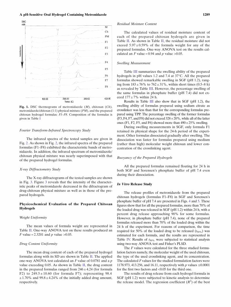
The DSC thermograms of metronidazole, medium molecu-lar weight chitosan, drug/polymer physical mixture (1:1), and theprepared formulas (F1–F8) are illustrated in Fig. 1. The DSCthermograms of metronidazole/chitosan physical mixture andthe prepared hydrogels revealed that the endothermic peak ofthe drug was reserved, but it became slightly shorter.
1288 El-Mahrouk et al.
Fourier Transform-Infrared Spectroscopy Study
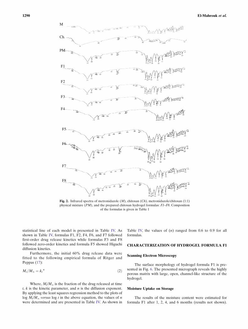
The infrared spectra of the tested samples are given inFig. 2. As shown in Fig. 2, the infrared spectra of the preparedformulas (F1–F8) exhibited the characteristic bands of metro-nidazole. In addition, the infrared spectrum of metronidazole/chitosan physical mixture was nearly superimposed with thatof the prepared hydrogel formulas.
X-ray Diffractometry Study
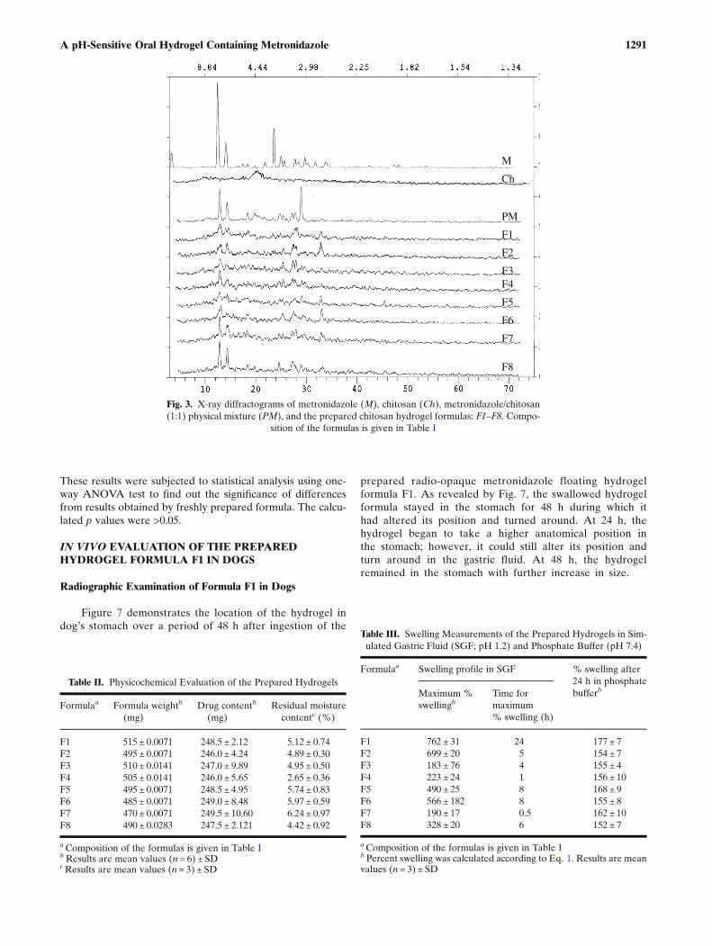
The X-ray diffractograms of the tested samples are shownin Fig. 3. Figure 3 reveals that the intensity of the character-istic peaks of metronidazole decreased in the difractogram ofdrug-chitosan physical mixture as well as in those of the pre-pared hydrogels.
Physicochemical Evaluation of the Prepared ChitosanHydrogels
Weight Uniformity
The mean values of formula weight are represented inTable II. One-way ANOVA test on these results produced anF value = 2.3201 and p value >0.05.
Drug Content Uniformity
The mean drug content of each of the prepared hydrogelformulas along with its SD are shown in Table II. The appliedone-way ANOVA test calculated an F value of 0.0781 and a pvalue exceeding 0.05. As shown in Table II, the drug contentin the prepared formulas ranged from 246± 4.24 (for formulaF2) to 249.5 ± 10.60 (for formula F7); representing 98.4± 1.70% and 99.8 ± 4.24% of the initially added drug amount,respectively.
Residual Moisture Content
The calculated values of residual moisture content ofeach of the prepared chitosan hydrogels are given inTable II. As shown in Table II, the residual moisture did notexceed 5.97 ± 0.59% of the formula weight for any of theprepared formulas. One-way ANOVA test on the results cal-culated an F value = 0.94 and p value >0.05.
Swelling Measurement
Table III summarizes the swelling ability of the preparedhydrogels in pH values 1.2 and 7.4 at 37°C. All the preparedformulas showed remarkable swelling in SGF (pH 1.2), rang-ing from 183 ± 76% to 762± 31%, within short times (0.5–8 h)as revealed by Table III. However, the percentage swelling ofthe same formulas in phosphate buffer (pH 7.4) did not ex-ceed 177 ± 7% within 24 h.
Results in Table III also show that in SGF (pH 1.2), theswelling ability of formulas prepared using sodium citrate ascrosslinker was less than that for the corresponding formulas pre-pared using TPP. The percentage swelling of the former formulas(F3, F4, F7, andF8) did not exceed 328±20%,while all of the latterones (F1, F2, F5, and F6) showed more than 490±25% swelling.
During swelling measurements in SGF, only formula F1retained its physical shape for the 24-h period of the experi-ment. Other formulas dissociated gradually after swelling. Thedissociation was faster for formulas prepared using medium(rather than high) molecular weight chitosan and lower con-centration of the crosslinking agent.
Buoyancy of the Prepared Hydrogels
All the prepared formulas remained floating for 24 h inboth SGF and Sorensen’s phosphate buffer of pH 7.4 evenduring their dissociation.
In Vitro Release Study
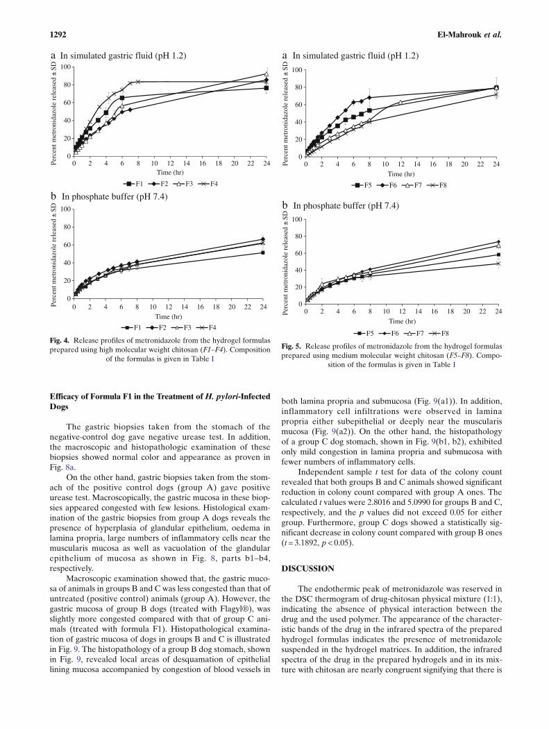
The release profiles of metronidazole from the preparedchitosan hydrogels (formulas F1–F8) in SGF and Sorensen’sphosphate buffer of pH 7.4 are presented in Figs. 4 and 5. Thesefigures show that for all the prepared formulas, more than 70%ofthe loaded drug was released in SGF (pH 1.2) within 24 h, with apercent drug release approaching 90% for some formulas.However, in phosphate buffer (pH 7.4), none of the preparedformulas released more than 70% of the loaded drug within the24 h of the experiment. For reasons of comparison, the timerequired for 50% of the loaded drug to be released (t50%) wasestimated for each formula, and the results are represented inTable IV. Results of t50% were subjected to statistical analysisusing two-way ANOVA test and Fisher’s PLSD.
The F values were calculated for the three studied formu-lation factors namely, the molecular weight of the used chitosan,the type of the used crosslinking agent, and its concentration.The calculated F values for the studied formulation factors were1178.973, 413.256, and 16.11, respectively with p values ≤0.0001for the first two factors and <0.05 for the third one.
The results of drug release from each hydrogel formula inSGF (pH 1.2) were subjected to kinetic analysis to determinethe release model. The regression coefficient (R2) of the best
M
Ch
PM
F1
F2
F3
F4
F5
F6
F7
F8
Fig. 1. DSC thermograms of metronidazole (M), chitosan (Ch),metronidazole/chitosan (1:1) physical mixture (PM), and the preparedchitosan hydrogel formulas: F1–F8. Composition of the formulas isgiven in Table I
1289A pH-Sensitive Oral Hydrogel Containing Metronidazole
statistical line of each model is presented in Table IV. Asshown in Table IV, formulas F1, F2, F4, F6, and F7 followedfirst-order drug release kinetics while formulas F3 and F8followed zero-order kinetics and formula F5 showed Higuchidiffusion kinetics.
Furthermore, the initial 60% drug release data werefitted to the following empirical formula of Ritger andPeppas (17):
Mt=M∞ ¼ kt n ð2Þ
Where, Mt/M∞ is the fraction of the drug released at timet, k is the kinetic parameter, and n is the diffusion exponent.By applying the least squares regression method to the plots oflog Mt/M∞ versus log t in the above equation, the values of nwere determined and are presented in Table IV. As shown in
Table IV, the values of (n) ranged from 0.6 to 0.9 for allformulas.
CHARACTERIZATION OF HYDROGEL FORMULA F1
Scanning Electron Microscopy
The surface morphology of hydrogel formula F1 is pre-sented in Fig. 6. The presented micrograph reveals the highlyporous matrix with large, open, channel-like structure of thehydrogel.
Moisture Uptake on Storage
The results of the moisture content were estimated forformula F1 after 1, 2, 4, and 6 months (results not shown).
M
Ch
PM
F1
F2
F3
F4
F5
F6
F7
F8
Fig. 2. Infrared spectra of metronidazole (M), chitosan (Ch), metronidazole/chitosan (1:1)physical mixture (PM), and the prepared chitosan hydrogel formulas: F1–F8. Composition
of the formulas is given in Table I
1290 El-Mahrouk et al.
These results were subjected to statistical analysis using one-way ANOVA test to find out the significance of differencesfrom results obtained by freshly prepared formula. The calcu-lated p values were >0.05.
IN VIVO EVALUATION OF THE PREPAREDHYDROGEL FORMULA F1 IN DOGS
Radiographic Examination of Formula F1 in Dogs
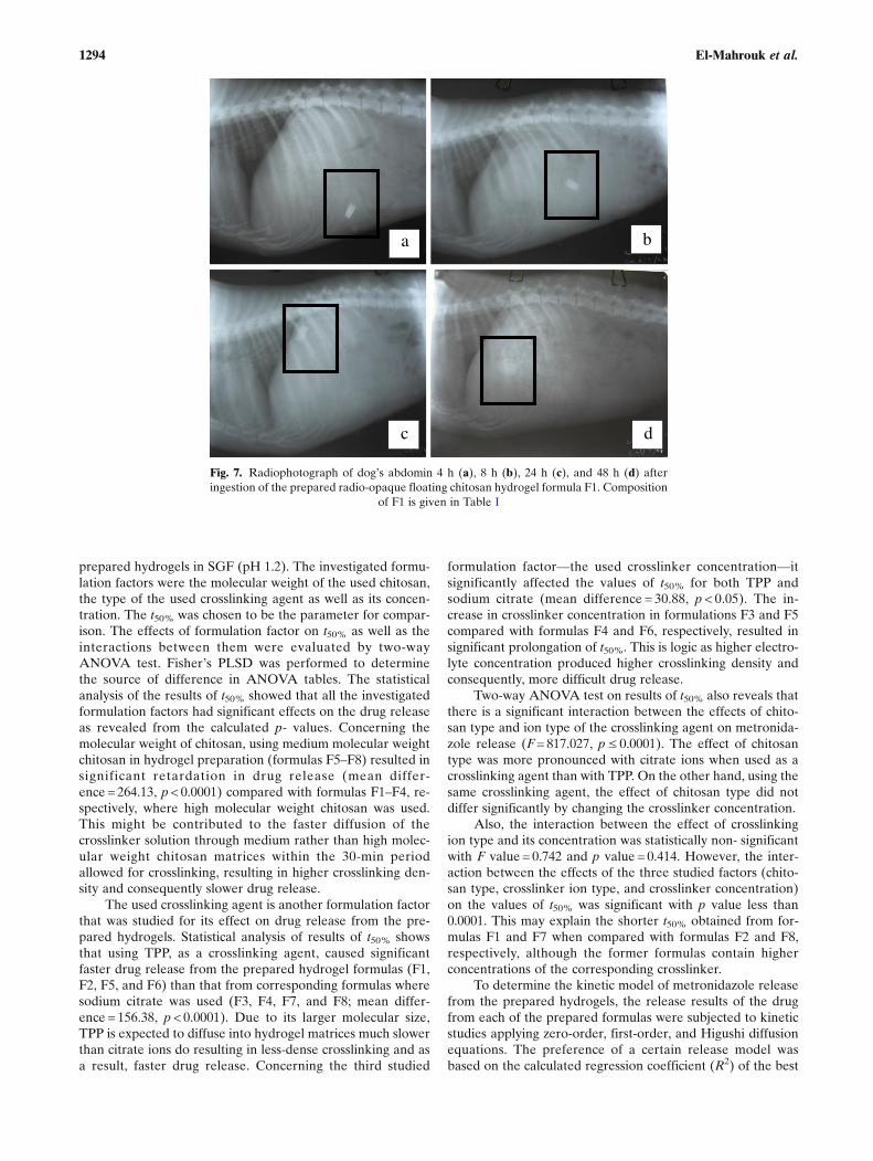
Figure 7 demonstrates the location of the hydrogel indog’s stomach over a period of 48 h after ingestion of the
prepared radio-opaque metronidazole floating hydrogelformula F1. As revealed by Fig. 7, the swallowed hydrogelformula stayed in the stomach for 48 h during which ithad altered its position and turned around. At 24 h, thehydrogel began to take a higher anatomical position inthe stomach; however, it could still alter its position andturn around in the gastric fluid. At 48 h, the hydrogelremained in the stomach with further increase in size.
M
Ch
PM
F1
F2
F3 F4
F5
F6
F7
F8
Fig. 3. X-ray diffractograms of metronidazole (M), chitosan (Ch), metronidazole/chitosan(1:1) physical mixture (PM), and the prepared chitosan hydrogel formulas: F1–F8. Compo-
sition of the formulas is given in Table I
Table II. Physicochemical Evaluation of the Prepared Hydrogels
Formulaa Formula weightb
(mg)Drug contentb
(mg)Residual moisture
contentc (%)
F1 515 ± 0.0071 248.5 ± 2.12 5.12 ± 0.74F2 495 ± 0.0071 246.0 ± 4.24 4.89 ± 0.30F3 510 ± 0.0141 247.0 ± 9.89 4.95 ± 0.50F4 505 ± 0.0141 246.0 ± 5.65 2.65 ± 0.36F5 495 ± 0.0071 248.5 ± 4.95 5.74 ± 0.83F6 485 ± 0.0071 249.0 ± 8.48 5.97 ± 0.59F7 470 ± 0.0071 249.5 ± 10.60 6.24 ± 0.97F8 490 ± 0.0283 247.5 ± 2.121 4.42 ± 0.92
aComposition of the formulas is given in Table IbResults are mean values (n = 6) ± SDcResults are mean values (n = 3) ± SD
Table III. Swelling Measurements of the Prepared Hydrogels in Sim-ulated Gastric Fluid (SGF; pH 1.2) and Phosphate Buffer (pH 7.4)
Formulaa Swelling profile in SGF % swelling after24 h in phosphatebufferbMaximum %
swellingbTime formaximum% swelling (h)
F1 762 ± 31 24 177 ± 7F2 699 ± 20 5 154 ± 7F3 183 ± 76 4 155 ± 4F4 223 ± 24 1 156 ± 10F5 490 ± 25 8 168 ± 9F6 566 ± 182 8 155 ± 8F7 190 ± 17 0.5 162 ± 10F8 328 ± 20 6 152 ± 7
aComposition of the formulas is given in Table Ib Percent swelling was calculated according to Eq. 1. Results are meanvalues (n = 3) ± SD
1291A pH-Sensitive Oral Hydrogel Containing Metronidazole
Efficacy of Formula F1 in the Treatment of H. pylori-InfectedDogs
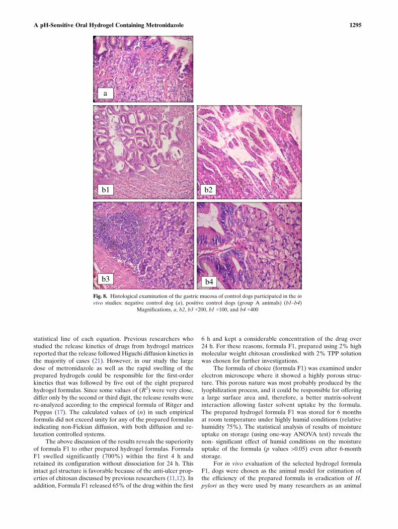
The gastric biopsies taken from the stomach of thenegative-control dog gave negative urease test. In addition,the macroscopic and histopathologic examination of thesebiopsies showed normal color and appearance as proven inFig. 8a.
On the other hand, gastric biopsies taken from the stom-ach of the positive control dogs (group A) gave positiveurease test. Macroscopically, the gastric mucosa in these biop-sies appeared congested with few lesions. Histological exam-ination of the gastric biopsies from group A dogs reveals thepresence of hyperplasia of glandular epithelium, oedema inlamina propria, large numbers of inflammatory cells near themuscularis mucosa as well as vacuolation of the glandularepithelium of mucosa as shown in Fig. 8, parts b1–b4,respectively.
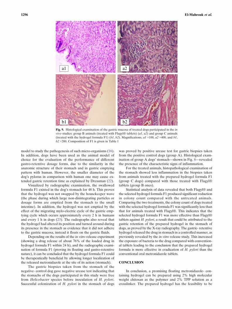
Macroscopic examination showed that, the gastric muco-sa of animals in groups B and C was less congested than that ofuntreated (positive control) animals (group A). However, thegastric mucosa of group B dogs (treated with Flagyl®), wasslightly more congested compared with that of group C ani-mals (treated with formula F1). Histopathological examina-tion of gastric mucosa of dogs in groups B and C is illustratedin Fig. 9. The histopathology of a group B dog stomach, shownin Fig. 9, revealed local areas of desquamation of epitheliallining mucosa accompanied by congestion of blood vessels in
both lamina propria and submucosa (Fig. 9(a1)). In addition,inflammatory cell infiltrations were observed in laminapropria either subepithelial or deeply near the muscularismucosa (Fig. 9(a2)). On the other hand, the histopathologyof a group C dog stomach, shown in Fig. 9(b1, b2), exhibitedonly mild congestion in lamina propria and submucosa withfewer numbers of inflammatory cells.
Independent sample t test for data of the colony countrevealed that both groups B and C animals showed significantreduction in colony count compared with group A ones. Thecalculated t values were 2.8016 and 5.0990 for groups B and C,respectively, and the p values did not exceed 0.05 for eithergroup. Furthermore, group C dogs showed a statistically sig-nificant decrease in colony count compared with group B ones(t= 3.1892, p< 0.05).
DISCUSSION
The endothermic peak of metronidazole was reserved inthe DSC thermogram of drug-chitosan physical mixture (1:1),indicating the absence of physical interaction between thedrug and the used polymer. The appearance of the character-istic bands of the drug in the infrared spectra of the preparedhydrogel formulas indicates the presence of metronidazolesuspended in the hydrogel matrices. In addition, the infraredspectra of the drug in the prepared hydrogels and in its mix-ture with chitosan are nearly congruent signifying that there is
0
20
40
60
80
100
0 2 4 6 8 10 12 14 16 18 20 22 24Perc
ent m
etro
nida
zole
rel
ease
d ±
SD
Time (hr)
F1 F2 F3 F4
0
20
40
60
80
100
0 2 4 6 8 10 12 14 16 18 20 22 24Perc
ent m
etro
nida
zole
rel
ease
d ±
SD
Time (hr)
F1 F2 F3 F4
a In simulated gastric fluid (pH 1.2)
b In phosphate buffer (pH 7.4)
Fig. 4. Release profiles of metronidazole from the hydrogel formulasprepared using high molecular weight chitosan (F1–F4). Composition
of the formulas is given in Table I
0
20
40
60
80
100
0 2 4 6 8 10 12 14 16 18 20 22 24Perc
ent m
etro
nida
zole
rel
ease
d ±
SD
Time (hr)
F5 F6 F7 F8
0
20
40
60
80
100
0 2 4 6 8 10 12 14 16 18 20 22 24Perc
ent m
etro
nida
zole
rel
ease
d ±
SD
Time (hr)
F5 F6 F7 F8
a In simulated gastric fluid (pH 1.2)
b In phosphate buffer (pH 7.4)
Fig. 5. Release profiles of metronidazole from the hydrogel formulasprepared using medium molecular weight chitosan (F5–F8). Compo-
sition of the formulas is given in Table I
1292 El-Mahrouk et al.

no chemical interaction between metronidazole and chitosanin the prepared formulas. The X-ray diffractogram of puremetronidazole proves its crystalline nature. However, the de-crease in the intensity of the characteristic peaks of the drug inthe diffractogram of drug-chitosan physical mixture indicatesthe decrease in drug purity by virtue of mixing with chitosan.The further decrease in the intensity of the characteristicpeaks of metronidazole in the diffractograms of the preparedhydrogels indicates that the preparation procedure caused adecrement in drug crystallinity although it remained in thecrystalline state. The lyophilization process is probably re-sponsible for this decrease in drug crystallinity in the preparedhydrogels as previously explained by Hawe and Frie in theirstudy on the effect of lyophilization on the crystallinity ofmannitol hydrate (18).
The results of one-way ANOVA test on the values offormula weight revealed no significant difference betweenthe prepared formulas indicating their weight uniformity.Also, the statistical analysis of data of drug content of theprepared hydrogels proved the uniformity of drug contentamong the prepared formulas.
The drug content of all the prepared formulas met theEuropean Pharmacopoeia’s (2002) specifications for single-dose preparations as it was in the range of 90–110% of thetheoretical drug content. In addition, one-way ANOVA testrevealed that the differences in the results of the residualmoisture content of the prepared hydrogel formulas werenon-significant. Concerning hydrogel swelling, results revealthat the swelling ability of the prepared hydrogels is highly pHdependent; as they remarkably swelled in SGF (pH 1.2), buttheir swelling in phosphate buffer of pH 7.4 was comparativelypoor. The extensive swelling ability of the prepared hydrogelsin SGF (pH 1.2) could be attributed to poor ionization of thecrosslinking agents (sodium citrate and TPP) at such acidic pHvalue. As a result, the electrostatic interaction with chitosanchains was weakened or disappeared. Moreover, protonationof chitosan free ammonium groups at such acidic pH resultedin their repulsion and favored the swelling of the formulas oreven their dissociation. Similarly, the poor swelling ability ofthe prepared hydrogels in phosphate buffer (pH 7.4) could beattributed to the reservation of the electrostatic attractionbetween the crosslinker ions and chitosan at alkaline pH.However, the slight increase in weight of the hydrogels in thismedium might be explained by hydration of the unboundamine groups of chitosan.
The difference in the swelling behavior of formulas pre-pared using the same chitosan, but different crosslinkingagents might indicate that when citrate ions were used theyproduced higher crosslinking density within the 30 minallowed for the crosslinking step. This came in accordancewith the results of Shu et al. (15) who reported the pH sensi-tivity of citrate-crosslinked chitosan films and Shu and Zhu(19), who studied the swelling behavior of ionicallycrosslinked chitosan beads. The high ability of the preparedhydrogel formulas to float over the 24 h of the experiment canbe mainly attributed to the highly porous nature of the formu-las probably due to the lyophilization process (20).
The release profiles of metronidazole from the preparedhydrogel formulas clearly signify that the hydrogels exhibitedpH-dependent release patterns, where drug release was pref-erable in SGF (pH 1.2) than in phosphate buffer of pH 7.4.
A 23 factorial experiment was designed to study the effectof different formulation factors on the drug release from the
Table IV. Metronidazole In Vitro Release Study from the Prepared Hydrogels
Formulaa t50% (h)b Regression coefficient (R2) Release exponent (n)d
Zero order First order Higushi
F1 4.167 ± 0.59 0.9881 0.9985c 0.9956 0.5947F2 6.025 ± 0.035 0.9860 0.9955c 0.9939 0.6627F3 5.39 ± 0.0825 0.9980c 0.9836 0.9720 0.7310F4 2.68 ± 0.023 0.9940 0.9978c 0.9900 0.7720F5 7.17 ± 0.24 0.9773 0.9946 0.9964c 0.6263F6 4.5 ± 0.24 0.9935 0.9968c 0.9822 0.7801F7 11.38 ± 0.06 0.9911 0.9987c 0.9943 0.6300F8 12.83 ± 0.25 0.9988c 0.9986 0.9779 0.8693
aComposition of the formulas is given in Table IbTime required for 50% of the loaded drug to be released. Results are mean values (n = 3) ± SDcThe highest values of R2 represent the release modeldThe release exponent (n) was calculated according to Eq. 2
Fig. 6. Scanning electron micrograph of hydrogel formula F1. Com-position of F1 is given in Table I
1293A pH-Sensitive Oral Hydrogel Containing Metronidazole
prepared hydrogels in SGF (pH 1.2). The investigated formu-lation factors were the molecular weight of the used chitosan,the type of the used crosslinking agent as well as its concen-tration. The t50% was chosen to be the parameter for compar-ison. The effects of formulation factor on t50% as well as theinteractions between them were evaluated by two-wayANOVA test. Fisher’s PLSD was performed to determinethe source of difference in ANOVA tables. The statisticalanalysis of the results of t50% showed that all the investigatedformulation factors had significant effects on the drug releaseas revealed from the calculated p- values. Concerning themolecular weight of chitosan, using medium molecular weightchitosan in hydrogel preparation (formulas F5–F8) resulted insignificant retardation in drug release (mean differ-ence = 264.13, p< 0.0001) compared with formulas F1–F4, re-spectively, where high molecular weight chitosan was used.This might be contributed to the faster diffusion of thecrosslinker solution through medium rather than high molec-ular weight chitosan matrices within the 30-min periodallowed for crosslinking, resulting in higher crosslinking den-sity and consequently slower drug release.
The used crosslinking agent is another formulation factorthat was studied for its effect on drug release from the pre-pared hydrogels. Statistical analysis of results of t50% showsthat using TPP, as a crosslinking agent, caused significantfaster drug release from the prepared hydrogel formulas (F1,F2, F5, and F6) than that from corresponding formulas wheresodium citrate was used (F3, F4, F7, and F8; mean differ-ence = 156.38, p< 0.0001). Due to its larger molecular size,TPP is expected to diffuse into hydrogel matrices much slowerthan citrate ions do resulting in less-dense crosslinking and asa result, faster drug release. Concerning the third studied
formulation factor—the used crosslinker concentration—itsignificantly affected the values of t50% for both TPP andsodium citrate (mean difference = 30.88, p < 0.05). The in-crease in crosslinker concentration in formulations F3 and F5compared with formulas F4 and F6, respectively, resulted insignificant prolongation of t50%. This is logic as higher electro-lyte concentration produced higher crosslinking density andconsequently, more difficult drug release.
Two-way ANOVA test on results of t50% also reveals thatthere is a significant interaction between the effects of chito-san type and ion type of the crosslinking agent on metronida-zole release (F= 817.027, p≤ 0.0001). The effect of chitosantype was more pronounced with citrate ions when used as acrosslinking agent than with TPP. On the other hand, using thesame crosslinking agent, the effect of chitosan type did notdiffer significantly by changing the crosslinker concentration.
Also, the interaction between the effect of crosslinkingion type and its concentration was statistically non- significantwith F value = 0.742 and p value = 0.414. However, the inter-action between the effects of the three studied factors (chito-san type, crosslinker ion type, and crosslinker concentration)on the values of t50% was significant with p value less than0.0001. This may explain the shorter t50% obtained from for-mulas F1 and F7 when compared with formulas F2 and F8,respectively, although the former formulas contain higherconcentrations of the corresponding crosslinker.
To determine the kinetic model of metronidazole releasefrom the prepared hydrogels, the release results of the drugfrom each of the prepared formulas were subjected to kineticstudies applying zero-order, first-order, and Higushi diffusionequations. The preference of a certain release model wasbased on the calculated regression coefficient (R2) of the best
a b
c d
Fig. 7. Radiophotograph of dog’s abdomin 4 h (a), 8 h (b), 24 h (c), and 48 h (d) afteringestion of the prepared radio-opaque floating chitosan hydrogel formula F1. Composition
of F1 is given in Table I
1294 El-Mahrouk et al.
statistical line of each equation. Previous researchers whostudied the release kinetics of drugs from hydrogel matricesreported that the release followed Higuchi diffusion kinetics inthe majority of cases (21). However, in our study the largedose of metronidazole as well as the rapid swelling of theprepared hydrogels could be responsible for the first-orderkinetics that was followed by five out of the eight preparedhydrogel formulas. Since some values of (R2) were very close,differ only by the second or third digit, the release results werere-analyzed according to the empirical formula of Ritger andPeppas (17). The calculated values of (n) in such empiricalformula did not exceed unity for any of the prepared formulasindicating non-Fickian diffusion, with both diffusion and re-laxation controlled systems.
The above discussion of the results reveals the superiorityof formula F1 to other prepared hydrogel formulas. FormulaF1 swelled significantly (700%) within the first 4 h andretained its configuration without dissociation for 24 h. Thisintact gel structure is favorable because of the anti-ulcer prop-erties of chitosan discussed by previous researchers (11,12). Inaddition, Formula F1 released 65% of the drug within the first
6 h and kept a considerable concentration of the drug over24 h. For these reasons, formula F1, prepared using 2% highmolecular weight chitosan crosslinked with 2% TPP solutionwas chosen for further investigations.
The formula of choice (formula F1) was examined underelectron microscope where it showed a highly porous struc-ture. This porous nature was most probably produced by thelyophilization process, and it could be responsible for offeringa large surface area and, therefore, a better matrix-solventinteraction allowing faster solvent uptake by the formula.The prepared hydrogel formula F1 was stored for 6 monthsat room temperature under highly humid conditions (relativehumidity 75%). The statistical analysis of results of moistureuptake on storage (using one-way ANOVA test) reveals thenon- significant effect of humid conditions on the moistureuptake of the formula (p values >0.05) even after 6-monthstorage.
For in vivo evaluation of the selected hydrogel formulaF1, dogs were chosen as the animal model for estimation ofthe efficiency of the prepared formula in eradication of H.pylori as they were used by many researchers as an animal
a
b4b3
b2b1
Fig. 8. Histological examination of the gastric mucosa of control dogs participated in the invivo studies: negative control dog (a), positive control dogs (group A animals) (b1–b4)
Magnifications, a, b2, b3 ×200, b1 ×100, and b4 ×400
1295A pH-Sensitive Oral Hydrogel Containing Metronidazole
model to study the pathogenesis of such micro-organisms (16).In addition, dogs have been used as the animal model ofchoice for the evaluation of the performance of differentgastro-retentive dosage forms, due to the similarity in theanatomic structure of their stomach and in gastric emptyingpattern with human. However, the smaller diameter of thedog’s pylorus in comparison with human one may cause ex-tended gastric retention time as explained by Dressman (22).
Visualized by radiographic examination, the swallowedformula F1 existed in the dog’s stomach for 48 h. This provesthat the hydrogel was not swapped by the housekeeper wave(the phase during which large non-disintegrating particles ordosage forms are emptied from the stomach to the smallintestine). In addition, the hydrogel was not emptied by theeffect of the migrating mylo-electric cycle of the gastric emp-tying cycle which occurs approximately every 2 h in humansand every 1 h in dogs (23). The radiographs also reveal thatthe hydrogel had altered its position and turned around duringits presence in the stomach as evidence that it did not adhereto the gastric mucosa, instead it floats on the gastric fluids.
Depending on the results of the in vitro release experiment(showing a drug release of about 76% of the loaded drug inhydrogel formula F1 within 24 h), and the radiographic exami-nation of formula F1 (proving its floating and gastro-retentivenature), it can be concluded that the hydrogel formula F1 couldbe therapeutically beneficial by allowing longer localization ofthe released metronidazole at the site of its action (stomach).
The gastric biopsies taken from the stomach of thenegative- control dog gave negative urease test indicating thatthe stomachs of the dogs participated in this study were freefrom Helicobacter species before inoculation of H. pylori.Successful colonization of H. pylori in the stomach of dogs
was proved by positive urease test for gastric biopsies takenfrom the positive control dogs (group A). Histological exam-ination of group A dogs’ stomach—shown in Fig. 8—revealedthe presence of the characteristic signs of inflammation.
For the treated animals, histopathological examination ofthe stomach showed less inflammation in the biopsies takenfrom animals treated with the prepared hydrogel formula F1(group C dogs) compared with those treated with Flagyl®tablets (group B ones).
Statistical analysis of data revealed that both Flagyl® andthe selected hydrogel formula F1 produced significant reductionin colony count compared with the untreated animals.Comparing the two treatments, the colony count of dogs treatedwith the selected hydrogel formula F1 was significantly less thanthat for animals treated with Flagyl®. This indicates that theselected hydrogel formula F1 was more effective than Flagyl®tablets against H. pylori, a result that could be attributed to thegastric retention of the prepared hydrogel in the stomach ofdogs, as proved by the X-ray radiography. The gastric- retentivehydrogel released the drug in stomach in a controlledmanner, aspreviously revealed by the in vitro release study. This increasedthe exposure of bacteria to the drug compared with convention-al tablets leading to the conclusion that the prepared hydrogelformula is more effective in eradication of H. pylori than theconventional oral metronidazole tablets.
CONCLUSION
In conclusion, a promising floating metronidazole- con-taining hydrogel can be prepared using 2% high molecularweight chitosan as the polymer and 2% TPP solution as acrosslinker. The prepared hydrogel has the feasibility to be
a1 a2
b1 b2
Fig. 9. Histological examination of the gastric mucosa of treated dogs participated in the invivo studies: group B animals (treated with Flagyl® tablets) (a1, a2) and group C animals(treated with the hydrogel formula F1) (b1, b2). Magnifications, a1 ×100, a2 ×400, and b1,b2 ×200. Composition of F1 is given in Table I
1296 El-Mahrouk et al.
retained in the stomach and to release metronidazole in acontrolled manner over a period of 24 h. Furthermore,the prepared hydrogel was more effective than oral met-ronidazole (Flagyl® tablets) in the eradication of H. py-lori in infected dog’s stomach. Keeping in mind thatmetronidazole in the prepared formula was used at alower dose level (250 mg, once daily) than in the conven-tionally used oral triple therapy (500 mg, three timesdaily), the results of this paper indicated that the preparedhydrogel could be considered as a promising site-specificdelivery system for the treatment of peptic ulcer causedby H. pylori. For further improvement of the efficiency ofthe prepared formula, the treatment duration can be elon-gated, accompanied by the incorporation of another anti-biotic along with a proton pump inhibitor in the preparedhydrogel.
ACKNOWLEDGMENTS
The authors would like to acknowledge The NationalHepatology and Tropical Medicine Research Institute, Cairo,Egypt, for providing gastric biopsies from patients with gastri-tis and gastric and duodenal ulcers. The authors also thankThe Faculty of Veterinary Medicine Cairo University, forproviding the facilities during the in vivo study.
REFERENCES
1. Marshall BJ, Warren JR. Unidentified curved bacilli in the stom-ach of patients with gastritis and peptic ulceration. Lancet.1984;1:1311–5.
2. Wolle K, Malfertheiner P. Treatment of Helicobacter pylori. BestPract Res Clin Gastroenterol. 2007;21:315–24.
3. Sweetman SC. Martindale; the complete drug reference. 33rd ed.London: The Pharmaceutical Press; 2002. p. 759–67.
4. Bardonnet PL, Faivre V, Pugh WJ, Piffaretti JC, Falson F.Gastroretentive dosage forms: overview and special case ofHelicobacter pylori. J Control Release. 2006;111:1–18.doi:10.1016/j.jconrel.2005.10.031.
5. Desai S, Bolton S. A floating controlled release drug deliverysystem: in vitro-in vivo evaluation. Pharm Res. 1993;10:1321–5.
6. Peppas NA. Hydrogels and drug delivery. Curr Opin ColloidInterface Sci. 1997;2:531–7. doi:10.1016/S1359-0294(97)80103-3.
7. Ishak RAH, Awad GAS, Mortada ND, Nour SAK. Preparation,in-vitro and in-vivo evaluation of stomach-specific metronidazoleloaded alginate beads as local anti-Helicobacter pylori therapy. JControl Release. 2007;119:207–14.
8. Qiu Y, Park K. Environment-sensitive hydrogels for drug deliv-ery. Adv Drug Deliv Rev. 2001;53:321–39.
9. Rajinikanth PS, Balasubramaniam J, Mishra B. Developmentand evaluation of a novel floating in situ gelling system of amox-icillin for eradication of Helicobacter pylori. Int J Pharm.2007;335:114–22.
10. Boriwanwattanarak P, Ingkaninan K, Khorana N, Viyoch J. De-velopment of curcuminoids hydrogel patch using chitosan fromvarious sources as controlled-release matrix. Int J Cosmet Sci.2008;30:205–18. doi:10.1111/j.1468-2494.2008.00437.x.
11. Ueno H, Mori T, Fujinaga T. Topical formulations and woundhealing applications of chitosan. Adv Drug Deliv Rev.2001;52:105–15.
12. Burkatovskaya M, Castano AP, Demidova-Rice TN, Tegos GP,Hamblin MR. Effect of chitosan acetate bandage on woundhealing in infected and noninfected wounds in mice. WoundRepa i r Regen . 2008 ;16 :425–31 . do i : 10 .1111 / j . 1524 -475X.2008.00382.x.
13. Malaekeh-Nikouei B, Sajadi Tabassi SA, Jaafari MR. Prepara-tion, characterization, and mucoadhesive properties of chitosan-coated microspheres encapsulated with cyclosporine A. DrugDev Ind Pharm. 2008;34:492–8. doi:10.1080/03639040701744004.
14. Bergera J, Reista M, Mayera JM, Feltb O, Peppasc NA, GurnybR. Structure and interactions in covalently and ionicallycrosslinked chitosan hydrogels for biomedical applications. EurJ Pharm Biopharm. 2004;57:19–34.
15. Shu XZ, Zhu KJ, Song W. Novel pH-sensitive citrate cross-linkedchitosan film for controlled drug release. Int J Pharm.2001;212:19–28.
16. Rossi G, Rossi M, Vitali CG, Fortuna D, Burroni D, Pancotto L,et al. A conventional beagle dog model for acute and chronicinfection with Helicobacter pylori. Infect Immun. 1999;67:3112–20.
17. Ritger PL, Peppas NA. A simple equation for description ofsolute release. II. Fickian and anomalous release from swellabledevices. J Control Release. 1987;5:37–42. doi:10.1016/0168-3659(87)90035-6.
18. Hawe A, Frie W. Impact of freezing procedure and annealing onthe physico-chemical properties and the formation of mannitolhydrate in mannitol–sucrose–NaCl formulations. Eur J PharmBiopharm. 2006;64:316–25.
19. Shu XZ, Zhu KJ. Controlled drug release properties of ionicallycross-linked chitosan beads: the influence of anion structure. Int JPharm. 2002;233:217–25. doi:10.4103/0250-474X.40326.
20. Jiankang H, Dichen L, Yaxiong L, Bo Y, Bingheng L, Qin L.Fabrication and characterization of chitosan/gelatin porous scaf-folds with predefined internal microstructures. Polymer.2007;48:4578–88. doi:10.1007/s13726-012-0019-0.
21. Agnihotri SA, Aminabhavi TM. Novel interpenetratingnetwork chitosan-poly(ethylene oxide-g-acrylamide) hy-drogel microspheres for the controlled release of capecit-abine . Int J Pharm. 2006;324 :103–15. doi :10.1016/j.ijpharm.2006.05.061.
22. Dressman JB. Comparison of canine and human gastrointestinalphysiology. Pharm Res. 1986;3 :123–31. doi :10.1023/A:1016353705970.
23. Arora S, Ali J, Ahuja A, Khar RK, Baboota S. Floating drugdelivery systems: a review. AAPS Pharm Sci Tech. 2005;6:372–90.doi:10.1208/pt070117.
1297A pH-Sensitive Oral Hydrogel Containing Metronidazole